Embed Size (px)
Citation preview

2nd Year Pathology 2010
Vascular Disturbances III Infarction & Shock

2nd Year Pathology 2010
Infarction

2nd Year Pathology 2010
Infarction
Tissue necrosis due to ischaemia vascular insufficiency of any cause usually arterial occlusion due to thrombosis/embolism
Mainly due to oxygen deficiency, but toxin accumulation & reperfusion injury may contribute
Number of determining factors Size of vessel and size of vascular territory Partial / total vascular occlusion Duration of ischaemia
2nd Year Pathology 2010
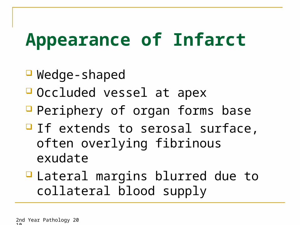
Appearance of Infarct
Wedge-shaped Occluded vessel at apex Periphery of organ forms base If extends to serosal surface, often
overlying fibrinous exudate Lateral margins blurred due to collateral
blood supply

2nd Year Pathology 2010
Appearance of Infarct
ARTERY
OCCLUSION
NORMAL TISSUE
INFARCTED TISSUE
SURFACE FIBRINOUS EXUDATE
ILL-DEFINED INFARCT BORDERS

2nd Year Pathology 2010
Types of Infarct Red (haemorrhagic) infarcts
1. Venous occlusion/congestion e.g. torsion2. Loose tissues where haemorrhage can occur and blood can
collect in infarcted zone e.g. lung 3. Tissues with dual blood supply e.g. lung small intestine
(permitting blood flow from unobstructed vessel into infarcted zone – note flow is insufficient to rescue ischaemia)
4. Tissues that were previously congested due to sluggish venous outflow
5. When flow is re-established e.g. fragmentation of an occlusive embolus, angioplasty
White infarcts1. arterial occlusion2. solid tissues, where haemorrhage limited e.g. spleen, heart,
kidney

2nd Year Pathology 2010
Types of Infarct
Red pulmonary infarcts - dual pulmonary / bronchial arterial supply

2nd Year Pathology 2010
Types of Infarct
White splenic infarct

2nd Year Pathology 2010
Event Sequence
1. Coagulative necrosis
2. Infiltration by neutrophils
3. Infiltration by macrophages
4. Phagocytosis of debris
5. Granulation tissue formation
6. Scar formation

2nd Year Pathology 2010
Event Sequence
Necrosis Neutrophils Macrophagephagocytosis
Granulationtissue
Fibrosis
Day 1 3 7 14 90

2nd Year Pathology 2010
Time Microscopic Features Gross Features
0 – 4 hr None None
4 – 12 hr Early coagulation necrosis (nucleus: pyknosis, cytoplasm: eosinophilia)
None
12 – 24 hr Further necrosis, haemorrhage, early neutrophil infiltrate
Dark mottling
1 – 3 days Marked neutrophil infiltrate and necrosis
Mottled with yellow-tan necrotic centre
3 – 7 days Early phagocytosis of dead cells by macrophages (at border)
Hyperaemic border, central yellow-tan softening
7 – 10 days Well-developed phagocytosis, early granulation tissue formation
Maximal yellow-tan softening, depressed red-tan margins
10 – 14 days Well-developed granulation tissue, early collagen deposition
Red-gray depressed infarct borders
2 – 8 wk Increased collagen deposition, decreased cellularity
Grey-white scar progresses from border toward centre
> 2 months Acellular collagenous scar Dense gray scar

2nd Year Pathology 2010
Infarct, day 0
Fibre eosinophilia & contraction band necrosis

2nd Year Pathology 2010
Infarct, day 1
Haemorrhage, necrosis and early neutrophil infiltrate

2nd Year Pathology 2010
Infarct, day 3
Myocyte necrosis, pyknosis and marked neutrophil infiltrate

2nd Year Pathology 2010
Infarct, day 7
Yellow necrotic infarct with hyperaemic border

2nd Year Pathology 2010
Infarct, day 10
Granulation tissue after macrophage phagocytosis of infarcted cells

2nd Year Pathology 2010
Infarct, day 90+
Subendocardial acellular fibrous scar

2nd Year Pathology 2010
Infarct Development Dependent on a number of factors
Nature of vascular supply Dual supply e.g. lungs, liver End arteries e.g. kidneys, spleen
Rate of vascular occlusion Time for development of collateral circulation
Vulnerability to hypoxia Neurons – 2-3mins, Myocardium – 20-30mins, Fibroblasts – hours.
Oxygen content of blood Anaemia, cyanosis, congestive heart failure
Can result in infarction due to otherwise inconsequential blockage Size of vessel and size of vascular territory Partial / total vascular occlusion Duration of ischaemia

2nd Year Pathology 2010
Reperfusion Injury
Possible effects of re-establishing blood flow: prevention of all necrosis salvage of reversibly injured cells accentuation of damage to irreversibly injured cells new cellular damage
Latter two constitute reperfusion injury Accentuated or new damage due to re-establishing blood
flow Many effects of ischaemic injury only seen when perfusion
re-established

2nd Year Pathology 2010
Reperfusion Injury
Can result in accelerated transition through stages of infarct development
Timing of infarcts unreliable post reperfusion Inevitable if reperfusion occurs after optimum
salvage time e.g. usually 6 hours after myocardial infarct
Characterised histologically by: Marked haemorrhage Marked contraction band necrosis
2nd Year Pathology 2010
Reperfusion Injury
Causes: delivery of oxygen and calcium ions to damaged tissue interior of cells with damaged cell membranes exposed to
high Ca++ conc cell lysis generation of oxygen-dependent free radicals by damaged
cells and phagocytes cell lysis accentuation of O2-dependent damage
Anti-oxidants have only small effect on tissue loss Acceleration of damage to irreversibly damaged cells more
than new cellular damage

2nd Year Pathology 2010
Shock

2nd Year Pathology 2010
Shock (cardiovascular collapse)
Final Common Pathway for a umber of potentially lethal clinical events: Severe Haemorrhage Burns Trauma Large MI (massive) Pulmonary Embolism Microbial Sepsis

2nd Year Pathology 2010
Shock (cardiovascular collapse)
Circulatory failure resulting in inadequate tissue perfusion (systemic hypoperfusion)
Results in: hypotension impaired tissue perfusion cellular hypoxia reversible cellular injury irreversible cell injury and cell death

2nd Year Pathology 2010
Types of Shock
Cardiogenic - due to myocardial pump failure Intrinsic damage (MI) Ventricular arrhythmias Extrinsic compression (Tamponade) Outflow obstruction (e.g. pulmonary embolism)
Hypovolaemic - due to loss of blood or plasma volume Haemorrhage Fluid Loss from severe burns Trauma

2nd Year Pathology 2010
Septic Systemic microbial infection
Neurogenic Loss of vascular tone –
spinal cord injury
Anaphylactic Generalised IgE
hypersensitivity response- systemic vasodilation
- due to reduction in effective circulating blood volume
peripheral pooling secondary to vasodilation and
leakage of fluid due to increased vascular permeability

2nd Year Pathology 2010
Types of Shock
Cardiogenic myocardial pump failure e.g. myocardial infarction, ventricular rupture, ventricular
arrhythmia, cardiac tamponade, pulmonary embolism Hypovolaemic
loss of blood or plasma volume e.g. haemorrhage, trauma, burns, vomiting, diarrhoea

2nd Year Pathology 2010
Types of Shock
Neurogenic shock peripheral pooling of blood due to loss of vascular tone e.g. anaesthetic accident / spinal cord injury
Anaphylactic shock systemic IgE-mediated hypersensitivity reaction to
allergens e.g. bee stings, peanut release of mast cell mediators systemic vasodilation and increased vascular permeability

2nd Year Pathology 2010
Types of Shock
Septic shock overwhelming microbial infection gram negative sepsis
due to lipopolysaccharide (LPS / endotoxin) in walls gram-positive / fungal septicaemia
due to molecules similar to LPS in walls Super-antigen release

2nd Year Pathology 2010
Septic Shock
Usually due to lipopolysaccharide (LPS/endotoxin) in walls of gram negative bacteria
LPS consists of fatty acid core and complex carbohydrate coat
Similar molecules in walls of gram positive bacteria or fungi
results in endothelial damage complement activation activation of macrophages with cytokine release

2nd Year Pathology 2010

2nd Year Pathology 2010
Event Sequence Low doses: Local effects of LPS & primary
mediators (IL-1, TNF) Complement activation by LPS Monocyte/macrophage
activation by LPS binding to surface receptors production of low doses of IL-
1 and TNF Endothelial cell activation by
IL-1 & TNF production of IL-6 & 8 by
endothelium increased adhesion molecule
expression Recruitment of inflammatory
cells and cytokine cascade

2nd Year Pathology 2010
Event Sequence Intermediate doses: Local effects of LPS & secondary
mediators (NO, PAF) Systemic effects of primary
mediators (IL-1, TNF) Endothelial cell injury by LPS
triggering of coagulation cascade
increased vascular permeability
production of secondary mediators by endothelium
Local vasodilation due to secondary mediators: nitric oxide, platelet activating factor
Systemic effects of IL-1 and TNF Fever Acute-phase reactant
production (CRP, fibrinogen)

2nd Year Pathology 2010
Event Sequence High doses Systemic effects of LPS,
primary & secondary mediators Widespread endothelial cell
injury (LPS, cytokines) Acute respiratory distress
syndrome Widespread activation of
coagulation (LPS, cytokines) Disseminated intravascular
coagulation Peripheral vasodilation,
decreased cardiac contractility (NO) Hypotension Multiorgan failure due to
hypoperfusion

2nd Year Pathology 2010
Multiorgan Failure• Multiple organ damage due to ischaemia
secondary to hypoperfusion• brain: ischaemic encephalopathy• heart: subendocardial infarcts• kidney: acute tubular necrosis• GIT: haemorrhagic enteropathy / ischaemia• liver: fatty change / centrilobular haemorrhagic
necrosis
• ARDS in lungs commonly present concurrently• Due to microvascular injury, not ischaemia
2nd Year Pathology 2010
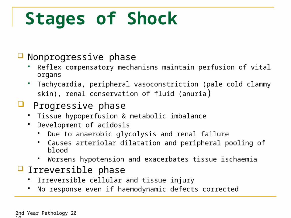
Stages of Shock
Nonprogressive phase Reflex compensatory mechanisms maintain perfusion of vital organs Tachycardia, peripheral vasoconstriction (pale cold clammy skin), renal
conservation of fluid (anuria) Progressive phase
Tissue hypoperfusion & metabolic imbalance Development of acidosis
Due to anaerobic glycolysis and renal failure Causes arteriolar dilatation and peripheral pooling of blood Worsens hypotension and exacerbates tissue ischaemia
Irreversible phase Irreversible cellular and tissue injury No response even if haemodynamic defects corrected

2nd Year Pathology 2010
Consequences of Shock
Pink hyaline membranes lining alveolar spaces in ARDS

2nd Year Pathology 2010
Consequences of Shock
Normal glomerulus and tubules ATN – swollen, sloughed and flattened regenerating tubular epithelium, normal glomerulus

2nd Year Pathology 2010
Consequences of Shock
Prognosis varies with cause and duration If circulatory disturbance corrected during nonprogressive
phase full recovery If progress to irreversible phase high mortality
Hypovolaemic shock > 80 – 90% survival in young healthy adults (10 - 20%
mortality) Cardiogenic shock due to MI / Septic shock
up to 75% mortality even with appropriate management

2nd Year Pathology 2010
Summary
Infarction Definition Infarct Types Timing of Infarcts Reperfusion injury
Shock Types of shock and aetiology Stages of shock Consequences