Embed Size (px)
DESCRIPTION
hi
Citation preview

Chapter 3
Biomaterials: Design, Development and Biomedical Applications
Gownolla Malegowd Raghavendra1*, Kokkarachedu Varaprasad2*, Tippabattini Jayaramudu1.
1 Synthetic Polymer Laboratory, Department of Polymer Science & Technology, Sri Krishnadevaraya University, Anantapuramu-515003, India.
2 Departamento de Ingeniería de Materiales, Facultad de Ingeniería Universidad de Concepción, Concepción, Chile
Corresponding authors: Gownolla Malegowd Raghavendra, Kokkarachedu VaraprasadE-mail: [email protected], [email protected], [email protected],
Abstract
The long been explorations in medical sciences have provided innumerable biomaterials that can
perform, augment or replace the natural function of a defective organ by interacting with the
biological system. These materials represent a unique class of biomedical functional materials,
potentially perform broad spectrum of biological activities in the absence of the original living
tissue/organ, thereby replace the problems encountered with the defective tissue/organ and
support smooth functioning of the organ and the living organism. The day-to-day enormous
increased demand in the medical field for the bio-alternatives that could able to perform the
living activities of bodily organs has raised the interest of the researchers to design novel
biomaterials. Hence, the study of biomaterials has become predominantly crucial for the
emerging researchers, tissue engineers and material scientists to understand much about the
biomaterial subject. In that point of view, the present chapter focuses on the design and
development of biomaterials besides their biomedical applications.
Keywords: Biomaterials; Defective organ; Biomedical functional materials; Living organism;
Bio-alternatives

Chapter outline
3.1. Overview
3.2. Design of biomaterials
3.2.1. Polymers
3.2.2. Metals
3.2.3. Composite materials
3.2.4. Ceramics
3.3. Basic considerations to design biomaterial
3.4. Characteristics of biomaterials
3.4.1. Non-toxicity
3.4.2. Biocompatible
3.4.3. Absence of foreign body reaction
3.4.4. Mechanical properties and performance
3.5. Fundamental aspects of tissue responses to biomaterials
3.5.1. Injury
3.5.2. Material interactions and initiation of the inflammatory response
3.5.3. Provisional matrix formation
3.5.4. Acute inflammation
3.5.5. Chronic inflammation
3.5.6. Granulation tissue
3.5.7. Foreign body reaction
3.5.8. Fibrosis and fibrous encapsulation
3.6. Evaluation of biomaterial behavior

3.6.1. Mechanical assessment
3.6.2. In vitro assessment
3.6.3. In vivo assessment
3.7. Properties assessed through in vivo experiments
3.7.1. Sensitization, irritation, and intracutaneous reactivity
3.7.2. Systemic toxicity, subacute and subchronic toxicity
3.7.3. Genotoxicity
3.7.4. Implantation
3.7.5. Hemocompatibility
3.7.6. Chronic toxicity
3.7.7. Carcinogenicity
3.7.8. Reproductive and developmental toxicity
3.7.9. Biodegradation
3.7.10. Immune responses
3.8. Applications of biomaterials
3.8.1. Orthopedic applications
3.8.2. Ophthalmologic applications
3.8.3. Cardiovascular Applications
3.8.4. Dental Applications
3.8.5. Wound dressing applications
3.8.6. Other applications
3.9. Future directions in biomaterials
3.10. Conclusions

Acknowledgements
References
3.1. Overview
Trauma, degeneration and diseases often make surgical repair or replacement necessary. This
usually requires replacement of the skeletal parts that include knees, hips, finger joints, elbows,
vertebrae, teeth and other bodily vital organs. These essential materials that are replaced in the
body are termed as “Biomaterials”. The Clemson University Advisory Board for biomaterials
has formally defined a biomaterial as “a systemically and pharmacologically inert substance
designed for implantation within or incorporation with living systems” [1]. It is also defined as
“a nonviable material used in a medical device, intended to interact with biological systems” [2].
Other definitions of biomaterial include, “materials of synthetic as well as of natural origin in
contact with tissue, blood, and biological fluids, and intended for use for prosthetic, diagnostic,
therapeutic, and storage applications without adversely affecting the living organism and its
components” [3] and “any substance (other than drugs) or combination of substances, synthetic
or natural in origin, which can be used for any period of time, as a whole or as a part of a system
which treats, augments, or replaces any tissue, organ, or function of the body” [4]. As the
definition for the term 'biomaterial' has been difficult to formulate, the more widely accepted
working definitions include: "A biomaterial is any material, natural or man‐made, that comprises
whole or part of a living structure or biomedical device which performs, augments, or replaces a
natural function" [5]. By contrast, a biological material is a material such as skin or artery,
produced by a biological system.
The study of biomaterials is called Biomaterials Science. Biomaterials Science encompasses the
elements of medicine, biology, chemistry, tissue engineering and materials science. A number of

factors, including the ageing population, an increasing preference by younger to middle aged
candidates to undertake surgery, improvements in the technology and life style, a better
understanding of body functionality, improved aesthetics and need for better function resulted
enormously expansion of Biomaterial Science from day to day and it is supposed to be a
continuous process. As the field of biomaterials experienced steady and strong growth, many
companies are investing larger amounts of money for the development of new products.
Biomaterial is not a recent origin. The introduction of nonbiological materials into the human
body was noted many centuries ago, far back in prehistory. The remains of a human found near
Kennewick, Washington, USA (often referred to as the “Kennewick Man”) was dated to be 9000
years old [6]. Some of the earliest biomaterial applications were as far back as ancient Phoenicia
where loose teeth were bound together with gold wires for tying artificial ones to neighbouring
teeth. The Mayan people fashioned nacre teeth from sea shells in roughly 600 AD and apparently
achieved what we now refer to as bone integration. Similarly, a corpse dated 200 AD with an
iron dental implant, found in Europe [7] was described as properly bone integrated. Though there
was no materials science, biological understanding, or medicine behind the followed procedures,
still their success is impressive and highlights two points: the forgiving nature of the human body
and the pressing drive, even in prehistoric times, to address the loss of physiologic/anatomic
function with an implant [6]. It is understood from the sources that though there were no medical
device manufacturers, no formalized regulatory approval processes, no understanding of
biocompatibility, and certainly no academic courses on biomaterials, yet crude biomaterials have
been used, generally with poor to mixed results, throughout history.
In the modern times, early in the 1900s, to aid in the fixation of long bone fractures, bone plates
were introduced [8]. Many of these early plates broke as a result of unsophisticated mechanical

design, as they were too thin and had stress concentrating corners. Also, materials such as
vanadium steel though chosen as biomaterial owing to its good mechanical properties, but
corroded rapidly in the body and caused adverse effects on the healing processes. Hence, better
designs and materials was soon followed. With the introduction of stainless steels and cobalt
chromium alloys in the 1930s, greater success was achieved in fracture fixation, and the first
joint replacement surgeries were performed [9]. As for polymers, Polymethyl methacrylate has
became widely used for replacements of sections of damaged skull bones. Following further
advances in materials and in surgical technique, in 1950s blood vessel replacements were tried
and during 1960s, heart valve replacements and cemented joint replacements were came in to
usage. Recent years have seen many further advances [10-12]. At the dawn of the 21st century,
biomaterials are widely used throughout medicine, dentistry and biotechnology. Just 50 years
ago biomaterials as we think of them today did not exist [6] as they are but replaced by newer
ones with much more comfort indicating the day to day advances in the biomaterials field.
Hence, keeping all in to the consideration, the chapter was aimed to describe majorly on the
design and development of biomaterials. In addition, to these biomedical applications were also
discussed.
3.2. Design of Biomaterials
Biomaterial is a nonviable substance intended to interact with biological systems. Their usage
within a physiologic medium requires the characteristic features such as efficient and reliable
[13]. These characteristic features are provided with a suitable combination of chemical,
mechanical, physical and biological properties [14] to design well established biomaterials.
These well established biomaterials are specifically designed by utilizing the classes of materials:
polymers, metals, composite materials and ceramics. The most of the biomaterials available

today are developed either singly or in combination of the materials of these classes. These
classes of materials have different atomic arrangement which present the diversified structural,
physical, chemical and mechanical properties and hence offer various alternative applications in
the body. The classes of the materials are illustrated below:
3.2.1. Polymers
Polymers are the attractive materials for biomedical applications and are used as cardiovascular
devices, replacement and proliferation of various soft tissues. There are a large number of
polymeric materials that have been used as implants or part of implant systems. The current
applications of them include cardiac valves, artificial hearts, vascular grafts, breast prosthesis,
dental materials [15], contact and intraocular lenses [16], fixtures of extracorporeal oxygenators,
dialysis and plasmapheresis systems, coating materials for medical products, surgical materials,
tissue adhesives etc. [17]. The composition, structure and organization of constituent
macromolecules specify the properties of polymers [13]. Further, the versatility in diverse
application requires the production of polymers that are prepared in different structures and
compositions and appropriate physicochemical, interfacial and biomimetic properties to meet
specific purpose.
The advantages of the polymeric biomaterials over other classes of materials are
• Ease to manufacture
• Ease of secondary processability
• Availability with desired mechanical and physical properties and
• Reasonable cost
The polymeric systems include acrylics, polyamides, polyesters, polyethylene, polysiloxanes,
polyurethane, etc. These are synthetic polymers. The main disadvantage of these synthetic

polymers is the general lack of biocompatibility in the majority of cases and hence their utility is
often associated with inflammatory reactions [18]. For this reason, the recent researches have
focused on the usage possibilities of natural origin polymers such as chitosan, carrageenan,
alginate etc.
3.2.2. Metals
Metallic implant materials have gained immense significant clinic importance in the medical
field since for a long time. Many of metal and metal alloys which were used for medical
requirements include: stainless steel (316L), titanium and alloys (Cp-Ti, Ti6Al4V), cobalt-
chromium alloys (Co-Cr), aluminium alloys, zirconium-niobium and tungsten heavy alloys. The
rapid growth and development in biomaterial field has created scope to develop many medical
products such as dental implants, craniofacial plates and screws; parts of artificial hearts,
pacemakers, clips, valves, balloon catheters, medical devices and equipments, bone fixation
devices, dental materials, medical radiation shielding products, prosthetic and orthodontic
devices for biomedical applications [13]. Though there are other classes of materials from which
biomaterials can be prepared, engineers prefers metals as a crucial one to design the required
biomaterial. The main criteria in selection of metal-based materials for biomedical applications
are their excellent biocompatibility, convenient mechanical properties, good corrosion resistance
and low cost [19].
The type of metal used in biomedical applications depends on functions of the implant and the
surrounded by biological environment. 316L type stainless steel (316L SS) is the mostly used
alloy in all implants ranging from cardiovascular to otorhinology.
The mechanical properties of metals have a great importance during the design of the load-
bearing dental and orthopaedic implants. However, when the implant requires high wear

resistance such as artificial joints, Co-Cr-Mo alloys are used to serve the purpose. The properties
of high tensile strength and fatigue limit of the metals allow them the possibility to design the
implants that can able to carry good mechanical loads compared with ceramics and polymeric
materials. In comparison to polymers, metals have higher ultimate tensile strength and elastic
modulus but lower strains at failure. However, in comparison to ceramics, metals have lower
strengths and elastic modulus with higher strains to failure [20].
In the biologic medium, when the metal-based biomaterial is implanted, the surface of material
can change and degrade to release some by-products. Owing to this releasing process,
interactions between metallic implant surface and cell or tissues occur. This factor has stimulated
the present day researchers to give great importance in understanding the surface properties of
metallic products in order to develop biocompatible materials.
3.2.3. Composite Materials
Composites are engineering materials which contain two or more physical and/or chemical
distinct, properly arranged or distributed constituent materials that have different physical
properties than those of individual constituent materials. Composite materials have a continuous
bulk phase called the matrix and one or more discontinuous dispersed phases called the
reinforcement, which usually has superior properties than the matrix. Separately, there is a third
phase named as interphase between the matrix and reinforced phases [21]. Composites have
unique properties and are usually stronger than any of the single materials from which they are
made, hence are applied to some difficult problems where tissue in-growth is necessary. In recent
years, scientific research has been focused to develop verity of biomedical composite materials
because they are new alternative solutions for load-bearing tissue components.

Composite scaffolds with porous structure tailored from combinations of bioglass particles and
biodegradable polymers with mechanical properties that are close to cancellous bone are
potentially in use. Hard-tissue applications such as skull reconstruction, bone fracture repair,
total knee, ankle, dental, hip and other joint replacement applications [22] are possible with fibre
reinforced composite materials.
The main advantage of the composite biomaterials is, though the individual metals or ceramic
materials suffer from disadvantages like exhibition of low biocompatibility and corrosion by
metals and brittleness and low fracture strength by ceramic materials, the composite materials
provide alternative route to improve many undesirable properties of homogenous materials
(metals or ceramics).
The properties of the constituent materials have significant influence on composite biomaterials.
One of the factor ‘linear expansion’ plays a crucial role in designing composite biomaterial.
Often composites are made from constituents that have similar linear expansion constants. If the
constituent materials possess distinct linear expansion constants, contact area (interface) between
reinforcement and matrix materials can generates large voids through the contact surface, which
blots the purpose of the implant. Therefore more care is required in selection of individual
constituents while processing the composite biomaterial by bone tissue engineers.
3.2.4. Ceramics
Ceramics are another class of materials used for designing biomaterials. The use of ceramics was
motivated by:
• Their inertness in the body,
• Their assay formability into a variety of shapes and porosities,
• Their high compressive strength, and

• Their excellent wear characteristics.
Ceramics are used as parts of the musculoskeletal system, hip prostheses, artificial knees, bone
grafts, dental and orthopaedic implants, orbital and middle ear implants, cardiac valves, coatings
to improve the biocompatibility of metallic implants. Though ceramics are utilized for designing
biomaterials, these have been preferred less commonly than either metals or polymers.
Applications of ceramics in some cases are severely restricted due to brittleness and poor tensile
strength properties. However, bioceramics of phosphates are widely used to manufacture ideal
biomaterials due to their high biocompatibility and bone integration, as well as being the
materials the most similar to the mineral component of the bones [23].
Among the ceramics, apatites occupied a prominent role. The Calcium phosphate-based
biomaterials are used in a number of different applications throughout the body, covering all
areas of the skeleton. A few of its applications include dental implants, transdermic devices and
use in periodontal treatment, treatment of bone defects, fracture treatment, total joint
replacement, orthopaedics, cranio-maxillofacial reconstruction, otolaryngology and spinal
surgery. Secondly, hydroxyapatite has been used as filler for bone defects and as an implant in
load-free anatomic sites such as nasal septal bone and middle ear. It is also used to develop bio-
eye hydroxyapatite orbital implants [24] and hydroxyapatite block ceramic [25]. In addition to
these applications, hydroxyapatite has been used as a coating material for stainless steels,
titanium and its alloys implants and on metallic orthopaedic and dental implants to promote their
fixation in bone. In this case, the fundamental metal surfaces to the surrounding bone strongly
bonds to hydroxyapatite. However, care has to be taken to avoid delamination. Since,
delamination of the ceramic layer from the metal surface cause serious problems and results in
the implant failure [26].

The classes of the material that are used for designing biomaterials and their advantages and
disadvantages are shown in Table 3.1.
Class of the material Advantages Disadvantages
Polymers
Nylon, silicones, PTFE Resilient
Easy to fabricate
Not strong,
Deform with time,
May degrade
Metals
Titanium, stainless steels,
CoCr alloys, gold
Strong,
Tough,
Ductile
May corrode,
High density
Composites
Various combinations Strong,
Tailor-made
Difficult to make
Ceramics
Aluminum oxide, carbon,
hydroxyapatite
Highly biocompatible,
Inert,
High modulus,
Compressive strength,
Good esthetic properties
Brittle,
Difficult to make,
Poor fatigue resistance

Table 3.1: The classes of the material that are used for designing biomaterials and their advantages and disadvantages.
3.3. Basic considerations to design biomaterial
Though a variety of devices and implants are designed to treat a disease or injury from the above
mentioned classes of materials, the fundamental aspects involved in designing of all the
biomaterials are viewed from the following basic considerations:
1. A proper specification for which a biomaterial is necessarily opted to design.
2. An accurate characterization of the environment in which the biomaterial is desired to
function and the effects that environment exhibit on the properties of the biomaterial.
3. A delineation of the length of time up to which the material must function.
4. A clear understanding of the biomaterial with respect to the safety concerns prior to
usage.
3.4. Characteristics of biomaterials
The fundamental characteristics that a biomaterial should possess in order to run successfully as
an implant in the living system are mentioned below:
3.4. 1. Non-toxicity
A designed biomaterial should serve the purpose of its being in the living environment of the
body. For that a biomaterial should be non-toxic. Toxicity for biomaterials deals with the
substances that migrate out of the biomaterials. It is reasonable to say that a biomaterial should
not give off anything from its mass unless it is specifically engineered to do so. In some cases the
biomaterials is designed to release necessary amounts of masses that is considered as toxic. This
toxicity of the designed biomaterials gives an advantage. Example a "smart" bomb" drug
delivery system that targets cancer cells and destroy them.

3.4.2. Biocompatible
Biocompatibility is generally defined as the ability of a biomaterial, prosthesis, or medical device
to perform with an appropriate host response in a specific application. All materials intended for
application in humans as biomaterials, medical devices, or prostheses undergo tissue responses
when implanted into living tissue. "Appropriate host response" includes lack of blood clotting,
resistance of bacterial colonization and normal heating. For a biomaterial implant to function
properly in the patient’s body, the implant should be biocompatible, in other words, "the patient
is to be alive, biomaterial must be biocompatible".
3.4.3. Absence of foreign body reaction
The reaction sequence that generates due to the presence of a foreign body in the living
biological system is referred as "foreign body reaction". This reaction will differ in intensity and
duration depending upon the anatomical site involved. Practically, the medical device should
perform as intended and presents no significant harm to the patient or user, however, always
there will be chance to develop foreign body reaction, since any material other than auto-
developed bodily material is a foreign material. Hence, the biomaterial should show nil foreign
body reaction.
3.4.4. Mechanical properties and performance
The most important requirement of the biomaterial is the matching of its physical properties with
the desired organ/tissue in the living system where it is to be implanted. Biomaterials and devices
should necessarily possess suitable mechanical and performance requirements equivalent to that
of the replacing organ/tissue. Hence, the materials are designed according to the tissue features
where they are going to be used. The basic mechanical and physical requirements for the

designed biomaterial are categorized in three ways that are mentioned below and tabulated in
Table 3.2:
i. Mechanical Performance
ii. Mechanical durability
iii. Physical Properties
Mechanical performance
Biomaterial Mechanical characteristics
Hip prosthesis Strong and rigid
Tendon material Strong and flexible
Heart valve leaflet Flexible and tough
Articular cartilage Soft and elastomeric
Dialysis membrane Strong, flexible and non-elastomeric
Mechanical durability
Biomaterial Mechanical durability
Catheter 3 days
Bone plate 6 months or longer
Leaflet in heart valve Must flex 60 times per minute without tearing
for the lifetime of the patient
Hip joint Must function under heavy loads for more than
10 years
Physical properties
Biomaterial Physical characteristic
Dialysis membrane Permeability
Articular cup of the hip joint High lubricity

Intraocular lens Clarity and refraction
Table 3.2: The basic mechanical and physical requirements for the designed biomaterial.
3.5. Fundamental aspects of tissue responses to biomaterials
All materials intended for application in humans as biomaterials, medical devices, or prostheses
when implanted into living tissue undergo tissue responses. The fundamental aspects of tissue
responses to materials are commonly described as the tissue response continuum. The tissue
response continuum is the series of actions that are usually initiated by the implantation
procedure and by the presence of the biomaterial, medical device, or prosthesis. These actions
involve fundamental aspects of tissue responses including injury, inflammatory and wound
healing responses, foreign body reactions, and fibrous encapsulation of the biomaterial, medical
device, or prosthesis.
3.5.1. Injury
The process of implantation of a biomaterial, prosthesis, or medical device results in injury to the
tissues or organs [27,28]. The response to injury is dependent on multiple factors including the
extent of injury, the loss of basement membrane structures, blood-material interactions,
provisional matrix formation, the extent or degree of cellular necrosis and the extent of the
inflammatory response. In situations where injury occurs with exudative inflammation and
without cellular necrosis or loss of basement membrane structures, then the process of resolution
occurs. Resolution is the restitution of the pre-existing architecture of the tissue or organ. On the
other hand, if the injury occurs with necrosis then the granulation tissue grows into an
inflammatory exudate and the process of organization with development of fibrous tissue occurs
leading to formation of fibrous capsule at the tissue/material interface. The process of resolution

or organization formation is determined on the basis of the proliferative capacity of cells within
the tissue or organ.
3.5.2. Blood-material interactions and initiation of the inflammatory response
Early responses to the injury mainly involve blood-material interactions which occur majorly
through vasculature [27-30]. Regardless of the tissue or organ into which a biomaterial is
implanted, the initial inflammatory response is activated by injury to vascularized connective
tissue. Apart from inflammatory response, changes in the vascular system induce changes in
blood and its components and cellular events [29-32]. As blood and its components are involved
in the initial inflammatory responses, thrombi and/or blood clots are often formed. Thrombus or
blood clot formation on the surface of a biomaterial is related to the well-known Vroman effect
of protein adsorption [33].
3.5.3. Provisional matrix formation
Injury to vascularized tissue in the implantation procedure leads to immediate development of
the provisional matrix at the implant site. From a wound-healing perspective, blood protein
deposition on a biomaterial surface is described as ‘provisional matrix formation’. The
provisional matrix consists fibrin, produced by activation of the coagulative and thrombosis
systems and inflammatory products. It is expected to be released by the complement system,
activated platelets, inflammatory cells, and endothelial cells [34-36]. Fibrin initiates the
resolution, reorganization, and repair processes such as inflammatory cell and fibroblast
recruitment [33]. Fibrin, also been shown to play a key role in the development of
neovascularization, i.e. angiogenesis. New vessel growth is expected to develop within four days
from the implanted porous surfaces which are filled with fibrin. The provisional matrix may be
viewed as a naturally derived, biodegradable, sustained release system where mitogens,

chemoattractants, cytokines, and other growth factors are released to control subsequent wound
healing processes [37-41] during biomaterial device implant.
3.5.4. Acute inflammation
Acute inflammation is of relatively short duration, lasting from minutes to days, depending on
the extent of injury. The main characteristics of acute inflammation are the exudation of fluid
and plasma proteins (edema) and the emigration of leukocytes (predominantly neutrophils).
Neutrophils and other motile white cells emigrate or move from the blood vessels to the
perivascular tissues and the injury (implant) site [42-44]. The major role of the neutrophils in
acute inflammation is to phagocytose microorganisms and foreign materials. The tissue injury
and fibrosis are usually mild and self-limited.
3.5.5. Chronic inflammation
Chronic inflammation is less uniform histologically than acute inflammation. In general, chronic
inflammation is characterized by the presence of macrophages, monocytes, and lymphocytes,
with the proliferation of blood vessels and connective tissue [29,30,45,46]. It must be noted that
many factors modify the course and histological appearance of chronic inflammation. Persistent
inflammatory stimuli lead to chronic inflammation. Although the chemical and physical
properties of the biomaterial may lead to chronic inflammation, motion in the implant site by the
biomaterial may also produce chronic inflammation. The chronic inflammatory response to
biomaterials is confined to the implant site. The tissue injury and fibrosis are usually severe and
progressive.
3.5.6. Granulation tissue
Within one day following implantation of a biomaterial (i.e. injury), the healing response is
initiated by the action of monocytes and macrophages, followed by proliferation of fibroblasts

and vascular endothelial cells at the implant site, leading to the formation of granulation tissue
[33]. Granulation tissue is the hallmark of healing inflammation. Granulation tissue’s
characteristic histological features include the proliferation of new small blood vessels and
fibroblasts. Depending on the extent of injury, granulation tissue may be seen as early as three to
five days following implantation of a biomaterial.
3.5.7. Foreign body reaction
If the foreign body, sometimes implant or biomaterial, remains inside the body is biocompatible,
it can get along well with the surrounding tissues and may survive inside the organism. On
contrary, if the chemical signals of implanted material are recognized as a threat and cannot be
terminated, defence system in the body activates rejection mechanism to withdraw the foreign
body. This rejection mechanism is called foreign body reaction [13]. The foreign body reaction
consisting mainly of macrophages and/or foreign body giant cells, may persist at the tissue-
implant interface for the lifetime of the implant [27,28,47-49]. The macrophages are activated
upon adherence to the material surface at the early stage in the inflammatory and wound healing
response. Although it is considered that the chemical and physical properties of the biomaterial
are responsible for macrophage activation, the nature of the subsequent events regarding the
activity of macrophages at the surface is not clear. High surface-to-volume implants such as
fabrics or porous materials will have higher ratios of macrophages and foreign body giant cells in
the implant site than will smooth-surface implants, which will have fibrosis as a significant
component of the implant site.
3.5.8. Fibrosis and fibrous encapsulation
The end-stage healing response to biomaterials is generally fibrosis or fibrous encapsulation.
Repair of implant sites involves two distinct processes: regeneration, which is the replacement of

injured tissue by parenchymal cells of the same type, or replacement by connective tissue that
constitutes the fibrous capsule [29,50,51]. Based on the regenerative capacity, cells can be
classified into three groups: labile, stable (or expanding), and permanent (or static) cells. Labile
cells continue to proliferate throughout life, stable cells retain this capacity but do not normally
replicate, and permanent cells cannot reproduce themselves after birth. The perfect repair with
restitution of normal structure theoretically occurs only in tissues consisting of stable and labile
cells, whereas all injuries to tissues composed of permanent cells may give rise to fibrosis and
fibrous capsule formation, with very little restitution of the normal tissue or organ structure.
Tissues composed of permanent cells (e.g. nerve cells, skeletal muscle cells, and cardiac muscle
cells) most commonly undergo an organization of the inflammatory exudate, leading to fibrosis.
Tissues composed of stable cells (e.g. parenchymal cells of the liver, kidney, and pancreas),
mesenchymal cells (e.g. fibroblasts, smooth musclecells, osteoblasts, and chrondroblasts), and
vascular endothelial and labile cells (e.g. epithelial cells and lymphoid and hematopoietic cells)
may also follow fibrosis or may undergo resolution of the inflammatory exudate, leading to
restitution of the normal tissue structure.
3.6. Evaluation of biomaterial behavior
Prior to clinical use of any biomaterial, which is going to be used as an implant, in contact with
living conditions of an organism should be strictly tested and proven to be non-hazardous. The
obvious implication of that statement is that the body receiving the implant should not reject it
physiologically. The evaluation of the biomaterial with regard to its applicability into the
biological system is significantly assessed through three ways before to its use.
3.6.1. Mechanical assessment

The first requirement is the matching physical properties of the substance, desired to be the same
or at least compatible with the organism. These materials, therefore, are produced according the
tissue features, where they are going to be used. According to tissue type for which the
biomaterial is utilized; physical strengths (tensile, compressive, shear strength, elasticity
modulus), thermal properties, photoreactivity or translucency, colour, calcification potency,
surface structure, chemical features, or degradation resistance are modified for ideal adaptation
to the biological environment. Those features are examined under laboratory conditions before
biologic behavioral tests [52].
3.6.2. In vitro assessment
In vitro term is used to define a test setup that produces cells extracted from a living organism
outside the body in controlled laboratory conditions [13]. Initially cells are harvested from a
living organism (animal, human or even plants), kept in proper environmental conditions
containing all necessary organic and inorganic substances, water, temperature etc., to maintain
their survival. The designed biomaterials which are to be used in the biological systems are
necessarily evaluated for their biologic performance through in vitro methods. The in vitro
methods are followed prior to in vivo methods. The in vitro methods are helpful to assess the
biologic behavior of a material without killing plenty of animals used in in vivo experiments. As
the in vitro study settings result rapidly and can be performed in many different models, the time
taken to conduct in vitro experiments is lesser. Additionally, these can be repeated more number
of times within a short time if necessary.
The in vitro pattern of biomaterial assessment involves the initial selection of the tissue cells that
form the host environment for the implant material. The evaluation is conducted to find if the
material has a potential to damage them or not. Even materials which are previously proven to be

safe to be used inside the body are also tested to check any chemical or major physical
modifications. By following the cell culture systems (in vitro), toxicity assessment of the
material is done for all the related cell types. Besides cell cultures; bacterial or fungi cultures can
also be used to evaluate antimicrobial effect of the test material. In this typical process,
microorganisms are cultured at a medium (on a surface or in a solution) are exposed to the test
material and surviving microorganisms are counted to evaluate their lethal efficacy [53,54].
Mutation effect on cell DNA due to exposure of test material may also change cellular behavior
or character and cause cancer. Such mutagenity evaluations can also be made through in vitro
studies by observing the morphological change of the cells that are taken for the in vitro studies.
The cells are scanned with scanning electron microscopy and ultrastructural transformations are
visualized with transmission electron microscopy. In some cases, gene modification is used to
transform those cells to cancer cells and work is preceded on such illnesses. Then cultured cells
are passaged and exposed to test material or stored at -196°C for further studies. Time depended
toxicity rate can be measured as the count of surviving cells, their physical appearance, functions
like replication rate or energy production ability (mitochondrial activity) [55].
3.6.3. In vivo assessment
The Third step in assessing biologic behavioral of biomaterial is in vivo (animal) experiments.
The concluded approved substances from in vitro examinations are subjected to in vivo
experiments. This minimizes the excessive scarification of experiment subjects. The goal of the
in vivo assessment of biomaterial medical devices is to determine and predict whether such
devices present potential harm to the patient or user by evaluations under conditions simulating
clinical use. FDA and regulatory bodies, i.e. ASTM, ISO, USP, have provided procedures,

protocols, guidelines, and standards that are used in the in vivo assessment of tissue
compatibility of medical devices [56-59].
Depending on the purpose of biomaterial, the substance is tested for allergic reaction
(sensitization), tissue inflammation and rejection reaction (irritation). Depending on the
requirement, the material is also implanted under the skin (intracutaneously) or in a prepared
bone cavity (intrabony) to evaluate local tissue response. The acute or subacute damages to all
organ systems are evaluated through microscopic imaging or biochemical analyses. Genotoxicity
potential of the material is investigated by observing changes of genetic structure of the cells that
are exposed to the material. Local reaction to the material can be measured with comparison of
healthy tissue, physiologic tissue healing or healing process of a standard material well-known
and documented previously. Comparison of those tissue samples exposed to the substance with
healthy tissue are made to detect any morphological changes via light microscopy, scanning
electron microscopy, transmission electron microscopy, confocal microscopy etc. some physical
tests (tensile strength, shear strength or compression tests) are used to evaluate physical strength
of hard tissues like bone and tooth, or even (sometimes) soft tissues. Calcified tissues like bone
or tooth are followed up via X-ray images (radiographically). Study designs are based on time
dependent comparative research methodology and can use assessment of half-life of
radioactively singed atoms implanted to the study substance to be placed in to the body [60]. It
should be noted that the samples which are selected for in vivo examination should be pure, free
from contamination and sterilized according their structure.
The various animals that are used for the in vivo experimentations to assess the medical devices
are presented in the Table 3.3[33].
Device classification Animal

Cardiovascular
Heart valves Sheep
Vascular grafts Dog, pig
Stents Pig, dog
Ventricular assist devices Calf
Artificial hearts Calf
Ex-vivo shunts Baboon, dog
Orthopedic/bone
Bone regeneration/substitutes Rabbit, dog, pig, mouse, rat
Total joints—hips, knees Dog, goat, non-human primate
Vertebral implants Sheep, goat, baboon
Craniofacial implants Rabbit, pig, dog, non-human primate
Cartilage Rabbit, dog
Tendon and ligament substitutes Dog, sheep
Neurological
Peripheral nerve regeneration Rat, cat, non-human primate
Electrical stimulation Rat, cat, non-human primate
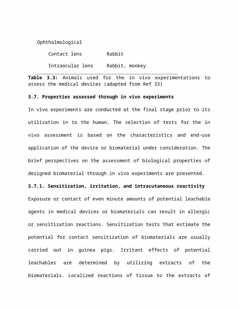
Ophthalmological
Contact lens Rabbit
Intraocular lens Rabbit, monkey
Table 3.3: Animals used for the in vivo experimentations to assess the medical devices (adapted from Ref 33)
3.7. Properties assessed through in vivo experiments
In vivo experiments are conducted at the final stage prior to its utilization in to the human. The
selection of tests for the in vivo assessment is based on the characteristics and end-use
application of the device or biomaterial under consideration. The brief perspectives on the
assessment of biological properties of designed biomaterial through in vivo experiments are
presented.
3.7.1. Sensitization, irritation, and intracutaneous reactivity
Exposure or contact of even minute amounts of potential leachable agents in medical devices or
biomaterials can result in allergic or sensitization reactions. Sensitization tests that estimate the
potential for contact sensitization of biomaterials are usually carried out in guinea pigs. Irritant
effects of potential leachables are determined by utilizing extracts of the biomaterials. Localized
reactions of tissue to the extracts of biomaterials are determined through intracutaneous
(intradermal) reactivity tests. Preparation of the test material and/or extract solution and the
choice of the solvents chosen used to determine sensitization or irritation or intracutaneous
reactivity must have physiological relevance.
3.7.2. Systemic toxicity, subacute and subchronic toxicity
Systemic toxicity tests estimate the potential harmful effects of either single or multiple
exposures of the medical devices, biomaterials and/or their extracts, during a period of less than
24 h. These tests also include pyrogenicity (fever-producing) testing. The basic considerations
involved in testing protocol are the form and area of the material, the thickness and the surface
area to extraction vehicle volume.

Depending on the intended application of the biomaterial, oral, dermal, inhalation, intravenous,
intraperitoneal, or subcutaneous application, animals of the choice mice, rats, or rabbits are usual
and, of the test substance may be used. Acute toxicity is considered to be the adverse effect,
which occurs after administration of a single dose or multiple doses of a test sample given within
24 h. If the adverse effects occur after administration of a single dose or multiple doses of a test
sample per day given during a period of from 14 to 28 days then it is called subacute toxicity
(repeat dose toxicity) and the adverse effects occurring after administration of a single dose or
multiple doses of a test sample per day given during a part of the life span, usually 90 days but
not exceeding 10% of the life span of the animal is called subchronic toxicity.
3.7.3. Genotoxicity
If in vitro test results indicate potential genotoxicity, in vivo genotoxicity tests are carried out.
Initially, at least three in vitro assays are used, and two of these assays should utilize mammalian
cells. While the initial in vitro assays cover the three levels of genotoxic effects: DNA effects,
gene mutations, and chromosomal aberrations, the in vivo genotoxicity tests cover the
micronucleus test, the in vivo mammalian bone marrow cytogenetic tests, chromosomal analysis,
the rodent dominant lethal tests, the mammalian germ cell cytogenetic assay, the mouse spot test,
and the mouse heritable translocation assay tests. Genotoxicity tests are performed with
appropriate extracts or dissolved materials using media as suggested by the known composition
of the biomaterial. The most common vivo genotoxicity test is the rodent micronucleus test.
3.7.4. Implantation
Implantation tests assess the local pathological effects on living tissue of a sample of a material
that is surgically implanted in to tissue appropriate for the intended application of the device.
Evaluation of the local pathological effects is carried out both at the gross level and the

microscopic level. Histological (microscopic) evaluation is utilized to characterize various
biological response parameters. Mice, rats, guinea pigs, or rabbits are the usual animals utilized
for short-term implantation evaluation out to 12 weeks. For longer-term testing in subcutaneous
tissue, muscle or bone, animals such as rats, guinea pigs, rabbits, dogs, sheep, goats, pigs and
other animals with relatively long-life expectancy are usually chosen. To evaluate a medical
device, larger species are utilized. In general, sheep are chosen for testing substitute heart valves,
whereas for ventricular assist devices and total artificial hearts calves are usually the animal of
choice.
3.7.5. Hemocompatibility
Hemocompatibility tests in in vivo examination evaluate the effects on blood and/or blood
components by blood-contacting medical devices or materials. Thrombosis, coagulation,
platelets, hematology, and immunology (complement and leukocytes) are the five test categories
indicated from the ISO standards perspective for hemocompatibility evaluation. In in vivo testing
of animals, species differences in blood reactivity are considered, and these differences may limit
the predictability of any given test in the human clinical situation. Although species differences
may complicate hemocompatibility evaluation, the utilization of animals in short- and long-term
testing is considered to be appropriate for evaluating thrombosis and tissue interaction.
3.7.6. Chronic toxicity
Chronic toxicity tests in in vivo experiments are considered as an extension of subchronic
(subacute) toxicity testing, and both may be evaluated in an appropriate experimental protocol or
study. Chronic toxicity tests determine the effects of either single or multiple exposures to
medical devices, materials, and/or their extracts during a period of at least 10% of the life span of
the test animal, e.g. over 90 days in rats [33].
3.7.7. Carcinogenicity
Carcinogenicity tests determine the tumorigenic potential of medical devices, materials, and/or
their extracts from either single or multiple exposures or contacts over a period of the major
portion of the life span of the test animal [33]. If data from other sources suggest a tendency for
tumor induction then the carcinogenicity tests will be conducted. In a single experimental study,
both carcinogenicity (tumorigenicity) and chronic toxicity may be studied. Controls of
comparable form and shape are generally used in carcinogenicity testing. The use of appropriate
controls is essential, since the animals when spontaneously develop tumors, a statistical
comparison between the test biomaterial/device and the controls is always necessary. The
commonly used control materials are polyethylene implants.
3.7.8. Reproductive and developmental toxicity
The concerned in vivo tests evaluate the potential effects of medical devices, materials, and/or
their extracts on reproductive function, embryonic development (teratogenicity), and prenatal
and early postnatal development. The application site of the device must be considered and tests
and/or bioassays should only be conducted when the device has potential impact on the
reproductive potential of the subject [33].
3.7.9. Biodegradation
The effects of a biodegradable material and its biodegradation products on the tissue response are
determined through biodegradation tests. The amount of degradation; the nature, the origin and
the qualitative and quantitative assessment of the degradation products (e.g. impurities, additives,
corrosion products, bulk polymer, etc); and leachable agents in adjacent tissues and distant
organs are specially focused through biodegradation tests. The test materials comparable to that
of degradation products may be prepared and studied to determine the anticipated biological

responses of the products in long-term implants. This provides in advance supporting
information on the effects of the implant with in short duration rather than anticipation for longer
time.
3.7.10. Immune responses
Determination of immune response is needed with modified natural tissue implants or some
other biomaterial device. Immunotoxicity is any adverse effect on the function or structure of the
immune system or other systems as a result of an immune system dysfunction. The potential
immunological effects include hypersensitivity, chronic inflammation, immunosuperession,
immunostimulation, autoimmunity. The potential immunological responses are histopatholoical
changes, humoral responses, host resistance, clinical symptoms, cellular responses.
3.8. Applications of biomaterials
Although the tissues and structures of the body perform for an extended period of time in most
people, they do suffer from a variety of destructive processes, including fracture, infection, and
cancer that cause pain, disfigurement, or loss of function. Hence, under these circumstances, it
has become necessary to remove the diseased tissue and replace it with some suitable synthetic
materials that perform the normal activity of the regular living tissue. The purpose of this is
fulfilled through biomaterials. The primary reasons using biomaterials is to physically replace
hard or soft tissues that have become damaged or destroyed through some pathological process
[61]. Some of the areas of applications where the biomaterials are successfully utilized are dealt
here.
3.8.1. Orthopedic applications
The use of biomaterials for orthopedic implant devices is one of the major achievements in the
field of medicine. The structure of freely movable (synovial) joints, such as the hip, knee,

shoulder, ankle and elbow are affected by both osteoarthritis and rheumatoid arthritis. It has been
possible to replace these joints with prostheses since the advent of anesthesia, antisepsis, and
antibiotics, and the relief of pain and restoration of mobility is well known to hundreds of
thousands of patients. Some of the biomaterials used for the orthopedic applications are shown in
Fig 3.1.
Fig 3.1: Orthopedic applications of biomaterials (a) artificial hip (b) artificial knee.
3.8.2. Ophthalmologic applications
The tissues of the eye can suffer from several diseases, leading to reduced vision and eventually,
blindness. The devices, such as spectacles used to correct the eye vision are the external devices.
However, the contact lenses being intimate contact with the tissues of the eye, are subject to the
same regulations that govern the use of implant materials. Apart from this, artificial cornea,
artificial endothellum, intraocular lenses and implants for vitreous and glaucoma are also
available. Artificial cornea and intraocular lenses are shown in Fig 3.2.
Fig 3.2: Ophthalmologic applications of biomaterials (a) artificial cornea hip (b) intraocular lenses.
3.8.3. Cardiovascular applications
One of the most prominent application areas for biomaterials is cardiovascular applications. The
problems that arise from failure of heart valves and arteries can be successfully treated with
implants. The heart valves suffer from structural changes that prevent the valve from either fully
opening or fully closing, and the diseased valve can be replaced with a variety of substitutes. The
problem of atherosclerosis, occurs by blocking of fatty deposits in the vessels and the blocking of
arteries can be solved by replacing segments with artificial arteries. Some of the biomaterial
devices used for cardiovascular purpose are cardiopulmonary bypass system, heart valves,
vascular grafts, stents, pacemakers and complete artificial heart. Heart valve and artificial heart
are shown in Fig 3.3.

Fig 3.3: Cardiovascular applications of biomaterials (a) heart valve (b) artificial heart.
3.8.4. Dental applications
The tooth and supporting gum tissues has maximum chances to get destroyed by bacterially
controlled diseases, since the tissues are present in the mouth which is the only passage for food
and liquids. Dental caries (cavities), the demineralization and dissolution of teeth associated with
the metabolic activity in plaque (a film of mucus that traps bacteria on the surface of the teeth),
can cause extensive tooth loss. Teeth in their entirety and segments of teeth both can be replaced
or restored by a variety of materials that form the biomaterials. Depending upon requirement
there are many verities of dental implants are available. The endosteal root form dental implant
and biomaterial tooth gums are presented in Fig 3.4.
Fig 3.4: Dental applications of biomaterials (a) the endosteal root form dental implant (b) biomaterial tooth gums
3.8.5. Wound dressing applications

Wound dressings are usually the external medical support given to the wound by the surgical
team to prevent the flowing of the blood, exudates and the proteins. The scaffolds that provide
the relief at the wound site such as cellulosic materials or the hydrogels or the films developed
either from natural or artificial materials are broadly considered as wound dressing materials [62-
66]. The antimicrobial wound dressings also came in to existence which not only act as normal
wound dressing but also provide hygienic atmosphere around the wound. Apart from this,
multiple therapies employing biomaterials for wound management have been developed. One of
the example for commercially available biosynthetic wound dressing is Biobrane® (Smith &
Nephew, London, UK), a biocomposite dressing composed of a silicon/nylon matrix in which
porcine dermal collagen has been embedded. Fig 3.5 showing biosynthetic wound dressing
Biobrane and its application on wound.
Fig 3.5: Wound dressing applications of biomaterials (a) biosynthetic wound dressing Biobrane (b) application of Biobrane as dressing on the wound.
3.8.6. Other applications
The other applications of biomaterials include drug delivery applications, organ implantations
like breast and development of artificial organs like artificial skin, etc. For drug delivery
applications, the products are designed to increase the duration of orally administered drugs and
consisted of small spheres with a soluble coating. By using coatings having varying thicknesses,

dissolution times could be varied, prolonging the action of the therapeutic agent. The artificial
skin and biomaterial breast implant material are shown in Fig 3.6.
Fig 3.6: other applications of biomaterials (a) artificial skin (b) breast implant biomaterial.
3.9. Future directions in biomaterials
Biomaterials are the backbone of the medical device industry, a critical element of health care.
Biomaterials have made a great impact on medicine. Currently, there are thousands of hard and
soft tissue products, biomedical devices, pharmaceutical and diagnostic products and disposable
materials are available at the medical market for human beneficial. However, numerous
challenges are there to be solved. Nowadays, modern clinical procedures such as preventing and
curing main genetic diseases are become significant and new medical demands cause the change
of the biomaterial products. Hence, the future trend has to combine the mechanically superior
metals and the excellent biocompatibility and biofunctionality of ceramics and polymers to
obtain the most desirable clinical performance of the implants. The biomaterial scientists and
engineers should increase the vicinity of applications by integration of biomaterials with
molecular biology, biochemistry, genetics, physics and other areas of sciences. This integration
supports the researchers, material scientists and tissue engineers to design the products from
molecular level from cell to tissue.

3.10. Conclusions
The day-to-day enormous increased demand in the medical field for the bio-alternatives which
could able to perform the living activities of bodily organs in their absence has raised the interest
of the researchers to design novel biomaterials. Based on this, in this chapter, discussion on
biomaterials and its design and development has been carried out. Apart from this, the necessary
requirements or characteristics of the biomaterials are also discussed. Further, the assessment of
designed biomaterial devices or implants and the applications of the biomaterials, which play a
significant one, are also discussed. In over all, information has been covered ranging from the
basic definition of biomaterial to the design, development and applications. In over all, the
crucial study on biomaterials has been made which provides the necessary basics required for
designing biomaterial devices and implants by the emerging researchers, tissue engineers and
material scientists.
Acknowledgements
The author (IF 110192) wish to acknowledge the Department of Science & Technology (DST,
INIDA) and Ministry of Science & Technology for providing financial assistance through
Innovation In Science Pursuit for Inspired Research (INSPIRE) programme. The authors (KVP)
thank the Fondecyt Proyecto No.: 3130748 Chile (South America).
References
[1] Black J.The education of the biomaterialist: Report of a survey, 1980-81. J Biomed Mater
Res 1982;16(2):159-67.
[2] Kalita SJ. Nanostructured Biomaterials. Springer. Nan Sci Tec 2008,168-219.
[3] Bronzino JD. The biomedical engineering handbook, second edition. 2000;2.

[4] Helmus MN, Gibbons DF, Cebon D. Biocompatibility: meeting a key functional requirement
of next-generation medical devices. Toxicol Pathol 2008;36:70-80.
[5] Tathe A, Ghodke M, Nikalje AP. A brief review: biomaterials and their application.
International Journal of Pharmacy and Pharmaceutical Sciences 2010;2(4):19-23.
[6] Ratner BD, Hoffman AS. Biomaterials Science: An introduction to materials in medicine.
Academic press 1996.
[7] Crubezy E, Ludes B, Poveda JD, Clayton J, Crouau-Roy B, Montagnon D, Identification of
mycobacterium dna in an egyptian pott’s disease of 5,400 years old. C R Acad Sci III
1998;321:941–51.
[8] Parida P, Mishra SC. Biomaterials in medicine, UGC sponsored national workshop on
innovative experiments in physics 2012;9-10.
[9] Pramanik S, Agarwal AK, Rai KN; Chronology of total hip joint replacement and materials
development, Trends Biomater. Artif. Organs 2005;19(1):15-26.
[10] Chakrabarty GV. Biomaterials: Metallic Implant Materials, technology, review essays 2011.
[11] Batista G, Ibarra M, Ortiz J, Villegas M. Engineering biomechanics of knee replacement,
applications of engineering mechanics in medicine, GED- University of Puerto Rico,
Mayagüez; 2004,1-12.
[12] Hwal suh. Recent advances in biomaterials. Yonsei medical journal 1998;39(2),87-96.
[13] Yoruç ABH and Şener BC. Biomaterials, A roadmap of biomedical engineers and
milestones 2012.
[14] Williams DF. Review: Tissue-biomaterial interactions. J Mat Sci 1987;22(10):3421-45.
[15] Isa ZM, Hobkirk IA. Dental implants: Biomaterial, biomechanical and biological
considerations. Annal Dent Univ Malaya 2000;7: 27-35.

[16] Anselme K. Osteoblast adhesion on biomaterials, Review. Biomaterials 2000;21:667-81.
[17] Mehdizadeh M, Yang J. Design strategies and applications of tissue bioadhesives Macromol
Biosci 2013;13:271–288
[18] Anderson JM. Biological responses to materials. Annu Rev Mater Res 2001;31:81–110.
[19] Niinomi M, Recent metallic materials for biomedical applications. Metal Mater Transac A
2002;33A:477-86.
[20] Silver FH, Christiansen DL. Biomaterials science and biocompatibility, Springer-Verlag
1999;87-120.
[21] Iftekhar A. Standard handbook of biomedical engineering and design. Chapter12:
Biomedical Composites McGraw-Hill Companies 2004.
[22] Dorozhkin SV. Biocomposites and hybrid biomaterials based on calcium orthophosphates.
Biomatter 2011;1(1):3-56.
[23] Vallet-Regí M. Ceramics for medical applications. J Chem Soc Dalton Trans 2001; 2:97-
108.
[24] Jordan DR, Munro SM, Brownstein S, Gilberg SM, Grahovac, SZ. A synthetic
hydroxyapatte implant: The so-called counterfeit implant. Ophthalmic plast rec
1998;14(4):244-49.
[25] Orlovskii VP, Komlev VS, Barinov SM. Hydroxyapatite and Hydroxyapatite-Based
Ceramics. Inorg Mater 2002;38(10):973–984.
[26] Bermejo R, Danzer R. High failure resistance layered ceramics using crack bifurcation and
interface delamination as reinforcement mechanisms. Engng Fract Mech 2010;77(11):2126–
2135.

[27] Anderson JM. Mechanism of inflammation and infection with implanted devices.
Cardiovasc Pathol 1993;2:33S-41S.
[28] Anderson JM. Inflammatory response to implants. ASAIO J 1988;11:101–7.
[29] Cotran RZ, Kumar V, Robbins SL. Pathologic basis of disease. Philadelphia: Saunders. 6 th.
ed. 1999:50–112.
[30] Gallin JI, Synderman R. Inflammation: Basic principles and clinical correlates. New York:
Raven. 2nd. ed. 1999.
[31] Weissman G, Smolen JE, Korchak HM. N Engl J Med 1980;303:27–34.
[32] Salthouse TN. Cellular enzyme activity at the polymer-tissue interface: A reviewJ Biomed
Mater Res 1976;10:197–229.
[33] Clark RA, Lanigan JM, DellePelle P, Manseau E, Dvorak HF, Colvin RB. J Invest Dermatol
1982;79:264-69.
[34] Tang L, Eaton JW. Fibrinogen mediates acute inflammatory responses to biomaterials. J
Exp Med 1993;178:2147-56.
[35] Tang, L. Mechanisms of fibrinogen domains: biomaterial interactions. J Biomat Sci Polym
Ed 9, 1998:1257-66.
[36] Broadley KN, Aquino AM, Woodward SC, Buckley-Sturrock A, Sato Y, et al. Lab Invest
1989;61:571-75.
[37] Sporn MB, Roberts AB. Peptide growth factors are multifunctional. Nature 1988;332:217-
19.
[38] Muller G, Behrens J, Nussbaumer U, B¨ohlen P, BirchmeierW. Proc Natl Acad Sci
1987;84:5600-4.
[39] Madri JA, Pratt BM, Tucker AM. Phenotypic modulation of endothelial cells by
transforming growth factor-beta depends upon the composition and organization of the
extracellular matrix. J Cell Biol 1988;106:1375-84.
[40] Wahl SM, Hunt DA, Wakefield LM, Roberts AB, Sporn MB. Transforming growth factor
type beta induces monocyte chemotaxis and growth factor production. Proc Natl Acad Sci
1987;84:5788-92.
[41] Ignotz R, Endo T, Massagu´e J. Regulation of fibronectin and type I collagen mRNA levels
by transforming growth factor-p. J Biol Chem 1987;262:6443-46.
[42] Ganz T. Neutrophils and host defense. Ann Intern Med 1988;109:127–42.
[43] Henson PM, Johnston RB Jr. Tissue injury in inflammation: oxidants, proteinases, and
cationic proteins. 1987. J. Clin. Invest. 79:669–74.
[44] Malech HL, Gallin JI. Current concepts: immunology. Neutrophils in human diseases.. N
Engl J Med 1987;317:687–94.
[45] Johnston RB Jr. Current concepts: immunology. Monocytes and macrophages. N Engl J
Med 1988;318:747–52.
[46] Williams GT, Williams WJ. Granulomatous inflammation--a review.J Clin Pathol
1983;36(7):723–33.
[47] Rae T. The macrophage response to implant materials. Biocompatibility. Crit Rev
1986;2:97–126.
[48] Anderson JM. Biologicalresponses to materials. Annu Rev Mater Res 2001;31:81–110.
[49] Chambers TJ, Spector WG. Review Inflammatory giant cells. Immunobiology
1982;161:283–89.

[50] Clark RAF, Henson PM. The molecular and cellular biology of wound Repair. Plenum
Press, New York 1988:3-23.
[51] Hunt TK, Heppenstall RB, Pines E, Rovee D. Soft and hard tissue repair: biological and
clinical aspects, New York: Praeger Scientific 1984;2:283-92.
[52] Davis JR. Handbook of Materials for Medical Devices. ASM International 2003.
[53] Lönnroth EC, Dahl JE. Cytotoxicity of dental glass ionomers evaluated using dimethyl
thiazoldiphenyltetrazolium and neutral red tests. Acta Odontol Scand 2001;59(1):34-39.
[54] Cory AH, Owen TC, Barltrop JA, Cory JG. Use of an aqueous soluble tetrazolium/formazan
assay for cell growth assays in culture. Cancer Commun. 1991;3(7):207-212.
[55] MacGregora JT, Collinsa JM, Sugiyamab Y, et al. In vitro human tissue models in risk
assessment: Report of a consensus-building workshop. Toxicol Sci 2001;59 (1):17-36.
[56] Ashammakhi N, Reis R, Chiellini F; Topics in tissue engineering, Chen Q, Roether JA and
Boccaccini AR. Chapter 6: Tissue Engineering Scaffolds from Bioactive Glass and
Composite Materials 2008.
[57] Scholz MS, Blanchfield JP, Bloom LD, Coburn BH, Elkington M, Fuller JD, Gilbert ME,
Muflahi SA, Pernice MF, Rae SI, Trevarthen JA, White SC, Weaver PM, Bond IP. The use
of composite materials in modern orthopaedic medicine and prosthetic devices: A review.
Compos Sci Technol 2011;71: 1791–1803.
[58] Berne RM, Levy MN, Koeppen BM, Stanton BA. Berne and Levy Physiology. 6 th Ed. St.
Louis: Mosby 2009.
[59] Guyton AC, Hall JE. Textbook of medical physiology. 12 th Ed. Philadelphia: Elsevier
Saunders 2010.

[60] Gartner LP, Hiatt JL, Strum JM. Cell Biology and Histology. Baltimore, Lippincott
Williams & Wilkins 2011.
[61] Benson JS, Boretos JW, Biomaterials and the future of medical devices. Med Device Diag
Ind 1995;17(4):32–37.
[62] Jayaramudu T, Raghavendra GM, Varaprasad K, Sadiku R, Raju KM. Development of
novel biodegradable Au nanocomposite hydrogels based on wheat: for inactivation of
bacteria. Carbohydr Polym 2013;92:2193–200.
[63] Varaprasad K, Murali Mohan Y, Vimala K, Mohana Raju K, Synthesis and characterization
of hydrogel‐silver nanoparticle‐curcumin composites for wound dressing and antibacterial
application. J Appl Polym Sci 2011;121, 784–96.
[64] Raghavendra GM, Jayaramudu T, Varaprasad K, Sadiku R, Ray SS, Raju KM. Cellulose–
polymer–Ag nanocomposite fibers for antibacterial fabrics/skin scaffolds. Carbohydr Polym
2013;93:553–60.
[65] Raghavendra GM, Jayaramudu T, Varaprasad K, Ramesh S, Raju KM. Microbial resistant
nanocurcumin-gelatin-cellulose fibers for advanced medical applications. RSC Adv 2014;4:
3494–501.
[66] Jayaramudu T, Raghavendra GM, Varaprasad K, Sadiku R, Ramam K, Raju KM. Iota-
Carrageenan-based biodegradable Ag0 nanocomposite hydrogels for the inactivation of
bacteria. Carbohydr Polym 2013;95:188–94.