Embed Size (px)
Citation preview
Number 10 Mar 2012
3-DIMENSIONAL REPRODUCTION TECHNIQUES TO PRESERVE AND SPREAD
PALEONTOLOGICAL MATERIAL – A CASE STUDY WITH A DIPLODOCID SAUROPOD NECK
Tschopp, Emanuel1,2 and Dzemski, Gordon3
1- CICEGe, Faculdade de Ciências e Tecnologia, FCT, Universidade Nova de Lisboa, 2829-516 Caparica, Portugal 2- Museu da Lourinhã, Rua João Luis de Moura, 2530-158 Lourinhã, Portugal. 3- Institut für Biologie und Ihre Didaktik, Universität Flensburg, Germany.
ABSTRACT The protection and preservation of irrecoverable, sensitive and fragile objects in museums, exhibitions and collections is critical in various research fields like cultural heritage, human sciences and paleontology. Lately, digitization of such endangered specimens proved to be a valuable tool to convert the objects into a digital form. However, in order to exploit all possibilities that such data could provide for educational and research purposes, it can be useful to transform the digital material back into a physical form. In this case study, a neck of a diplodocid sauropod dinosaur was digitally reproduced by 3D printing (a variant of rapid prototyping). The process is described in detail, and compared to the more classical reproduction using CNC-mills. CNC-milling is an inexpensive and accurate reproduction technology for large objects, and especially well-designed for producing durable casts. On the other hand, 3D printing is highly accurate to create small or complex objects, but is more expensive and yields more fragile physical objects. As accuracy of the complex shapes of the diplodocid cervical vertebrae was required in order to use them for research, 3D printing was preferred over CNC-milling in this case.
Keywords: reproduction, preservation, 3D modelling, reverse engineering, rapid prototyping, casting, real
virtuality
RESUMO [in Portuguese]
A protecção e preservação de objectos irreparáveis, sensíveis e frágeis pertencentes a museus, exibições e colecções é crítica em várias áreas de investigação como património cultural, humanidades, paleontologia. Ultimamente, a digitalização de espécimes raros tem-se revelado um método eficaz para transformar objectos num formato digital, portanto, fisicamente indestructível. Contudo, de modo a se poderem explorar todas as possibilidades que este tipo de informação digital pode proporcionar para fins de educação e investigação, pode ser útil transformar o material digital de volta à sua forma física. Neste caso de estudo, uma sequência parcial de vértebras cervicais de um dinossauro saurópode diplodocóide foi reproduzida através de impressão 3D (uma variante de prototipagem rápida). Esse processo é descrito em detalhe, e comparado com o método clássico de reprodução CNC-milling. CNC-milling é um método barato e preciso para reproduces de objectos de amplas dimensões, e especialmente bem conseguido para reproduzir réplicas duráveis. Por outro lado, a impressão 3D é extremamente precisa a criar objectos pequenos e complexos, mas é mais cara e produz objectos mais frágeis. Devido ao facto de as vertebras cervicais dos diplodocóides serem extremamente complexas, o método de impressão 3D é preferível em relação ao método de CNC-milling neste caso. How to cite this article: Tschopp, E. and Dzemski, G., 2012. 3-Dimensional Reproduction Techniques to preserve and spread paleontological material – a case study with a diplodocid Sauropod neck. Journal of Paleontological Techniques, 10: 1-8.
www.jpaleontologicaltechniques.org ISSN: 1646-5806
Tschopp and Dzemski, 2012: 3-DIMENSIONAL REPRODUCTION TECHNIQUES
2 ● Journal of Paleontological Techniques
INTRODUCTION Access to original material or specimens is limited for palaeontologists due to a number of reasons. Fossil material is rare and difficult to access (e.g. distance, museum policies, intense scientific interest). Fossil bones can be heavy or fragile to transport, or the desired element is mounted and on public display in a museum. Three-dimensional reproduction techniques therefore offer a great solution for archiving significant data of paleontological objects (Breithaupt et al., 2004; Remondino et al., 2005). Whereas in the early years of paleontology, physical casting of fossils has proved a valuable alternative way for researchers to undertake their investigations, as casts are usually lighter, less fragile, and easier to handle than original material, in the last 15 to 20 years alternative technologies have been developed to produce digital copies of original specimens (see Zollikofer and Ponce de León, 1995; Chapman et al., 1998; or Andersen et al., 2001 for some of the earliest attempts in Paleontology). Laser, x-ray, or magnet scanner, mechanical digitizers and photogrammetry methods transform matter into bits and bytes to create, manipulate, recreate and change objects assisted by a computer with editing software. For paleontological purposes the digital models can be stored and used for scientific research, spread via internet, or can be displayed virtually in museum exhibitions (Johnston et al., 2004; Mallison et al., 2009). Entire specimens can be measured providing accurate results for further research (Deck et al., 2004). Using simulation methods, bones can be articulated with each other without having to handle them manually – and thus without subjecting them to possible abrasion or damage due to real bone to bone contact (Chapman et al., 2001). They can be subjected to retrodeformation methods in order to re-establish a shape that is supposed to be closer to the original morphology (Motani et al., 2005; Kahzdan et al., 2009; Tschopp and Dzemski, in review now). Moreover, digital models of skeletal elements can be virtually connected with tendons and muscles to reconstruct the entire animal and to study in vivo motion patterns and biomechanical hypotheses (Walters et al., 2001; Bimber et al., 2002; Dzemski and Christian, 2007, 2010).
On the other hand, scientists often stress the importance of studying three-dimensional physical objects to manipulate them by hand and realize the dimensions of the dynamic
elements. Physical models can furthermore be used for education and in museum exhibits, and have a great potential to indirectly protect the original objects (Mallison, 2007; Remondino, 2007; Schlader et al., 2007). Instead of the traditional casting process described above, there are now several ways to transform a digital object to a physical model. They can be subdivided into additive (layer by layer “printing”) and subtractive (e.g. carving, milling) techniques. The additive method used for this case study (3D printing), as well as CNC-milling as example for a subtractive technique will herein be described, and their usage, advantages, and possibilities will be compared and discussed regarding the reproduction of the neck of the diplodocid sauropod SMA 0004 for research purposes.
Abbreviations: CAD: Computer Aided Design; CAM: Computer Aided Manufacturing; CNC: Computer Numerical Controlled; SMA: Sauriermuseum Aathal, Switzerland; STL: Standard Triangulation Language (a file format for 3D models).
METHODS
In order to reproduce a physical copy from the digital model, the following steps are necessary: scanning the object with a 3D-scanner, prepare the digital objects with 3D-software for the reproduction process, and then transform the digital model to physical models.
Scanning
Numerous ways to to create a digital model of the fossil have been reported (see Wilhite, 2003; Hohloch and Mallison, 2005; Mallison et al., 2009; Möller et al., 2009; Dzemski and Christian, 2010). The most commonly used techniques, as well as their advantages and disadvantages, are listed in Table 1.
In order to obtain proper physical models based on scanning data, it is crucial to be able to scan the original material with great accuracy (preferably below 1 mm, otherwise minute structures like small foramina would get lost). The structured light scanner Atos I from GOM, Germany, has proved a highly valuable tool in various research projects (Dzemski and Christian, 2007, 2010; Christian and Dzemski, 2007, 2011), and was thus also used for the scanning of SMA 0004. This industrial optical measuring machine is based on the principle of triangulation. Projected fringe patterns are
Tschopp and Dzemski, 2012: 3-DIMENSIONAL REPRODUCTION TECHNIQUES
3 ● Journal of Paleontological Techniques
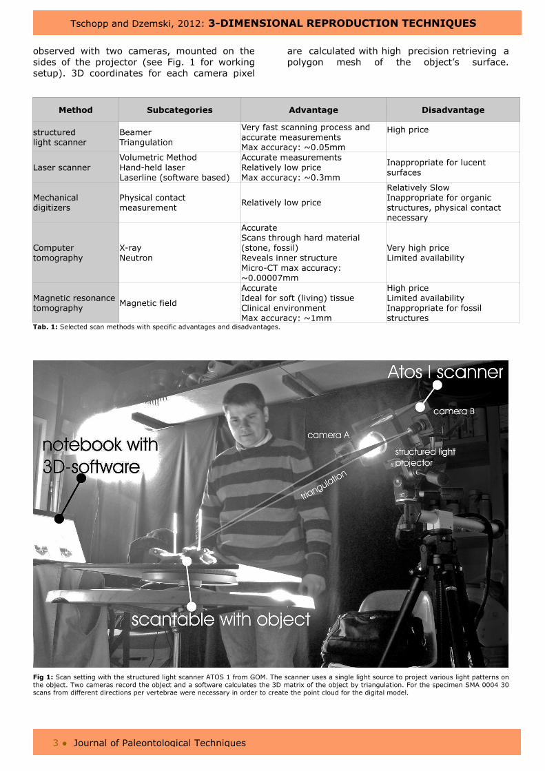
observed with two cameras, mounted on the sides of the projector (see Fig. 1 for working setup). 3D coordinates for each camera pixel
are calculated with high precision retrieving a polygon mesh of the object’s surface.
Method Subcategories Advantage Disadvantage
structured light scanner
Beamer Triangulation
Very fast scanning process and accurate measurements Max accuracy: ~0.05mm
High price
Laser scanner Volumetric Method Hand-held laser Laserline (software based)
Accurate measurements Relatively low price Max accuracy: ~0.3mm
Inappropriate for lucent surfaces
Mechanical digitizers
Physical contact measurement
Relatively low price
Relatively Slow Inappropriate for organic structures, physical contact necessary
Computer tomography
X-ray Neutron
Accurate Scans through hard material (stone, fossil) Reveals inner structure Micro-CT max accuracy: ~0.00007mm
Very high price Limited availability
Magnetic resonance tomography
Magnetic field
Accurate Ideal for soft (living) tissue Clinical environment Max accuracy: ~1mm
High price Limited availability Inappropriate for fossil structures
Tab. 1: Selected scan methods with specific advantages and disadvantages.
Fig 1: Scan setting with the structured light scanner ATOS 1 from GOM. The scanner uses a single light source to project various light patterns on the object. Two cameras record the object and a software calculates the 3D matrix of the object by triangulation. For the specimen SMA 0004 30 scans from different directions per vertebrae were necessary in order to create the point cloud for the digital model.
Tschopp and Dzemski, 2012: 3-DIMENSIONAL REPRODUCTION TECHNIQUES
4 ● Journal of Paleontological Techniques
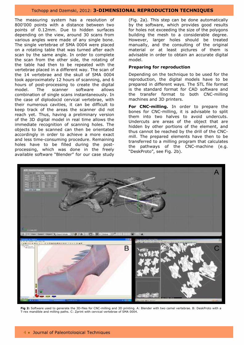
The measuring system has a resolution of 800'000 points with a distance between two points of 0.12mm. Due to hidden surfaces depending on the view, around 30 scans from various angles were made of any single bone. The single vertebrae of SMA 0004 were placed on a rotating table that was turned after each scan by the same angle. In order to complete the scan from the other side, the rotating of the table had then to be repeated with the vertebrae placed in a different way. The scan of the 14 vertebrae and the skull of SMA 0004 took approximately 12 hours of scanning, and 6 hours of post-processing to create the digital model. The scanner software allows combination of single scans instantaneously. In the case of diplodocid cervical vertebrae, with their numerous cavities, it can be difficult to keep track of the areas the scanner did not reach yet. Thus, having a preliminary version of the 3D digital model in real time allows the immediate recognition of scanning holes. The objects to be scanned can then be orientated accordingly in order to achieve a more exact and less time-consuming procedure. Remaining holes have to be filled during the post-processing, which was done in the freely available software “Blender” for our case study
(Fig. 2a). This step can be done automatically by the software, which provides good results for holes not exceeding the size of the polygons building the mesh to a considerable degree. However, larger holes should be treated manually, and the consulting of the original material or at least pictures of them is advisable in order to obtain an accurate digital model.
Preparing for reproduction
Depending on the technique to be used for the reproduction, the digital models have to be prepared in different ways. The STL file format is the standard format for CAD software and the transfer format to both CNC-milling machines and 3D printers.
For CNC-milling. In order to prepare the bones for CNC-milling, it is advisable to split them into two halves to avoid undercuts. Undercuts are areas of the object that are hidden by other portions of the element, and thus cannot be reached by the drill of the CNC-mill. The prepared elements have then to be transferred to a milling program that calculates the pathways of the CNC-machine (e.g. “DeskProto”, see Fig. 2b).
Fig 2: Software used to generate the 3D-files for CNC-milling and 3D printing. A: Blender with two camel vertebrae. B: DeskProto with a T-rex mandible and milling paths. C: Zprint with cervical vertebrae of SMA 0004.
Tschopp and Dzemski, 2012: 3-DIMENSIONAL REPRODUCTION TECHNIQUES
5 ● Journal of Paleontological Techniques
CNC-mills (Fig. 3b) are classified in devices from 3-axes up to 7-axes. The more axes a machine has, the more degrees of freedom are possible: 6- or 7-axes CNC-mills are capable of milling a 360 degree object without interruptions, and are thus ideal to produce precise replicates out of wood, synthetic material, or metal for educational and scientific purposes (Deck et al., 2007). If one has only a 3-axes device at his disposal, an extra step in the preparation of the digital models is needed: the limited mobility requires to split the virtual model in several parts and load it into a control program (e.g. “WINPC-NC”) to control the CNC machines. Subsequent to the milling process, the different parts of the models have to be assembled in order to conclude the 3D replicas (Fig. 4c).
For Rapid Prototyping. As 3D printers are assembling the models layer for layer, the digital 3D object has to be divided into thin, printable slices. For this, the 3D-printer software Zprint from Z-Corp. was used (Fig. 2c). Given the limited size for a printable object in the Z-Corp. device (Fig. 3a), larger objects have to be subdivided into smaller parts for this process as well – or they can be reproduced at a smaller scale. Especially for research purposes it can be very handy to have small physical models of large bones. This is particularly useful for elements of sauropod dinosaurs, and was therefore also done in the case of the vertebrae of SMA 0004. The digital models were rescaled by 1:4 before slicing them virtually, and loading them into Zprint. However, one has to take into consideration that by scaling elements down, small details on the bones get lost.
Reproduction process
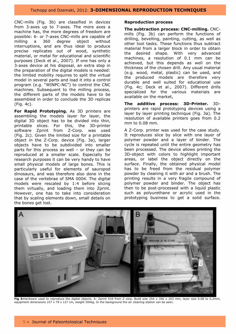
The subtraction process: CNC-milling. CNC-mills (Fig. 3b) can perform the functions of drilling, bevelling, painting, cutting, as well as other tool tasks. These functions thus subtract material from a larger block in order to obtain the desired shape. With very advanced machines, a resolution of 0.1 mm can be achieved, but this depends as well on the thickness of the chosen drill. Any usual material (e.g. wood, metal, plastic) can be used, and the produced models are therefore very durable and well suited for educational use (Fig. 4c; Deck et al., 2007). Different drills specialized for the various materials are available on the market,
The additive process: 3D-Printer. 3D-printers are rapid prototyping devices using a layer by layer printing technique (Fig. 3a). The resolution of available printers goes from 0.2 mm to 0.08 mm.
A Z-Corp. printer was used for the case study. It reproduces slice by slice with one layer of polymer powder and a layer of binder. The cycle is repeated until the entire geometry has been processed. The device allows printing the 3D-object with colors to highlight important areas, or label the object directly on the surface. Finally, the obtained physical model has to be freed from the residual polymer powder by cleaning it with air and a brush. The printing results in a very fragile compound of polymer powder and binder. The object has then to be post-processed with a liquid plastic such as polyurethane or acrylic used in the prototyping business to get a solid surface.
Fig 3:Hardware used to reproduce the digital objects. A: Zprint 510 from Z corp. Build size 254 x 356 x 203 mm, layer size 0.08 to 0.2mm, equipment dimensions 107 x 79 x 127 cm, weight 204kg. In the background the air cleaning station can be seen.
Tschopp and Dzemski, 2012: 3-DIMENSIONAL REPRODUCTION TECHNIQUES
6 ● Journal of Paleontological Techniques
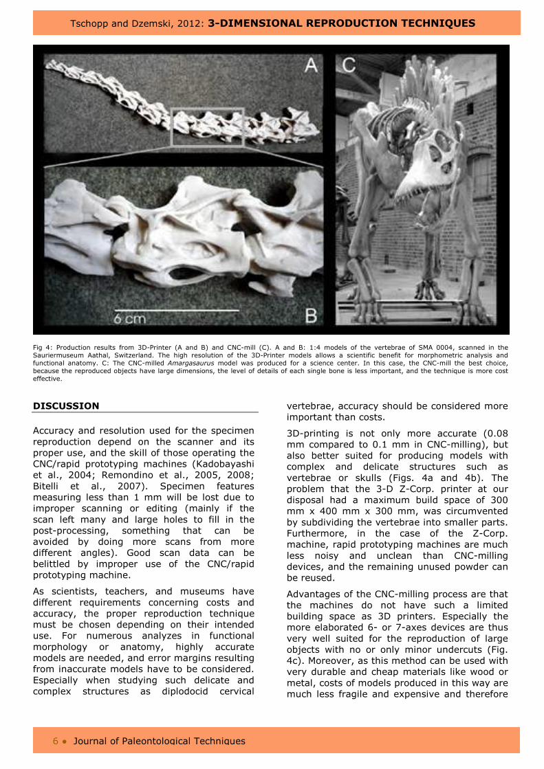
Fig 4: Production results from 3D-Printer (A and B) and CNC-mill (C). A and B: 1:4 models of the vertebrae of SMA 0004, scanned in the Sauriermuseum Aathal, Switzerland. The high resolution of the 3D-Printer models allows a scientific benefit for morphometric analysis and functional anatomy. C: The CNC-milled Amargasaurus model was produced for a science center. In this case, the CNC-mill the best choice, because the reproduced objects have large dimensions, the level of details of each single bone is less important, and the technique is more cost effective.
DISCUSSION
Accuracy and resolution used for the specimen reproduction depend on the scanner and its proper use, and the skill of those operating the CNC/rapid prototyping machines (Kadobayashi et al., 2004; Remondino et al., 2005, 2008; Bitelli et al., 2007). Specimen features measuring less than 1 mm will be lost due to improper scanning or editing (mainly if the scan left many and large holes to fill in the post-processing, something that can be avoided by doing more scans from more different angles). Good scan data can be belittled by improper use of the CNC/rapid prototyping machine.
As scientists, teachers, and museums have different requirements concerning costs and accuracy, the proper reproduction technique must be chosen depending on their intended use. For numerous analyzes in functional morphology or anatomy, highly accurate models are needed, and error margins resulting from inaccurate models have to be considered. Especially when studying such delicate and complex structures as diplodocid cervical
vertebrae, accuracy should be considered more important than costs.
3D-printing is not only more accurate (0.08 mm compared to 0.1 mm in CNC-milling), but also better suited for producing models with complex and delicate structures such as vertebrae or skulls (Figs. 4a and 4b). The problem that the 3-D Z-Corp. printer at our disposal had a maximum build space of 300 mm x 400 mm x 300 mm, was circumvented by subdividing the vertebrae into smaller parts. Furthermore, in the case of the Z-Corp. machine, rapid prototyping machines are much less noisy and unclean than CNC-milling devices, and the remaining unused powder can be reused.
Advantages of the CNC-milling process are that the machines do not have such a limited building space as 3D printers. Especially the more elaborated 6- or 7-axes devices are thus very well suited for the reproduction of large objects with no or only minor undercuts (Fig. 4c). Moreover, as this method can be used with very durable and cheap materials like wood or metal, costs of models produced in this way are much less fragile and expensive and therefore
Tschopp and Dzemski, 2012: 3-DIMENSIONAL REPRODUCTION TECHNIQUES
7 ● Journal of Paleontological Techniques
ideal for educational use in schools or museums (Deck et al. 2007).
The complex shape of the cervical vertebrae of SMA 0004 required a large number of polygons to properly describe them. A 3D printer capable of generating slices small enough to replicate those polygons was therefore preferred in this case study. Using CNC-milling, the machine output would not have shown all the specimen features required for further studies. CONCLUSION The combination of digitizing and reproduction makes paleontological specimens accessible to a broader scientific audience on a worldwide scale. Research on specific paleontological material would no longer have to be carried out in the institution housing the specimen, but can be conducted at the researcher’s workplace. Although 3D technology for the reproduction of digital objects remains still a limiting element
for the obtainable complexity, size, surface condition, and price of the physical models, with the fast improvement of scanning and reproduction devices, an ubiquitous digital object exchange will be even easier in near future than it is today.
ACKNOWLEDGMENTS We thank Hans-Jakob Siber (SMA) for the possibility to scan specimens under his care, as well as Jan-Thomas Möller, Martin Kistler, and Ben Pabst for assistance during the scanning process of SMA 0004. Ralph Chapman's and Art Anderson's careful reviews greatly improved an earlier version of this paper.
Emanuel Tschopp is supported by the Fundação para a Ciência e a Tecnologia doctoral fellowship SFRH / BD / 66209 / 2009 (Ministério da Ciência, Tecnologia e Ensino superior, Portugal).
REFERENCES CITED
ANDERSEN, A., CHAPMAN, R. E., DICKMAN, J. & HAND, K. (2001). Using rapid prototyping technology in vertebrate paleontology. Journal of Vertebrate Paleontology, 21(Suppl. 3), 28A.
BIMBER, O., GATESY, S. M., WITMER, L. M., RASKAR, R. & ENCARNACÃO, L. M. (2002). Merging fossil specimens with computer-generated information. IEEE Computer, 35(9), 25–30.
BITELLI, G., GIRELLI, V. A., REMONDINO, F., & VITTARI, L. (2007). The potential of 3d techniques for cultural heritage object documentation. Videometrics IX, 6491, 1-8.
BREITHAUPT, B., MATTHEWS, N., & NOBLE, T. (2004). An integrated approach to three-dimensional data collection at dinosaur tracksites in the Rocky Mountain West. Ichnos, 11(1), 11–26.
CHAPMAN, R. E., SNYDER, R. A., JABO, S. & ANDERSEN, A. (2001). On a new posture for the horned dinosaur Triceratops. Journal of Vertebrate Paleontology, 21(Suppl. 3), 39A-40A.
CHAPMAN, R. E., WEISHAMPEL, D. B. & RASSKIN-GUTMAN, D. (1998). Three-dimensional modeling and analysis of function of fossil vertebrates. Journal of
Vertebrate Paleontology, 18(Suppl. 3), 33A.
CHRISTIAN, A. & DZEMSKI, G. (2007). Reconstruction of the cervical skeleton posture of Brachiosaurus brancai
Janensch, 1914 by an analysis of the intervertebral stress along the neck and a comparison with the results of different approaches. Fossil Record, 10(1), 38–49.
CHRISTIAN, A. & DZEMSKI, G. (2011). Neck posture in sauropods, pp: 251 – 260, in Klein, N., Remes, K., Gee, C. G. & Sander, P. M. (eds.). Biology of the sauropod dinosaurs: Understanding the life of giants. Indiana University Press, Bloomington.
DECK, L. T., CHAPMAN, R. E. & ANDERSEN, A. (2004). Conservation of vertebrate fossils and collections data using virtualization. Journal of Vertebrate Paleontology, 24(Suppl. 3), 51A.
DECK, L. T., SCHLADER, R., CLEMENT, N., GIBBS, W. & CHAPMAN, R. E. (2007). Especially durable prototypes of fossil specimens and replicas for use in public programming. Journal of Vertebrate Paleontology, 27(Suppl. 3), 67A.
DZEMSKI, G. & CHRISTIAN, A. (2007). Flexibility along the neck of the ostrich (Struthio camelus) and consequences for the reconstruction of dinosaurs with extreme neck length. Journal of Morphology, 268(8), 701–714.
Tschopp and Dzemski, 2012: 3-DIMENSIONAL REPRODUCTION TECHNIQUES
8 ● Journal of Paleontological Techniques
DZEMSKI, G. & CHRISTIAN, A. (2010). Analyses of ligament tensions in long necked animals indicates possible neck posture. Symposium of Vertebrate Palaeontology and Comparative Anatomy, Abstracts, unpaginated.
HOHLOCH, A. & MALLISON, H. (2005). Digitizing dinosaurs: new techniques for the Microscribe 3D digitizer. Journal of Vertebrate Paleontology, 25(Suppl. 3), 70A.
JOHNSTON, R. A., BARNES, K., LOVELL-SMITH, T. & PRICE, N. B. (2004). Use of a hand-held laser scanner in palaeontology: a 3D model of a plesiosaur fossil. Image and Vision Computing, 4.
KADOBAYASHI, R., KOCHI, N., OTANI, H. & FURUKAWA, R. (2004). Comparison and evaluation of laser scanning and photogrammetry and their combined use for digital recording of cultural heritage. International Archives of the Photogrammetry, Remote Sensing and Spatial Information Sciences, 35(5), 401–406.
KAHZDAN, M., AMENTA, N., GU, S., WILEY, D. F. & HAMANN, B. (2009). Symmetry restoration by stretching. Canadian Conference on Computational Geometry, Vancouver.
MALLISON, H. (2007). Virtual dinosaurs-Developing computer aided design and computer aided engineering modeling methods for vertebrate paleontology. Dissertation. Tübingen: Eberhard Karls Universität Tübingen.
MALLISON, H., HOHLOCH, A. & PFRETZSCHNER, H. U. (2009). Mechanical digitizing for paleontology-new and improved techniques. Palaeontologia Electronica, 12(2), 1–41.
MÖLLER, J. T., DZEMSKI, G. & CHRISTIAN, A. (2009). Testing an efficient laser scan method to digitize sauropod vertebrae for anatomical and kinematic neck movement simulations. Journal of Vertebrate Paleontology, 29(Suppl. 3), 150A.
MOTANI, R., AMENTA, N. & WILEY, D. F. (2005). Possibilities and limitations of
three dimensional retrodeformation of a trilobite and plesiosaur vertebrae. PaleoBios, 25(2), 88.
REMONDINO, F. (2007). Detailed image-based 3D geometric reconstruction of heritage objects. DGPF Tagungsband, 16, 483–492.
REMONDINO, F., GIRARDI, S., GONZO, L. & RIZZI, A. (2008). Multi-resolution modeling of complex and detailed Cultural Heritage. Proc. of 9th Int. Symp. on Virtual Reality, Archaeology and Cultural Heritage (VAST 2008), 1-8.
REMONDINO, F., GUARNIERI, A. & VETTORE, A. (2005). 3D modeling of close-range objects: photogrammetry or laser scanning. Proceedings of SPIE-IS&T Electronic Imaging: Videometrics VIII 5665, 216–225.
SCHLADER, R., BREITHAUPT, B. H., CLEMENT, N., CHAPMAN, R. E. & PETERSEN, C. (2007). High-resolution virtualization of dinosaur footprints and rock art in the field using a low-cost, high-resolution laser-scanner. Journal of Vertebrate Paleontology, 27(Suppl. 3), 141A.
TSCHOPP, E. & DZEMSKI, G. (in review). How retrodeformation changes morphology in 3D: an example from diplodocid sauropod vertebrae and its implications on taxonomy. Palaeontologia Electronica.
WALTERS, R., CHAPMAN, R. E. & SNYDER, R. A. (2001). Fleshing-out Triceratops: adding muscle and skin to the Virtual Triceratops. Journal of Vertebrate Paleontology, 21(Suppl. 3), 111A.
WILHITE, R. (2003). Digitizing large fossil skeletal elements for three-dimensional applications. Palaeontologia Electronica, 5(1), 1-10.
ZOLLIKOFER, C. P. E. & PONCE DE LEÓN, M. S. (1995). Tools for rapid prototyping in the biosciences. IEEE Computer Graphics and Applications, 15, 48–55.
Additional images and material can be downloaded at http://www.jpaleontologicaltechniques.org/