Embed Size (px)
Citation preview

30th Annual
Combined Orthopaedic
Spring Symposium
April 17-18, 2015
Hawaii Prince Hotel - Honolulu

Welcome from HOA President
2007 HOA Officers
Aloha & Welcome to the 30th Annual Combined OrthopaedicSpring Symposium! This is a great opportunity for our Hawaiiorthopaedic community to learn about the latest advances inorthopaedic surgery. As always, the program features visitingsurgeons who are dynamic speakers and nationally renownedexperts in their fields. The symposium provides an outstandingforum for discussions among HOA members, residents, medicalstudents and allied health professionals from across our state. Italso provides an opportunity to highlight the research beingconducted by University of Hawaii and Tripler Army MedicalCenter residents. Last, but not least, the symposium providesopportunities to network with fellow specialists at the Allen B.Richardson Memorial Golf Tournament on Thursday, cocktailreception on Friday and the awards banquet Saturday. This istruly a winning package, providing opportunities to gainknowledge and earn CME credit, participate in discussions andcatch-up with our local orthopaedic ohana over the course ofjust a few days. Mahalo for joining us!
John Jiuliano, MDHOA President & Symposium Chair
HOA Membership InformationContact HOA Executive Director Cathy Iwai at 808-630-1586 or [email protected] if
you are interested in becoming a member of the Hawaii Orthopaedic Association.
Hawaii Orthopaedic AssociationP.O. Box 61207Honolulu, HI 96839Fax: 808-536-4141.
Americans with Disability Act (ADA)Participants with special needs should contact Cathy Iwai at 808-630-1586 or [email protected] todiscuss desired accommodation(s).
PresidentJohn Jiuliano, MD
Vice-PresidentDarren Egami, MD
Secretary/TreasurerWei Chin Chen, MD
Immediate Past PresidentElizabeth Ignacio, MD
Board of CouncilorsJerry Van Meter, MD
Executive DirectorCathy Iwai
2015HOAOfficers
1

CME CreditsThis activity has been planned and implemented in accordance with the Essential Areas and policies of the AccreditationCouncil for Continuing Medical Education through the joint sponsorship of the Hawaii Consortium for CME and HawaiiOrthopaedic Association. The Hawaii Consortium for CME is accredited by the ACCME to provide continuing medicaleducation to physicians. The Hawaii Consortium for CME designates this educational activity for a maximum of 12.0AMA PRA Category 1 Credit(s)TM. Physicians should only claim credit commensurate with the extent of their participa-tion in the activity.
Disclosure DeclarationThe following have no relevant financial relationships with any commercial interest:
PlannersJohn Jiuliano, MDDarren Egami, MDWei Chin Chen, MDElizabeth Ignacio, MDLinda Rasmussen, MDJerry Van Meter, MDByron Izuka, MDPaul M. Ryan, MDCraig Ono, MDCathy Iwai
Learning ObjectivesThe program content of the Hawaii Orthopaedic Association Spring Symposium is designed to identify and address theadvances and changes that occur throughout the many areas of orthopaedic surgery, to include oncology, sportsmedicine, joint arthroplasty, hand, pediatric, general orthopaedics, pain, and healthcare reform. The topics are intendedto relate both directly and indirectly to the practice of each practitioner. At the conclusion of this educational activity,participants will be able to:
1. Incorporate knowledge and understanding of orthopaedic conditions and treatment options and apply to currentpractice in the care of patients.
2. Understand current state of clinical research in Hawaii and utilize to improve and advance orthopaedic care.3. Recognize recent and future changes in healthcare reform as it applies to the field of orthopaedics and integrate
those changes into daily practice and management of patients.
ResidentsNicholas Foeger, MDDana Hensley, MDLiang Zhou, MDSean Brugman, MDAlex Sweet II, MDMegan Kuba, MDBryan Armitage, MDKim Driftmier, MDJulian Ku, MDJoshua Dworkin, MDShawn Gee, MD
The following have relevant financial relationships to disclose:
Joel Matta, MD DePuy, A Johnson & Johnson Company – ConsultantInvuity Inc. – Consultant, Stock or stock optionsMedtronic – ConsultantMedtronic – ConsultantMedtronic Sofamor Danek – ConsultantMizuhoOSI – IP RoyaltiesRadlink Corp – Stock or stock optionsStryker – Consultant
Edward McPherson, MD Biomet/Biocomposite - ConsultantMark Rekant, MD Axogen = Consultant
Biomet - Royalties
David Ring, MD, PhD Acumed LLC – ConsultantAmerican Shoulder and Elbow Surgeons – Board or committee memberAmerican Society for Surgery of the Hand – Board or committee memberBiomet – IP royalties, consultantClinical Orthopaedics and Related Research – Editorial or governing boardIlluminos – Stock or stock optionsJournal of Hand Surgery – American: Editorial or governing boardJournal of Orthopaedic Trauma – Editorial or governing boardJournal of Shoulder and Elbow Surgery – Editorial or governing boardMedartis – IP royaltiesSkeletal Dynamics – IP royaltiesWright Medical Technology, Inc. – IP royalties
Guest SpeakersCarlo Bellabarba, MDRobert Durkin, MDPhillip Wilson MD
Jeremy McCallum, MDMatthew Cage, MDChristopher Belyea, MDJJ Johnson, MDRobert Cole Tiurmer, MDDaniel Derosa, MD
2

AcknowledgementsThank you for the Support of all of Our Exhibitors...
All Island Surgical
Automated Healthcare Solutions
Biomet Orthopedics
Central Pacific Bank
Ceterix Orthopaedics
Depuy Synthes Unit Orthopaedics
DJO Global
Ferring Pharmaceutical Co.
First Hawaiian Bank
Special Thanks to...
Shriners Hospital for Children - Honolulu
Tripler Army Medical Center Orthopaedic Residency Program
University of Hawaii Orthopaedic Residency Program
...and a Big Mahalo to...
HOA Executive Director Cathy Iwai for all of your work
in overseeing another successful year!
Kaimana Orthopaedics
Northwestern Mutual
SegWAY Orthopaedics
Smith & Nephew Inc.
Sportstek Mecial/Arthrex
Stryker Endoscopy
Vericel Corporation
Zimmer
Best Resident Paper AwardsRichardson Awards: The Richardson Fund was established in 1982 to honor the memory of B. AllenRichardson, MD. Dr. Richardson was one of the first Board-Certified Orthopaedic Surgeons in Honolulu,where he practiced for nearly 30 years. He was an active member of the teaching staff of the University ofHawaii Orthopaedic Residency Training Program from its inception in the mid-1960s, and was a staunchsupporter for the creation of the John A. Burns School of Medicine. The proceeds of the Richardson Fund areused to award first, second and third place prizes for the best resident papers presented at the Annual Com-bined Orthopaedic Spring Symposium.
Shriners Award: The Shriners Award is presented annually and was established to honor an orthopaedicresident who has completed a rotation at the Shriners Hospital for Children in Honolulu. Residents present theircompleted papers to medical staff and allied health professionals at the Shriners Hospital's patient care confer-ence. The paper must be written to meet standards for publishing in clinical publications.
3

30th Annual Combined Orthopaedic Spring SymposiumFriday, April 17, 2015
7:00 Registration / Continental Breakfast / Exhibits7:30 Welcome and Opening Remarks - John Jiuliano, MD7:45 “Disease X”: Peripheral Mononeuropathy - David Ring, MD8:30 The Impact of Juncturae Tendona on Extensor Tendon Distribution and Angle of
Approach to the Metacarpophalangeal Joint: An Anatomic Study - Nicholas Foeger, MD8:40 Muscle Atrophy at Presentation of Cubital Tunnel Syndrome: Demographics and Duration of
Symptoms - Dana Hensley, MD8:50 Detection of Occult Scaphoid Fractures Using Digital Tomography - Liang Zhou, MD9:00 Repair of Nerve Injuries - Mark Rekant, MD9:45 Moderators: David Ring, MD & Mark Rekant, MD10:00 Break / PLEASE VISIT EXHIBITS10:30 ACL Reconstruction in the Skeletally Immature: Where Have We Been, Where Are We Going?
- Philip Wilson, MD11:20 Early Outcomes After Hybrid ACL Reconstruction - Sean Brugman, MD11:30 Efficacy of Femoral Nerve Block for Anterior Cruciate Ligament Reconstruction in Pediatric Patients
- Alex Sweet II, MD11:40 Biomechanical Profile of the Young, Healthy Female Adult - Megan Kuba, MD11:50 Moderator: Philip Wilson, MD12:00 Hip Torque, Sport Function and Quality of Life Differences in Patients with Symptomatic Femoro-
Acetabular Impingement and Labral Tears versus Controls - Robert Durkin, MD12:10 ICD-10 Presentation - Jeri Leong12:20 PAC Discussion - Byron Izuka, MD
HOA Website Presentation - Bryan Armitage, MD12:30 Lunch1:30 The Growth of Anterior Approach THA - Joel Matta, MD2:10 Effect of Pelvic Tilt on Acetabular Components in THA - Bryan Armitage, MD2:20 Efficacy of Radiation Attenuation of Radiopaque Gloves and Cream: A Comparative Study
- Kim Driftmier, MD2:30 Initial Conservative vs. Operative Treatment of Grade V Acromioclavicular Dislocations
- Julian Ku, MD2:40 Revision Total Hip Arthroplasty: Pelvic Recon & Femoral Recon Options
- Edward McPherson, MD3:20 Moderators: Joel Matta, MD & Edward McPherson, MD3:30 AAOS Healthcare Reform: Thomas J. Grogan, MD4:20 HMSA Representative4:45 Welcome Reception
4
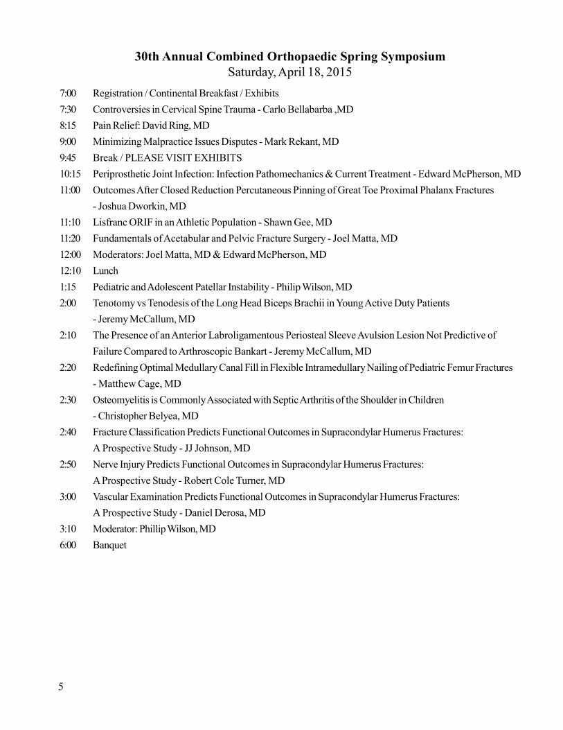
30th Annual Combined Orthopaedic Spring SymposiumSaturday, April 18, 2015
7:00 Registration / Continental Breakfast / Exhibits7:30 Controversies in Cervical Spine Trauma - Carlo Bellabarba ,MD8:15 Pain Relief: David Ring, MD9:00 Minimizing Malpractice Issues Disputes - Mark Rekant, MD9:45 Break / PLEASE VISIT EXHIBITS10:15 Periprosthetic Joint Infection: Infection Pathomechanics & Current Treatment - Edward McPherson, MD11:00 Outcomes After Closed Reduction Percutaneous Pinning of Great Toe Proximal Phalanx Fractures
- Joshua Dworkin, MD11:10 Lisfranc ORIF in an Athletic Population - Shawn Gee, MD11:20 Fundamentals of Acetabular and Pelvic Fracture Surgery - Joel Matta, MD12:00 Moderators: Joel Matta, MD & Edward McPherson, MD12:10 Lunch1:15 Pediatric and Adolescent Patellar Instability - Philip Wilson, MD2:00 Tenotomy vs Tenodesis of the Long Head Biceps Brachii in Young Active Duty Patients
- Jeremy McCallum, MD2:10 The Presence of an Anterior Labroligamentous Periosteal Sleeve Avulsion Lesion Not Predictive of
Failure Compared to Arthroscopic Bankart - Jeremy McCallum, MD2:20 Redefining Optimal Medullary Canal Fill in Flexible Intramedullary Nailing of Pediatric Femur Fractures
- Matthew Cage, MD2:30 Osteomyelitis is Commonly Associated with Septic Arthritis of the Shoulder in Children
- Christopher Belyea, MD2:40 Fracture Classification Predicts Functional Outcomes in Supracondylar Humerus Fractures:
A Prospective Study - JJ Johnson, MD2:50 Nerve Injury Predicts Functional Outcomes in Supracondylar Humerus Fractures:
A Prospective Study - Robert Cole Turner, MD3:00 Vascular Examination Predicts Functional Outcomes in Supracondylar Humerus Fractures:
A Prospective Study - Daniel Derosa, MD3:10 Moderator: Phillip Wilson, MD6:00 Banquet
5
Carlo Bellabarba, MD
* Acting Chief of Service. Orthopaedics, Harborview Medical Center, Seattle, WA
* Director, Spine ACE Program, Harborview Medical Center, Seattle, WA
Thomas Grogan, MD
* Chairman, Practice Management Committee, American Academy of Orthopaedic Surgeons
* Orthopedic Surgeon, Pacific Coast Sports Medicine, Los Angeles, CA
Joel M. Matta, MD
* Founder and Director, Hip & Pelvis Institute, Los Angeles, CA
* Orthopedic Surgeon, Providence Saint John's Health Center, Los Angeles, CA
Edward J. McPherson, MD, FACS
* Director of Orthopedic Surgery, L.A. Orthopedic Institute, Los Angeles, CA
* Designer, Vanguard Knee System and Medallion Revision Hip System
Mark S. Rekant, MD
* Orthopedic Surgeon, The Philadelphia Hand Center, Philadelphia, PA
* Associate Profesor, Dept of Orthopaedic Surgery, Thomas Jefferson University, Philadelphia, PA
David C. Ring, MD, PhD
* Chief, Hand & Upper Extremity Service, Massachusetts General Hospital, Boston MA
* Associate Professor of of Orthopaedic Surgery, Harvard Medical School, Boston, MA
Philip L. Wilson MD
* Associate Prof of Orthopedic Surgery, Texas Scottish Rite Hospital Sports Medicine Center, Plano, TX
* Pediatric Orthopaedic Surgeon, UT Southwestern Medical Center, Dallas, TX
###
6
GUEST SPEAKERS

Sy
mp
os
ium
Ab
str
ac
ts -
Fri
da
y,
Apr
il 17
“Disease X”: Peripheral Mononeuropathy
David Ring, MD
Abstract available upon request.
7###

Sy
mp
os
ium
Ab
str
ac
ts -
Fri
da
y,
Apr
il 17
8
The Impact of Juncturae Tendona on Extensor Tendon Distribution and Angle ofApproach to the Metacarpophalangeal Joint: An Anatomic Study
Nicholas Foeger, MD, PhD and Robert Atkinson, MD
OBJECTIVE: Rupture of the sagittal bands at the metacarpophalangeal joint (MCPJ) results in aloss of centralizing forces on the extensor tendons to the hand and a risk of lateral tendondislocation, pain and inability to actively extend the fingers. The size of the juncturae tendona hasbeen correlated with the potential for tendon dislocation and congenital absence of juncturae has bedescribed in a case of spontaneous extensor tendon dislocation, suggesting that the juncturaefunction as a secondary checkrein to prevent dislocation of the tendons after sagittal band injury.The impact of the juncturae on the angle of extensor tendon approach to the MCPJ, however,remains unclear. The present studies were performed to delineate the angle of approach for thevarious extensor tendons to the MCPJ and to test directly the hypothesis that the juncturae impactthe angle of approach.
METHODS: The extensor tendon anatomy in ~20 cadaveric specimens will be explored withmeasurement of the angle of approach from the wrist to the MCPJ for each extensor tendon.Following classification, the juncturae will be sectioning and these measurements will be repeated.
RESULTS/ CONCLUSIONS: Pending. Insights gained from these investigations may suggest anadditional area of surgical intervention to supplement sagittal band repair in the treatment ofextensor tendon dislocation.
###

Sy
mp
os
ium
Ab
str
ac
ts -
Fri
da
y,
Apr
il 17
9
Muscle Atrophy at Presentation of Cubital Tunnel Syndrome:Demographics and Duration of Symptoms
Matthew L. Drake, Dana T. Hensley, Wei C. Chen, Kenneth F. TaylorTripler Army Medical Center, Honolulu, HI
PURPOSE: To describe the demographics and duration of symptoms of patients with cubital tunnelsyndrome who present with muscle atrophy.
METHODS: We identified 146 patients who presented to the hand surgery clinic at a singlemilitary institution over a five-year period with an initial diagnosis of cubital tunnel syndrome.Medical records were retrospectively reviewed to determine if there was a difference indemographic data, physical examination findings, and duration of symptoms in patients whopresented with muscle atrophy from those with sensory complaints alone.
RESULTS: 11.6% of patients presented with muscle atrophy, all of which were men. 17.2% ofmen presented with atrophy versus no women. Age by itself was not a predictor of presentationwith atrophy, however younger patients with atrophy presented with significantly shorter duration ofsymptoms. Patients with atrophy under 29 years on average had symptoms for 2.4 monthscompared with 16.2 months of symptoms for those over 55 years of age.
DISCUSSION: Men with cubital tunnel syndrome are more likely to present with muscle atrophythan women. Age is not necessarily a predictor of presentation with atrophy. There exists a subsetpopulation of younger patients who presents with extremely short duration of symptoms that rapidlydevelops muscle atrophy. Early surgical intervention may be warranted in the younger patient withnew onset of motor symptoms to avoid potential permanent muscle atrophy.
###

Sy
mp
os
ium
Ab
str
ac
ts -
Fri
da
y,
Apr
il 17
10
Digital Tomography for Detection of Acute Occult Scaphoid Fractures
Barcia AM, Liang Z, Cook JB, Lindell KK, Dykstra AD, Lachky RJ,Shaha S, Taylor KF
INTRODUCTION: Diagnosis of occult scaphoid fractures remains a challenge with variousmodalities advocated. One of the proposed imaging techniques to assess for scaphoid fractureswhen there is high clinical suspicion but initial radiographs are negative is the digital tomogram. Thismodality quickly provides fine-cut visualization of the carpus with minimal radiation exposure. Ourstudy set out to determine the ability of digital tomography to detect acute occult scaphoid fracturesand to compare this modality against MRI, a much more expensive, but well-established technique.
METHODS: This was an IRB approved, prospective series. All patients age 18 years and olderwith clinical suspicion for acute scaphoid fracture and negative initial radiographs were included.Patients had to be within 96 hours of the acute trauma and all were evaluated by orthopedicsurgeons. Both the digital tomogram and a wrist MRI were obtained. The wrists were thenimmobilized and re-evaluated at 10-14 days with repeat radiographs as per standard of care. Thestudies were interpreted by a musculoskeletal radiologist in a blinded fashion. Repeat radiographsand clinical follow up were used as a control to compare the efficacy of both digital tomogram andMRI.
RESULTS: There were 40 extremities in 39 patients included in the study. Six of the 40 scaphoids(15%) were determined to be fractured as determined by repeat radiographs and clinical follow up.Digital tomogram was positive in 4 of the 40 extremities (10%). MRI was positive in 8 of the 40extremities (20%). The sensitivities were 67% and 100% for digital tomogram and MRIrespectively (p=0.14). The positive predictive value (PPV) was 100% for tomogram and 75% forMRI (p=0.15).
DISCUSSION/CONCLUSIONS: The optimal algorithm for diagnosing occult scaphoid fractureshas yet to be determined. Traditional management of two weeks immobilization and repeatradiographs is often inconvenient for the patient and results in unnecessary immobilization of manypatients without fracture. MRI has been shown to be very sensitive in the acute setting and is alsocapable of detecting ligamentous injury, but it is an expensive test and difficult to obtain in manycenters. While very sensitive, the MRI may over-call some contusion injuries as fracture, a findingreported previously by other authors. Digital tomography has been proposed for this use but hasnever been compared to MRI. Our findings indicate that digital tomography will detect mostscaphoid fractures not seen on initial radiographs but may miss some entirely non-displacedfractures. Our study shows that digital tomography is less sensitive than MRI for detecting occultscaphoid fractures.
###
Sy
mp
os
ium
Ab
str
ac
ts -
Fri
da
y,
Apr
il 17
11
Repair of Nerve Inuries
Mark Rekant, MD
Abstract available upon request.
###

Sy
mp
os
ium
Ab
str
ac
ts -
Fri
da
y,
Apr
il 17
###12
ACL Reconstruction in the Skeletally Immature:Where Have We Been, Where Are Going?
Phillip Wilson, MD
Abstract available upon request.

Sy
mp
os
ium
Ab
str
ac
ts -
Fri
da
y,
Apr
il 17
###13
Outcomes After Allograft Augmentation of Autograft HamstringAnterior Cruciate Ligament Reconstruction
CPT Sean Brugman, Craig Bottoni, CDR Douglas Rowles
BACKGROUND: Anterior cruciate ligament reconstruction is a commonly performed surgery torestore normal anatomic function and stability of the knee. Autogenous quadrupled hamstring graftshave been shown to be an excellent graft choice, especially in younger patients, as long as the graftsize is adequate. When graft size is insufficient, less than 8mm, it is common practice to augment thedonor’s own tissue with allograft tissue. Recent literature has shown that pure allograft ACLreconstructions have a higher failure rate in patients less than 25 years old. This project seeks todetermine the incidence allograft augmentation, and understand the short and mid-term outcomes ofthese hybrid ACL reconstructions. To our knowledge, no studies have ever evaluated this subset ofpatients.
METHODS: Chart review was performed from 2012 to 2014 for all patients undergoingAutograft ACL reconstruction requiring allograft augmentation. Operative reports were used todetermine patient demographics, size of autograft at harvest, reason for allograft, type of allograftused and final graft construct diameter. Pre- and post-operative Lysholm, KOOS, SOMOS andVAS pain scores were obtained.
RESULTS: over the two year collection period, 113 ACL hamstring autografts were performed,30 requiring allograft augmentation 22.6%. Of those, 14 were posterior tibial tendons, 12Semitendinosus and 4 could not be determined from op report. The most common reason forusewas diminutive size of harvested autograft. Remainder of Data is in collection and analysis.
CONCLUSIONS: Rates of allograft augmentation have not been reported on, but in our series therate is 22.5% with posterior tibial and semitendinosus. Further research is required to determine theoutcomes of this subset of patients.

Sy
mp
os
ium
Ab
str
ac
ts -
Fri
da
y,
Apr
il 17
14
Efficacy of Femoral Nerve Block for Anterior Cruciate LigamentReconstruction in Pediatric Patients
PI/Supervising Professor: Byron Izuka, MD; Co Investigators: Alex Sweet, MD;Kendra Dilcher, MD; Greg Atkinson, MS III, Eunjung Lim, Ph.D.
Abstract available upon request.
###
Sy
mp
os
ium
Ab
str
ac
ts -
Fri
da
y,
Apr
il 17
15
Anatomic Alignment, Strength and Balance in the Youth Female Soccer Player
Megan H.M. Kuba, MD
COAUTHORS: Eileen M. Kirk, BS*; Arabella Leet, MD (deceased *); Ross Oshiro, MS, ATC+;Kevin Chang, ATC+; Elizabeth Ignacio, MD- 1010 South King St, Suite 401 Honolulu, HI 96814;
John J. Chen, PhD#;Yongjun Cheng, MS #
* Shriners Hospital for Children- Honolulu 1310 Punahou St. Honolulu HI 96826+ Hawaii Optimum Performance- 94-1410 Moaniani St, Waipahu, HI 96797# University of Hawaii John A. Burns School of Medicine Biostatistics Core-Biosciences Building, Suite
211F 651 Ilalo St. Honolulu, HI 96813
INTRODUCTION: Anterior cruciate ligament (ACL) injuries in female athletes are an establishedproblem. Multiple studies have looked at causes for the discrepancy between male and femalenoncontact ACL injuries and have attempted to design prevention programs to decrease theincidence. The majority of these studies have focused on collegiate level athletes. With a rise inyouth sports participation, the incidence of ACL injuries in the skeletally immature patient hasincreased. Thanks in part to Title IX and the rise in female sports participation; there has also beena significant increase in girls’ injuries. A myriad of both intrinsic and extrinsic factors have beenlinked with increased risk of ACL injury in the adult population, however it is not definitively knownwhether these translate to the pediatric patient.
Anatomical and structural factors are typically considered nonmodifiable once skeletal maturity isreached. However, repetitive training motions have been shown to alter bony alignment in thedominant arms of professional pitchers and the hips of ballet dancers. In the skeletally immaturepatient, it is known that anatomical alignment and the rotational profile change with growth anddevelopment. There is limited data on how these changing factors may play a role in injurymechanics. Maturation has also been shown to influence neuromuscular factors and strength ratiosknown to influence injury rate in the adult, such as the quadriceps-to-hamstring ratio and jumplanding biomechanics. Recent studies have looked at individual risk factors in the adult to see if theymay be extrapolated to the pediatric athlete. Our study aimed to expand on this topic by lookingspecifically at the rotational profile, lower extremity strength and balance of young, competitivefemale soccer players.
METHODS: 126 female soccer players, age 10-14 years old, from ten competitive teams of twoHawaii Youth Soccer Association (HYSA) clubs during the 2013 summer season were included inthis study. All participants underwent baseline rotational profile measurements (e.g., hip internal andexternal rotation, and foot-thigh angle), as well as measurement of trochanteric prominence angle,quadriceps (Q) angle, leg length, strength testing of the hip and proximal thigh musculature, and Y-balance testing. These measurements were performed by trained professionals. The pubertal statuswas obtained via questionnaire from the parents as a marker of physiologic age. Physiologic
(continued on next page)

Sy
mp
os
ium
Ab
str
ac
ts -
Fri
da
y,
Apr
il 17
16###
developmental status for each girl was classified as prepubescent (no breast buds and no menses),premenarchal (with breast buds, but no menses), or postmenarchal (with menses).
Descriptive statistics were used to summarize each measurement, stratified by age, both chronologicand physiologic (prepubescent, premenarchal, and postmenarchal). For each measurement, theaverage of the bilateral legs and the difference between right and left legs were calculated. Foraverages and differences of each measurement, the change with age was analyzed using trend testand the variation among developmental stages was compared using one-way analysis of variancewith post hoc Student-Newman-Keuls comparisons. Two-sided p-value of less than 0.05 wasregarded as statistically significant.
RESULTS: There were expected significant gains in leg length and strength of all musculature withincreasing chronologic and developmental age. No significant difference was found in the rotationalprofile when compared for the three developmental categories. A significant difference was foundbetween the measurements of right and left leg external rotation when compared for pubertal statuswith a difference of 7.8°, 4.9°, and 1.2° for prepubescent, premenarchal, and postmenarcalrespectively (p=0.018). Quadriceps (Q) angle was not found to correlate with chronologic ordevelopmental age. The largest Q angle was seen in the 11 year-old and premenarchal girl ascompared to the smallest angle in the 14-year-old and prepubertal girl. There was no significantdifference for the quadriceps:hamstring ratio when both chronological and developmental age werecompared. The average ratios for all participants ranged from 1.33-1.43. Y-balance scores showeda significant increase on the left side with increasing age and a non-significant trend towardincreasing on the right leg.
CONCLUSIONS: The results demonstrate that within the range of our study group there is nospecific age or pubertal period that correlates to dramatic changes in the rotational profile, strength,or balance of the young female soccer player. The difference in external rotation between the rightand left leg may be explained by leg dominance and could theoretically lead to increased injury riskin a certain limb. This is an area that necessitates further study. As compared to adult literature andother pediatric studies, we found no difference in quadriceps: hamstring ratio in our study populationof girls two years postmenarchal or younger. This may show that the increase in this ratio is morerelated to chronologic aging then the onset of merche and associated hormonal changes. Whileincreasing ACL injuries are being seen in younger girls, the results of our study lead to theconclusion that, like in their adult counterparts, anatomic changes cannot be independently blamedand static strength training alone may not be enough to account for the gender inequality.

Sy
mp
os
ium
Ab
str
ac
ts -
Fri
da
y,
Apr
il 17
###17
Hip Torque, Sport Function and Quality of Life Differences in Patients withSymptomatic Femoro-acetabular Impingement and Labral Tears Versus Controls
Freemyer BG*†, Durkin RC†, Crawford SN‡, Stickley CD*
* Department of Kinesiology and Rehabilitation Science, University of Hawaii, Manoa,† Kapiolani Medical Center for Women and Children, Honolulu, HI,‡ Straub Hospital; Honolulu, HI
CONTEXT: The Hip Outcome Score (HOS) is a widely used, 28-item questionnaire for assessingpatient related outcomes following hip surgery. The relationship between hip and knee torque andHOS in symptomatic femoro-acetabular impingement (FAI) has not been established.
OBJECTIVE: This study’s purpose was to compare torque production between symptomatic FAIpatients and controls and determine the relationship between HOS and hip and knee torque. Wehypothesized that patients with symptomatic FAI would have decreased torque and lower quality oflife and sports function as measured by the HOS compared to controls and that torque measureswould correlate with HOS scores. Design: Case-Control. Setting: Research Laboratory. Patients:Eleven patients with unilateral FAI, scheduled to undergo hip arthroscopy for labral repair (1M,10F, age = 31.2±7.9 years, height = 1.72±0.08 m, mass = 73.6±13.7 kg), were anthropometricallymatched with healthy controls (2M, 12F, age = 31.1±6.6 years, height = 1.72±0.08 m, and mass =70.5±15.2 kg). Interventions: Peak isometric hip and knee torques were measured via hand helddynamometry using standardized procedures and compared between the FAI and control groups.The FAI group completed data collection within two weeks prior to hip arthroscopy. MainOutcome Measures: Peak isometric force production (lbs of force) of the FAI patients’ involvedlimb was compared to the dominate limb of controls for hip flexion (HF), extension (HE), abduction(HAB), adduction (HAD), internal rotation(HIR), and external rotation (HER), and knee flexion(KF) and extension(KE). All participants completed the Hip Outcome Score (HOS) survey whichproduces activities of daily living (HOSADL) and sports (HOSSS) subscale scores.
RESULTS: One-tailed, independent sample t-tests indicated controls were stronger (lbs of force)than FAI patients in HF (54.6±14.5, 31.7±14.6, P<.001), HE (44.8±9.5, 27.3±15.2, P=0.001),HAB (37.1±8.6, 25.1±8.1, P=.001), HAD (33.8±11.6, 19.3±9.6, P=.002), HIR (26.0±5.1,13.1±8.5, P<.001), KF (35.7±12.5, 23.3±12.7, P=.012), and KE (44.1±14.5, 28.2±15.4, P=.008) and that HOSADL and HOSSS scores were higher for controls than FAI patients (99.8±0.1,64.8±11.7, P<.001 and 99.8±0.1, 42.7±12.7, P<.001, respectively). Significant (p<0.05)Pearson’s product-moment correlations were found between peak isometric torque and theHOSADL and HOSSS , respectively, for HF (r=.649, r=.623), HE(r=.665, r=.637), HAB(r=.617, r=.564), HAD (r=.610, r=.579), HIR (r=.720, r=.710), KF (r=.521, r=.444), KE(r=.561, r=.491).
CONCLUSIONS: Symptomatic FAI leads to significant pre-operative torque deficits compared tocontrols. These deficits are correlated with lower self-reported ability in activities of daily living andsports performance compared with controls, as measured by the HOS. The HOSADL and HOSSSwere most correlated with hip internal rotation torque, which is consistent with clinical findingscommonly reporting painful hip internal rotation in symptomatic FAI. Progressive impairmentassociated with FAI leads to considerable weakness in the hip musculature and is accuratelyrepresented by the HOS.

Sy
mp
os
ium
Ab
str
ac
ts -
Fri
da
y,
Apr
il 17
###18
The Growth of Anterior Approach THA
Joel Matta, MD
Abstract available upon request.

Sy
mp
os
ium
Ab
str
ac
ts -
Fri
da
y,
Apr
il 17
###19
Effect of Pelvic Tilt on Acetabular Components in THA
Bryan Armitage, MD
Abstract available upon request.

Sy
mp
os
ium
Ab
str
ac
ts -
Fri
da
y,
Apr
il 17
20
Efficacy of Radiation Attenuation of Radiopague Gloves and Cream:A Comparative Study
Driftmier, K; Scarcella, N; Patel, M; Izuka, B
OBJECTIVE: Recently, the FDA approved a radiation attenuation hand cream for use to limit theexposure of a surgeon’s hands to radiation scatter during intraoperative fluoroscopy. Currently thereare many radiation gloves approved for the same use. It is the purpose of this study to measure theattenuation of radiation to the surgeon’s hands of two brands of gloves and the cream in a simulatedoperating room environment.
METHODS: A standard C-Arm fluoroscopic unit was used to expose a simulated surgeon hand toradiation. A commercially available radiation detector was used for this. Standard settings on the C-Arm were set and then used throughout the trials. A cadaver upper extremity was placed in thedirect path of the beam to simulate the patient. The radiation sensor was exposed in the direct pathof the beam and 6 inches outside the beam to measure scatter. The exposures occurred under thefollowing 6 scenarios: 1) covered with standard operating room gloves; 2) covered with radiationattenuation cream; 3) covered with first brand of radiation attenuation gloves; 4) covered with thesecond brand of radiation attenuation gloves; 5) covered with cream and the first brand of radiationattenuation gloves; 6) covered with cream and the second brand of gloves. Multiple exposures weretaken of each scenario and the mean attenuation was recorded.
RESULTS/DISCUSSION/CONCLUSIONS: Forthcoming
###

Sy
mp
os
ium
Ab
str
ac
ts -
Fri
da
y,
Apr
il 17
21
Initial Conservative vs. Operative Treatment ofGrade V Acromioclavicular Dislocations
Julian Ku, MD
INTRODUCTION: Acromioclavicular (AC) joint injuries are common and constituteapproximately 9% of all shoulder injuries. Traditionally Rockwood Types I and II are treatedconservatively, Type III dislocations are controversial, and Types IV, V, and VI AC dislocations areindicated for surgery. The basis for surgical recommendation for type V dislocations is limited. Littleis known about non-operative treatment of Type V AC dislocations especially in active populations.The purpose of this study was to compare the outcomes between Type V AC dislocations treatedinitially with acute surgical intervention versus those treated conservatively.
METHODS: A retrospective review was conducted using an automated search of electronicpatient medical records from January 2007 through December 2012 for patients diagnosed with anAC dislocation in the Tripler Army Medical Center Department of Orthopedics. Patients wereexcluded who were not active military at the time of injury or who were tertiary referrals.Radiographs were reviewed and comparison was made to the contralateral shoulder. A Type Vinjury was defined as greater than 100% increase in the coracoclavicular (CC) distance comparedto the contralateral side or greater than 2cm of displacement of the superior border of the clavicleunilaterally. Acute repair was defined as repair within 90 days without a trial of conservative therapy.Failure of conservative therapy was defined as being unable to return to full duty or requiringsurgical intervention. A good outcome was defined as a return to full duty without limitations.
RESULTS: 111 patients were identified with 60 patients having a bilateral shoulder films. NormalCC distances of the uninjured shoulder ranged from 4.3 mm to 18.66 mm with a mean of 9.09 mmand a standard deviation of 2.30 mm. 59 patients had a complete AC dislocation with the lateralend of the clavicle being displaced completely above the superior border of the acromion. 41patients were Type V dislocations. 9 patients were tertiary referrals and were excluded. Acutesurgical AC reconstruction was selected in 10 patients, initial conservative therapy wasrecommended for 22. In the conservative group: 11 patients (57%) returned to duty without surgery(average 97.8 days); 6 patients (31%) had delayed surgery and returned to full duty (average 131.3days after surgery, 2 revisions); 2 refused surgery and changed careers; and 3 patients were lost tofollow up. In the acute surgical group: 8 patients (88%) returned to full duty in an average of 190.6days after surgery (mean time to surgery 28.29 days) with 4 of those requiring revision surgery; 1patient was lost to follow up, and 1 patient failed to return to full duty. In the conservatively treatedgroup, patients who failed conservative treatment and had a mean increase in CC distance of 129%(range 104%-166%) with a mean 20.2mm displacement (12.5-22.7mm). In the successfully treatedgroup there was an average 150% increase in CC distance (range 102% - 217%) with a mean19.34 mm displacement (12.73-24.22). There was no significance between the two groups for theincrease in CC distance or mm of displacement (p= 0.32 and 0.69 respectively).
(continued on next page)

Sy
mp
os
ium
Ab
str
ac
ts -
Fri
da
y,
Apr
il 17
###22
CONCLUSIONS: While numerous studies have evaluated the operative versus non-operativetreatment of type III injuries in both a prospective and retrospective manner, no study to date hasreported on the conservative treatment of type V AC dislocations. In this study we report onconservative treatment being successful in a majority of patients and that the average time to returnto duty was not improved in an acute versus delayed surgical intervention. While more study isneeded, this suggests that type V AC dislocations may be given a trial of conservative therapy.Secondarily we report on an increased range of the normal CC interspace (previously reported 1.1-1.3cm) and emphasize the need for a contralateral shoulder radiograph for accurate classification.

Sy
mp
os
ium
Ab
str
ac
ts -
Fri
da
y,
Apr
il 17
23
Revision Total Hip Arthroplasty: Pelvic Recon & Femoral Recon Options
Edward McPherson, MD
Abstract available upon request.
###

24
Sy
mp
os
ium
Ab
str
ac
ts -
Sat
urd
ay
, A
pril
18
###
Controversies in Cervical Spine Trauma
Carlos Bellabarba, MD
Abstract available upon request.

25
Sy
mp
os
ium
Ab
str
ac
ts -
Sat
urd
ay
, A
pril
18Pain Relief
David Ring, MD
Abstract available upon request.
###

26
Sy
mp
os
ium
Ab
str
ac
ts -
Sat
urd
ay
, A
pril
18Minimizing Malpractice Issues Disputes
Mark Rekant, MD
Abstract available upon request.
###

27
Sy
mp
os
ium
Ab
str
ac
ts -
Sat
urd
ay
, A
pril
18Periprosthetic Joint Infection: Infection Pathomechanics & Current Treatment
Edward McPherson, MD
Abstract available upon request.
###

###28
Sy
mp
os
ium
Ab
str
ac
ts -
Sat
urd
ay
, A
pril
18Fractures of the Great Toe: Acceptable Alignment and Outcomes
Joshua Dworkin, MD
INTRODUCTION: Proximal phalanx fractures of the hallux are a common injury. While themajority of fractures are typically managed conservatively angulated or severely comminutedfractures may require operative intervention. There is limited literature to support either method oftreatment. The purpose of this study is to evaluate the outcome of patients with proximal phalanxfractures.
METHODS: A retrospective review was conducted using an electronic medical record searchfrom 2010 through 2014 at a single institution. Patients were included if they had a completebicortical fracture through the diaphysis with angulation. Unicortical fractures or corner fractureswere excluded. Pain scores were evaluated at a minimum of 10 weeks. Radiographs wereevaluated by two fellowship trained foot and ankle surgeons and pre and post interventionangulation was measured. RESULTS: In total, 10 patients with complete fractures through the proximal phalanx of the halluxunderwent closed reduction and percutaneous pinning with Kirschner wires or closed reductionalone. Eight patients underwent closed reduction and percutaneous pinning while two patients weretreated with closed reduction alone. The mean injury angle on the lateral radiograph for patientsundergoing percutaneous pinning was 38o versus 25o for closed reduction alone. All fractures withgreater than 30 degrees of angulation underwent closed reduction percutaneous pinning. Two ofthree patients with less than 30 degrees angulation underwent closed reduction alone. The meanangulation at final follow up in the percutaneous pinning group was 11 degrees versus 15 degrees inthe conservative group. All patients in the closed reduction group had minimal pain at follow-up.Only 50% of patients in the closed reduction percutaneous pinning group had no or minimal pain atfinal follow up and 2 of 8 (25%) patients in the closed reduction percutaneous pinning grouprequired secondary procedures.
CONCLUSIONS: There is a paucity of literature on indications or techniques for operativeintervention of this particular injury. Overall patients were more likely to undergo operativeintervention with higher angulation. A majority of patients, regardless of treatment had good painoutcome scores. Both groups demonstrated an improvement in final angulation regardless oftreatment. While percutaneous pinning resulted in a more anatomic union, only half the patients inthis group had no or minimal pain. This small series highlights the fact that more research is neededfor indications and techniques of operative fixation of proximal phalanx hallux fractures.

Sy
mp
os
ium
Ab
str
ac
ts -
Sat
urd
ay
, A
pril
18
29
Lisfranc ORIF in an Athletic Population
Shawn Gee, MD
BACKGROUND: This study assessed the outcomes and return to duty rate following ORIF ofLisfranc injuries in an active duty military population.
METHODS: A retrospective chart review was performed of all Lis Franc ORIFs performed at ourinstitution from 2009-2012 on active duty personnel.Inclusion criteria included active duty at the time of surgery, Lisfranc injuries undergoing ORIF, andminimum 6 month follow-up. Fifteen patients were identified that met inclusion criteria. No patientswere excluded from this study.
RESULTS: Of the fifteen patients, three had fractures or fracture-dislocations and twelve hadprimarily ligamentous Lisfranc injuries. Of the primarily ligamentous Lisfranc injuries, six underwentscrew fixation (5 males, 1 female, mean age 25.7) and six underwent suture button fixation (5 males,1 female, mean age 29.7). Only half of the patients in the screw fixation group returned to fullactivity, whereas all patients in the suture button fixation group returned to full activity. Average timeto return to full activity was 358 days in the screw fixation group and 181 days in the suture buttongroup. The hardware removal rate was 83% in the screw fixation group, with no hardware removalsin the suture button group.
CONCLUSIONS: All patients with suture button fixation returned to full activity, however only50% of patients with screw fixation were able to return to full activity. Return to full activity afterLisfranc ORIF averaged 358 days with screw fixation, and 181 days with suture button fixation.Our results demonstrate that button fixation serves as a viable alternative to screw fixation in anathletic population.
###

Sy
mp
os
ium
Ab
str
ac
ts -
Sat
urd
ay
, A
pril
18
###30
Fundamentals of Acetabular and Pelvic Fracture Surgery
Joel Matta, MD
Abstract available upon request.

Sy
mp
os
ium
Ab
str
ac
ts -
Sat
urd
ay
, A
pril
18
31###
Pediatric and Adolescent Patellar Instability
Phillip Wilson, MD
Abstract available upon request.

Sy
mp
os
ium
Ab
str
ac
ts -
Sat
urd
ay
, A
pril
18
###32
Biceps Tenotomy vs Tenodesis of the Long Head of the Biceps Brachiiin Young, Active Duty Patients
Jeremy McCallum MD CPT MC; Shawn Gee MD CPT MC; Jay Cook MD CPT MC; CraigBottoni MD; John Tokish MD; Douglas Rowles MD CDR USN
INTRODUCTION: A standard in treatment of intra-articular Biceps tendon pathology remainscontroversial. Tenotomy is simple but may result in cosmetic deformity or functional deficit. Forthese reasons, many recommend tenodesis, especially in the younger, more active patient. No datacomparing outcomes from these procedures in a patient population less than forty exist. Thepurpose of this study is to directly compare the clinical outcomes of proximal biceps tendonpathology treated with either a tenotomy or tenodesis in a young active duty military population.METHODS: A retrospective, age-matched case control study was performed at a single militarymedical center. All patients who had undergone surgery for recurrent biceps tendinopathy by one ofthree fellowship trained orthopedic sports medicine surgeons were identified. Inclusion criteriaconsisted of: (1) Diagnosis of bicep pathology by MRI, physical exam, and arthroscopicexamination of the biceps tendon (2) Age less than or equal to 40 (3) Active duty status at the timeof surgery. Patients were excluded if they had (1) prior shoulder surgery or (2) a concomitantrotator cuff or labral repair. The patients were contacted and completed a detailed web-basedassessment. Primary outcome measures included ASES, SANE, patient satisfaction, VR 12 scoresand biceps specific questions about cramping, weakness, and deformity.
RESULTS: During the study period, 113 patients underwent tenotomy and 154 underwenttenodesis. Of these, 26 tenotomy patients and 28 tenodesis patients met the inclusion criteria. Theaverage age was 33 years (range: 23-40) and the follow up averaged 36 (11-77) months. Theaverage ASES score was 78.5 (range: 37 – 100) for tenodesis and 70.5 (range: 33 – 100) fortenotomy (p=0.26). The average SANE score was 61 (range: 7 –100) for tenodesis and 47 (range:0 – 100) for tenotomy (p=0.26). The average patient satisfaction was 70 (range: 6 – 100) and 60(range: 0 – 99) for tenodesis and tenotomy respectively (p=0.35). The average VR12 physicalscore for tenodesis was 46.03 (range: 22.13 – 58.74) and for tenotomy, 38.49 (range: 17.88 –55.74) (p=0.035). There were no significant differences in the VR12 Mental Score, weakness,cramping, or cosmetic deformity between the two groups. Neither group noted a high rate ofcosmetic deformity. Deformities, if any, were not concerning to patients in either group.
DISCUSSION: Surgical treatment of biceps pathology with either tenotomy or tenodesis resultedin similar results in a population of young active duty patients with the exception of a 7.5 pointstatistically significant difference in the VR12 Physical Score. Concerns of cramping, weakness, andcosmesis were infrequent and occurred equally with both procedures. Neither treatment appears tobe as effective in this patient population as in previous reports on older study cohorts.

Sy
mp
os
ium
Ab
str
ac
ts -
Sat
urd
ay
, A
pril
18
33
The Presence of An Anterior Labroligamentous Periosteal Sleeve AvulsionLesion Not Predictive of Failure Compared to Arthroscopic Bankart
Jeremy McCallum MD CPT MC
OBJECTIVES: Anterior labroligamentous periosteal sleeve avulsion lesions (ALPSA) have beenidentified as a potential risk factor for failure of an arthroscopic labral repair. The objective of thisstudy was to compare the failure rates and clinical outcomes of arthroscopic ALPSA repair toarthroscopic Bankart repair. Additionally, the role of glenoid bone loss on failure rates was analyzedwithin each group.
METHODS: This was a retrospective review of 72 consecutive patients with anterior shoulderinstability (73 shoulders) who underwent an anterior arthroscopic labral repair at a single militaryinstitution by one of three Sports medicine fellowship trained orthopaedic surgeons. At the time ofsurgery, a diagnostic arthroscopy identified 13 (17.8%) ALPSA lesions and 60 (82.2%) isolatedBankart lesions. All lesions were repaired and placed on standard post-operative protocol. Datawas collected on demographics, the Western Ontario Shoulder Instability (WOSI) score, SANEscore, and recurrence rates. Failure was defined as recurrent dislocation. Additionally, glenoidbone loss in all patients was calculated using a standardized technique on preoperative images.Outcomes were analyzed by type of initial lesion.
RESULTS: The average age at surgery was 26.3 years (range, 20-42) with average follow-up of53.3 months (range, 28-63). There were 13 distinct ALPSA lesions and 60 Bankart lesionsidentified on diagnostic arthroscopy. There were no significant differences between groups withrespect to any demographic data. There was 1 failure (7.7%) in the ALSPA group and 8 failures(13.3%) in the Bankart group (p=0.10). The ALPSA group had 13.1% glenoid bone losscompared to 13.5% in the Bankart group (p=0.88). There was no significant difference betweengroups for WOSI or SANE scores. When analyzing both groups using a previously establishedglenoid bone loss of 13.5%, there were significantly more failures in the Bankart group (7/31) thanthe ALSPA group (0/5)
CONCLUSIONS: Contrary to previously published data, we did not find patients with ALPSAlesions to be at an increased risk for failure of an arthroscopic repair compared to an isolatedBankart repair. Further, the presence of an ALPSA lesion was not predictive on increased glenoidbone loss. Additionally, there was no difference in functional outcomes between the two groups.
###

Sy
mp
os
ium
Ab
str
ac
ts -
Sat
urd
ay
, A
pril
18
###34
Flexible Intramedullary Nails for Femur Fracturesin Pediatric Patients Heavier Than 100 Pounds
James S. Shaha, MD; J. Matthew Cage, DO; Sheena R. Black, MD; Robert L. Wimberly, MD;Steven H. Shaha, PhD; Anthony I. Riccio, MD
PURPOSE: The utility of flexible intrameduallary nailing (FIMN) for fixation of femoral shaftfractures in children greater than 100 pounds remains controversial. The purpose of this study is toassess the relationship between patient weight and alignment at radiographic union following FIMNof pediatric femoral shaft fractures.
METHODS: An IRB approved, retrospective review of all patients who sustained a femoral shaftfracture treated by retrograde, stainless steel FIMN was performed at a single level 1 pediatrictrauma center from 2005-2012. Preoperative radiographs were analyzed to determine fracturepattern, location, and isthmic canal diameter. Patient weight was measured on initial presentation tothe ER. Radiographs at the time of bony union were reviewed to measure shortening, coronalangulation and sagittal angulation. Data was analyzed in 10 pound increments beginning at 100pounds to assess the impact of weight on final radiographic outcome.
RESULTS: 274 children underwent stainless steel retrograde FIMN for femoral shaft fracturesduring the study period. There were 25 patients who weighed e” 100 lbs and 249 patients <100lbs. There were no significant differences in gender (76% vs. 73% male), fracture stability (40.0%vs. 41.8% length unstable), or fracture patterns between the two groups. The e”100 lbs. group wassignificantly older (10.7 vs. 8.1 years, p<0.01). There were no significant differences in final coronalangulation (1.5 vs. 2.9 degrees), sagittal angulation (3.0 vs. 3.0 degrees) or shortening (3.4 vs. 3.4millimeters) between the two groups. There were significantly more nail removals in the <100 lbsgroup (79.9% vs. 68%, p=0.02). Four percent of the population (11 patients) weighed e”120 lbs.Aside from age (11.6 vs. 8.2 years, p>0.01), there were no significant demographic or fracturedifferences between this group and the remaining population. This group demonstrated no significantdifference in shortening (3.3 vs. 3.4 millimeters), coronal angulation (0.7 vs. 2.9 degrees) or sagittalangulation (1.0 vs. 3.1 degrees) at radiographic union. There was a significant increase in the use ofa fracture table over a flat-top table in the heavier population (p=0.01).
DISCUSSION: Stainless steel FIMN is an effective treatment modality for pediatric femoral shaftfractures in patients >100 pounds with excellent radiographic outcomes and no increased risk forcomplications. There was an increased incidence of nail removal in patients weighing <100 lbs.
SIGNIFICANCE: Stainless steel flexible IM nails are able to maintain fracture alignment withoutan increase in complications in a population weighing more than 100 lbs.

Sy
mp
os
ium
Ab
str
ac
ts -
Sat
urd
ay
, A
pril
18
###
Osteomyelitis is Commonly Associated with Septic Arthritisof the Shoulder in Children
Christopher Belyea, MD
PURPOSE: To describe the clinical presentation, management, and outcomes of surgically treatedseptic arthritis of the shoulder in a pediatric population.
METHODS: A retrospective chart review over 5 years of children with operatively managedseptic arthritis of the shoulder was completed. Demographics, clinical presentation, symptomsduration, antibiotic regimen and duration, number of surgical procedures, and evaluation oflaboratory value improvements were collected. Pre-treatment and final radiographs were assessed.Causative organisms were reviewed. Patients were stratified in age groups to determine clinicalvariability based upon patient age.
RESULTS: 22 children, ages 15 days to 14 years (average 37.3 months), were treated for septicarthritis of the shoulder from 2006-2010 at a single pediatric institution. All patients were managedwith open anterior arthrotomy at an average of 1.95 days after initial orthopaedic consultation(range 0-15 days). Multiple presenting signs were noted; the most common was decreased use(54%). Average admission laboratory values include crp 10.6 (0.3-41.6), ESR 62.8 (11-107), andWBC 14.9 (5.9-31.7). Initial radiographs were read as normal in 12 patients, concern forosteomyelitis in 5, cortical irregularity in 1, fracture in 3, and neoplasm in a single child. 20 patientshad a pre-operative MRI and 15 demonstrated an effusion, 15 had evidence of humeralosteomyelitis, 5 had a subperisoteal abscess, and 4 had soft tissue abscesses. 8 patients remainedculture negative. The most commonly identified organism was methcillin resistant staphylococcusaureus (MRSA) (22.7%). The patients under 12 months of age revealed more diverse organisms atculture and were less likely to have MRSA. All patients averaged 1.55 (1-5) surgical proceduresand had an average hospital stay of 13.5 days. Intravenous (IV) antibiotics averaged 16.3 daysfollowed by an average of 34 days of oral treatment. MRSA patients were significantly more likelyto require multiple operations to eradicate the infection (p<0.02) and had a longer duration of IVantibiotic use (p<0.003). MRSA patients were more likely to have abnormal radiographs at finalfollow-up (p<0.03).
CONCLUSIONS: Septic arthritis of the shoulder in children is commonly associated with adjacentosteomyelitis. Pediatric septic arthritis of the shoulder due to MRSA bacteria can have a morevirulent course than other bacterial causes, but is a less commonly identified organism in theyoungest patients.
SIGNIFICANCE: To our knowledge, this is the largest series published concerning the treatment,course, and outcomes of pediatric septic arthritis of the shoulder.
35

Sy
mp
os
ium
Ab
str
ac
ts -
Sat
urd
ay
, A
pril
18
###
Fracture Classification Predicts Functional Outcomes inSupracondylar Humerus Fractures: A Prospective Study
JJ Johnson, MD
PURPOSE: To prospectively evaluate the relationship between fracture classification and functionaloutcome in children with supracondylar humerus fractures (SCHFX) using validated outcomemeasures.
METHODS: An IRB approved prospective enrollment of consecutive patients with operativeSCHFX was performed over a 3-year period. Fracture pattern and Gartland classification wererecorded by the treating surgeon at the time of surgery. Functional outcome was assessed at finalfollow-up using the Pediatric Outcomes Data Collection Instruments (PODCI) and the quickDisabilities of the Arm, Shoulder, and Hand (QuickDASH) Outcome Measure. Multiple regressionanalysis was used to determine the relationship between fracture classification/pattern and functionaloutcome while controlling for other injury parameters including patient age, neurologic deficit,vascular abnormality and presence of an open fracture.
RESULTS: 752 patients were enrolled during the study period of which 199 (average age 6.7years) completed functional outcome measures at final follow-up. Of these, 10 patients (5%)sustained flexion injures and 189 (95%) sustained extension injuries of which 62 (33%) were TypeII fractures and 127 (67%) were Type III fractures. 65 (34%) of the extension injuries wereposteromedially displaced, 58 (31%) were posterolaterally displaced, 54 (29%) were posteriorlydisplaced without coronal plane deformity, and 12 (6%) were multidirectionally unstable. Theaverage PODCI global functioning scale score and QuickDASH scores for the entire cohort were93.5 and 10.5 respectively indicating excellent function. No differences in outcome scores werenoted between patients with Type II fractures, Type III fractures, and those with multidirectionalinstability. For extension injuries, no difference in outcome was identified based upon fracturepattern. Flexion injuries demonstrated significantly lower PODCI transfer and basic mobility (93.9vs. 98.7) (p<0.001), and PODCI pain and comfort scores (77.8 vs. 94.8) (p<0.03) than Type IIIextension injuries. As a whole, extension injuries demonstrated significantly higher PODCI pain andcomfort scores (94.8 vs. 77.8) (p<0.02) than flexion injuries.
CONCLUSIONS: While children generally have excellent functional outcomes following theoperative treatment of SCHFX, flexion injuries may be predictive of poorer outcomes with regardsto pain and mobility when compared to extension injuries at final follow-up.
SIGNIFICANCE: This is the first study to prospectively determine an association betweenfracture classification and functional outcome using validated outcome measures following theoperative treatment of children with SCHFX.
36
Sy
mp
os
ium
Ab
str
ac
ts -
Sat
urd
ay
, A
pril
18Vascular Examination Predicts Functional Outcomes in
Supracondylar Humerus Fractures: A Prospective Study
Robert Cole Turner, MD
PURPOSE: To prospectively evaluate the relationship between vascular abnormality atpresentation and functional outcome in children with supracondylar humerus fractures (SCHFX)using validated outcome measures.
METHODS: An IRB-approved prospective enrollment of consecutive patients with operativeSCHFX was performed over a 3-year period. Among other injury parameters, the presence andsymmetry of the radial pulse in comparison to the uninjured extremity was documented by thetreating surgeon at presentation. Doppler examination of all non-palpable pulses was documentedas was the perfusion status of the hand. Functional outcome was assessed at final follow-up usingthe Pediatric Outcomes Data Collection Instruments (PODCI) and the quick Disabilities of theArm, Shoulder, and Hand (QuickDASH) Outcome Measure. Multiple regression analysis was usedto determine the relationship between the presence of a vascular abnormality and functionaloutcome while controlling for other injury parameters including patient age, fracture classification,fracture pattern, neurologic deficit and presence of an open fracture.
RESULTS: 752 patients were enrolled during the study period of which 199 (average age 6.7years) completed functional outcome measures at final follow-up. Of these, 24 (12%) patients hadan abnormal vascular exam at initial presentation: 11 (5.5%) with a palpable asymmetric pulse and13 (6.5%) with a non-palpable pulse. Of those with a non-palpable pulse, 10 (5%) weredopplerable and 3 (1.5%) had no identifiable Doppler signal. Patients with a symmetric, palpablepulse demonstrated better outcomes in PODCI pain and comfort scale scores (95.2 vs. 85.2)(p<0.002), PODCI upper extremity scores (93.4 vs. 87.2) (p<0.05), and QuickDASH scores(10.9 vs. 21.6) (p<0.003) compared to those with any abnormal vascular examination. Patientswith a palpable pulse, regardless of symmetry, demonstrated significantly higher PODCI pain andcomfort scale scores (94.6 vs. 84.7) (p<0.02) compared to those with nonpalpable pulses. Noother statistically significant differences in outcome scores were found between patients withdifferent types of abnormal examinations (palpable asymmetric vs. nonpalpable dopplerable vs.nonpalpable nondopplerable).
CONCLUSIONS: In children with operative supracondylar humerus fractures, the presence of anabnormal vascular examination at presentation is predictive of poorer outcomes with regards to painand upper extremity function at final follow-up.
SIGNIFICANCE: This is the first study to prospectively determine an association betweenvascular examination at presentation and functional outcome using validated outcome measuresfollowing the operative treatment of children with SCHFX
###37

Sy
mp
os
ium
Ab
str
ac
ts -
Sat
urd
ay
, A
pril
18Nerve Injury Predicts Functional Outcomes in
Supracondylar Humerus Fractures: A Prospective Study
Daniel Derosa, MD
PURPOSE: To prospectively evaluate the relationship between neurologic deficit at presentationand functional outcome in children with supracondylar humerus fractures (SCHFX) using validatedoutcome measures.
METHODS: An IRB approved prospective enrollment of consecutive patients with operativeSCHFX was performed over a 3-year period. Among other injury parameters, the presence andtype of any neurologic deficit was documented by the treating surgeon at presentation andthroughout the follow-up period. Functional outcome was assessed at final follow-up using thePediatric Outcomes Data Collection Instruments (PODCI) and the quick Disabilities of the Arm,Shoulder, and Hand (QuickDASH) Outcome Measure. Multiple regression analysis was used todetermine the relationship between the presence/type of nerve injury and functional outcome whilecontrolling for other injury parameters including patient age, fracture classification, fracture pattern,vascular abnormality and presence of an open fracture.
RESULTS: 752 patients were enrolled during the study period of which 199 (average age 6.7years) completed functional outcome measures at final follow-up. Of these 22 (11%) patients had aneurologic deficit at the time of initial presentation with 25 nerve injuries noted: 10 (40%) anteriorinterosseous nerve (AIN) deficits, 5 (20%) posterior interosseous nerve (PIN) deficits, 4 (16%)ulnar nerve deficits, 3 (12%) median nerve deficits, and 3 (12%) radial nerve deficits. As a group,patients with neurologic injury demonstrated significantly lower. There were no significantdifferences in outcomes scores when comparing different types of nerve injuries. All nerve injuriesresolved without further intervention with no residual nerve deficits in at final follow-up.
CONCLUSIONS: In children with operative supracondylar humerus fractures, the presence of anerve deficit at presentation is predictive of poorer outcomes with regards to pain, function, mobilityand satisfaction at final follow-up despite complete spontaneous resolution.
SIGNIFICANCE: This is the first study to prospectively determine an association betweenneurologic deficit and functional outcome using validated outcome measures following the operativetreatment of children with SCHFX.
###38

In order to receive CME credit,
symposium participants must complete the evaluation form.
Please see Cathy Iwai for more information.
CME Requirement: Evaluation Form
Mahalo for attending the30th Annual Combined Orthopaedic Spring Symposium!