Embed Size (px)
Citation preview

310 IEEE JOURNAL OF SELECTED TOPICS IN SIGNAL PROCESSING, VOL. 2, NO. 3, JUNE 2008
Signal Processing in Sequence Analysis:Advances in Eukaryotic Gene Prediction
Mahmood Akhtar, Julien Epps, Member, IEEE, and Eliathamby Ambikairajah, Member, IEEE
Abstract—Genomic sequence processing has been an activearea of research for the past two decades and has increasinglyattracted the attention of digital signal processing researchers inrecent years. A challenging open problem in deoxyribonucleicacid (DNA) sequence analysis is maximizing the prediction ac-curacy of eukaryotic gene locations and thereby protein codingregions. In this paper, DNA symbolic-to-numeric representationsare presented and compared with existing techniques in terms ofrelative accuracy for the gene and exon prediction problem. Novelsignal processing-based gene and exon prediction methods arethen evaluated together with existing approaches at a nucleotidelevel using the Burset/Guigo1996, HMR195, and GENSCANstandard genomic datasets. A new technique for the recognitionof acceptor splice sites is then proposed, which combines signalprocessing-based gene and exon prediction methods with anexisting data-driven statistical method. By comparison with theacceptor splice site detection method used in the gene-finding pro-gram GENSCAN, the proposed DSP-statistical hybrid techniquereveals a consistent reduction in false positives at different levelsof sensitivity, averaging a 43% reduction when evaluated on theGENSCAN test set.
Index Terms—Autoregressive processes, correlation, deoxyri-bonucleic acid (DNA), discrete cosine transforms (DCTs), discreteFourier transforms (DFTs), Gaussian mixture models.
I. INTRODUCTION
DEOXYRIBONUCLEIC acid (DNA), the material ofheredity in most living organisms, consists of genic
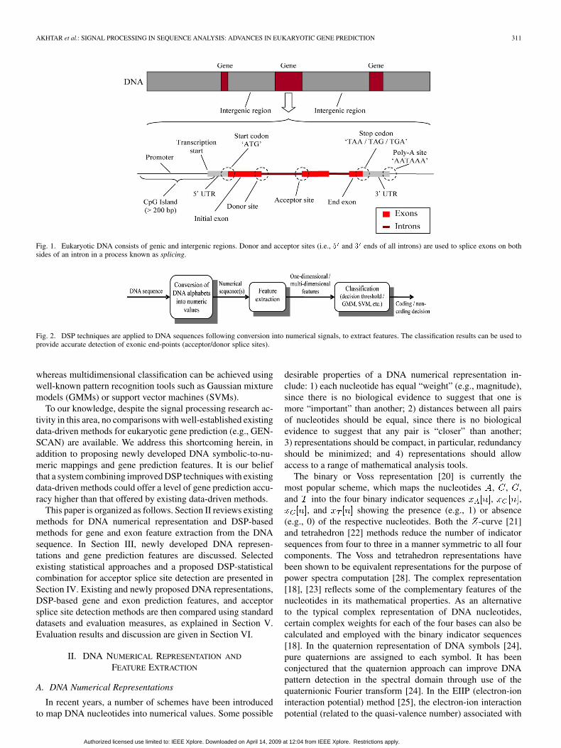
and intergenic regions, as shown in Fig. 1. In eukaryotes,genes are further divided into relatively small protein codingsegments known as exons, interrupted by noncoding spacersknown as introns. In eukaryotes such as human, the intergenicand intronic regions often make up more than 95% of theirgenomes. Codons (i.e., triplets of possible four types of DNAnucleotides , , , and ) in exons encode 20 amino acidsand 3 terminator signals, known as stop codons (i.e., TAA,TAG, and TGA). Initial exons of the genes begin with a start
Manuscript received September 11, 2007; revised March 8, 2008. This workwas supported in part by the National University of Sciences and Technology(NUST), Pakistan, and in part by the University of New South Wales (UNSW),Australia, under a UNSW Faculty Research Grant 2007 for genomic signal pro-cessing research. The associate editor coordinating the review of this manuscriptand approving it for publication was Dr. Ioan Tabus.
M. Akhtar is with the National University of Sciences and Technology,Rawalpindi Cantt, Pakistan, and also with the University of New South Wales,Sydney, NSW 2052, Australia (e-mail: [email protected]).
J. Epps and E. Ambikairajah are with the School of Electrical Engineeringand Telecommunications, University of New South Wales, Sydney, NSW 2052,Australia (e-mail: [email protected], [email protected]).
Color versions of one or more of the figures in this paper are available onlineat http://ieeexplore.ieee.org.
Digital Object Identifier 10.1109/JSTSP.2008.923854
codon “ATG.” Looking from the end of DNA (upstream) toits end (downstream), the exon-to-intron border is known asthe donor splice site and consists of a consensus dinucleotide“GT” as the first two nucleotides of the intron, whereas theintron-to-exon border is known as the acceptor splice site,which consists of a consensus dinucleotide “AG” as the lasttwo nucleotides of the intron. The accurate identification ofgenomic protein coding regions, along with the recognition ofother signals and/or regions (shown in Fig. 1) would result inan ideal gene finding and annotation system.
Despite the existence of various data-driven gene findingprograms, such as AUGUSTUS [1], FGENES [2], geneid [3],GeneMark.hmm [4], Genie [5], GENSCAN [6], HMMgene[7], Morgan [8], and MZEF [9], the accuracy of gene predic-tion is still limited. Previous investigations of computationalgene finding programs [10]–[13] reveal that these data-drivenapproaches seem to rely more on compositional statistics of thesequences (e.g., content) than the genomic signals (e.g.,promoters, acceptor/donor sites, start/stop codons) involved inthe translation process from DNA to protein, and are heavilydependent on the statistics of the sequences they learn fromand are, thus, not equally suitable for all types of sequences.Furthermore, the accuracy is dependent on the length andposition of the exons [14], [15]. High prediction accuracy canoften be attributed to friendly training and test sequences, inwhose formation certain rules were followed, such as includingsequences consisting of one complete gene with consensusintronic dinucleotides “GT” and “AG,” respectively, for theirdonor and acceptor splice sites, excluding those containing al-ternatively spliced genes and having any in-frame stop codons.
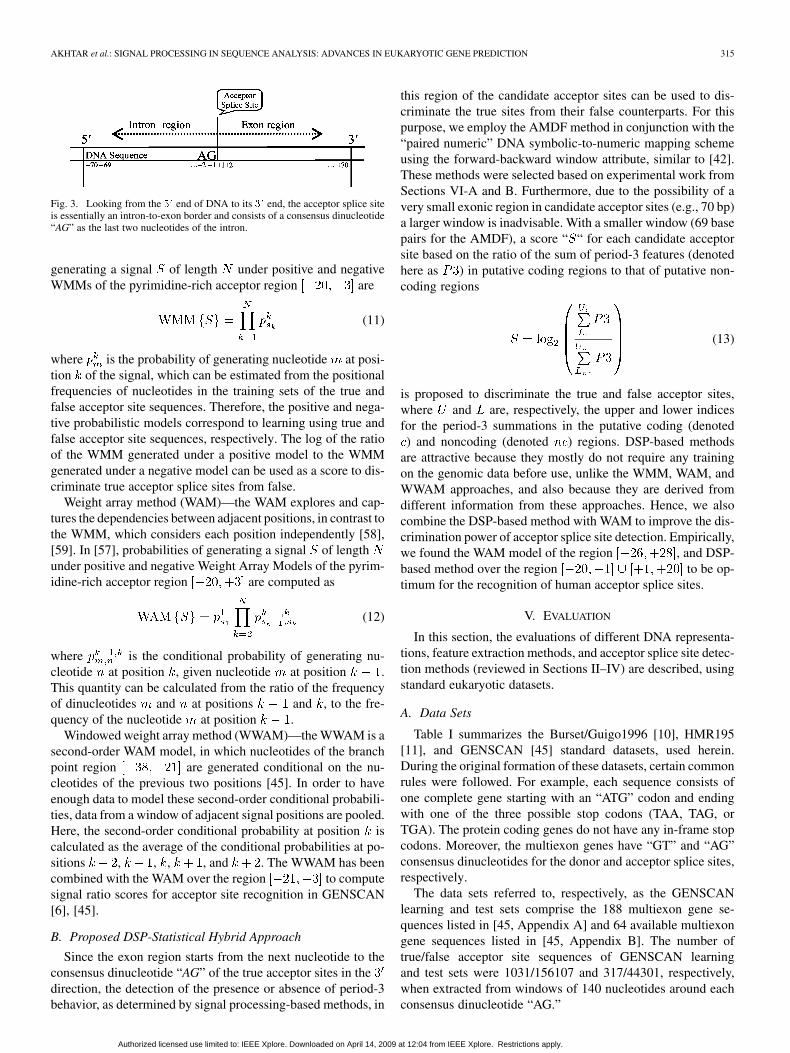
However, it has been observed that gene prediction accuracycan be substantially increased by combining different methods[16], [17]. The discrete nature of the DNA information, beingdiscrete in both “time” and “amplitude,” invites investigationby digital signal processing (DSP) techniques. The conversionof DNA nucleotide symbols into discrete numerical values en-ables novel and useful DSP-based applications for the solu-tion of different sequence analysis related problems such asgene finding and annotation, and such applications have beenoverviewed by previous authors [18], [19]. The present roleof DSP applications in this area is summarized in Fig. 2. Inorder to apply DSP methods, the DNA sequences are first con-verted into suitable numeric values. DSP-based methods for pe-riodicity detection are then applied to the numerical sequencesto obtain 1-D or multidimensional features. The resultant fea-tures are then passed on for back-end processing to classify be-tween protein coding and noncoding regions. An empiricallyderived decision threshold can be used for 1-D classification,
1932-4553/$25.00 © 2008 IEEE
Authorized licensed use limited to: IEEE Xplore. Downloaded on April 14, 2009 at 12:04 from IEEE Xplore. Restrictions apply.
AKHTAR et al.: SIGNAL PROCESSING IN SEQUENCE ANALYSIS: ADVANCES IN EUKARYOTIC GENE PREDICTION 311
Fig. 1. Eukaryotic DNA consists of genic and intergenic regions. Donor and acceptor sites (i.e., 5 and 3 ends of all introns) are used to splice exons on bothsides of an intron in a process known as splicing.
Fig. 2. DSP techniques are applied to DNA sequences following conversion into numerical signals, to extract features. The classification results can be used toprovide accurate detection of exonic end-points (acceptor/donor splice sites).
whereas multidimensional classification can be achieved usingwell-known pattern recognition tools such as Gaussian mixturemodels (GMMs) or support vector machines (SVMs).
To our knowledge, despite the signal processing research ac-tivity in this area, no comparisons with well-established existingdata-driven methods for eukaryotic gene prediction (e.g., GEN-SCAN) are available. We address this shortcoming herein, inaddition to proposing newly developed DNA symbolic-to-nu-meric mappings and gene prediction features. It is our beliefthat a system combining improved DSP techniques with existingdata-driven methods could offer a level of gene prediction accu-racy higher than that offered by existing data-driven methods.
This paper is organized as follows. Section II reviews existingmethods for DNA numerical representation and DSP-basedmethods for gene and exon feature extraction from the DNAsequence. In Section III, newly developed DNA represen-tations and gene prediction features are discussed. Selectedexisting statistical approaches and a proposed DSP-statisticalcombination for acceptor splice site detection are presented inSection IV. Existing and newly proposed DNA representations,DSP-based gene and exon prediction features, and acceptorsplice site detection methods are then compared using standarddatasets and evaluation measures, as explained in Section V.Evaluation results and discussion are given in Section VI.
II. DNA NUMERICAL REPRESENTATION AND
FEATURE EXTRACTION
A. DNA Numerical Representations
In recent years, a number of schemes have been introducedto map DNA nucleotides into numerical values. Some possible
desirable properties of a DNA numerical representation in-clude: 1) each nucleotide has equal “weight” (e.g., magnitude),since there is no biological evidence to suggest that one ismore “important” than another; 2) distances between all pairsof nucleotides should be equal, since there is no biologicalevidence to suggest that any pair is “closer” than another;3) representations should be compact, in particular, redundancyshould be minimized; and 4) representations should allowaccess to a range of mathematical analysis tools.
The binary or Voss representation [20] is currently themost popular scheme, which maps the nucleotides , , ,and into the four binary indicator sequences , ,
, and showing the presence (e.g., 1) or absence(e.g., 0) of the respective nucleotides. Both the -curve [21]and tetrahedron [22] methods reduce the number of indicatorsequences from four to three in a manner symmetric to all fourcomponents. The Voss and tetrahedron representations havebeen shown to be equivalent representations for the purpose ofpower spectra computation [28]. The complex representation[18], [23] reflects some of the complementary features of thenucleotides in its mathematical properties. As an alternativeto the typical complex representation of DNA nucleotides,certain complex weights for each of the four bases can also becalculated and employed with the binary indicator sequences[18]. In the quaternion representation of DNA symbols [24],pure quaternions are assigned to each symbol. It has beenconjectured that the quaternion approach can improve DNApattern detection in the spectral domain through use of thequaternionic Fourier transform [24]. In the EIIP (electron-ioninteraction potential) method [25], the electron-ion interactionpotential (related to the quasi-valence number) associated with
Authorized licensed use limited to: IEEE Xplore. Downloaded on April 14, 2009 at 12:04 from IEEE Xplore. Restrictions apply.

312 IEEE JOURNAL OF SELECTED TOPICS IN SIGNAL PROCESSING, VOL. 2, NO. 3, JUNE 2008
each nucleotide is used to map DNA character strings intonumerical sequences. The EIIP is just one example of a realnumber representation. Another can be obtained by attachingdigits to the four nucleotides: , , ,and [23]. However, this structure implies that purines( or ) are in some respect “greater than” pyrimidines ( or
). Similarly, the representation , , , andsuggests that and . This representation is
an example of a Galois field assignment, upon which symbolicGalois field operations are possible [26]. Another real-numberrepresentation maps , , , and
, similar to the complementary property of thecomplex method. These assignments of real numbers to eachof the four DNA characters do not necessarily reflect thestructure present in the original DNA sequences. Alternatively,to calculate weights representing the actual participation ofeach symbol in the detected pattern, a linear transform andoptimization can be performed on the DNA sequences [29].The internucleotide distance method [27] replaces each DNAnucleotide with an integer representing the distance betweenthe current nucleotide and the next similar nucleotide.
Each of the existing DNA representations offer differentproperties, and map the DNA sequences into between one andfour numerical sequences. Many existing methods, such asVoss [20], -curve [21], and tetrahedron [22], map the DNAsequence to three or four numerical sequences, potentiallyintroducing redundancy in the representation. The assignmentof arbitrary numbers to each of the four DNA characters inEIIP [25] and other real number representations [23], [26]does not necessarily reflect the structure present in the originalDNA sequence. Representations such as quaternions [24] arelimited to specific mathematical analysis tools. For example,a discrete quaternion Fourier transform (DQFT) [41] basedspectral analysis is required to detect certain DNA patterns.Furthermore, existing DNA representations do not fully exploitthe structural differences of protein coding and noncodingregions to facilitate digital signal processing based gene andexon prediction features. These issues are addressed in theDNA representations proposed in Section III-A.
B. DSP-Based Features for Gene and Exon Prediction
Periodicities of 3, 10.5, 200, and 400 have been reported ingenomic sequences [30]. In exons, the occurrence of identicalnucleotides in identical codon positions is the basis for a period-icity of three interpretation in these regions [31]. The period-3behavior of exons has been widely used to identify these regionsusing DSP-based methods, following conversion to numericalsequences.
The discrete Fourier transform (DFT), the most commonlyused method for spectrum analysis of a finite-length numericalsequence of length , is defined as [36]
(1)
Equation (1) can be used to calculate DFTs for numericalsequences representing DNA sequence portions, for exampleeach of the four binary indicator sequences (i.e., , ,
, and ). The periodicity of 3 in exon regions of aDNA sequence suggests that the DFT coefficient correspondingto (where is chosen to be a multiple of 3) in eachDFT sequence should be large [18]. Note that the calculationof the DFT at a single frequency is sufficient, sothat the Goertzel algorithm [37], which reduces the cost ofsingle point DFT computation by almost a factor of two, canbe employed. Various DFT based spectral measures exploitingthe period-3 behaviour of exons for the identification of theseregions have been proposed. The spectral content (SC) measure[32] combines the individual DFTs (i.e., , , ,and ) to obtain a total Fourier magnitude spectrum of theDNA sequence, as follows:
(2)
The GeneScan program [32], based on the SC measure, com-putes the signal-to-noise ratio of the peak at as
, where represents a longer-term average of thespectral content defined in (2). Regions having are as-sumed to be protein coding (exon) regions. The optimized SCmeasure [18] assigns complex weights , , , and to each ofthe four DFTs , , , and in (2). Theseweights are calculated using an optimization technique appliedto the known genes of a given organism. However, one can alsoapply complex conjugate pairs and . The spec-tral rotation (SR) measure [33] rotates four DFT vectors ,
, , and clockwise, each by an angle equiva-lent to the average phase angle value in coding regions, to makeall of them “point” in the same direction. The SR measure alsodivides each term by the corresponding phase angle deviationsto give more weight to exonic distributions. The feature
(3)
where and are the means and standard deviations ofthe phase angle value in coding regions, has been used for thedetection of exons, and was shown to give better performancethan the SC (2) measure at a 10% false positive gene detectionrate [33]. Note that all DFT-based techniques suffer from spec-tral leakage, due to the finite-length analysis window, which in-troduces small contributions from signal frequencies other thanthose at the frequency .
Autoregressive (AR) methods provide an alternative, morecompact characterization of the signal spectrum. Particular ad-vantages of the AR technique are that it requires relatively fewbase pairs to calculate the AR model (which is convenient ifthe exon regions are short and/or closely spaced), and that itprovides a compact estimate of the signal spectrum. It has beenshown that AR spectral estimation using the Burg algorithm andimproved covariance analysis performs better than the DFT forthe detection of period-3 behaviour in short genomic sequences[34]. However, the selection of the model order is crucial in thisapproach, since choosing too low or too high will result in un-necessarily smoothed or spurious modeling of spectral peaks,respectively. Note also that AR models cannot reasonably be ap-plied to binary indicator sequences, since these could not have
Authorized licensed use limited to: IEEE Xplore. Downloaded on April 14, 2009 at 12:04 from IEEE Xplore. Restrictions apply.

AKHTAR et al.: SIGNAL PROCESSING IN SEQUENCE ANALYSIS: ADVANCES IN EUKARYOTIC GENE PREDICTION 313
resulted from an AR process. A possible solution to the problemis bandpass filtering of numerical sequences before their appli-cation to AR modeling.
In order to reduce the spectral leakage present in DFT-basedexon prediction, a larger window size is required, which implieslonger computation time and also compromises the base-do-main resolution. The infinite impulse response (IIR) antinotch(AN) filter approach in [35] attempts to address these problems.The magnitude response of the antinotch filter has a sharp peakat , which preserves only period-3 components. Theindividual outputs of the four binary indicator sequences canbe combined in a sum-of-squares manner. It has been shown in[35] that digital filter based period-3 detection results are com-parable to those of the DFT-based SC measure given by (2).
The autocorrelation function (ACF) is a measure of how wella signal matches a time-shifted version of itself, as a function ofthe time shift. Practically, the ACF will produce a peak if sig-nificant correlation exists at . Besides period-3 detection,DNA sequences have been widely analyzed for other correla-tions in [20], [31], [38], [39]. Li [40] gives a critical review ofthe study of correlation structures.
The identification of protein coding regions is difficultmainly due to the noncontiguous and noncontinuous nature ofeukaryotic genes. Despite the existence of many DSP-basedapproaches and also data-driven approaches, the accuracy ofexon prediction is still limited and needs to be improved. More-over, the existing approaches only rely on the identification ofperiod-3 property of exons, and do not fully exploit efficientDNA representations and other complementary features re-quired to separate protein coding and noncoding nucleotides.These problems have been addressed in new DSP-based geneand exon prediction features, proposed in Section III-B.
III. NEW DNA REPRESENTATIONS AND DSP-BASED FEATURES
FOR GENE AND EXON PREDICTION
A. DNA Numerical Representations
1) Paired Numeric: The paired numeric representation forgene and exon prediction [42] exploits one of the differentialproperties of exons and introns, according to which introns arerich in nucleotides “ ” and “ ” whereas exons are rich in nu-cleotides “ ” and “ ” [43]. Furthermore, the DFT phase anglehistogram distributions for coding and noncoding regions ofhuman datasets have been shown to give smaller and nearlyequal values of angular mean for distributions of nucleotides“ ” and “ ” than those of “ ” and “ ” [42]. To fully exploitboth of these properties, these nucleotides - - can bepaired in a complementary manner and values of and canbe used to denote - and - nucleotide pairs, respectively. Asimilar approach was used by Datta and Asif [44]; however, nomotivation for the “ - ” and “ - ” pairing was given. “ - ”and “ - ” are complementary pairs, joining opposite strandsof double helix DNA through hydrogen bonds. However, this isnot the reason for their pairing here, as only one strand is usedfor the computational analysis of DNA. This representation in-corporates a very useful DNA structural property, in addition toreducing complexity.
2) Frequency of Nucleotide Occurrence: It has been shownin [42] that the four DNA nucleotides differ considerably intheir occurrence in exonic regions, and that the fractional oc-currence of any particular nucleotide is reasonably consistentacross the Burset/Guigo1996 [10], HMR195 [11], and GEN-SCAN learning [45] datasets considered therein. It has beenfurther observed that the frequency of nucleotide occurrence inexons is a key parameter for any DNA representation to be usedfor the detection of these regions. According to the frequencyof nucleotide mapping [42], nucleotides are represented by theirfractional occurrences in exons of a training database.
B. DSP-Based Features for Gene and Exon Prediction
1) Paired and Weighted Spectral Rotation (PWSR) Measure:The PWSR measure [46] incorporates a statistical property ofeukaryotic sequences, according to which introns are rich in thenucleotides “ ” and “ ” whereas exons are rich in nucleotides“ ” and “ .” This information leads to an alternative propertyto the well-known period-3 behavior of exons. In this method,the DNA sequences are first converted into two binary indicators,
- and - . Using training data from DNA sequencesof the same organism, the means and standard deviationsof the distributions of DFT phase angle averaged over coding re-gions, i.e., one phase angle value per coding region, are calcu-lated. Weights based on the frequency of occurrence of nu-cleotides “ or ” and “ or ” in coding regions of the trainingdata are also calculated. The expression given in (4) can then beused as a feature, along one direction of the DNA sequence
-
-- -
-
-- - (4)
where denotes the forward and reverse directions ofDNA sequence, and - - are the meanand standard deviation values obtained from distributions of theDFT phase angle averaged over coding regions of the trainingdata, are frequency of occurrence weights from trainingdata, and are the sliding DFT windows of two indicatorsequences. The PWSR is calculated in both directions of theDNA sequence, and combined as
(5)
Note that due to paired indicators, a DFT in the reverse di-rection of the same DNA strand is equivalent to a DFT on itscomplementary strand.
2) Paired Spectral Content (PSC) Measure: The PWSRmeasure is a data-driven frequency domain method for gene andexon prediction, which requires training from DNA sequences ofthe same organism. A more general method, known as the pairedspectral content (PSC) measure [47], first converts the DNA se-quence into singlenumerical sequences using the paired-numericrepresentation, as discussed in Section III-A-I, then combinesforward and backward DFTs on the same DNA sequence
(6)
Authorized licensed use limited to: IEEE Xplore. Downloaded on April 14, 2009 at 12:04 from IEEE Xplore. Restrictions apply.

314 IEEE JOURNAL OF SELECTED TOPICS IN SIGNAL PROCESSING, VOL. 2, NO. 3, JUNE 2008
where and are DFTs of the indicator sequencein the forward and reverse directions. Contrary to the SR
and PWSR measures, the PSC measure can be applied to the se-quences taken from any organism, i.e., PSC is not an organism-specific measure.
3) “Time-Domain” Algorithms: In the following approaches[48], DNA sequences are first converted into Voss indicator se-quences, which are passed through a second-order resonant filterwith a center frequency of (similar to [35]) before beinginput to either algorithm. This prefiltering helps to remove spec-tral components at , , , which arise fromthe application of correlation-based approaches to a binary in-dicator sequence at a base-domain lag of 3.
Average magnitude difference function (AMDF)—the av-erage magnitude difference function (AMDF) has long beenused in speech processing, and is defined for a discrete signal
as a function of the period as [49]
(7)
where is the window length. The AMDF, with , is anefficient time-domain algorithm for gene and exon prediction[48]. Practically, the AMDF will produce a deep null if signifi-cant correlation exists at period .
Time domain periodogram (TDP)—the time domain peri-odogram (TDP) is an algorithm used for periodicity detectionin sunspots and pitch detection for speech processing [50]. Ac-cording to this algorithm, the -point data are first arranged in amatrix, with rows containing subsequences of length equalto the period being tested, where is the window length.The columns of the matrix are summed to obtain the TDP vectorof size , as follows:
(8)
The final estimate of the degree of periodicity at period isderived as follows:
(9)
It has been shown in [50] that for large , has a verysharp peak if correlation exists at period , enabling accuratedetection of periodicity.
4) Singular Value Decomposition (SVD): The singular valuedecomposition (SVD) can be applied to a rectangular datamatrix , decomposing it into matrices , , and [51], asfollows:
(10)
where , and , i.e., and are orthog-onal. The singular values of are square roots of the eigen-values from or , where here comprises the framesof numeric DNA sequence values organized into a rectan-gular matrix, where we choose . A linear combination of
the largest singular values of all frames obtained using all fourbinary indicator sequences can then be used for the coding/non-coding decision. SVD-based period-3 detection has also beenenhanced using bandpass filtering of the individual binary indi-cator sequences, emphasizing the period-3 behavior [52].
5) Time-Frequency Hybrid (TFH) Measure: The time-fre-quency hybrid (TFH) measure [46] combines magnitude andphase-based features, acknowledging earlier results by Kotlarand Lavner [33] showing that additionally considering the DFTphase angle is more informative than the magnitude alone. Fea-tures from the time-domain AMDF method and frequency-do-main PWSR measure are normalized to the range [0, 1] and thensummed to produce a TFH feature.
6) Multidimensional Features: For all existing methods andnew methods discussed in Sections III-B1–V, features are com-bined, typically using a sum-of-squares approach as in (2), toproduce a 1-D feature for comparison with some predeterminedthreshold. Since a sum-of-squares approach will not necessarilyproduce optimal feature fusion, multidimensional features havebeen recently proposed in [53] and [54]. One such scheme usesthe PWSR and AMDF features from the TFH measure in [46],and transforms each separately using the DCT, to decorrelatethem with energy localized to the first few coefficients. A 5-Dfeature, comprising one transformed PWSR coefficient and allfour transformed AMDF coefficients, is then formed. In an-other such scheme, the linear predictor coefficients are treated asmultidimensional AR-based features, modeling coding and non-coding regions in terms of their spectral characteristics withinthe given window length. The optimized AR based feature, withmodel order 12 and window size 180 bp is then concatenatedwith the 5-D TFH feature, as shown in [54]. The resultant higherdimensional feature set is then used for training and testing ofthe multidimensional feature based classification system.
IV. ACCEPTOR SPLICE SITE DETECTION METHODS
The accurate prediction of eukaryotic protein coding regionsrequires methods for the detection of their end-points. Theintron-exon border is known as the acceptor splice site (orend of the intron) and consists of a consensus dinucleotide“AG” as the last two nucleotides of the intron. Due to thecommon occurrences of this dinucleotide at locations otherthan acceptor sites throughout a gene sequence, detection isvery difficult. In order to apply data-driven and other methodsherein, the candidate acceptor site sequences were extractedas windows of 140 nucleotides around each consensus dinu-cleotide “AG,” similarly to [55]. The nucleotide positions werethen labeled relative to the consensus dinucleotide “AG,” whichwas assumed to occupy the positions and . From theend to the end of a genomic sequence, these labels wouldbe: . In the caseof a true acceptor splice site, the first 70 positions representintronic nucleotides, while the last 70 labels can be treated asexonic nucleotides, as shown in Fig. 3.
A. Existing Methods
Weight matrix method (WMM)—this method assumes thatthe probabilities of the nucleotides at each position are indepen-dent of each other [56]. According to [57], the probabilities of
Authorized licensed use limited to: IEEE Xplore. Downloaded on April 14, 2009 at 12:04 from IEEE Xplore. Restrictions apply.
AKHTAR et al.: SIGNAL PROCESSING IN SEQUENCE ANALYSIS: ADVANCES IN EUKARYOTIC GENE PREDICTION 315
Fig. 3. Looking from the 5 end of DNA to its 3 end, the acceptor splice siteis essentially an intron-to-exon border and consists of a consensus dinucleotide“AG” as the last two nucleotides of the intron.
generating a signal of length under positive and negativeWMMs of the pyrimidine-rich acceptor region are
(11)
where is the probability of generating nucleotide at posi-tion of the signal, which can be estimated from the positionalfrequencies of nucleotides in the training sets of the true andfalse acceptor site sequences. Therefore, the positive and nega-tive probabilistic models correspond to learning using true andfalse acceptor site sequences, respectively. The log of the ratioof the WMM generated under a positive model to the WMMgenerated under a negative model can be used as a score to dis-criminate true acceptor splice sites from false.
Weight array method (WAM)—the WAM explores and cap-tures the dependencies between adjacent positions, in contrast tothe WMM, which considers each position independently [58],[59]. In [57], probabilities of generating a signal of lengthunder positive and negative Weight Array Models of the pyrim-idine-rich acceptor region are computed as
(12)
where is the conditional probability of generating nu-cleotide at position , given nucleotide at position .This quantity can be calculated from the ratio of the frequencyof dinucleotides and at positions and , to the fre-quency of the nucleotide at position .
Windowed weight array method (WWAM)—the WWAM is asecond-order WAM model, in which nucleotides of the branchpoint region are generated conditional on the nu-cleotides of the previous two positions [45]. In order to haveenough data to model these second-order conditional probabili-ties, data from a window of adjacent signal positions are pooled.Here, the second-order conditional probability at position iscalculated as the average of the conditional probabilities at po-sitions , , , , and . The WWAM has beencombined with the WAM over the region to computesignal ratio scores for acceptor site recognition in GENSCAN[6], [45].
B. Proposed DSP-Statistical Hybrid Approach
Since the exon region starts from the next nucleotide to theconsensus dinucleotide “AG” of the true acceptor sites in thedirection, the detection of the presence or absence of period-3behavior, as determined by signal processing-based methods, in
this region of the candidate acceptor sites can be used to dis-criminate the true sites from their false counterparts. For thispurpose, we employ the AMDF method in conjunction with the“paired numeric” DNA symbolic-to-numeric mapping schemeusing the forward-backward window attribute, similar to [42].These methods were selected based on experimental work fromSections VI-A and B. Furthermore, due to the possibility of avery small exonic region in candidate acceptor sites (e.g., 70 bp)a larger window is inadvisable. With a smaller window (69 basepairs for the AMDF), a score “ “ for each candidate acceptorsite based on the ratio of the sum of period-3 features (denotedhere as ) in putative coding regions to that of putative non-coding regions
(13)
is proposed to discriminate the true and false acceptor sites,where and are, respectively, the upper and lower indicesfor the period-3 summations in the putative coding (denoted) and noncoding (denoted ) regions. DSP-based methods
are attractive because they mostly do not require any trainingon the genomic data before use, unlike the WMM, WAM, andWWAM approaches, and also because they are derived fromdifferent information from these approaches. Hence, we alsocombine the DSP-based method with WAM to improve the dis-crimination power of acceptor splice site detection. Empirically,we found the WAM model of the region , and DSP-based method over the region to be op-timum for the recognition of human acceptor splice sites.
V. EVALUATION
In this section, the evaluations of different DNA representa-tions, feature extraction methods, and acceptor splice site detec-tion methods (reviewed in Sections II–IV) are described, usingstandard eukaryotic datasets.
A. Data Sets
Table I summarizes the Burset/Guigo1996 [10], HMR195[11], and GENSCAN [45] standard datasets, used herein.During the original formation of these datasets, certain commonrules were followed. For example, each sequence consists ofone complete gene starting with an “ATG” codon and endingwith one of the three possible stop codons (TAA, TAG, orTGA). The protein coding genes do not have any in-frame stopcodons. Moreover, the multiexon genes have “GT” and “AG”consensus dinucleotides for the donor and acceptor splice sites,respectively.
The data sets referred to, respectively, as the GENSCANlearning and test sets comprise the 188 multiexon gene se-quences listed in [45, Appendix A] and 64 available multiexongene sequences listed in [45, Appendix B]. The number oftrue/false acceptor site sequences of GENSCAN learningand test sets were 1031/156107 and 317/44301, respectively,when extracted from windows of 140 nucleotides around eachconsensus dinucleotide “AG.”
Authorized licensed use limited to: IEEE Xplore. Downloaded on April 14, 2009 at 12:04 from IEEE Xplore. Restrictions apply.

316 IEEE JOURNAL OF SELECTED TOPICS IN SIGNAL PROCESSING, VOL. 2, NO. 3, JUNE 2008
TABLE ISUMMARY OF DATASETS
B. System Configurations
For the comparison of DNA symbolic to numeric mappings,evaluated on the exon detection problem, the GENSCANdatasets (learning and test sets) were used for training (whereneeded) and testing of DNA representations. The GENSCANtest set was mapped into all DNA representations, and theDFT-based SC measure was then applied for gene and exonprediction in each case. A constant length rectangular window( [18]) was used for all types of DFT calculations. Forthe quaternion representation, the discrete quaternion Fouriertransform (DQFT) [41] was employed to calculate the SCmeasure.
The second evaluation compared the various DSP-based exondetection methods discussed in Sections II and III. The Burset/Guigo1996, HMR195, and GENSCAN datasets were all used.Note that the SR, PWSR, and TFH methods are organism-spe-cific and can only be trained and tested on datasets consistingof gene sequences taken from one particular organism, such asGENSCAN in our case. In 1-D feature extraction, a rectangularwindow of constant size (consistent with previouswork [18], [32], [33]) was again used in DFT-based methods.The AMDF, TDP, and SVD methods were prefiltered with abandpass filter tuned at , to emphasize the period-3 compo-nent and de-emphasize all other components. In AR model im-plementation, a model order of 40 and frame size of 135 wereused, as were found suitable in the preliminary work of [52].A frame size of 81 was used for the SVD method, similar to[52]. Empirically, we found a frame length of 117 suitable forAMDF and TDP. A frame size of 117 was used for ACF, consis-tent with the frame sizes for the other time-domain algorithms.In multidimensional feature extraction, a constant window sizeof was used for DFT-related features and frame sizeof 117 was used for AMDF calculations. For AR modeling, amodel order of 12 and window length of 180 were used, as de-termined in [54].
For exon prediction using multidimensional features, twoGMMs were trained, based on protein coding and noncodingfeatures of the GENSCAN learning set, respectively, fromwhich likelihood estimates were extracted as features duringtesting on the GENSCAN test set, as explained in [54]. Em-pirically, we found 32 mixtures optimal for training the GMMparameters, and a diagonal covariance matrix was used.
Fig. 4. Nucleotide level measures of prediction accuracy.
In the evaluation of acceptor splice site detection, true andfalse acceptor sites from the GENSCAN datasets (learning andtest sets) were used for training and testing of WMM, WAM,WWAM, and the proposed DSP-based method.
Note that in all cases we do not actually perform the classifi-cation to derive an exon/intron decision. Instead, we take advan-tage of the fact that this is a 2-class classification problem andgive results across a range of different threshold settings/deci-sion rules.
C. Evaluation Measures
In these evaluations, results are compared at the nucleotidelevel, contrary to existing comparisons at exon level or genelevel, e.g., [33]. In exon-level detection, the feature value for onepoint (i.e., nucleotide) in an exon being greater than a decisionthreshold is sufficient for the detection of that particular exon.The following measures were employed.
Sensitivity and Specificity—The prediction accuracy mea-sures of sensitivity, specificity (similar to [10]) can be explainedwith the aid of Fig. 4, where true positive (TP) is the numberof coding nucleotides correctly predicted as coding, falsenegative (FN) is the number of coding nucleotides predictedas noncoding, true negative (TN) is the number of noncodingnucleotides correctly predicted as noncoding, and false positive(FP) is the number of noncoding nucleotides predicted ascoding. The sensitivity gives the measure of the propor-tion of coding nucleotides that have been correctly predictedas coding. The specificity is the proportion of predictedcoding nucleotides that are actually from the coding region.
Receiver operating characteristic (ROC) curves—The re-ceiver operating characteristic (ROC) curves were developedin the 1950s as a technique for visualizing, organizing andselecting classifiers based on their performance [60]. In theexon-intron separation problem, an ROC curve explores theeffects on TP and FP as the position of an arbitrary decisionthreshold is varied. The curve can be characterized as a singlenumber using the area under the ROC curve (AUC), with largerareas indicating more accurate detection methods.
False positive (and specificity) versus sensitivity—in thismeasure, the percentage of false positives and percentage speci-ficity are calculated at different levels of percentage sensitivity.A threshold output feature value Th at a particular level ofpercentage sensitivity is the minimum value for which ofthe exonic nucleotides have feature values greater than Th [45].
Detection of exonic nucleotides for false positive—thepercentage of exonic nucleotides detected for false positives(where , 20, and 30) can also be calculated, generatingcurves when the decision threshold is varied. False positives are
Authorized licensed use limited to: IEEE Xplore. Downloaded on April 14, 2009 at 12:04 from IEEE Xplore. Restrictions apply.
AKHTAR et al.: SIGNAL PROCESSING IN SEQUENCE ANALYSIS: ADVANCES IN EUKARYOTIC GENE PREDICTION 317
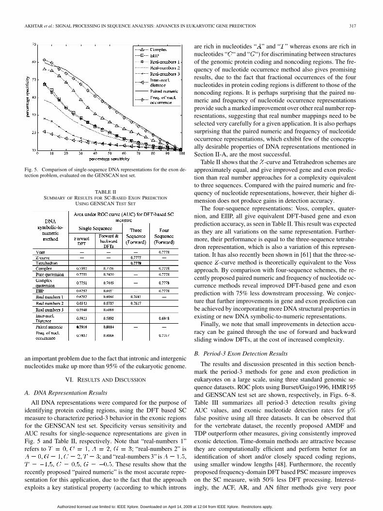
Fig. 5. Comparison of single-sequence DNA representations for the exon de-tection problem, evaluated on the GENSCAN test set.
TABLE IISUMMARY OF RESULTS FOR SC-BASED EXON PREDICTION
USING GENSCAN TEST SET
an important problem due to the fact that intronic and intergenicnucleotides make up more than 95% of the eukaryotic genome.
VI. RESULTS AND DISCUSSION
A. DNA Representation Results
All DNA representations were compared for the purpose ofidentifying protein coding regions, using the DFT based SCmeasure to characterize period-3 behavior in the exonic regionsfor the GENSCAN test set. Specificity versus sensitivity andAUC results for single-sequence representations are given inFig. 5 and Table II, respectively. Note that “real-numbers 1”refers to , , , ; “real-numbers 2” is
, , , ; and “real-numbers 3” is ,, , . These results show that the
recently proposed “paired numeric” is the most accurate repre-sentation for this application, due to the fact that the approachexploits a key statistical property (according to which introns
are rich in nucleotides “ ” and “ ” whereas exons are rich innucleotides “ “ and “ “) for discriminating between structuresof the genomic protein coding and noncoding regions. The fre-quency of nucleotide occurrence method also gives promisingresults, due to the fact that fractional occurrences of the fournucleotides in protein coding regions is different to those of thenoncoding regions. It is perhaps surprising that the paired nu-meric and frequency of nucleotide occurrence representationsprovide such a marked improvement over other real number rep-resentations, suggesting that real number mappings need to beselected very carefully for a given application. It is also perhapssurprising that the paired numeric and frequency of nucleotideoccurrence representations, which exhibit few of the conceptu-ally desirable properties of DNA representations mentioned inSection II-A, are the most successful.
Table II shows that the -curve and Tetrahedron schemes areapproximately equal, and give improved gene and exon predic-tion than real number approaches for a complexity equivalentto three sequences. Compared with the paired numeric and fre-quency of nucleotide representations, however, their higher di-mension does not produce gains in detection accuracy.
The four-sequence representations: Voss, complex, quater-nion, and EIIP, all give equivalent DFT-based gene and exonprediction accuracy, as seen in Table II. This result was expectedas they are all variations on the same representation. Further-more, their performance is equal to the three-sequence tetrahe-dron representation, which is also a variation of this represen-tation. It has also recently been shown in [61] that the three-se-quence -curve method is theoretically equivalent to the Vossapproach. By comparison with four-sequence schemes, the re-cently proposed paired numeric and frequency of nucleotide oc-currence methods reveal improved DFT-based gene and exonprediction with 75% less downstream processing. We conjec-ture that further improvements in gene and exon prediction canbe achieved by incorporating more DNA structural properties inexisting or new DNA symbolic-to-numeric representations.
Finally, we note that small improvements in detection accu-racy can be gained through the use of forward and backwardsliding window DFTs, at the cost of increased complexity.
B. Period-3 Exon Detection Results
The results and discussion presented in this section bench-mark the period-3 methods for gene and exon prediction ineukaryotes on a large scale, using three standard genomic se-quence datasets. ROC plots using Burset/Guigo1996, HMR195and GENSCAN test set are shown, respectively, in Figs. 6–8.Table III summarizes all period-3 detection results givingAUC values, and exonic nucleotide detection rates forfalse positive using all three datasets. It can be observed thatfor the vertebrate dataset, the recently proposed AMDF andTDP outperform other measures, giving consistently improvedexonic detection. Time-domain methods are attractive becausethey are computationally efficient and perform better for anidentification of short and/or closely spaced coding regions,using smaller window lengths [48]. Furthermore, the recentlyproposed frequency-domain DFT based PSC measure improveson the SC measure, with 50% less DFT processing. Interest-ingly, the ACF, AR, and AN filter methods give very poor
Authorized licensed use limited to: IEEE Xplore. Downloaded on April 14, 2009 at 12:04 from IEEE Xplore. Restrictions apply.

318 IEEE JOURNAL OF SELECTED TOPICS IN SIGNAL PROCESSING, VOL. 2, NO. 3, JUNE 2008
Fig. 6. ROC curves for period-3 detection, using the Burset/Guigo1996 dataset.
Fig. 7. ROC curves for period-3 detection, using the HMR195 dataset.
Fig. 8. ROC curves for period-3 detection, using the GENSCAN test set.
identification of period-3 regions, and a likely cause is the lackof bandpass filtering as used in the AMDF and TDP methods,
as discussed in Section III-B3. In results using the HMR195dataset, the AMDF and TDP also give improved performancecompared with other methods, similar to the results obtainedusing Burset/Guigo1996 dataset.
Since the Burset/Guigo1996 (vertebrate) and HMR195(mammalian) datasets contain mixed genomic sequences (i.e.,sequences taken from different organisms), the SR, PWSR,and TFH methods, which require training data, can not beapplied to these datasets in a straightforward manner. Hence,for comparison between all methods, the GENSCAN testset was employed. It is quite clear from the results in Fig. 8that the data-driven, DFT-based PWSR measure outperformswell-known 1-D frequency-domain methods, giving consis-tently fewer false positives (and higher levels of specificity)at each sensitivity level and improved nucleotide detection.By comparison with other DFT-based measures, the PWSRmethod reveals relative improvements of 15.2% and 10.7%,respectively, over the SC and SR measures in the detection ofexonic nucleotides at a 10% false positive rate. The recentlyproposed paired spectral content (PSC) method also improveson the SC and SR measures. One reason for DFT-basedmethods (i.e., SC, SR, PWSR, and PSC measures) givingpoorer accuracy than time-domain algorithms (e.g., AMDF,and TDP), is their relatively large window size (351). Recentinvestigations [62] suggest that the optimum window length forDFT-based methods depends to a large extent on the averagelength of exon regions of the dataset being used, whereasfor time-domain algorithms, this length lies within a shortrange. The time-frequency hybrid (TFH), which combines thecomplementary PWSR and AMDF methods, provides a furthersmall gain in accuracy over the individual PWSR and AMDFmethods.
Finally, the multidimensional feature-based methods givemore accurate gene and exon prediction than all 1-D methods.By comparison with the best 1-D method (TFH), the recentlyproposed multidimensional TFH and AR-TFH methods revealrelative improvements of 4.7% and 11.4%, respectively, in thedetection of exonic nucleotides at a 10% false positive rate.
C. Acceptor Splice Site Results
After training on the GENSCAN learning set, the proposedDSP-statistical hybrid acceptor site detection method fromSection IV-B was compared with WMM, WAM and WWAMusing the GENSCAN test set. Fig. 9 shows ROC curves for allmethods using the GENSCAN test set, from which it can beobserved that the ROC curve for the proposed method exhibitsbetter discrimination power for the detection of acceptor splicesites. The DSP-based method alone is notably poorer thandata-driven methods, presumably due to the fact that it reliessolely on the accurate identification of the period-3 behaviouron one side of the “AG” junction (i.e., consensus dinucleotidefor acceptor sites). The periodicity of three in exons is oftenweak, and existing DSP-based methods are not well equippedto identify this periodicity over a short length of the sequence(e.g., 69 in our case). However, DSP-based methods combinedwith data-driven methods still improve the accuracy of predic-tion, because the two approaches exploit different information.Table IV summarizes the comparison, giving results for AUC,
Authorized licensed use limited to: IEEE Xplore. Downloaded on April 14, 2009 at 12:04 from IEEE Xplore. Restrictions apply.

AKHTAR et al.: SIGNAL PROCESSING IN SEQUENCE ANALYSIS: ADVANCES IN EUKARYOTIC GENE PREDICTION 319
TABLE IIISUMMARY OF PERIOD-3 DETECTION RESULTS ON THREE DATASETS
Fig. 9. ROC plot for acceptor site detection, using GENSCAN test set.
false positive and percentage specificity at different levels ofpercentage sensitivity, for all methods, using the GENSCANtest set. The proposed DSP-statistical hybrid method clearlyachieves a larger area under the ROC curve, consistently fewerfalse positives and higher percent specificities compared withall three existing methods. By comparison with the WWAMmethod used in gene-finding program GENSCAN [6], thenumber of false positives across different sensitivity levels inthe proposed method shows an average relative improvementof 43%.
According to the results of subsection B, further gainsmight be expected from using multidimensional feature-basedmethods; however, these require suitable training data forestimating the GMM parameters and, hence, suffer similardrawbacks to existing data-driven techniques in terms of re-quiring sufficient organism-specific training data.
TABLE IVSUMMARY OF ACCEPTOR SPLICE SITE DETECTION
RESULTS USING GENSCAN TEST SET
VII. CONCLUSION
In summary, a number of digital signal processing-basedmethods for eukaryotic gene prediction have been proposed,and these have been evaluated alongside many other DSP-basedmethods. Firstly, DNA symbolic-to-numeric mappings werecompared in terms of both computational complexity andrelative accuracy for the gene and exon prediction problem.From these experiments, the recently proposed paired numericrepresentation was shown to give an improvement of 2% overthe Voss binary indicator sequences in terms of area underthe ROC curve for gene and exon prediction, with 75% lessdownstream processing, when evaluated on the GENSCAN testset.
All 1-D output feature methods for gene and exon predic-tion were then evaluated on the standard genomic datasetsBurset/Guigo1996, HMR195 and GENSCAN. In terms ofgene and exon prediction accuracy, the recently proposedTFH, AMDF, TDP, SVD, PWSR, and PSC methods exhibitedrelatively more accurate gene and exon prediction, improvingon the well-known DFT based-SC measure by 4% to 9% interms of area under the ROC curve. In light of the weaknesses
Authorized licensed use limited to: IEEE Xplore. Downloaded on April 14, 2009 at 12:04 from IEEE Xplore. Restrictions apply.

320 IEEE JOURNAL OF SELECTED TOPICS IN SIGNAL PROCESSING, VOL. 2, NO. 3, JUNE 2008
and strengths of the 1-D genomic period-3 detection methods,we recommend the AMDF and TFH for nondata-driven anddata-driven gene detection, respectively. Furthermore, recentlyproposed multidimensional output feature methods were shownto give improved gene and exon prediction over their 1-Dcounterparts. By comparison with the most accurate 1-D mea-sure, the multidimensional methods yielded improvements of5% to 11% in terms of relative increase in exonic nucleotidesdetected at a 10% false positive rate, when evaluated on theGENSCAN test set. Evaluations of all schemes herein havebeen performed on large databases and using metrics calculatedat the nucleotide level, in contrast to much of the previousliterature on the topic.
Finally, we have also proposed a new DSP-statistical hybridtechnique for acceptor splice site detection. Results show thatDSP-based approaches to gene and exon prediction aloneare unlikely to rival current data-driven techniques such asGENSCAN or AUGUSTUS. The proposed DSP-statisticalcombination for the detection of acceptor splice sites, whichachieves a performance improvement of 43% over WWAM(used in GENSCAN), is illustrative of the potential DSP-basedtechniques still offer in terms of improving the state of the art.Future directions may include more accurate identification ofexonic/intronic end-point signals (i.e., start codon, donor splicesite, acceptor splice site, and stop codons) using multidimen-sional DSP-based features, and combining signal processingbased work with data-driven methods to advance the state ofthe art in eukaryotic gene prediction algorithms.
ACKNOWLEDGMENT
The authors would like to thank the anonymous reviewersand editor whose helpful suggestions resulted in substantial im-provement of this paper.
REFERENCES
[1] M. Stanke, R. Steinkamp, S. Waack, and B. Morgenstern, “AU-GUSTUS: A web server for gene finding in eukaryotes,” Nucl. AcidsRes., Web Server Issue, vol. 32, pp. W309–W312, 2004.
[2] V. V. Solovyev, A. A. Salamov, and C. B. Lawrence, “Identification ofhuman gene structure using linear discriminant functions and dynamicprogramming,” in Proc. 3rd Int. Conf. Intelligent Systems for Molec-ular Biology, 1995, pp. 367–375.
[3] G. Parra, E. Blanco, and R. Guigo, “GeneID in drosophila,” GenomeRes., vol. 10, no. 4, pp. 511–515, 2000.
[4] A. V. Lukashin and M. Borodovsky, “GeneMark.hmm: New solutionsfor gene finding,” Nucl. Acids Res., vol. 26, no. 4, pp. 1107–1115, 1998.
[5] D. Kulp, D. Haussler, M. G. Reese, and F. H. Eeckman, “A generalizedhidden Markov model for the recognition of human genes in DNA,” inProc. 4th Int. Conf. Intelligent Systems for Molecular Biology, 1996,pp. 134–142.
[6] C. Burge and S. Karlin, “Prediction of complete gene structure inhuman genomic DNA,” J. Mol. Biol., vol. 268, no. 1, pp. 78–94, 1997.
[7] A. Krogh, “Two methods for improving performance of an HMM andtheir applications for gene-finding,” in Proc. 5th Int. Conf. IntelligentSystems for Molecular Biology, 1997, pp. 179–186.
[8] S. Salzberg, A. L. Delcher, K. H. Fasman, and J. Henderson, “A deci-sion tree system for finding genes in DNA,” J. Comput. Biol., vol. 5,no. 4, pp. 667–680, 1998.
[9] M. Q. Zhang, “Identification of protein coding regions in the humangenome by quadratic discriminant analysis,” Proc. Nat. Acad. Sci., vol.94, no. 2, pp. 565–568, 1997.
[10] M. Burset and R. Guigo, “Evaluation of gene structure prediction pro-grams,” Genomics, vol. 34, pp. 353–367, 1996.
[11] S. Rogic, A. K. Mackworth, and B. F. Ouellette, “Evaluation of gene-finding programs on mammalian sequences,” Genome Res., vol. 11, no.5, pp. 817–832, 2001.
[12] V. Makarov, “Computer programs for eukaryotic gene prediction,”Briefings Bioinf., vol. 3, no. 2, pp. 195–199, 2002.
[13] A. Nagar, S. Purushothaman, and H. Tawfik, “Evaluation and fuzzyclassification of gene finding programs on human genome sequences,”FSKD, pp. 821–829, 2005.
[14] S. Logeswaran, E. Ambikairajah, and J. Epps, “A method for detectingshort initial exons,” in Proc. IEEE Workshop Genomic Signal Pro-cessing and Statistics, 2006, pp. 61–62.
[15] Y. Saeys, P. Rouze, and Y. V. de Peer, “In search of the short ones:Improved prediction of short exons in vertebrates, plants, fungi andprotists,” Bioinformatics, vol. 23, no. 4, pp. 414–420, 2007.
[16] K. Murakami and T. Takagi, “Gene recognition by combination ofseveral gene-finding programs,” Bioinformatics, vol. 14, no. 8, pp.665–675, 1998.
[17] V. Pavlovic, A. Garg, and S. Kasif, “A Bayesian framework for com-bining gene predictions,” Bioinformatics, vol. 18, no. 1, pp. 19–27,2002.
[18] D. Anastassiou, “Genomic signal processing,” IEEE Signal Process.Mag., vol. 18, no. 4, pp. 8–20, Apr. 2001.
[19] X. Zhang, F. Chen, Y. Zhang, S. C. Agner, M. Akay, Z. Lu, M. M.Y. Waye, and S. K. Tsui, “Signal processing techniques in genomicengineering,” Proc. IEEE, vol. 90, no. 12, pp. 1822–1833, Dec. 2002.
[20] R. F. Voss, “Evolution of long-range fractal correlations and 1/f noise inDNA base sequences,” Phy. Rev. Lett., vol. 68, no. 25, pp. 3805–3808,1992.
[21] R. Zhang and C. T. Zhang, “Z curves, an intuitive tool for visualizingand analyzing the DNA sequences,” J. Biomol. Struct. Dyn., vol. 11,no. 4, pp. 767–782, 1994.
[22] B. D. Silverman and R. Linsker, “A measure of DNA periodicity,” J.Theor. Biol., vol. 118, pp. 295–300, 1986.
[23] P. D. Cristea, “Genetic signal representation and analysis,” in Proc.SPIE Inf. Conf. Biomedical Optics Symp., 2002, vol. 4623, pp. 77–84.
[24] A. K. Brodzik and O. Peters, “Symbol-balanced quaternionic period-icity transform for latent pattern detection in DNA sequences,” in Proc.IEEE ICASSP, 2005, vol. 5, pp. v/373–v/376.
[25] J. Ning, C. N. Moore, and J. C. Nelson, “Preliminary wavelet analysisof genomic sequences,” in Proc. IEEE Bioinformatics Conf., 2003, pp.509–510.
[26] G. L. Rosen, “Signal processing for biologically-inspired gradientsource localization and DNA sequence analysis,” Ph.D. dissertation,Georgia Inst. Technol., Atlanta, 2006.
[27] A. S. S. Nair and T. Mahalakshmi, “Visualization of genomic datausing inter-nucleotide distance signals,” presented at the IEEE Int.Conf. Genomic Signal Processing, 2005.
[28] E. Coward, “Equivalence of two Fourier methods for biological se-quences,” J. Math. Biol., vol. 36, pp. 64–70, 1997.
[29] W. Wang and D. H. Johnson, “Computing linear transforms of sym-bolic signals,” IEEE Trans. Signal Process., vol. 50, no. 3, pp. 628–634,Mar. 2002.
[30] E. N. Trifonov, “3-, 10.5-, 200- and 400-base periodicities in genomesequences,” Phys. A, vol. 249, pp. 511–516, 1998.
[31] J. W. Fickett, “Recognition of protein coding regions in DNA se-quences,” Nucl. Acids Res., vol. 10, pp. 5303–5318, 1982.
[32] S. Tiwari, S. Ramaswamy, A. Bhattacharya, S. Bhattacharya, and R.Ramaswamy, “Prediction of probable genes by Fourier analysis of ge-nomic sequences,” Comput. Appl. Biosci., vol. 13, pp. 263–270, 1997.
[33] D. Kotlar and Y. Lavner, “Gene prediction by spectral rotation mea-sure: A new method for identifying protein-coding regions,” GenomeRes., vol. 18, pp. 1930–1937, 2003.
[34] N. Rao and S. J. Shepherd, “Detection of 3-periodicity for small ge-nomic sequences based on AR techniques,” in Proc. IEEE Int. Conf.Comm., Circuits Syst., 2004, vol. 2, pp. 1032–1036.
[35] P. P. Vaidyanathan and B.-J. Yoon, “Gene and exon prediction usingallpass-based filters,” presented at the IEEE Workshop Genomic SignalProcessing and Statistics, Raleigh, NC, 2002.
[36] S. K. Mitra, Digital Signal Processing: A Computer-Based Approach,2nd ed. Singapore: McGraw-Hill, 2002.
[37] G. Goertzel, “An algorithm for the evaluation of finite trigonometricseries,” Amer. Math. Monthly, vol. 65, no. 1, pp. 34–35, 1958.
[38] W. Li and T. G. Marr, “Understanding long-range correlations in DNAsequences,” Phys. D, vol. 75, pp. 392–416, 1994.
[39] H. Herzel and I. Große, “Correlations in DNA sequences: The role ofprotein coding segments,” Phys. Rev. E, vol. 55, no. 1, pp. 800–810,1997.
Authorized licensed use limited to: IEEE Xplore. Downloaded on April 14, 2009 at 12:04 from IEEE Xplore. Restrictions apply.

AKHTAR et al.: SIGNAL PROCESSING IN SEQUENCE ANALYSIS: ADVANCES IN EUKARYOTIC GENE PREDICTION 321
[40] W. Li, “The study of correlation structure of DNA sequences: A criticalreview,” Comput. Chem., vol. 21, no. 4, pp. 257–271, 1997.
[41] S. J. Sangwine, “The discrete quaternion Fourier transform,” in Proc.6th Int. Conf. Image Processing and its Applications, 1997, vol. 2, pp.790–793.
[42] M. Akhtar, J. Epps, and E. Ambikairajah, “On DNA numerical repre-sentations for period-3 based exon prediction,” presented at the IEEEWorkshop on Genomic Signal Processing and Statistics, Tuusula, Fin-land, 2007.
[43] P. D. Cristea, “Conversion of nucleotides sequences into genomic sig-nals,” J. Cell. Mol. Med., vol. 6, no. 2, pp. 279–303, 2002.
[44] S. Datta and A. Asif, “A fast DFT based gene prediction algorithm foridentification of protein coding regions,” in Proc. IEEE ICASSP, 2005,vol. 5, pp. 653–656.
[45] C. Burge, “Identification of genes in human genomic DNA,” Ph.D. dis-sertation, Stanford Univ., Stanford, CA, 1997.
[46] M. Akhtar and J. E. E. Ambikairajah, “Time and frequency domainmethods for gene and exon prediction in eukaryotes,” in Proc. IEEEICASSP, 2007, pp. 573–576.
[47] M. Akhtar, J. Epps, and E. Ambikairajah, “Paired spectral content mea-sure for gene and exon prediction in eukaryotes,” in Proc. IEEE Int.Conf. Information and Emerging Technologies, 2007, pp. 127–130.
[48] E. Ambikairajah, J. Epps, and M. Akhtar, “Gene and exon predictionusing time-domain algorithms,” in Proc. IEEE 8th Int. Symp. SignalProcessing and its Applications, 2005, pp. 199–202.
[49] M. Ross, H. Shaffer, A. Cohen, R. Freudberg, and H. Manley, “Averagemagnitude difference function pitch extractor,” IEEE Trans. Acoustics,Speech, Signal Process., vol. ASSP-22, no. 5, pp. 353–362, May 1974.
[50] E. Ambikairajah and M. J. Carey, “The time-domain periodogram al-gorithm,” Signal Process., vol. 5, pp. 491–513, 1983.
[51] P. P. Kanjilal, J. Bhattacharya, and G. Saha, “Robust method for pe-riodicity detection and characterization of irregular cyclical series interms of embedded periodic components,” Phys. Rev. E, vol. 59, no. 4,pp. 4013–4025, 1999.
[52] M. Akhtar, E. Ambikairajah, and J. Epps, “Detection of period-3 be-havior in genomic sequences using singular value decomposition,” inProc. IEEE Int. Conf. Emerging Technologies, 2005, pp. 13–17.
[53] M. Akhtar, E. Ambikairajah, and J. Epps, “GMM-based classificationof genomic sequences,” in Proc. IEEE 15th Int. Conf. Digital SignalProcessing, 2007, pp. 103–106.
[54] M. Akhtar, E. Ambikairajah, and J. Epps, “Comprehensive autoregres-sive modeling for classification of genomic sequences,” presented at theIEEE 6th Int. Conf. Information, Communications, Signal Processing,2007.
[55] P. Pollastro and S. Rampone, “HS D: Homo sapiens splice site dataset,” Nucl. Acids Res. Annu. Database Issue, 2003.
[56] R. Staden, “Computer methods to locate signals in nucleic acid se-quences,” Nucl. Acids Res., vol. 12, pp. 505–519, 1984.
[57] C. Burge, “Modeling dependencies in pre-mRNA splicing signals,”in Computational Methods in Molecular Biology, S. L. Salzberg, D.B. Searls, and S. Kasif, Eds. New York: Elsevier, 1998, ch. 8, pp.129–164.
[58] M. Q. Zhang and T. G. Marr, “A weight array method for splicingsignal analysis,” CABIOS, vol. 9, no. 5, pp. 499–509, 1993.
[59] S. L. Salzberg, “A method for identifying splice sites and translationalstart sites in eukaryotic mRNA,” Comput. Appl. Biosci., vol. 13, no. 4,pp. 365–376, 1997.
[60] T. Fawcett, ROC Graphs: Notes and Practical Considerationsfor Researchers HP Laboratories, 2003 [Online]. Available:http://www.hpl.hp.com/personal/Tom Fawcett/papers/ROC101
[61] A. Rushdi and J. Tuqan, “Gene identification using the Z-curve repre-sentation,” in Proc. IEEE ICASSP, 2006, vol. 2, pp. 1024–1027.
[62] M. Akhtar, E. Ambikairajah, and J. Epps, “Optimizing period-3methods for eukaryotic gene prediction,” in Proc. IEEE ICASSP,2008, pp. 621–624.
Mahmood Akhtar received the B.Sc. (Honors)degree in electrical engineering from the Universityof Engineering and Technology (UET), Lahore,Pakistan, in 2003, and the M.S. degree in computerengineering from the National University of Sciencesand Technology (NUST), Pakistan, in 2005. He iscurrently pursuing the Ph.D. degree in the Schoolof Electrical Engineering and Telecommunications,University of New South Wales, Australia.
His main research interest is in genomic signal pro-cessing with specific focus on DNA representations,
feature extractions, and classifications of genomic protein coding and noncodingregions. Other research interests lie in the areas of public health bio-surveillancemodeling, speech, audio, and image processing. He has authored or coauthoredaround 16 publications.
Julien Epps (M’99) received the B.E. and Ph.D. de-grees from the University of New South Wales, Aus-tralia, in 1997 and 2001, respectively.
After an appointment as a Postdoctoral Fellow atthe University of New South Wales, he worked onspeech recognition and language processing researchas a Research Engineer at Motorola Labs and thenas a Senior Researcher at National ICT Australia. Hejoined the UNSW School of Electrical and Telecom-munications as a Senior Lecturer in 2007. He has au-thored or coauthored around 80 publications and has
served as a reviewer for several IEEE, IET, and other journals and numerousconferences. His research interests include speaker verification, speech recog-nition, speech and audio coding, auditory modeling, speech enhancement, andgenomic signal processing.
Eliathamby Ambikairajah (M’90) received thePh.D. degree from Keele University, U.K.
He was appointed as Head of Electronic En-gineering and later Dean of Engineering at theAthlone Institute of Technology, Ireland. He wasan invited Research Fellow with British TelecomLaboratories (BTL), Martlesham Heath, U.K., forten years (1989–1999). He joined the Universityof New South Wales, Australia, in 1999, wherehe is currently the Deputy Head of School andthe Director of Academic Studies in the School of
Electrical Engineering and Telecommunications. His research interests includespeech and audio compression, speech enhancement and recognition, biometrictechnology, and biomedical signal processing. He has authored and coauthoredaround 150 conference and journal papers and is also a regular reviewer forseveral IEEE, IET, and other journals and conferences.
Prof. Ambikairajah received the Vice-Chancellor’s Award for Teaching Ex-cellence in April 2004 for his innovative use of educational technology. He iscurrently a Fellow and a Chartered Engineer of IET (UK) and EA (Australia).
Authorized licensed use limited to: IEEE Xplore. Downloaded on April 14, 2009 at 12:04 from IEEE Xplore. Restrictions apply.