Embed Size (px)
Citation preview
1
3.13 Amino acids, proteins and DNA
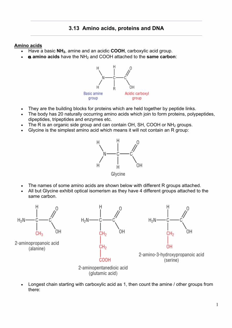
Amino acids • Have a basic NH2, amine and an acidic COOH, carboxylic acid group. • a amino acids have the NH2 and COOH attached to the same carbon:
• They are the building blocks for proteins which are held together by peptide links. • The body has 20 naturally occurring amino acids which join to form proteins, polypeptides,
dipeptides, tripeptides and enzymes etc. • The R is an organic side group and can contain OH, SH, COOH or NH2 groups. • Glycine is the simplest amino acid which means it will not contain an R group:
• The names of some amino acids are shown below with different R groups attached. • All but Glycine exhibit optical isomerism as they have 4 different groups attached to the
same carbon.
• Longest chain starting with carboxylic acid as 1, then count the amine / other groups from there:
2
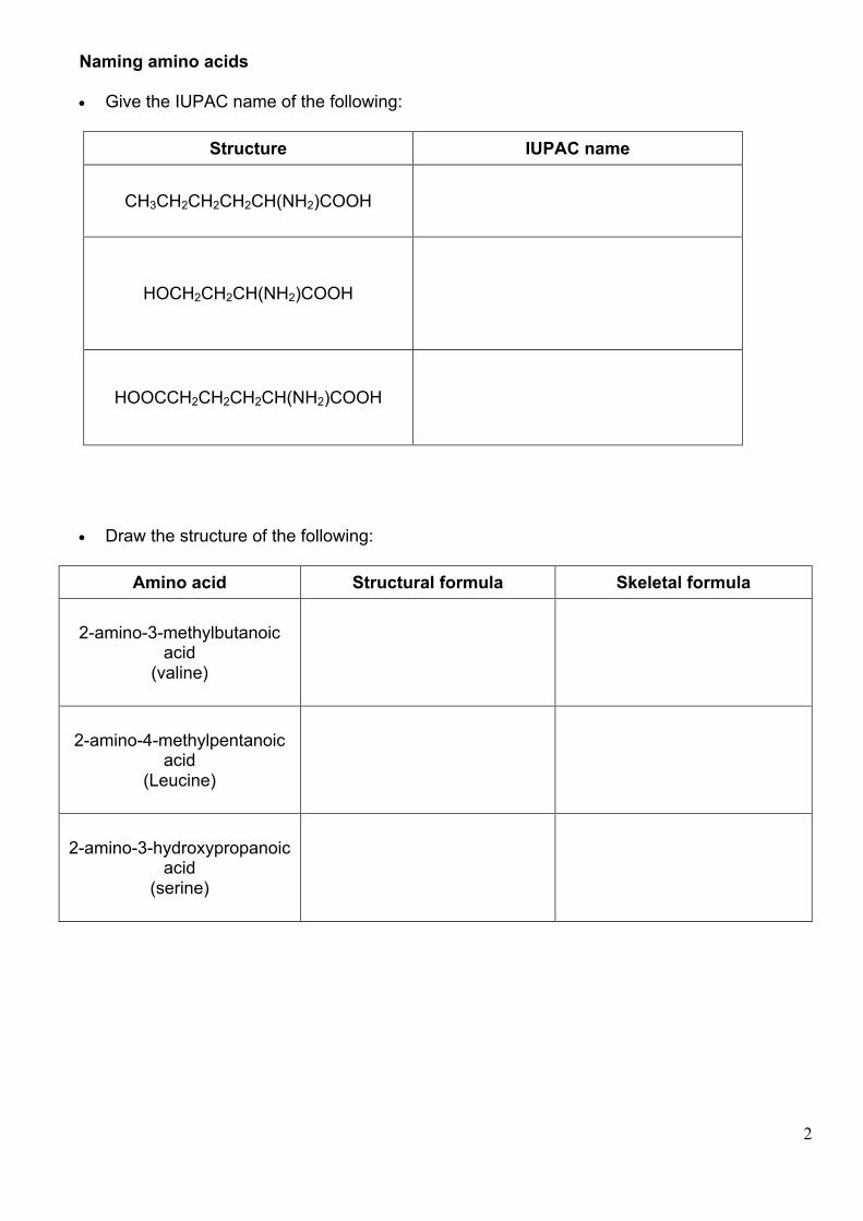
Naming amino acids • Give the IUPAC name of the following:
Structure IUPAC name
CH3CH2CH2CH2CH(NH2)COOH
HOCH2CH2CH(NH2)COOH
HOOCCH2CH2CH2CH(NH2)COOH
• Draw the structure of the following:
Amino acid Structural formula Skeletal formula
2-amino-3-methylbutanoic acid
(valine)
2-amino-4-methylpentanoic acid
(Leucine)
2-amino-3-hydroxypropanoic acid
(serine)
3
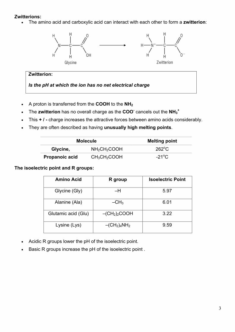
Zwitterions: • The amino acid and carboxylic acid can interact with each other to form a zwitterion:
Zwitterion: Is the pH at which the ion has no net electrical charge
• A proton is transferred from the COOH to the NH2 • The zwitterion has no overall charge as the COO- cancels out the NH3
+
• This + / - charge increases the attractive forces between amino acids considerably. • They are often described as having unusually high melting points.
The isoelectric point and R groups:
Amino Acid R group Isoelectric Point
Glycine (Gly) –H 5.97
Alanine (Ala) –CH3 6.01
Glutamic acid (Glu) –(CH2)2COOH 3.22
Lysine (Lys) –(CH2)4NH2 9.59
• Acidic R groups lower the pH of the isoelectric point. • Basic R groups increase the pH of the isoelectric point .
Molecule Melting point Glycine, NH2CH2COOH 262oC
Propanoic acid CH3CH2COOH -21oC
4
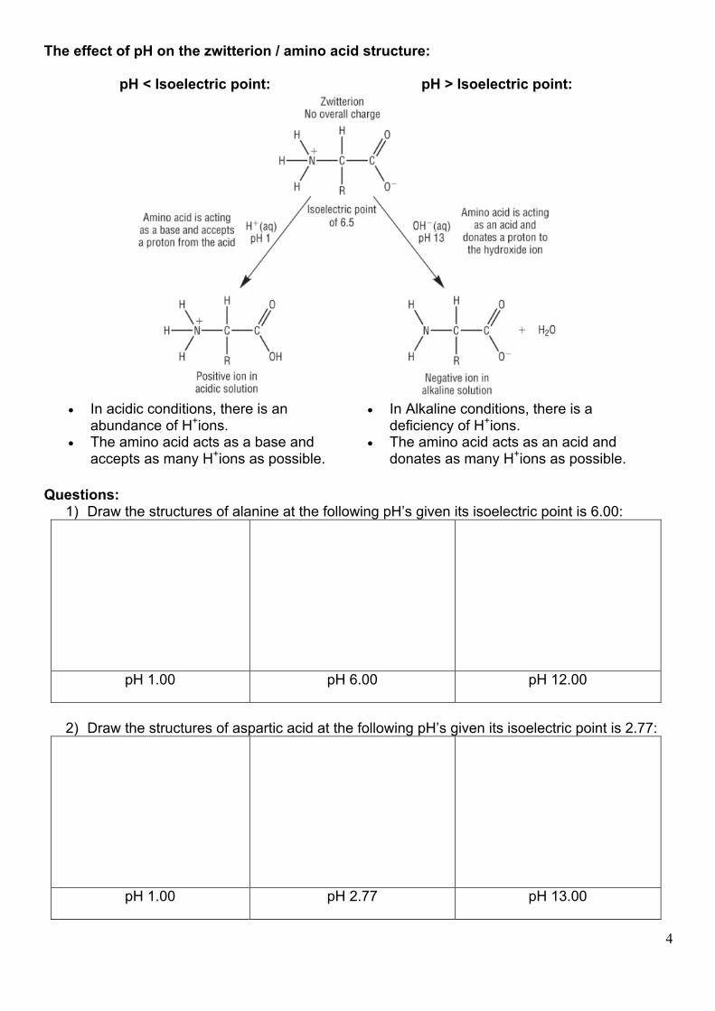
The effect of pH on the zwitterion / amino acid structure:
Questions: 1) Draw the structures of alanine at the following pH’s given its isoelectric point is 6.00:
pH 1.00 pH 6.00 pH 12.00
2) Draw the structures of aspartic acid at the following pH’s given its isoelectric point is 2.77:
pH 1.00 pH 2.77 pH 13.00
pH < Isoelectric point: pH > Isoelectric point:
• In acidic conditions, there is an abundance of H+ions.
• The amino acid acts as a base and accepts as many H+ions as possible.
• In Alkaline conditions, there is a deficiency of H+ions.
• The amino acid acts as an acid and donates as many H+ions as possible.
5
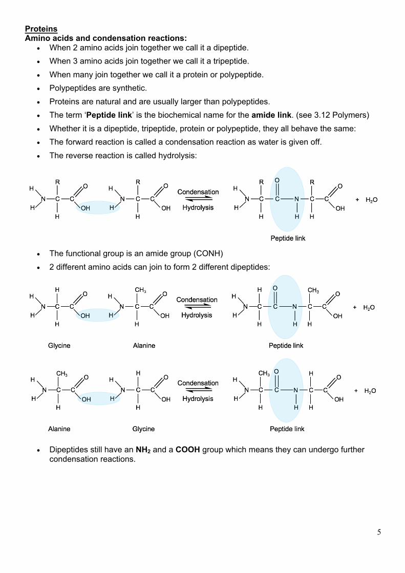
Proteins Amino acids and condensation reactions:
• When 2 amino acids join together we call it a dipeptide. • When 3 amino acids join together we call it a tripeptide. • When many join together we call it a protein or polypeptide. • Polypeptides are synthetic. • Proteins are natural and are usually larger than polypeptides. • The term ‘Peptide link’ is the biochemical name for the amide link. (see 3.12 Polymers) • Whether it is a dipeptide, tripeptide, protein or polypeptide, they all behave the same: • The forward reaction is called a condensation reaction as water is given off. • The reverse reaction is called hydrolysis:
• The functional group is an amide group (CONH) • 2 different amino acids can join to form 2 different dipeptides:
• Dipeptides still have an NH2 and a COOH group which means they can undergo further condensation reactions.
6
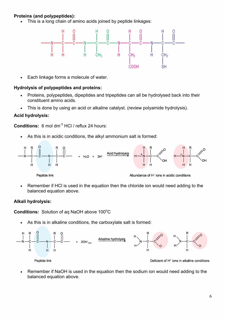
Proteins (and polypeptides): • This is a long chain of amino acids joined by peptide linkages:
• Each linkage forms a molecule of water. Hydrolysis of polypeptides and proteins:
• Proteins, polypeptides, dipeptides and tripeptides can all be hydrolysed back into their constituent amino acids.
• This is done by using an acid or alkaline catalyst. (review polyamide hydrolysis). Acid hydrolysis: Conditions: 6 mol dm-3 HCl / reflux 24 hours:
• As this is in acidic conditions, the alkyl ammonium salt is formed:
• Remember if HCl is used in the equation then the chloride ion would need adding to the balanced equation above.
Alkali hydrolysis: Conditions: Solution of aq NaOH above 100oC
• As this is in alkaline conditions, the carboxylate salt is formed:
• Remember if NaOH is used in the equation then the sodium ion would need adding to the balanced equation above.
7
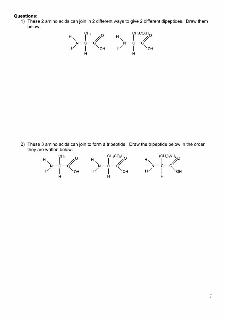
Questions: 1) These 2 amino acids can join in 2 different ways to give 2 different dipeptides. Draw them
below:
2) These 3 amino acids can join to form a tripeptide. Draw the tripeptide below in the order they are written below:
8
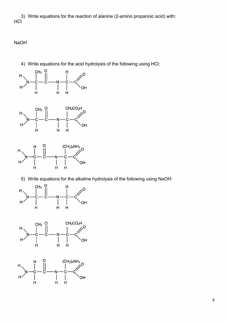
3) Write equations for the reaction of alanine (2-amino propanoic acid) with: HCl NaOH
4) Write equations for the acid hydrolysis of the following using HCl:
5) Write equations for the alkaline hydrolysis of the following using NaOH:
9
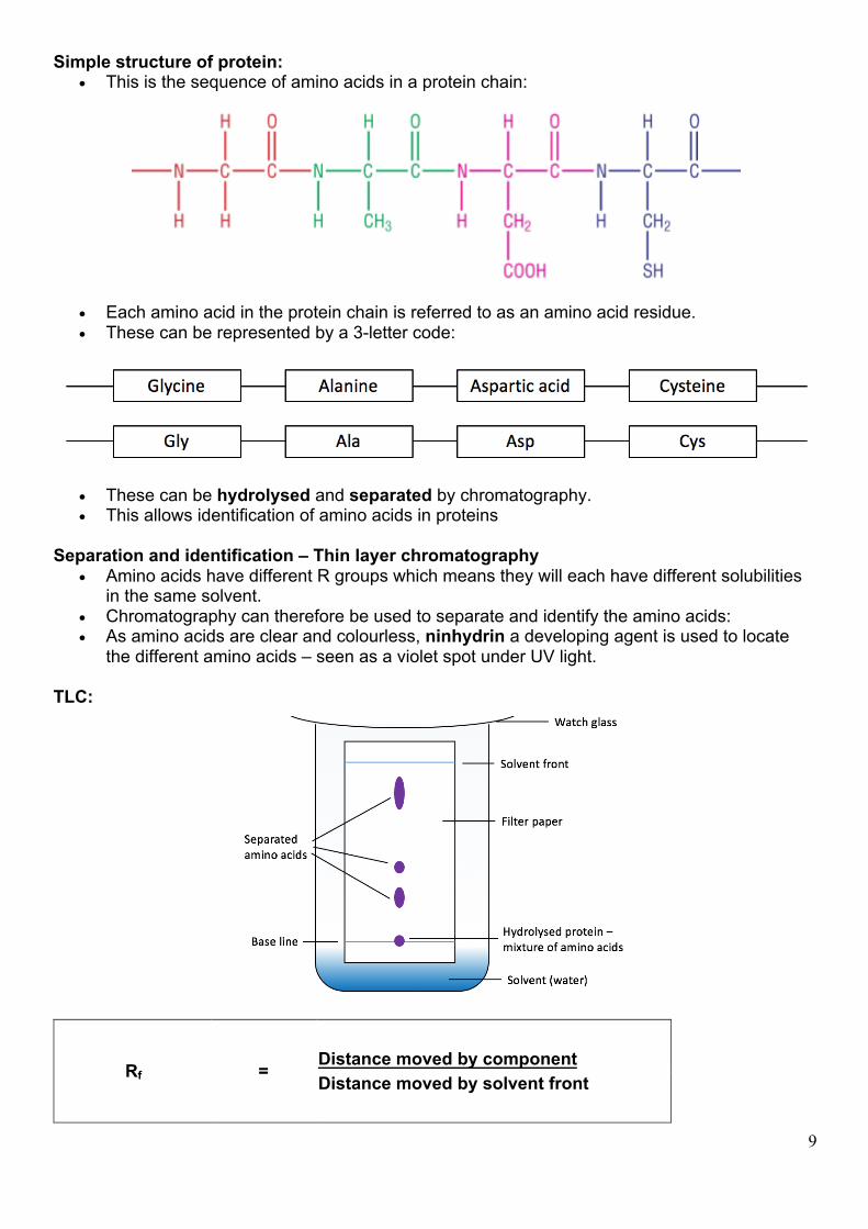
Simple structure of protein: • This is the sequence of amino acids in a protein chain:
• Each amino acid in the protein chain is referred to as an amino acid residue. • These can be represented by a 3-letter code:
• These can be hydrolysed and separated by chromatography. • This allows identification of amino acids in proteins
Separation and identification – Thin layer chromatography
• Amino acids have different R groups which means they will each have different solubilities in the same solvent.
• Chromatography can therefore be used to separate and identify the amino acids: • As amino acids are clear and colourless, ninhydrin a developing agent is used to locate
the different amino acids – seen as a violet spot under UV light. TLC:
Rf = Distance moved by component Distance moved by solvent front
10
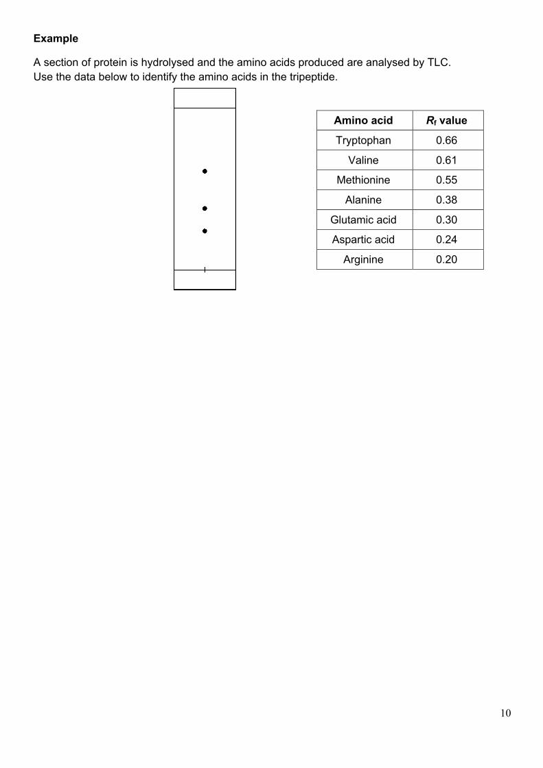
Example
A section of protein is hydrolysed and the amino acids produced are analysed by TLC. Use the data below to identify the amino acids in the tripeptide.
Amino acid Rf value
Tryptophan 0.66
Valine 0.61
Methionine 0.55
Alanine 0.38
Glutamic acid 0.30
Aspartic acid 0.24
Arginine 0.20
11
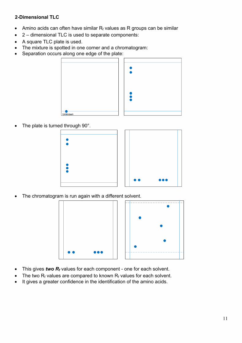
2-Dimensional TLC
• Amino acids can often have similar Rf values as R groups can be similar • 2 – dimensional TLC is used to separate components: • A square TLC plate is used. • The mixture is spotted in one corner and a chromatogram: • Separation occurs along one edge of the plate:
• The plate is turned through 90°.
• The chromatogram is run again with a different solvent.
• This gives two Rf values for each component - one for each solvent. • The two Rf values are compared to known Rf values for each solvent. • It gives a greater confidence in the identification of the amino acids.
12
Structure of protein: • This long chain of amino acids have complex structures. • Best thought of in 4 levels:
Ø Primary Ø Secondary Ø Tertiary Ø Quartenary (although this structure isn’t required)
1) Primary Structure:
• This is the sequence of amino acids in the protein chain:
• Each amino acid in the protein chain is referred to as an amino acid residue. • These can be represented by a 3-letter code:
2) Secondary structure:
• The peptide links can form hydrogen bonds with each other in one of 2 ways:
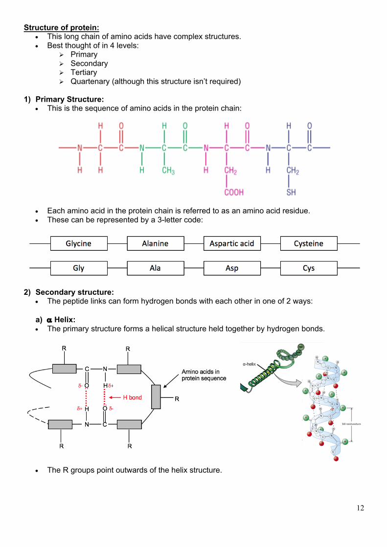
a) a Helix: • The primary structure forms a helical structure held together by hydrogen bonds.
• The R groups point outwards of the helix structure.
13
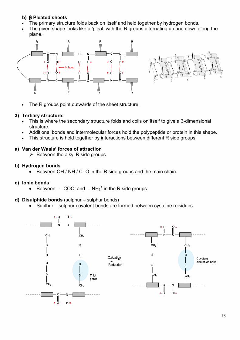
b) b Pleated sheets • The primary structure folds back on itself and held together by hydrogen bonds. • The given shape looks like a ‘pleat’ with the R groups alternating up and down along the
plane.
• The R groups point outwards of the sheet structure. 3) Tertiary structure:
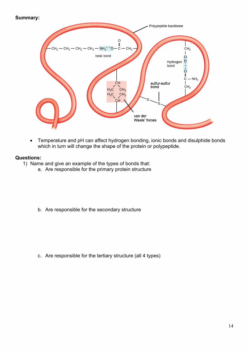
• This is where the secondary structure folds and coils on itself to give a 3-dimensional structure.
• Additional bonds and intermolecular forces hold the polypeptide or protein in this shape. • This structure is held together by interactions between different R side groups:
a) Van der Waals’ forces of attraction
Ø Between the alkyl R side groups
b) Hydrogen bonds • Between OH / NH / C=O in the R side groups and the main chain.
c) Ionic bonds
• Between – COO- and – NH3+ in the R side groups
d) Disulphide bonds (sulphur – sulphur bonds)
• Suplhur – sulphur covalent bonds are formed between cysteine reisidues
14
Summary:
• Temperature and pH can affect hydrogen bonding, ionic bonds and disulphide bonds which in turn will change the shape of the protein or polypeptide.
Questions:
1) Name and give an example of the types of bonds that: a. Are responsible for the primary protein structure
b. Are responsible for the secondary structure
c. Are responsible for the tertiary structure (all 4 types)
15
Enzymes
• These are proteins that catalyse chemical reactions in living things. • They often contain non – protein components which are called co – factors. • These co – factors are usually small organic molecules or metal ions.
Stereospecific nature of enzymes:
• This means that they only react with specific substances which are called substrates. Active site:
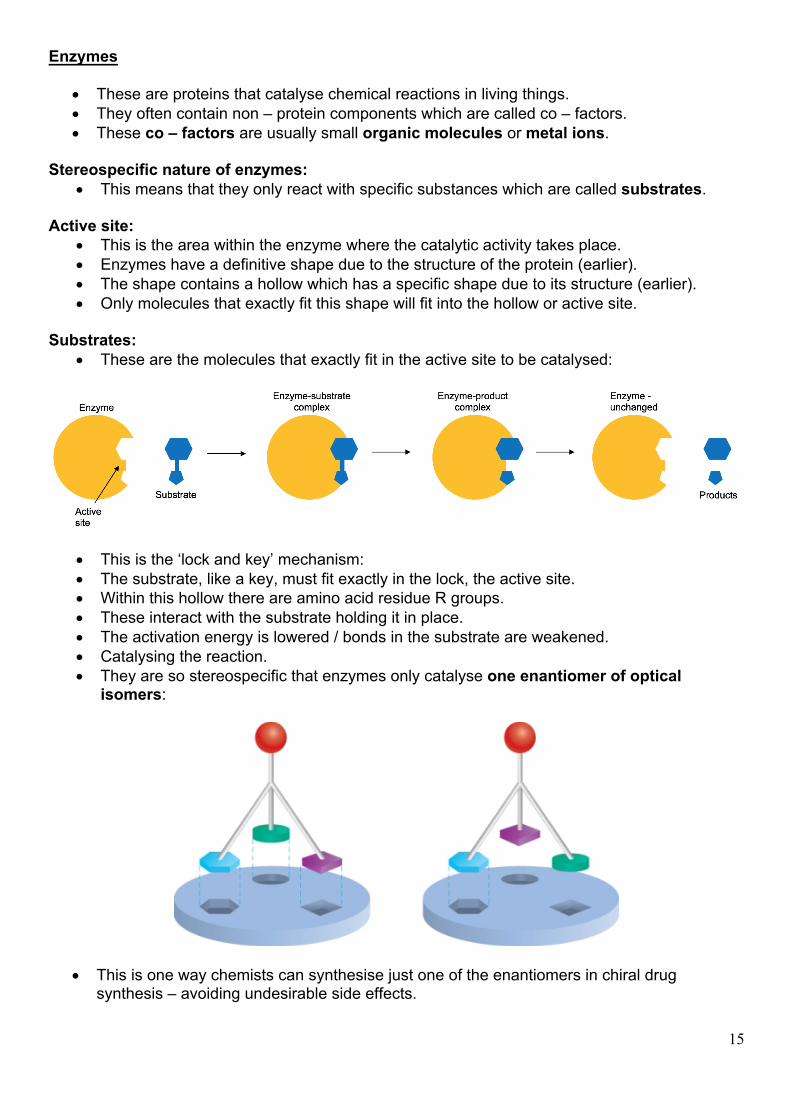
• This is the area within the enzyme where the catalytic activity takes place. • Enzymes have a definitive shape due to the structure of the protein (earlier). • The shape contains a hollow which has a specific shape due to its structure (earlier). • Only molecules that exactly fit this shape will fit into the hollow or active site.
Substrates:
• These are the molecules that exactly fit in the active site to be catalysed:
• This is the ‘lock and key’ mechanism: • The substrate, like a key, must fit exactly in the lock, the active site. • Within this hollow there are amino acid residue R groups. • These interact with the substrate holding it in place. • The activation energy is lowered / bonds in the substrate are weakened. • Catalysing the reaction. • They are so stereospecific that enzymes only catalyse one enantiomer of optical
isomers:
• This is one way chemists can synthesise just one of the enantiomers in chiral drug synthesis – avoiding undesirable side effects.
16
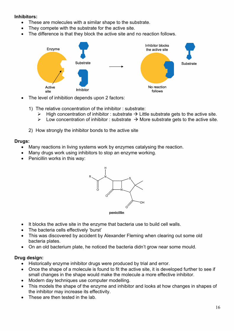
Inhibitors: • These are molecules with a similar shape to the substrate. • They compete with the substrate for the active site. • The difference is that they block the active site and no reaction follows.
• The level of inhibition depends upon 2 factors:
1) The relative concentration of the inhibitor : substrate:
Ø High concentration of inhibitor : substrate à Little substrate gets to the active site. Ø Low concentration of inhibitor : substrate à More substrate gets to the active site.
2) How strongly the inhibitor bonds to the active site
Drugs:
• Many reactions in living systems work by enzymes catalysing the reaction. • Many drugs work using inhibitors to stop an enzyme working. • Penicillin works in this way:
• It blocks the active site in the enzyme that bacteria use to build cell walls. • The bacteria cells effectively ‘burst’ • This was discovered by accident by Alexander Fleming when clearing out some old
bacteria plates. • On an old bacterium plate, he noticed the bacteria didn’t grow near some mould.
Drug design:
• Historically enzyme inhibitor drugs were produced by trial and error. • Once the shape of a molecule is found to fit the active site, it is developed further to see if
small changes in the shape would make the molecule a more effective inhibitor. • Modern day techniques use computer modelling. • This models the shape of the enzyme and inhibitor and looks at how changes in shapes of
the inhibitor may increase its effectivity. • These are then tested in the lab.
17
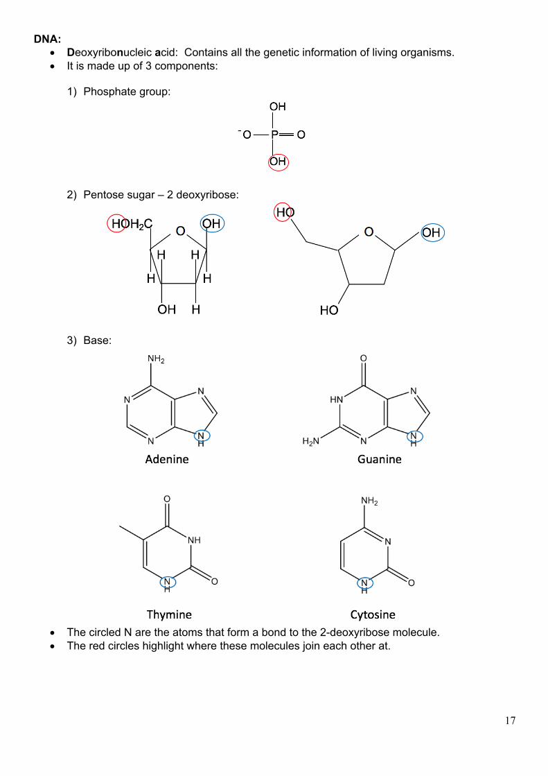
DNA: • Deoxyribonucleic acid: Contains all the genetic information of living organisms. • It is made up of 3 components:
1) Phosphate group:
2) Pentose sugar – 2 deoxyribose:
3) Base:
• The circled N are the atoms that form a bond to the 2-deoxyribose molecule. • The red circles highlight where these molecules join each other at.
18
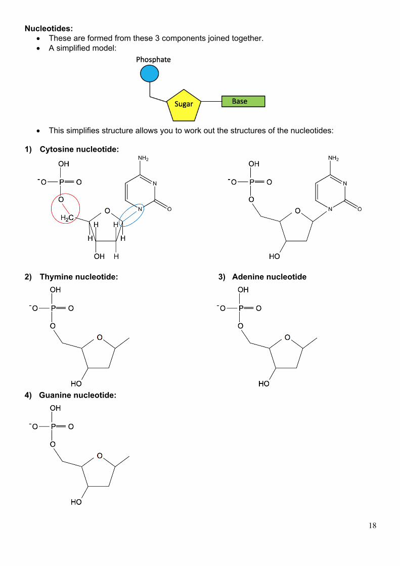
Nucleotides: • These are formed from these 3 components joined together. • A simplified model:
• This simplifies structure allows you to work out the structures of the nucleotides:
1) Cytosine nucleotide:
2) Thymine nucleotide: 3) Adenine nucleotide
4) Guanine nucleotide:
19
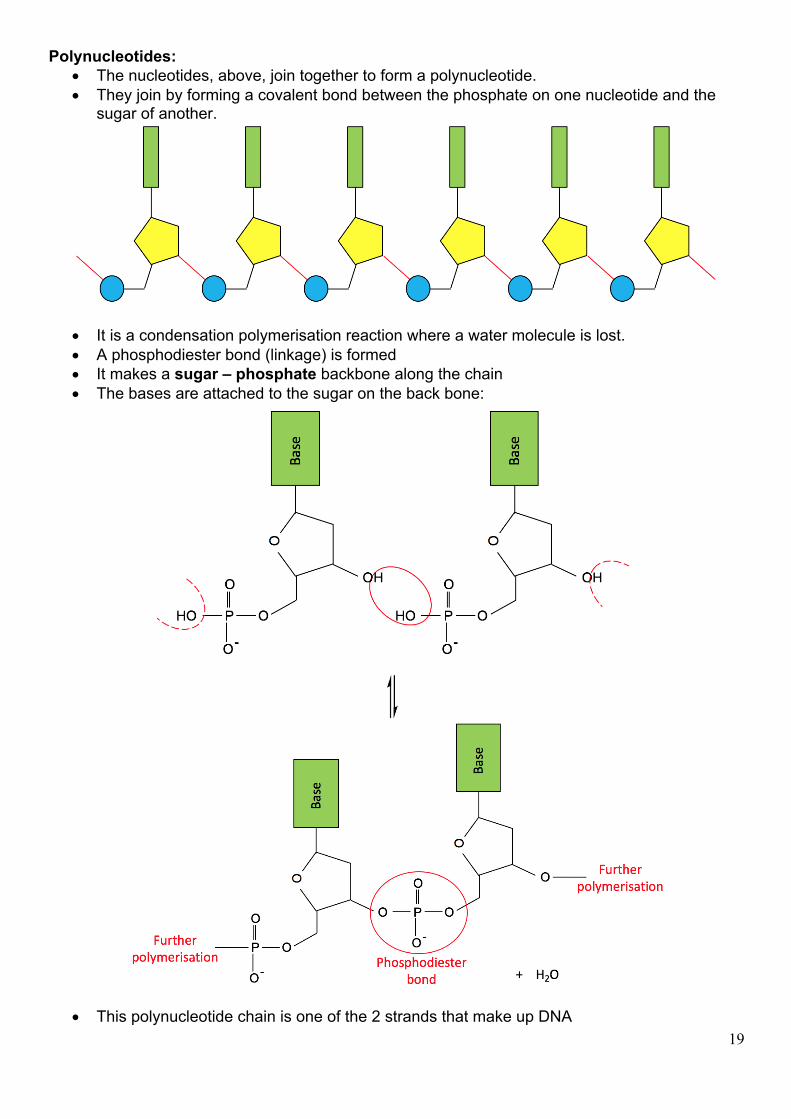
Polynucleotides: • The nucleotides, above, join together to form a polynucleotide. • They join by forming a covalent bond between the phosphate on one nucleotide and the
sugar of another.
• It is a condensation polymerisation reaction where a water molecule is lost. • A phosphodiester bond (linkage) is formed • It makes a sugar – phosphate backbone along the chain • The bases are attached to the sugar on the back bone:
• This polynucleotide chain is one of the 2 strands that make up DNA
20
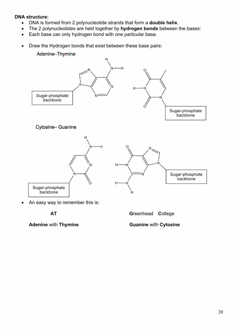
DNA structure: • DNA is formed from 2 polynucleotide strands that form a double helix. • The 2 polynucleotides are held together by hydrogen bonds between the bases: • Each base can only hydrogen bond with one particular base.
• Draw the Hydrogen bonds that exist between these base pairs:
• An easy way to remember this is:
AT Greenhead College
Adenine with Thymine Guanine with Cytosine
21
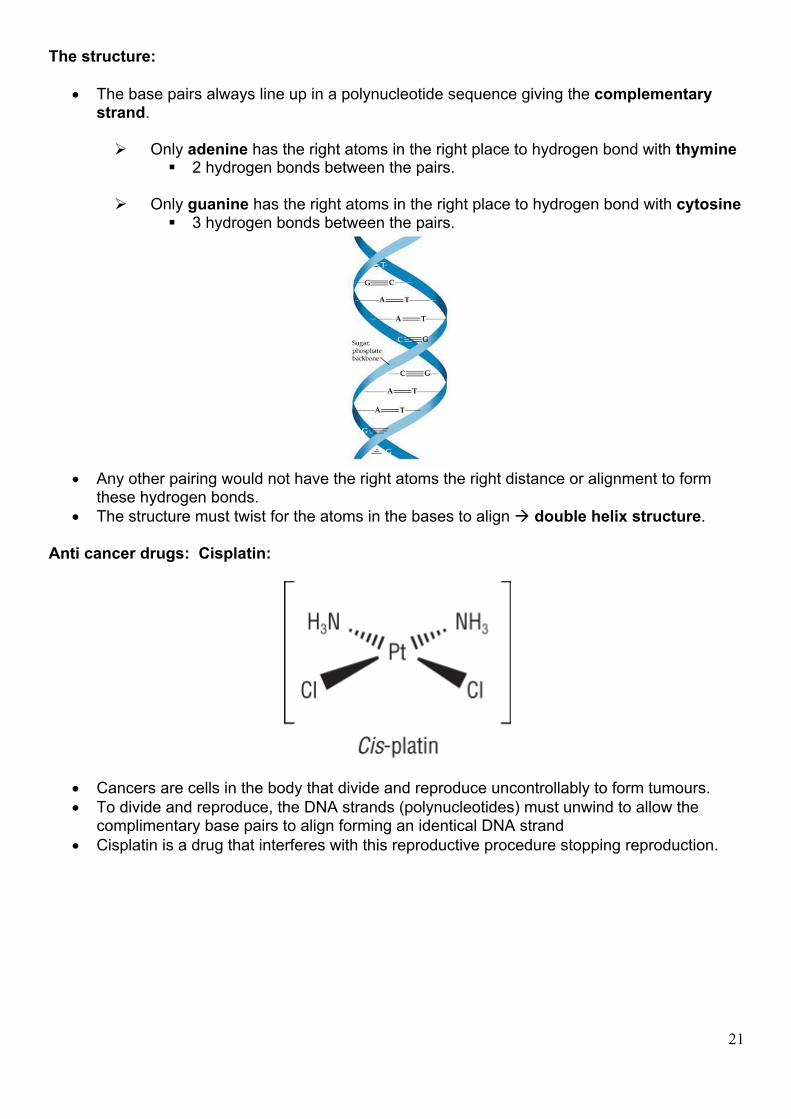
The structure:
• The base pairs always line up in a polynucleotide sequence giving the complementary strand.
Ø Only adenine has the right atoms in the right place to hydrogen bond with thymine
§ 2 hydrogen bonds between the pairs.
Ø Only guanine has the right atoms in the right place to hydrogen bond with cytosine § 3 hydrogen bonds between the pairs.
• Any other pairing would not have the right atoms the right distance or alignment to form
these hydrogen bonds. • The structure must twist for the atoms in the bases to align à double helix structure.
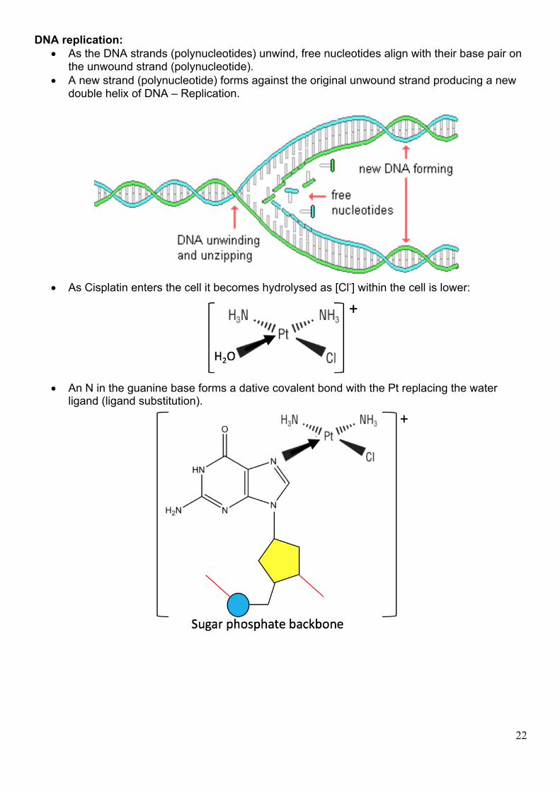
Anti cancer drugs: Cisplatin:
• Cancers are cells in the body that divide and reproduce uncontrollably to form tumours. • To divide and reproduce, the DNA strands (polynucleotides) must unwind to allow the
complimentary base pairs to align forming an identical DNA strand • Cisplatin is a drug that interferes with this reproductive procedure stopping reproduction.
22
DNA replication: • As the DNA strands (polynucleotides) unwind, free nucleotides align with their base pair on
the unwound strand (polynucleotide). • A new strand (polynucleotide) forms against the original unwound strand producing a new
double helix of DNA – Replication.
• As Cisplatin enters the cell it becomes hydrolysed as [Cl-] within the cell is lower:
• An N in the guanine base forms a dative covalent bond with the Pt replacing the water
ligand (ligand substitution).
23
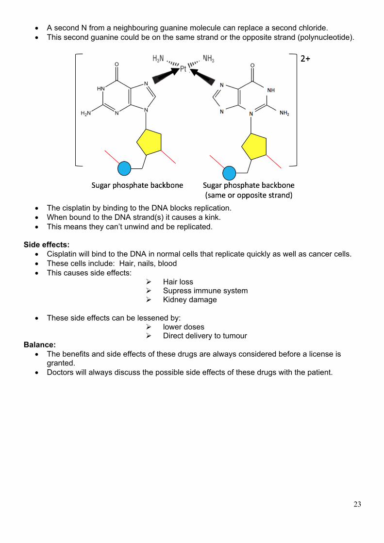
• A second N from a neighbouring guanine molecule can replace a second chloride. • This second guanine could be on the same strand or the opposite strand (polynucleotide).
• The cisplatin by binding to the DNA blocks replication. • When bound to the DNA strand(s) it causes a kink. • This means they can’t unwind and be replicated.
Side effects:
• Cisplatin will bind to the DNA in normal cells that replicate quickly as well as cancer cells. • These cells include: Hair, nails, blood • This causes side effects:
Ø Hair loss Ø Supress immune system Ø Kidney damage
• These side effects can be lessened by:
Ø lower doses Ø Direct delivery to tumour
Balance: • The benefits and side effects of these drugs are always considered before a license is
granted. • Doctors will always discuss the possible side effects of these drugs with the patient.
24
Questions: 1) Name and draw the structures of the 3 components that make up DNA:
2) Use your data sheet to combine these 3 components making a nucleotide using the guanine base:
3) This question is about polynucleotides. Ø What type of polymerisation makes a polynucleotide?
Ø What is the name of the bond formed between the monomers?
Ø Draw a section of 2 nucleotides with cytosine and thymine:
25
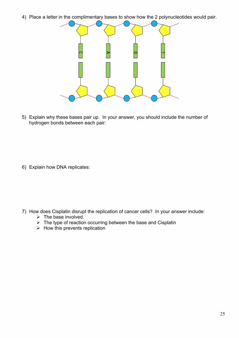
4) Place a letter in the complimentary bases to show how the 2 polynucleotides would pair.
5) Explain why these bases pair up. In your answer, you should include the number of hydrogen bonds between each pair:
6) Explain how DNA replicates:
7) How does Cisplatin disrupt the replication of cancer cells? In your answer include: Ø The base involved. Ø The type of reaction occurring between the base and Cisplatin Ø How this prevents replication