Embed Size (px)
Citation preview

3/17/08
Respiratory System
Chapter 21 – Day 2

3/17/08
Pathway of Air/ O2
Nose – external nares → nasal cavity → internal nares
Pharynx – nasopharynx → oropharynx → laryngopharynx
Larynx – epiglottis → larynx Trachea – trachea Bronchi – primary bronchi → secondary bronchi → tertiary bronchi → bronchioles
Lungs – alveoli → blood stream

3/17/08
Pathway of Air/ O2
Each component is composed of special tissues which aid in their function
Passageways & accessory structures in the nose and pharynx = upper respiratory system
Nose: external & internal nose♦Outside: cartilage & skin structure with 2 openings =
external nares♦Directs air into nasal passageways♦External = object of “vanity’
•“nose job” = Rhinoplasty – surgery to repair or alter nose structure – involves addition or removal of tissue
•Involves inconspicuous incisions & local anesthesia

3/17/08
Pathway of Air/ O2 - SkullNostrils lead into 2 chambers separated by the nasal
septumRight and left chambers of the nasal cavityWalls of the nasal septum = ethmoid & vomer bonesProtrusions of the bone = nasal conchae (superior,
middle, inferior)♦Allows air to swirl in the nasal cavity – airborne particles get
trapped in mucus♦Cavity is lined with mucous membrane♦Mucus is secreted by the paranasal sinuses (=air cavity with
epithelium in the skull)Floor of the nasal cavity = hard palate (roof of the
mouth)

3/17/08
Function of the NoseDirects air into the nasal passagewayWarms air via blood circulationLocation for olfaction sensationTraps particles & microbes = non-specific defense
♦These then get swallowed with mucous

3/17/08
Pathway of Air/ O2 – Pharynx and on From the nasal cavity, air
enters internal nares then the pharynx (throat)
Pharynx♦Shared by digestive and
respiratory systems♦It is a muscular tube lined with
mucous epithelium Just behind the nasal cavity =
nasopharynx The oropharynx is located right
under the soft palate – base of the tongue at the opening of the throat
♦It is lined with stratified squamous epithelium (b/c it is shared with the digestive system
Fig. 21.3

3/17/08
Pathway of Air/ O2 – Pharynx and on
Fig. 21.3
Laryngopharynx♦Below oropharynx = cavity♦Opening to esophagus & trachea♦Common area for air & food♦Stratified squamous epithelium
Next air enters the Larynx♦Top of the trachea♦Epiglottis = elastic cartilage
•Allows distinguish food vs. air•It closes the glottis (opening of
the larynx) if food is passing into the pharynx
•The ligaments stretch during swallowing to prevent food from entering the nasal passageway
•Fig. 21.5

3/17/08
Pathway of Air/ O2 – Larynx (& sound)
Fig. 21.4
Larynx♦Cylinder made of cartilage,
ligaments & skeletal muscle♦Note large pieces of hyaline
cartilage♦The larynx contains 2 ligaments
stretched from cricoid cartilage to thyroid cartilage = vocal ligaments (commonly called “vocal cords”
♦Air brushes against the vocal cords and creates vibrations = sound
The Larynx is important for air passage and speech
Skeletal muscles lengthen and shorten vocal cords – produces different sounds

3/17/08
Pathway of Air/ O2 – Trachea
Fig. 21.6
aka “windpipe” Tube made of smooth muscle
from larynx to bronchi Supported by cartilage rings
♦C-shaped cartilage – open at back = flexibility & expansion
♦Posterior wall – pseudostratified epithelium
Bifurcation at bottom of trachea (into 2)
Ridge @ the bifurcation =carina
♦Contains the cough reflex center 2 branches = Primary bronchi
(to right and left lung)♦Cartilage rings support the bronchi

3/17/08
Pathway of Air/ O2 – Bronchi
Fig. 21.9
Primary bronchi Each Primary bronchi branches into
secondary (lobar) bronchi♦These go to individual lobes of the lung
These then branch into the tertiary (segmental) bronchi
♦These are branches within each lobe Branch to bronchioles
♦End in lobules The bronchi regulate the air flow through
the lungs Bronchitis = infection/inflammation of the
bronchi♦Acute bronchitis is usually caused by a virus
(sometimes by bacteria)♦Cough, mild fever, yellow/green mucous

3/17/08
Pathway of Air/ O2 – Lung
Fig. 21.7
Collection of lobules containing alveoli Air sacs = light consistency Whole lung = paired organ - Right and left side Each lung is divided into lobes
♦Right = divided into 3 lobes by fissures ♦Left = divided into 2 lobes (know names of fissures and lobes for lab)
The shape of the lungs accommodates neighboring organs♦Heart is slightly to left = left lung is less broad & has cardiac notch♦Right side – liver is just below diaphragm = right lung is shorter
3/17/08
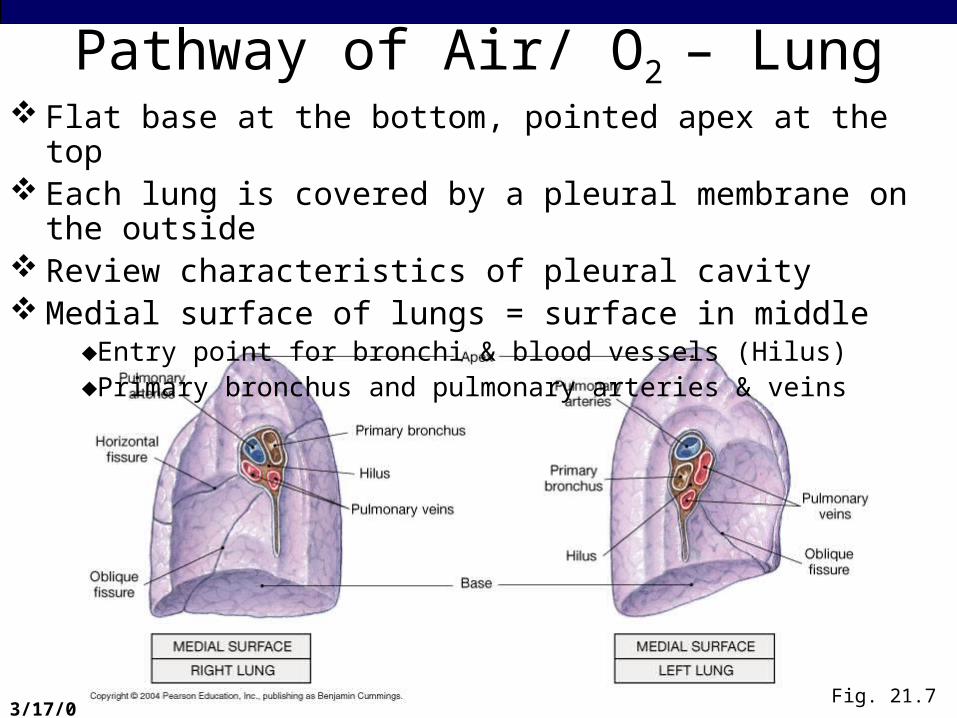
Pathway of Air/ O2 – Lung
Fig. 21.7
Flat base at the bottom, pointed apex at the top Each lung is covered by a pleural membrane on the outside Review characteristics of pleural cavity Medial surface of lungs = surface in middle
♦Entry point for bronchi & blood vessels (Hilus)♦Primary bronchus and pulmonary arteries & veins
3/17/08
Pathway of Air/ O2 – Lung
Fig. 21.9
Bronchus divides further inside the lungs – to each lobe Divided into segments within each lobe
♦Bronchopulmonary segment Visceral pleura extends into the lung – divides segments into
smaller sections♦Each section = Pulmonary lobule
Each lobule♦Contains a cluster of alveoli♦Receives air from the Bronchioles♦Lymph vessel circulation♦Pulmonary arteries and venules
Each alveolus within each lobule♦Hollow air sac♦Two or more air sacs may share a common opening = alveolar sac♦Recall what you saw in lab…

3/17/08
Alveoli In Pulmonary Lobule
Fig. 21.9

3/17/08
Pathway of Air/ O2 – Alveoli Alveoli = location of gas exchange Each alveolus – is confined – consists of a layer of simple
squamous epithelium Epithelium also contains
♦Macrophages- to engulf any escaped pathogens♦Septal cells – special cells that secrete a liquid called surfactant
•A surfactant keeps alveoli from collapsing shut
There is a continuous capillary adjacent to each alveolus These are connected by fused basement membranes of
epithelial cells & endothelial cells
Gas crosses 3 layers CO2 enters alveolus & O2 enters capillary

3/17/08
Alvioli – Capillary Interface
Fig. 21.11

3/17/08
Mechanics of RespirationVentilation
♦= mechanical process♦involves the diaphragm and skeletal muscles (intercostal
muscles)
Breathing consists of 2 phases: ♦Inspiration
•air is taken into the lungs
♦Expiration•Air passes out of the lungs

3/17/08
Mechanics of RespirationAir is moving in & out because of pressure gradientsAir flows from high pressure to low pressure…For air to enter the lungs, pressure should be low in
the lungs♦EXPANSION of lungs lowers pressure♦The Diaphragm contracts – pushed down = opens space in
the lungs♦External intercostal muscles contract – elevates chest
•Pulls on parietal pleura
•Pulls on visceral pleura
•Expands space into the lung
The pressure gradient forces air into the lungs (insp)ACTIVE process – powered by muscle

3/17/08
Mechanics of RespirationInspiration = ACTIVE process – powered by muscleExpiration = PASSIVE processObjective = increase pressure in lungs to create high
pressure gradient in TO out…♦The diaphragm and intercostals relax♦Pushes against pleura – close in on lungs♦Imagine a full balloon with hands pushing against it♦Increased pressure in lungs forces air out
Process of regular breathingDeep breathing & forced expiration require additional
muscles – abdominal muscles

3/17/08
Mechanics of RespirationThe amount of air entering during breathing depends
on several factorsCOMPLIANCE – degree of expandability of lungs
♦Large compliance – more air enters♦Elastic fibers surround alveoli & surfactant in alveoli
contribute to compliance & mobility of thoracic cage•Less surfactant – alveoli collapse – decreases comp.•Less elasticity = increases compliance
♦Emphysema•Shortness of breath, weak at exertion•Destruction of alveolar surface = loss of elasticity•Merged alveoli = larger space, but not enough capillary
support, so gas exchange does not support demand♦Skeletal disorders
•Arthritis or rib injuries reduce compliance

3/17/08
Lung mechanics
Fig. 21.13

3/17/08
Lung mechanics
Fig. 21.14

3/17/08
Lung mechanics
Fig. 21.15

3/17/08
Lung mechanics
Fig. 21.16

3/17/08
Lung mechanics
Fig. 20.18

3/17/08
Lung mechanics
Fig. 20.19

3/17/08
Lung mechanics
Fig. 20.14
3/17/08
Mechanics of Respiration

3/17/08
Mechanics of RespirationAir is moving in and

3/17/08
Mechanics of RespirationAir is moving in and out of the lungs due to pressure
gradients We will cover the mechanism in more detail in lecture,
but be sure you read the introduction to each lab and to each section/activity…you will be responsible for the information presented there in lab and lecture!
Happy breathing

3/17/08
Nose
Fig. 20.14