Embed Size (px)
Citation preview

3/2017

Bibliografische Information der Deutschen Nationalbibliothek
Die Deutsche Nationalbibliothek verzeichnet diese Publikation in der
Deutschen Nationalbibliografie; detaillierte bibliografische Daten sind im Internet über http://dnb.dnb.de abrufbar.
Bibliographic information published by the Deutsche Nationalbibliothek
The Deutsche Nationalbibliothek lists this publication in the Deutsche
Nationalbibliografie; detailed bibliographic data are available on the Internet at http://dnb.dnb.de .
Information bibliographique de la Deutsche Nationalbibliothek La Deutsche Nationalbibliothek a répertorié cette publication dans la
Deutsche Nationalbibliografie; les données bibliographiques détaillées
peuvent être consultées sur Internet à l'adresse http://dnb.dnb.de .
Informazione bibliografica della Deutsche Nationalbibliothek
La Deutsche Nationalbibliothek registra questa pubblicazione nella Deutsche Nationalbibliografie; dettagliati dati bibliografici sono disponibili
in internet in http://dnb.dnb.de .
Библиографическая информация Немецкой Национальной
Библиотеки
Немецкая Национальная Библиотека вносит эту публикацию в Немецкую национальную библиографию; подробные
библиографические данные можно найти в интернете на странице:
http://dnb.dnb.de .
Información bibliográfica de la Deutsche Nationalbibliothek
La Deutsche Nationalbibliothek recoge esta publicación en la Deutsche
Nationalbibliografie. Los datos bibliográficos están disponibles en la
dirección de Internet http://dnb.dnb.de . .

Deutscher Wissenschaftsherold • German Science Herald, N 3/2017
ISSN 2509-4327 (print) ISSN 2510-4780 (online)
Deutscher Wissenschaftsherold
German Science Herald
№ 3/2017
Die Zeitschrift „Deutscher Wissenschaftsherold“ ist eine Veröffentlichung mit dem Ziel ein breites Spektrum der
Wissenschaft allgemeinverständlich darzustellen. Die Redaktionsleitung versteht sich als Vermittler zwischen
Wissenschaftlern und Lesern. Durch die populärwissenschaftliche Bearbeitung wird es möglich unseren Lesern
neue wissenschaftliche Leistungen am besten und vollständigsten zu vermitteln. Es werden Untersuchungen,
Analysen, Vorlesungen, kurze Berichte und aktuelle Fragen der modernen Wissenschaft veröffentlicht.
Impressum Deutscher Wissenschaftsherold – German Science Herald Wissenschaftliche Zeitschrift Herausgeber: InterGING Sonnenbrink 20 31789 Hameln, Germany Inhaber: Marina Kisiliuk Tel.: + 49 51519191533 Fax.:+ 49 5151 919 2560 Email: [email protected] Internet:www.dwherold.de Chefredakeur/Editor-in-chief: Marina Kisiliuk Korrektur: O. Champela Gestaltung: N. Gavrilets
Auflage: № 3/2017 (August) – 23 Redaktionsschluss August, 2017 Erscheint vierteljährlich Editorial office: InterGING Sonnenbrink 20 31789 Hameln, Germany Tel.: + 49 51519191533 Fax.:+ 49 5151 919 2560 Email: [email protected] Deutscher Wissenschaftsherold - German Science Herald is an international, German/English language, peer-reviewed, quarterly published journal. № 3/2017 Passed in press in August 2017 Druck: WIRmachenDRUCK GmbH Muhlbachstr. 7 71522 Backnang Deutschland
Der Abdruck, auch auszugsweise, ist nur mit ausdrücklicher Genehmigung der InterGING gestattet. Die Meinung der Redaktion oder des Herausgebers kann mit der Meinung der Autoren nicht übereinstimmen. Verantwortung für die Inhalte übernehmen die Autoren des jeweiligen Artikels.
INDEXING: Google Scolar, WorldCat, InfoBase Index, Journal Index, Citefactor, International Scientific Indexing, JIFACTOR, Scientific Indexing Services, International Institute of Organized
Research. © InterGING © Deutscher Wissenschaftsherold – German Science Herald

Deutscher Wissenschaftsherold • German Science Herald, N 3/2017
2
REDAKTIONSKOLLEGIUM / INTERNATIONAL EDITORIAL BOARD:
Jurga Bernatoniene, Dr., Prof. Physics Lithuania [email protected] Arvaidas Galdikas, Dr. habil., professor Physics Lithuania, [email protected] Kristina Ramanauskienė, Ph.dr., Prof. Pharmacy, Lithuania [email protected] Khpaliuk Alexander, Dr. med. habil., Prof. Pharmakologie, Belorus [email protected] Arnold M. Gegechkori, Dr., full Prof. Biology, Georgia [email protected] Omari Mukbaniani, Prof., DSc. Chemistry, Georgia [email protected] Teimuraz Lezhava, Prof. Genetics, Georgia [email protected] Shota A. Samsoniya, Prof. Chemistry, Georgia [email protected] Mdzinarashvili Tamaz, DSc., Prof. Biophysics, Georgia [email protected] Aliaksandr V.Prokharau, MD, PhD, MSc Prof. Oncology, Belarus [email protected] Pyrochkin V., MD, PhD, MSc Prof. Theraphy, Belarus [email protected] Golubev A.P., BD, Prof. Ecology, Belarus [email protected] Makarevich A., MD, PhD, Prof. Theraphy, Belarus [email protected] Kanunnincova N., BD, Prof. Physiology, Belarus [email protected] Giedrius Vanagas, Prof. Internal Medicine, Lithuania [email protected] Armuntas Baginskas, Prof. Neurofiziologija, Lithuania [email protected] Ricardas Radisauskas, MD., Ph.D., Prof. Cardiology, Lithuania [email protected] Meyramov Gabit, Prof. Cytology and Histology, Kazakhstan [email protected] Aisha Mohammed Abd al-salam Shahlol Ph.D. in Medical Bacteriology, Libya [email protected]
Edmundas Kadusevicius, MD, PharmD, PhD, Prof. Pharmacology, Lithuania [email protected] Ivo Grabchev, Prof., PhD. Chemistry, Bulgaria [email protected] [email protected] Mariyana Ivanova Lyubenova, Prof., PhD. Ecology, Bulgaria [email protected] [email protected] Tsvetanka Tsankova Marinova, MD, PhD, DMedSci, Biology, Bulgaria [email protected] Evgueni D. Ananiev, Prof PhD, Biology. Bulgaria [email protected] Plamen G. Mitov, Prof., PhD. Biology, Bulgaria [email protected] Atanas Dimov Arnaudov, Ph.D. Physiology, Bulgaria [email protected] Iliana Georgieva Velcheva, PhD, Ecology, Bulgaria [email protected] Osman Demirhan, Prof. Biology, Turkey [email protected] Jharna Ray, M. Sc., PhD, Prof. Neurogenetics, India [email protected] Marián Halás doc. RNDr, Ph.D. Human geography, Сzech [email protected] Ayfer Pazarbasi Prof.Dr. Biology, Turkey [email protected] Tusharkanti Ghosh Prof. Physiology, India [email protected] Khudaverdi Gambarov Gambarov, Prof. Microbiology, Azerbaijan [email protected] Rovshan Ibrahimkhalil Khalilov, Prof. Biophysics, Azerbaijan [email protected] Svitlana Antonyuk, Dr.phil. Stony Brook University, USA Linguistics Samuel M.Johnson, Prof.Dr.phil. Theology, Wells, Maine, USA [email protected] Satanovsky Leon MD/PhD. Perio-odontologie, Israel [email protected]
Lists of references are given according to the Vancuver style

Deutscher Wissenschaftsherold • German Science Herald, N 3/2017
3
DDC-UDC 616.2-098.3-085.22:616.127-005.8-06 DOI:10.19221/201731
Grechko S.І., Trefanenko І.V.,
Shumko G.I., Shuper V.O.,
Reva T.V. Department of Internal Medicine, Physical Rehabilitation and Sport Medicine, Higher State Educational Establishment of
Ukraine “Bukovinian State Medical University”, Chernivtsi, Ukraine
COMBINED CONTROL OF THE HEART RHYTHM IN PATIENTS WITH ACURE CORONARY
SYNDROME Abstract. One of the main factors influencing on a short-term and remote prognosis of patients experienced acute myocardial infarction is heart rate. Administration of Ivabradine decreases HR at the expense of inhibition of electric activity of the sinoatrial node (Keith-Flack node) resulting in reduction of heart rhythm, increase of diastolic time during perfusion as a result of decreased oxygen supply to the myocardium without any harmful changes – arterial pressure values, coronary blood supply and contractile capacity of the myocardium. Monotherapy with Bisoprolol is indicative of an effective control of the heart rhythm in patients with ACS, but after a combined therapy with Ivabradine and Bisoprolol better results were found during the first 3-4 days of treatment. Insufficient decrease of HR in patients with ACS during the first 3-7 days of hospitalization is associated with an increased risk of post-infarction angina or relapse of myocardial infarction. Key words: congenital developmental defects; heart rhythm; acure coronary syndrome.
Introduction. A high mortality rate due to ischemic heart diseases in Ukraine outside in-patient departments is caused by low levels of detection and diagnostics of acute coronary syndromes (ACS). One of the main factors influencing on a short-term and remote prognosis of patients experienced acute myocardial infarction (MI) is heart rate (HR). Administration of Ivabradine decreases HR at the expense of inhibition of electric activity of the sinoatrial node (Keith-Flack node) resulting in reduction of heart rhythm, increase of diastolic time during perfusion as a result of decreased oxygen supply to the myocardium without any harmful changes – arterial pressure values, coronary blood supply and contractile capacity of the myocardium [1,3]. In patients with coronary failure and reduced ejection fraction Ivabradine demonstrated a positive effect in improvement of clinical results in addition to standard therapy [2]. However, a clear value concerning administration of Ivabradine in case of acute coronary syndrome has not been found.
Objective: to detect a potential value of Ivabradine in clinical context of treatment of
patients with ACS. Materials and methods. 135 patients with ACS
were included into the study. The possibility to improve treatment and clinical-prognostic role of ACS reduction was assessed with administration of Ivabradine. The patients were divided into the following groups: patients with the diagnosis of unstable angina – 62 individuals (45,9%), patients with MI without ST elevation – 52 individuals (38,5%), patients with MI and ST elevation – 21 individuals (15,5%), including 4 cases when thrombolysis had been performed (33%). Pharmacological therapy correlated with the national recommendations concerning management of patients with ACS. In addition to clinical and hemodynamic indices, the causes when β-adrenoreceptor blocking agents (β-AB) had not been indicated were analyzed, the frequency of achieving target HR values, dynamics of HR and BP against the ground of treatment by means of β-AB, causes limiting the titration of β-AB doses, frequency of side-effects after β-AB administration, the character of the disease at the hospital stage depending on the peculiarities of β-AB indication have been detected. The rates of
© Grechko S.І., Trefanenko І.V., Shumko G.I., Shuper V.O., Reva T.V., 2017 Anzeige – [email protected]

Deutscher Wissenschaftsherold • German Science Herald, N 3/2017
4
HR, BP, ECG indices were assessed after admission to the hospital: during an acute period (on the 2-4th day, the 3rd day on an average), during sub-acute period (on the 14th day) of staying in the hospital. The patients were divided into two groups: the one included patients receiving Bisoprolol with the aim to control HR (group І, 93 patients), and another one - patients receiving Bisoprolol in the combination with Ivabradine (group ІІ, 42 patients). The groups were similar by their major clinical-demographic indices. Mathematic analysis of the results obtained estimating the mean value and standard deviation of the mean value. Probability of quantitative indices was detected by means of the method of “null hypothesis” control using Student t-criterion (for equal and unequal dispersions – checked according to Fisher criterion), reliable results were considered with the index р<0,05.
Results and discussion. Analysis of the main parameters of the clinical-instrumental examination was not indicative of reliable differences between the patients of the examined groups at the beginning of treatment. On admission to the hospital the rates of HR and BP and on the 3rd day of treatment in both groups did not differ considerably. More than in half of the cases in both groups HR decreased – 49 patients (44,6%) of І group and 24 (57,1%) - ІІ group (р=0,55) respectively. In comparison with Bisoprolol as monotherapy, a combined administration of Ivabradine and Bisoprolol at the beginning of treatment was associated with more frequent achievement of a target value of HR and less probability of side-effects [2,3]. More considerable decrease of HR in 62% of patients was found in combination with Ivabradine. The target rate of systolic pressure (SP) was achieved in 54 (57,2%) and 31 (73,8%) patients (р=0.21), diastolic pressure (DP) – in 65 (68,9%) and 34 (81,9%) patients (р=0.47), and SP and DP – у 54 (54,1%) and 28 (68,6%) patients (р=0,39) of І and ІІ groups respectively. On the 14th day the target HR was achieved in 58 (62,4%) patients from І group and 34 (83%) patients of ІІ group (р=0.05). The target levels of SP and DP were achieved in all the patients of the examined groups. Inconsiderable decrease of HR during the first week of hospitalization was associated with an increased relapse risk of angina or myocardial
infarction without consideration of a reduced regimen of a medical agent. While analyzing the causes restricting the possibility to reach a target value of HR the following results were obtained: in І group in 14 (13%) cases symptomatic hypotension was found (in 1 patient was associated with the development of AB-block, 1st degree), in 5 (4,3%) – development of bronchial-obstructive syndrome, in 16 (17,7%) – only disorders of AB-conductivity to the 1-2nd degree. Bisoprolol was not cancelled in any of the cases. Hypotension, disorders of AB-conductivity and bronchial obstruction were eliminated by means of decreasing the dose of the drug, correction of doses of APP inhibitor and diuretics, a short-term administration of bronchodilators. In ІІ group transient visual signs were the cause to cancel further increase of the dose of Ivabradine only among 4 (9,5%) patients after achieving an average value of HR 66 b/min.
Complicated curse of ACS in the form of relapse of MI and/or post-infarction angina was registered in 33 (35,5%) patients of І group and 13 (18,5%) patients of ІІ group (р=0.70). At the same time, with reliably higher values of HR during the study the patients with complicated course of ACS (subgroup A) demonstrated reliably lower decrease of HR, than those without variant angina and/or relapse of MI (subgroup B) during all the stages of the hospital investigation. Similar dynamics of HR changes can be found in both groups of HR correction. Maximal decrease of HR was found since the first days of the study, which was similar for both groups of comparison. Considering selective decrease of HR without loss of the myocardial contractility, Ivabradine can be recommended as an effective agent to treat ACS without decrease of ejection fraction.
Conclusion. Monotherapy with Bisoprolol is indicative of an effective control of the heart rhythm in patients with ACS, but after a combined therapy with Ivabradine and Bisoprolol better results were found during the first 3-4 days of treatment. Insufficient decrease of HR in patients with ACS during the first 3-7 days of hospitalization is associated with an increased risk of post-infarction angina or relapse of myocardial infarction.
Prospects of further studies. Further improvement of therapeutic tactics,

Deutscher Wissenschaftsherold • German Science Herald, N 3/2017
5
determination of possible combination of Ivabradine and β-adrenoreceptor blocking agents will enable to reduce side effects and improve a remote prognosis in patients with acute coronary syndrome.
References. 1. Belousova ІP, Afonіna TV, Sterіonі ІV.
Ocіnka efektivnostі kombіnovanogo zastosuvannja nebіvololu ta іvabradinu v terapії pacієntіv zі stabіl'noju stenokardієju naprugi v poєdnannі z hronіchnimi obstruktivnimi zahvorjuvannjami legen'. Ukraїns'kij medichnij al'manah. 2011;14(1):32-3.
2. Parhomenko AN, Lutaj JaM, Irkin OI. Jeffektivnost' i bezopasnost' primenenija ingibitora If-kanalov ivabradina u bol'nogo s ostrym Q-infarktom miokarda s sinusovoj tahikardiej na fone terapii blokatorami β –adrenoreceporov. Ukr. med. chasopis. 2012;87(1):103-10.
3. Moiseev V, Abdulasisov O, Dyatchuk L, Meray I, Kiyakbaev G, Kobalava Z. Efficacy of Ivabradine in patients with chronic heart failure after myocardial infarction. European Journal of Heart Failure. 2011;10:234.
Deutscher Wissenschaftsherold • German Science Herald, N 3/2017
6
DDC-UDC 611.061.1+004.93 DOI:10.19221/201732
Dudenko V.G., MD, Prof., Department of Operative Surgery and Topographic Anatomy, Kharkiv National Medical University, Kharkiv,
Ukraine Avrunin О.G.,
Tymkovych М.Yu., Department of Biomedical Engineering Kharkiv National University of Radio Electronics, Kharkiv, Ukraine,
Kurinnyi V.V. PhD Student, Department of Operative Surgery and Topographic Anatomy, Kharkiv National Medical University, Kharkiv,
Ukraine
CONSTRUCTION OF A STATISTICAL THREE-DIMENSIONAL MODEL OF THE HUMAN
DIAPHRAGM ON THE BASIS OF TOMOGRPAHY FINDINGS Abstract. The work is devoted to the construction of a statistical three-dimensional model of the human diaphragm based on computed tomography data. As a result of the study, a statistical computer model of the human diaphragm was obtained, which in the future will allow us to classify the anatomical structure of the patient's diaphragm and identify possible deviations, as well as the peculiarities of their occurrence. Keywords: diaphragm, computed tomography, statistical modelling, reconstruction
Introduction. When a surgeon performs surgery his awareness and knowledge of the internal anatomical structure of a patient and personal structure in particular play a crucial role. The diaphragm occupies a special position among such structures. Its function is very important while performing surgery on certain kinds of hernias. Although, there are a number of evidences concerning general anatomical structure of the diaphragm [1-2] and its analysis with the use of intrascopic methods [4], the data concerning the peculiarities of its structure depending on the type of the body, gender, age and other factors are not much available. Modern studies in the field of medical anatomy require application of mathematical methods as to formalization of description of the anatomical structures [4-5]. Their use should level certain subjectivity of investigations and the quality of the description of an examined structure [6].
Therefore, construction of statistical models describing three-dimensional structure of the diaphragm and its peculiarities is uninvestigated multi-disciplinary area having great theoretical and practical value.
Objective: to construct statistical three-dimensional model of the human diaphragm on the basis of volumetric tomographic examinations
of patients. To achieve the goal the key points of the diaphragm should be measured enabling to describe it and visualize by means of three-dimensional graphics.
Materials and methods. The study was carried out at the Department of Operative Surgery and Topographic Anatomy, Kharkiv National Medical University. The reference data were specimens of computed tomography presented in DICOM format obtained by means of the computer scanner Toshiba Aquilion 16. Tomographic examination was performed at the supply voltage on the tube equal 120 kW, and current – 300 mА. Pixel range was 0,8 mm ×0,8 mm, and the distance between sections – 0,8 mm.
The sample of tomographic examination in three planes is presented on Fig. 1.
A three-dimensional model of the diaphragm was suggested to introduce by means of the use of 55 apices of the diaphragmatic surface located on the parallel sagittal sections and one additional apex – a basic one. In this way the diaphragmatic surface is formed by 84 triangles consisting of an appropriate connection of the apices (Fig. 2).
Having conducted appropriate constructions applying original software designed by the authors three-dimensional models of the diaphragm were obtained (Fig. 3).
© Dudenko V.G., Avrunin О.G., Tymkovych М.Yu., Kurinnyi V.V., 2017 Anzeige – [email protected]

Deutscher Wissenschaftsherold • German Science Herald, N 3/2017
7
a b c Fig. 1. The sample of examined tomographic investigation : а – axial section; b – coronary section; c – sagittal
section
Fig. 2. Schematic structure of apical connections of a
three-dimensional diaphragmatic model
Indicating the three-dimensional model for the
first patient as M1, the whole set of examinations will be marked as M:
1 1
1 1
1 1
1
56 56
56 561
,..., ,...,... ...
n
n
x x
y y
z zM M M
y y
z z
where x1, y1, z1, ..., y56, z56 – coordinates of apices of an appropriate investigation model;
n – number of patients. Therefore, having the whole set of
investigations available we have got the vector of mean values (μ) and covariant matrix (Σ):
1
1 n
i
i
Mn
1
1( )( )
1
nT
i i
i
M Mn
These two constituents together with normal
distribution (N) form a statistical three-dimensional model of the human diaphragm described in the following way:
1 1 1 1 1 1 56 1 561
1 1 1 1 1 1 56 1 561
1 1 1 1 1 1 1 56 56 56
56
56
2
1 2
12
1
56
56
...
...
...,
... ... ... ... ... ... ... ...
x x y x z x y x zx
y x y y z y y y zy
z z x z y z z y y z
yy
z
x
y
zN
y
z
56 1 56 1 56 1 56 56 56
56 1 56 1 56 1 56 56 56
2
2
...
...
x y y y z y y z
z x z y z z z y z
where σ – the element of covariant matrix Σ.
Fig. 3. The sample of a three-dimensional model of
the diaphragm: а – visualization with sagittal section; b – visualization without sagittal section
Results and discussion. Therefore, a statistical model of the human diaphragm was constructed on the basis of 30 intrascopic examinations of patients. Numerous values of the statistical model are obtained including the vector of mean values (μ) 168×1 in size and covariant matrix (Σ) 168×168 in size. The values obtained correspond to the normal law of distribution which is indicative of

Deutscher Wissenschaftsherold • German Science Herald, N 3/2017
8
further possibility to apply the model. Conclusions. 1. Three-dimensional models of
patients on the basis of tomographic data with the use of an original software are constructed.
2. The statistical model of the human diaphragm is obtained on the basis of three-dimensional models. This model will promote to determine and visualize a typical model of the human diaphragm.
3. The statistical model of the human diaphragm is obtained enabling to conduct mathematical modeling of different deviations from the normal values using Gauss distribution and at the same time to construct a three-dimensional structure of the obtained diaphragm. It enables to simulate pathologies of different nature both for training of students and further scientific investigations.
Prospects of further studies. A promising direction of further studies is construction of statistical models considering different pathological deviations which will enable to apply the data obtained for preliminary analysis of patient’s condition, and moreover, to determine anatomical signs and peculiarities promoting
occurrence of these deviations from the norm. One more prospective direction is the research directed to the analysis of the diaphragm according to the gender, age, constitution of the body, and, for example, for sportsmen to solve the tasks of sport medicine.
References. 1. Dufour M. Muscle diaphragm.
Kinésithérapie. la Revue. 2015;15(168):16-7. 2. Downey R. Anatomy of the Normal
Diaphragm. Thoracic Surgery Clinics. 2011;21(2):273-9.
3. Dianbo C, Wei L, Bolduc J-P, Deslauriers J. Correlative Anatomy of the Diaphragm. Thoracic Surgery Clinics. 2011;21(2):281-7.
4. Lorenz C. Generation of Point-Based 3D Statistical Shape Models for Anatomical Objects. Computer Vision and Image Understanding. 2000;77:175–91.
5. Dean D. Statistical Shape Analysis. Journal of Human Evolution. 2000;38(3) :455-7.
6. Zhang M. Statistical shape analysis : From landmarks to diffeomorphisms. Medical Image Analysis. 2016;33:155-8.

Deutscher Wissenschaftsherold • German Science Herald, N 3/2017
9
DDC-UDC 615.322:633.88:339.133 DOI:10.19221/201733
Sakhatska I.M. Candidate of Pharmaceutical Sciences
Higher State Educational Establishment of Ukraine «Bukovinian State Medical University», Chernivtsi, Ukraine
MARKET ANALYSIS ON MEDICINAL PLANT RAW MATERIAL
Abstract. The advantage of medicinal plants is their high biological activity in combination with rather low toxicity, good tolerance of phyto-preparations by patients, and as a rule, the absence of considerable side effects, and possibility of long administration. According to the study conducted chamomile flowers as medicinal raw material is in the greatest demand among the population. Keywords: medicinal plant raw material.
Introduction. The beginning of the ХХІ century is characterized by a wide use of phytotherapy and a very fast spread of assortment of plant medicinal remedies at the world pharmaceutical market [3]. According to the UN Food Agricultural Organization sales volume of plant medicinal remedies has exceeded 1 billion US dollars at the end of the last century [2].
In spite of rapid development of chemistry and appearance of new more effective synthetic medicines medicinal plants remain to be on a leading position in the arsenal of medical agents and their popularity in the whole world has been increasing. At the pharmaceutical markets of developed countries the share of medicines of a plant origin constitutes 50%. For example, 80% of physicians of all specialties use phyto-preparations in their practical work in Germany [3]. By the WHO prognosis their share during the following decade will constitute over 60% [4].
In recent years in Ukraine a tendency has been observed to the increase in prices for medical preparations making people more often use plant medicinal remedies. In this situation during the last 10 years in Ukraine a number of enterprises cultivating medicinal plants has been renewed and developed.
The advantage of medicinal plants is their high biological activity in combination with rather low toxicity, good tolerance of phyto-preparations by patients, and as a rule, the absence of considerable side effects, and possibility of long administration.
In addition, phyto-therapeutic means possess wider therapeutic action and other advantages as compared to synthetic medical agents. As a result, phyto-preparations have been successfully
competed with drugs obtained by means of synthesis for several years already [6, 10].
Preparations of a plant origin are able to participate organically in biochemical processes of the human body, change these processes and reveal therapeutic action even in small doses of their administration [9]. One more considerable difference of preparations of a plant origin from their synthetic analogues is that pharmacological effect of a synthetic drug is based on the action of one or several purified active substances. In their turn, preparations of a plant origin contain several groups of biologically active substances with various pharmacological actions. Every compound contained in a plant possesses its individual pharmacological effect, and therapeutic action of plants is based on the combination of effects of several compounds. This effect of medicinal raw material is achieved by means of synergism of biologically active substances in the content of plants [9].
In addition, administration of drugs of a natural origin enables to avoid acquired tolerance or addiction and medical dependence, stimulate individual adaptive and protective systems of the body. Administration of preparations of a plant origin for many years in clinical practice is indicative of their efficacy in treatment of many diseases [1, 7, 8].
One more factor influencing a growing trust of customers in preparations of a plant origin is continuous increase of the quality and safety at the expense of introduction of standards concerning proper industrial practice [5].
Objective: to detect the kinds of medicinal raw material being in demand among the customers of chemist’s shops and are profitable.
© Sakhatska I.M., 2017 Anzeige – [email protected]

Deutscher Wissenschaftsherold • German Science Herald, N 3/2017
10
Materials and methods. Every year the need of pharmaceutical enterprises in medicinal herbs becomes 20-25% higher at the expense of increased sales of the old ones and creation of new preparations of a plant origin. Among the plants used in medicine in Ukraine medicinal are 250 species including 150 used in traditional medicine and the rest – in folk medicine only [5].
At present the Ukrainian pharmaceutical market includes more than 20 Ukrainian companies and enterprises working with medicinal raw material. The leaders producing medicinal plant material and pharmaceutical products are joint-stock companies «Liktravy», «Viola», «Lubnypharm», and the public corporation «Ternopil Pharmaceutical Plant».
Dispensing chemists at chemist’s shops were
surveyed with the aim to detect the demand for medicinal plant material and analyze price-lists with the purpose to determine demand/cost ratio.
The survey and detection of average prices were conducted at chemist’s shops in the towns of Berdychiv and Chernivtsi, including the company «Med-service», «Low prices pharmacy», the pharmaceutical network «Harmony», the pharmacy network «D.S.», pharmaceutical enterprises «Tkachuk», «Universytetska №1», «PharmMix» №1 and «Akizum-Pharm» (Table 1).
The price-lists of the major suppliers of medicines to chemist’s shops were analyzed, including joint ventures «BaDM», «Optima-pharm, Ltd», «Venta Ltd» and the company «Pharmplaneta» (Table 1).
Table 1 Analysis of prices
Items Average retail price in chemist’s shops, hrn
Average wholesale price of suppliers, hrn
Chamomile flowers 40 g 12,90 10,52
Flax seeds 100 g 8,0 6,42
Pepper mint leaves 50 g 12,50 10,63
Marigold herb 50 g 11,80 9,7
Melissa herb 50 g 16,00 11,33
Oak-tree bark 100 g 11,10 10,75
Marshmallow roots 75 g 14,76 11,7
Calendula flowers 50 g 17,95 12,99
Wild rose fruit 130 g 17,75 13,82
Sage leaves 50 g 18,00 15,25
Results and discussion. The questionnaire of dispensing chemists determined that the customers of chemist’s shops most often ask for such medicinal plant as chamomile leaves 40 g, and then in a descending order: Flax seeds 100 g, Pepper mint leaves 50 g, Marigold herb 50 g, Melissa herb 50 g, Oak-tree bark 100 g, Marshmallow roots 75 g, Calendula flowers 50 g, Wild rose fruit 130 g, Sage leaves 50 g.
On an average chemist’s shops sale 8-20 packages of Chamomile flowers every day. The demand for this medicinal plant is associated with a wide spectrum of its action and minimal number of contraindications. Chamomile flowers possess anti-inflammatory, spasmolytic, diuretic, anti-allergic, mild sedative, antimicrobial action, increase the activity of the digestive glands, and promote appetite [4].
Chamomile preparations are used internally and externally. They are indicated internally in case of intestinal spasms, flatulence, diarrhea, gastritis, colitis, bronchial asthma, rheumatism, menstrual disorders, liver and urinary bladder diseases, insomnia.
Externally they are used for gargling in case of inflammation of the oral cavity, inflammatory processes of the urinary tract, syringing, irrigation of ulcers, purulent wounds, hemorrhoid nodes, dermatitis, and eczema.
As to the cultivation of chamomile (Matricaria chamomilla), keeping to the major technological agricultural methods (winter sowing, planting depth of 0,5 cm, soil moisture) ensures heavy yields of inflorescences with minimal labour inputs.
Conclusion. Therefore, according to the study

Deutscher Wissenschaftsherold • German Science Herald, N 3/2017
11
conducted chamomile flowers as medicinal raw material is in the greatest demand among the population.
References 1. Viktorov AP. Fitopreparaty: racional'nyj
podhod k medicinskomu primeneniju. Fitoterapija. Chasopis. 2011;(3):1-10.
2. Mіrzoєva T. Osoblivostі vіtchiznjanogo rinku lіkars'kih roslin v umovah s'ogodennja. Іnnovacіjna ekonomіka. 2013;44(6):209-12.
3. Degtjar'ova KO, Vishnevs'ka LI, Jarnih TG, Tkachuk OJu. Perspektivi vikoristannja roslinnoї sirovini garbuza dlja stvorennja lіkars'kogo preparatu na jogo osnovі. Ukraїns'kij zhurnal klіnіchnoї ta laboratornoї medicini. 2013;8(2):31-35.
4. Kislichenko VS, editor. Farmakognozіja: bazovij pіdruch dlja stud vishh farmac navch zakl IV rіvnja akreditacії. Harkіv: NFaU: Zolotі storіnki; 2015. 736 p.
5. Jakovenko VK. Naukovo-teoretichne obґruntuvannja pіdhodіv do upravlіnnja jakіstju
pri rozrobcі ta virobnictvі roslinnih lіkars'kih zasobіv [dis. ... doktora farm. Nauk]. Harkіv; 2015. 384 p.
6. Korsun VF, Korsun AF. Psoriaz. Sovremennye i starinnye metody lechenija. SPb: DILJa; 2000. 208 p.
7. Reshetnikov VN, Gapanovich VN, Volod'ko IK. Gosudarstvennaja narodno-hozjajstvennaja programma razvitija syr'evoj bazy i pererabotki lekarstvennyh i prjano-aromaticheskih rastenij na 2005-2010 gody «Fitopreparaty» – inovacii v dejstvii. Trudi BGU. 2010;5(2):10-15.
8. Abd EI-Aziz AB, Abd EI-Kalek HH. Antimicrobial proteins and oil seeds from pumpkin (Cucurbita moschata). Nature and Science. 2011;3(9):105-18.
9. Lieberman S. Natural Interventions for Treating Psoriasis. Alternative and Complementary Therapies. 2002;19(6):355-8.
9. Seong-Tae L. Herbal Remedies for Psoriasis. Journal of Ethnopharmacology. 2010;127(8):11-8.

Deutscher Wissenschaftsherold • German Science Herald, N 3/2017
12
DDC-UDC 371.315:372.8:613.71 DOI:10.19221/201734
Kondratiuk О.S., Korshun М.М.,
Garkavyi S.І. Department of Hygiene and Ecology № 3 O.O. Bogomolets National Medical University, Kyiv, Ukraine,
ADAPTIVE CAPACITY ASSESSMENT OF PRIMARY SCHOOL CHILDREN IN CASE OF VARIOUS FORMS OF ORGANIZAION OF PHYSICAL TRAINING CLASSES
Abstract. The state of adaptation capacity (AC) of the -4th form pupils has been evaluated. The experimental group (E) included 183 pupils who studied at the school where one of the three physical training classes was held in the swimming pool. Pupils from the neighboring school without a pool, visited three traditional classes of physical culture in the gym and were included into the control group (C, n = 184). The arithmetic mean from Robinson’s, Rohrer’s and Kerdo’s indices was calculated and after obtained values AC was estimated. The children from E group have lower Robinson’s and Kerdo’s indices in comparison with C group (t = 2, 65 and t = 8, 36, respectively, p <0, 01). In group E higher proportion of pupils with a high level of AC as compared to the pupils from K group (14,21 and 5,97%, respectively, t = 2, 64, p <0, 05). The percentage of schoolchildren with low AC level in E group was lower in comparison with K group (50, 27 and 66, 85%, respectively, t = 3, 27, p <0, 01). Thus, swimming exercises during one of the three physical training classes facilitates pupils’ efficient and economical adaptation to the educational process. Key words: adaptation capacity, primary school pupils, swimming.
Introduction: Health of children is a guarantee of a healthy nation. For a long time high sickness rate and occurrence of diseases had been an issue of great concern, and only in recent 4 years these indices have been marked with a tendency to decrease [1]. This state of affairs has stipulated elaboration and fulfillment of certain measures directed to strengthening of health. Although in the majority of cases health-improving initiatives have been introduced without proper hygienic substantiation. One of such innovations is organization of educational process concerning physical training classes. One of the three classes stipulated by the standard curriculum for primary school was held on the basis of a swimming pool.
An effective preventive measure prevents occurrence of pre-morbid conditions without any pronounced clinical signs and reflect disorders of the adaptive process to the environment due to unfavourable effect of certain school factors [2]. Therefore, in order to provide hygienic substantiation of health-improving effect of the classes held in the swimming pool, adaptive capacity of the body should be assessed.
Objective: to assess adaptive capacity of the 1-4th-form pupils in different forms of organization
of physical training classes. Materials and methods. On the basis of
sanitary examination of 7 comprehensive secondary schools in the city of Kyiv 2 educational establishments have been chosen including one with a swimming pool. Water in the pool is disinfected by means of the most spread method in Ukraine – chlorination.
Similar groups of the study were formed by the results of a questionnaire. Children attending sport clubs after school were not included into the study. Those primary school children who have their physical training classes in the gym were included into the control group (C, n=184). The experimental group (E, n=183) included the schoolchildren from the comprehensive secondary school where classes in the swimming pool are organized once a week.
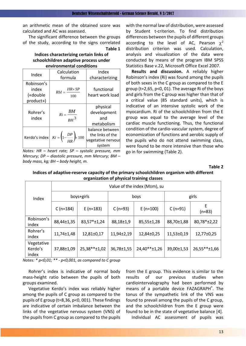
Adaptive capacity (AC) of the organism was investigated by means of the suggested methods [3]. The study was conducted in two stages: during the first stage anthropometric and physiometric indices were measured, the general score of indices characterizing certain links of adaptive process under conditions of the environment were calculated (Table 1). During the second stage
© Kondratiuk О.S., Korshun М.М., Garkavyi S.І., 2017 Anzeige – [email protected]
Deutscher Wissenschaftsherold • German Science Herald, N 3/2017
13
an arithmetic mean of the obtained score was calculated and AC was assessed.
The significant difference between the groups of the study, according to the signs correlated
Table 1 Indices characterizing certain links of
schoolchildren adaptive process under environmental conditions
Index Calculation
formula Index
characterizing Robinson’s
index («double product»)
100
SPHRRbI
functional heart work load
Rohrer’s index 3
BH
RiBM
physical development
and metabolism
Kerdo’s index 1001 HR
DPKi
balance between the links of the
vegetative nervous system
Notes: HR – heart rate; SP – systolic pressure, mm Mercury; DP – diastolic pressure, mm Mercury; BM – body mass, kg; BH – body height, m.
with the normal law of distribution, were assessed by Student t-criterion. To find distribution differences between the pupils of different groups according to the level of AC, Pearson 2 distribution criterion was used. Calculation, analysis and visualization of the data were conducted by means of the program IBM SPSS Statistics Base v.22, Microsoft Office Excel 2007.
Results and discussion. A reliably higher Robinson’s index (Ri) was found among the pupils of both sexes in the C group as compared to the E group (t=2,65, p<0, 01). The average Ri of the boys and girls from the C group was higher than that of a critical value (85 standard units), which is indicative of an intensive systolic work of the myocardium. Ri of the schoolchildren from the E group was equal to the average level of the cardiac muscle functioning. Thus, the functional condition of the cardio-vascular system, degree of economization of functions and aerobic supply of the pupils who do not attend swimming class, were found to be more intensive than those who go in for swimming (Table 2).
Table 2 Indices of adaptive-reserve capacity of the primary schoolchildren organism with different
organization of physical training classes
Index
Value of the index (М±m), su
boys+girls boys girls
C (n=184) Е (n=183) C (n=93) Е (n=100) C (n=91) Е
(n=83)
Robinson’s index
88,44±1,35 83,57*±1,24 88,18±1,9 85,55±1,28 88,70±1,88 80,78*±2,22
Rohrer’s index
11,74±1,48 12,81±0,17 11,94±2,19 12,84±0,25 11,53±0,19 12,77±0,25
Vegetative Kerdo’s index
37,88±1,09 25,38**±1,02 36,78±1,55 24,40**±1,26 39,00±1,53 26,55**±1,66
Notes: * p<0,01; ** - p<0,001, as compared to C group
Rohrer’s index is indicative of normal body
mass-height ratio between the pupils of both groups examined.
Vegetative Kerdo’s index was reliably higher among the pupils of C group as compared to the pupils of E group (t=8,36, p<0, 001). These findings are indicative of certain imbalance between the links of the vegetative nervous system (VNS) of the pupils from C group as compared to the pupils
from the E group. This evidence is similar to the results of our previous studies when cardiointervalography had been performed by means of a portable device FAZAGRAPH®. The tonus of the sympathetic link of the VNS was found to prevail among the pupils of the C group, and the schoolchildren from the E group were found to be in the state of vegetative balance [4].
Individual AC assessment of pupils was

Deutscher Wissenschaftsherold • German Science Herald, N 3/2017
14
conducted by the results of arithmetic mean calculation from the previously calculated and converted into the points according to the scale stipulating 4 levels of the body functioning: satisfactory adaptation (≥ 2,67 points), intensification of adaptive mechanisms (1,67–2,66 ponits), unsatisfactory adaptation (1,35–1,66 points) and adaptive failure (≤ 1,34 points), which correlates with high, moderate, low and critically low assessment of AC.
Among junior schoolchildren from the both examined groups there was no one found with adaptive failure. The reliable difference was found in the distribution of pupils from the C and E groups by AC level (χ2 = 12,50, df = 2, p<0,01). In the C group a part of pupils with high AC level was 2,4 times less as compared to the E group (t=2,64, p<0,05), and the children with low adaptive reserves prevail (t=3,27, p<0,01) (Fig. 1).
Fig. 1. Distribution of junior pupils with different
organization of physical training classes according to the level of adaptive-reserve capacity of the organism.
* p<0, 05, ** - p<0, 001
Conclusions. 1. Attending swimming classes by
schoolchildren promotes better functioning of the cardio-vascular system and vegetative homeostasis, and Robinson’s and Kerdo’s indices
were reliably lower than those of the pupils attending swimming classes as compared to the pupils from the control group.
2. At the school where one out of three classes of physical training was held on the basis of the swimming pool, a part of pupils with high individual level of adaptive capacity was bigger, and the percentage of pupils with low adaptive reserves was lower as compared to the control educational establishment.
Thus, organization of physical training classes with one out of the three ones on the basis of a swimming pool promotes effective and economic adaptation of pupils to the environmental factors including school surroundings, and therefore, it is health-improving.
References 1. Shafrans'kogo VV, editor. Shhorіchna
dopovіd' pro stan zdorov’ja naselennja, sanіtarno-epіdemіologіchnu situacіju ta rezul'tati dіjal'nostі sistemi ohoroni zdorov’ja Ukraїni. 2015 rіk. K: 2016; 452 p.
2. Podrіgalo LV. Doslіdzhennja rіvnja funkcіonuvannja organіzmu u razі ocіnki і prognozuvannja donozologіchnih stanіv zdorov'ja dіtej, pіdlіtkіv і molodі. Dovkіllja ta zdorov'ja. 2013;(3):69-74.
3. Kvashnіna LV, Pol'ka NS, Kalinichenko ІO, Makovkіna JuA. Ocіnka adaptacіjnih і funkcіonal'no-rezervnih mozhlivostej organіzmu dіtej shkіl'nogo vіku. Metodichnі rekomendacії. Kiїv; 2010. 15 p.
4. Kondratjuk OS, Fajnzil'berg LS, Garkavij SІ. Ocіnka funkcіonal'nogo stanu uchnіv pochatkovoї shkoli v dinamіcі urokіv fіzichnogo vihovannja ta plavannja. Gіgієna naselenih mіsc'. 2014;(64):303-7
0
20
40
60
80
100
К Е
66,8550,27**
27,1735,52
5,97 14,21*

Deutscher Wissenschaftsherold • German Science Herald, N 3/2017
15
DDC-UDC: 616.314. – 17-008.18-002 DOI:10.19221/201735
Kononova O.V.
Department of Therapeutic Dentistry at O.O.Bogomolets National Medical University, Kyiv, Ukraine
INFLUENCE OF PSYCHOSOMATIC CONDITIONS ON THE PERIODONTAL TISSUE OF PATIENTS
Abstract. The severity and character of the course of periodontal diseases depend on a number of factors and psychological stress in particular. Considering these circumstances determination of possible relationships between the psychosomatic condition of patients and the condition of their periodontal tissues has become of a certain interest. The measurement of anxiety level determines the characteristics of the organism activity, affects its health status and periodontal tissue. Objective: to determine the influence of psychosomatic condition on the periodontal tissue of the individuals examined. To study subjective human responses to the effect of various environmental factors a specially designed questionnaire is advisable to be used. The diagnosis of anxiety level was made by means of a self-determination test including reactive and personal anxiety according to Spielberger. The assessment of periodontal tissues status was based on clinical signs and index scoring. More than a half of the respondents - 204 (58.29%) were found to consider their health insufficient subjectively. According to the questionnaire they complained of general somatic diseases available. Testing by means of Spielberger test showed a moderate level of reactive anxiety among the respondents - 34.722.45 and a high level of personal anxiety - 50.643.58. In patients with a high level of personal anxiety a significantly higher prevalence of periodontal diseases, especially generalized periodontitis, has been detected. The results of the survey showed that more than a half of the respondents - 204 (58.29%) subjectively consider their state of health to be inadequate. Testing by means of Spielberger test showed that the respondents had a high level of personal anxiety - 50.643.58. These factors associated together lead to a significant increase in the prevalence of periodontal diseases - 95,096,7%, especially generalized periodontitis - 88,726,5%. Key words: reactive and personal anxiety, periodontal diseases.
Introduction. The submitted scientific study is a part of the planned scientific investigation of the Department of Therapeutic Dentistry at O.O.Bogomolets National Medical University “Peculiarities of Diagnostics, Treatment and Prevention of Caries, Periodontal Diseases and Oral Mucosa Occurring Against the Ground of Somatic Pathology”, state registration number 0107 U002 901.
Periodontal diseases are widely spread human diseases. They are (especially generalized periodontitis) the cause of a number of teeth extraction. Occurrence of periodontal diseases in different countries is considerable and practically similar except certain regions in Asia. Among Ukrainian population aged from 35 to 44 and older occurrence of periodontal diseases is 92% -98% [5, 8]. An increasing tendency of general amount of these diseases among young people and growth of the number of patients with generalized periodontitis is of a special concern. Occurrence of periodontal diseases among young people (19-24
years of age) reaches up to 30%, and at the age of 25-30 – more than 60% [2, 7, 13, 15].
Degree of severity and the character of periodontal diseases depend on a number of factors. In addition to local irritants (dental deposits, periodontal pathogenic microflora etc.) general condition of the body, general somatic diseases available, environmental effect, and chronic stress occupy an important position as well [17]. The studies conducted are indicative of the fact that periodontal diseases occur more frequently among people older than 30 with systemic diseases, inadequate oral hygiene, high level of stress and low social-economic status [11, 12, 16].
A number of studies deal with a possible correlation between psychological stress and periodontal diseases. Stress has been suggested to play a certain provocative role in the development of periodontal diseases. Individuals in the condition of psychological stress are prone to the development of generalized periodontitis
Kononova O.V., 2017 Anzeige – [email protected]

Deutscher Wissenschaftsherold • German Science Herald, N 3/2017
16
more than those without it [11, 12, 16]. The results of the studies obtained after
investigation of psychological stress effect on young people are of a certain interest. The researches performed evidenced a high anxiety level among students taking their exams [19, 21]. Considerably more dental plaques and higher degree of periodontal tissue inflammation were found among them. The researchers have drawn a conclusion concerning a possible negative effect of psychological stress on the periodontal tissue condition among young people [18].
Anxiety measurement as a personal characteristic is especially important as it considerably stipulates individual behavior. An appropriate anxiety level is natural and ever present feature of an active personality [1].
Considering all the mentioned above detection of possible interrelations between psychosomatic condition of patients and condition of their periodontal tissue has become of a certain interest.
Objective: to determine the effect of psychosomatic condition on the periodontal tissue of the individuals examined.
Materials and methods. To study general state of health of the individuals examined a specially designed questionnaire was used that was filled according to they had said. The diagnosis of anxiety level was made by means of a self-determination test including reactive and personal anxiety according to Spielberger [9, 10, 14, 22]. The test enables to assess emotional condition and the level of emotional stress in particular. Reactive and personal aspects of anxiety are assessed. The examined individuals filled in Spielberger’s questionnaire that helped to assess personal and situational anxiety. Their answers were assessed according to the keys and general score was estimated by all the statements separately according to the scales (reactive anxiety and personal anxiety).
350 residents from different districts of Kyiv, Vinnitsa and Dnipro were examined and involved into the survey. The cohort included mostly young people (an average age - 31, with maximal 68 and minimal 18). Women prevailed among those involved in the survey – 63,14%, and men constituted only 36,86%.
The finding obtained enabled to create electronic data base in Excel format. The qualitative analysis of the answers obtained from the respondents was made as well.
Examination of the oral cavity included assessment of colour and consistency of the mucous membrane of the vestibule, its depth, condition and height of the frenula attachment. Condition of the mucous membrane of the cheeks, soft palate, hard palate, tongue, mouth bottom was assessed. The gums were examined from the vestibular and oral sides. Their colour, presence or absence of swelling, consistency, and relief of the gingival border were assessed. Availability, localization and intensity of inflammatory process were evaluated by means of Shiller-Pisarev test [4]. Dental deposits were of special concern: their appearance, consistency, amount and localization. To find dental deposits (dental plaques) diagnostic dyes were used.
The oral cavity of patients with generalized periodontitis was carefully clinically examined: condition of the hard dental tissue, dentition, anatomical peculiarities of the vestibular structure, the level of attachment of frenula, condition of the gingival mucosa, periodontal pockets, the width of the gums attached, condition of the gums and osseous tissue of the alveolar processes. The whole examination included anamnesis, clinical examination and X-ray examination. The diagnosis was made on the basis of periodontal disease classification by M.F. Danilevsky (1994) [3].
Oral hygiene was assessed by means of hygienic index of Fedorov-Volodkina and Green-Vermillion index (1964) [4]. PMA index was used to determine the degree of gingival inflammation [3, 4, 20].
The results obtained were statistically processed in the package “STATISTICA 6.1” applying parametric and non-parametric methods. The accuracy of distribution of signs by every variational series, mean values by every sign and their standard errors and deviations were estimated [6].
Results. The results of the survey showed that more than a half of the respondents - 204 (58.29%) subjectively consider their state of health to be inadequate, and 146 (41,71%) of them – satisfactory.
More accurate findings concerning the sickness rate of the patients demonstrated that deterioration of health by one and more signs (sickness, adaptation, physical condition, psychoemotional status) was found in 301 (86,0%) of the individuals involved into the study. 126 (36,0%) individuals suffered from various chronic

Deutscher Wissenschaftsherold • German Science Herald, N 3/2017
17
diseases (digestive, cardio-vascular, diabetes etc.) at the stage of compensation and sub-compensation. 221 (63,14%) patients admitted negative levels of adaptation, 94 (26,86%) ones considered their state of health insufficient. Therefore, according to the subjective assessment of health 204 (58,29%) individual involved into the study were not satisfied with their health, and 146 (41,71%) patients considered it to be satisfactory.
Testing by means of Spielberger’s questionnaire determined a moderate level of reactive anxiety in - 34,72+2,45 and a high level of personal anxiety in - 50,64+3,58. 98 (28,0%) individuals demonstrated a low level of reactive anxiety – 25,86+1,83 on an average, 42 (12,0%) patients admitted a high level of reactive anxiety – 52,33+3,68 on an average. Concerning personal anxiety 21 (6,0%) individuals demonstrated a low level of anxiety – 29,33+2,07 on an average, and 224 (64,0%) of the individuals admitted a high level of personal anxiety – 54,44+3,85 on an average.
To assess the effect of psychosomatic condition on the periodontal tissue the patients were divided into two groups: I group - 204 (58,29%) of those who considered their health to be insufficient and II group - 146 (41,71%) of those who were satisfied with their health.
Tables 1and 2 present the following regularities of distribution of reactive anxiety among the individuals examined.
The results obtained are indicative of practically similar level of reactive anxiety in both groups of the examined patients.
The analysis of findings obtained concerning personal anxiety was indicative of their close
Table 1 Level of reactive anxiety depending on diseases
among the individuals of I group Diseases available
Number % Reactive anxiety
Deterioration of health by one or more signs
149 73,39 55,93+3,95
Chronic diseases available
57 28,08 51,27+3,62
Those considering their health to be unsatisfactory
24 11,82 58,22+4,11
Table 2 Level of reactive anxiety depending on diseases
among the patients of II group Diseases available
Number % Reactive anxiety
Deterioration of health by one or more signs
108 73,46 54,86+3,88
Chronic diseases available
42 28,57 49,73+3,51
Those considering their health to be unsatisfactory
17 11,56 55,33+3,91
relations with diseases available. Thus, in I group considerably higher level of personal anxiety was found (Table 3). Those who considered their health to be satisfactory demonstrated the following regularities of personal anxiety distribution (Table 4).
Table 3 demonstrates that diseases available increase the level of personal anxiety among the patients involved into the study. 32 (22,76%) patients who did not admit evident clinical signs of diseases possessed a low level of personal anxiety – 28,33.
Comparison of personal anxiety of the individuals depending on their subjective assessment demonstrated certain differences (Table 5). The findings are indicative of the fact that reliable differences (<0,05) were found in the indices of personal anxiety between the patients
Table 3 Level of personal anxiety depending on diseases
among the individuals of I group Diseases available
Number % Personal anxiety
Deterioration of health by one or more signs
192 94,58 65,83+4,65
Chronic diseases available
84 41,38 61,33+4,33
Those considering their health to be unsatisfactory
77 37,93 68,14+4,81

Deutscher Wissenschaftsherold • German Science Herald, N 3/2017
18
Table 4 Level of personal anxiety depending on diseases
among the patients in II group Diseases available
Number
% Personal anxiety
Deterioration of health by one or more signs
139 94,56 49,13+ 3,47
Chronic diseases available
61 41,49 49,67+ 3,51
Those considering their health to be unsatisfactory
56 38,09 56,22+ 3,97
from I and II groups. A certain correlation between the level of
personal anxiety and sickness rate among the examined individuals was found. The level of reactive anxiety and diseases available in different groups was practically similar. Diseases available increase the level of personal anxiety. Reliable differences (<0,05) were found among the indices of personal anxiety in case diseases were available.
The epidemiological examination conducted was indicative of a wide occurrence of periodontal diseases among the patients of I group - 95,096,7% (Table 6). Approximately similar occurrence of periodontal lesions was found among the patients of II group - 81,517,6%. The difference between these indices was statistically reliable (р>0,05).
The analysis of the structure of periodontal diseases demonstrated that among the patients of I group generalized periodontitis was the most spread among other periodontal diseases. It was found in 181 patients (88,726,5%). 13 (6,381,7%) were diagnosed with chronic catarrhal gingivitis, 7 (3,431,3%) – with parodontosis. Clinically healthy periodontal tissues were found only in 3 (1,470,7%) examined individuals of I group.
Table 5 Comparison of the level of personal anxiety in I
and II groups Diseases available
Personal anxiety
І group ІІ group р
Deterioration of health by one or more signs
65,83+4,65 49,13+3,47 <0,05
Chronic diseases available
61,33+4,33 49,67+3,51 <0,05
Those considering their health to be unsatisfactory
68,14+4,81 56,22+3,97 <0,05
Practically similar structure of periodontal sickness was found among the patients of II group: generalized periodontitis in 108 of patients - 73,976,9%, chronic catarrhal gingivitis – in 11 patients (7,532,3%), parodontosis – in 7 patients (4,790,9%), and clinically healthy periodontal tissue was found in 20 examined individuals - 13,693,9%. It can be stated that in spite of approximately similar occurrence of periodontal diseases the level of generalized periodontitis among the patients of this group was statistically lower (р<0,05) in case of practically similar rate of inflammatory periodontal diseases.
Conclusions. Investigation of psychosomatic condition of the examined patients and occurrence and structure of periodontal diseases demonstrated a certain relations between them. Higher level of personal anxiety of patients produces a negative effect on the rate of occurrence of periodontal diseases and growth of generalized periodontitis. In case of improved psychosomatic condition of patients sickness rate
Table 6 Occurrence and structure of periodontal diseases among the individuals examined (%)
Group Number of
the examined
Chronic catarrhal gingivitis
Generalized periodontitis
Total periodontal diseases
abs. % abs. % abs. % І group 204 13 6,381,7 181 88,726,5* 194 95,096,7*
ІІ group 146 11 7,532,3 108 73,976,9* 119 81,517,6* * - reliability (р 0 ,05) between the finding of I and II groups of the examined individuals

Deutscher Wissenschaftsherold • German Science Herald, N 3/2017
19
on generalized periodontitis was lower. Therefore, investigation of the effect of
psychoemotional stress on the course of periodontal diseases and further development of appropriate diagrams of rational medical treatment of periodontal diseases (generalized periodontitis) among the individuals with psychoemotional instability is a perspective topical task of therapeutic dentistry.
References 1. Astapov VM, editor. Funkcional'nyj podhod
k izucheniju sostojanija trivogi. Trevoga i trevozhnost'. SPb: Piter; 2001. 156-65.
2. Danilevskij NF, Sidel'nikova LF, Tkachenko AG. Rasprostranennost' osnovnyh stomatologicheskih zabolevanij i sostojanie gigieny polosti rta u naselenija razlichnyh regionov Ukrainy. Sovremennaja stomatologija. 2006;(2):14-6.
3. Borisenka AV, editor. Zahvorjuvannja parodonta. K: Medicina; 2008. 614 p.
4. Ivanov VS, Barannikova IA, Balashov AN. Diagnostika sostojanija parodonta s ispol'zovaniem standartnyh pokazatelej (indeksov). M: 1982. 21 p.
5. Kosenko KM. Epіdemіologіja osnovnih stomatologіchnih zahvorjuvan' u naselennja Ukraїni і shljahi їh profіlaktiki [avtoref. dis. na zdobuttja nauk. stupenja doktora med. nauk]. K: 1994. 45 p.
6. Mіncer OP, Voronenko JuV, Vlasov VV. Obroblennja klіnіchnih і eksperimental'nih danih u medicinі. K: Vishha shk; 2003. 350 p.
7. Ostapko OІ. Naukove obґruntuvannja shljahіv ta metodіv profіlaktiki osnovnih stomatologіchnih zahvorjuvan' u dіtej v regіonah z rіznim rіvnem zabrudnennja dovkіllja [avtoref. dis. na zdobuttja nauk. stupenja doktora med. nauk]. Kiїv: 2011. – 38 p.
8. Pavlenko OV, Antonenko MJu, Sіdel'nikov P.V. Planuvannja lіkuval'no-profіlaktichnoї dopomogi hvorim na generalіzovanij parodontit na osnovі ocіnki riziku urazhennja parodontu. Sovremennaja stomatologija. 2009;(1):56-61.
9. Radjuk OM. Vos'mifaktornyj lichnostnyj oprosnik Spilbergera-Radjuka. Minsk: RIVSh; 2009. 96 p.
10. Astapova VM, editor. Konceptual'nye i metodologicheskie problemy issledovanija trivogi. Trevoga i trevozhnost'. SPb: Piter; 2001. 88-103.
11. Tarasenko LM. Patogenez povrezhdenija parodonta pri stresse [avtoref. dis. na zdobuttja nauk. stupenja doktora med. nauk]. M: 1986. 32 p.
12. Tarasenko LM, Petrushanko TA. Stress i parodont. Poltava: 1999. 192 p.
13. Tkachenko AG. Osoblivostі klіnіchnogo perebіgu, lіkuvannja ta profіlaktiki generalіzovanogo parodontitu u osіb molodogo vіku 18–25 rokіv [avtoref. dis. na zdobuttja nauk. stupenja kand. med. nauk]. Kiїv: 2006. – 20 p.
14. Shorohova EV, Levkovich VP, editors. Hanin JuL. Lichnostnye i social'no-psihologicheskie oprosniki v prikladnih issledovanijah: problemy i perspektivy. Social'naja psihologija i obshhestvennaja praktika. M: Nauka; 1985. p. 163-177.
15. Chizhevs'kij ІV. lіnіchne ta gіgієnіchne obgruntuvannja profіlaktiki karієsu zubіv u dіtej u promislovo rozvinutomu regіonі [avtoref. dis. na zdobuttja nauk. stupenja doktora med. nauk]. Kiїv: 2010. 38 p.
16. Akhter R, Hannan M, Okhuba R, Morita M. Relationship between stress factor and periodontal disease in a rural area population in Japan. Eur J Med Res. 2005;10(8):352-7.
17. Breivik T, Thrane PS. Psychoneuroimmune interaction in periodontal disease. Psychoneuroimmunology. In: Ader R, Fetten DL, Cohen N. 3nd ed. San Diego: Academic Press, 2001. – P. 627-644.
18. Deinzer R, Granrath N, Spahl M, Linz S, Waschul B, Herforth A. Stress, oral health behavior and clinical outcome. Br J Health Psychol. 2005;10(2):269-83.
19. Omigbodun OO, Odukogbe AT, Omigbodun AO, Yusuf OB, Bella TT, Olayemi O. Stressors and physiological symptoms in students of medicine and allied health professions in Nigeria. Soc Psychiatry Psychiatr Epidemiol. 2006;41(5):415-21.
20. Parma C. Parodontopathien. Leipzig: 1960; 203 p.
21. Smith CK, Peterson DF, Degenhardt BF, Johnson JC. Depression, anxiety, and perceived hassels among entering medical students. Psychol Health Med. 2007;12(1):31-9.
22. Spielberger СD. Test Anxiety Inventory. Sampler Set. Manual, Test, Scoring. Redwood City: Mind Garden; 1980. – 240 p.

Deutscher Wissenschaftsherold • German Science Herald, N 3/2017
20
DDC-UDC: 616.833-031.37/.38-02:616.379-008.64]-092-08 DOI:10.19221/201736
Pavlovych L.B.,
Bilous I.I. Higher State Educational Establishment of Ukraine “Bukovinian State Medical University”, Chernivtsi, Ukraine,
PATHOGENETIC TREATMENT OF DIABETIC POLYNEUROPATHY
Abstract. The authors have studied the effect of mildronat and thiotriazolin on the processes of lipid peroxidation, the oxidative modification of proteins and the state of the blood antioxidant system 3 and 6 months following a course of multimodality treatment in patients with diabetes mellitus and diabetic polyneuropathy. Key words: diabetic polyneuropathy, diabetеs.
Introduction. There are nearly 1 million diabetic patients in Ukraine, and it is believed that approximately the same number has undiagnosed DM. Thus, the real number of cases is around 2-2.5 million of people [3, 4]. Over the past 10 years, the incidence of diabetes has increased more than 1.5 times, and mortality has increased 2 times [5]. The economic and social damage caused by this disease is enormous because of its prevalence and disability it leads to. One of the most common and the most widespread neurological complications of the diabetes mellitus (DM) is a diabetic polyneuropathy (DPN) (the incidence according to various literary sources ranges from 20% to 93% depending on the type of diabetes and diagnostic methods) [1, 2]. It is one of the most common diseases, and it remains one of the most difficult health and social problems.
The aim of the study. To investigate the effect of mildronat and thiotriazolin on the processes of lipid peroxidation (LP), proteins oxidative modification and the state of the antioxidant system of blood 3 and 6 months after multimodality treatment in diabetic patients with DPN.
Objectives of the study. To study the effect of the mildronat and thiotriazolin on the processes of lipid peroxidation, proteins oxidative modification and the state of the blood antioxidant system 3 and 6 months after multimodality treatment in diabetic patients with DPN.
Materials and methods. We examined 32 patients with diabetes of type II, who were hospitalized in Chernivtsi Regional Clinical Endocrinology Dispensary. Among the patients there were 20 women and 12 men, the age of the
patients ranged from 36 to 65 years old. Moderate diabetes was observed in 30 patients whereas 2 patients were in critical condition. 9 patients were in a position to compensate for the disease, 23 had subcompensation. Patients were divided into 2 groups. Group I consisted of patients receiving basic therapy; it included diet № 9, 5 mg of maninil twice a day or insulin (2/3 of daily dose in the morning and 1/3 of dose in the evening, 0.7 - 1.0 U / kg of body weight), pentoxifylline taken intravenously 5 ml per 250 ml of the isotonic sodium chloride, vitamins B6, B12 (14 patients); Group II consisted of patients that along with basic treatment received TTZ (2 ml of intramuscularly 2.5% solution 1 time per day for two weeks) and MD (5 ml of bolus intravenous solution 10% 1 time per day) (18 patients). The control group comprised 20 almost healthy individuals.
Research results discussion. The evolution of lipid peroxidation and protein as well as the state of the blood antioxidant system 3 and 6 months after basic treatment in patients with diabetic polyneuropathy is shown in Table 1. Patients with DPN who took basic treatment have the activation of lipid peroxidation and protein and inhibition of the state the blood antioxidant system 3 months after treatment which is shown by reduction of the glutathione content, HS-groups, increasing activity of ceruloplasmin, malonic aldehyde content, decreased activity of catalase, G-6-PD and an increase in content of ketones and aldehydes of neutral character (λ 370) and main character (λ 430). 6 months after treatment, these figures hardly differed from the corresponding parameters the patients had shown before taking treatment.
Pavlovych L.B., Bilous I.I., 2017 Anzeige – [email protected]

Deutscher Wissenschaftsherold • German Science Herald, N 3/2017
21
Table 1 The evolution of lipid peroxidation and protein and the state of blood antioxidant system 3 and 6
months after the basic treatment in diabetic polyneuropathy patients (M ± m)
Indexes The Control Before treatment
In 2 weeks In 3 months In 6 months
The activity of ceruloplasmin (mg / l)
167 ± 8,2 317 ± 7,1 (р<0,01)
305 ± 9,3 (р>0,05)
313 ±8,7 (р>0,05)
322 ±8,9 (р>0,05)
The content of reduced glutathione (mmol / mL)
2,02 ± 0,08 0,86 ± 0,06 (р<0,01)
0,96 ± 0,07 (р>0,05)
0,92 ± 0,07 (р>0,05)
0,89 ± 0,07 (р>0,05)
The content of HS-groups (mmol / 1 ml er. weight)
2,59± 0,08 1,61 ± 0,05 (р<0,01)
1,68 ± 0,04 (р>0,05)
1,65 ± 0,06 (р>0,05)
1,62 ± 0,08 (р>0,05)
The content of malonic aldehyde (mmol / L)
20,4±0,43 33,1±0,51 (р<0,01)
32,7±1,2 (р>0,05)
32,9±1,4 (р>0,05)
33,8±1,7 (р>0,05)
The activity of catalase (Mkkat / g of protein)
5,3 ±0,3 3,6 ±0,2 (р<0,01)
3,8 ±0,2 (р>0,05)
3,7 ± 1,2 (р>0,05)
3,6 ± 1,4 (р>0,05)
The activity of G-6-FDG (In mmol / min (g Hb)
4,21± 0,11 2,76 ± 0,23 (р<0,01)
2,88 ± 0,12 (р1>0,05)
2,85 ± 0,13 (р>0,05)
2,78 ± 0,14 (р>0,05)
ketones and aldehydes of neutral character (λ 370) (mmol / g protein)
1,51 ± 0,12 3,26±0,12 (р<0,01)
2,89±0,15 (р>0,05)
2,99±0,14 (р>0,05)
3,23±0,17 (р>0,05)
ketones and aldehydes of main character (λ 430)
19,48 ± 2,6 41,88±2,8 (р<0,01)
38,43±2,1 (р>0,05)
39,67±2,9 (р>0,05)
41,45±2,3 (р>0,05)
Note: p - the probability is compared with patients before treatment; Table 2
The evolution of lipid peroxidation and protein and the state of blood antioxidant system 3 and 6 months after the prescription of additional Mildronat and Thiotriazoline in diabetic polyneuropathy
patients (M ± m)
Indexes The Control Before treatment In 2 weeks In 3 months In 6 months
The activity of ceruloplasmin (mg / l)
167 ± 8,2 316±8,5 (р<0,01)
185 ± 8,7 (р<0,01)
192 ± 6,2 (р<0,01)
295± 8,9 (р>0,05)
The content of reduced glutathione (Mmol / mL)
2,02 ± 0,08 0,86 ± 0,06 (р<0,01)
1,80 ± 0,06 (р<0,01)
1,65 ± 0,05 (р<0,01)
1,12 ± 0,07 (р<0,05)
The content of HS-groups (mmol / 1 ml er. weight)
2,59± 0,08 1,61 ± 0,05 (р<0,01)
2,49 ± 0,09 (р<0,01)
2,37 ± 0,06 (р<0,01)
1,88 ± 0,08 (р<0,05)
The content of malonic aldehyde (mmol / L)
20,4±0,43 33,1±0,51 (р<0,01)
23,2±1,5 (р<0,01)
24,8±1,3 (р<0,01)
27,9±1,7 (р<0,05)
The activity of catalase (mkkat / g protein)
5,3 ±0,3 3,6 ±0,2 (р<0,01)
4,8± 0,3 (р<0,01)
4,6± 0,4 (р<0,05)
3,9± 0,5 (р>0,05)
The activity of G-6-FDG (Mmol / min (g Hb)
4,21± 0,11 2,76 ± 0,23 (р<0,01)
4,09 ± 0,22 (р<0,01)
3,78 ± 0,18 (р<0,01)
3,25± 0,28 (р>0,05)
ketones and aldehydes neutral character (λ 370) (mmol / g protein)
1,51 ± 0,12 3,26±0,12 (р<0,01)
1,77±0,16 (р<0,01)
1,82±0,18 (р<0,01)
2,94±0,9 (р>0,05)
ketones and aldehydes of main character (λ 430), (o. O. H / g protein)
19,48 ± 2,6 41,88±2,8 (р<0,01)
23,54±2,5 (р<0,01)
25,68± 1,9 (р<0,01)
34,89±2,5 (р>0,05)
Note: p - the probability is compared with patients before treatment;

Deutscher Wissenschaftsherold • German Science Herald, N 3/2017
22
The evolution of lipid peroxidation and protein and the state of the blood antioxidant system 3 and 6 months after the addition of MD and TTZ in patients with DPN is shown in Table 2. 3 months after treatment with the addition of MD and TTZ in patients with DPN there was no significant alteration of lipid peroxidation and protein indicators and the state of the antioxidant system of the blood in comparison with the patients after the discharge. Thus, there was only a tendency for increasing the activity of ceruloplasmin, content of malonic aldehyde, a slight decrease of glutathione, HS-groups, catalase activity, G-6-FDG and increasing of ketones and aldehydes of neutral character (λ 370) and the main character (λ 430) in comparison with the patients after discharge. 6 months after treatment with simultaneous use of MD and TTZ there was an increase in activity of ceruloplasmin by 59.5%, malonic aldehyde content by 20.3%, a decrease of glutathione content by 37,8%, HS-groups by 24.5 %, catalase activity reduction by 18.8%, G-6-FDG by 20.5% and an increase of ketones and aldehydes of neutral character (λ 370) by 66.1% and ketones and aldehydes of the main character (λ 430 ) is by 48.2%.
Conclusions 1. 3 months after basic therapy there is activation of lipid peroxidation and
protein and inhibition of the state of the blood antioxidant system. 6 months after treatment, these figures hardly differ from the corresponding parameters the patients had before taking the treatment.
2. When taking basic treatment accompanied by MD and TTZ, there is activation of lipid peroxidation and protein and inhibition of the state of the blood antioxidant system only 6 months after the therapy, indicating the need to go through re-treatment.
Further research in this area will significantly improve the treatment of diabetes patients complicated by neuropathy.
References: 1. Balabolkin MI, Klebanova EM, Kreminskaja
VM. Lechenie saharnogo diabeta i ego oslozhnenij: Rukovodstvo dlja vrachej. M: Medicina; 2005. — 512 p.
2. Burchinskij SG. Vozmozhnosti, antioksidantnoj farmakoterapii v nevrologicheskoj praktike. Ukr nevrol zh. 2007;(2):68-73.
3. Burchinskij SG. Nejroprotektornaja farmakoterapija v geriatrii: zashhita ot chego i dlja chego. Zdorov'ja Ukraїni. 2006;(8):42-3.
4. Galstjan GR. Porazhenija nizhnih konechnostej u bol'nyh saharnym diabetom. Consilium medicus. 2006;8(9):4-8.
Deutscher Wissenschaftsherold • German Science Herald, N 3/2017
23
DDC-UDC612.821:159.922:355.12 DOI:10.19221/201737
Badiuk M.I.,
Chief of medical support department of the Ukrainian military medical academy, doctor of medical sciences, professor, Kyiv, Ukraine, [email protected]
Shevchuk O.S., chief of clinic of eye illnesses of the National military medical clinical center is a main ophthalmologist of Ministry of
defense of Ukraine, candidate of medical sciences, associate professor, Kyiv, Ukraine, [email protected]
Birуuk I.G., Head of Disaster and Military Medicine, PhD, associate professor, Higher State Educational Establishment of Ukraine
“Bukovinian State Medical University”, Chernivtsi, Ukraine, [email protected]
Kukovska I.L., Associate Professor, PhD, Department of Disaster and Military Medicine, Higher State Educational Establishment of
Ukraine “Bukovinian State Medical University”, Chernivtsi, Ukraine, [email protected]
Kovalchuk P.E., PhD, Associate Professor, Department of Disaster and Military Medicine Higher State Educational Establishment of Ukraine
“Bukovinian State Medical University”, Chernivtsi, Ukraine, [email protected]
Sykyrytska T.B. PhD, Associate Professor of sovereign oftalmologії, Higher State Educational Establishment of Ukraine “Bukovinian State
Medical University”, Chernivtsi, Ukraine, [email protected]
DEVELOPMENTAL FEATURES OF UP-TO-DATE COMBATANTS PSYCHOLOGICAL
SUPPORT
Abstract. Extreme conditions that accompany the professional tasks accomplishment often cause specific violations (from minor maladjustment to pathological manifestations of mental disorders) almost in all categories of military specialists. This article describes the urgency of the problem, an analysis of key challenges of organization of psychological rehabilitation for combatants with post traumatic stress disorders (PTSD) has been provided. The ways and methods of psychological rehabilitation of combatants with PTSD and possible ways to optimize medical care, prevention of pathological phenomena and prevention of PTSD after their professional and personal disintegration have been suggested. Key words: Post-traumatic stress disorder, psychological rehabilitation, combatants, psyco-traumatic event.
Introduction. One of the most important components of combat readiness is moral and psychological state of servicemen taking direct part in hostilities, as is warranted the attention with which experts refer to the evaluation and correction of their mental status. This issue is extremely relevant also because the "tail" of adverse mental states, which lasts for years after the return of former soldiers to civilian life. "Vietnamese", "Afghan", "Chechen" syndromes were observed in 15 - 20% of the participants (1, 2).
Continuum of altered mental states that arise during military warfare includes combat stress response (initial manifestation disadaptation disorders), combat fatigue and post-traumatic stress disorder (PTSD) and reactive states as the most severe forms of combat mental pathology. Experience shows that non-adaptive psychological reactions to combat conditions
ranged from 10 to 50% of health loss in modern local wars. However, recent conflicts in the Persian Gulf and Somalia were characterized by a low level of mental disease in the US Army and allies. Thus, the conduct of active hostilities from February to April 1991, in the 15th mobile hospital, which served mainly US Marine units, the diagnosis of "battle fatigue" was raised only 1.3% of patients (from a total of 86% of the victims had diagnosis directly not related to warfare) (4). Experts explain this, first of all, well-established work on psyhoprevention and correction in terms of engagement, and, second, relatively low deadweight losses among personnel. On the other hand, psychological exhaustion was one of the main causes of vulnerability of Iraqi troops during the Gulf War. A clear proof of this are the facts of
Badiuk M.I., Shevchuk O.S., Birуuk I.G., Kukovska I.L., Kovalchuk P.E., Sykyrytska T.B., 2017 Anzeige – [email protected]

Deutscher Wissenschaftsherold • German Science Herald, N 3/2017
24
mass delivery of entire units captured Iraqis. Combat stress has been described more than a
hundred years ago. During the American Civil War, this condition was called "nostalgia" for a long time when people were cut off from home. "Shell shock" (bomb shocks) - so was characterized the condition of the people that came under bombardment during World War II, and "war neurosis" - the state of those who did not suffer bombings, but also took part in the fighting. Later they began to use the term "combat stress" and "combat fatigue." In 1980, the American Psychiatric Association noted that the symptoms experienced by the combatants were similar to those that occurred in civilians who have suffered an accident or natural disaster. In the Diagnostic and Statistical Manual of the American Psychiatric Association (The Diagnostic and Statistical Manual of the American Psychiatric Association DSM - 111 - R) lists these symptoms as the criteria for post-traumatic stress disorder (PTSD). Experience the rehabilitation of military personnel with this type of disorder indicates that PTSD is a pathological finale, which develops as a result of chronic stressors intense action of war and preceded by a number of transitional states.
The term "combat stress reaction" (CSR) (combat stress reaction) and combat fatigue (battle fatigue) are used to characterize normal initial reaction to the fight. That is, these conditions may develop to PTSD, are not pathological by their nature but demonstrate a significant stress compensatory and adaptive human systems. CSR - a general term that covers all possible reactions to combat. This reaction can be both positive adaptive effect, inspiring warrior acts of heroism and negative, making him unable to participate in the battle. CSR - a combat stress reaction in which combat stressors and other personal stressors combined with psychological overload protection mechanisms and make people temporarily unable to perform his duties. It should be noted that CSR is not a mental diagnosis. In the literature, published after the Second World War, emphasizes that the diagnosis of "neurosis" shall not apply to persons who have symptoms
CSR. This division of psychiatric disorders can determine CSR as a normal condition that occurs in normal people under the influence of abnormal circumstances (8, 9).
Special significance is the ability of military
personnel and their immediate environment (especially commanders, doctors, psychologists) to assess the human condition adequatly, to identify those symptoms that suggest the development of stress. You must know and take into account several features: sleep disorders, anxiety, depression, fear, irritability, tension and tremor. CSR first appears as individual human inability to perform their duties. Such manifestations are very early and are associated with increasing intensity of combat exposure and stress factors. Among the most characteristic features should be noted leaving the battlefield, unexplained absence and others. Those combatants who experience combat fatigue are more likely recurrence of symptoms. Psychological factors (personal characteristics of a person) could delay or hinder rehabilitation after such states. More serious manifestations of the impact of combat on soldiers conditions are dissociative and conversion disorders, self-injury, suicidal behavior. For example, during the Gulf War in the US Army, which is involved in the operation have been six suicides, 3 - when landing in Haiti and 1 - in Somalia (6, 9).
Principles of medical and psychological rehabilitation of combatants with CSR signs were first formulated by Salmon during the WWI. These features are proximity, urgency, expectancy (PIE). Further development and complement the principles of rehabilitation of combatants with signs of combat stress reaction amounted acronym BICEPS. BICEPS - acronym composed of English words Brevity (short duration), Immediacy (urgency), Centrality (centralized), Expectancy (expectations), Proximity (proximity), Simplicity (ease).
The principle of promptness means the need to assist victims of CSR symptoms within minutes or hours after the manifestation of symptoms. CSR manifestations become more resistant to therapeutic effects in cases of delay in assistance. It is much easier to remove signs of frustration and turn combatant in action in the early stages. From this principle it follows that first aid should not be provided by medical officer. The most appropriate option should be considered to assist soldiers with equal status.
The third principle, which called hope, is perhaps the most important. Confidence that the victim back to the system must be demonstrated at every stage of rehabilitation. Numerous studies

Deutscher Wissenschaftsherold • German Science Herald, N 3/2017
25
have shown that if the victim is treated as a patient and is sent to the rear, his symptoms kept longer and he rarely return to duty, or it does not happen at all.
A very important question is wording of diagnosis. If the victim of CBS has a psychiatric diagnosis, it may affect the forecast. Analysis of cases of unsuccessful treatment suggests that victims who are aware of psychiatric diagnosis, reduce the control of behavior, they have exacerbated symptoms of injury. In this case, there is a great risk of iatrogenic. The wording of the diagnosis, such as "war neurosis", "battle shock", "psychoneurosis" or "hysterical reaction" can instill the idea of chronic disease. Neutral terms such as "combat stress reaction" or "combat fatigue" are much better.
Victims should receive rehabilitation not as patients, but such as military personnel. It is important to separatethe victims from somatic sick or injured. Almost always leaving this group of military uniforms instead of hospital pajamas is preferred. There should be supported military ethics and discipline. Combatants may be allowed to leave the personal weapon after it is discharged. As a rule, drugs should be avoided, making an exception only for sedatives. The main means of rehabilitation is rest, renewal of relations in the military and help the team to adapt to the combat environment. Providing a shower, food, warm dry clothing and sleeping are simple and very effective means of rehabilitation. It is important for the rehabilitation period to be short (48-72 hours) and simple. Individual and group psycho sessions are used to allow victims to express and share their anger, grief and fear, both with captains, and with the same victims. This process is aimed at relaxation and awareness (catharsis) negative emotional reactions to combat stress. Simple psychotherapeutic techniques such as awareness, suggestion, persuasion, can be effectively used for rehabilitation of people with symptoms of CSR. Understanding, the process of processing the unpleasant experience of traumatic events through discussion with others plays a significant role in restoring optimal condition. This helps eliminate feelings of helplessness. Awareness as a therapeutic method can be used to provide primary care and prevent further deterioration of the victim. Providing of rehabilitation assistance in
the event of BSR provides the framework to organize a three-tiered system. The first stage is based on the control by others, assistance is provided on site. If the first stage measures were insufficient, the victim is sent through a sorting point for the second phase. He is represented by the Center of Combat Stress (Battle stress management center), where is a well-trained personnel of psychiatric profile. The choice of deploying such a center is a rather serious problem. Usually it is some distance from the front line (about 2-4 miles), in an area, which is relatively safe in terms of reach of the enemy. At this stage, resulting in shorter screening individuals with signs of CSR are selected.
The Israeli army has a rich experience in treating of combat stress reaction. They applied this above principles in the war with Lebanon in 1982 to assist victims. This was creating of so-called recovery unit combat capability (Combat Fitness Retraining Unit), which personnel consisted of psychiatrists, social workers, clinical psychologists, trainers in sports and combat training. An important feature, which point out the organizers of these units is that the physician or psychologist must be a person with compulsory military experience that will establish more trust in the process of treatment (7, 10).
The third stage is usually located in central areas at air bases, it carried a deep but short and immediate examination and psychiatric care with the prospect of a quick return to the system. It should be noted that persons with serious psychiatric disorders are usually evacuated from the theater of operations once the fourth tier of assistance. It is given to patients with posttraumatic stress disorder and other serious diseases. An example of a specialized medical institution that deals with military psychiatric disorders are Tripler Army Medical Center (Hawaii), where most studied problem of post-traumatic stress disorder, patients being treated using drugs psychotherapeutic procedures (3, 9). The Center for 5 years, since the Gulf War, have been successful rehabilitation of 632 patients. It must be emphasized that in specialized centers only get those with severe symptoms. Additional problems arise due to the significant increase in the number of women performing military service (if in 1973 the share of female US Army was 2%, now – 9%). They course of post-traumatic stress

Deutscher Wissenschaftsherold • German Science Herald, N 3/2017
26
disorder is different number of specific features that are not yet fully understood. All this creates certain social problems.
Importantly, the treatment of post-traumatic stress disorder preference psychotherapeutic techniques, and on the intended pharmacological medicines which reduce impulsivity, aggressiveness. In the second place destined drugs that normalize sleep. It is noted that the best results posttraumatic stress disorder therapy gives in when the symptoms can not remove hyperactivation.
Calculations show that using the principles BICEPS in assisting victims with symptoms of combat stress response allows the expected return in order to 85% of personnel. Of these, approximately 7% expect repeated reactions. In the Israeli army during the war in Lebanon used three of the six principle. Comparing the units in which these principles are adhered to, showed a return to the system was 60%; and only 22% where these principles are not respected. In addition, the incidence of post-traumatic stress disorder was lower by 30%.
In conclusion, we can not say about another important condition to ensure the high level of mental health of military personnel, which has no relation to medicine and psychology, but which depends very much - propaganda providing military campaigns. Reference democratic regime - USA - forget freedoms, including freedom of speech, when it comes to warfare. For example, the access of foreign correspondents in the US forces deployed in Saudi Arabia during the war, "Desert Storm" was limited to a visit to a small number of units, the list of which was approved by the US command. To communicate with correspondents allowed only specially selected and trained soldiers. Command strictly dose information on the operation. The situation repeated itself during the fighting in Afghanistan. This is intended to provide "attractive" image war in the eyes of the population - the initiator of hostilities, and therefore a positive attitude to the soldiers who took part in the operations. The soldier who feels like a hero, after returning home soon overcome the adverse psychological effects
of participation in war (9). Referenses:
1. Tarasa AE, editor. Krys'ko VG. Secrets of psychological warfare (goals, objectives, methods, experience). Mn: Harvest; 1999. 448 p.
2. Kokun OM, Pіshko ІO, Lozіns'ka NS. Osoblivostі negativnih psihіchnih stanіv vіjs'kovosluzhbovcіv. Vіsnik Nacіonal'nogo unіversitetu oboroni Ukraїni. 2014;5(42):185-90.
3. Badiuk MI, Shevchuk OS, Gutchenko KS, Biryuk IG, Moldovan TE. The combat stress reaction as a scientific problem of the world, and its social and medical consequences. Clinical and Experimental Pathology. 2016;15№4(58):10-14.
4. Popeljushko RP. Dіagnostichne doslіdzhennja kombatantіv z vіddalenimi naslіdkami stresogennih vplivіv. In: Aktual'nі pitannja teorії ta praktiki psihologo-pedagogіchnoї pіdgotovki majbutnіh fahіvcіv. Tezi dop. V Vseukr. nauk.-prakt. konf. (Hmel'nic'kij, 30-31 march 2017). – Hmel'nic'kij: HNU; 2017. S. 155-7.
5. Bove AA, Oxler SJ. Medical department operations in a fleet hospital during operation Desert Storm. Military Medicine. 1995;(8):391-5.
6. Budd F. Helping the helpers after the bombing in Dhahran: critical-incident stress services for an Air Rescue Squadron. Military Medicine. 1997;(8):515-20.
7. Sajko OV. Osoblivostі perebіgu gostrogo perіodu strusu golovnogo mozku, otrimanogo vіjs'kovosluzhbovcjami v zonі provedennja antiteroristichnoї operacії. Medicina neotlozhnyh sostojanij. – 2016;77(6):65-71.
8. Kokun OM, Pіshko ІO, Lozіns'ka NS. Osoblivostі vijavu negativnih psihіchnih stanіv u vіjs'kovosluzhbovcіv, jakі otrimali bojovі fіzichnі ta psihіchnі travmi. Problemi ekstremal'noї ta krizovoї psihologії. 2016;19(1):146-152.
9. de Montleau F, Boussaud M, Dascalescu D, Granier C, Allagil C, Daudin M. Rehabilitation and reintegration of soldiers injured on deployment. Soins Psychiatr. 2015 Mar-Apr;(297):15-8.
10. Takla NK, Koffman R, Bailay DA. Combat stress, Combat Fatigue and psychiatric disability in aircrew. Aviation, Space and environment Medicine. 1994;65:858-65.

Deutscher Wissenschaftsherold • German Science Herald, N 3/2017
27
DDC-UDC 611.311.2.068:612.273.2:612.014.45 DOI:10.19221/201738
Dmytrenko R.R., Galagdina А.А.
Department of Surgical and Pediatric Dentistry Higher State Educational Establishment of Ukraine “Bukovinian State Medical University”, Chernivtsi, Ukraine
AGE FUNCTIONAL PECULIARITIES OF THE GINGIVAL TISSUE RESPONSE IN RATS TO
DISCONTINUOUS HYPOBARIC HYPOXIA AND PHOTOPERIOD OF A DIFFERENT DURATION
Abstract. The experiments conducted on albino rats of different ages and gender have found that systemic incontinuous long-term hypobaric hypoxia causes inconsiderable reduction of intensity of lipid peroxide oxidation and proteins in the gingival tissues, and considerable reduction of enzymatic activity in them. Antioxidant-prooxidant index is indicative of the advantage of the antioxidant activity in the gingival tissues according to the intensification of resistance processes of these tissues. Key words: gums, hypoxia, photoperiod, oxidized-modified proteins, gender.
Introduction. Any stressful situation including hypoxia within the norm is associated with generation of oxygen active forms (OAF) [1], participating in the processes of activation of the transcription factors and appropriate genes including those coding antioxidant enzymes [2, 3]. Protein molecules are targets for the action of OAF resulting from the formation of oxidized products of radical and non-radical nature. Destruction of such oxidized proteins is considered as a sign of antioxidant protection [4]. Moreover, contrary to lipids nucleic acids are considered to be oxidized first. First of all OAF is oxidized and therefore oxidation-modified proteins are suggested to be one of the earliest and most accurate markers of tissue damage [5]. Our previous studies detected gender peculiarities of the response of lipid peroxide oxidation (LPO) molecular products and oxidation-modified proteins (OMP) of the gingival tissues in immature rats to the photoperiod of a different duration [6, 7].
Objective: to detect age peculiarities of response of the pro- and antioxidant, proteolytic and fibrinolytic systems of the gingival tissue and blood plasma in rats under conditions of a combined action of the systemic incontinuous hypobaric hypoxia and the photoperiod of a different duration.
Materials and methods. The study was conducted on 28 albino nonlinear mature male rats and mature and immature female rats. The examinations were performed according to the main regulations of GLP (1981 р.), Requirements of work with the use of experimental animals (1977 р.), the Council of Europe Convention on protection of vertebrate animals used in experiments and other scientific purposes dated 18.03.1986, the Directive of EEC №609 dated 24.11.1986 and the Order of the Ministry of Public
Health of Ukraine №281 dated 01.11.2000. To conduct experimental studies an original
method was designed with simultaneous application of a long-term incontinuous hypoxic effect with photoperiod changes of different directions. Hypobaric hypoxia was simulated, equivalent to the height of 4 000 m above sea level created in a transparent flowing hermetic camera by means of air suction with the help of a vacuum compressor. The speed of “rise” of animals to this value was 24 km/hour. Hypoxia serials 2 hour long were conducted every day from 9 to 11 a.m. during 14 days against the ground of three light regimens: natural light peculiar for spring-summer period with duration of day light of 15 hours; continuous 24-hour light with the intensity of 500 lux and complete darkness during 24 hours. A changed regimen of light was introduced since the first series of hypoxia, and the animals were under changed photoperiod during 15 days.
On the following day after the last series of hypoxia the animals were removed from the experiment by means of decapitation under ether narcosis. Euthanasia was performed in the morning from 9 to 12 a.m. for all the groups of animals. After exsanguination the samples of the gingival tissue were quickly taken, weight on the torsiobalance and homogenized in 1,2 ml of cool TPIC-buffer and 2 ml of cool borate buffer. The homogenate was frozen and kept in the freezer at the temperature of – 20˚С before performing laboratory examinations.
Results and discussion. In the intact animals the processes of free radical lipid oxidation and accumulation of molecular LPO products manifested more in mature male rats as compared to immature male rats: diene conjugate and Malone aldehyde
Dmytrenko R.R., Galagdina А.А., 2017 Anzeige – [email protected]

Deutscher Wissenschaftsherold • German Science Herald, N 3/2017
28
became in 1,5 and 1,3 times higher. Differences in the antioxidant system of the gums were the following: catalase activity in the immature rats was 16,64% higher than that of the mature ones. At the same time both in immature and mature rats superoxide dismutase (SOD)/catalase balance was practically the same. Hence, antioxidant-prooxidant index (API)/peroxide oxidation in the gingival tissues of immature male rats was in 1,6 times higher than that of mature ones. It is indicative of a well-developed system of the antioxidant protection, that is, before the production of testosterone by the testes.
Interval hypobaric hypoxia (4000 m above the sea level) produced more intensive effect on the immature male rats as compared to the mature male rats. In immature male rats as compared to the mature male rats, interval hypoxia reduced the processes of free radical lipid oxidation and accumulation of LPO molecular products (diene conjugate and Malone aldehyde became in 1,6 and 1,4 times lower) and SOD activity - in 1,3 times. In mature male rats the integral index (API) did not undergo considerable changes and was higher than that of the intact mature male rats and those suffered from interval hypoxia, which can be considered as a high level of the antioxidant system tension.
Staying of animals during 15 days under conditions of a continuous light (“physiological” epiphysectomy) intensified the processes of free radical lipid oxidation and more considerable accumulation of LPO products in the gingival tissues as compared to immature animals. At the same time, AO/PO index in the mature rats was 1/3 more than that of immature male rats. Interval hypoxia against the ground of light intensified even more the age difference in the response of the gingival tissues.
The results of a series of experiments with continuous light and hypoxia are indicative of the fact that in mature male rats adaptive abilities are more developed that those in the immature ones.
The experiments with melatonin-forming function of the epiphysis demonstrated that a long-term keeping of rats in darkness resulted in an increased accumulation of LPO products in the gingival tissues of the immature animals, reduced the activity of antioxidant enzymes and SOD/catalase balance as compared to the mature animals. The integral index of correlations between the antioxidant activity and the content of LPO products (AO/PO) was identical both in immature and mature male rats. Although, in immature animals the condition was maintained by means of increasing enzymatic activity in response to a high level of LPO products. Keeping mature male rats in darkness reduced the level of LPO products and activity of SOD and catalase in the gingival tissues. We
suggest that, first of all, it was the result of anti-stress and antioxidant effect of melatonin. Interval hypoxia in the period of keeping male rats in darkness affected the gingival tissues of both age groups approximately in a similar way as the effect of hypoxia alone. Comparing the results of changes of pro- and antioxidant processes in the gingival tissues in the three series of the experiment a positive protective effect of keeping the animals in darkness is seen, that is melatonin effect.
In intact immature animals LPO products and enzymatic activity were found to be less and higher respectively than those in mature rats. The interval hypoxia (14 days) does not affect much on the pro- and antioxidant processes in the gums of mature rats, although it reduced their levels in immature rats. A long-term light “physiological epiphysectomy” in mature rats intensifies pro- and antioxidant processes in the gums, and immature male rats it does not affect the content of LPO products and reduces activity of antioxidant enzymes. The interval hypoxia against the ground of a long-term light in mature rats increases the integral index of correlation between antioxidant activity and the content of LPO products in the gingival tissues, and in immature rats it decreases the value of this index. A long-term keeping in darkness (stimulation of melatonin production by the epiphysis) in mature animals reduces and in immature animals increases the level of LPO products in the gums and activity of antioxidant enzymes.
The comparative analysis of proteolytic processes in the gingival tissues under the action of a long-term hypobaric incontinuous hypoxia (2 hours a day) and changed photoperiod during 15 days is presented in
Under the action of incontinuous hypoxia the values of proteolytic activity of all the three types of proteins in immature male rats were less than those in mature male rats. It was especially manifested for collagen – 64,25%, high molecular proteins (35,82% ) and less – for low molecular prteins – 19,83%).
The change of photoperiod duration, especially continuous darkness found a number of age peculiarities. Thus, under the action of light only low molecular lysis in the gums of immature male rats was 12,69% less than that of mature male rats, other indices of lysis were similar in both age groups. Under conditions of darkness a considerable intensification of proteolytic activity in immature male rats was found as compared to mature ones.
Certain attention is drawn to the fact that collagenolysis was the most pronounced sign of proteolysis age differences in the gums. Thus, under conditions of continuous light alone and under conditions of its action together with hypoxia collagenolysis was similar in mature and immature

Deutscher Wissenschaftsherold • German Science Herald, N 3/2017
29
male rats. At the same time, under conditions of darkness and its combination with hypoxia collagenolysis was stimulated much more 51,82% up in immature male rats as compared to mature ones.
Conclusions. There are age peculiarities in physiological system of gum protection. In mature male rats under conditions of continuous light the balance of superoxide dismutase and catalase is preserved, and the integral index of the gum protective system remains unchanged together with increasing level of oxidized-modified proteins and reduced proteolytic processes. In immature rats the balance of antioxidant enzymes is disturbed, and the index of the gum protective system is reduced together with a reduced content of oxidized-modified proteins and active proteolysis.
Prospects of further studies. Further comparative studies of proteolysis processes in the gingival tissues of mature and immature male rats can give additional information concerning the suggested differentiation of factors of the gingival tissue gender peculiarities.
References. 1. Saprin AN, Kalinina EV. Okislitel'nyj stress i ego
rol' v mehanizmah apoptoza i razvitija patologicheskih processov. Uspehi biol him. 1999;39:289-326.
2. Kalinina EV, Chernov NN, Saprin AN. Izmenenie jekspressii genov antioksidantnih fermentov, gemoksigenazy-1, bcl-2, bcl-xl i urovnja aktivnyh form kisloroda pri formirovanii rezistentnosti opuholevyh kletok k doksorubicinu. Biohimija. 2006;71(11):1475-87.
3. Ljahovich VV, Vavilin VA, Zenkov NK. Aktivnaja zashhita pri okislitel'nom stresse. Antioksidan responsivnyj jelement. Biohimija. 2006;71(9):1183-97.
4. Dunlop RA, Rodgers KJ, Dean RT. Recent developments in the intracellular degradation of oxidized proteins. Free Radic Biol Med. 2002.33(7):894-906.
5. Pacifici RE, Davies KJA. Protein degrad ation as an index of oxidative stress. Methods Enzymol. 1990;186:485-502.
6. Hodorovs'kij GІ, Dmitrenko PP, Jasіns'ka OV. Vpliv pіneal'noї zalozi na bіomarkeri funkcіonal'nogo stanu tkanin jasen statevozrіlih samcіv shhurіv. Eksperim. ta klіn fіzіol і bіohіmіja. 2013;63(3):49-54.
7. Hodorovs'kij GІ, Dmitrenko RR, Jasіns'ka OV. Vpliv fotoperіodu rіznoї trivalostі na prookicno-antioksidantnij gomeostaz u tkaninah jasen statevonezrіlih samok shhurіv. Biomed and biosoc Anthropology. 2013;(21):8-12.

Deutscher Wissenschaftsherold • German Science Herald, N 3/2017
30
DDC-UDC: 576.8.095 DOI:10.19221/201739
Masoumikia R.Y., Ganbarov Kh.G.,
Abdullayeva N.A., Department of microbiology, Baku State University, Baku, Azerbaijan
Youshari N. Student Research Committee, School of Nutrition, Tabiz University of Medical Sciences, Tabriz, Iran
SCREENING, ISOLATION AND IDENTIFICATION LACTIC ACID BACTERIA WITH PROBIOTIC POTENTIAL FROM TRADITIONAL DAIRY PRODUCTS OF AZERBAIJAN
Abstract Probiotics are defined as live bacterial preparation with clinical documented health effects in human. Human health is deemed to be maintained by the crosstalk among the body and probiotic bacteria. Thus the search for isolation and identification of friendly human bacteria from traditional fermented foods is important in medicine. Lactic acid bacteria (LAB) are a major group of probiotics. In this research, as five traditional dairy product (home-made cheese, suzme) from Dashkasan, Ismailli and khachmaz regions in Republic of Azerbaijan was characterized for the isolation 15 species of Lactic acid bacteria with probiotic potentiality. Afterwards, the selected strains were examined for their tolerance to acidic pH=3 and 0.3% bile salt. Finally, the isolates were identified by 16s rDNA sequencing.The results clearly revealed two species with higher homology to the L.brevis and L.plantarum with high probiotic potentiality were isolated.This study showed that the Traditional Dairy Products of Azerbaijan contained probiotic bacteria, hence, isolate and evaluate probiotic bacteria from traditional fermented foods which can be used as probiotics as well as starter cultures in food industry and in medicine, which are capable of fighting against pathogenic bacteria and living in the digestive tract. Key words: Probiotic, lactic acid bacteria, acid and bile resistant bacteria, 16s rDNA sequencing
Introduction. Probiotic terms derived from Greek words Pro (favor) and bios (life) [1]. Probiotics are a subgroup of microorganisms with positive effects such as the improvement of human immune system, rearrangement of intestinal microflora, and establishment of antagonistic effect on the growth of harmful bacteria [2,3]. On the host health through improving the gut bacterial balance. These bacteria were first discovered by Metchnikoff in 1907 [4]. Lactic acid bacteria (LAB) are the most common types of probiotics. These bacteria have a long-term survival in fermented products [5]. Lactobacillus is a Gram-positive, non-spore-forming, rarely motile bacteria, while Lactococcus is a Gram-positive, spherical and rarely motile bac-teria, both of which are present in considerable amounts in dairy products [6]. LAB make an acidic condition and prevent the growth of pathogens by converting the milk sugar (lactose) into lactic acid [7]. In the food industry, LAB is widely employed as starter cultures and has been indexed as part of human microbiota. Yogurt, cheese and fermented
milk products are mentioned as the main food sources of probiotics. The use of Lactic Acid Bacteria (LAB) in foods and food supplements has a long history and most strains are considered commensal microorganisms with no pathogenic potential lactic acid bacteria (LAB) are widely used in fermented food production and are considered as generally recognized as safe (GRAS) organisms which is safely applied in medical and veterinary functions. Today, the probiotic human-friendly bacteria are isolated from foods, cheese yogurt [8] as well as human himself, human milk [9] infant feces [10] women vagina [11] etc. According to WHO guidelines for evaluation of probiotics, putative strains should be screened for resistance to gastric acidity and bile salts, antimicrobial compound production and safety properties such as antibiotic resistance. To analyses and rapidly identify bacteria from microbial communities, classical physiological and biochemical tests are not adequately efficient, since bacterial population involved often has similar nutritional requirements and grows under similar
© Masoumikia R.Y., Ganbarov Kh.G., Abdullayeva N.A., Youshari N., 2017 Anzeige – [email protected]

Deutscher Wissenschaftsherold • German Science Herald, N 3/2017
31
environmental conditions. Therefore, a clear identification within the species by simple phenotypic tests may sometimes be difficult. The development of molecular techniques has opened up new perspectives for characterizing strains from fermented dairy foods [12].
Objective: isolation and identification lactic acid bacteria with probiotic potential from traditional dairy products of Azerbaijan.
Materials and methods. Sampling and isolation of bacteria. 5 cheese and suzme (curds) samples were collected from Dashkasan, Ismailli and khachmaz regions, and then 1 g of each sample was homogenized into 10 ml sodium citrate. Then, 1 ml was inoculated with MRS broth (Fluka, Buchs, Switzerland) and incubated in aerobic condition for 48 h at 37 °C. For screening the tolerance of lactobacilli to acidic condition (harsh condition of gastrointestinal tract), 1 ml of each enriched culture was inoculated in 10 ml PBS buffer (pH = 2.5) [12] and incubated for 3 h. After centrifugation, survived organisms were resuscitated by addition to 10 ml MRS broth and incubation for 24 h at 37 °C. Additionally, the modified method was used for LAB screening against bile salt [13]. Briefly, the overnight cultures of LAB were inoculated in MRS broth containing 0.3% (w/v) oxgall (Sigma, Louis, USA) and incubated for 4h at 37 °C. Serial dilutions were prepared from acid and bile resistant cultures, then 0.01 ml of 10-5 dilution were spread onto MRS-agar plates and incubated for 24-48 h at 37 °C. Several single colonies were randomly picked up and incubated in 10 ml MRS broth. Preliminary screening of isolates was performed by morphological evaluation (gram staining, cell morphology) of the single clones. The isolates were subcultured in MRS broth and then conserved in MRS broth with skim milk and glycerol (25%) at 70 °C.
Antibiotic susceptibility of potentially probiotic isolates.The resistance of the isolates were determined using the NCCLS modified Kirby–Bauer disc diffusion method [14] for the following clinically important antibiotics: chloramphenicol (30 μg), vancomycin (30 μg), tetracycline (30 μg), erythromycin (15 μg), Ampicillin (10 μg), and methicillin (10 μg). All antibiotic discs were purchased from Padtan Teb Co (Tehran, Iran). Antibiotic susceptibility assays were performed
according to the producer’s guideline and the isolates were classified into mediate andsensitive. Then the sensitive isolates were subjected to further characterization.
DNA extraction and molecular identification of probiotic bacteria.The bacterial genomic DNA was extracted according to a previously published method [15].
Amplification of the 16s rDNA was carried out using the primer pair reported previously as: 16 16lacF5ˊ- AGAGTTTGATCMTGGCTCAG-3ˊ 1 6 lacR5ˊ - TACCTTGTTAGGACTTCACC-3ˊ[16]. Reactions were performed in an automatic thermal cycler (Bio-Rad, Hercules, CA, USA) under the following conditions: initial denaturation at 94°C for 4 min; 32 cycles of 94°C for 50 s, 59°C for 50 s and 72°C for 90 s and final extension at 72°C for 10 min and holding at 4°C. PCR products were ligated to the pGEM T/A cloning vector (Promega, Madison, WI, USA) according to the manufacturing instruction. Then, they were transformed to the E. coli DH5α according to the literature [17]. The plasmids were then sent to a commercial sequencing facility (Macrogene, Seoul, Korea). The sequences were compared to those reported in GenBank, using Basic Local Alignment Search Tool (BLAST) algorithm. The isolates were identified by similarity with standard strains in GenBank.
Results and discussion. The screening of isolates (strains) in simulated condition of human gastrointestinal system (i.e., pH=3 for 2.5 h and 0.3% bile salts for 4 h) led to the attainment of acid fig.1 and bile fig. 2 resistant rod-shaped isolates.
Antibiotic susceptibility of potentially probiotic isolates. As shown in fig. 3, approximately 100% of the selected strains were sensitive or semi-sensitive to the entire routinely used antibiotics in the inhibition zone evaluation [18].
A B Fig. 1. Screening of lactobacilli tolerance to acidic
condition in simulated condition of human gastrointestinal system at pH=3 for 2.5 h. A: the
colonies are mix of resistant and unresistant bacteria in acidic condition, that many of them should be removed in screening process.B: the colonies are
resistant bacteria in acidic condition.

Deutscher Wissenschaftsherold • German Science Herald, N 3/2017
32
Fig. 2. The growth inhibitory of oxgal (0.3%) for candidate probiotics at overnight incubation. Strain TG5 was
very sensitive to bile as compared with strains PF6, PG1, and PD3.
Fig.3. The inhibition zone of routinely used antibiotics (Vancomycin, Tertacycline, Methicillin, Ampicillin, Erythromycin, Chloramphenicol) against selective strains.
Identification of Lactoacilli by 16s rDNA pattern. The 16s rDNA PCR product of the isolates with high probiotic potential and physiological characterization of selected Lactobacillus, namely, PF6 and PD3 were cloned in plasmids and sequenced fig. 4, Then the sequencing results were aligned using BLAST (http://
blast.ncbi.nlm.nih.gov/Blast.cgi) and compared with the sequences deposited in NCBI GenBank for different lactobacillus species. The isolates had 100% similarity with L.brevis and L.plantarum, with high probiotic potentiality were isolated.
It is recommended that human friendly bacteria be isolated with respect to native foods
0
0,5
1
1,5
2
2,5
0 h 3 h 6 h 9 h 12h
15h
18h
21h
24hA
bso
rban
ce (
OD
62
0 n
m)
Time (h)
PG1 (Oxgal 0.3 %) PG1 (Cnt)
0
0,5
1
1,5
2
2,5
3
0 h 3 h 6 h 9 h 12h
15h
18h
21h
24hA
bso
rban
ce (
OD
62
0 n
m)
Time (h)
PD3 (Oxgal 0.3 %) PD3(Cnt)
0
0,5
1
1,5
2
2,5
3
0 h 3 h 6 h 9 h 12h
15h
18h
21h
24hA
bso
rban
ce (
OD
62
0 n
m)
Time (h)
TG5 (Oxgal 0.3 %) TG5 (Cnt)
0
0,5
1
1,5
2
2,5
0 h 3 h 6 h 9 h 12h
15h
18h
21h
24h
Ab
sorb
ance
(O
D 6
20
nm
)
Time (h)
PF6(Oxgal 0.3 %) PF6(Cnt)
0
0,5
1
1,5
2
2,5
PF1 PF3 PF6 PG1 PG3 PG5 PD3 ZC1 ZC2 ZC3 ZC4 ZC5 ZC6 PJ2 PJ4
Inh
ibit
ion
zo
ne
(mm
)
Selective strains
Antibiotic susceptibility of potentially probiotic strains
Vancomycin Tertacycline Methicillin Ampicillin Erythromycin Chloramphenicol

Deutscher Wissenschaftsherold • German Science Herald, N 3/2017
33
Fig. 4. PCR electrophoresis of 16s rDNA. After amplification the PCR product was inserted on pGEM vector and
sequenced . The sequence of our strains was blasted on NCBI for identification.
[19] due to their efficacy in the same population [20] in this study, we found out that in the rural regions of Dashkasan, Ismailli and khachmaz in Republic of Azerbaijan, homemade cheese could be a valuable source to get the probiotics. It could be applied in designing starter culture for industrial dairy products to save the natives’ health and prevent modern diseases that the world suffers from as a result of industrial lifestyles. Also, according to the WHO guideline, probiotic bacteria such as Lactobacillus are expected to display high sensitivity to conventional antibiotics. This implies that use/abuse of antibiotics can change the bacterial resistance patterns in different regions. In this region due to large traditional medications, no antibiotic resistance was detected in any of the isolates. Another feature of health beneficial probiotic LAB in WHO guideline is its inhibitory effect on the growth of pathogenic bacteria. The health beneficial impacts from probiotics can be merely stemmed from the effect of bacteriocin secretion. Therefore in this study, pronase treatment was applied for the degradation of bacteriocin and discrimination of bacteriocin and non-bacteriocin effects.
Conclusion. The acid- and bile-resistant lactobacilli strains from traditional dairy product (home-made cheese, suzme) from Dashkasan, Ismailli and khachmaz regions in Republic of Azerbaijan, where people have a traditional life-style and continue to follow largely the traditional medications, were identified by 16s rDNA as L.brevis and L.plantarum. These bacteria were
preserved in a biobank for future studies for medicinal applications and food industry.
References: 1. Masoumikia R,Ganbarov Kh. Antogonistic
activity of probiotic lactobacilli against human enteropathogenic bacteria in homemade tvorog curd cheese from Azerbaijan. Biolmpacts. 2015;(5) p. 151-4.
2. Setyawardani T, Rahayu W, Maheswari R, Palupi N. Identification and Characterization of Probiotic Lactic Acid Bacteria Isolated from Indigenous Goat Milk. Animal Production. 2011;13:57-63.
3. Liao Q, Hang X, Liu X, Pan J, Zhang H. The influence of pH on heat stress response by probiotic Lactobacillus plantarum. Ann Microbiol. 2010;60:341-8.
4. Rashid S, Hassanshahian M. Screening, Isolation And Identification of Lactic Acid Bacteria From a Traditional Dairy Product of Sabzevar. Inter Jour Enteric Pathog. 2014;2:183-93.
5. Barakat O, Ibrahim G, Tawfik N, El-Kholy W. Identification and probiotic characteristics of Lactobacillus strains isolated from traditional Domiati cheese Inter.Jour. Microbiol Res. 2011;3:59-66.
6. Buyukyoruk S, Cibik R, Cetinkaya F, Soyutemiz G, Goksoy E. Isolation, phenotypic and molecular identification of Lactococcus lactis isolates from traditionally produced village cheeses. Jour Anim Vet Adv. 2010;9:2154-8.
7. Simova E, Beshkova D, Angelov M. Bacetriocin production by strain Lactobacillus delbrueckii ssp. bulgaricus BB18 during continuous

Deutscher Wissenschaftsherold • German Science Herald, N 3/2017
34
prefermentation of yogurt starterculture and subsequent batch coagulation of milk. Jour Ind Microbiol Biotechnol. 2008;35:559-67.
8. Houshang J, Hassan S. Isolation and Identification of Lactobacilli Found in Nomads Tradional Yogurt in the City of Jahrom Using PCR Method and, the Study of their Interactional Effects on Streptococcus mutans as Cuase of Tooth decay Using dise and Auger Hole Methods. Advances in Environmental Biology. 2014;8:421-7.
9. Chiu Y, Tsai J, Lin S, Chotirosvakin C, Lin M. Characterisation of bifidobacetria with immunomodulatory properties isolated from human breast milk. Jour of Functional Foods. 2014;7:700-8.
10. Rubio R, Jofre A, Martin B, Aymerich T, Garriga M. Caracterization of lactic fermented sausages. Food Microbiology. 2014;38:303-11.
11. Al Kassaa I, Hamze M, Hober D, Chihib N, Drider D. Identification of vaginal lactobacilli with potential probiotic properties isolated from women in North Lebanon. Microbial Ecology. 2014;67:722-34.
12. Erkkila S, Petaja E. Screening of commercial meat starter cultures at low pH and in the presence of bile salts for potential probiotic use. Meat Science. 2000;55:297-300.
13. Gilliland S, Staley T, Bush L. Importance of bile tolerance of Lactobacillus acidophilus used as a dietary adjunct. Jour. of Dairy Science.
1984;67:3045-51. 14. Bauer A., Kirby W., Shersis J., Turck M.
Antibiotic susceptibility testing by a standardized single disk method. American Jour. of Clinical Pathology. 1966;45:493-6.
15. Atashpaz S, Khani S, Barzegari A, Barar J, Vahed S, Azerbaijani R. A robust universal method for extraction of genomic DNA from bacterial species. Mikrobiologiia. 2010;79:562-6.
16. Lane D, Pace B, Olsen G, Stahl D, Sogin M, Pace N. Rapid determination of 16S ribosomal RNA sequences for phylogenetic analyses. Proceedings of the National Academy of Sciences of the United States of America. 1985;82:6955-9.
17. Snanehbandi D, Saeib A, Zarredar H, Barzegari A. Vibration and glycerolmediated plasmid DNA transformation for Escerichia coli. FEMS Microbiology Letters. 2013;348:74-8.
18. Temmerman R., Pot B., Huys G., Swings J. Identification and antibiotic susceptibility of bacterial isolates from probiotic products. International Jour. of Food Microbiology 2003;81:1-10.
19. Barzegari A, Saei A. Designing probiotics with respect to the native microbiome. Future Microbiology. 2012;7:571-5.
20. Mojarad Khanghah S, Ganbarov Kh. Lactobacillus with probiotic potential from Azerbijan. Biolmpacts. 2014;(4):49-52.

Deutscher Wissenschaftsherold • German Science Herald, N 3/2017
35
DDC-UDC: 577.112.386: 546.221.1:611.13:678.048:615.27 DOI:10.19221/2017310
Melnik A.V.
PhD, Associate professor of department of biological and general chemistry, National Pirogov Memorial Medical University, Pirogov str., 56, Vinnytsya, Ukraine, 21018, [email protected]
EFFECT OF POLYPHENOL COMPOUNDS ON THE AORTA STATE IN MALE AND FEMALE RATS UNDER CONDITIONS OF HYPERHOMOCYSTEINAEMIA
Abstract Hyperhomocysteinemia is associated with the development of oxidative stress in the heart and vessels. The ability of different bioflavonoids to correct the processes of free radical oxidation in the aorta in male and female rats under this pathology remains unknown. Therefore, the objective of the work was to study the effect of genistein and quercetin bioflavonoids on the development of endothelial dysfunction and changes of the pro-antioxidant system in the aorta of different gender rats induced by hyperhomocysteinemia. Administration of genistein under conditions of hyperhomocysteinemia was found to restrain the development of endothelial dysfunction, reduce superoxide dismutase activity, prevent hyperactivity of NADPH-oxidase and the processes of free radical lipid and protein oxidation in the aorta of male and female rats. Under these conditions the effect of quercetin on the above processes was considerable lower than that of genistein. Key words: hyperhomocysteinemia, oxidative stress, aorta, genistein, quercetin, gender.
Introduction. Hyperhomocysteinemia (HHC) is a common independent risk factor of cardio-vascular pathology [1]. One of the molecular mechanisms of endotheliotoxic action of homocystein high doses is development of oxidative stress [1]. Under conditions of hyperhomocysteinemia pro-antioxidant imbalance develops in the aorta, the processes of free radical lipid and protein oxidation intensify which is associated with the development of endothelial dysfunction. Nowadays with the purpose to correct the functional state of vessels polyphenol compounds are widely used (genistein, quercetin etc.) which manifest antioxidant and anti-inflammatory properties [2, 3]. At the same time genistein is found to demonstrate hyperhomocysteinemic action under conditions of experimental methionine HHC [4]. Although, the ability of bioflavonoids to correct the processes of lipid and protein peroxidation in the aorta in male and female rats under HHC conditions remains unknown.
Objective: to study the effect of genistein and quercetin bioflavonoids on the development of endothelial dysfunction and changes of the pro-antioxidant system in the aorta of male and female rats induced by hyperhomocysteinemia.
Materials and methods. The experiments were conducted on 80 albino laboratory rats of both genders with the body weight of 220-280 g. The animals were kept under standard conditions with
natural light regimen day/night, water and food were supplied ad libitum. The animals were fed on semisynthetic starch-casein diet with a balanced content of all the macro- and micronutrients. The study was conducted according to general ethical principles of experiments on animals due to the First National Congress of Ukraine on Bioethics (Kyiv, 2001) and European Convention on Protection of Vertebrate Animals Used for Experimental and Scientific Purposes (Strasburg, 1986).
Hyperhomocysteinemia model was designed by means of introduction of thiolactone D, L-homocysteine (Sigma, USA) intragastrically in the dose of 100 mg/kg of the body weight on 1% starch solution once a day during 28 days [5]. The two groups of animals (10 males and females in each) in addition to thiolactone homocysteine received genistein (2,5 mg/kg of the body weight intragastrically on 1% starch solution once a day) [4], and a part of them (10 males and females) quercetin (25 mg/kg of the body weight intragastrically on 1% starch solution once a day) during 28 days. The animals were killed by decapitation under propofol narcosis [6].
Activity of NADPH-oxidase (КФ 1.6.3.1), superoxide dismutase (КФ 1.15.1.1), the content of Malone dialdehyde (MDA) and carbonyl protein groups (CPG) were detected by means of spectrophotometric method [7]. The content of homocysteine in the blood serum was determined
© Melnik A.V., 2017 Anzeige – [email protected]

Deutscher Wissenschaftsherold • German Science Herald, N 3/2017
36
by means of the set «Homocysteine EIA» (Axis-Shield, England). The content of vascular cell adhesive molecule-1(sVCAM-1) in the blood serum was detected by means of immune enzymatic method with the kit “sVCAM-1 ELISA KIT” (Diaclone, France) according to the instructions supplied by the producer. The results were statistically processed by means of the program SPSS Statistica 17.0. Difference reliability between the indices was assessed by the parametric Student t-criterion (in case of normal distribution) and non-parametric Mann-Whitney U-criterion (in case of contradiction to normal distribution). The data were considered reliable with р<0,05.
Results and discussion. Administration of thiolactone homocysteine is associated with 2,1 and 1,8 increase of homocysteine content in males and females respectively (р˂0,05), as compared to the control. The use of genistein under these conditions demonstrates hyperhomocysteinemic effect: the level of homocysteine in malea and females was 50,3 and 37,1% down respectively (р˂0,05), as compared to the “HHC” group. On the other hand, administration of quercetin does not effect the level of homocysteine in the blood.
Applied pharmacotherapy restrained imbalance of pro-antioxidant enzymes in the aorta of rats under HHC conditions with different efficacy (Table 1). Thus, 28-day introduction of thiolactone homocysteine causes reliable increase of NADPH-oxidase activity in the aorta of males to 46,2% (р˂0,05), and in females – to 31,6% (р˂0,05) and decrease of superoxide dismutase activity on 34,1 and 20,7% (р˂0,05) in males and
females respectively as compared to the control indices. At the same time, in the “HHC + genistein” group activity of the pro-oxidative enzyme NADPH-oxidase in the aorta appeared to be 26,8% less (p<0,05) in males and 17,8% less - in females (p<0,05), as compared to the group of untreated animals. At the same time, activity of antioxidant enzyme superoxide dismutase in the aorta was 47,6% up in males (p<0,05), and 22,8% up – in females (p<0,05), as compared to the group of untreated animals. Administration of quercetin less than genistein contradicts the development of imbalance between pro- and antioxidant enzymes in the aorta: NADPH-oxidase activity appeared to be 17,2% less in males (p<0,05) and19,0% in females (p<0,05), while superoxide dismutase activity was 24,7% more in males (p<0,05), and 20,9% more in females (p<0,05), as compared to the group of untreated animals.
Pharmacotherapy restrained the development of HHC-induced oxidative stress in the aorta, although it differed considerably by its efficacy depending on the drug of choice (Table 2). In animals with HHC a reliable increase of MDA and CPG was found in the aorta: 83,1 and 99,7% up in males (р˂0,05), and 60,5 and 76,4% up in females (р˂0,05) respectively as compared to the control. Genistein anticipates quercetin by its antioxidant action in males and females. Introduction of genistein was associated with a reduced activity of lipid peroxidation and protein oxidative destruction in the aorta under HHC conditions: the content of MDA and CPG in males became 43,6 and 45,2% less (p˂0,05), and in females 33,6 and 35,5% less (p˂0,05), as compared to the untreated animals. Under conditions of genistein administra-
Table 1 Genistein and quercetin effect on the activity of pro- and antioxidant enzymes in the aorta of rats of
both genders under HHC conditions (M±m, n=10) №
Groups of animals Gender NADPH-oxidase,
nmol/min·mg protein Superoxide dismutase,
s.u./mg protein
1 Control
Males 0,85±0,04 2,25±0,09 2 Females 0,61±0,03 2,71±0,11 3
HHC Males 1,24±0,08* 1,48±0,04*
4 Females 0,80±0,04* 2,15±0,09* 5 HHC+
Genistein Males 0,91±0,05# 2,19±0,07#
6 Females 0,66±0,02# 2,64±0,08# 7
HHC+ quercetin Males 1,03±0,04*# 1,85±0,05*#
8 Females 0,65±0,03# 2,60±0,06# Notes: 1. * - statistically reliable difference (p<0,05) relatively to the appropriate control group; 2. # - statistically reliable difference (p<0,05) relatively to the appropriate group with HHC.

Deutscher Wissenschaftsherold • German Science Herald, N 3/2017
37
Table 2 Genistein and quercetin effect on the content of products of lipid and protein peroxidation in the
aorta of rats of both genders under HHC conditions (M±m, n=10) № Groups of animals Gender МDА, mcmol/g tissue CPG, nmol/ml protein
1 Control
Males 6,45±0,21 0,74±0,03 2 Females 5,08±0,14 0,51±0,02 3
HHC Males 11,8±0,34* 1,48±0,06*
4 Females 8,15±0,22* 0,90±0,03* 5
HHC+genistein Males 6,66±0,23# 0,81±0,05#
6 Females 5,41±0,17# 0,58±0,04# 7
HHC+quercetin Males 7,30±0,19*# 1,05±0,08*#
8 Females 5,31±0,13# 0,66±0,07# Notes: 1. * - statistically reliable difference (p<0,05) relatively to the appropriate control group; 2. # - statistically reliable difference (p<0,05) relatively to the appropriate group with HHC.
tion the indices of MDA and CPG were close to those of the control group. Administration of quercetin produces less pronounced antioxidant action in males and females under conditions of modeled pathology: the content of MDA and CPG in males was 38,2 and 29,0% down respectively (p˂0,05), and in females - 34,8 and 26,6% down (p˂0,05), as compared to untreated animals. Under these conditions the values of MDA and CPG in males are reliably higher than those in the control group of animals.
Administration of genistein and quercetin is associated with endotheliotropic action under HHC conditions, although their effect differs considerable in males and females (Figure). On the 28th day of administration of thiolactone homocystein the content of sVCAM-1 in males
becomes 50,3% higher (р˂0,05), and in females - 35,6% higher (р˂0,05). At the same time, in “HHC+genistein” group sVCAM-1 content in the blood of males is reliable less on 32,2% (p<0,05), and in females – on 23,0% (p<0,05), as compared to untreated animals. Under this pathology quercetin demonstrated reliably less endothelial protection action. In “HHC+quercetin” group sVCAM-1 in males was 20,7% less (p<0,05), and in females – 19,7% less (p<0,05), as compared to untreated animals. Administration of quercetin in male rats does not correspond to those values of sVCAM-1 in the control group. Therefore, introduced pharmacotherapy corrected the processes of free radical oxidation and disorders of functional vascular state induced by HHC in rats of both genders with different
Figure – Effect of genistein and quercetin on the content of VCAM-1 in the blood of rats of both genders under
HHC conditions. Notes: * - statistically reliable difference (p<0,05) concerning the appropriate control group; # - statistically reliable difference (p<0,05) concerning the group with HHC.

Deutscher Wissenschaftsherold • German Science Herald, N 3/2017
38
efficacy. Administration of genistein prevents HHC-induced decrease of the activity of superoxide dismutase, hyperactivation of NADPH-oxidase, processes of lipid peroxidation and protein oxidation modification in the aorta which is associated with restoration of the endothelial functional state. On the contrary, quercetin efficacy under these conditions was not so high as that of genistein.
High antioxidant activity of polyphenol compounds of quercetin and genistein is proved by literary data [2, 3, 6]. A question arises concernignpossible causes of higher antioxidant activity of genistein as compared to quercetin. Among polyphenols studied only genistein demonstrates its hyperhomocysteinemic activity. At the same time, high concentrations of homocysteine cause the developemnt of oxidative stress [1]. Therefore, ability of genistein to reduce the level of homocysteine in the blood is one of the factors of more pronounced antioxidant action of this bioflavonoid.
Conclusions. 1. Administration of the examined bioflavonoids restrains HHC-induced activation of NADPH-oxidase activation, processes of free radical oxidation of lipids and proteins, prevents reduced activity of superoxide dismutase in the aorta of male and female rats. Under these conditions genistein efficacy is higher than that of quercetin.
2. Under HHC conditions genistein demonstrates more pronounced endothelial protective action in males and females than quercetin.
Prospects of further studies. Investigations in this direction will enable to elaborate effective approaches concerning the correction of pathological changes in the vessels associated
with metabolic disorders of sulfur-containing amino acids.
References 1. Lai WK, Kan MY. Homocysteine-Induced
Endothelial Dysfunction. Ann Nutr Metab. 2015;67(1):1-12.
2. Wenzel U, Fuchs D, Daniel H. Protective effects of soy-isoflavones in cardiovascular disease: identification of molecular targets. Hamostaseologie. 2008;28(1-2):85-88.
3. Han S, Wu H, Li W, Gao P. Protective effects of genistein in homocysteine-induced endothelial cell inflammatory injury. Mol Cell Biochem. 2015;403(1-2):43-49.
4. Zhen P, Zhao Q, Hou D, Liu T. Jiang D, Duan J, Lu L. et al. Genistein attenuates vascular endothelial impairment in ovariectomized hyperhomocysteinemic rats. J Biomed Biotechnol. [Internrt]. 2012. [cited 2017 aipril 10]; Available from: http://dx.doi.org/10.1155/2012/730462
5. Stangl GI, Weisse K, Dinger C. Hirche F, Brandsch C, Eder K.. Homocysteine thiolactone-induced hyperhomocysteinemia does not alter concentrations of cholesterol and SREBP-2 target gene mRNAS in rats. Exp Biol Med. 2007;232(1):81-7.
6. Roslan J, Giribabu N, Karim K, Salleh N. Quercetin ameliorates oxidative stress, inflammation and apoptosis in the heart of streptozotocin-nicotinamide-induced adult male diabetic rats. Biomed Pharmacother. 2017;86:570-82.
7. Zaichko NV, Olhovskiy AS, Melnik AV. Effect of the polymicroelement drug "esmin" on aging changes of hydrogen sulfide content and markers of pro-antioxidant system in the myocardium of rats. Ukrainian Biochemical Journal. 2014;86(3):69-76.

Deutscher Wissenschaftsherold • German Science Herald, N 3/2017
39
DDC-UDC: 617.51-001-06: 616.1/.9-091: 599.323.4 DOI:10.19221/2017311
Kholodkova O.,
Odessa National Medical University, Odessa, Ukraine, [email protected]
Prus R., Odessa National Medical University, Odessa, Ukraine Academika Vorobyova str., 11, 204 r., 65006, [email protected]
Sadovska Y., Horiuk I.,
Ternovyi D. Odessa National Medical University, Odessa, Ukraine
PECULIARITIES OF STRUCTURAL CHANGES IN THE LIVER, MYOCARDIUM AND KIDNEYS OF RATS AT DIFFERENT AGE UNDER CONDITIONS OF CRANIOCEREBRAL INJURY
Abstract Morphological changes in the liver, kidneys and myocardium of mature and immature rats were studied 1 hour after mild craniocerebral injury. Clear changes in the type of microcirculatory disorders were found in all the examined organs of mature rats associated with stasis, infiltration, and dilated vessels. In immature rats morphological changes were more expressed and of mainly ischemic and necrotic nature. Key words: craniocerebral injury, histologic examination, morphological changes of the inner organs of immature rats.
Introduction. Craniocerebral injury (CCI) is one of the most important issues of modern medicine playing a dominating role in sickness and mortality rates of the population of economically developed countries. The indicated medical problem is multi-disciplinary in the field of practical and experimental medicine and biology, and additionally to medical it is of great social value considering a young age of patients experienced traumatic effects and considerable cost of their treatment [1].
The WHO estimates in an average 2% increase of annual CCI frequency, and at the beginning of the XXI century the number of people with limited abilities due to traumatic injuries has achieved 5 million [2]. Among all the injuries of childhood CCI is known to constitute 37,6% of cases [3].
Liver and kidney failure and certain pathological changes in the myocardium and lungs are proved to occur more frequently in case of CCI, especially in its acute period.
In spite of the fact that disorders in the internal organs of experimental animals in case of CCI were studied long ago, still dynamic peculiarities of multiple-organ pathology with CCI depending on age remain inconsiderably studied [4].
Objective: to investigate morphological peculiarities of the liver, myocardium and kidneys
of mature and immature rats after CCI in the period of acute response to injury (1 hour later).
Materials and methods. The study was carried out on 20 mature (3-month, body weight 180-230 g) and 20 immature (20-day, body weight 20-25 g) albino laboratory rats. Intact groups of comparison of an appropriate age were selected for both groups. Mechanical mild CCI was simulated for rats of both groups by means of a free load fall in the parietal-occipital area of the skill to reconstruct diffuse brain injury; the load of 5 g was chosen for mature rats by the common method [5], and for immature rats – the load of 2 g. 1 hour after CCI the animals were taken out from the experiment by means of euthanasia (inhalation overdosage with ether). The block of organs were fixed in the neutral formalin solution with 10% concentration and 7,5% nitric acid, after that they were dipped into the alcohols of an increasing concentration followed by paraffin-waxy mixture. Microtomic cuts 5 mcm thick were stained with hematoxylin and eosin. The organs were examined microscopically by means of the light microscope «Leica-DMLS» and standard morphometric methods.
Results and discussion. Histologic examination of the liver, myocardium and kidneys performed 1 hour after craniocerebral injury in mature and
© Kholodkova O., Prus R., Sadovska Y., Horiuk I., Ternovyi D., 2017 Anzeige – [email protected]

Deutscher Wissenschaftsherold • German Science Herald, N 3/2017
40
immature rats found clear morphological changes. Thus, in the liver of mature rats vascular hyperemia with the signs of diapedetic hemorrhage and hepatocyte disintegration were found (Fig. 1).
In the liver of immature rats 1 hour after CCI ischemic disorders were found manifested by focal areas of empty vessels and well pronounced macrophage and lympohistocytic infiltration (Fig. 2).
The vascular bed of the kidneys of mature rats underwent morphological changes as well with expressed lymphohistocytic infiltration of glomeruli and diapedetic infiltration of the stroma (Fig. 3).
In the kidneys of immature rats 1 hour after CCI more expressed changes were found which manifested by deterioration of the glomerular capsule and their contraction, considerable diapedetic hemorrhages on the border between the cortical and medullary substances, as well as ischemic necrosis of the tubular epithelium and clearly pronounced interstitial swelling (Fig. 4).
In the myocardium of mature rats 1 hour after CCI single myosymplasts are found which is indicative of microcirculatory disorders of the muscle proper and vascular disorders available characterized by dilation of middle and small vessels (Fig. 5).
In immature rats more pronounced morphological changes were found in the myocardium as compared to those of mature rats, which manifested by focal necrosis under the endocardium and spasm of small arterioles and capillaries (Fig. 6).
Many leading specialists consider that in case of craniocerebral injury a number of cerebral-visceral disorders occur which provoke the
Fig. 1. The liver of the mature rat 1 hour after CCI.
Disintegration of hypatocytes. Diapedetic infiltration of the stroma. Staining with hematoxylin and eosin.
Magnification х200.
Fig. 2. The liver of the immature rat 1 hour after CCI.
Moderate lymphohistocytic infiltration. Hydropic dystrophy. Staining with hematoxylin and eosin.
Magnification х100.
Fig. 3. The kidney of the mature rat 1 hour after CCI.
Vascular hyperemia. Diapedetic infiltration of the stroma. Staining with hematoxylin and eosin.
Magnification х100.
Fig. 4. The kidney of the immature rat 1 hour after
CCI. Ischemic necrosis of the tubular epithelium. Clearly expressed interstitial swelling. Staining with
hematoxylin and eosin. Magnification х200.
whole cascade of molecular changes – secondary lesions resulting in hypoxia, release of endogenous irritable amino acids, formation of proinflammatory substances and free radicals.
Combination of neurodynamic and destructive processes in different areas of the brain in case of CCI causes functional disorders of the internal organs as well [6-8].
At the same time one should understand that any shock (traumatic) situation is interpreted in the aspect of development of non-specific resistance (protective) reaction of the body, which is similar to adaptation syndrome in an acute phase [4].

Deutscher Wissenschaftsherold • German Science Herald, N 3/2017
41
Fig. 5. The myocardium of the mature rat 1 hour after CCI. Single myosymplasts are found. Vascular dilation.
Staining with hematoxylin and eosin. Magnification х100.
Fig. 6. The myocardium of the immature rat 1 hour
after CCI. Spasm of the arterioles and capillaries. Foci of necrosis. Staining with hematoxylin and eosin.
Magnification х40. It was demonstrated that in mature rats after
CCI in the period of acute reaction of the body to injury non-specific morphological disorders were found in the parenchymal organs (liver, kidneys, myocardium and lungs) manifested by circulatory disorders with further dystrophic changes [9]. These data in general are similar to those obtained by us. Therefore, not only pathophysiological but non-specific morphological disorders occur during the period of acute reaction. They progress not only in the focus of a direct mechanical lesion, but in different internal parenchymal organs [9, 10].
The literature available does not contain data concerning disorders of immature rats under conditions of CCI. The data obtained in our experiments demonstrate principal differences in the adaptation reaction of immature and mature rats, which enable to carry out further studies of these organs after CCI depending on the time after injury.
Conclusions. Therefore, the results of our investigation demonstrate that in all the examined organs (liver, myocardium and kidneys) after craniocerebral injury in an acute phase (1 hour later) in mature rats specific morphological changes are present in the form of microcirculatory disorders, that is, stasis, infiltration and dilation of vessels. In immature
rats morphological changes were more pronounced than those of mature ones. These changes are mostly of ischemic and necrotic character.
Prospects of further studies. Morphological changes in the internal organs of immature and mature rats after CCI depending on the tine after injury will be further studied.
References: 1. Kirichenko AG. Klіnіko-epіdemіologіchnі
aspekti pervinnoї іnvalіdnostі vnaslіdok cherepno-mozkovoї travmi. Problemy jekologii i mediciny. 2012; 1-2(16):30-33.
2. Bailes JE, Petraglia AL. Role of subconcussion in repetitive mild traumatic brain injury. Journal of Neurosurgery. 2013;119(5):1235-45.
3. Karpov SM, Lubenec AE, Shevchenko PP. Diagnostika kognitivnyh narushenij v ostrom periode detskoj cherepno-mozgovoj travmy Kubanskij nauchnyj medicinskij vestnik. 2011;(5):73-5.
4. Chepkij LP, Mіnov SV, Chernishov VІ. Patogenez, klіnіka, prognozuvannja ta profіlaktika polіorgannoї nedostatnostі pri cherepno-mozkovіj travmі. Ukraїns'kij nejrohіrurgіchnij zhurnal. 2011;(2):56-9.
5. Merec'kij VM, inventor; Ternopіl's'kij derzhavnij medichnij unіversitet іm. І.Ja. Gorbachevs'kogo, assignee. Sposіb modeljuvannja cherepno-mozkovoї travmi. 74935 Ukraine. 2012 dec 12.
6. Heegaard W, Biros M. Traumatic brain injury. Emerg Med Clin N Am. 2007;25(3):655-78.
7. Fursov IV, Mogila VV. Vnecherepnye oslozhnenija tjazheloj cherepno-mozgovoj travmy. Tavricheskij mediko-biologicheskij vestnik. 2013;16(3):146-9.
8. Aarabi B, Simard JM. Traumatic brain injury. Current Opinion in Critical Care. 2009;15(6):548-53
9. Mereckij VM. Strukturno-morfologіchnі zmіni vnutrіshnіh organіv shhurіv pri cherepno-mozkovіj travmі. Shpital'na hіrurgіja. 2012; 58(2):43-5.
10. Prus RV, Ternovoj DP, Sadovskaja JuA Morfologicheskie izmenenija vnutrennih organov nepolovozrelyh krys v ostryj period ostroj cherepno-mozgovoj travmy. In: Vzajemodіja lіkarja zagal'noї praktiki ta lіkarja-specіalіsta v lіkuvannі polіmorbіdnogo pacієnta: materialy nauch-prakt Konferencii. Odesskij nacional'nyj medicinskij universitet. Odessa; 2017 apr 22.

Deutscher Wissenschaftsherold • German Science Herald, N 3/2017
42
DDC-UDC: 615.322:582.683.2:615.451 DOI:10.19221/2017312
Arzu Kaska,
Department of Science and Mathematics, Faculty of Education, Pamukkale University, Denizli, Turkey, [email protected]
Nahide Deniz, Ramazan Mammadov
Department of Biology, Faculty of Arts and Science, Pamukkale University, Denizli, Turkey
ANTIOXIDATIVE CAPACITIES AND PHENOLIC COMPOUNDS OF VARIOUS EXTRACTS OF AUBRIETA DELTOIDEA
Abstract This study was designed to examine the phenolic compounds and antioxidant capacities of various extracts (ethanol, acetone and water) of Aubrieta deltoidea. The antioxidant activities of these extracts were evaluated by phosphomolybdenum and reducing power methods. In addition, total phenolic, flavonoid and tannin contents were also determined. In phosphomolybdenum method, among the three different extracts of A. deltoidea evaluated, acetone extract (52.76 ± 0.57 µg/mg), showed the highest activity. Ethanol extract showed the highest amount of power reducing activity (0.137 ± 0.005 mg/mL). The total phenolic and flavonoid content of A. deltoidea extracts ranged from 15.21 ± 2.29 to 58.08 ± 8.10 mgGAE/g and 27 ± 0.87 to 93.18 ± 0.36 mg QEs/g respectively. Total tannins content of A. deltoidea extracts varied from 2.20 ± 0.38 to 56.94 ± 9.89 mgCE/g. Key words: Aubrieta deltoidea, Phosphomolybdenum, Phenolic compounds, Antioxidant.
Introduction. Use of plants as medicines has been known for centuries. Nowadays plants are basic food source and a way to prevent illnesses especially in developing country for the vast majority of the world population (Pezzuto, 1997). Large number of plants has been investigated for their antioxidant properties since natural antioxidants are very effective to prevent the destructive processes caused by oxidative stress (Zengin et al., 2011). Brassicaceae family has number of biological active compounds such as phenolic acids, flavonoids and vitamins which are associated with antioxidant and anticancer properties. Therefore, Brassicaceae family possess both antioxidant and anticarcinogenic properties (Vaughn and Berhow, 2005; Cohen et al., 2000; Chu et al., 2002). Aubrieta genus, in the Brassicaceae family, consist of about 12 species (Al-Shehbaz et al., 2006) and genus Aubrieta comprises perennial herbs distributed in Southwest Asia (mostly Аnatolia) and South and Southeast Europe. Aubrieta deltoidea is a species of flowering plant in the Aubrieta genus and it has an Anatolian-Balkan-Appennines area of distribution (Ancev and Goranova, 2009). Many papers have been published in which antioxidant properties and phenolic compounds of different plant species in Brassicaceae family especially
Brassica vegetables are studied but as far as our literature survey could ascertain, there are no reports on the biological activities of Aubrieta deltoidea. Therefore, the present study is the first in this area.
Objective. This study was designed to evaluate antioxidant properties, phenolic, flavonoid and tannin content of extracts isolated from Aubrieta deltoidea by different polarity solvents, such as ethanol, acetone and water.
Materials and methods: Plant material and plant extract. A. deltoidea was collected in the flowering stage from Honaz Mountain in Denizli (1300-1400 m) and identified in our laboratory. The aerial parts were air-dried and powdered. The extracts of A. deltoidea were prepared according to Ozay et al. (2015) and all extracts were lyophilized (Labconco FreeZone, Kansas City, MO) and stored at -20 oC until use.
Determination of total antioxidant activity. Phosphomolybdenum method.
Antioxidant activities of acetone, ethanol and water extracts were evaluated by phosphomolybdenum method according to Prieto et al. (1999). In phosphomolybdenum method, different concentration of extracts (0.3 mL) were combined with 3 mL reagent solution (0.6 M sulfuric acid, 28 mM sodium phosphate and 4 mM
© Arzu Kaska, Nahide Deniz, Ramazan Mammadov, 2017 Anzeige – [email protected]

Deutscher Wissenschaftsherold • German Science Herald, N 3/2017
43
ammonium molybdate). The reaction mixture was placed in test tubes and the tubes were incubated at 95 oC for 90 min. Then the absorbance of the solution was measured at 695 nm against a blank.
Ferric reducing power methods. The reducing power activity carried out with slight modifications of the method of Oyaizu (1996). Different concentration of extracts were mixed with 0.2M phosphate buffer (pH:6.6) and 1% potassium ferricyanide. The mixture was incubated at 50oC for 20 min and then 10% trichloroacetic acid was added. Reaction mixture (2.5 mL) was mixed with 2.5 mL distilled water and 0.5 ml of 0.1% ferric chloride. The solution absorbance was measured at 700 nm.
Determination of bioactive components. Tannin content. Tannin content was determined by the vanillin method of Bekir et. al. (2013) with slight modification. The extracts (0.5 mL) were mixed with vanillin reagent (1% in 7M H2SO4) in test tubes that are placed in an ice bath. Absorbance of the solution was measured at 500 nm after 15 min incubation at room temperature.
Total phenolic content. Total phenolic content was determined with Folin-Ciocalteu method (Slinkard and Singleton 1977). In this method, extract (1 mg/mL) was mixed with Folin-Ciocalteu reagent (1 mL) and distilled water (46 mL). After 3 min, 2% sodium carbonate (Na2CO3) solution was added. The mixture was incubated in dark for 2 h at room temperature and absorbance measured at 760 nm. Gallic acid was used for calibration and the results were expressed as mg of gallic acid equivalents (mg GAE g -1 extract).
Total flavonoid content. Total flavonoid content of extracts was determined according to Arvouet-Grand et al. (1994). Briefly, 1mL of 2% AlCl3 was mixed with the extract solution (2 mg/mL). After 10 min incubation at room temperature absorbance of the reaction mixtures were measured at 415 nm. The flavonoid content was calculated from a quercetin standard curve (mg QEs/g extract).
Statistical analysis. All analyses were performed in triplicate and The results obtained were analysed by using MINITAB Statistical Package program. The differences between the different extracts were tested with Analyses of Variance (ANOVA) and to see which groups are different from the others tested with Tukey
(P<0.05). Results and discussion. Phosphomolybdenum
and reducing power activity. There is no standard method for determining the antioxidant activity of a compound due to antioxidant activity determination methods depend upon several parameters such as the reaction condition in the system studied and the structure of the compound to be analyzed. Thus, it is recommended that the antioxidant measurements should be evaluated using a several methods, at least two methods (Brand-Williams et al., 1995; Zengin and Aktümsek, 2014). Consequently, we applied two antioxidant methods (phosphomolybdenum and reducing power method) to evaluate true antioxidant potential of the extracts. In this study, phosphomolybdenum activity of acetone, ethanol and water extract of A. deltoidea were 52.76 ± 0.57, 40.52 ± 4.37 and 34.40 ± 2.61 µg/mg respectively. There was a statistically significant difference between acetone, ethanol and water extracts (Table 1). The higher activity in acetone extracts were may due to high contents of antioxidant components. In previously studies phosphomolybdenum activity were determined from different species of Brassicaceae family (Ozay and Mammadov 2016; Savran et al., 2016). Savran et al. (2016) extracted Pseudosempervivum sempervivum using different solvents (acetone, metanol and water) and they found that phosphomolybdenum activity were found to be different according to the solvents used and this results were in aggrement with our results.
As can be seen from Table 1, the highest reducing power activity (0.137 ± 0.005 mg/mL) was observed in ethanol extract of A. deltoidea also the lowest reducing power activity (0.13 ± 0.003 mg/mL) was observed in water extract. In previously study, total reducing power activity was determined from Zilla macroptera of Brassicaceae family (Keffous et al., 2016). In compared results of this study and our study, reducing power activity of water extract of A. deltoidea is higher than water extract of Z. macroptera.
Total phenolic, flavonoid and tannin contents. Phenolic acids are phenolic compounds that have been extensively studied over the last years

Deutscher Wissenschaftsherold • German Science Herald, N 3/2017
44
Table 1 Antioxidative potentials of the extracts of A.
deltoidea
Sample Phospho-
molydenum (µg/mg)
Power reducing (mg/mL)
Acetone 52.76 ± 0.57 a 0.132 ± 0.004ab
Ethanol 40.52 ± 4.37 b 0.137 ± 0.005 a
Water 34.40 ± 2.61 c 0,13 ± 0.003b *Values are mean of three replicate determinations
(n=3) ± standard deviation.
Mean values follwed by different superscripts in a column are significantly different (p<0.05).
waterethanolacetone
60
50
40
30
20
10
0
solvent
ph
osp
ho
mo
lyb
de
nu
m (µ
g/
mg
)
Figure 1. Comparison of phosphomolybdenum activity
of different extracts of A. deltoidea
because of their potency in protecting against various diseases such as cancer, dermatological and coronary heart diseases. (Piasek et al., 2009; Mammadov 2014). Phenolic compounds are good indicators of antioxidant activity because of their hydrogen donor and radical scavenging properties (Miguel, 2010) and chelating ability (Michalak, 2006). In present study, total phenolic content of A. deltoidea extracts ranged from 15.21 ± 2.29 to 58.08 ± 8.10 mgGAE/g. Acetone extract contained the highest total phenolic content (58.08 ± 8.10 mgGAE/g) followed by ethanol extract (22.34 ± 2.22 mgGAE/g ), and water extract (15.21 ± 2.29 mgGAE/g) (Table 2). Previous reports have demonstrated that water extracts from several plants possess lowest phenolic contents, which is in agreement the present results (Zengin et al., 2015, Savran et al., 2016).
Flavonoids are secondary metabolite that are abundant and commonly found in plant foods. Flavonoids also have significant antioxidant properties and they can act as free radical scavenger and metal chelators (Aktümsek et al.,
2013; Carocho and Ferreira, 2013). In the present study, acetone extract (93.18 ± 0.36 mgQEs/g) possessed highest content of flavonoid compared to ethanol and water extracts with 77.59 ± 1.29 and 27 ± 0.87 mg QEs/g respectively (Table 2). In previously studies total flavonoid content were determined from different species of Brassicaceae family (Ozay and Mammadov, 2016; Savran et al., 2016). Savran et al. (2016) indicated that total flavonoid content of acetone, metanol and water extracts of Pseudosempervivum sempervivum were 30.9 ± 0.6, 41.5 ± 0.2 and 13.2 ± 0.4mg QEs/g
respectively. In compared results of this study and our study, flavonoid content of acetone and water extracts of A. deltoidea (93.18 ± 0.36 and 27 ± 0.87 mg QEs/g ) are higher than acetone and water extracts of P. sempervivum and flavonoid contents were found to be different according to the solvents used.
In the present study, total tannins content of A. deltoidea extracts was presented in Table 2 and varied from 2.2 ± 0.38 to 56.94 ± 9.89 mgCE/g. Acetone extract contained the highest value of total tannins content (56.94 ± 9.89 mgCE/g) followed by ethanol (22.76 ± 4.26 mgCE/g), and water (2.20 ± 0.38 mgCE/g). There was a statistically significant difference between acetone, ethanol and water extracts (Table 2). Bekir et al., (2013) extracted Punica granatum using different solvents and they found that tannin contents were found to be different according to the solvents used. This was in aggrement with our results, tannin content varied
Table 2.
Total flavonoid, phenolic and tannin content of A. deltoidea extracts
Sample
Total flavonoid content
(mgQEs/g)
Total phenolic content
(mgGAEs/g)
Total tannin content
(mgCEs/g)
Acetone 93.18 ±
0.36a 58.08 ± 8.10 a
56.94 ± 9.89 a
Ethanol 77.59 ± 1.29b
22.34 ± 2.22 b
22.76 ± 4.26 b
Water 27 ± 0.87c
15.21 ± 2.29 c
2.2 ± 0.38 c
*Values are mean of three replicate determinations (n=3) ± standard deviation. Mean values followed by
different superscripts in a column are significantly different (p<0.05).

Deutscher Wissenschaftsherold • German Science Herald, N 3/2017
45
waterethanolacetone
0,15
0,14
0,13
0,12
0,11
0,10
solvent
red
ucin
g p
ow
er
acti
vit
y (
mg
/m
L)
Figure 2. Comparison of reducing power activity of
different extracts of A. deltoidea
according to the solvent used.
Conclusions. Interest in antioxidants have also increased considerably in recent years because the use of antioxidants is an effective way of protecting against free radical-related diseases (Babu et al., 2013). In the present study, antioxidant capacities, total phenolic, flavonoid and tannin content of ethanol, acetone and water extracts of A. deltoidea were evaluated. To the best our knowledge, this study is the first undertaken on the antioxidant properties and phenolic compunds of A. deltoidea. We think that the results showed here will supply new information for further studies in this species.
Prospects of further studies. The results presented here will help us to understand the antioxidant capacity and phenolic compounds of A. deltoidea and might provide additional information for the further studies about evaluate biological activities of this species and also help us to understand the importance of A. deltoidea. However, further indepth studies, such as the study of additional antioxidant capacities, phenolic compound and also study of anthelmintic, antimicrobial activities are needed.
References: 1. Aktümsek A, Zengin G, Güler GO, Cakmak YS,
Duran A. Antioxidant potentials and anticholinerase activities of methanolic and aqueous extracts of three endemic Centaurea L. species. Food and Chemical Toxicology. 2013;55:290-6.
2. Al-Shehbaz IA, Belstein MA, Kellogg EA. Systematics and polyploidy of the Brassicaceae (Cruciferae): an overview. Plant Systematics and Evolutions. 2006;259:89-120.
3. Ančev M, Goranova V. Trichome morphology of eleven genera of the tribe Alysseae (Brassicaceae) occuring in Bulgaria. Willdenowia, 2006;36:193-204.
4. Arvouet-Grand A, Vennat B, Pourrat A, Legret P. Standardization of a propolis extract and identification of the main constituents. Journal de pharmacie de Belgique. 1994;49:462-8.
5. Babu D, Gurumurthy P, Borra SK, Cherian KM. Antioxidant and free radical scavenging activity of triphala determining by using different in vitro models. Journal of Medicinal Plant Research. 2013;39(7):2898-905.
6. Bekir J, Mars M, Souchard JP, Bouajila J. Assessment of antioxidant, anti-inflammatory, anti-cholinesterase and cytotoxic activities of pomegranate (Punica granatum) leaves. Food and Chemical Toxicology. 2013;55:470-5.
7. Brand-Williams W, Cuvelier ME, Berset C. Use of free radical method to evaluate antioxidant activity. Lebensmittel Wissenschaft und Technologie. 1995;28:25-30.
8. Carocho M, Ferreira, I.C.F.R. A review on antioxidants, prooxidants and related controversy: natural and synthetic compounds, screening and analysis methodologies and future perspectives. Food and Chemical Toxicology. 2013;51: 15-25.
9. Cohen J, Kristal R, Stanford J. Fruit and vegetable intakes and prostate cancer. Journal of the National Cancer Institute. 2000;9:61-8.
10. Chu Y-F, Sun J, Wu X, Liu RH. Antioxidant and antiproliferative activities of common vegetables. Journal of Agriculture and Food Chemistry. 2002;50: 6910-6.
11. Keffous F, Nasser B, Houria D, Abdelkrim C, Khaled S, Hassan YA. Total antioxidant capacity, reducing and cyclic voltammetry of Zilla macroptera (Brassicaceae) aqueous extract. Current bioactive compounds. 2016;12(1):39-45.
12. Mammadov R. Fenolik bileşikler. Tohumlu bitkilerde sekonder metabolitler. Nobel Yayıncılık. 2014; p. 173-274.
Michalak A. Phenolic compounds and their antioxidant activity in plants growing under heavy metal stress. Polish J. of Environ. Stud. 2006;15(4):523-30.
13. Miguel M. Antioxidant activity of medicinal and aromatic plants. A review. Flavour Fragr J. 2010;25:291-312.
14. Oyaizu M. Studies on product on browning

Deutscher Wissenschaftsherold • German Science Herald, N 3/2017
46
reaction prepared from glucose amine. Japanese Journal of Nutrition. 1986;44:307-15.
15. Ozay C, Mammadov R, Tasdelen G, Karagur ER, Akça H. Potential antioxidant, antiproliferative and hepatoprotective effects of Crataegus meyeri. Journal of Food Biochemistry. 2015;39:548.
16. Ozay C, Mammadov R. Assessment of some biological activities of Alyssum L. known as madwort. Natural Drugs. 2016;5(73):1213-20.
17. Pezzuto JM. Plant derived anticancer agents. Biochemical Pharmacology, 1997;(2):121-133.
18. Piasek A, Bartoszek A, Namiesnik J. Phytochemicals that counteract the cardiotoxic side effects of cancer chemotherapy. Postepy Hig Med Dosw. 2009;63:142-58.
19. Prieto P, Pineda M, Aguilar M. Spectrophotometric quantitation of antioxidant capacity through the formation of a phosphomolybdenum complex: Specific application to the determination of vitamin E. Analytical Biochemistry. 1999;269:337-41.
20. Savran A, Zengin G, Aktümsek A, Mocan A, Glamoclija J, Ciric A, Sokovic M. Phenolic compounds and biological effects of edible Rumex scatatus and Pseudodempervivum sempervivum:
potential sources of natural agents with health benefits. Food & Function. 2016;7:3252.
21. Slinkard K, Singleton VL. Total phenol analyses: Automation and comparison with manual methods. American Journal of Enology and Viticulture. 1977;28:49–55.
22. Vaughn SF, Berhow MA. Industrial. Crops and Products. 2005;21:193.
23. Zengin G, Cakmak YS, Guler GO, Aktumsek A. Antioxidant properties of methanolic extract and fatty acid composition of Centaurea urvillei DC. subsp. hayekiana Wagenitz. Records of Natural Products. 2011;(5):123-32.
24. Zengin G, Aktumsek A. Investigation of antioxidant potentials of solvent extracts from different anatomical paerts of Asphodeline anatolica E. Tuzlaci an endemic plant to Turkey. African Journal of Traditional, Complementary and Alternative Medicines. 2014;11:481-8.
25. Zengin G, Uysa, S, Ceylan R, Aktümsek A. Phenolic constituent, antioxidative and tyrosinase inhibitory activity of Ornithogalum narbonense L. from Turkey: A phytochemical study. Industrial Crops and Products. 2015;70:1-6.

Deutscher Wissenschaftsherold • German Science Herald, N 3/2017
47
DDC-UDC: 618.29:618.398 DOI:10.19221/2017313
Goshovska A.V.,
Assistant, Department of Obstetrics, Gynecology and Perinatology, Higher State Educational Establishment of Ukraine “Bukovinian State Medical University”, Chernivtsi, Ukraine
Goshovskyi V.M., Deputy Chief Doctor, Municipal Maternity Home №1, Chernivtsi, Ukraine
Proniayev D.V., Candidate of Medical Sciences, Associated Professor, Higher State Educational Establishment of Ukraine “Bukovinian State
Medical University”, Chernivtsi, Ukraine
Shargan V.I. Student, Higher State Educational Establishment of Ukraine “Bukovinian State Medical University”, Chernivtsi, Ukraine
ASSESSMENT OF INTRAUTERINE FETAL CONDITION IN WOMEN WITH PROLONGED
PREGNANCY
Abstract The article presents the main indicators of diagnostics of intrauterine fetal condition in women with prolonged pregnancy. Cardiotocography diagnostics is one of the first links reflecting the intrauterine condition of the fetus that serves as a signal for further observation and management tactics of women in the period of gestation more than 40 weeks.
More significant disturbances of the parameters diagnosed during recording cardiotocograms in women with prolonged pregnancy, namely, lower momentary oscillation amplitude (MOA) and the number of oscillations, reduced number of accelerations, simultaneous increase of variable, deep, long-term decelerations during pregnancy and occurrence of late decelerations in childbirth (dipІІ, dipІІІ), increased percentage of a stable rhythm with a considerable amount of monotony and sinus rhythm were indicative of disorders of the intrauterine fetal condition: hypoxia, fetal distress and disturbance of placental circulation in 87,6% of cases in women with prolonged pregnancy. It should be noted that according to the CTG, a satisfactory fetal condition was diagnosed in most women in the control group. Key words: fetus, women, deltoidea, prolonged pregnancy.
Introduction. The issue of prolonged pregnancy and delayed labour so far presents a great scientific interest for obstetricians-gynecologists and perinatologists, since functional disorders of the placental complex are the main cause of gestational complications in case the term of gestation is more than 40 weeks. The importance and topicality of the issue is explained by a number of complications during pregnancy, labour and postnatal period. To diagnose disorders of the placental complex and intrauterine fetal condition there are numerous methods, however, one of the first and reliable one is cardiotocogrpahy.
Objective of the study is to diagnose peculiarities of cardiotocography indices in women with the signs of prolonged pregnancy and compare the results obtained with the indices of women before 40 weeks of gestation.
Materials and methods. To asses the results of
cardiotocographic findings we have divided pregnant women into 2 groups: the main group included 30 pregnant with prolonged pregnancy in the term of 41-42 weeks, and the control group included 20 healthy pregnant in the term of 37-40 weeks of gestation. To diagnose prolonged pregnancy the following data were considered: calculation of gestation period by menstrual cycle, ovulations, USD screening methods of examination, first fetal movements, beginning of the maternity leave. The complex of the examinations performed included: echography examination (fetometry, placentography, Doppler findings of circulation rate curves), cardiotocography, and amnioscopy. It should be noted that distribution of groups according to age was from 20 to 35 years including primiparas – 18(30%), secundiparas – 42(70%). The most frequent extragenital diseases in women of the main group were: anemia of I and II degree
© Goshovska A.V., Goshovskyi V.M., Proniayev D.V., Shargan V.I., 2017 Anzeige – [email protected]

Deutscher Wissenschaftsherold • German Science Herald, N 3/2017
48
12(40%), gastrointestinal diseases - 6(20%), urinary tract diseases -5 (16,6%), varicose dilation of veins of the lower limbs –8(26,7%), diseases of the thyroid gland – 4 (13,3%). Conducting retrospective analysis of medical documents it was found that in women of the main group the following gestational complications were registered: threat of miscarriage without blood smears – 16(53,3%), threat of miscarriage with blood smears at an early embryonic period with partial exfoliation of the chorion -8 ( 26,7%), early gestosis-9(30%), retarded fetal development syndrome (RFDS) of І and ІІ degree – 7 (23,3%). To diagnose the intrauterine fetal condition all the pregnant women (the main and control groups) underwent cardiotocographic examination.
Results and discussion. Cardiotocographic (CTG) examination was conducted for all the women of the main and control groups. The following parameters were assessed according to CTG findings: basal heart rate (BHR), variability of HR (amplitude and oscillation rate), availability and type of temporary changes in BHR in the form of acceleration or deceleration of the heart rhythm. Changes of BHR among women of the main group (Table 1) into the side of tachycardia (р>0,05) were found to reach 175,8 beats per minute and more, although in 32,5% cases this index was on the rate of 110-170 beats per minute. Stable bradycardia was registered in 2 women (6,6%). Reactivity of the autonomous system of the fetus is added by the assessment of variability. Variability is indicative of deviations from the mean rate of the basal rhythm in the
form of oscillations. Variability of the basal rhythm was assessed by the amplitude and frequency. In women of the main group with the signs of functional disorders of the placental complex the momentary oscillation amplitude (MOA) decreased as compared to the normal one, and CTG usually possessed smooth, monotonous low amplitude and low frequency rhythm. MOA decreased to 4,2 ±0,2mm (р<0,001) in women with the signs of prolonged pregnancy, although in women from the control group this index was within the limits of 10,8±0,52 mm. The frequency of oscillations in women from the main group was 3,1±0.12 oscillations per minute, while in women from the control group this index was within the limits of 7,5±0,18 oscillations per minute. Manifestation of the registered monotonous rhythm of the curve is indicative of hypoxic disorders of the fetus in women from the main group.
Accelerations are the index of stable functioning of the fetal condition found mostly in the control group. This number of accelerations in healthy women (in the term to 40 weeks of pregnancy) during 20 minutes of the examination was 6,9±0,24, amplitude -22,6±1,20, duration – 14,6±0,57 seconds. It should be noted that in women from the main group as compared to the control group the number of accelerations decreased and was 1,5±0,37 (р<0,001). The amplitude was 15,6±1,5( р<0,01), and duration was 8,9±0,58 seconds respectively (р>0,05).
A negative index reflecting pathological fetal condition in the form of pronounced hypoxia was
Table Indices of cardiotocograms in examined pregnant women (М±m)
CTG indices Groups of pregnant women
Main group (n =30)
Control group (n =20)
BHR, beats per minute 175,8±2,3 145,6±1,03
MOA, beats per minute 4,2±0,2 10,8±0,52
Oscillation rate per minute 3,1±0.12 7,5±0,18
Accelerations
Number 1,5±0,37 6,9±0,24
Amplitude, beats per minute 15,9±1,5 22,6±1,2
Duration, seconds 8,9±0,58 14,6±0,57
Decelerations
Number 4,6±0,29 1,3±0,2
Amplitude, beats per minute 25,1±0,81 15,4±1,32
Duration, seconds 3,62±0,41 0,6±0,006
Movements 3-6 5-10

Deutscher Wissenschaftsherold • German Science Herald, N 3/2017
49
found mainly in women of the main group. The number of decelerations in women of the main group was 4,6±0,29(р<0,001), amplitude was 25,1±1,81 (р<0,05), duration 3,62±0,41seconds (р<0,001). However, the number of decelerations in women from the control group was diagnosed to be inconsiderable: 1,3±0,2, amplitude reached 1,54±1,32, duration – 0,6±0,06 seconds.
Estimating the data of CTG of women in birth it should be noted that late dip ІІ, dip ІІІ decelerations were found in women of the main group which was indicative of fetal distress and indications for urgent delivery by means of cesarean section. Thus, in women from the main group the number of decelerations ranged between 4,6 - 5,7±0,12; amplitude was within the range of 16-23 beats per minute – 19,6±0,41 beats per minute, duration was 18-31 с – 24,2±0,77 seconds. However, in women from the control group single, sporadic, short-term decelerations were found occurring in response to fetal movements. The number of decelerations was 1,3 – 2,1±0,08, amplitude ranged within the limits from 16 to 19 beats per minute – 17,4± 0,12 beats per minute, and duration ranged within the range of 15-19 seconds and in an average it was 17,2±0,16 seconds. At the beginning of labour activity in healthy women single, periodical (early) decelerations dip 0, dip І were found with duration no more than 15 seconds.
Estimating the motor activity of the fetus in women of the main and control groups on the moment of CTG registration (20 minutes) the number of fetal movements in women from the main group was diagnosed like 3-6, although in women from the control group the motor activity of the fetus was 5-10.
More considerable disorders of the parameters diagnosed during registration of cardiotocography in women of the main group including decreased MOA and number of oscillations, decreased number of accelerations, simultaneous increase of variable, deep, long-term decelerations during pregnancy and appearance of late decelerations during labour (dipІІ, dipІІІ), increased percentage of a stable rhythm with considerable areas of monotonous and sinusoid rhythm, which was indicative of disorders in the intrauterine fetal condition: hypoxia, fetal distress and disorders of the placental circulation in 87,6% cases in women
with prolonged pregnancy. It should be noted that according to CTG findings a satisfactory fetal condition was diagnosed in the majority of women from the control group.
Conclusions. Therefore, the most informative indices reflecting intrauterine fetal condition according to CTG findings are: basal heart rate, amplitude, momentary oscillation amplitude, the number, amplitude, duration of accelerations and decelerations, and reactivity to stress test. CTG findings enable to make timely diagnostics, prevent threatening condition of the fetus (hypoxia, distress) during pregnancy, labour, and choose the right obstetrical tactics concerning management of a certain patient.
Prospects of further studies. Timely diagnostics of the placental complex and intrauterine fetal condition by means of instrumental methods of diagnostics.
References: 1. Besedіn VM, Doroshenko-Kravchik MV.
Stan gormonal'noї funkcії placenti u nemolodih pershorodjachih, mozhlivostі kompleksnoї terapії fetoplacentarnoї nedostatnostі. Vіsn. nauk. Doslіdzhen'. 2006;35(2):227-9.
2. Dashkevich VЄ, Janjuta SM. Placentarna nedostatnіst': suchasnі aspekti patogenezu, dіagnostiki, profіlaktiki ta lіkuvannja. Mistectvo lіkuvannja. 2011;(4):20-36
3. Kalіnovs'ka ІV. Doslіdzhennja rіvnja £-mіkroglobulіnu v materins'kіj sirovatcі krovі pri placentarnіj formі fetoplacentarnoї nedostatnostі u rіznі termіni vagіtnostі. Odes med zh. 2008;(2):51-6.
4. Reznіchenko GІ, Besarabov JuM. Patogenetichnі ta klіnіchnі aspekti hronіchnoї placentarnoї nedostatnostі, profіlaktika і lіkuvannja. Zaporіzhs'kij med zhurn. 2013:75-79.
5. Rec JuV. Gormonal'no-gistometricheskie korreljacii pri hronicheskoj placentarnoj nedostatochnosti. Vopr ginekol, akush i peritol. 2008;7(1):12-5.
6. Filippov OS, Kazanceva AA. Prognosticheskaja znachimost' razlichnyh metodov diagnostiki fetoplacentarnoj nedostatochnosti. Problemy reprodukcii. 2007;(3):60-4.
7. Chepka JuL. Prognozuvannja fetoplacentarnoї nedostatnostі na osnovі statistichnogo bagatofaktornogo komp′juternogo analіzu. Ukraїns'kij medichnij chasopis. 2009;33(1):105-8.

Deutscher Wissenschaftsherold • German Science Herald, N 3/2017
50
DDC-UDC: 616-092+616.24+616.37-002+616-08 DOI:10.19221/2017314
Cherkasova V.V.
Department of Pathophysiology of the SHEE “Ivano-Frankivsk National Medical University”, 42, Pivdennyi Bulvar str., apt. 124, Ivano-Frankivsk, 76010, Ukraine, [email protected], [email protected]
OXIDATIVE STRESS IN CASE OF ACUTE PANCREATITIS AND UNDER CONDITIONS OF DEXAMETHASONE CORRECTION
Abstract. The study was performed on 82 white male rats of Wistar line weighing 180-220 g, with modeled acute pancreatitis (AP) and dexamethasone correction. The following samples for analysis were taken: the blood serum on 12, 24 and 48 hours to determine the level of malondialdehyde, diene conjugates and catalase in order to evaluate the intensity of oxidative stress. The main pathophysiological link of AP was found to be a release of pancreatic enzymes to the blood. General inflammatory response occurs with formation of free radicals accumulating in many tissues and organs, and may cause irreversible changes. The introduction of dexamethasone in the early stages of AP is evidenced to slow down the process of lipid peroxidation, whereas injection in the later stages of pancreatitis is not accompanied by a significant decrease of oxidative processes in the blood serum of animals. Key words: acute pancreatitis, lipid peroxidation, diene conjugates, catalase, rats.
Introduction. One of the main issues of abdominal surgery is acute pancreatitis (AP), especially its destructive form. In spite of introduction of new methods of treatment lethal outcome in case of AP remains stably high and reaches 85% in severe cases. The highest mortality rate is associated with various complications of pancreatitis, such as purulent-septic ones and multiple organ failure. In case of AP a great number of pro-inflammatory mediators are released and systemic inflammatory response syndrome develops (SIRS) [11].
Release of numerous inflammatory mediators is closely connected with the processes of lipid peroxide oxidation (LPO). The pancreas possesses the lowest level of antioxidants in the body. Under conditions of inflammation and ischemia peroxide reactions are imbalanced, anaerobic way of glycolysis is triggered, a number of under-oxidized metabolites and concentration of free oxygen radicals (FOR) increase, and antioxidant systems (AOS) become quickly depleted (catalase, superoxide dismutase, glutathione system) [2]. In case of AOS insufficiency peroxidation of membrane lipids occurs resulting in an increased passive permeability of the membranes at the expense of formation of through polar canals, changes of the flow and charge of the lipid layer, and eventually – damage of exocrine pancreatic cells and disorders of intercellular contacts [15].
Damaged exocrine pancreatic cells release FOR and LPO products able to activate neutrophils with further intensification of SIRS and microcirculatory disorders [14].
Severe course of acute pancreatitis is often associated with an increased permeability of micro-vessels resulting in great loss of intravascular fluid into the tissues, and thus decreasing perfusion of the lungs, kidneys and other organs [16].
Excessive production of LPO products leads to cytotoxic action manifested by damage of phospholipids of the cellular membranes of many organs. At the same time, the structure of the lipid biological layer changes even to its rupture, cytochromoxidase activity is inhibited, stable toxic waste products are released, such as Malone dialdehyde (MDA), able to increase cellular damage of those acting as chemoattractants in case of SIRS with participation of a cascade of complements, various cytokines and other substances of an acute phase [9]. At the same time, MDA inhibits prostacyclin promoting aggregation of platelets and clot formation. Therefore, intensity of LPO processes can be determined by MDA level [7].
Thus, highly toxic compounds such as diene conjugates (DC) and active products of thiobarbituric acid (TBA-AP) formed in case of POL activation result in damage of the membranes and
Cherkasova V.V., 2017 Anzeige – [email protected]

Deutscher Wissenschaftsherold • German Science Herald, N 3/2017
51
cellular structures, and together with energy deficiency and metabolic acidosis promote affliction of the majority of parenchymal organs, and the lung tissue in particular [3].
One of the most effective medicines inhibiting inflammatory response is glucocorticosteroids (GCS). The drug Dexamethasone is a synthetic GCS widely used in clinical practice. This group of medicines possesses a powerful anti-inflammatory, immunosuppressive and anti-allergic action. These steroid hormones affect all the stages of inflammatory process. Dexamethasone effect promotes reduced permeability of the blood vessels, inhibition leukocyte migration as a result of inhibited expression of adhesion molecules, and blockage of arachidonic acid products formation. Immunosuppressive action is manifested in reduced formation of NFκβ, which is a transcription factor for the main stimulators of inflammation, such as TNF-α, IFN-γ and numerous interleukins. Due to extended inhibiting action of the drug on neutrophils and macrophages, where oxygen explosion occurs, dexamethasone is able to decrease oxidative stress [8].
Objective of the study was to find regularities of LPO development in case of acute L-arginine induced pancreatitis and under conditions of dexamethasone correction.
Materials and methods. The study was performed on 82 white male rats of Wistar line weighing 180-220 g, kept on a standard diet with free access to water. The animals were distributed into 4 groups: I – intact group of animals (n = 10); II – control group (n = 10) receiving physiological solution in the dosage of 1 ml per 100 g of the body weight; III – rats with experimental acute pancreatitis (n = 32), IV – animals with experimental acute pancreatitis and correction by means of dexamethasone (“Darnytsia”, Kyiv, Ukraine) (n = 30). All the experiments were conducted under general anaesthesia using ketamine (40 mg/kg). The animals were kept and all the manipulations performed according to the regulations of the Law of Ukraine “On protection of Animals against Cruel Treatment” (N 1759-VI dated 15.12.2009). After the experiment was over, all the animals were euthanized.
Experimental pancreatitis was modeled by means of two intraabdominal injections with 20% L-arginine solution in the total dose of 5 g/kg with
one hour interval. Dexamethasone solution was injected i/m in the dose of 1 ml per 1 kg. The drug was injected to animals with modeled AP 1 hour before the material for examination was taken (on 11, 23 and 47 hours). The blood was taken for biochemical analysis in 12, 24 and 48 hours after the experiment was initiated.
LPO processes in the blood serum were studied by means of diene conjugates detection method of unsaturated higher fatty acids (V.G. Gavrylov et al., 1998). The method is based on the following principles: in the process of LPO related double bonds are formed in the molecules of oxidized substrates. At the same time, the maximum appears in the spectrum of optic radiation absorption with the wave length of 232 nm [5].
The method to detect TBA-active products (Е.N. Korobeynikova, 1989) consists in the following: with heating a part of LPO products, which belong to the class of endoperoxides, in acid medium are hydrolyzed with formation of Malone aldehyde. Interaction of its molecules with two molecules of thiobarbituric acid results in the formation of a stained complex [6].
The method of detection of catalase activity (А.N. Bakh, І.S. Zubkova, 1968) is based on the following: a certain amount of hydrogen peroxide is added to a sample containing an enzyme, and after a certain period of time by means of titration with potassium permanganate the amount of undamaged peroxide is detected [1].
The data obtained were statistically processed by means of non-parametric criteria on a personal computer and the program «Statistica 7» («Statsoft, Inc.» – USA). The reliability was determined by Wolcoxon’s criterion and Sign-test. Differences were considered reliable in case the value of Р was 95% and more (р<0,05).
Results and discussion. With the aim to characterize oxidative-antioxidative imbalance in the organism of animals with experimental AP to determine LPO intensity, the concentrations of diene conjugates (Fig. 1), TBA-AP (Fig. 2), and catalase (Fig. 3) in the blood serum were examined. The biochemical analyses conducted are indicative of intensification of LPO processes in the blood serum even during the first 12 hours, demonstrating reliable increase of the levels of diene conjugates, TBA-AP and catalase in comparison with the control values in all the experimental groups. Thus, in III group of rats

Deutscher Wissenschaftsherold • German Science Herald, N 3/2017
52
diene conjugates became 31,6% as much, and TBA-AP – 78,3% as much. Catalase level also increased and became 40,3% as much (p<0,05), as with increased formation of hydrogen peroxide this antioxidant is the most active for its neutralization.
In IV group 12 hours later after the beginning of the experiment inconsiderable growth of oxidative reactions occurred (р>0,05) as compared to that of the control. It should be noted that with dexamethasone correction lower values were found as compared to the experimental rats with AP: diene conjugates – 18,7% lower, TBA-AP – 32,5% lower (p<0,05), and catalase – 22,5% lower (р>0,05). Due to inhibition of neutrophil activation by means of dexamethasone there was no need to considerable increase of antioxidant level such as catalase.
It should be noted that at the following stages of the experiment LPO values continued to increase which was indicative of imbalance between the rate of the processes of active oxygen formation and antioxidant system. In its turn, it promoted activation of peroxide oxidation processes and resulted in a complete breaking down of unsaturated lipids.
The most intensive changes concerning the content of LPO products occurred 24 hours later in animals with modeled AP. Their reliable increase was found in the blood serum, thus TBA-AP and diene conjugates – by 1,98 and 2,7 times, and catalase – by 2,2 times respectively. As compared to the previous stage of the experiment 24 hours after the beginning of AC development the values of oxidative reactions increased very rapidly. Since with this experimental model 24 hours later exocrine pancreatic cells are necrotized it results in a powerful activation of inflammatory processes [12].
DC, con un.
Fig. 1. Graphic analysis of diene conjugates level in rats in case of experimental acute pancreatitis
Fig. 2. Graphic analysis of thiobarbituric acid active products (TBA-AP) in rats with experimental acute
pancreatitis
Catalase mg H2O2/L
Fig. 3. Graphic analysis of catalase level in rats in
experimental acute pancreatitis
In rats with dexamethasone correction a
considerable intensification of the processes of free radical oxidation was found at this stage of the experiment. As compared to the control IV group of animals demonstrated a reliable increase of oxidative processes and antioxidants respectibely: TBA-AP – 95,2% as much (p<0,05), DC – twice as much (p<0,05), catalase – by 2,1 times (p<0,05). These results can be indicative of the fact that with administration of GCS 24 hours later when pancreonecrosis develops the drug is not able to inhibit SIRS considerably. Thus, changes occurred in case of AP are irreversible, uncontrollable potentiation of oxidative reactions is progressing.
While comparing rats in the groups with dexamethasone correction on the 12th and 24th hour lipid oxidation is clearly found to occur much more intensively with progressing of pancreatitis. Thus, TBA-AP becomes 28,4% as much (p<0,05), DC increase twice as much (p<0,05), catalase – 56,9% as much (p<0,05). The results of LPO

Deutscher Wissenschaftsherold • German Science Herald, N 3/2017
53
products presented similar variants of a reliable growth: TBA-AP – 87,8% as much (p<0,05), DC – 88,5% as much (p<0,05), catalase – 91,7% as much (p<0,05) as compared to glucocorticoid therapy on the 12th hour of acute pancreatitis. It can be suggested that administration of dexamethasone on the 24th hour does not produce any considerable correcting effect on progressing of AP, and prevention of complications becomes less possible [13].
The next stage of the experiment was accompanied by slow rates of LPO growth evidenced by the following values. Thus, on the 48th hour of the experiment in III group the following values were found: TBA-AP – 5,1% as much (р>0,05), DC – 3,3% as much (р>0,05), catalase level – 43,4% as much (p<0,05), as compared to the results on the 24th hour. Similar tendency of unreliable elevation of biochemical parameters was found under conditions dexamethasone correction in IV group. Although, the results obtained in the blood increased reliably as compared to the control: TBA-AP – twice as much, diene conjugates – by 2,6 times, catalase – same value (2,6 times). Thus, on the second day of the experiment the level of antioxidant system activity remains high, which is indicative of the body attempt to inhibit impetuous reactions with formation of FOR.
Intensification of the processes of free radical lipid oxidation plays an important role in pathogenesis of complications of acute pancreatitis. A cascade of complicated pathobiochemical reactions forms the basis for the mechanisms of multiple organ failure. Reduced supply of oxygen molecules stimulates production of superoxide-anion in the respiratory chain of mitochondria with further formation of free radicals. Under conditions of neutrophil activation the lungs become the first line affected organ due to a developed flow of microcirculation, where in case of hypoxia sequestration of activated neutrophils occurs. Metabolic activity of neutrophil granulocytes in the blood results in respiratory explosion and generation of free radicals, which harmful action is directed to proteins and lipids of the basal membranes [10].
Blockage of SIRS by means of dexamethasone is an effective prophylactic measure to prevent auto-destructive processes in the tissues of target organs. Protection of macrophages, lymphocytes,
neutrophils, endothelial cells against their hyperactivity and exhaustion is the main pathogenic link of the drug action [4].
Conclusions. The results obtained demonstrated that acute L-arginine induced pancreatitis is associated with intensive processes of LPO able to potentiate the development of multiple organ failure in case of pancreas inflammation.
Investigation of the parameters of oxidative stress occurring due to imbalance between LPO and antioxidant protection enables to detect pathogenesis of pathological processes, evaluate the degree of risk of their occurrence, and prognosticate peculiarities of complications of AP, which substantiate the topicality of this study.
Experimental AP during the whole period of the investigation is accompanied by intensification of lipoperoxidation processes which is manifested by increased LPO production, especially pronounced on the 24th hour of the experiment.
Administration of dexamethasone at the initial stages of AP development are proved to inhibit LPO processes, while untimely administration of the drug is not accompanied by a reliable decrease of oxidative processes in the blood of animals.
Prospects of further studies. Further investigations can reveal still unknown mechanisms of damaging target organs in case of AP and give the possibility to use glucocorticoids as an early prevention of severe complications in clinical practice.
References 1. Babenko GO. Viznachennja
mіkroelementіv і metalo fermentіv u klіnіchnih laboratorіjah. K: 1968. – 138 p.
2. Bernik OO, Savchuk OM, Dvorshhenko KO, Beregova TV, Ostapchenko LІ. Peroksidne okisnennja bіlkіv pechіnki za umov gіpoacidnogo stanu. Fіzika zhivogo. 2010;18(3):89–92.
3. Bes'kij VO, Grishhuk LA, Marushhak MІ. Pokazniki vіl'noradikal'nogo okisnennja krovі ta bronhoal'veoljarnogo lavazhu pri sindromі gostrogo ushkodzhennja legen'. Suchasnі medichnі tehnologії. 2014;(2):48-54.
4. Blagovestnov DA, Hvatov VB, Upyrev AV. Kompleksnoe lechenie ostrogo pankreatita i ego oslozhnenij. Hirurgija. 2004;(5):68-75.
5. Kamyshnikov VS. Spravochnik po kliniko-biohimicheskim issledovanijam i laboratornoj diagnostike. 3-rd ed. – M: MEDpress-inform; 2009.

Deutscher Wissenschaftsherold • German Science Herald, N 3/2017
54
896 p. 6. Korobejnikova EN. Modifikacija izmerenija
produktov perekisnogo okislenija lipidov v reakcii s tiobarbiturovoj kislotoj. Lab Delo. 1989;(7):8-10.
7. Chuklіn SM, Bіgal's'kij ІJu, Perejaslov AA. Markeri oksidacіjnogo stresu jak pokazniki tjazhkostі gostrogo pankreatitu Ukraїns'kij zhurnal hіrurgії. 2011;15(6):159-163.
8. Bertorelli G, Pesci A, Peveri S, Mergoni M, Corradi A. Alpha glucocorticoid receptor expression in different experimental rat models of acute lung injury. Pulmonary Pharmacological Therapy. 2008;21:214–21.
9. Goode HF, Cowley HC, Walker BE, Howdle PD, Webster NR. Decreased antioxidant status and increased lipid peroxidation in patients with septic shock and secondary organ dysfunction. Crit Care Med. 1995 Apr;23(4):646-51.
10. Di Filippo A, Ciapetti M, Prencipe D, Tini L, Casucci A, Ciuti R, Messeri D, Falchi S, Dani C. Experimentally-induced acute lung injury: the protective effect of hydroxyethyl starch. Ann Clin Lab Sci. 2006 Summer;36(3):345-52.
11. Granger J, Remick D. Acute pancreatitis:
models, markers, and mediators. Shock. 2005;(24):45-51.
12. Gyorgy Biczo, Peter Hegyi, Sandor Berczi. Inhibition of arginase activity ameliorates L -arginine induced acute pancreatitis in rats. Pancreas. 2010;(6):868–874.
13. Hegyi P, Rakonczay ZJr, Sári R, Góg C, Lonovics J, Takács T, Czakó L. L-arginine-induced experimental pancreatitis. World J Gastroenterol. 2004 Jul 15;10(14):2003-9.
14. Anna Dziurkowska-Marek, Tomasz A. Marek, Andrzej Nowak, Andrzej Nowak, Teresa Kacperek-Hartleb, Ewa Sierka, Ewa Nowakowska-Dulawa. The dynamics of the oxidant-antioxidant balance in the early phase of human acute biliary pancreatitis Pancreatology. 2004;.(4):215-22.
15. Shi C, Andersson R, Zhao X. Potential role of reactive oxygen species in pancreatitis associated multiple organ dysfunction. Pancreatology. 2005;(5):492-500.
16. Wilmer A. ICU management of severe acute pancreatitis. European Journal of Internal Medicine. 2004;15:274–80.

Deutscher Wissenschaftsherold • German Science Herald, N 3/2017
55
DDC-UDC 616.381-002.2-056.7-008.9:612.017.1 DOI:10.19221/2017315
Polianskyi І.Yu., Moroz P.V.
Department of Surgery №1, Higher State Educational Establishment of Ukraine “Bukovinian State Medical University”, Chernivtsi, Ukraine
PECULIARITIES OF IMMUNOLOGICAL AND METABOLIC DISORDERS IN CASE OF
DIFFUSE PERITONITIS WITH DIFFERENT VARIANTS OF IL1Β (-511 С/Т) GENE Abstract. The changes of immunological reactivity, functional activity of proteolytic, fibrinolytic, pro - and antioxidant systems in the development of widespread peritonitis depending on the genotype IL1β (-511 C/T) encoding the synthesis IL1β are investigated. Key words: acute peritonitis, genotype, cytokines, IL1β, fibrinolysis, proteolysis, lipid peroxidation, antioxidant protection
Introduction. Acute peritonitis is one of the most topical issues in surgery. In spite of numerous studies of its causes, mechanisms of development, ways of progressing, many aspects of pathogenesis of an inflammatory process in the abdominal cavity are not investigated sufficiently making the choice of therapeutic tactics complicated and decreasing its efficacy [3, 6, 10]. In the first turn it is stipulated by the character of disorders of non-specific resistance, immunological reactivity, their correlation with marked morphological changes in the peritoneum, processes of formation and generalization of toxic metabolites resulting in considerable homeostasis disorders [2, 6, 7]. The correction of these processes would enable to make a positive influence on the course of general process in the peritoneum and improve the results of treatment of such patients.
In pathogenesis of peritonitis an important role is played by the mediators of inflammation – cytokines. There is a balance between pro- and anti-inflammatory mediators, and disorders of this equilibrium create preconditions for the development, spread and progress of the inflammatory process.
Recent studies have demonstrated that certain allele associations of genes of IL-1 family are responsible for the changed character of expression and production of appropriate proteins. IL1β gene allele carrying point alteration in the promoter area in the position (-511) is associated with an increased production of this cytokine and its effect on the character of
inflammatory reaction. Three determinant genes of IL1β (-511С/Т) are known: СС-, СТ- and ТТ-variant [1,4,9,10]. However, their connection with the course of the inflammatory process is not practically investigated. The blood plasma is known to contain a complex set of proteolytic enzymes, which correlation makes the basis of hemocoagulation, fibrinolysis, kininogenesis, immune reactions, regulation of circulation [2, 5, 6, 8]. Excessive activation of proteolysis is an important pathogenic link in the development of inflammatory reactions and disorders of hemostasis processes [2, 8]. Fibrinolytic system (FS) factors as inductors, mediators and regulators play an important role at all the stages of inflammatory process development [2, 8]. In this respect investigation of these systems with the development of inflammatory process in the abdominal cavity depending on the functional polymorphism of cytokine genes is rather topical.
A necessity arises to study gene polymorphism coding proteins of IL1β family, their effect on the character of development and expressiveness of inflammatory reactions in case of diffuse forms of peritonitis.
The therapeutic tactics in case of diffuse peritonitis in patients with various variants of genetic determination of interleukin activity should be obviously different, although the studies available are not sufficient.
Objective: to investigate the correlation between the variants of IL1β (-511 С/Т) IL1β (-511 С/Т) and changes of immunological reactivity, functional activity, proteolytic, fibrinolytic, pro-
© Polianskyi І.Yu., Moroz P.V., 2017 Anzeige – [email protected]

Deutscher Wissenschaftsherold • German Science Herald, N 3/2017
56
and anti-oxidant systems in the development of diffuse peritonitis and the variant of IL1β (-511 С/Т) genotype coding IL1β synthesis.
Materials and methods. A comprehensive examination of 37 patients admitted to the hospital with the signs of diffuse peritonitis was conducted. The diagnosis was confirmed surgically.
All the patients underwent examination of the variants of IL1β -511С/Т gene polymorphism. The material for molecular-genetic examination was DNA isolated from the lymphocytes of the peripheral venous blood of patients by means of the set of reagents «DNA-sorb-В». Polymerase chain reaction (PCR) was conducted using Таq-DNA-polymerase and specific primers. The alleles were discriminated by means of specific endonucleases of AVAI and AVAII restriction («Fermentas», Lithuania) in hydrolysis reaction. PCR restriction products were divided by means of electrophoresis in 2% agarous gel with tris borate buffer (TBB) concentrated with ethidium bromide for 30-45 minutes: “mutant” AVAII-resistance T-allele was divided into “wild” C-allele [1]. The fragments were visualized by means of transilluminator with the molecular mass marker available 100-1000 bp («SibEnzym», Russia).
The level of cytokines in the blood serum was evaluated by means of immunoenzyme method on the analyzer STAT-Fax Plus-303 (USA); test systems DIACLON were used (France), DRG (Germany).
Fibrinolytic activity of the blood plasma was detected by means of examination of enzymatic fibrinolytic activity (EFA), non-enzymatic fibrinolytic activity (NFA) and total fibrinolytic activity (TFA). The main protein fractions constituted proteolytic activity: azoalbumin, azocasein, azacol. Peroxide oxidation and antioxidant defense were detected by means of examining Malone aldehyde in the blood plasma and erythrocytes, reduced glutathione, glutathione –S-transferase, glutathione peroxidase. These values were estimated by means of the standard sets of reagents produced by “Simko Ltd” (Lviv) according to the methods elaborated by О.L. Kukharchuk (1996), B.М. Bodnar et al. (2000).
The patients were distributed into three groups depending on IL1β -511С/Т gene polymorphism.
І group of patients included 3 individuals with СС-variant of IL1β -511С/Т gene polymorphism. ІІ group included 28 patients with СТ-variant of IL1β -511С/Т gene polymorphism. ІІІ group included 6 patients with TT-variant of IL1β -511С/Т gene polymorphism.
The control group included 15 practically healthy volunteers.
The results obtained were statistically processed by means of Student and Fisher criteria and probability ratio.
Results and discussion. The analysis of IL1β concentration in the blood found that in patients with diffuse peritonitis this index increased reliably (209,29 ± 5,47 pg/ml against 94,92 ± 2,04 pg/ml in the control; р<0,01). It is indicative of an important role of IL1β in progressing of the general process. At the same time, a clear correlation is found between the concentration of IL1β in the blood and IL1β 511С/Т genotype variant. The lowest concentration of IL1β was found to be at СС-variant (192,71 ± 5,08 pg/ml against 94,92 ± 2,04 pg/ml in the control; р<0,05). It was reliably higher than that of the control value but it was considerably lower than that of the general value and similar values at СТ- and ТТ-variants. The concentration of IL1β was reliably higher in patients with СТ-variant (232,31 ± 4,08 pg/ml; р<0,05). The highest IL1β concentration was found in patients with TT-variant of IL1β 511 С/Т gene (263,45 ± 6,15 pg/ml), being reliably higher the similar value in the control, general index and that of the patients with CC- and CT-variants (Table 1).
Considering the fact that among the patients with diffuse peritonitis (Figure) the patients with CT- and TT-variants prevail (75,6% and 16,3% respectively) we have developed the method to prognosticate the course of peritonitis by the detection of variants of IL1β 511С/Т genotype – with CT- and TT-variants unfavourable course is predicted with quick spread of inflammatory process in the abdominal cavity [9].
Considering the fact that interleukins are triggers of different mechanism of inflammation we have examined the correlation between their concentration in the blood plasma and expressiveness of peroxide oxidation processes, antioxidant defense, proteolysis, fibrinolysis, as well as peculiarities with different variants of IL1β

Deutscher Wissenschaftsherold • German Science Herald, N 3/2017
57
Table 1 IL1β concentration with different variants of IL1β (-511С/Т) gene in patients with diffuse forms of
peritonitis
№
Index Control
General index
1 group (СС- variant)
2 group (СТ- variant)
3 group (ТТ- variant)
1 2 3 4 5
1 IL1β (pg/ml) 94,92 ± 2,04 209,29 ± 5,47 p 1-2**
192,71 ± 5,08 p 1-3*
p 2-3**
232,31 ± 4,08 p 1-4*** p 2-4** p 3-4**
263,45 ± 6,15 p 1-5*** p 2-5**
p 3-5*** p 4-5*
Note: * - probability ratio р <0,05; ** - < 0,01; *** - < 0,001 (only statistically reliable differences are presented).
Figure. Frequency of detection of the variants of 1β -511(С/Т) interleukin gene polymorphism in case of
diffuse peritonitis
511 С/Т gene.
The investigations conducted are indicative of the fact that redox-system imbalance plays an important role in realization of damages in case of diffuse peritonitis, that is, correlation between pro- and antioxidant systems. The level of Malone aldehyde in the erythrocytes of the examined patients (Table 2) was found to be 22,37% higher (р<0,05), that was reliably higher than that of the control index (10,12 ± 0,25 mcМ/L against 8,27 ± 0,34 mcМ/L in the control; р<0,05). At the same time, the activity of the antioxidant system enzymes either increased inconsiderably (glutathione-S-transferase by 7,65%; р<0,05), or decreased considerably (activity of reduced glutathione – by 20%; р<0,05), which is indicative of imbalance in the redox-system.
We have found considerable differences of the redox-potential disorders in patients with different variants of IL1β (-511С/Т) gene. The most pronounced increase of Malone aldehyde
concentration was found at CT- and TT-variants (25,88%) and (25,27%; р<0,05) respectively, while in patients with CC-variant this index was only 20,31% higher (р<0,05). Activity of reduced glutathione in patients with CT-variant was 46,55% lower (р<0,05), and in patients with TT-variant was 14,12% lower (р<0,05), while in patients with CC-variant this index did not practically differ from that of the control.
It is indicative of the fact that in case of diffuse peritonitis in patients with CT- and TT-variants the activity of peroxide oxidation processes becomes reliably higher against the ground of reduced activity of antioxidant defense enzymes, first of all reduced glutathione. This imbalance in the redox-system stipulates the necessity of its correction in the treatment of patients with diffuse peritonitis.
One of the mechanisms of damage in case of diffuse peritonitis is the system of unlimited proteolysis realizing its effect through excessive splitting of low molecular structures, distortion of proteolysis of middle molecular peptides including hormone regulatory substances, and excessive activation of collagen structures proteolysis. Thus, in case of diffuse peritonitis (Table 3) proteolytic activity of low molecular substrates increase according to the data obtained (up to 2,95 ± 0,25 Е440/ml/hour against 2,47 ± 0,17 Е440/ml/hour in the control; р<0,05). Although considerable differences of such increase are found in patients with different variants of IL1β (-511С/Т) genotype. Thus in patients with CC-variant proteolytic activity by azoalbumin became 0,81% higher, with СС-variant– 6,88% higher (р<0,05), and with ТТ-variant 19,43% higher (р<0,01). It is excessive activation of proteolysis of low molecular structures that is
8,1%
75,6%
16,3%Diffuse peritonitis
СС- variant СТ - variant ТТ - variant

Deutscher Wissenschaftsherold • German Science Herald, N 3/2017
58
Table 2 Dynamics of indices of peroxide oxidation and antioxidant defense of the blood plasma in the
groups of patients examined
№
Index Control
General index
1 group (СС-
variant)
2 group (СТ-
variant)
3 group (ТТ-
variant)
1 2 3 4 5
1 Malone dialdehyde in plasma (mcМ/L)
3,48 ± 0,15
3,33 ± 0,16
3,55 ± 0,21
2,88 ± 0,27 p 1-4* p 2-4* p 3-4*
3,31 ± 0,42
2 Malone dialdehyde in erythrocytes (mcМ/L)
8,27 ± 0,34
10,12 ± 0,25 p 1-2*
9,95 ± 0,33 p 1-3*
10,41 ± 0,41 p 1-4*
10,36 ± 1,04 p 1-5*
3 Reduced glutathione (mcM/ml)
0,85 ± 0,05
0,68 ± 0,03 p 1-2*
0,73 ± 0,02 p 1-3* p 2-3
0,58 ± 0,05
p 1-4** p 3-4*
0,73 ± 0,03 p 1-5* p 4-5*
4 Glutathione-S-transferase (nmol)
142,59 ± 4,83
154,42 ± 6,23
149,15 ± 8,16
166,36 ± 0,52
p 1-4* p 2-4* p 3-4*
150,75 ± 3,85
5 Glutathione peroxidase (nmol)
205,52 ± 7,23
301,03 ± 9,01
p 1-2**
321,26 ± 1,91
p 1-3** p 2-3*
274,11 ± 1,44
p 1-4* p 2-4* p 3-4*
315,6 ± 14,81
p 1-5** p 4-5*
Note: * - probability ratio р <0,05; ** - < 0,01; *** - < 0,001 (only statistically reliable differences are presented). Table 3
Dynamics of indices of the blood plasma proteolytic activity in the groups of patients examined
№
Index Control
General index
1 group (СС-
variant)
2 group (СТ-
variant)
3 group (ТТ-
variant)
1 2 3 4 5
1 Azoalbumin (Е440/ml/hour)
2,47 ± 0,17
2,56 ± 0,09
2,49 ± 0,09
2,64 ± 0,21
2,95 ± 0,25
p 1-5* p 2-5*
p 3-5**
2 Azocasein (Е440/ml/hour)
1,21 ± 0,21
1,42 ± 0,08
1,38 ± 0,09
1,62 ± 0,14
p 1-4* p 3-4*
1,68 ± 0,14
p 1-5* p 2-5* p 3-5*
3 Azocol (Е440/ml/hour) 0,46 ± 0,03
0,62 ± 0,05
p 1-2*
0,57 ± 0,06
p 1-3*
0,61 ± 0,06
p 1-4*
0,69 ± 0,09
p 1-5* Note: * - probability ratio р <0,05; ** - < 0,01; *** - < 0,001 (only statistically reliable differences are presented).

Deutscher Wissenschaftsherold • German Science Herald, N 3/2017
59
one of the causes of more pronounced signs of endotoxicosis.
Activity of proteolysis of middle molecular peptides in case of diffuse peritonitis increase as well (to 1,42 ± 0,08 Е440/ml/hour against 1,21 ± 0,21 Е440/ml/hour in the control; р<0,05). Although in patients with CC-variant it increased only by 14,05% (р<0,05), with СТ-variant - 33,88% (р<0,05), and ТТ-variant - 38,84% (р<0,05). This excessive activation of proteolysis of middle molecular peptides which greater part plays a regulatory role of different mechanisms of inflammation, can be considered as a cause of discoordination between activators and inhibitors of damaging mechanisms, and it can be a cause promoting endotoxicosis.
While examining proteolytic activity to collagen structures we have found that in case of diffuse peritonitis this index increases by 25,81% (to 0,69 ± 0,09 Е440/ml/hour against 0,46 ± 0,03 Е440/ml/hour in the control; р<0,05). At the same time, its biggest growth was found in patients with TT-variant (50%; р<0,05), a little less with СТ-variant – 32,61% (р<0,05), and the least - with СС-variant – 23,91% (р<0,05). It is activation of collagen structures proteolysis that is an important mechanism promoting the spread of inflammatory process in the abdominal cavity due to disorders of differentiation processes occurring with the participation of the connective tissue elements.
Activity of fibrinolytic system plays an important role in the processes of spread of inflammatory process in the abdominal cavity.
We have found (Table 4) that in case of diffuse peritonitis the total fibrinolytic activity increases by 76,03% (to 2,82 ± 0,42 Е440/ml/hour against 1,46 ± 0,07 Е440/ml/hour in the control; р<0,01). At the same time, the most pronounced fibrinolytic activity was found in patients with TT-variant (93,15%; р<0,01), less – with CT-variant (91,09%; р<0,01) and the least – with CC-variant (63,01%; р<0,01). It was fibrin splitting and destruction of fibrin junctions occurring in case of inflammation that did not enable to differentiate the focus of inflammation from other portions of the abdominal cavity promoting its spread. It is important to note that activation of fibrinolytic activity occurs mainly at the expense of non-enzymatic fibrinolysis that became 106,17% higher in the examined patients (р<0,01), and the biggest growth of this index was found in patients with TT-variant (270,37%; р<0,001), less in patients with CT-variant (212,35%; (р<0,01) and the least – with CC-variant – 95,06% (р<0,01). Although enzymatic fibrinolytic activity increased but it was not so marked (by 30,77%; р<0,01). And in patients with TT-variant it was even lower than that of the control, which can be a sign of disorders of synthesis of these enzymes in the liver.
Therefore, the investigations conducted are indicative of the fact that the leading mechanisms of inflammatory process progressing in the abdominal cavity are excessive activity of IL1β, carrying genetic determination. The examined cohort of patients with TT- and CT-variants dominates as IL1β activity is the highest. It
Table 4 Dynamics of indices of fibrinolytic activity of the blood plasma in the groups of patients examined
№
Index Control
General index
1 group (СС- variant)
2 group (СТ- variant)
3 group (ТТ- variant)
1 2 3 4 5
1 TFA (Е440/ml/hour) 1,46 ± 0,071 2,57 ± 0,13 p 1-2**
2,38 ± 0,14 p 1-3**
2,79 ± 0,25 p 1-4**
2,82 ± 0,42 p 1-5**
2 NFA (Е440/ml/hour) 0,81 ± 0,031 1,67 ± 0,12 p 1-2**
1,58 ± 0,15 p 1-3**
1,72 ± 0,19 p 1-4** p 2-4**
2,19 ± 0,59 p 1-5***
3 EFA (Е440/ml/hour) 0,65 ± 0,051 0,85 ± 0,06 p 1-2*
0,80 ± 0,05 p 1-3*
1,07 ± 0,12 p 1-4** p 2-4* p 3-4*
0,63 ± 0,17 p 4-5**
Note: * - probability ratio р <0,05; ** - < 0,01; *** - < 0,001 (only statistically reliable differences are presented).

Deutscher Wissenschaftsherold • German Science Herald, N 3/2017
60
enables to suggest about genetic determination of inflammatory process in case of peritonitis. A clear dependence between the concentration of IL1β (-511С/Т) and activity of peroxide oxidation processes, antioxidant defense, proteolysis and fibrinolysis are indicative of close interrelations of these mechanisms in realization of inflammatory process in the abdominal cavity.
Conclusions 1. Intensity of inflammatory reactions and metabolic disorders in case of peritonitis is of genetically determined character and depends on the variants of IL1β -511С/Т gene. 2. The dependence found between the concentration of IL1β (-511С/Т) and activity of peroxide oxidation processes, antioxidant defense, proteolysis and fibrinolysis are indicative of close interrelations of these mechanisms in realization of inflammatory process in the abdominal cavity.
Prospects of further scientific search: to investigate other mechanisms of inflammatory reaction in patients with acute peritonitis depending on the variants of IL1β (-511С/Т) gene polymorphism.
References. 1. Klimkowicz-Mrowiec A. Interleukin-1 gene
-511 CT polymorphism and the risk of Alzheimer’s disease in a Polish population. Dement Geriatr Cogn Disord. 2009;28(5):461-4.
2. Peritoneal fibrinolytic activity and adhesiogenesis. Chirurgia (Bucur). 2013;108(3):331–40.
3. Shkaruba N, Siikov A, Goreva Е, Lopatnikova J, Sennikova ., Sizikov A, Kozlov V, Sennikov S. Association of single nucleotide polymorphism in the TNF-a and IL-lb genes with production of proteins by mononuclear cells from
healthy donors. In: Annual EULAR congress; 25-28 May; London. United Kingdom abstract. Ann Rheum Dis. 2011;70(3):538.
4. Treutner KH, Schumpelick V. Prevention of adhesions. Wish and reality. Chirurg. 2000;71(5):510-7.
5. Van Veen SQ, Levi M, Van Vliet AK, Florquin S, van Gulik TM, Boermeester MA. Peritoneal lavage with activated protein C alters compartmentalized coagulation and fibrinolysis and improves survival in polymicrobial peritonitis. Crit Care Med. 2006 Nov;34(11):2799-805.
6. Pіdruchnjak DB, Kushnіr LD, Kobіl'nik MM. Zmіni oksidantnogo gomeostazu ta proteolіtichnoї sistemi krovі pri erozivno-virazkovih urazhennjah slizovoї obolonki shlunka ta dvanadcjatipaloї kishki u hvorih na hronіchne obstruktivne zahvorjuvannja legen'. Hist: vseukraїns'kij medichnij zhurnal molodih vchenih. 2015;17:75.
7. Korot'ko GF. Proteoliz v reguljacii funkcij sistemy pishhevarenija. Jeksperimental'naja i klinicheskaja gastrojenterologija. 2013;10:23-7.
8. Poljans'kij ІJu, Moroz PV, inventor; BDMU. Sposіb prognozuvannja viniknennja rіznih form peritonіtu u hvorih z gostroju hіrurgіchnoju patologіeju. 93421 Ukraina. 25.09.14.
9. Poljans'kij ІJu, Moroz PV. Vibіr lіkuval'noy taktiki pri rіznih formah gostrogo peritonіtu zalezhno vіd genetichno determіnovanih porushen' іmunologіchnoї reaktivnostі. Arhіv klіnіchnoї medicini. 2014;(2):96-8.
10. Poljans'kij ІJu., Moroz PV. Genetichnі aspekti virazhenostі zapal'nogo procesu pri gostromu peritonіtі. Klіnіchna ta eksperimental'na patologіja. 2014;(1):108-12.

Deutscher Wissenschaftsherold • German Science Herald, N 3/2017
61
DDC-UDC 371.322: 378.147 DOI:10.19221/2017316
Kryvetska I.I. Candidate of Medical Sciences, Associated Professor, S.M.Savenko Department of Nervous Diseases, Psychiatry and
Medical Psychology of the Higher State Educational Institution of Ukraine “Bukovinian State Medical University”, Chernivtsi, Ukraine
PEDAGOGICAL INNOVATIONS PERSONALITY ORIENTED APPROACH IN THE DOCTOR'S
PROFESSIONAL TRAINING SYSTEM Abstract. Personality oriented approach involves organizing process , learning how to organize learning activities of students and reorientation of the process for the formulation and solution of educational problems by studentsю. The use of interactive lecture gives an opportunity of modeling the scientific or professional problems that are related to specific content of the educational material. Formulation of the problem motivates the students for studying activity and for finding the answers to their questions independently. Key words: personality oriented approach, learning, students.
Introduction. The innovative way of society’s development can be ensured by the forming a generation of people who think and act in new ways. Hence a lot of attention to the overall development of personality, her communication skills, learning, autonomy in decision-making, criticality and the culture of thinking, the development of the information and social skills [3]. The basic principles that lead to the development of higher education in Ukraine in modern conditions is to create innovative space-based educational and scientific support. [2]
Personality oriented teaching and upbringing is the basis for the creation of school educational environment, organizations pedagogically appropriate verbal and nonverbal communication and a high pedagogical skills of teaching staff [1]. A learner-centered approach determines the change of the nature of the object and the process of learning, but also the basic pattern of interaction between the teacher and the student. Instead of the pattern of interaction between the teacher and the student, where the teacher is the subject of pedagogical influence and control, and the student – the object of influence, must find place the scheme of subject – subject equal partnerships with academic cooperation between the teacher and students in a joint didactically organized by the teacher the decision of educational tasks.
The increase of hours for independent work, with reduced classroom lecture hours and require
rebuilding of all forms of educational process and the search for new approaches to learning.
Objective: рerson-oriented approach claims that the learning center is a student – his motives, goals, unique psychological warehouse. Based on the student interest, level of knowledge and skills, the teacher defines the purpose of knowledge and adjusts the educational process, guided the development of the individual student. All methodological tools are refracted through the prism of the student's personality, his motives, needs, abilities, intelligence, addressed to the student questions, assignments should stimulate their personal intellectual activity without undue fixing of errors, mistakes, failures. Thus formulated further development of the technology learner, their cognitive processes, personality traits, activity-related characteristics. The personality-oriented approach involves organizing the learning process as the organization of educational activity of students and the shift of the process on the formulation and solution of learning tasks by the students. The teacher will determine the range of learning tasks and activities, their hierarchy, provide the students with a focused framework and algorithm execution. With such tasks can cope a teacher with a high level of pedagogical skills. Pedagogical skills based on a high professional level of the teacher, his general culture and pedagogical experience. Prerequisites of the teacher’s pedagogical skills of are humanistic position:
© Kryvetska I.I., 2017 Anzeige – [email protected]

Deutscher Wissenschaftsherold • German Science Herald, N 3/2017
62
professionally important personality traits and qualities. The psychological climate in terms of an educational environment should be focused on the high moral values, which are knowledge, hard work, success in scientific work.
In recent years in the pedagogical process there was a fundamentally new and important direction – the theory of innovation which include the interactive teaching methods and computer technology [2, 5]. Of course, the computer technology is very varied and their use depends on technical support – access to a server of remote training, the quantity and quality of its content, software and the like. Therefore, in our opinion, the more important and promising is the role of interactive lectures that provide the acquisition of knowledge by the students through their active and direct participation, that is, to continue personally oriented interaction between the teacher and the student with the use of modern tools of educational process. This lecture gives you the opportunity to simulate the scientific or professional issues associated with specific content of educational material. The formulation of the problem encourages students for active mental activity, trying to answer the question, there is an interest in the material covered and activates attention. Lectures should be the basis of discipline, to determine the level and extent of information load adapted to the relevant course.
Preparing to the practical classes, the students worked as teaching material, written notes, answered, worked at the bedside and have been evaluated. And, of course, for some time before the lecture I forgot a part of specific knowledge, remained in the memory of the basic information and that is based on the logic, associations. In such situation the lecturer is no need to repeat all the content material of the lecture. He may on some issues brief, descriptive, fast paced and focus students on more complex issues. The most important thing to attract students to actively participate in the work and not just the best students. Deliberately involving the discussion of the "weak" students, we encourage them to learn only in the case when given the chance to express themselves correct concrete answer to the question, but after hearing their attitude to the problem without his condemnation, and at the same time and given [believers or praising the
student. The role of the lecturer to use previously acquired knowledge of the student in constructing the logical explanation and complement those with which they do not possess.
During the lecture must necessarily be a place for the control of knowledge. The goal is to identify the causes of deficiencies in the learning, not be a way of detecting errors, i.e. to provide feedback for the elaboration of correcting actions. Even more interesting and productive is the current remote control using short test questions, which are highlighted on the screen, and to respond to that need with the help of the remote control.
This form of evaluation has several advantages: a) the student must listen carefully to the lecturer, because the answer to the question is contained in the material of the lecture; b) independently read the question carefully because the answer is not always approving, curiosity can be added, creating the conditions of competition between groups, rows and the like; g) the effect of satisfaction with the outcome of their work, even though it was additional 15-20 minutes of attention of a student, and when his answer is still correct, then this is the best driving force in the learning process; d) the minimum amount of time. However, the quality of the lecture depends not only on the scientific and professional, but also on the pedagogical skills of the teacher. After all, the highest quality of learning, which form the satisfaction of the student, determined by the teacher's personality, ability to motivate student, to engage him in the learning process to create a positive perception of the discipline, its value for the future of the profession. This type of lectures will be useful to improve the skills of the lecturer, because the preparation of such lectures require careful selection, structuring and polishing of the material, selecting relevant illustrative material, his own creation schemes and algorithms. Students are always more interested in what is not in the textbook, or the material easier and more interesting structured. In the preparation process to clarity of the lecture should involve the students. This form of cooperation of the teacher and student is the basis of the activity method of teaching, when a student gets not only knowledge, but specific skills while performing community service [5].

Deutscher Wissenschaftsherold • German Science Herald, N 3/2017
63
Conclusions. The interactive, innovative teaching methods increase the efficiency and effectiveness of learning with engaging personality oriented-warped approach to the pedagogical process because they not only form the knowledge, abilities and skills, but also create preconditions for the development of future specialists' ability of independent decision-making, challenging situations, professional mobility
Referenses 1. Avdєєva І. Model' organіzacіi osobistіsno
zorієntovanoї pedagogіchnoї osvіti. Psihologіja і suspіl'stvo. 2008;(1):127-9.
2. Volosovec' AO. Vikoristannja vіdeoprezentacіj u navchal'nomu procesі na kafedrі nevrologії. In: Rozvitok naukovih
doslіdzhen'. materіali VII naukovo-praktichnoї konferencії; 2011 november 28-30; Poltava. Poltava: Vid-vo «Іnter-Grafіka»; 2011;(6):32-3.
3. Koval'chuk LJa. Novіtnі shljahi vdoskonalennja pіdgotovki fahіvcіv u Ternopіl's'komu derzhavnomu medichnomu unіversitetі іmenі І. Ja. Gorbachevs'kogo. Medichna osvіta. 2010;(2):27-30.
5. Sulіma E. Nevіdkladnі zavdannja sistemi vishhoї osvіti na novomu etapі Bolons'kogo procesu. Vishha shkola. 2010;(1):5-13.
6. Sus' B.A., Pavelko T.M. Dіjal'nіsnij metod jak sposіb aktivnogo zaluchennja studentіv do tvorchoї roboti v procesі navchannja. Vіsnik NTUU «KPІ»: Fіlosofіja. Psihologіja. Pedagogіka. 2004;11(2):207-10.
Deutscher Wissenschaftsherold • German Science Herald, N 3/2017
64
DDC-UDC 616.311.3-007.23- 084 + 616.314-089.843 + 616.31-089-07+ 004.9:378.147 DOI:10.19221/2017317
Fochuk P., Vice-rector (science and international cooperation), Inorganic Chemistry Department, Yu. Fed’kovych Chernivtsi National
University, Chernivtsi, Ukraine
Kasiyanchuk M., Department of Prosthetic Dentistry, Higher State Educational Institution of Ukraine “Bukovinian State Medical University”,
Private Specialized Medical Practice, Chernivtsi, Ukraine
Kasiyanchuk R., Department of Molecular Genetics and Biotechnology, Yu. Fed’kovych Chernivtsi National University, Private Specialized
Medical Practice, Chernivtsi, Ukraine
Kramer B. Private Specialized Medical Practice, Meppen, Germany
MORPHOLOGICAL BACKGROUND SAVING OPPORTUNITIES FOR ADAPTIVE SOFT TISSUE TO THE SECOND STAGE OF DENTAL IMPLANTATION
Abstract. Morphological analysis of clinical and anatomical structures pryimplantatnyh zones after dental implantation compared with neutral zones with a detailed study of soft tissue in the area of the periosteum. Established efficacy own methods of conservation of tested material in a clinical setting (patent №40621). Grounded account of morphological features of alveolar bone structure of the jaw to preserve soft tissue adaptive capacity in the second phase of dental implantation. Key words: dental implants, implants-gingival pocket, teeth-gingival pocket, protetyka gingival tissues.
Introduction. According to the literature loss or change architectonics anatomical structures in the area at Mucosa periosteum, periosteal injury is commonplace (J. Zoeller, 2006, 2015; R. Ewers, M. Nevins, 2016) A significant loss of tissue volume required more complex surgical intervention, and sometimes additional tissue augmentation and sufficiently high qualification of doctor [1-3]. Atrophic changes of anatomical structures found in 30-35% of cases [3-6]. Although prosthetic rehabilitation patients to date in 86.1% of cases of tooth loss appropriate use of dental implants of different options, but the use of standardized methods on implant does not always meet patients [2-5]. The use of internal bone dental implants not in all cases lead to sustainable success guaranteed. Unsatisfactory results according to various authors range from 7 to 50% (Zoeller J., 20013, Pyuryk VP Prots GB, 2008; Buser D., 2007; Ewers R. 2016 and others.). There are diagnostic tests as success criteria (risk) dental implant to some extent subjective, and are used to finding already peryimplantatnoyi progression of bone loss or are outdated (OV Pavlenko, 2015). Formation pryimplantatnyh gingival tissue is considered by scientists in terms depending on the surface structure implantatnyh components
and compared with the intact tooth-gingival structure (tooth-gingival cleft) [5-7]. But the difficulty of diagnosis morphology of tissue structures in medical practice in specific clinical situations leads to the introduction of new diagnostic techniques [7-10]. Therefore, the scientist is an urgent search for alternative known methods of diagnosis (examination) protetyky and gingival structures in terms atrophy and remodeling of bone and gingival tissue jaws, with no security of commonly accepted approach to the problem [2, 10]. For practitioners topical analysis of known knowledge of anatomy, morphology, physiology and biology surrounding implant tissue structures, search for clues to the processes of healing tissue adaptation and operation of artificial supports in the mouth (Zoeller J., 2013,2015; Pyuryk VP ., Prots G. et al. 2008; Buser D., 2007, 2016, 2014,. 2003).
We believe that the dental clinic one way to optimize diagnostic methods at Mucosa-periosteal injury is the use and improvement of morphological and visualizing techniques, which will open new opportunities improving the quality of patients [3, 7, 10].
Objective: in connection with the above objective of this research is to study
© Fochuk P., Kasiyanchuk M., Kasiyanchuk R., Kramer B., 2017 Anzeige – [email protected]

Deutscher Wissenschaftsherold • German Science Herald, N 3/2017
65
morphological features of alveolar bone structure of the jaw to preserve soft tissue adaptive capacity in the second phase of dental implantation. Proposal mode imaging in clinical research and method of saving the test material for possible use of research results in dentistry.
Material and methods. To achieve this goal we have developed, implemented and patented in Ukraine, (Pat. Number 97953), the applicant Bukovina State Medical University) own method of preserving tissues (Figure 1) obtained in terms of dental medical practice through the use of containers as polymeric capsules antydeformatsiynymy tubular shape with edges of chemically inert sterile internal environment [16-19].
In order to monitor structures in the anatomical features of the test site in the skeleton Mucosa-periosteal injury, we used one of visualizing techniques - radioviziohrafiyu on the basis of a single crystal of Cd (Zn) What (Fig. 2) [16-19].
Fig. 1. Container antydeformatsiynymy tubular shape
with edges of chemically inert sterile internal environment
Fig. 2. Experimental version of the sensor viziohrafa
(Based on single crystal Cd (Zn) Te)
In the study sample was 42 patients after surgery, which examined during 2011-2016 years at the Private Specialized Medical Practice, Chernivtsi, Ukraine and Private Specialized Medical Practice, Meppen, Germany. Man-woman ratio amounted to 1:2, the average age of patients 43,0 ± 4,7 years. Used deskryptyv¬nyy study design with the requirements of biomedical ethics (informed consent). Patients were divided into 2 groups: the group I (10 patients) included patients who performed the operation (root implant). To the second group included 10 patients who performed tissue augmentation in the area of alveolar bone in the area of intact teeth. Patients in both groups were examined by standard procedures. The criteria of exclusion, poor oral hygiene, diabetes, Allergic status, blood diseases, cancer.
We analyzed radioviziohrafichni photos (42 clinical cases) after the dental implants will continue with detailed histological study of bone and soft tissue in the periosteum. Cloth material prepared forced surgery. Histological preparations were produced in the department of forensic histology at the regional bureau of forensic medical examination in. Chernovtsy (Head. Prof. Bachinskiy VT). Histological samples were positioned in marginally-apical direction, fixed, zafarbovuvaly hematoxylin-eosin.
Based on Department: of Chemistry, of Molecular Genetics and Biotechnology Yu. Fed'kovych Chernivtsi National University, of Prosthetic Dentistry Bukovinian State Medical University conducted a clinical and morphological analysis of anatomical structures pryimplantatnyh zones after dental implantation compared with neutral zones with a detailed study of soft tissue in the area of dental periosteum after implantation. We analyzed 42 histo-morphological tissue slices drugs in implant-gum and tooth-gingival pockets (fissur); In the projection retynovanyh teeth and tissue sections in the absence of dental germs; projections of cells lost teeth.
Results and discussion. The results of comparative study of the efficacy of the method of preservation and examination of tissue obtained in terms of dental medical practice we found: The total number of observations - 42. The effectiveness of innovation is 93.33%: -

Deutscher Wissenschaftsherold • German Science Herald, N 3/2017
66
optimization of the diagnostic process; - The adoption of rational tactics surgery; - Reduction in the incidence of complications; - Reducing energy consumption; - No additional costs for specialized equipment sterilization, storage and disposal of materials; positive motivation dentist for the histological examination.
In the clinical part, examining the effectiveness bioprotetychnoho approach in addressing acquired bone defects and analysis of experimental shots radioviziohrafichnyh sensor we installed in 34 cases out of 42 higher diagnostic efficiency of the proposed method of visualizing tissues in the periosteum.
In this case, traced the fine structure of tissues not visible with standard method radioviziohrafiyi and slit-like defects in areas adjacent to the implant (Fig. 3).
Fig. 3. Clinical example radioviziohrafichnoho
monitoring of the state of bone and soft tissue in the area of obtaining material for morphological analysis.
In the study of anatomical specimens in 22 (73%) cases after dental implantation, we observed the presence of wedge-shaped defects affect by different degrees. all cases 100%, we observed the presence of implants, gingival pockets *, with varying degrees of their affect. Implant gingival pocket * covered circular implant with no uniform deepening, with different densities adjacent to the implant. Clinical statistics prompted us to conduct morphological studies.
Fig. 4. The cut tissue in the area of implants, gingival
pockets;
Histological examination of soft tissues in the
alveolar bone in the jaws pryimplantatniy area we saw: the test section in marginally-apical direction adjacent connective basis throughout includes chronic inflammatory infiltrate with the presence of plasma cells. Collagen fibers are swollen, focal-homogenized. Existing large hemorrhage with hemosiderin deposition, a hotbed of granulation tissue. Smaller arteries, arterioles full-fledged. There sclerosis of the arteries and arterioles of intimal thickening, swelling subintimalnyh departments hiperelastozom, razvoloknennyam media, swelling, focal desquamation of the endothelium. Severe perivascular sclerosis and narrowing of the blood vessels. The lumen of some vessels obliterovanyy. In the nerve fibers marked vacuolization and fragmentation. Covering squamous epithelium with a picture of pseudo epitelioznoyi hyperplasia, focal desquamation. There germination of stratified squamous epithelium In the adjoining connective tissue with its loosening (Fig. 4). We have seen that in the test section of the soft tissue areas of the tooth - gingival pockets in marginally-apical

Deutscher Wissenschaftsherold • German Science Herald, N 3/2017
67
direction: in the adjacent connective basis squamous epithelium with focal desquamation. Collagen fibers are swollen, focal homogenized. Existing large hemorrhage with hemosiderin deposition, a hotbed of granulation tissue. Smaller arteries, arterioles full-fledged. There sclerosis of the arteries and arterioles of intimal thickening, swelling subintimalnyh departments hiperelastozom, razvoloknennyam media, swelling, focal desquamation of the endothelium. Severe perivascular sclerosis and narrowing of the blood vessels. The lumen of some vessels obliterovanyy. In the nerve fibers marked vacuolization and fragmentation (Fig. 5).
The cut derived from the field of alveolar bone in the jaws adjacent tooth and implant placement represented: sometimes mature, and sometimes not mature connective tissue, almost all along existing cracks - like capillaries of intimal thickening; marked focal clusters of red blood cells. Partly visible focal infiltrates of plasma, lymphoid, histiocytic cells. A separate area of observe osteoid tissue (Fig. 6).
Morphological analysis of drugs: slices of tissue in the area of the tooth-gingival and implant-gingival pockets confirming their similarity (Fig. 4-6).
Histological examination of sections of soft tissues in the alveolar bone in the jaws projections former cells of the roots of teeth (Fig. 3): available fibrous bands, including connective tissue. This is consistent with the results of observations of other researchers and not relevant in the present.
Histological examination of sections of soft tissue in the projections retenovanyh teeth and
Fig. 5. The cut tissue in the area of the tooth-gingival
Fig. 6. The cut tissue from the area adjacent tooth and
implant placement
adjacent zones morphological differences are not differentiated. This is also consistent with the results of observations of other researchers and not relevant in the present.
Conclusions. 1. The implants-gingival pocket, teeth-gingival pocket is post traumatic anatomical and functional structures and morphologically similar.
2. The intact soft tissue alveolar bone jaws are not differentiated specific morphological structures forming the tooth-gingival slot (intact anatomical and physiological formation).
3. Viziohrafichnyy method study of thin bone structures segment in combination with histological examination makes it possible to adequately assess the impact of trauma or surgery in size and nature of defect acquired and the need for a differentiated approach to the diagnosis of reparative capacity of tissues in the treatment area.
4. Proposed containers to preserve biological tissues under conditions appropriate dental medical practice is environmentally and socially conditioned by means of medical service.
Prospects for research. The study of morphological features of alveolar bone structure of the jaw to preserve soft tissue adaptive capacity in the second phase of dental implantation leads to the development of improved methods of forming anatomical structures for similarity to the physiological process of tooth eruption and methods of service osteointehrovanyh implants.
Referenses 1. Karapetian E, Neuqebauer J, Zoller J.
Deutscher Wissenschaftsherold • German Science Herald, N 3/2017
68
Immidiate implant loading in auqmented upper and lover. University of Cologne, Germany. 2007;85.
2. Astrand P, Engquist B, Dahlgren S, Gröndahl K, Engquist E, Feldmann H. Marginal bone reaction to oral implants: аprospective comparative study of Astra Tech and Branemark System implants. Clin Oral Implants Res. 2004 Aug;15(4):413-20.
3. Zoeller J, Kasiyanchuк M, Fochuk P. Сonservation method of bone adaptive opportunities during oral osteoplastic surgical intervention. in: conference materials International Osteology Symposium in Monaco; 2016; Monaco: Osteology Foundation, Switzerland; 2016. p.143
4. Ewers R, Schicho K, Undt G, Wanschitz F, Truppe M, Seemann R, Wagner А. Basic research and 12 years of clinical experience in computer assisted navigation technology: A review. J Oral Maxillofac Surg. 2005;34(1):1-8.
5. Romanos GE, Testori T, Degidi M, Piattelli A. Histologic and Histomorphometric Findings from Retrieved, Immediately Occlusally Loaded Implants in Humans. J Periodontol. 2005 Nov; 76(11):1823-32.
6. Nevins M, Kim DM, Sc DM, Jun SH, Guze K, Schupbach P, Nevins ML. Histologic evidence of a connective tissue attachment to laser microgrooved abutments: a canine study. Accepted for publication. IJPRD. 2010;30(3):245-55.
7. Shibli JA, Feres M, de Figueriedo LC, Lezzi G, Piatelly A. Histological comparison of bone to implant contact in two types of dental implant surfaces: a single case study. J Cont Dent Pract. 2007;8(3)2-7.
8. Degidi M, Iezzi G, Perrotti, Piattelli A. Comparative Analysis of Immediate Functional Loading and Immediate Nonfunctional Loading to Traditional Healing Periods: A 5-Year Follow-Up of Dental Implants. Clin Implant Dent Relat Res. 2009 Dec;11(4):257-66
9. Irinakis T. Rationale for socket preservation after extraction of a single-rooted tooth when planning for future implant placement. J Can Dent Assoc. 2006;72(10):917-22.
10. Kasiyanchuk M, Fochuk P, Zoeller J. Тhe method of clinical trial optimization at oral osteoplastic surgical intervention. International Poster. J Dent Oral Medicine. 2013;(1):19.
Deutscher Wissenschaftsherold • German Science Herald, N 3/2017
69
DDC-UDC 616.314.17-008.1-08:615.844.6 DOI:10.19221/2017318
Batih V.M., Ivanitska O.V.,
Higher State Educational Institution of Ukraine “Bukovinian State Medical University”, Chernivtsi, Ukraine
Borysenko A.V., Lynovytska L.V.
O.O. Bohomolets National Medical University, Kiyv, Ukraine
TREATMENT OF CHRONIC APICAL PERIODONTITIS IN PATIENTS WITH PREVALENT
PARASYMPATHIC VEGETATIVE NERVOUS SYSTEM Abstract. Caries complications are a major cause of early loss of teeth. The effectiveness of conservative treatment of apical periodontitis is quite low. The solution is to improve treatment and implementation in the dental practice. The problem of increasing the effectiveness of treatment of chronic apical periodontitis is urgent and further study of the possibilities of its optimal application in patients with prevalent parasympathetic vegetative nervous system is necessary. Objective: to determine the effectiveness of chronic apical periodontitis treatment in patients with the prevalent parasympathetic autonomic nervous system. Clinical studies were conducted on a group of 60 patients with apical chronic periodontitis. Treatment of patients with chronic apical periodontitis was carried out according to treatment protocols approved by the Ministry of Public Health of Ukraine (2005). To normalize the condition of the autonomic nervous system of patients the developed scheme of medical preparations of treatment was used. The root canals were obturated and caries cavity was filled with permanent composite restoration. Efficacy of treatment was assessed on the basis of clinical and radiological data immediately after treatment, and 18 months later. According to this method 60 cases with chronic apical periodontitis were treated. In most cases – 56 (93.33%) – any exacerbation of process after treatment was not observed. X-ray examination indicated the tendency to restore the bone in periapical areas in 57 patients (95.0%). Comparison of destruction areas before treatment and after treatment showed its decrease in 53 (83.33%) patients. The results suggest high efficiency during treatment of patients with chronic apical periodontitis with prevalent parasympathetic autonomic nervous system. Key words: chronic apical periodontitis, patients with prevalent parasympathetic autonomic nervous system, results of treatment.
The scientific research conducted is a part of a planned scientific study of the Department of Therapeutic Dentistry at Higher State Educational Establishment of Ukraine “Bukovinian State Medical University” on the issue “Elaboration of Methods of Diagnostics, Therapeutic Treatment and Rehabilitation of Dental Patients”, State Registration № 0115U002765 and a planned scientific study of the Department of Therapeutic Dentistry, O.O.Bohomolets National Medical University “Peculiarities of Diagnostics, Treatment and Prevention of Caries, Periodontal Diseases and Oral Mucous Membrane Occurring Against the Ground of Somatic Pathology”, State Registration № 0107 U002 901.
A considerable spread of dental caries in Ukraine reaching in some regions 96-98% is an
important problem in dentistry. Due to this fact a high level of complications after dental caries is observed in Ukraine, that is, pulpitis and periodontitis. Their occurrence is 75-78%. Patients with these caries complications constitute 30-40% of all the individuals consulting dentists. Inadequate treatment of its absence results in considerable early loss of teeth and occurrence of various inflammatory diseases of the maxillary-facial area. Endodontic treatment of pulpitis and periodontitis is rather complicated, and unfortunately it efficacy is not high. In addition, foci of chronic periodontitis are the cause of occurrence of a number of systemic diseases of the body [4, 5, 8-10, 17, 21, 22].
Considering this fact the problem of treatment of different forms of periodontitis remains
© Batih V.M., Ivanitska O.V., Borysenko A.V., Lynovytska L.V., 2017 Anzeige – [email protected]

Deutscher Wissenschaftsherold • German Science Herald, N 3/2017
70
extremely topical for Ukrainian dentistry. Nowadays up-to-date instruments and medicines are available in Ukraine for endodontic treatment, although inadequate financing limits their use. The level of complications after endodontic treatment of periodontitis remains high [14, 15, 25]. Therefore increasing efficacy of periodontal treatment is an urgent problem. Its solution includes improvement of endodontic treatment methods and their introduction into the dental practical work. Traditional methods of treatment are not always adequate to achieve success [3-5, 8-10, 18]. To enhance the efficacy of endodontic treatment physiotherapeutic methods of treatment are used, for example, the method of copper-calcium hydroxide (CCH) depoforesis or cupral depoforesis [2, 7, 11-13, 20, 24].
Reaction of the vegetative or parasympathetic autonomous nervous system is one of the mechanisms of the body adjustment to environmental changes and occurrence of pathological processes. It is considered as one of the constitutional characteristics of the body stipulating the type of reaction to various physiological and pathological irritants. Analysis of literary data demonstrated a considerable effect of the vegetative nervous system to stressful situations, especially in young and elderly people. A substantial influence of the vegetative nervous system on development of different diseases is indicated [1, 6, 26].
Objective: to determine the efficacy of medical treatment in patients with chronic periodontitis (vagotonic) considering condition of the vegetative nervous system.
Materials and methods. Condition of the vegetative nervous system was considered in treatment of patients with chronic periodontitis. Condition of the vegetative nervous system was assessed by means of Kerdo index detection [23]. 60 patients were chosen for this study suffering from chronic periodontitis with prevalent parasympathetic nervous system.
To normalize the condition of the vegetative nervous system of patients a scheme of medical preparation of patients was elaborated. Thus, two days before treatment at the dentist patients were administered to an appropriate medical treatment including: buskopan in tablets 0,01 g in the dose of 1 tablet three times a day during 2
days; valerian tincture 25 drops three times a day for 2 days. After dental treatment the following treatment was indicated: ibuprofen 0,2 g 2 tablets three times a day for 3 days; valerian tincture 20 drops three times a day for 3 days; tablets of buskopan 0,01 g 1 tablet three times a day for three days.
In this group of patients 60 teeth were treated afflicted with chronic periodontitis. Among the forms of periodontitis chronic granulating periodontitis prevailed – 45 teeth (75,0%), in 15 (25,0%) teeth chronic granulomatous periodontitis was diagnosed. There were 34 teeth of the upper jaw (56,67%) and 26 (43,33%) teeth were on the lower jaw including 35 (58,33%) molars, 16 (26,67%) premolars and 9 (15,0%) upper incisors.
Clinical and radiological examinations of patients and teeth afflicted by periodontitis were performed. The diagnosis was made on the basis of the clinical data obtained and radiographic results [19]. X-ray of the afflicted teeth enabled to assess accurately condition of a carious cavity, its connection with the dental cavity, shape and number of roots and root canals, pathological changes in the periapical tissues. In case of necessity thermodiagnostics, electrodiagnostics of the afflicted teeth were made. The diagnosis of periodontitis was made according to I.G.Lukomsky classification [16]. According to anamnesis data general condition of patients was satisfactory.
The control group included 15 patients who underwent treatment of 20 teeth by means of generally accepted methods of treatment.
Methods of treatment of patients suffering from chronic periodontitis. Preliminary examination of patients demonstrated that among them vagotonics with chronic periodontitis prevailed (62,65%), and sympathotonics were 37,35%. Two days before treatment at the dentist patients were administered to an appropriate medical treatment. Surgical treatment of chronic periodontitis proper in patients was performed according to the protocols of treatment approved by the Ministry of Public Health of Ukraine (2005). For medical treatment of the root canal 3% sodium hypochloride solution and 3% of hydrogen peroxide solution were used. Instrumental treatment of the root canals was conducted by the crown-down method. To make the root canals

Deutscher Wissenschaftsherold • German Science Herald, N 3/2017
71
wider (in case they are narrowed) the preparation Largal Ultra containing EDTA was used. To promote passing medicines into the periapical tissues the apical opening was opened 0,2-0,3 mm wide. A turunda with antiseptic solution or 1% metronidazole was inserted into the root canal. Carious cavity was closed by a hermetic dressing. In case obliterated root canals were available depoforesis of the root canals and periapical foci with cupral was performed by Knappworst method: 3 sessions with the interval of 8-10 days [11, 12]. “Atatsamit” was left in the root canal, and the cavity was covered with hermetic dressing. Several days later in case a patient did not complain of inflammatory signs the root canal and the carious cavity were filled.
To assess the efficacy of the therapy conducted the patients were examined in the nearest terms and 12-18 months later. Clinical and radiological examinations of patients were carried out. In case the treatment was successful the patients did not complain and clinical signs of inflammation were absent. X-ray images detected decreased foci of affliction of the periapical tissues.
Results and discussion. 69 teeth with chronic periodontitis were treated according to this method. Among the forms of periodontitis chronic granulating periodontitis prevailed – 45 teeth (75,0%), in 15 (25,0%) teeth chronic granulomatous periodontitis was diagnosed. In the process of treatment exacerbation of periodontitis was not found and there was no need to remove hermetic dressing. In the nearest terms after filling of root canals painful sensations of patients were assessed as well as availability of possible exacerbation of the process in the periapical tissues. In 56 (93,33%) teeth exacerbation of the process was not found (positive percussion, hyperemia and swelling of the gums in the projection of the tooth apex, etc.). The teeth were painless with percussion, chewing, the gums in the area of the teeth treated were without pathological changes. In 4 (6,67%) patients pathological process was exacerbated that had been eliminated after 2-3 sessions of UHF-therapy. In 2 (3,33%) patients periodical dull ache in the treated teeth was found lasting for a year.
After filling the root canals intraoral contact radiography of teeth to control the quality of filling
was performed. In 52 (86,67%) teeth the root canals were filled within the limits of the apical opening, in 8 (13,33%) patients filling material did not reach 1-2 mm to the apical opening.
A control X-ray examination of patients in a year demonstrated a tendency to restoration of the osseous tissue of the periapical area in 57 patients (95,0%). The patients did not complain of ache or discomfort sensations in the treated teeth. Decreased foci of affliction were found in radiographic images of 53 (83,33%) teeth (Fig. 1, 2). In 7 (16,67%) patients stated unreliable decrease of the afflicted area in the periapical tissues. In all the cases filling material did not reach to the apical opening of the dental root.
Conclusion. The results obtained enable to state a high efficacy of the suggested treatment of patients with chronic periodontitis in those with prevalent parasympathetic vegetative nervous system.
The prospects of further investigations are elaboration of differential methods of treatment of various forms of periodontitis in patients with prevalent parasympathetic and sympathetic vegetative nervous system.
А Б
В Fig. 1. Patient N. Radiograms of 31, 32, 42 teeth.
Diagnosis: chronic granulomatous periodontitis: А – before treatment, Б – after treatment; В – 18 months
after treatment

Deutscher Wissenschaftsherold • German Science Herald, N 3/2017
72
А Б
В
Fig. 2. Patient М. radiogram of 46 tooth. Diagnosis: chronic granulomatous periodontitis: А – after
treatment, Б – after treatment; В – 18 months after treatment. Considerable decrease of sizes of afflicted
periapical tissues.
References 1. Agadzhanjan NA. Baevskij RM, Berseneva
AP. Agadzhanjan NA. Uchenie o zdorov'e i problemy adaptacii. – M: Stavropol'; 2000. 203 p.
2. Bogdanova NG. Vlijanie depoforeza gidrookisi medi-kal'cija na sosotojanie periapikal'nyh tkanej kornej zubov. Ural's'kij stomatologicheskij zhurnal. 2001;(3):21-2.
3. Borisenko AV, Cipan SB. Primenenie 2% rastvora sul'fata medi v lechenii periodontitov Sovremennaja stomatologija. 2005;(1):25-6.
4. Borovskij EV, Zhohova NS. Problemy jendodontii: analiz faktorov, opredeljajushhih kachestvo jendodonticheskogo lechenija. In: Konf Dental Jekspo. Tez. dokl. nauch.-prakt. konf. M: 1998; p. 6-9.
5. Borovskij EV. Klinicheskaja jendodontija. 2nd ed. M: Simvol Pljus; 2003. 176 p.
6. Vejn AM. Zabolevanija vegetativnoj nervnoj sistemy. M: Medicina; 1991. 624 p.
7. Dieva MB. Jeffektivnost' jendodonticheskogo lechenija metodom depoforeza s ispol'zovaniem «Mediksida» [avtoref. dis. na soiskanie nauchnoj stepeni kand. med. Nauk] Moskva: 2005. 22 p.
8. Dubova MA, Kornetova IV. Sovremennye
tehnologii v jendodontii. Uchebnoe posobie. S-PbGU: Sankt-Peterburg; 2005. 94 p.
9. Dubrova NA. Ocenka riska i individual'noe prognozirovanie rezul'tatov lechenija oslozhnennogo kariesa [avtoref. dis. na soiskanie nauchnoj stepeni kand. med. nauk]. Ekaterinburg: 2010. 24 p.
10. Zhohova NS. Oshibki i oslozhnenija jendodonticheskogo lechenija i puti ih ustranenija [avtoref. dis. na soiskanie nauchnoj stepeni d-ra med. Nauk]. M: 2002. 44 p.
11. Knappvost A. Depoforez gidrookisi medi-kal'cija. Nauchno obosnovannaja al'ternativa v jendodontii. Klinicheskaja stomatologija. 1998;(2):12-5.
12. Knappvost A. Teoreticheskoe i jeksperimental'noe obosnovanie metoda «depoforez gidrookisi medi-kal'cija». Majestro stomatologii. 2000;(1):31-5.
13. Knappvost A. Novі eksperimental'nі і klіnіchnі danі pіdtverdzhujut' neobhіdnіst' zastosuvannja Kupral-depoforeza dlja lіkuvannja endodontichnoї patologії. Maestro. 2003;10(3):101-6.
14. Kop'ev DA. Lechenie zubov, soderzhashhih fragmenty jendodonticheskih instrumentov v kornevyh kanalah: (kliniko-laboratornoe issledovanie) [avtoref. dis. na soiskanie nauchnoj stepeni kand. med. nauk] M: 2012. 21 p.
15. Leus LI. Hronicheskij oral'nyj sepsis kak faktor riska sistemnyh boleznej. Zdravoohranenie. 2002;(2):26-9.
16. Lukomskij IG. Bolezni zubov i polosti rta. Moskva: Medgiz; 1949.336 p.
17. Mamedova JIA, Olesova VN. Sovremennye tehnologii jendodonticheskogo lechenija. Moskva: Med.Kniga; 2002. p. 21-23.
18. Mitronin AV, Carev VN, Jasnikova EJa, Cherdzhieva DA. Osobennosti kontaminacii jekosistemy kornevyh kanalov na jetapah jendodonticheskogo lechenija ostrogo periodontita. Jendodontija today. 2008;(1):26-32.
19. Rabuhina NA, Arzhancev AP. Rentgenodiagnostika v stomatologii. M: OOO MIA; 2003. 452 p.
20. Sadovs'kij VV. Laboratorne doslіdzhennja dії gіdrookisu kal'cіju, zmіshanoju z jodom і depoforeza gіdrookisu mіdі – kal'cіju na zhittєzdatnіst' bakterіj v dentinnih kanal'cjah. Maestro. 2003;10(1):111-2.
Deutscher Wissenschaftsherold • German Science Herald, N 3/2017
73
21. Jalovij LM. Optimіzacіja kontrolju plombuvannja korenevih kanalіv zubіv. Vestnik problem biologii i mediciny. 1998;(2):131-9.
22. Jalovij LM. Optimіzacіja effektivnostі ta kontrolju obrobki korenevih kanalіv [avtoref. dis. na zdobuttja naukovogo stupenja kand. med. nauk]. Kiїv: 2002. 17 s.
23. Kérdö I. Ein aus Daten der Blutzirkulation kalkulierter Index zur Beurteilung der vegetativen Tonuslage. Acta neurovegetativa. 1966;29(2):250-68.
24. Knuppwost AA. Kupfer-Calciumhydroxid start Calciumhydroxid in der Endodontic. Zahn-arzte. 1993;44(4):136.
25. Loftus JJ, Keating AP, McCartan BE. Periapical status and quality of endodontic treatment in an adult Irish population Int Endod J. 2005;38(2):81-6.
26. Spurt MJ. Stress-related mucosal disease: Risk factors and prophylactic therapy. Clinical Therapoutics. 2004;26(2):197-213.

Deutscher Wissenschaftsherold • German Science Herald, N 3/2017
74
DDC-UDC 611.715.6.013.068-053.13/.3 DOI:10.19221/2017319
Boychuk О.М., Candidate of Medical Sciences, Associated Professor, N.H. Turkevych Department of human anatomy of the Higher State
Educational Institution of Ukraine “Bukovinian State Medical University”, Chernivtsi, Ukraine
Bambuliak А.V., Galagdina А.А., Dmytrenko R.R.
Department of Surgical and Pediatric Dentistry, Higher State Educational Establishment of Ukraine “Bukovinian State Medical University”, Chernivtsi, Ukraine
ASSESSMENT OF THE ETHMOID BONE SIZE IN THE PERINATAL PERIOD OF HUMAN ONTOGENESIS AND INFANTS
Abstract: The anterior-posterior, transverse sizes of the perforated lamina, the number of cells, shape of the ethmoid bone and sizes of the adjacent structures by means of comprehensive morphometry were determined which will promote the efficacy of diagnostics of congenital developmental defects. Key words: ethmoid bone, perinatal period, infancy, ontogenesis, man.
Introduction. To specify the time of occurrence of different intrauterine transformations ensuring the whole systemic genesis of the fetus is very important for practical health care [1,2]. Despite the fact that the period of the intrauterine development is relatively short, the body transformations in this period of time are much more substantial than during the whole following life. Therefore, the structure of the organs and systems should be studied in association with the principal forms of morphogenesis [3]. Annually the WHO estimates 140 million children in the world born, many of them have congenital disabilities. Therefore, one of the most serious tasks of modern perinatology is substantiation of effective methods of prevention and early diagnostics of congenital pathology [4]. Investigation of the peculiarities of the structure and variant anatomy of the ethmoidal labyrinth at different ages is an important issue of practical ENT specialists (otorhinolaryngologists). The greatest individual variability is peculiar for the cells of the ethmoidal labyrinth of the ethmoid bone and frontal sinuses [5]. Topographic development and formation of lateral nasal walls in the intrauterine period of human ontogenesis have been studied (6-7-month, 9-10-month old). The main form-building processes of the nasal cavity walls are found to occur during the fetal development. Syntopy is changed and morphometric parameters of the nasal walls become larger in 9-10-month fetuses which is
indicative of incompletion of their morphogenesis in the prenatal period of human development [6]. Literary review is indicative of the fact that the data concerning regulations of development and formation of topography of the human ethmoidal labyrinth derivatives during intrauterine period and infancy of human ontogenesis are fragmentary and do not reveal the succession of their transformations.
Objective: to determine anterior-posterior, transverse sizes of the perforated lamina, the number of cells, shape of the ethmoid bone and sizes of the adjacent structures by means of comprehensive morphometry.
Materials and methods. At first the fetuses were fixed in 5% formalin solution during 7 days, followed by fixation in 10% formalin solution during 30 days. Before putting into formalin solution the thorax and abdomen of fetuses were dissected. All the fetuses were examined by means of macro- and microdissection methods, and histological sections of the fetal nasal areas were made. During dissection certain structures were drawn, and specimens with anatomical peculiarities after completion of dissection photos were made. Specimens of dead fetuses, newborns, heads or separate complexes of organs of different age groups fixed in formalin solution after external examination were washed during 1-2 days under running water. Afterwards sagittal section of the head or complex of organs was made in that way the section passed close to the
© Boychuk О.М., Bambuliak А.V., Galagdina А.А., Dmytrenko R.R., 2017 Anzeige – [email protected]

Deutscher Wissenschaftsherold • German Science Herald, N 3/2017
75
wall of the nasal septum. On the anatomical specimens obtained in this way the structures of the ethmoid bone and opening of the paranasal sinuses were described. Then the ethmoidal labyrinth was opened and its sizes were measured (anterior-posterior, transverse and vertical), ethmoidal cells were examined, and structural peculiarities of the ethmoid bone were described.
Results and discussion. In fetuses of the fourth month of development (81,0-134,0 mm of PCL) the perforated lamina of the ethmoid bone is presented by the cartilaginous tissue. Its anterior-posterior size increases from 4,9 to 7,8 mm, and the transverse one – from 1,5 to 2,2 mm. The ethmoidal labyrinth is also presented by the cartilaginous tissue. Its anterior-posterior size increases from 4,7 to 6,2 mm, and the vertical one – from 2,5 to 3,2 mm.
In fetuses of the fifth month of development (135,0-185,0 mm of PCL) the perforated lamina of the ethmoid bone is of cartilaginous structure as well. Its anterior-posterior size is 11,5 mm, the transverse one – 2,7 mm. The length of the ethmoidal bulla is 3,4 mm, the width – 1,7 mm. The uncinate process of ethmoid bone is 4,4-5,1 mm long and 1,4 mm wide. Anteriorly it bounds the semilunar opening 4,3 mm long.
In fetuses of the sixth month of development (186,0-230,0 mm of PCL) the border in the cartilaginous part between the septum cartilage and perpendicular lamina of the ethmoid bone is not found. The anterior-posterior size of the ethmoid bone perpendicular lamina is 13,2 mm, and the transverse one – 3,3 mm. The labyrinth of the ethmoid bone is of an osseous basis. Its anterior-posterior size is 6,9-8,4 mm, and vertical one – 4,4-5,2 mm. The length of the semilunar opening is 4,3-4,9 mm. The ethmoidal bulla looks like a roller (3,7×1,4 mm), the length of the uncinate process of ethmoid bone is 5,1 mm and its width – 1,9 mm. Anterior and middle ethmoidal cells are opened higher of the ethmoidal bulla. The labyrinth of the ethmoid bone is of an osseous basis.
In fetuses of the seventh-eighth months of development (231,0-310,0 mm of PCL) the structure of the nasal septum does not differ from the fetuses of the preliminary stage. The anterior-posterior size of the labyrinth of the ethmoid bone is 11,2-12,4 mm, its height – 5,4-6,2 mm. The
length of the semilunar opening is 7,7 mm. The ethmoidal bulla looks like a roller as in previous case (5,1×2,2mm). The length of the uncinate process of ethmoid bone is no more than 7,2 mm, the width – 2,4 mm. In the fetuses of the described age the ethmoidal labyrinth cells are well expressed, but their number is not big – from 3 to 6. The cells are covered with the mucous membrane 0,24-0,35 mm thick, they are oval in shape and different in size. The largest of them is 1,4×1,12 mm, and the smallest one – 0,83×0,55 mm.
In fetuses of the ninth-tenth months of development (311,0-378,0 mm of PCL) the islets of the osseous tissue appear in the perforated lamina of the ethmoid bone. Its anterior-posterior size increases to 17,2-18,5 mm, the transverse – to 7,5 mm. The anterior-posterior size of the ethmoidal labyrinth is 13,5 mm, the vertical one – 6,7 mm. Posterior ethmoidal cells open in the posterior third of the upper nasal passage. The length of the semilunar opening is no longer than 8,6 mm. The ethmoidal bulla is 6,2 mm long and 2,3 mm wide. The length of the uncinate process of ethmoid bone is 8,1-8,7 mm, the width – 2,2 mm. The ethmoidal cells are well seen, their number ranges from 4 to 6. They are oval in shape and of different sizes. The largest of them is 1,7×1,5 mm, and the smallest - 1,0×0,7 mm.
In newborns the length of the semilunar opening is no longer than 8,2±0,7 mm. The ethmoidal bulla is 4,0±0,13 mm long and 2,4±0,05 mm high. The length of the uncinate process of ethmoid bone is 5,8±0,1 mm, and the width – 2,0±0,05 mm. Protrusion of the frontal inlet on the specimens examined is 1,8±0,07 mm. The ethmoidal cells are well marked. There are 4-8 of them. They are oval in shape and different in size. The largest of them is 2,2 х1,6 mm, and the smallest – 1,6х1,0 mm. All the cells are covered with the mucous membrane, the blood vessels and mucous glands are found in them.
In newborns the ethmoidal cells are variable in shape: oval (52%), spherical (32%), spherical-oval (16%). On computed tomograms their number ranges from 4 to 8. Anterior and middle ethmoidal cells open in the area of the osteomeatal complex, and the posterior ones – in the upper nasal passage. All the ethmoidal cells are divided by septa. There are no connections between the

Deutscher Wissenschaftsherold • German Science Herald, N 3/2017
76
anterior, middle and posterior cells found. In 15% of cases anatomical variability of shape, number and size of the ethmoidal cells are found.
Investigations of development and topographic-anatomical interrelations of the nasal walls with the adjacent structures during the period of infancy (10 days – one year) have found the following: well marked ethmoidal cells in the amount of 4-6. They are oval in shape and different in size. The largest of them is 2,5х1,8 mm, and the smallest – 1,2х0,8 mm. Posteriorly from the ethmoidal labyrinth the hyoid perpendicular lamina is located, 7,6±0,14 mm high and 4,8±0,05 mm wide. The posterior portion of the lateral wall is supplied by the middle lamina of the pterygoid process 7,6±0,05 mm high and 5,6±0,1 mm wide.
Conclusions: the anterior-posterior, transverse sizes of the perforated lamina, the number of cells, shape of the ethmoid bone and sizes of the adjacent structures by means of comprehensive morphometry were determined which will promote the efficacy of diagnostics of congenital developmental defects.
Perspective of further studies. Investigations of the ethmoid bone at other age periods are
planned to be performed. References.
1. Ahtemіjchuk JuT, Slobodjan OM, Pronjaev DV. Vstanovlennja perinatal'nih parametrіv organіv і struktur – profіlaktika prirodzhenoї patologії. In: Perinatal'na ohorona ploda: problemi, naslіdki, perspektivi. Mater nauk-prak konf z mіzhnar uch;. Chernіvcі; 2011. p. 15-16.
2. Ahtemіjchuk JuT, Slobodjan OM, Hmara TV. Narisi perinatal'noї anatomії. Chernіvcі: BDMU; 2011. 300 p.
3. Kostilenko JuP. Osnovnye formy organizacii arterial'nogo rusla. Vіsn problem bіol і med. 2003;1(3):16-18.
4. Sorokman TV, Lastіvka ІV, Sokol'nik SV. Epіdemіologіja ta shljahi profіlaktiki urodzhenih vad rozvitku. Chernіvcі: BDMU; 2010. 272 p.
5. Pronіna OM, Serbіn SІ. Topografo-anatomіchne obґruntuvannja viniknennja ta shljahіv rozpovsjudzhennja patologіchnih procesіv lobovoї pazuhi v sumіzhnі dіljanki. Vіsnik problem bіologії і medicini. 2011;1(2):38-42.
6. Jakovec' KІ. Morfogenez і stanovlennja topografії bіchnih stіnok nosa v plodovomu perіodі ontogenezu ljudini. Klіnіchna anatomіja ta operativna hіrurgіja. 2005;4(2):37-40.

Deutscher Wissenschaftsherold • German Science Herald, N 3/2017
77
DDC-UDC 616.61-085.835.3:611.6:612.46.15]-092.9 DOI:10.19221/2017320
Fedoruk О.S., Doctor of Medical Sciences (MD), Professor, Department of Urology and Neurosurgery, Higher State Educational
Establishment of Ukraine “Bukovinian State Medical University”, Chernivtsi, Ukraine, [email protected] Vizniuk V.V.
Assistant of the Department of Urology and Neurosurgery,of Higher State Educational Establishment of Ukraine “Bukovinian State Medical University”, Chernivtsi, Ukraine, [email protected]
ANALYSIS OF MORPHOLOGICAL EXAMINATION OF ANIMAL KIDNEYS UNDER
CONDITIONS OF OZONE THERAPY Abstract. The results of experimental morphological studies of kidneys in case of acute pyelonephritis and therapeutic effect of ozone therapy in the experiment conducted on rats have been analyzed. Changes in the kidney tissues most frequently damaged in case of complicated acute pyelonephritis have been determined. The reasonability of ozone therapy application in the treatment of infectious-inflammatory diseases of the kidneys is evidenced, and its addition to postoperative therapeutic complex is considered to be the most reasonable. Key words: morphofunctional kidney changes, ozone therapy, urolithiasis.
Introduction. Nowadays the tactics of management of patients suffering from urolithiasis complicated with pyelonephritis does not always keep to the principles of detection of ureter occlusion and optimal diagnostic algorithm is not available. Its elaboration and further introduction into clinical practice will enable to make a comprehensive diagnostics of complication available as quick as possible keeping to the principle of priority in treatment of patients with application of distant shock-wave lithotripsy and ozone therapy [1, 7].
The character of pathological changes occurring in case of acute pyelonephritis is in general examined and widely presented in literature [2]. However, there are a number of regularities that should be noted in the context of comparison of clinical signs with the results of experimental studies of morphological groups where ozone therapy was applied as a therapeutic method. In this case changes occurring in different areas of the nephron and renal interstitial tissue should be isolated.
Certain evidences are available concerning favourable ozone effect on the damaged tissue structures after development of acute processes, the efficacy of ozone therapy method to renew the kidney functional state is indicated, certain recommendations are obtained concerning the choice of a rational range of ozone effective doses, detoxicative effects of ozone therapy in treatment
of acute infectious-inflammatory complications are determined [3, 6].
Since the investigation of morphofunctional condition and ultrastructural mechanisms of acute pyelonephritis in clinical conditions is next to impossible to be performed completely we have conducted experimental studies in simulating the disease in the experiment.
Objective: to assess experimentally the effect of ozone therapy on the morphofunctional indices of the kidneys with underlying pyelonephritis.
Materials and methods. Experimental acute pyelonephritis was modeled on 33 mature rats. Inflammatory process was simulated in the form of pyelonephritis according to methodical approaches developed by P.I. Remezov (1960).
The method of experimental modeling of acute pyelonephritis in animals included surgery imitating upper urinary tract obstruction followed by infection with the agents of acute pyelonephritis (Е.Соli - 2,5*108CFU/ml).
Results and discussion. The studies conducted enable to solve the tasks of the experiment from the positions of metabolic effects of ozone therapy. Generated dystrophic and necrotic changes in the kidney structure before ozone therapy are irreversible, therefore the effect of ozone therapy is assessed as a positive one preventing further development of acute pyelonephritis.
Morphological changes of the kidney tissues after development of acute experimental
© Fedoruk О.S., Vizniuk V.V., 2017 Anzeige – [email protected]

Deutscher Wissenschaftsherold • German Science Herald, N 3/2017
78
pyelonephritis and its treatment by means of parenteral introduction of ozonized solutions (ozone concentration in the solution 500 mcg/L) in the postoperative period differ much from the morphological manifestation of acute pyelonephritis of the control group of animals. Since the effect of conducted ozone therapy enables to eliminate hypoxia of the kidney tissue quickly, and as a result to activate metabolism in the kidney, it produces a positive effect on the course of the inflammatory process in the kidney.
On the 3rd day of acute experimental pyelonephritis and its treatment with parenteral introduction of ozonized solutions practically complete inhibition of inflammatory process is determined histologically, which is seen in elimination of an inflammatory infiltrate and interstitial swelling in the kidney medulla (Fig. 1).
On the 10th day of the experiment under conditions of correction of experimental pyelonephritis by means of ozonized solution use a relative normalization of the cortical substance structure is observed.
Similar reorganization of structural components is found in the medullar substance of the organ. Direct tubules are moderately widened, epithelial cells are clearly outlined, only in separate tubules destruction of the cellular cytoplasm is found (Fig. 2, Fig. 3).
Conducted electron microscopic examinations of the kidney cortical substance in this group of experimental animals on the 3rd day of the experiment have detected decrease of destructive
Fig. 1. Microscopic condition of the kidney cortical
substance in animals on the 3rd day after experimental pyelonephritis and application of ozonized solutions.
Hypertrophic kidney bodies with a clear structural organization of glomeruli, well structured tubules.
Staining with hematoxylin and eosin. х 200
Fig. 2. Microscopic condition of the cortical substance
in the animal kidney on the 10th day after experimental pyelonephritis and application of ozonized solutions. The kidney body with moderately enlarged capsule
space, partially changed twisting tubules. Staining with hematoxylin and eosin. х 200
Fig. 3. Microscopic condition of the cortical substance
in the animal kidney on the 10th day after experimental pyelonephritis and application of ozonized solutions. Direct tubules are moderately widened and epithelial
cells are clearly outlined. х 200.
changes of its components. The blood capillaries of the vascular glomerulus
in the kidney bodies and capillaries of the peritubular network are moderately widened. They include blood corpuscles, erythrocytes prevail. Endotheliocytes contain nucleli of an elongated shape with prevalent euchromatin in the karyoplasm. The membranes of the nuclear layer are clearly outlined. The cytoplasm of endotheliocytes, podocytes and cytotrabeculae is swollen and lucid in some places. Fenestration is well seen in the cytoplasmic areas of the endothelial cells. The basal membrane is clearly outlined and preserves three-layer structure (Fig. 4).

Deutscher Wissenschaftsherold • German Science Herald, N 3/2017
79
Fig. 4. Ultrastructural condition of the kidney body
components of animals on the 3rd day after experimental pyelonephritis and application of ozonized solutions. Blood capillary space (1),
endotheliocyte (2), cytotrabecula (3), cytopodia (4), basal membrane (5). х 17 000.
Submicroscopic examination on the 10th day of
the experiment under conditions of correction of experimental pyelonephritis with application of ozonized solutions determines normalization of the ultrastructure of the kidney bodies. Numerous cytopodia emerge from the cytotrabeculae; they are not wide, elongated, clearly outlined. They contact with the basal membrane, small ficcures are seen between them (Fig. 5). Electron microscopic examinations of the nephron canaliculi determined less marked structural changes and appearance of signs of epithelial cells reparation regeneration in their wall as compared to the group of animals without correction.
It should be noted that ultrastructural
Fig. 5. Submicroscopic organization of the kidney body
components in animals on the 10th day after experimental pyelonephritis and application of ozonized solutions. Blood capillary space (1),
cytotrabecula (2), cytopodia (3), basal membrane (4). х 17 000.
examination of the material of this group of animals detected decrease of dystrophic changes in the kidney tissues, appearance of nephrocytes in the cytoplasm, a number of major mitochondria with preserved stellate electronic-dense granules of glycogen. The profiles of the uneven endoplasmic network and preserved canals with free lumen and narrow intercellular spaces are found. The villi of the brush frame are found on the apical surface of cells. In the areas of the interstitial fibrosis in the space between nephrocytes young fibroblasts with elongated dark electronic dense nuclei are seen.
The results obtained enable to conclude that ozone therapy effectively corrects changes of the kidney tissue caused by acute inflammatory process manifested by the absence of dystrophic and necrotic structures of the kidney parenchyma and tubular apparatus. On the basis of the above mentioned data a conclusion can be drawn concerning reasonability to use ozone therapy for the treatment of infectious-inflammatory kidney diseases. It is the most reasonable to be applied in postoperative therapeutic complex of such patients.
Therefore, the experiment conducted has made our knowledge concerning biomechanics and pathophysiological ways of development and formation of kidney damage deeper, which is taken into account while improving the existing and elaborating new methods of minor invasive surgery, in prognosis of the course and management of patients with urolithiasis after initiation of the suggested therapy using the series of distant shock-wave lithotripsy in association with ozone therapy.
Conclusions. The results obtained enable to conclude that ozone therapy effectively corrects changes of the kidney tissue caused by acute inflammatory process manifested by the absence of dystrophic and necrotic structures in the kidney parenchyma and tubular apparatus.
Prospects of further studies. further investigation of the ultrastructural rebuilding of the kidney tissue under conditions of pyelonephritis and ozone therapy will serve as a morphological basis to choose optimal methods of its prevention and treatment.

Deutscher Wissenschaftsherold • German Science Herald, N 3/2017
80
References. 1. Bojagіna OD, Izmajlova LV, Movchan JuA.
Morfologicheskie osobennosti sosudov perikarda pri koronarnoj nedostatochnosti. In: Medicina tret'ogo tisjacholіttja. Zb. tez mіzhvuzіvs'koї konf molodih vchenih ta studentіv; 2014 jan 14; Harkіv: Harkіvs'kij nacіon. med. unіversitet. 2014. p. 8.
2. Grigorova MV, Zinov'ev IE, Izmajlova LV. Morfologicheskie osobennosti nervov perikarda cheloveka. Morfologіja organіv і tkanin u normі ta pіd vplivom dejakih chinnikіv. Mater nauk-prakt konf stud ta molodih vchenih, prisv 90-rіchchju prof VV. Bobіna; 2014 oct 21; Harkіv: Harkіvs'kij nacіon med unіversitet. 2014. p. 17-20.
3. Dzevul's'ka ІV. Gemomіkrocirkuljatorne ruslo perikarda ljudini v prenatal'nomu perіodі ontogenezu [avtoref. dis...kand. med.nauk.] Kiїv: 2004. 24 p.
4. Dunaev OV. Dodatkovі dzherela
kropostachannja sercja cherez veni subendokardіal'nogo sudinnogo rusla pri rozladah koronarnogo krovotoku. Ukr morfol al'manah. 2011;(4):21-4.
5. Ljashhenko DN. Morfometricheskie harakteristiki vzaimootnoshenij serdca cheloveka s okruzhajushhimi organami na jetape rannego plodnogo perioda ontogeneza. VNMT. 2011;(2):128-30.
6. Shalikova LO. Osobennosti izuchenija fetal'noj topograficheskoj anatomii serdca, vnutriserdechnyh struktur i krupnyh sredostennyh sosudov cheloveka klassicheskimi morfologicheskimi metodami. Vestnik OGU. 2011;16(135):370-2.
7. Shvecov IM. Hirurgicheskaja anatomija i vozrastnye osobennosti sosudov perikarda cheloveka [avtoref. diss…dokt. med. nauk.] M: 1967. 28 p.

Deutscher Wissenschaftsherold • German Science Herald, N 3/2017
81
DDC-UDC 621.891.22:621.892.094:621.892.099.6 DOI:10.19221/2017321
Kurta S.A., Doctor of Technical Sciences, Professor, Department of theoretical and applied chemistry, State university "Vasyl Stefanyk
Precarpathian national university", 57 Shevchenko str., Ivano-Frankivsk, 76018, Ukraine, [email protected]
Ribun V.S., Graduate student, Department of theoretical and applied chemistry, State university "Vasyl Stefanyk Precarpathian
national university", 57 Shevchenko str., Ivano-Frankivsk, 76018, Ukraine, [email protected] Fedorchenko S.V.
Candidate of Technical Sciences, Associated Professor, Department of theoretical and applied chemistry, State university "Vasyl Stefanyk Precarpathian national university", 57 Shevchenko str., Ivano-Frankivsk, 76018, Ukraine, [email protected]
DEWAXING OF MOTOR FUELS IS THE COMPLEX METHOD OF INCREASING THE OCTANE AND CETANE NUMBERS OF GASOLINE AND DIESEL
Abstract. The process of dewaxing of diesel fuel and gasoline with the objective of increasing the octane and cetane numbers of both types of motor fuel is considering in this paper. Conducted researches were made to show the possibility of increasing the octane number (ON) of gasoline due to the removal of low-octane n-paraffins and the introduction of separating of diesel, with extractive dewaxing with urea and thiourea. It is established that the increase of ON of gasoline is from 3 to 10 points by the motor method is possible. At the same time it shows that the introduction of n-paraffins are removed from gasoline by urea, and the removal separating thiourea in diesel fuel, allows to obtain diesel fuel with increased cetane number by 5-10 points. Key words: dewaxing, urea, thiourea, extraction, octane number, cetane number, n-paraffins, isoparaffins, gasoline, diesel fuel.
Introduction. Dewaxing is used to remove from petroleum fractions n-paraffin hydrocarbons that are crystallized from solution at low temperature. Among the hydrocarbons that can stand out in the crystalline state of the oil fractions are n-paraffin hydrocarbons (NPH), including naphthenic and aromatic hydrocarbons with long side alkeline substituents, normal and slightly branched structure. Low paraffin solid hydrocarbon at room temperature is hexadecane (C16) with the melting point of 18° C and the boiling point 287ºС[1]. Earlier studies have been conducted and presented data about the effect of fractional composition of n-paraffin hydrocarbons in low-temperature characteristics of diesel fuel (DF) and shown that the removal of the DF of n-paraffin hydrocarbons higher C18 significantly improves these characteristics [2].
One of the types of dewaxing is urea dewaxing, the so-called extractive crystallization. The formation of complexes with urea are formed by the following equilibrium system at Zimmirshed [1]:
urea(s)↔urea(d) (1)
n-paraffin hydrocarbons (l) ↔ n-paraffin hydrocarbons (d)
(2) urea(d) + n - paraffin hydrocarbons (d) ↔
complex (d) (3)
complex (d)↔complex (s) (4),
where indices s, l, and d corresponds to solid,
liquid and dissolved phases. In a homogeneous liquid phase the basic
equilibrium of the complexation of urea and the NPH is characterized with the equation (3) [1]. Adding to this mixture of 5-10% methanol causes a violent reaction of formation of the complex. Among such activators there also are other alcohols, ketones, and the reaction with the solid urea and water. The activator and its amount should be chosen experimentally [3]. When used in the complexation process as a solvent − activator methyl ethyl ketone (MEK) improves the filtering properties of the solid phase complex. The wax secreted, contains fewer impurities (8,8%) than with methanol [4]. Since the stability
© Kurta S.A., Ribun V.S., Fedorchenko S.V., 2017 Anzeige – [email protected]

Deutscher Wissenschaftsherold • German Science Herald, N 3/2017
82
of complexes decreases with increasing temperature, the complexation process is carried out at low temperatures, effect on the dewaxing concentration of ethanol is observed. While using 60% alcohol more NIP is separated and a pour point is reduced to a lesser degree than with 70% alcohol, but it makes possible to extract the NPH with a higher number of C atoms in the molecule of paraffin than in the original fuel NPH and removed with 60% solution of alcohol [5].
When the water content in crystalline urea is 0.1 - 10% wt., very small crystals of the complex are formed, that coalesce into balls with a diameter of 10 mm or more. The concentration of urea in solution with water or alcohols should be such that the solution is still saturated. Saturation of the solution with urea is carried out at higer temperatures. With increasing the degree of extraction of n-paraffins content of low-boiling paraffin hydrocarbons increases. Content of impurities increases too [5].
Objective. Our researches show the possibility of increasing the octane number (ON) of gasoline by the removal of the urea of low octane n-paraffin hydrocarbons of normal structure. The process is based on the use of catalysts, activators and adsorbents on the basis of highly dispersed pyrogenic silica. A number of process variants, are based on the use as raw material distilled gasoline as products of catalytic reforming. The increase in ON of gasoline may depend on the process conditions and the characteristics of the feedstock from 3 to 10 points by the motor method. Therefore, the study of conditions and improvement of technology of dewaxing motor fuels with increased octane and cetane numbers is an important issue.
Materials and methods. Urea dewaxing of petroleum feedstock can be carried out in a stationary layer of crystalline urea, and also in dynamic conditions of quasi-liquefaction or mechanical mixing.the latter carry oil dewaxing with a solution of urea. Exact amount of gasoline or diesel, and pre-crushed in a porcelain mortar urea or thiourea was taken, adding the solvent (isooktan, petroleum ether or water) placed in a flask and was intensely mixed . For dewaxing of diesel fuel and gasoline the mixture of the calculated amount of activator (methyl or ethyl alcohol, acetone, MEK or water) was placed in a
separatіng funnel. The number of urea, thiourea, solvent and activator for dewaxing of gasoline or diesel fuel, as well as the optimal initial and final temperatures of the experience were determined experimentally. For some time the mixture of motor fuels, solvent and urea or thiourea termostatic at an elevated temperature and continuous stirring, and then dropwise introduced activator. It is recorded that the induction period is equal to the time elapsed from the introduction of the first drops of activator to start the reaction of complex formation (crystallization). The latter is characterized by an increase in temperature of the reaction mixture, which was fixed thermometer at a constant temperature. The temperature gap between the temperature and the heat carrier depends on the content of complexing hydrocarbons in the raw material, which is subjected to dewaxing. The process of complexation is conducted at a reduced temperature and constant stirring and is completed in an hour. After the beginning of the complexation the temperature is adjusted within the specified limits.
After complexation the contents of the flask are transferred to a filter, filtered in vacuum and the complex washed twice with water, in the amount of 100% by mass of the initial urea or thiourea, and all of the filtrates collected together. The washed complex is transferred to a beaker and decomposed with water (at 80-90 ºC), with a volume of at least 1l. The decomposition of the complex form two layers: the upper – solution NPH or separating, in an appropriate solvent and a lower aqueous solution urea wich is separated with separating funnel. A solution of n-paraffins or separating which are washed with water to remove traces of urea or thiourea and the activator, filtered on a double filter paper to remove traces of water.
The removal of gasoline from NPH complex with urea use diesel fuel (DF). Soluble complex in the DF, and heated under stirring until complete dissolution and filtered the resulting solution. NPH of gasoline dissolves and passes into solvent (diesel fuel). Filter remains the only urea.
When extraction with water, a complex of urea with a NPH of gasoline is dissolved in boiling water. Urea dissolves. On the surface of the water is the layer of NPH. Filtration of hot solution is
Deutscher Wissenschaftsherold • German Science Herald, N 3/2017
83
carried out. Urea moves into the filtrate and on the filter the NPH remain.
To separate the removal of diesel fuel from their complex with thiourea gasoline is used. Soluble complex with separating isoparaffins and thiourea in gasolin, heated under stirring until complete dissolution and filtered the resulting solution. Isoparaffins dissolve and pass into the solvent (gasoline) and on the filter remains thiourea.
When extracting with water, the complex of thiourea with a diesel separated isoparaffins is dissolved in boiling water and hiourea dissolves. The layer of isoparaffins is formed on the surface of water. We carry out the filtration of hot solution. Thiourea passes into the filtrate, and on the filter separated isoparaffins remains [4].
Determination of the CN of diesel fuel brand L-0,2-40 and ON of gasoline A-76 and the pour point of diesel fuel was carried out by using the laboratory analyzer fuel and lubricants brand "Octanemeter" type Shatox sx-100k. [6].Evaluation of ON of gasolin on the motor way was made in the laboratory of quality control of the firm OKKO in Galich of Ivano-Frankivsk region of Ukraine.
Proposed by the authors [7-11] mathematical calculation of determining the ON allows with sufficient accuracy to predict the change in the ON of gasoline fraction of catalytic cracking of the mixed raw materials while changing the composition and quality characteristics .
Results and discussion. The essence of the work is to improve ON and CN gasoline and diesel motor fuels. As a result of preliminary studies a composition was found, which allows to increase significantly the octane number of gasoline and cetane number of diesel fuel. Detailed and elaborate mixtures of different compositions using activators (isopropyl alcohol, MEK, methyl alcohol, ethyl alcohol, etc.) and water as a catalyst were analized. The conducted research gave the chance to create an optimal composition, which would satisfy all the conditions of the process [12].
When specified below in tab.1 composition is a mixture of wet powder of urea and other components. There is important to prevent coalescence particles of the mixture that is obtained by adding experienced a certain amount of urea and thiourea for gasoline and diesel fuel respectively. Less than the subject content of urea
and thiourea in the compositions leads to deterioration of the removal of paraffins from gasoline and separating of diesel fuel. The greater of their number is not technologically suitable, because it makes impossible the process of mixing and contact phases, as well as significantly increase the loss of motor fuels. The composition is presented in table 1.
As it can be seen from table 1 for determination there such indicators of motor fuels as refractive index, density, before and after dewaxing. The data show that the density of diesel fuel after dewaxing decreases and refractive index increases. As for gasoline density, it partly increases, and the refractive index increases slightly, which can be explained by the changes in fractional composition of motor fuels.
Precipitation of complexes of urea and thiourea, obtained after dewaxing of diesel fuel and gasoline, were analyzed for paraffin and isoparaffin content and separated before drying, after drying and after extraction with water or gasoline [13]. Data from these experiments are presented in table 2.
As it can be seen from table 2 the number of n-paraffins from diesel fuel precipitated urea activator with ethanol and SiO2 wetted with water does not exceed 3.9% before drying, and 1.6% after drying and 1.19% after the extraction of urea with water. For DF cleared wiith thiourea the amount of isoparaffins are 8,3%, 2,2%, 2,0%.
For gasoline dewaxing which is performed by the urea, activated with ethanol and SiO2 wetted with water, the content of n-paraffins, which were deparaffinated amounted to drying of 4.52% for the dried precipitate was 0.6%, and after extraction with urea and water was 0.39 percent.
In general we can say that the number of n-paraffins and separating which is derived from the DF does not exceed 1.19 and 2%, and the number of n-paraffins from gasoline to 0.39-0.6 percent. Under such conditions, can add that separating in DF are much smaller, and the extraction of their gasoline results in partial contact with the thiourea in gasoline, which distorts the results.
Distillation and fractionation of gasoline and diesel fuel. To assess the quality of diesel and gasoline was additionally carried out their distillation into fractions in the lab. For this comparison dispersed of conventional motor fuel,

Deutscher Wissenschaftsherold • German Science Herald, N 3/2017
84
Table 1
The composition of the reactants used to dewax motor oil and their main characteristics before and after cleaning
№ DF (brent S-0,2-40) dewaxed
with thiourea Gasoline ( brent А-76 )
dewaxed with urea DF (brent S-0,2-40) dewaxed
with urea
The mixture of reagents for dewaxing a motor fuel
Reagent Weight,
g Content,
% Reagent
Weight, g
Content, % Reagent Weight,g Content,
%
1 DF 1000 85-84 Gasoline 1000 86,8 DF 1000 84,9
2 Thiourea 160-165
13,5-14 Urea 140 12,1 Urea 160 13,6
3 С2Н5ОН 8-9 0,67-0,75 С2Н5ОН 6 0,5 С2Н5ОН 8 0,67
4 Н2О 8-9 0,67-0,75 Н2О 6 0,5 Н2О 8 0,67
5 SiO2 2-4 0,16-0,3 SiO2 1 0,1 SiO2 2 0,16
1178- -1187
100 1153 100 1178 100
Basic indicators of motor fuels before dewaxing
n20=1,46771
=0,830
n 20(Н2О)=1,3344 n20(С2Н5ОН)=1,3642
n20=1,4285
=0,745
n20=1,46771
=0,830
Basic indicators of motor fuels after dewaxing
m= 860g msediment=188,0g (15,9%)
n20=1,46874
=0,822 mloss =130g
m=860g msediment =189,2г(16,4%)
n20=1,4287
=0,746 mloss =103,8g
m =960g msediment =183г(15,5%)
n 20=1,46874
=0,820 mloss =35g
Table 2
The results of an experiment dewax motor oil and wax extraction with water or gasoline
Reactants weight
for dewaxing, m,g
Sediment weight
before drying, m,g
A number of waxes before
drying, %
Sediment weight
after drying, m,g
A number of waxes
after drying,
%
sediment weight after
extraction m,g
A number of waxes
after extraction, %
Gasoline ( brent А-76 ) dewaxed with urea
1153 189,2 4,52 156,88 6,0 0,39 0,6 (Water)
DF (brent S-0,2-40) dewaxed with urea
1178 183 3,9 179,0 1,6 16,35 1,19(Water)
DF (brent S-0,2-40) dewaxed with thiourea
1178 188 8,3 184,0 2,2 157,33 2,0 (Gasoline)

Deutscher Wissenschaftsherold • German Science Herald, N 3/2017
85
dewaxed and enriched with n-paraffins and isoparaffins [14-17].
The results of the distillation of gasoline fraction are presented in table 3. As can be seen from table 3 and the graph (fig.1) change in % of volume fractions for gasoline dewaxed with urea is changed to decrease the light fractions and the increase of heavy fractions, but more than by 1-3%. Obviously this is due to the fact that urea takes out of gasoline while dewaxing of n-paraffins low-boiling light fractions, almost without changing the content of the medium fraction with a boiling point in the range of 100-185°C. From this it can be concluded that the concentration of urea, for more efficient dewaxing of gasoline should be higher than 12%.
At the same time dewaxed with urea gasoline enriched separate removed from diesel fuel with thiourea, significantly reduces (by 10%) the content of fractions with a boiling point in the range of 100-185°С and raises the content of heavy fractions of the 180-200°С twice (tab.3, fig.1.). This phenomenon can be explained by the entering in gasoline these medium and heavy fractions separating taken out of diesel fuel when dewaxing thiourea. Considering Fig.1, which shows the dependence of the content of the gasoline fractions from the distillation temperature (curve 3, reflecting that the enrichment of purified from gasoline isoparaffin hydrocarbons are withdrawn from the DF, we can say the following:
Table 3 The results of the experiment of the distillation of gasoline A-76 on fraction
№
Fraction weight
m, g
Fraction volume
V,ml
The refractive index of the fraction, n20
Number, wt. %
Distillation time, min.
Temperature of distillation,
°C
Gasoline А-76
1. 2. 3. 4. 5.
Loss:
Overall:
26,34 48,99
100,53 4,03 4,00
183,89 16,11 200
39 71
130 6 5
245 - -
1,389 1,406 1,442 1,474 1,483
1,4285 - -
13,17 24,495 50,265 2,015 2,55 92,0 8,0 100
13 37 54 55 55 55 55 55
20-67 67-100
100-185 185-200
>200 - - -
Gasoline dewaxed with urea А-76
1. 2. 3. 4. 5.
Loss:
Overall:
21,55 54,72 99,8 6,47 8,66
191,2 8,8 200
31 71
130 8
11 251
- -
1,419 1,406 1,446 1,475 1,481
1,4287 - -
10,775 27,36 49,9
3,235 4,33 95,4 4,6 100
13 10 35 39 39 39 39 39
20-67 67-100
100-185 185-200
>200 - - -
Dewaxed gasoline A-76 enriched separate taken thiourea with DF
1. 2. 3. 4. 5.
Loss:
Overall:
24,23 51,58
79 12,32 17,39
184,52 15,48 200
37 80 95 15 15
242 - -
1,386 1,401 1,445 1,476 1,483
- - -
12,115 25,79 39,5 6,16
8,695 92,26 8,39 100
15 20 25 25 25 25 25 25
20-67 67-100
100-185 185-200
>200 - - -

Deutscher Wissenschaftsherold • German Science Herald, N 3/2017
86
Fig.1. A graph of the % content of fractions in the gasoline A-76 to the temperature of distillation: 1 –gasoline A-76; 2 – dewaxed gasoline with urea; 3 – dewaxed gasoline with urea and enriched isoparaffins separated with
DF(tab. 3).
1) when dewaxing content increases the low temperature fractions of gasoline boiling in the range of 20-40°C, while there is the tendency of growth of low-boiling fractions compared to the normal (curve 1) and purified (curve 2) gasoline. This confirms the assumption that dewaxing with enrichment provides the fuel with improved combustion efficiency when you start the car, both under summer and winter conditions;
2) dewaxing at the same time decreases the content of gasoline fractions boiling in the range of 100-150°С. When their content exceeds 40% (curve 3), this gives us the opportunity to obtain gasoline which can be used in uprated engines with increased power, for example in sports cars when you need to quickly set the engine from small speed to large and vice versa That is, the gasoline provides an opportunity to increase the starting speed of the vehicle and shorten the time to gain the maximum power;
3) in addition to deparaffinization and at the same time enriched gasoline (fig.1, curve 3) decreases the contents of high boiling fractions, in the range of 150-180°C. This enables more efficient usage of gasoline, to achieve more completeness of the combustion in the cylinders of the engine, leading to fuel savings and reduction of environmental pollution.
Further analysis of the refractive index of the fкaction of gasoline purified urea in comparison with the untreated gasoline is shown in fig.3. As it can be seen from fig.3 there was a significant increase of the refractive index in the range of light fractions (boiling point=20-67°С), which
confirms the increase of light fractions in the range of 20-67°С, and do not change the characteristics of gasoline fractions in another bands.
Diesel fuel was also distillated into fractions. In table 4 and figure 2 are presented the results of the distillation of gasoline dewaxed using urea DF (curve 2, fig.2) and thiourea (curve 3, fig. 2) and DF-enriched congregation removed from gasoline A-76 (curve 4, fig.2) in comparison with the content of distillated fractions for DF (curve 1, fig. 2).
Further analysis of the refractive index of the fraction of gasoline purified urea in comparison with the untreated gasoline is shown in fig. 3. As it can be seen from fig.3 there was a significant increase of the refractive index in the range of light fractions (boiling point 20-67°С), which confirms the increase of light fractions in the range of 20-67°С, and do not change the characteristics of gasoline fractions in another bands.
Diesel fuel also was distillated into fractions. In table 4 and figure 2 are presented the results of the distillation of gasoline dewaxed using urea DF (curve 2, fig. 2) and thiourea (curve 3, fig. 2) and DF enriched congregation removed from gasoline A-76 (curve 4, fig. 2) in comparison with the content of fractions for DF (curve 1, fig. 2).
As it can be seen from the data presented in the curves (fig.2) and in table 4, in dewaxed with DF increases significantly (to 11%) content of light fractions, which are distilled in the range of 145-200°С and simultaneously decreased (12%) content of heavy fractions that are distilled in the
0
10
20
30
40
50
60
20-67 67-100 100-185 185-200 200<
1.Gasoline A-76
2.Dewaxed gasoline withurea
3. Dewaxed gasoline withurea and enrichedisoparaffins separated withDF
2
3
Co
nte
nt
of
frac
tio
ns
in t
he
gaso
line
А-7
6, %
Decomposition temperature, °С

Deutscher Wissenschaftsherold • German Science Herald, N 3/2017
87
Fig.2. A graph of the refractive index of the fraction of gasoline A-76 to the temperature of distillation: 1 –
gasoline A-76; 2-gasoline dewaxed with urea; 3-gasoline enriched isoparaffins separated from the DF.
Table 4 The results of the experiment of distillation of diesel fuel into fractions
№, п/п
Fraction weight
m, g
Fraction volume
V,ml
The refractive index of the fraction, n20
Number, wt. %
Distillation time, min.
Temperature of distillation,
°C Diesel fuel (brent S 0,2-40)
1 2 3 4
Loss: Overall
17,71 118,6 48,72 1,59
186,62 13,38 200
23,5 146 62 1.2
232,7 - -
1,447 1,465 1,480 1,497
1,46771 - -
8,86 83,66 0,795 0.31 93.0 6,69 100
10 55 75 75 75 75 75
145-200 200-290 290-340
>340 - -
Diesel fuel (brent S 0,2-40) dewaxed with urea 1 2 3 4
Loss: Overall
22 148,56
16 2,6
189,16 10,84 200
29 180 20 2.0
231.0 - -
1,452 1,467 1,483 1,489
1,46771 - -
11 74,28
8 1,3
94.58 5,42 100
5 20 35 35 35 35 35
145-200 200-290 290-340
>340 - - -
Diesel fuel (brent S 0,2-40) dewaxed with thiourea 1 2 3 4
Loss: Overall
16,1 104,4 61,86 9,31
191,67 8,33 200
21 130 78 12
241 - -
1,449 1,462 1,478 1,489
1,46771 - -
8,05 52,2
30,93 4,66
95,84 4,16 100
5 12 32 32 32 32 32
145-200 200-290 290-340
>340 - - -
Diesel fuel (brent S 0,2-40) dewaxed with thiourea and enriched n-paraffins taken by dewaing from gasoline
1 2 3 4
Loss: Overall
20,1 120,5 23,5 10,5
174,6 25,4 200
27 150 28 13
218 - -
1,447 1,464 1,480
в 1,46771
- -
10,05 60,25 11,75 5,25 87,3 12,7
12 55 70 70 70 70 70
145-200 200-290 290-340
>340 - - -
1,38
1,4
1,42
1,44
1,46
1,48
1,5
20-67 67-100 100-185 185-200 200<
1.Gasoline A-76
2.Dewaxed gasoline with urea
3. Dewaxed gasoline with ureaand enriched isoparaffinsseparated with DF1
2
Decomposition temperature, °С
Ref
ract
ive
ind
ex, n
20

Deutscher Wissenschaftsherold • German Science Herald, N 3/2017
88
range of 200-290°С, which indicates a high effect of dewaxing (removal) of the high-boiling fractions of n-paraffins with the DF.
In fig.4 the change dependence of the refractive index for different fractions of DF of distillation temperature is shown. (S 0,2-40); 2-DF dewaxed with urea; 3-DF dewaxed thiourea; DF dewaxed with thiourea and enriched n-paraffins taken by dewaing from gasoline
As for DF, which is purified with thiourea and enriched paraffins extracted from gasoline A-76 (curve 4, fig. 2), it is possible to say the following:
1) dewaxing and enrichment gives the possibility to obtain DF with a reduced content of the fractions, which boil in the range 250-340°C how it was like for DF dewaxed with urea (curve 2, fig. 2). This partially increases the content of the fractions, which boil in the range 145-200°C.
Fig.3. A graph of the % content of fraction of DF (S 0,2-40) to the temperature of distillation: 1 - DF (S 0,2-40); 2-DF dewaxed with urea; 3-DF dewaxed thiourea; DF dewaxed with thiourea and enriched n-paraffins taken by
dewaing from gasoline
Fig.4. A graph of the refractive index of the fractions of DF on the temperature of the distillation: 1 - DF
0
10
20
30
40
50
60
70
80
90
145-200 200-290 290-340 340<
1. DF (S 0,2-40)
2. DF dewaxed with urea
3. DF dewaxed thiourea
4. DF dewaxed withthiourea and enriched n-paraffins taken bydewaing from gasoline
1,44
1,45
1,46
1,47
1,48
1,49
1,5
145-200 200-290 290-340 340<
1. DF (S 0,2-40)
2. DF dewaxed with urea
3. DF dewaxed with thiourea
4. DF dewaxed with thioureaand enriched n-paraffins takenby dewaing from gasoline
Decomposition temperature, °С
Decomposition temperature, °С
Co
nte
nt
of
frac
tio
ns
in t
he
gaso
line
А-7
6, %
R
efra
ctiv
e in
dex
, n20

Deutscher Wissenschaftsherold • German Science Herald, N 3/2017
89
That will result in DF, which is more efficient to evaporate in the spray nozzle in a diesel engine, and simultaneously to decrease the content of high boiling fractions which boil in the range of 240-340°C and give us the fuel completely burnt in the cylinders [18-20]. Therefore, the combustion process is optimized in all temperature range. The data in Fig.3 and Fig.4 confirm the previously made conclusions, namely a slight increase of the refractiv e index and density of gasoline and diesel fuel that will give us the opportunity to get a better quality fuel that has more octane and cetane number, respectively.
Measurement octane number of dewaxed gasoline and cetane number of dewaxed diesel fuel:
In addition, dewaxed gasoline and diesel fuel were analyzed for octane and cetane number with the octanemeter PE 7300 and by motor method. As it can be seen from fig. 5 and fig.6, the obtained values of octane and cetane number allowes us to come to the conclusion that the way we have chosen their increase was justified. The octane number of dewaxed gasoline (curve 2, fig. 5) is higher than conventional gasoline A-76 (curve1, fig.5). Much higher is the octane number of gasoline enriched by separation of hydrocarbons that have been extracted when dewaxing with thiourea diesel fuel (curve 3, fig. 5). Overall , therefore, it is possible to increase the octane number of no milovanich gasoline direct distillation by 3-7%.
Fig.5. The dependence of the octane number of gasoline (defined PE-7300) of the method of dewaxing and
enrichment isoparaffins separated by dewaxing of diesel fuel (1-A-76 without treatment; 2 - A-76 dewaxed with urea; 3-A-76 dewaxed with urea and enriched separated by dewaxing of DF with thiourea
At the same time cetane number deparaffinizing DP-D(curve 2, fig.6.) also increases in comparison with the original DF (Fig.6.curve1).We even more increases of the cetane number of diesel fuel, dewaxed and enriched normal paraffins, separated by dewaxing of gasoline A-76 investigated. Therefore, it is possible to increase the cetane numbers of summer and winter grades of diesel fuel by 10-20%without adding special additives [21-24].
Сonclusions. 1. Gasoline and diesel fuel are the main types of motor fuels for internal combustion engines the most common in the world, that is why dewaxing of diesel fuel and gasoline can be carried out with the aim of changing the fractional composition and increase the octane and cetane
numbers of both types of motor fuel. 2. The obtained results indicate the possibility
of content regulation normal and separating as in gasoline and diesel fuels by dewaxing. An increase in the density and refractive index dewaxing of gasoline results is a positive result.
3. The detailed fractional analysis of dewaxing of gasoline and diesel fuel by distillation into fractions and identified the ratio of these fractions before and after dewaxing, is shown that they differ significantly from each other.
4. Therefore, the process of dewaxing of diesel fuel by processing it with thiourea removing isoparaffins can be combined with the dewaxing process of gasoline by urea with the removal of n-paraffins. The received diesel fuel isoparaffins we
72
74
77
69
70
71
72
73
74
75
76
77
78
1 2 3
oct
ane
nu
mb
er
А-76 А-76D А-76DЗ gasoline

Deutscher Wissenschaftsherold • German Science Herald, N 3/2017
90
can enter in the gasoline A-76. And, as the studies shows the octane number of gasoline may rise on average by 3-7% .
5. At the same time, if the n-paraffins which were removed from gasoline by dewaxing with urea, enter in the DF (to enrich it), it is possible to obtain diesel fuel with a high potential for spontaneous combustion and improve combustion efficiency, and higher cethane number by 10-20%.
6. By combining these two technological processes, namely, dewaxing of diesel fuel and gasoline with thiourea and urea, it is possible to conduct a comprehensive isoparaffins using the obtained n-paraffins to improve the octane number of straight run low-octane grades of gasoline and cethane number of summer and winter diesel fuel.
References 1. Chernozhukov NI. Tehnologija pererabotki
nefti i gaza. Ch.ІІІ.M: Himija; 1996. 220 p. 2. Jurchenko VV, Suvorov MJu. Sbornik
nauchnyh trudov. Leosk in-t nefti i gaza. 1986;205:8-13.
3. Babaev ID., Ismajlov AG., Gamidova SK, Buhman IF. Himija i tehnologija topliv i masel. 1985;5:4-5.
4. Biktimirov FS, Gorelov JuS. Problemy uglublenija pererabotki nefti. In: 6 Resp nauchn.-tehn konf. Tez dokl. Ufa; 1985. p. 11.
5. Abdullaev ESh, Ismajlov AG, Gadzhiev ASh, Balajan RD. Himija i tehnologija topliv i masel. 1987;2:18-19.
6. Jednačak M, Laktik A, Beck I. Deparafinirani rafinat kao komponenta motornog benzina. Nafta (SFRJ). 1990;41(1):49-52.
7. Schoonveld Gary A, Marshall William F. The total effekt of a reformulated gasoline on vehicle emissions by technology (1973 to 1989) SAE Techn. Pap Ser. 1991;910380:1-24.
8. Іnstrukcіja laboratornogo oktanometra і cetanometra «Oktanometor» tipu shatox sx-100k [Internet]. Available from: http://shatox.ru/catalog/i-1-oktanometr-SX-100K.html
9. Chen Shing-Bong. Measurement of cetane numbtr with a small-sample octane analyzer. Ind and Eng. Chem Prod. Res. and Dev. 1986;25(3):392-4.
10. Kulikov AA, Luk'janova MK, Nishljuk AV.
Neftepererabotka i neftehimija (Moskva). 1988;(9):31-2.
11. Tumanjan BP, Kolesnikov SI, Elashn DD, Gureev AA. Himija i tehnologija topliv i masel. 1989;(1):22-3.
12. Ismailov AG, Abdulaev ESh, Gadzhiev ASh, Balajan RD, Novruzov FN. Azerb him issled. 1982;(4):17-9.
13. Kaiser EW, Andino J. Hydrocarbon and aldehyde emissions from an engine fueled with ethyl-t-butyl ether. Air and Waste Wanag Assoc. 1991;41(2):195-7.
14. Levine Irling E, inventor; SShA, MKI S10 L1/32, C10 L1/10. N832263. Pat.4892561. 21.02.86.
15. Car Gordona, Ceriċ Emir, Marčec-Rahelič Neda, Jenuč Alenka. FCC benzin, komponenta za namješavahje motornih benzina. Nafta (SFRJ). 1990;41(1):37-47.
16. Lyle D Burns, inventor; Motor fuel, Phillips Petroleum Co. Pat.4445909 SShA. 24.10.80.
17. Sail Nasir M, inventor; Fuels: SShA, MKI S10 L1/00,C10 L1/32/. N13829. Pat.4892560. 10.06.85.
18. Talbert William L, inventor; Hydrocarbon fuel systems. N569367. 15.8.90.
19. Lykov OP. Uluchshenie jekspluatacionnyh harakteristik motornyh topliv s pomoshh'ju poverhnostno-aktivnyh prisadok. Himija i tehnologija topliv i masel. 1992;(1):16-25.
20. Additives for improving the oktane rating of liquid motor fuels Vizard David; Aldon Automotive Ltd Zajavka 2106933, Velikobritanija. Zajavl. 31.07.81, #8123503, opubl. 20.04.83. MKI S10 L1/10 C10 L1/22,NKI C5G.
21. Anderes ungiftiges Antiklopfmittec für der kraftstoff Benzin Karl. - #R3840069.3; Zajavl. 28.11.88; opubl. 31.06.90.
22. Ethil renews request for HiTEC 3000 apporoval by EPA. Oil and Gas J. 1991;89(29):37.
23. Carr Richard P., Corpuz Marcos V.; Nalco Chemical Co, inventors. Diesel fuel additive. Pat.4661120, SShA. Zajavl. 12.07.85, #754304, opubl. 28.04.87. MKI S10 1/18, NKI 44/57.
24. Cetane number of diesel fuel by incorporating polynitrate esters and stabilizers. Yan Tsoung Y., Mobil Oil Corp. Opubl. 10.11.87. MKI S10 L1/18 C10L1/22, NKI 44/57.
25. Vatanabe Josihide, Hasimoto Dziro, Nodzava Masaki; Kao K.K. Zajavka 62-18494,

Deutscher Wissenschaftsherold • German Science Herald, N 3/2017
91
Japonija. Zajavl. 16.07.85, #60-156527 opubl. 27.01.87. MKI S10L1/18.
26. Composition for cetane improvement of diesel fuels: Pat.4740215 SShA, MKI S10 L1/10 Dillon D.M., Jessup P.J.,Vnion Oil Co. - #6978; Zajavl. 27.01.87.;opubl. 26.04.88; NKI 44/57.
27. Diesel fuel additive: Pat. 4857073, MKI SO 1L 1/1 8, CO1L 1/22 Nataru Marcel, Filowitz Mark S.; Wynn Oil. - #182299; Zajavl. 28.03.88.; opubl. 15.08.89., NKI 44/57.
28. Fuel additive: Pat. 4920691 SShA, MKI5 S10 L1/18/Fainman Mortonz. - #355438; Zajavl. 22.05.89.; opubl. 01.05.90.; NKI 44/57.
29. Additive composition, for gasoline: Pat. 4797134 SShA, MKI S10 M1/14, C10 M1/22/Vataru Marcel; Wynn Oil Co. - #89598; Zajavl. 27.08.87.; opubl. 10.01.89; NKI 44/66.
30. Usachjov VV. Karbamidnaja deparafinizacija. M: Himija; 1967. p.7-75.
31. Smidovich EV, Lukashevich IP, editors. Praktikum po tehnologii pererabotki nefti. 3d ed. M: Himija; 1978.
32. Bogdanov NF, Pereverzev AN. Deparafinizacija neftjanyh produktov, Gostoptehizdat, 1961.
33. Bocard Christian Gilles Water-sensitivity of
diesel fuels: effects of composition and ageing. In: 3rd Int Conf Stab Sept. 1998; Conf Proc Vol.2. London; 1998. p. 337-353.
34. Verfahren zur Herstellung von Filterhilfe-I: Pat.287047 GDR, MKI5 S 01 G73/04/Bach Grete, Hänel Rolf, Holin Fridger, Kopinke Frank-Dieter, Walther Dieter, Zimmermann Gerhard; VEB Hydrierwerk zeitz - #3317414; Zajavl.14.8.89; opubl.14.2.91.
35. A.S. 1622807 Sposob opredelenija nizkotemperaturnyh svojstv dizel'nyh topliv SSSR, MKI5 G01 #25/04/Judaev A.N., LevinI.A., Sadevich A.M., Rudjak K.B.; PO Novopolocknefteorgsintez im.HHV s"ezda KPSS. - #4289695/25; Zajavl.27.07.87., opubl.23.01.91, Bjul. #3.
36. Sposob opredelenija temperatury zastyvanija topliv: A.S. 1679328 SSSR, MKI5 G01 N25/02/Avzalov A.F., Litvinenko A.N.; Man'shev D.V., Totolin V.P., Kaljakin A.V. - #4681459/25, Zajavl. 20.4.89., opubl. 23.9.91. Bjul. #35.
37. Parhomenko V.E., Tehnologija pererabotki nefti i gaza., Gostoptehizdat, 1959, str.437.
38. Vrednye veshhestva v promyshlennosti. Spravochnik dlja himikov, inzhenerov i vrachej. Pod obshhej red. N.V. Lazareva i Je.N. Levinoj, L.: Izd. “Himija”, 1976, t.1 – 3.

Deutscher Wissenschaftsherold • German Science Herald, N 3/2017
92
CONTENT: Grechko S.І., Trefanenko І.V., Shumko G.I., Shuper V.O., Reva T.V. Combined control of the heart rhythm in patients with acure coronary syndrome 3
Dudenko V.G., Avrunin О.G., Tymkovych М.Yu., Kurinnyi V.V. Construction of a statistical three-dimensional model of the human diaphragm on the basis of tomogrpahy findings 6
Sakhatska I.M. Market analysis on medicinal plant raw material 9
Kondratiuk О.S., Korshun М.М., Garkavyi S.І. Adaptive capacity assessment of primary school children in case of various forms of organizaion of physical training classes 12
Kononova O.V. Influence of psychosomatic conditions on the periodontal tissue of patients 15
Pavlovych L.B., Bilous I.I. Pathogenetic treatment of diabetic polyneuropathy 20
Badiuk M.I., Shevchuk O.S., Birуuk I.G., Kukovska I.L., Kovalchuk P.E., Sykyrytska T.B. Developmental features of up-to-date combatants psychological support 23
Dmytrenko R.R., Galagdina А.А. Age functional peculiarities of the gingival tissue response in rats to discontinuous hypobaric hypoxia and photoperiod of a different duration 27
Masoumikia R.Y., Ganbarov Kh.G., Abdullayeva N.A., Youshari N. Screening, isolation and identification lactic acid bacteria with probiotic potential from traditional dairy products of Azerbaijan
30
Melnik A.V. Effect of polyphenol compounds on the aorta state in male and female rats under conditions of hyperhomocysteinaemia 35
Kholodkova O., Prus R., Sadovska Y., Horiuk I., Ternovyi D. Peculiarities of structural changes in the liver, myocardium and kidneys of rats at different age under conditions of craniocerebral injury
39
Arzu Kaska, Nahide Deniz, Ramazan Mammadov. Antioxidative capacities and phenolic compounds of various extracts of Аubrieta Dltoidea 42
Goshovska A.V., Goshovskyi V.M., Proniayev D.V., Sharhan V.I. Assessment of intrauterine fetal condition in women with prolonged pregnancy 47
Cherkasova V.V. Oxidative stress in case of acute pancreatitis and under conditions of dexamethasone correction 50
Polianskyi І.Yu., Moroz P.V. Peculiarities of immunological and metabolic disorders in case of diffuse peritonitis with different variants of IL1β (-511 с/т) gene 55
Kryvetska I.I. Pedagogical innovations personality oriented approach in the doctor's professional training system 61
Fochuk P., Kasiyanchuk M., Kasiyanchuk R., Kramer B. Morphological background saving opportunities for adaptive soft tissue to the second stage of dental implantation 64
Batih V.M., Ivanitska O.V., Borysenko A.V., Lynovytska L.V. Treatment of chronic apical periodontitis in patients with prevalent parasympathic vegetative nervous system 69
Boychuk О.М., Bambuliak А.V., Galagdina А.А., Dmytrenko R.R. Assessment of the ethmoid bone size in the perinatal period of human ontogenesis and infants 74
Fedoruk О.S., Vizniuk V.V. Analysis of morphological examination of animal kidneys under conditions of ozone therapy 77
Kurta S.A., Ribun V.S., Fedorchenko S.V. Dewaxing of motor fuels is the complex method of increasing the octane and cetane numbers of gasoline and diesel 81
Deutscher Wissenschaftsherold
German Science Herald
Bibliographic information published by the Deutsche Nationalbibliothek The Deutsche Nationalbibliothek lists this publication in the Deutsche Nationalbibliografie; detailed
bibliographic data are available on the Internet athttp://dnb.dnb.de
№ 3/2017 – 23 Passed in press in August 2017