1
METHODS: C4-2, LNCaP, PC3, CWR22V1 and LAPC-4 cells PCa cells were cultured in phenol-red free RPMI 1640 with 1% charcoal stripped fetal bovine serum (1%CSF) and 10nM of 5-a ´ dihydrotestos- terone (DHT) for 3 days. Cells were then split into two plates with or without DHT and harvested after 3 days. EMT was evaluated on the basis of: 1) expression of epithelial markers E-Cadherin, and the mesenchymal markers N-Cadherin, Fibronectin or Vimentin, 2) cell morphology, 3) cell migration and 4) regulation of genes that are known to induce EMT such as ZEB1, ZEB2, Snail, Twist and FOXC2. RESULTS: Our results demonstrate that within six days of withdrawal of androgen, ADT decreases the expression of E-Cadherin and increases the expression of N-Cadherin, Fibronectin or vimentin in C4-2, LNCaP, CWR22V1 and LAPC-4 cells. In addition, these PCa cells became more mobile and transformed to a spindle, fibroblast-like morphology. Together, these data indicate that ADT rapidly induces EMT in PCa cells. Blockage of AR signaling with Casodex resulted in a suppression of ADT-induced EMT. In PC-3 cells, ADT did not induce EMT, however, in PC-3 cells stably transfected with AR, ADT increased the expression of N-Cadherin and Fibronectin. These data indicate that ADT-induced EMT in PCa cells requires the presence of AR. We have demonstrated that some genes involved in EMT-ZEB1, ZEB2, Snail, Twist and FOXC2 were increased after ADT. Of these, snail expression appears to be correlated with ADT, with increased expression of snail noted by both luciferase assays and QPCR following ADT. Finally, knockdown of snail expression with small interfering RNA can blocked ADT induced EMT. CONCLUSIONS: These data suggest that ADT can induce EMT in PCa cells as early as six days. ADT induced EMT appears to involve Snail, as evidenced by both its coordinated temporal expres- sion and the ability of its directed knockdown to block ADT-induced EMT. These data suggest even early on, ADT may incite and promote the aggressive and metastastic behavior of PCa and may explain the worse outcomes seen with early ADT. Source of Funding: Prostate Cancer Foundation 328 PS20 INHIBITS EXPANSION OF CD8-T CELLS AND NK CELLS IN THE PROSTATE CANCER MICROENVIRONMENT AND INHIBITS KILLING OF PROSTATE CANCER CELLS. Oliver Hickman*, Christine Galustian, Oussama Elhage, Richard Anthony Smith, London, United Kingdom; Osamu Ukimura, Inderbir Gill, Los Angeles, CA; Annapurna Vyakarnam, Prokar Dasgupta, London, United Kingdom INTRODUCTION AND OBJECTIVES: The protein ps20, en- coded by the WFDC1 gene, is a member of a family consisting of one or more whey-4-disulphide core domains (WFDCs). Ps20/WFDC1 ex- pression by prostate cancer epithelium may correlate with tumour progression. Ps20 is known to inhibit CD4 T cell effector function. The prostate cancer lines PC3 and LNCaP both express ps20 at low levels. The aim of this study was to determine whether increased soluble ps20 can inhibit CD8 and NK cell expansion in prostate cancer-lymphocyte co-cultures. METHODS: Irradiated prostate cancer cell-lines LNCaP and PC3 were incubated with non-adherent PBMCs at an 8:1 effector:target ratio. IL-15, which can expand NK and CD8-T cells in this model, was added at 1ng/ml to give sub-maximal NK and CD8 expansion. Soluble recombinant ps20 was added at 0.1-20g/ml. After 7 days, flow cytom- etry was carried out using anti-CD8, anti-CD3 and anti-CD56 to deter- mine numbers of NK and CD8-T cells. Percentages of dead/apoptotic tumour cells were determined by propidium-iodide/annexin staining. RESULTS: With LNCaPs, 2g/ml ps20 inhibited CD8 expan- sion by 36%/4% (p0.01 by 1-way anova and post-hoc Newman Keuls). NK cell expansion was inhibited by 50%/18% with LNCaPs, 38% /5% with PC3s and 33% /9% with non-adherent PBMCs alone (p0.01 by 1-way anova and post-hoc Newman Keuls). Killing of LNCaPs and PC3s was inhibited by upto 30% and 50% respectively without affects on PBMC viability. CONCLUSIONS: Ps20 inhibits NK and CD8-T cell expansion, and tumour cell killing in prostate cancer-PBMC co-cultures. Ps20 blockade should therefore be considered in immunotherapies for pros- tate cancer. Source of Funding: Heathside Trust, London, MRC UK 329 ERG ONCOPROTEIN EXPRESSION IN LOCALIZED PROSTATE CANCER IN JAPANESE POPULATION Takahiro Inoue*, Takehiko Segawa, Atushi Maeno, Shusuke Akamatsu, Takeshi Yoshikawa, Yousuke Shimizu, Kamba Tomomi, Koji Yoshimura, Kyoto, Japan; Albert Dobi, Shiv Srivastava, Rockville, MD; Osamu Ogawa, Kyoto, Japan INTRODUCTION AND OBJECTIVES: Gene fusion is a key molecular event in prostate cancer. Fusions of TMPRSS2, androgen receptor associated gene, and nuclear transcription factors in the ETS gene family, especially ERG, are frequently identified in prostate can- cer, resulting in over-expression of the oncogenic protein. Several reports showed almost 50% of patients with prostate cancer detected by PSA screening harbor TMPRSS2-ERG fusion in Caucasian, but ERG oncoprotein expression in prostate cancer patients in Japanese population still remains unknown. The aim of this study is to analyze ERG expression in localized prostate cancer in Japanese population using an anti-ERG monoclonal antibody (MAb) (Biocare Medical). METHODS: Tissue microarray derived from surgical speci- mens of 64 patients with localized prostate cancer without neoadjuvant hormonal therapy was analyzed by anti-ERG MAb. A total of 160 cancer spots were analyzed. The ERG-MAb staining was determined to percent of cells positive: negative (0), up to 50% (1), 50% (2). The intensity was scored as mild (1), moderate to strong (2). A combination of both measurements was calculated by multiplying the percent of positive cells with the degree of intensity, which resulted in a score. Clinical and pathological data was reviewed according to the medical records. Androgen receptor (AR) expression was also analyzed by anti-AR antibody (Novocastra) as previously described. RESULTS: Median age and initial PSA of 64 patients was 68 years (50-76) and 9.7 ng/ml (3.2-120), respectively. Among 64 patients 16 patients (25%) showed at least one positive cancer spot (ERG score1) for ERG expression. All benign epithelium showed negative in ERG expression. During follow-up period (median 109 months) 24 patients suffered PSA failure. There was no statistical difference in ERG positivity and the occurrence of PSA failure. Consideration of 160 cancer spots, very weak positive tendency existed in ERG and AR expression (p0.01, 0.304). But strong ERG expression spots (score4) were significantly higher in AR expression in comparison to ERG weak or negative spots (p0.01). There was no relation with Gleason score and ERG score in cancer spots. CONCLUSIONS: ERG positive rates were lower in Japanese prostate cancer patients in comparison to those in Caucasian. Although Vol. 187, No. 4S, Supplement, Sunday, May 20, 2012 THE JOURNAL OF UROLOGY e133

328 PS20 INHIBITS EXPANSION OF CD8-T CELLS AND NK CELLS IN THE PROSTATE CANCER MICROENVIRONMENT AND INHIBITS KILLING OF PROSTATE CANCER CELLS

  • Upload
    prokar

  • View
    212

  • Download
    0

Embed Size (px)

Citation preview

METHODS: C4-2, LNCaP, PC3, CWR22V1 and LAPC-4 cellsPCa cells were cultured in phenol-red free RPMI 1640 with 1% charcoalstripped fetal bovine serum (1%CSF) and 10nM of 5-a dihydrotestos-terone (DHT) for 3 days. Cells were then split into two plates with orwithout DHT and harvested after 3 days. EMT was evaluated on thebasis of: 1) expression of epithelial markers E-Cadherin, and themesenchymal markers N-Cadherin, Fibronectin or Vimentin, 2) cellmorphology, 3) cell migration and 4) regulation of genes that are knownto induce EMT such as ZEB1, ZEB2, Snail, Twist and FOXC2.

RESULTS: Our results demonstrate that within six days ofwithdrawal of androgen, ADT decreases the expression of E-Cadherinand increases the expression of N-Cadherin, Fibronectin or vimentin inC4-2, LNCaP, CWR22V1 and LAPC-4 cells. In addition, these PCacells became more mobile and transformed to a spindle, fibroblast-likemorphology. Together, these data indicate that ADT rapidly inducesEMT in PCa cells. Blockage of AR signaling with Casodex resulted in asuppression of ADT-induced EMT. In PC-3 cells, ADT did not induceEMT, however, in PC-3 cells stably transfected with AR, ADT increasedthe expression of N-Cadherin and Fibronectin. These data indicate thatADT-induced EMT in PCa cells requires the presence of AR. We havedemonstrated that some genes involved in EMT-ZEB1, ZEB2, Snail,Twist and FOXC2 were increased after ADT. Of these, snail expressionappears to be correlated with ADT, with increased expression of snailnoted by both luciferase assays and QPCR following ADT. Finally,knockdown of snail expression with small interfering RNA can blockedADT induced EMT.

CONCLUSIONS: These data suggest that ADT can induceEMT in PCa cells as early as six days. ADT induced EMT appears toinvolve Snail, as evidenced by both its coordinated temporal expres-sion and the ability of its directed knockdown to block ADT-inducedEMT. These data suggest even early on, ADT may incite and promotethe aggressive and metastastic behavior of PCa and may explain theworse outcomes seen with early ADT.

Source of Funding: Prostate Cancer Foundation

328PS20 INHIBITS EXPANSION OF CD8-T CELLS AND NK CELLS INTHE PROSTATE CANCER MICROENVIRONMENT AND INHIBITSKILLING OF PROSTATE CANCER CELLS.

Oliver Hickman*, Christine Galustian, Oussama Elhage,Richard Anthony Smith, London, United Kingdom; Osamu Ukimura,Inderbir Gill, Los Angeles, CA; Annapurna Vyakarnam,Prokar Dasgupta, London, United Kingdom

INTRODUCTION AND OBJECTIVES: The protein ps20, en-coded by the WFDC1 gene, is a member of a family consisting of oneor more whey-4-disulphide core domains (WFDCs). Ps20/WFDC1 ex-pression by prostate cancer epithelium may correlate with tumourprogression. Ps20 is known to inhibit CD4 T cell effector function. Theprostate cancer lines PC3 and LNCaP both express ps20 at low levels.The aim of this study was to determine whether increased soluble ps20can inhibit CD8 and NK cell expansion in prostate cancer-lymphocyteco-cultures.

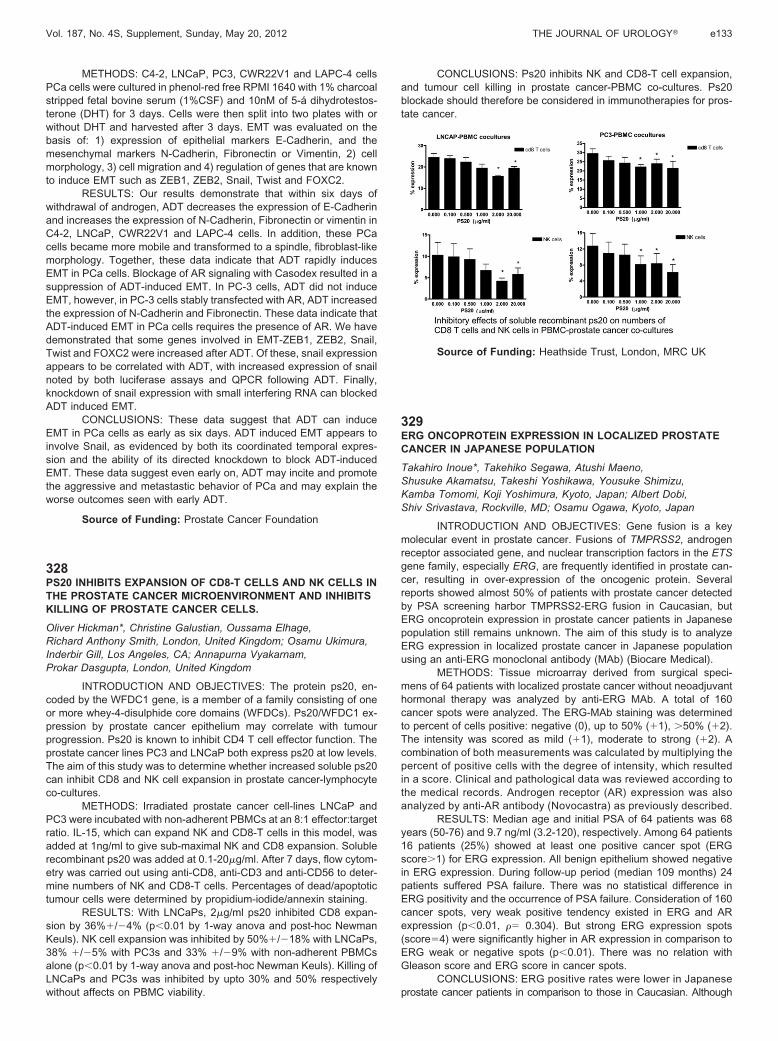
METHODS: Irradiated prostate cancer cell-lines LNCaP andPC3 were incubated with non-adherent PBMCs at an 8:1 effector:targetratio. IL-15, which can expand NK and CD8-T cells in this model, wasadded at 1ng/ml to give sub-maximal NK and CD8 expansion. Solublerecombinant ps20 was added at 0.1-20�g/ml. After 7 days, flow cytom-etry was carried out using anti-CD8, anti-CD3 and anti-CD56 to deter-mine numbers of NK and CD8-T cells. Percentages of dead/apoptotictumour cells were determined by propidium-iodide/annexin staining.

RESULTS: With LNCaPs, 2�g/ml ps20 inhibited CD8 expan-sion by 36%�/�4% (p�0.01 by 1-way anova and post-hoc NewmanKeuls). NK cell expansion was inhibited by 50%�/�18% with LNCaPs,38% �/�5% with PC3s and 33% �/�9% with non-adherent PBMCsalone (p�0.01 by 1-way anova and post-hoc Newman Keuls). Killing ofLNCaPs and PC3s was inhibited by upto 30% and 50% respectivelywithout affects on PBMC viability.

CONCLUSIONS: Ps20 inhibits NK and CD8-T cell expansion,and tumour cell killing in prostate cancer-PBMC co-cultures. Ps20blockade should therefore be considered in immunotherapies for pros-tate cancer.

Source of Funding: Heathside Trust, London, MRC UK

329ERG ONCOPROTEIN EXPRESSION IN LOCALIZED PROSTATECANCER IN JAPANESE POPULATION

Takahiro Inoue*, Takehiko Segawa, Atushi Maeno,Shusuke Akamatsu, Takeshi Yoshikawa, Yousuke Shimizu,Kamba Tomomi, Koji Yoshimura, Kyoto, Japan; Albert Dobi,Shiv Srivastava, Rockville, MD; Osamu Ogawa, Kyoto, Japan

INTRODUCTION AND OBJECTIVES: Gene fusion is a keymolecular event in prostate cancer. Fusions of TMPRSS2, androgenreceptor associated gene, and nuclear transcription factors in the ETSgene family, especially ERG, are frequently identified in prostate can-cer, resulting in over-expression of the oncogenic protein. Severalreports showed almost 50% of patients with prostate cancer detectedby PSA screening harbor TMPRSS2-ERG fusion in Caucasian, butERG oncoprotein expression in prostate cancer patients in Japanesepopulation still remains unknown. The aim of this study is to analyzeERG expression in localized prostate cancer in Japanese populationusing an anti-ERG monoclonal antibody (MAb) (Biocare Medical).

METHODS: Tissue microarray derived from surgical speci-mens of 64 patients with localized prostate cancer without neoadjuvanthormonal therapy was analyzed by anti-ERG MAb. A total of 160cancer spots were analyzed. The ERG-MAb staining was determinedto percent of cells positive: negative (0), up to 50% (�1), �50% (�2).The intensity was scored as mild (�1), moderate to strong (�2). Acombination of both measurements was calculated by multiplying thepercent of positive cells with the degree of intensity, which resultedin a score. Clinical and pathological data was reviewed according tothe medical records. Androgen receptor (AR) expression was alsoanalyzed by anti-AR antibody (Novocastra) as previously described.

RESULTS: Median age and initial PSA of 64 patients was 68years (50-76) and 9.7 ng/ml (3.2-120), respectively. Among 64 patients16 patients (25%) showed at least one positive cancer spot (ERGscore�1) for ERG expression. All benign epithelium showed negativein ERG expression. During follow-up period (median 109 months) 24patients suffered PSA failure. There was no statistical difference inERG positivity and the occurrence of PSA failure. Consideration of 160cancer spots, very weak positive tendency existed in ERG and ARexpression (p�0.01, � 0.304). But strong ERG expression spots(score�4) were significantly higher in AR expression in comparison toERG weak or negative spots (p�0.01). There was no relation withGleason score and ERG score in cancer spots.

CONCLUSIONS: ERG positive rates were lower in Japaneseprostate cancer patients in comparison to those in Caucasian. Although

Vol. 187, No. 4S, Supplement, Sunday, May 20, 2012 THE JOURNAL OF UROLOGY� e133