Embed Size (px)
Citation preview

37st European Congress of Cytology, September 2012, Cavtat– Croatia
INTRAOPERATIVE FINDINGS IN GYNECOLOGICAL CYTOLOGY GYNECOLOGICAL CYTOLOGY
Karmela Šentija
University Hospital “Merkur”, Zagreb, Croatia
j
Case study I
CLINICAL HYSTORYA 40‐year female patient was hospitalized at surgery Dpt for suspected obstructive uropathy of the left kidney and urolithiasis, and subsequently developed pyelonephritis symptoms.
Ultrasound findingEnlarged left adnexa, a tumor mass 69x60mm in diameter, with a cystic inclusion (25x22mm), suspected as endometrioma.
P l i i i i (MRI)Pelvic magnetic resonance imaging (MRI)
increased left ovary, with two cystic formations containing no solid parts
increased adnexa with the primary finding of a tumor formation with central necrosis, originating most likely in the lower part of the left ovary and extending into the retroperitoneum
Pelvic magnetic resonance imaging (MRI)
h i i l fib id h i• uterus showing an intramural fibroid, otherwise normal
• cervix showing a Naboth cyst, otherwise normal
• Iliac lymph nodes marginally increasedy p g y
Previous Pap smears:‐ 2 years ago: normal finding
h‐ 2 months ago: ASCUS
Due to a suspected tumor of the ovary, which performs compression on the ureter, theperforms compression on the ureter, the patient underwent surgery
INTRAOPERATIVE CYTOLOGYb f i i f h itwo tubes, from aspiration of the ovarian cyst
and of ascites, with few millilitres of blurry fluid i l d h l l b fmaterial send to the cytology lab for urgent
intraoperative cytological analysis
Cytological analysis – ovarian cyst
In the sample from the ovarian cyst, only well preserved erythrocytes were found, without any other cellular elements.
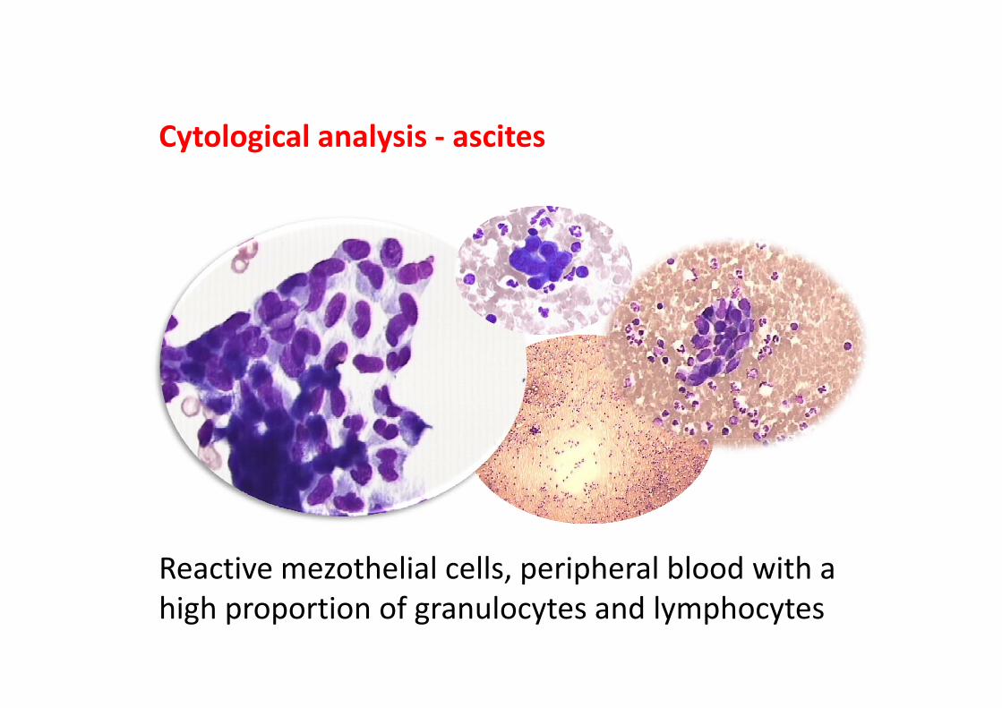
Cytological analysis ‐ ascites
Reactive mezothelial cells, peripheral blood with a high proportion of granulocytes and lymphocytes
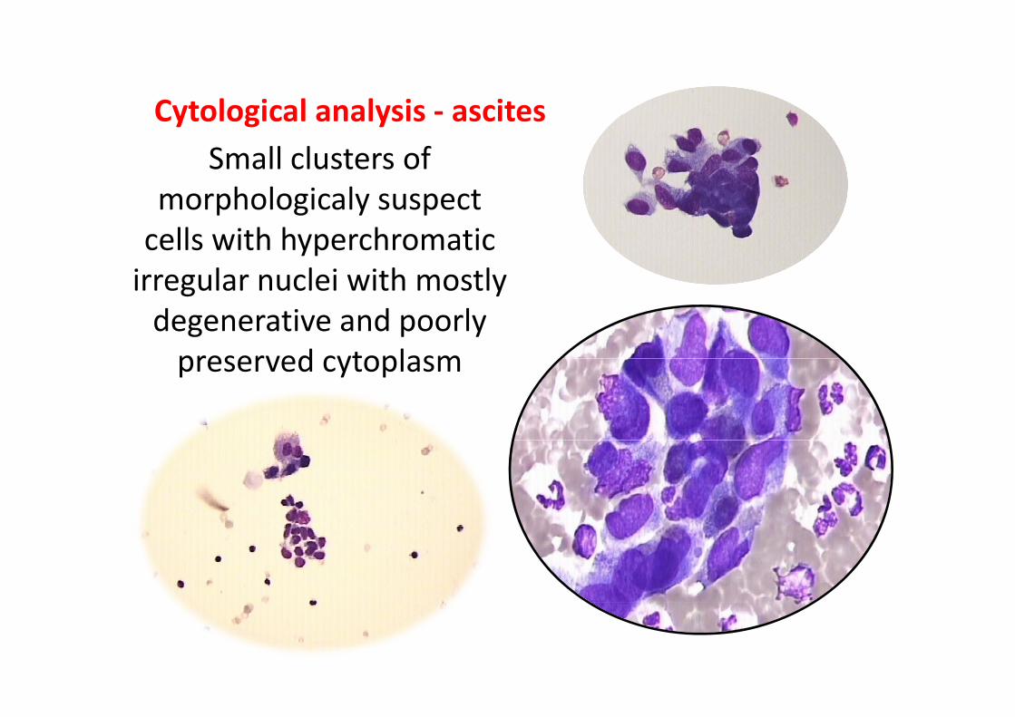
Cytological analysis ‐ ascitesSmall clusters of
morphologicaly suspect
Cytological analysis ‐ ascites
morphologicaly suspect cells with hyperchromatic irregular nuclei with mostlyirregular nuclei with mostly degenerative and poorly preserved cytoplasmpreserved cytoplasm
Differential diagnosis
Possible differential diagnostic procedures and PHD findings will be disscused at the slide seminar
Case study II
CLINICAL HYSTORY
A 32 year female patient was hospitalized forA 32‐year female patient was hospitalized for pelvic pain.
Ul d l d d i h i• Ultrasound: enlarged adnexa with a cystic inclusion in her right ovary
• Ca125marker: moderately increased
The patient underwent surgery to remove the cystic formation
INTRAOPERATIVE CYTOLOGY
ovarian cyst aspirate and abdominal lavage were sent to the cytology lab for intraoperativesent to the cytology lab for intraoperativecytological analysis, which showed markeddegenerative and necrotic changesdegenerative and necrotic changes
abdominal lavage sampleabdominal lavage sample single cells and small clusters of malignant
cells with prominent nucleoli and dispersedcells with prominent nucleoli and dispersed cytoplasm were found
ovarian cyst samplenecrosis and highly degenerative changesnecrosis and highly degenerative changes,
along with same malignant findings as from abdominal lavageabdominal lavage
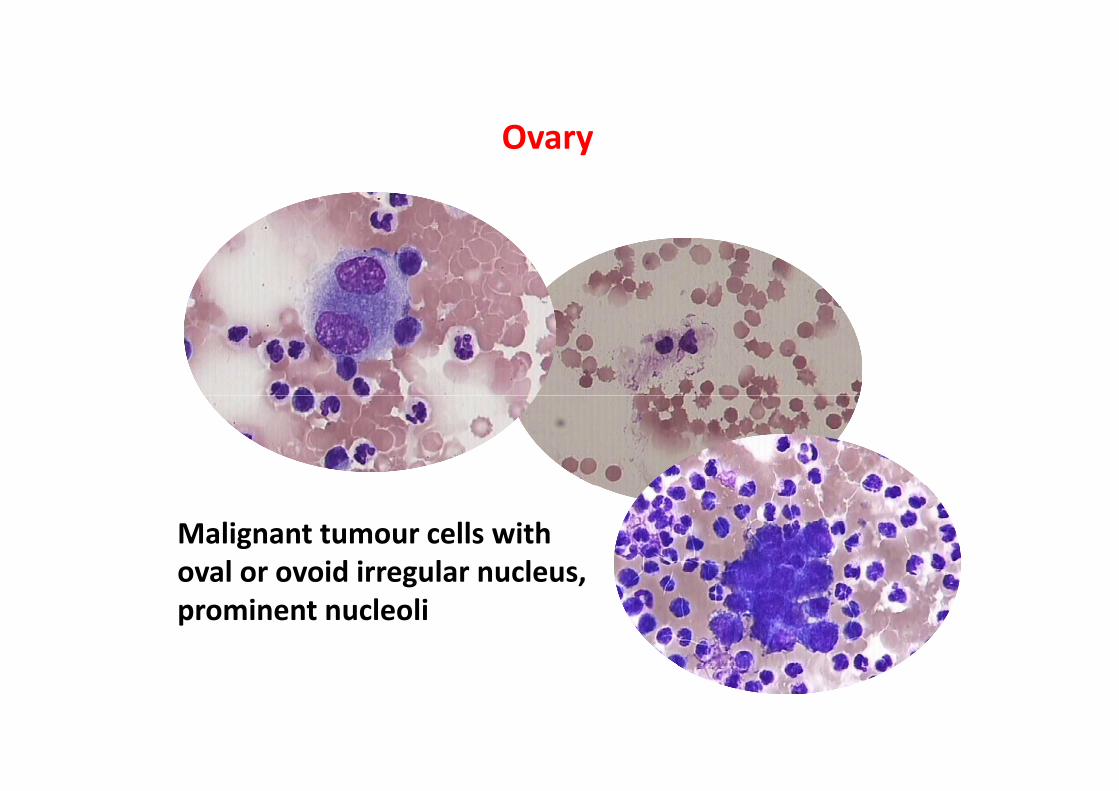
OOvary
Malignant tumour cells with oval or ovoid irregular nucleusoval or ovoid irregular nucleus, prominent nucleoli
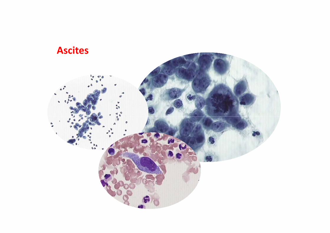
Ascites
Differential diagnosis
AdenocarcinomaCarcinoma clarocellulareMelanoma