Embed Size (px)
Citation preview
1/9/2018
1
3D Printing &Echocardiography
Echo HawaiiJan 18, 2018
Stephen H. Little, MDJohn S. Dunn Chair in Cardiovascular Research and Education, Associate professor, Weill Cornell Medicine
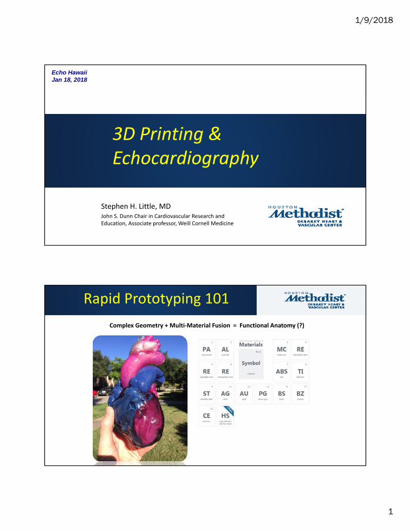
Rapid Prototyping 101
Complex Geometry + Multi‐Material Fusion = Functional Anatomy (?)
1/9/2018
2
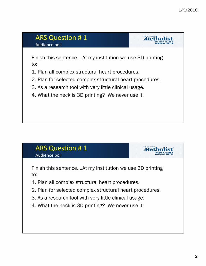
Finish this sentence….At my institution we use 3D printing to:1. Plan all complex structural heart procedures.2. Plan for selected complex structural heart procedures.3. As a research tool with very little clinical usage.4. What the heck is 3D printing? We never use it.
ARS Question # 1Audience poll
1/9/2018
3
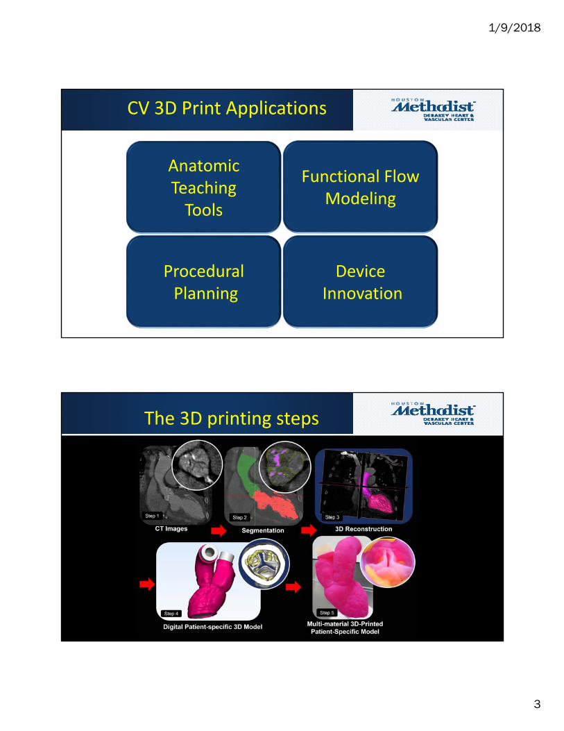
Anatomic Teaching Tools
DeviceInnovation
Functional Flow Modeling
ProceduralPlanning
CV 3D Print Applications
The 3D printing steps
1/9/2018
4
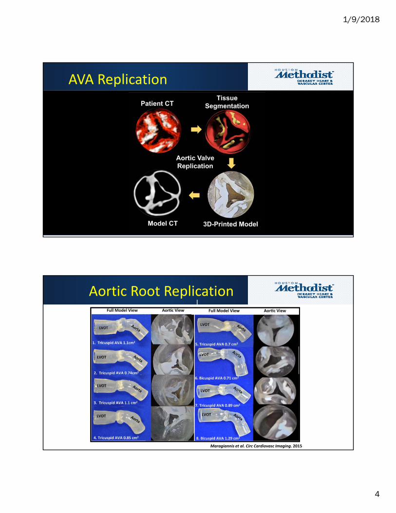
AVA Replication
Maragiannis et al. Circ Cardiovasc Imaging. 2015
Aortic Root Replication
1/9/2018
5
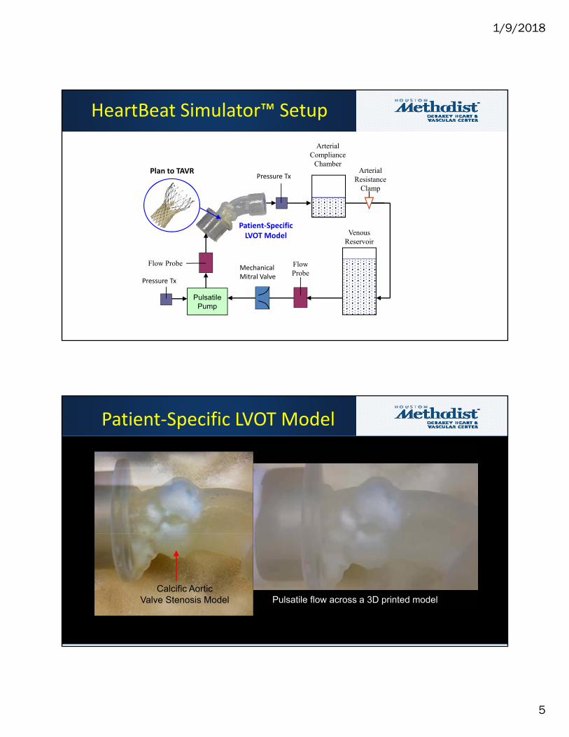
HeartBeat Simulator™ Setup
ArterialCompliance
ChamberArterial
ResistanceClamp
FlowProbe
VenousReservoir
PulsatilePump
Flow Probe
Patient‐Specific LVOT Model
Mechanical Mitral Valve
Pressure Tx
Pressure TxPlan to TAVR
Calcific AorticValve Stenosis Model
Patient‐Specific LVOT Model
Pulsatile flow across a 3D printed model
1/9/2018
6
0
1
2
3
4
5
6
7
8
9
0.00 0.01 0.02 0.04 0.05 0.06
Stress (MPa)
Engineering Strain
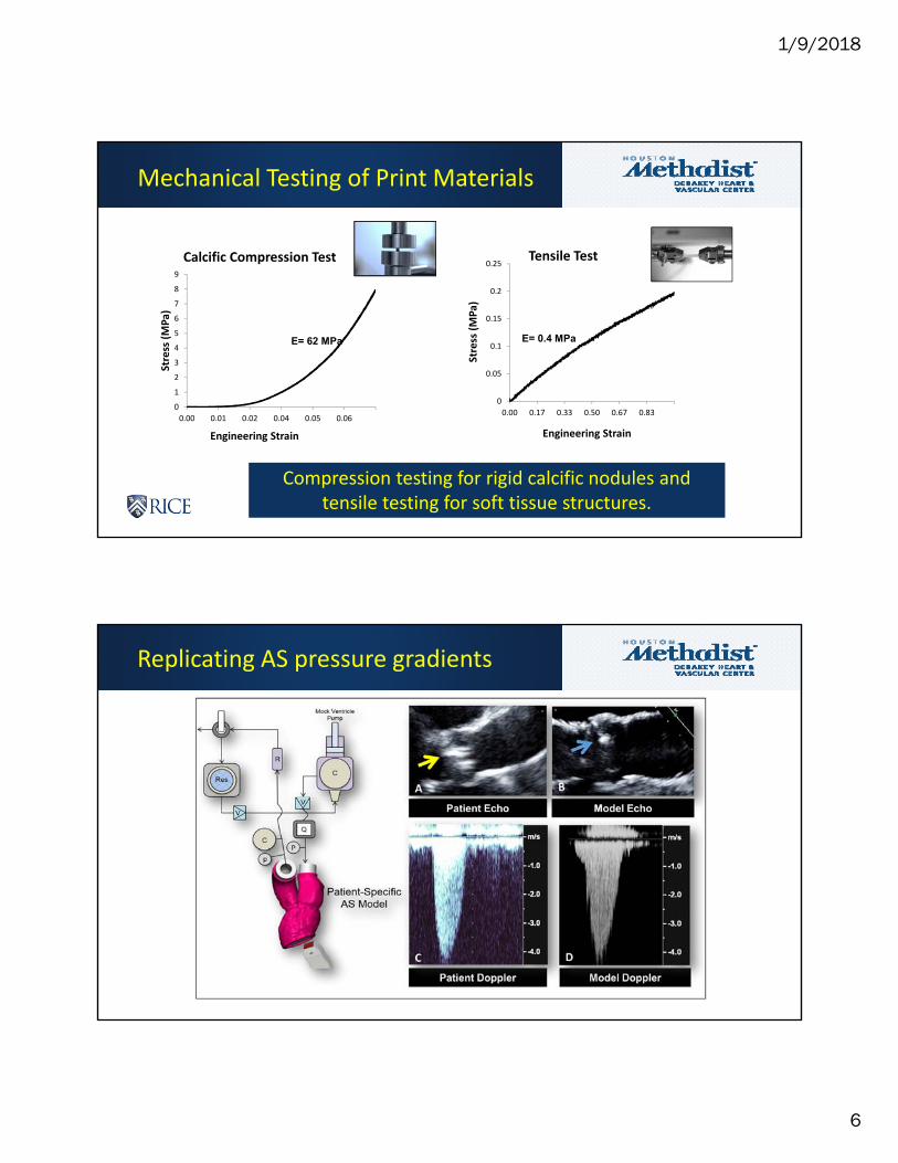
Calcific Compression Test
0
0.05
0.1
0.15
0.2
0.25
0.00 0.17 0.33 0.50 0.67 0.83
Stress (MPa)
Engineering Strain
Tensile Test
E= 62 MPa E= 0.4 MPa
Compression testing for rigid calcific nodules and tensile testing for soft tissue structures.
Mechanical Testing of Print Materials
Replicating AS pressure gradients
1/9/2018
7
0.30
0.45
0.60
0.75
0.90
1.05
1.20
1.35
1.50
30 50 70 90
AV
A (
cm2)
Stoke Volume (ml/beat)
Patient 1
0.30
0.45
0.60
0.75
0.90
1.05
1.20
1.35
1.50
30 50 70 90
AV
A (
cm2)
Stoke Volume (ml/beat)
Patient 2
0.30
0.45
0.60
0.75
0.90
1.05
1.20
1.35
1.50
40 75 110 145
AV
A (
cm2)
Stoke Volume (ml/beat)
Patient 3
0.30
0.45
0.60
0.75
0.90
1.05
1.20
1.35
1.50
30 50 70 90
AV
A (
cm2)
Stoke Volume (ml/beat)
Patient 4
0.30
0.45
0.60
0.75
0.90
1.05
1.20
1.35
1.50
30 50 70 90
AV
A (
cm2)
Stoke Volume (ml/beat)
Patient 5
0.30
0.45
0.60
0.75
0.90
1.05
1.20
1.35
1.50
30 50 70 90
AV
A (
cm2)
Stoke Volume (ml/beat)
Patient 6
0.30
0.45
0.60
0.75
0.90
1.05
1.20
1.35
1.50
40 75 110 145
AV
A (
cm2)
Stoke Volume (ml/beat)
Patient 7
0.30
0.45
0.60
0.75
0.90
1.05
1.20
1.35
1.50
40 75 110 145
AV
A (
cm2)
Stoke Volume (ml/beat)
Patient 8
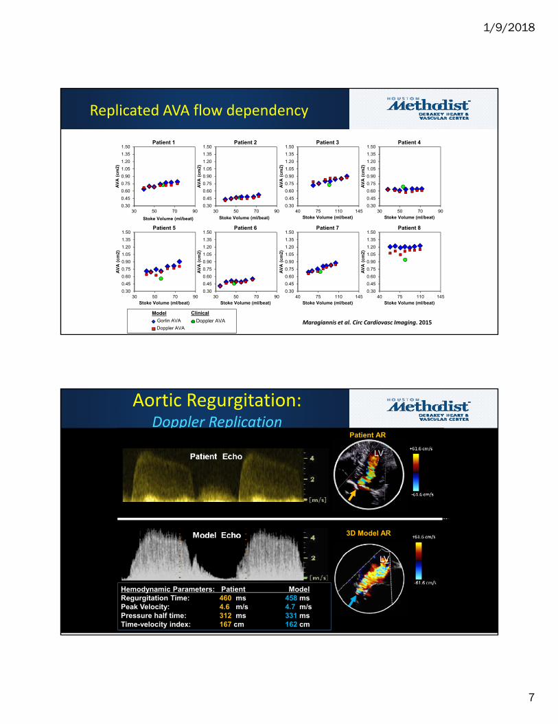
ClinicalModel
Gorlin AVA
Doppler AVA
Doppler AVA
Replicated AVA flow dependency
Maragiannis et al. Circ Cardiovasc Imaging. 2015
Aortic Regurgitation: Doppler Replication
3D Model AR
Hemodynamic Parameters: Patient ModelRegurgitation Time: 460 ms 458 msPeak Velocity: 4.6 m/s 4.7 m/sPressure half time: 312 ms 331 msTime-velocity index: 167 cm 162 cm
Patient AR
LV
LV
1/9/2018
8
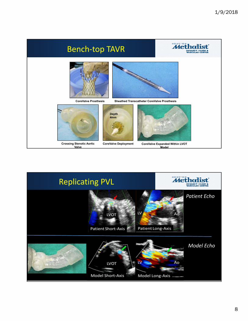
CoreValve Prosthesis Sheathed Transcatheter CoreValve Prosthesis
Crossing Stenotic Aortic Valve
CoreValve Deployment CoreValve Expanded Within LVOT Model
Depth 4mm
Bench‐top TAVR
Replicating PVL
Patient Echo
Model Echo
1/9/2018
9
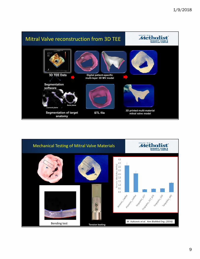
3D TEE Data
Mitral Valve reconstruction from 3D TEE
3D printed multi-material mitral valve model
Digital patient-specific multi-layer 3D MV model
AP
PL
Coronal planeAxial plane
Segmentation of target anatomy
STL file
Segmentation software
AL
PL
Mechanical Testing of Mitral Valve Materials
AL
PL
Bending test Tension testingM. Vukicevic et al. Ann BioMed Eng. (2016)
1/9/2018
10
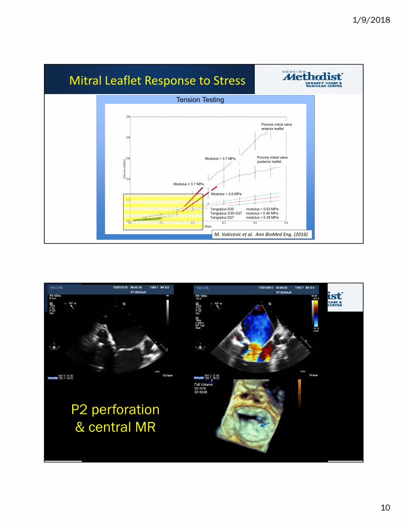
Porcine mitral valve anterior leaflet
Porcine mitral valve posterior leaflet
Tangoplus S35 modulus = 0.53 MPaTangoplus S35+S27 modulus = 0.46 MPaTangoplus S27 modulus = 0.38 MPa
Modulus = 3.7 MPa
Modulus = 2.6 MPa
Modulus = 3.7 MPa
Tension Testing
Mitral Leaflet Response to Stress
M. Vukicevic et al. Ann BioMed Eng. (2016)
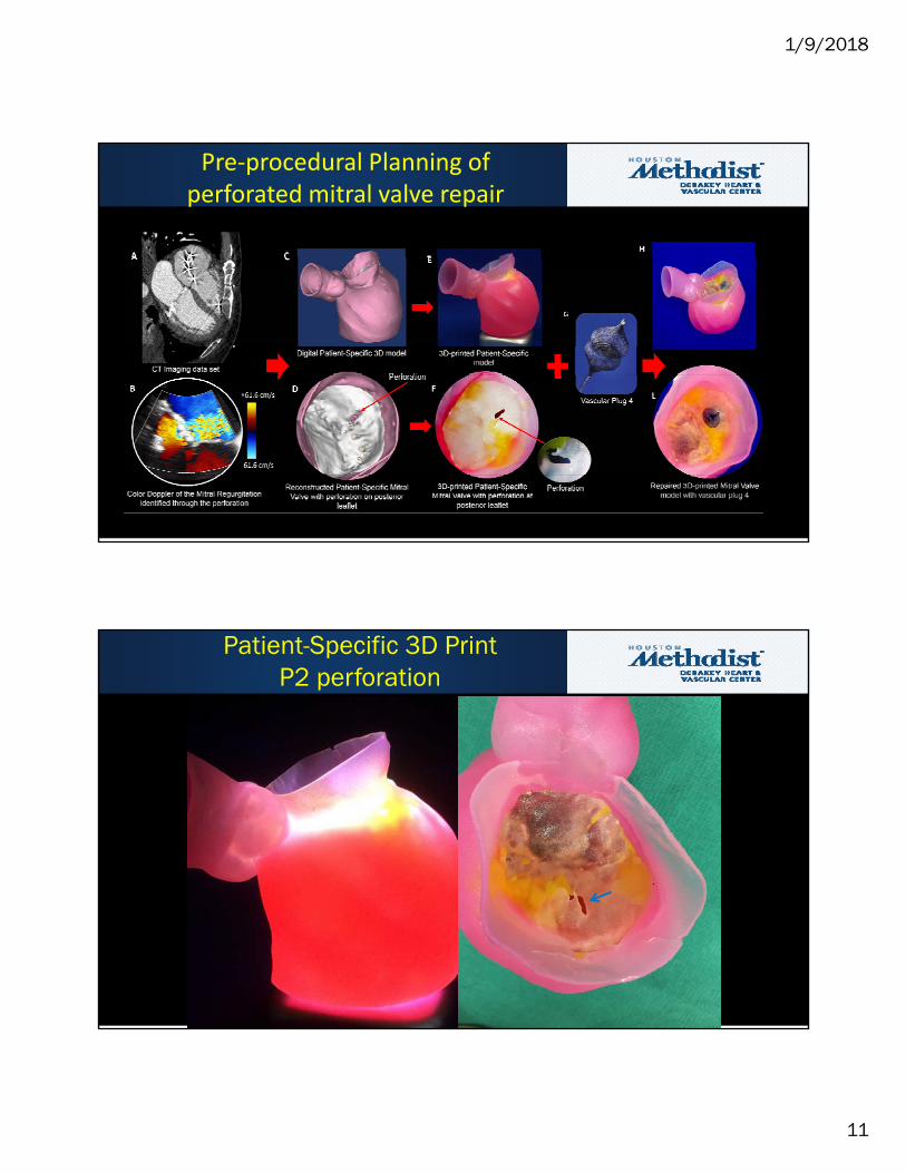
P2 perforation & central MR
1/9/2018
11
Pre‐procedural Planning of perforated mitral valve repair
Patient-Specific 3D PrintP2 perforation
1/9/2018
12
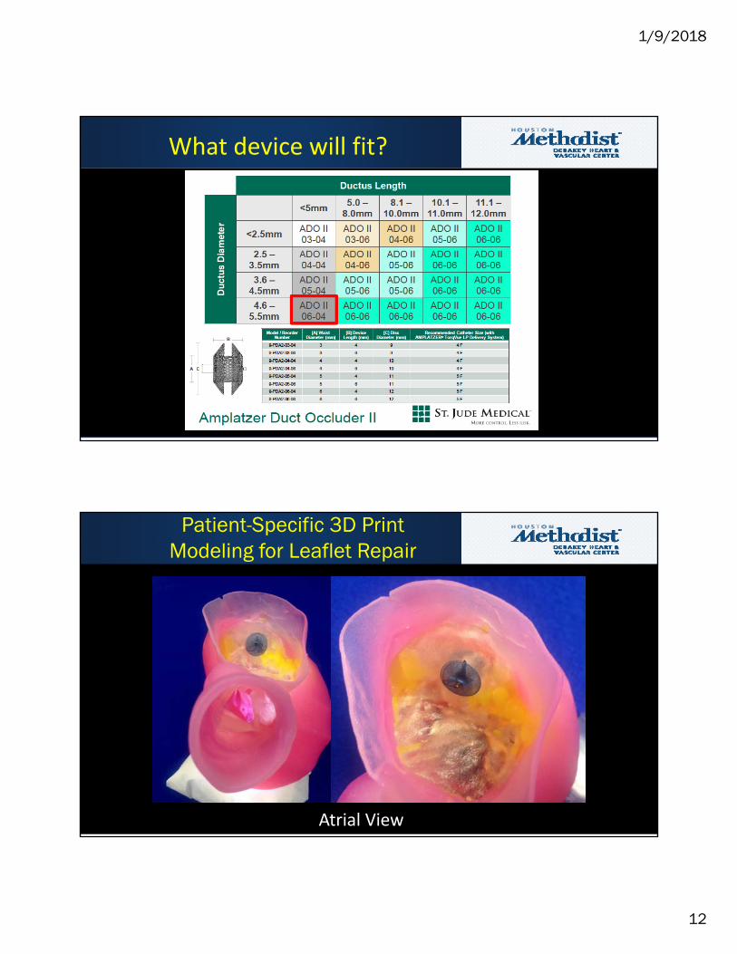
What device will fit?
Patient-Specific 3D PrintModeling for Leaflet Repair
Atrial View
1/9/2018
13
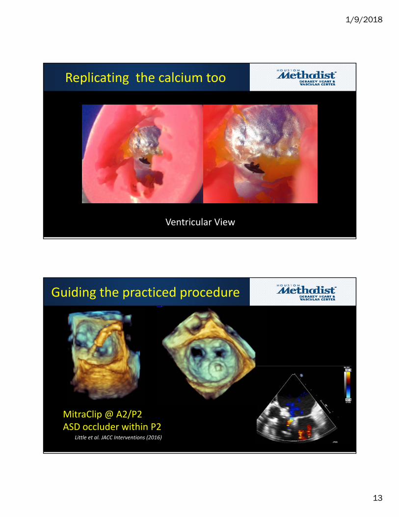
Ventricular View
Replicating the calcium too
MitraClip @ A2/P2ASD occluder within P2
Little et al. JACC Interventions (2016)
Guiding the practiced procedure
1/9/2018
14
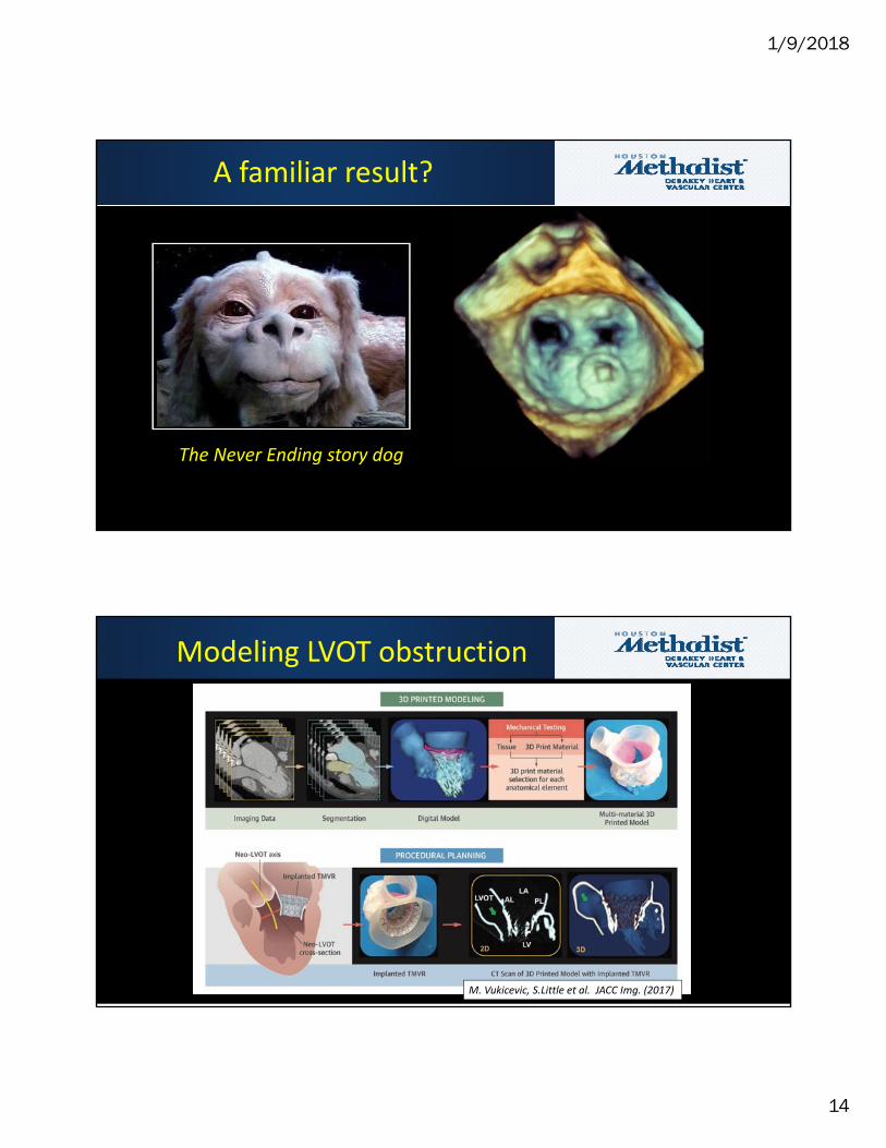
The Never Ending story dog
A familiar result?
Modeling LVOT obstruction
M. Vukicevic, S.Little et al. JACC Img. (2017)
1/9/2018
15
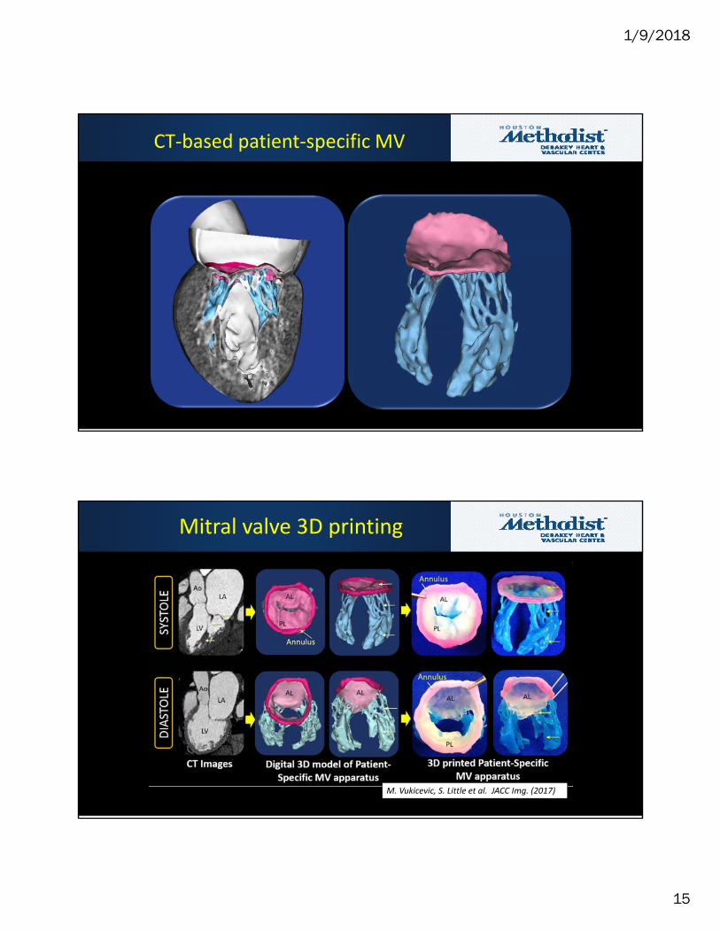
CT‐based patient‐specific MV
Mitral valve 3D printing
M. Vukicevic, S. Little et al. JACC Img. (2017)
1/9/2018
16
AL
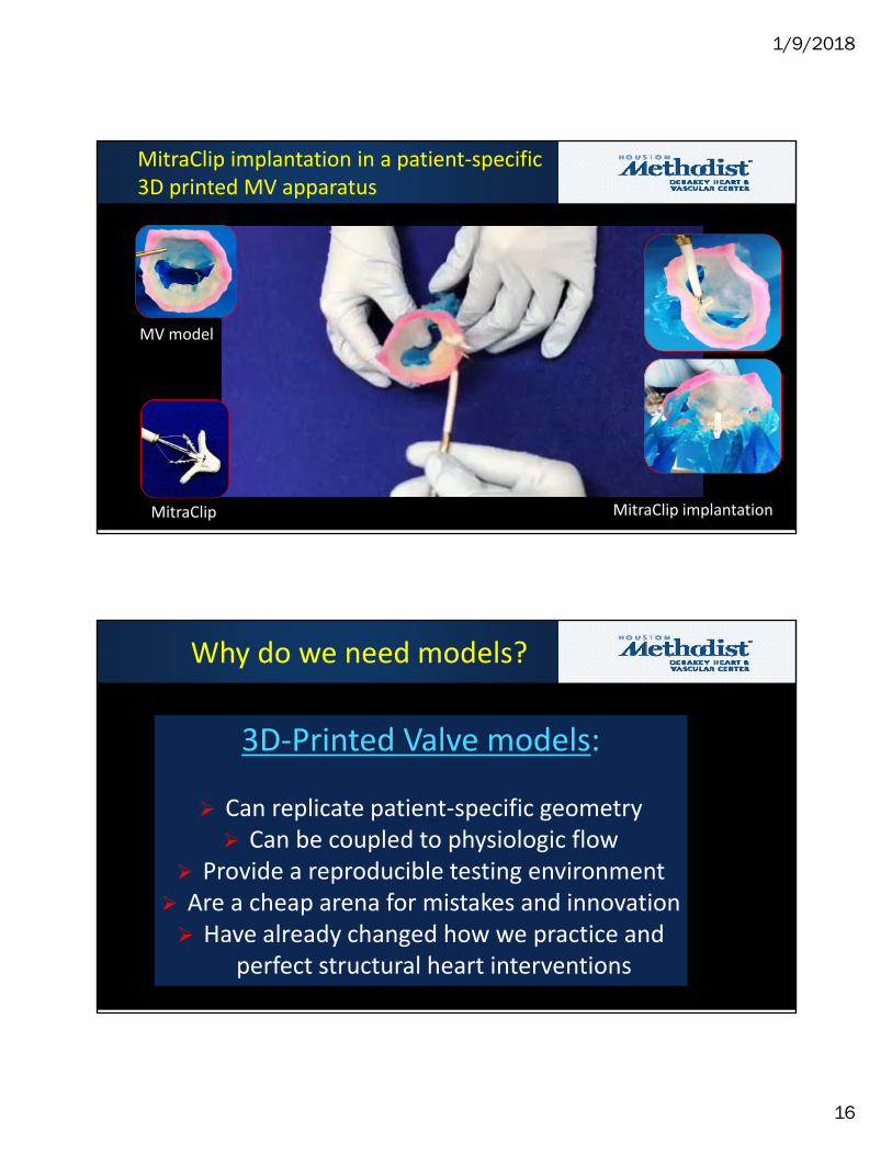
MV model
MitraClip MitraClip implantation
MitraClip implantation in a patient‐specific 3D printed MV apparatus
Why do we need models?
3D‐Printed Valve models:
Can replicate patient‐specific geometry Can be coupled to physiologic flow
Provide a reproducible testing environment Are a cheap arena for mistakes and innovation Have already changed how we practice and
perfect structural heart interventions