Embed Size (px)
Citation preview
3rd lecture
In
Anatomy
For
1st Class
dr.Ibtisam Khalaf
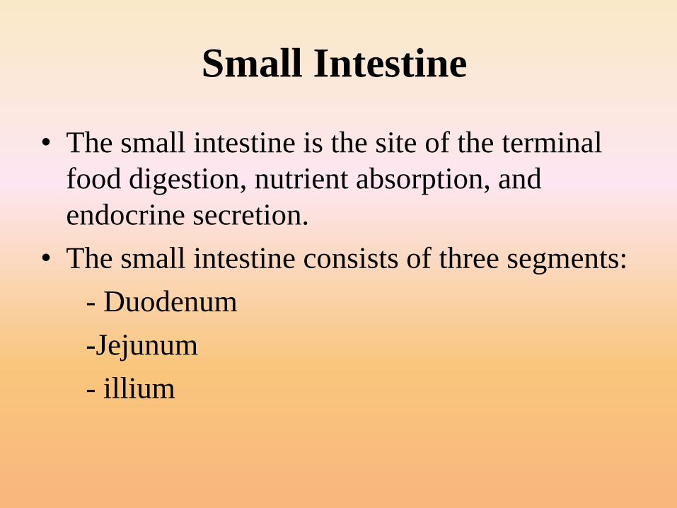
Small Intestine
• The small intestine is the site of the terminal
food digestion, nutrient absorption, and
endocrine secretion.
• The small intestine consists of three segments:
- Duodenum
-Jejunum
- illium
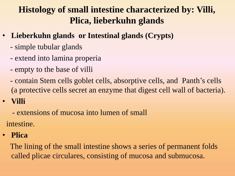
Histology of small intestine characterized by: Villi,
Plica, lieberkuhn glands
• Lieberkuhn glands or Intestinal glands (Crypts)
- simple tubular glands
- extend into lamina properia
- empty to the base of villi
- contain Stem cells goblet cells, absorptive cells, and Panth’s cells
(a protective cells secret an enzyme that digest cell wall of bacteria).
• Villi
- extensions of mucosa into lumen of small
intestine.
• Plica
The lining of the small intestine shows a series of permanent folds
called plicae circulares, consisting of mucosa and submucosa.
.
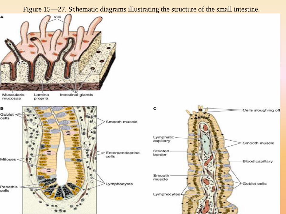
Figure 15—27. Schematic diagrams illustrating the structure of the small intestine.
Photomicrograph of the small intestine. Note the villi, intestinal glands, submucosa, muscle
layers, and serosa. PT stain. Low magnification.
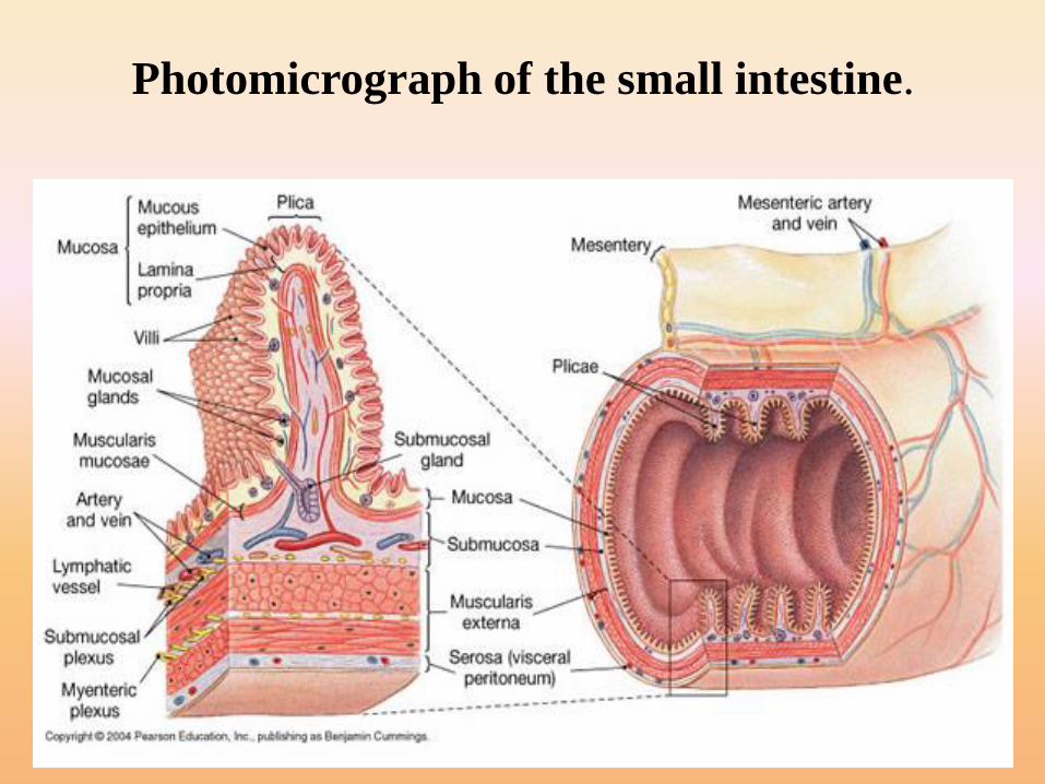
Photomicrograph of the small intestine.
Histology of the small Intestine
• The lining of the small intestine shows a series of permanent folds called plicae circulares, consisting of mucosa and submucosa.
• Plicae well developed in the jejunum, but do not constitute a significant feature of the duodenum and ileum.
• Intestinal villi are mucosa projecting into the lumen of the small intestine.
• Between the villi are small openings of simple tubular glands called intestinal glands(glands of lieberkuhn).
•Therefore the small intestine is modified for
dramatically increased surface area.
• The Doudenum has Brunner’s glands (submucosal doudenal
glands secret neutral alkaline mucus). There secretions protect
doudenum from erosion by acid entering from stomach.
• The jejunum has many long leaf like villi (plicae circularis).
And intermediate number of goblet cells (produce mucus).
• The ileum has numerous goblet cells and Peyer’s patchs
(aggregation of lymphocytes).
• Innervation of intestinal glands:
- Sympathatic stimulation leads to decrease
intestinal secretions
- Parasympathatic stimulation leads to
increase intestinal secretions.
Vessels of the small intestine
• The blood vessels that nourish the intestine and
remove absorbed products of digestion penetrate the
muscularis and form a large plexus in the submucosa.
Therefore each villus receives one or more branches
that form a capillary network just below its
epithelium
• Lacteals (capillary lymphatic vessel of villus).
- Important for the absorption of lipids because blood
circulation does not easily accept the lipoproteins
produced by the tall columnar during this process.
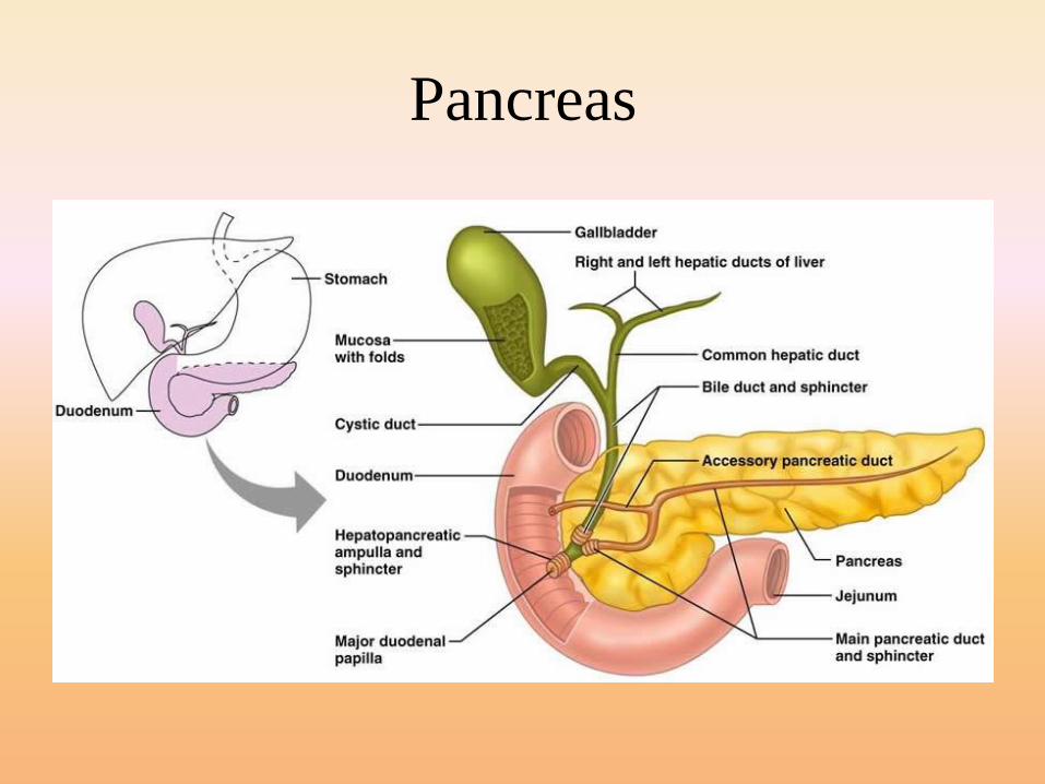
• In the duodenum there is a major duodenal papillain which the bile and pancreatic secretions enterthe intestine through Ampulla of Vater.
• Bile secretion leads to emulsify the fats.Emulsification of fats is important for fatdigestion by lipase enzyme which is produced bypancreas.
• Pancreas secrets amylase enzyme (forcarbohydrate digestion), lipase enzyme ( for fatdigestion) and proteolytic enzymes (for proteindigestion).
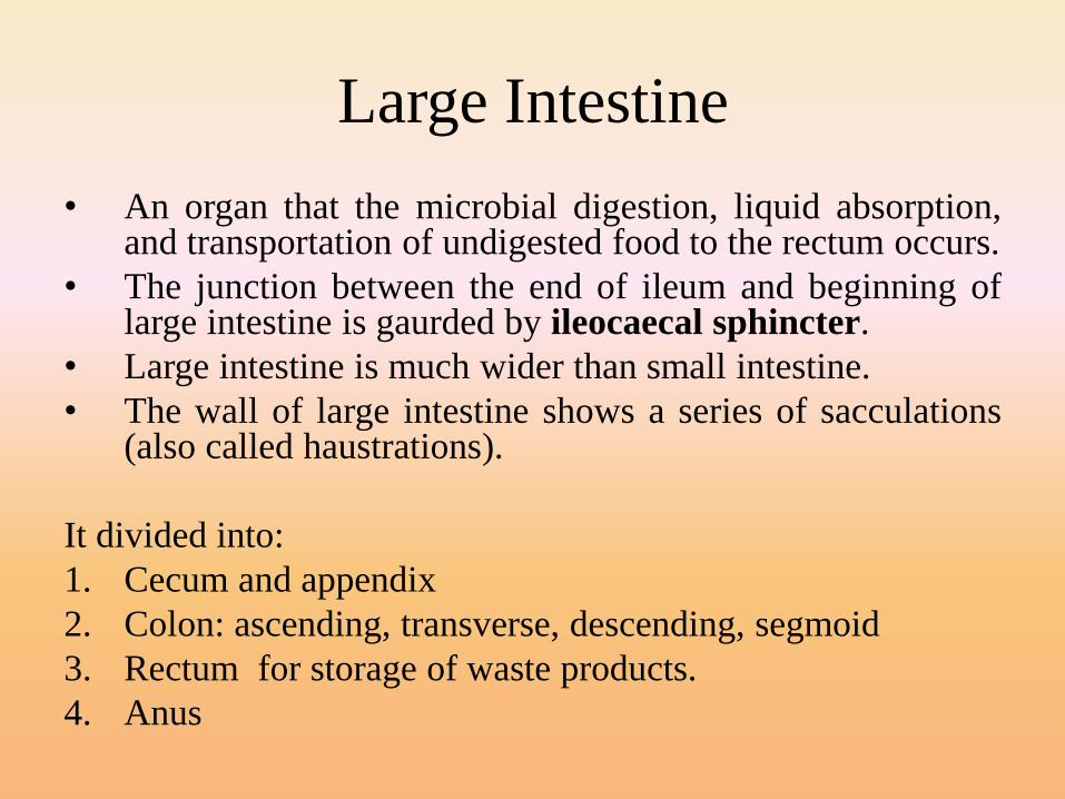
Large Intestine
• An organ that the microbial digestion, liquid absorption,and transportation of undigested food to the rectum occurs.
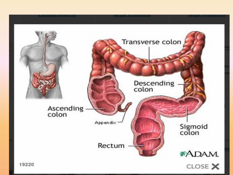
• The junction between the end of ileum and beginning oflarge intestine is gaurded by ileocaecal sphincter.
• Large intestine is much wider than small intestine.
• The wall of large intestine shows a series of sacculations(also called haustrations).
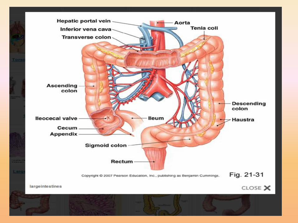
It divided into:
1. Cecum and appendix
2. Colon: ascending, transverse, descending, segmoid
3. Rectum for storage of waste products.
4. Anus
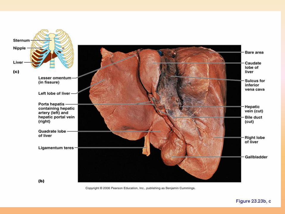
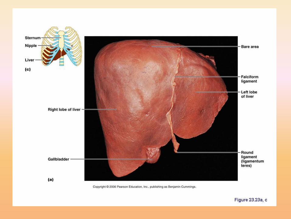
liver• It is the largest internal organ in the body.
• The nutrient absorbed in the digestive tract areprocessed (detoxification and metabolism) andstored in the liver for use by other parts of the body.Thus liver is an interface between digestive systemand blood.
• All materials absorbed via intestine reach the liverthrough the portal vein except the complex lipidswhich transport mainly by lymph vessel
• It consists of four lobes (1. left lobe 2. right lobe 3.Quadrate lobe 4. Caudate lobe)
HISTOLOGY OF THE LIVER
• Liver mainly consists from a large number oflobules (hexagonal lobule)
• three adjacent lobules form an area called portalarea (this area contains blood vessels, bile ductand lymph vessel).
• In the center of each lobule there is central vein.
• Hepatocytes arrange as a cord or plate fromcentral vein to the periphery of lobules.
• Sinusiods carry blood from portal vein andhepatic artery (in the portal area) to the centralvein.
• Kupffer’s cells
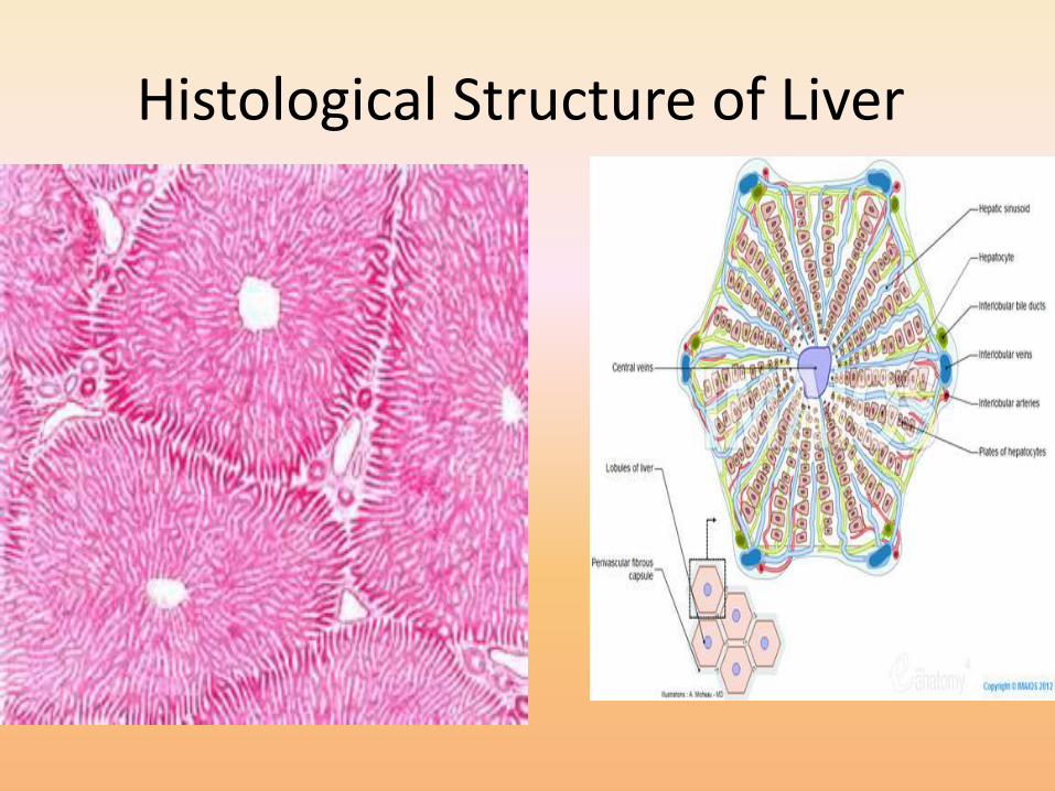
Histological Structure of Liver
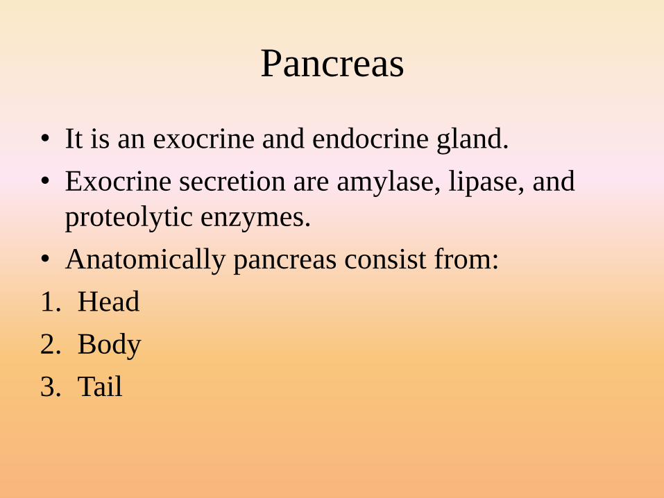
Pancreas
• It is an exocrine and endocrine gland.
• Exocrine secretion are amylase, lipase, and
proteolytic enzymes.
• Anatomically pancreas consist from:
1. Head
2. Body
3. Tail
• Secretion of pancreas carry by main pancreatic
duct.
• The main pancreatic duct units with common
bile duct to form ampula of vater
Pancreas
Pancreatic secretion is controlled by:
1. Hormones mainly secretin and cholecystokinin
are produced by enteroendocrine cells of duodenal
mucosa.
2. Parasympathatic stimulation