Embed Size (px)
Citation preview
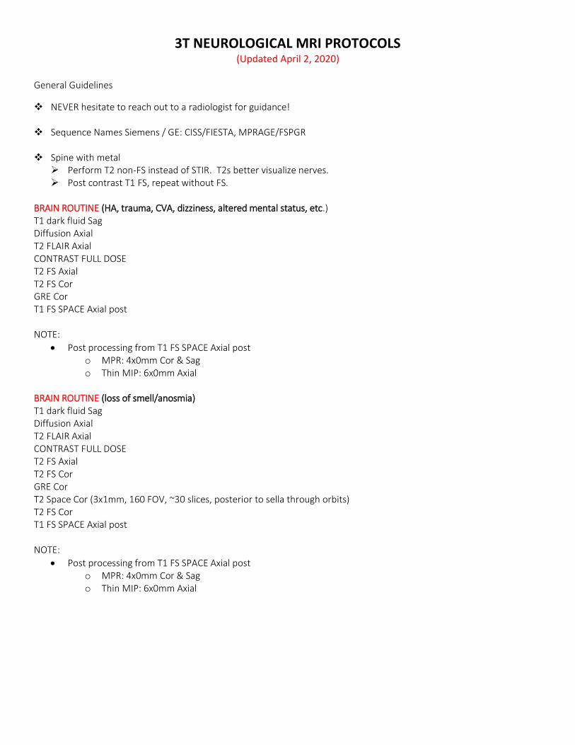
3T NEUROLOGICAL MRI PROTOCOLS (Updated April 2, 2020)
General Guidelines
NEVER hesitate to reach out to a radiologist for guidance!
Sequence Names Siemens / GE: CISS/FIESTA, MPRAGE/FSPGR
Spine with metal Perform T2 non-FS instead of STIR. T2s better visualize nerves. Post contrast T1 FS, repeat without FS.
BRAIN ROUTINE (HA, trauma, CVA, dizziness, altered mental status, etc.) T1 dark fluid Sag Diffusion Axial T2 FLAIR Axial CONTRAST FULL DOSE T2 FS Axial T2 FS Cor GRE Cor T1 FS SPACE Axial post NOTE:
Post processing from T1 FS SPACE Axial post o MPR: 4x0mm Cor & Sag o Thin MIP: 6x0mm Axial
BRAIN ROUTINE (loss of smell/anosmia) T1 dark fluid Sag Diffusion Axial T2 FLAIR Axial CONTRAST FULL DOSE T2 FS Axial T2 FS Cor GRE Cor T2 Space Cor (3x1mm, 160 FOV, ~30 slices, posterior to sella through orbits) T2 FS Cor T1 FS SPACE Axial post NOTE:
Post processing from T1 FS SPACE Axial post o MPR: 4x0mm Cor & Sag o Thin MIP: 6x0mm Axial

BRAIN TUMOR (mass, oncology, metastasis) T1 SPACE Axial pre (1mm, with 4x0mm Sag MPR) Diffusion Axial T2 FLAIR Axial CONTRAST FULL DOSE T2 FS Axial T2 FS Cor GRE Cor T1 FS SPACE Axial post (1mm, with 4x0mm Cor & Sag MPR) T1 FS SPACE Axial post NOTE:
Post processing from T1 FS SPACE Axial post o MPR: 4x0mm Cor & Sag o Thin MIP: 6x0mm Axial
BRAIN ROUTINE WITHOUT CONTRAST T1 dark fluid Sag Diffusion Axial T2 FLAIR Axial T2 FS Axial T2 FS Cor GRE Cor BRAIN ROUTINE WITH BRACES / SHUNT / SIGNIFICANT METAL IN MOUTH – NON-FS, FSE/TSE T1 dark fluid Sag Diffusion Axial T2 FLAIR Axial CONTRAST T2 Axial T2 Cor GRE Cor T1 FS SPACE Axial post NOTE:
Post processing from T1 FS SPACE Axial post o MPR: 4x0mm Cor & Sag o Thin MIP: 6x0mm Axial
SETON NETWORK ONLY * BRAIN ROUTINE/TUMOR (HA, trauma, CVA, dizziness, altered mental status, etc.) T1 dark fluid Sag Diffusion Axial T2 FLAIR Axial CONTRAST T2 FS Axial GRE Axial T2 FS Cor GRE Cor T1 FS SPACE Axial post NOTE: Create 2x2mm MPR Cor & Sag from the T1 FS SPACE Axial post.
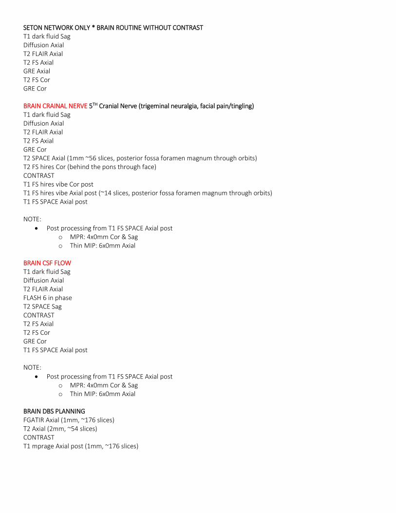
SETON NETWORK ONLY * BRAIN ROUTINE WITHOUT CONTRAST T1 dark fluid Sag Diffusion Axial T2 FLAIR Axial T2 FS Axial GRE Axial T2 FS Cor GRE Cor BRAIN CRAINAL NERVE 5TH Cranial Nerve (trigeminal neuralgia, facial pain/tingling) T1 dark fluid Sag Diffusion Axial T2 FLAIR Axial T2 FS Axial GRE Cor T2 SPACE Axial (1mm ~56 slices, posterior fossa foramen magnum through orbits) T2 FS hires Cor (behind the pons through face) CONTRAST T1 FS hires vibe Cor post T1 FS hires vibe Axial post (~14 slices, posterior fossa foramen magnum through orbits) T1 FS SPACE Axial post NOTE:
Post processing from T1 FS SPACE Axial post o MPR: 4x0mm Cor & Sag o Thin MIP: 6x0mm Axial
BRAIN CSF FLOW T1 dark fluid Sag Diffusion Axial T2 FLAIR Axial FLASH 6 in phase T2 SPACE Sag CONTRAST T2 FS Axial T2 FS Cor GRE Cor T1 FS SPACE Axial post NOTE:
Post processing from T1 FS SPACE Axial post o MPR: 4x0mm Cor & Sag o Thin MIP: 6x0mm Axial
BRAIN DBS PLANNING FGATIR Axial (1mm, ~176 slices) T2 Axial (2mm, ~54 slices) CONTRAST T1 mprage Axial post (1mm, ~176 slices)
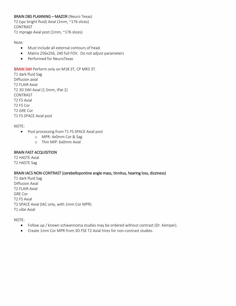
BRAIN DBS PLANNING – MAZOR (Neuro Texas) T2 (spc bright fluid) Axial (1mm, ~176 slices) CONTRAST T1 mprage Axial post (1mm, ~176 slices) Note:
Must include all external contours of head.
Matrix 256x256, 240 full FOV. Do not adjust parameters
Performed for NeuroTexas
BRAIN SWI Perform only on M18 3T, CP MR3 3T. T1 dark fluid Sag Diffusion axial T2 FLAIR Axial T2 3D SWI Axial (1.5mm, iPat 2) CONTRAST T2 FS Axial T2 FS Cor T2 GRE Cor T1 FS SPACE Axial post NOTE:
Post processing from T1 FS SPACE Axial post o MPR: 4x0mm Cor & Sag o Thin MIP: 6x0mm Axial
BRAIN FAST ACQUISITION T2 HASTE Axial T2 HASTE Sag BRAIN IACS NON-CONTRAST (cerebellopontine angle mass, tinnitus, hearing loss, dizziness) T1 dark fluid Sag Diffusion Axial T2 FLAIR Axial GRE Cor T2 FS Axial T2 SPACE Axial (IAC only, with 1mm Cor MPR) T1 vibe Axial NOTE:
Follow up / known schwannoma studies may be ordered without contrast (Dr. Kemper).
Create 1mm Cor MPR from 3D FSE T2 Axial hires for non-contrast studies.
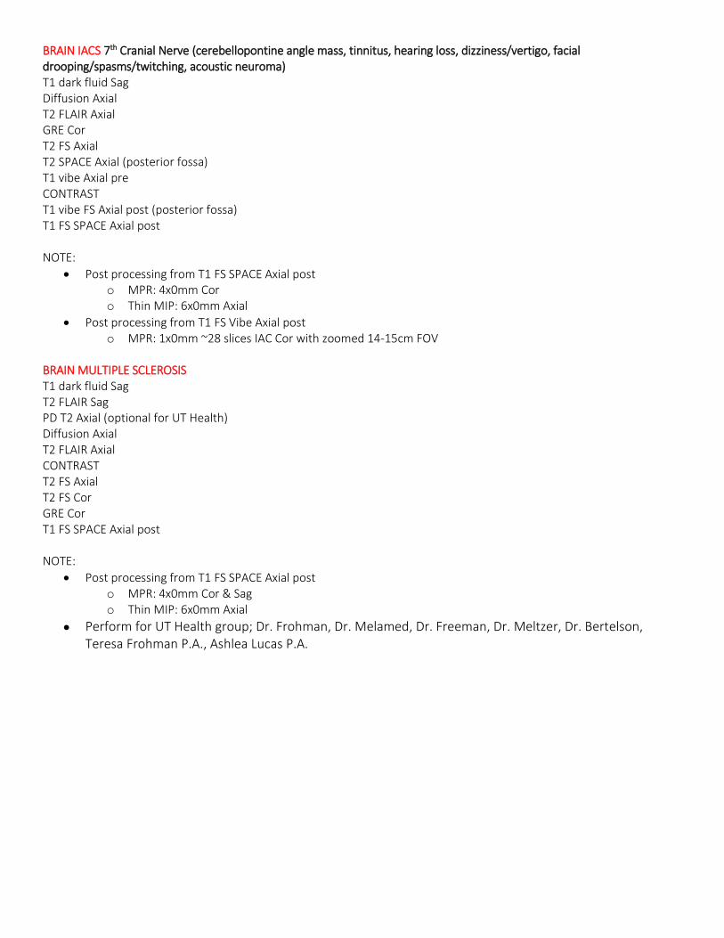
BRAIN IACS 7th Cranial Nerve (cerebellopontine angle mass, tinnitus, hearing loss, dizziness/vertigo, facial drooping/spasms/twitching, acoustic neuroma) T1 dark fluid Sag Diffusion Axial T2 FLAIR Axial GRE Cor T2 FS Axial T2 SPACE Axial (posterior fossa) T1 vibe Axial pre CONTRAST T1 vibe FS Axial post (posterior fossa) T1 FS SPACE Axial post NOTE:
Post processing from T1 FS SPACE Axial post o MPR: 4x0mm Cor o Thin MIP: 6x0mm Axial
Post processing from T1 FS Vibe Axial post o MPR: 1x0mm ~28 slices IAC Cor with zoomed 14-15cm FOV
BRAIN MULTIPLE SCLEROSIS T1 dark fluid Sag T2 FLAIR Sag PD T2 Axial (optional for UT Health) Diffusion Axial T2 FLAIR Axial CONTRAST T2 FS Axial T2 FS Cor GRE Cor T1 FS SPACE Axial post NOTE:
Post processing from T1 FS SPACE Axial post o MPR: 4x0mm Cor & Sag o Thin MIP: 6x0mm Axial
Perform for UT Health group; Dr. Frohman, Dr. Melamed, Dr. Freeman, Dr. Meltzer, Dr. Bertelson, Teresa Frohman P.A., Ashlea Lucas P.A.
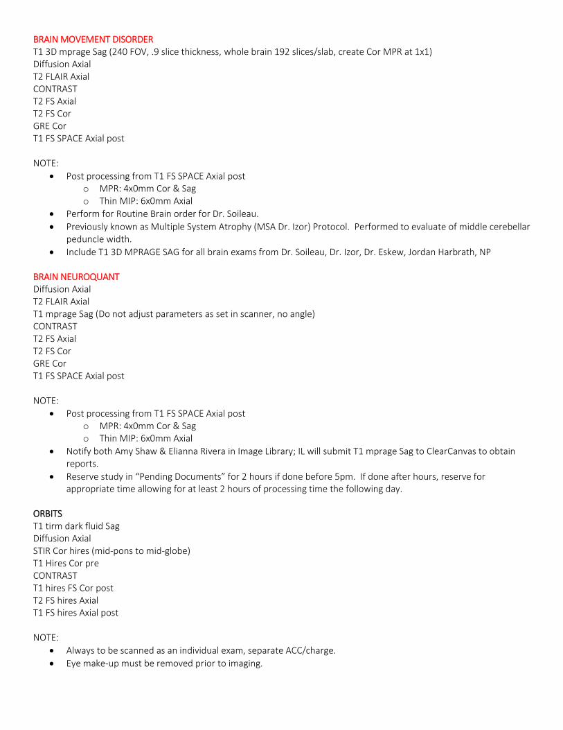
BRAIN MOVEMENT DISORDER T1 3D mprage Sag (240 FOV, .9 slice thickness, whole brain 192 slices/slab, create Cor MPR at 1x1) Diffusion Axial T2 FLAIR Axial CONTRAST T2 FS Axial T2 FS Cor GRE Cor T1 FS SPACE Axial post NOTE:
Post processing from T1 FS SPACE Axial post o MPR: 4x0mm Cor & Sag o Thin MIP: 6x0mm Axial
Perform for Routine Brain order for Dr. Soileau.
Previously known as Multiple System Atrophy (MSA Dr. Izor) Protocol. Performed to evaluate of middle cerebellar peduncle width.
Include T1 3D MPRAGE SAG for all brain exams from Dr. Soileau, Dr. Izor, Dr. Eskew, Jordan Harbrath, NP BRAIN NEUROQUANT Diffusion Axial T2 FLAIR Axial T1 mprage Sag (Do not adjust parameters as set in scanner, no angle) CONTRAST T2 FS Axial T2 FS Cor GRE Cor T1 FS SPACE Axial post NOTE:
Post processing from T1 FS SPACE Axial post o MPR: 4x0mm Cor & Sag o Thin MIP: 6x0mm Axial
Notify both Amy Shaw & Elianna Rivera in Image Library; IL will submit T1 mprage Sag to ClearCanvas to obtain reports.
Reserve study in “Pending Documents” for 2 hours if done before 5pm. If done after hours, reserve for appropriate time allowing for at least 2 hours of processing time the following day.
ORBITS T1 tirm dark fluid Sag Diffusion Axial STIR Cor hires (mid-pons to mid-globe) T1 Hires Cor pre CONTRAST T1 hires FS Cor post T2 FS hires Axial T1 FS hires Axial post NOTE:
Always to be scanned as an individual exam, separate ACC/charge.
Eye make-up must be removed prior to imaging.
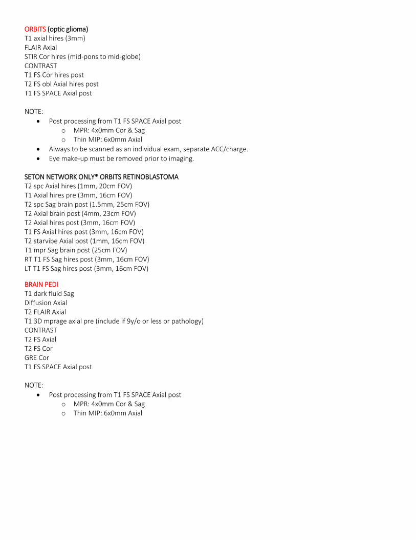
ORBITS (optic glioma) T1 axial hires (3mm) FLAIR Axial STIR Cor hires (mid-pons to mid-globe) CONTRAST T1 FS Cor hires post T2 FS obl Axial hires post T1 FS SPACE Axial post NOTE:
Post processing from T1 FS SPACE Axial post o MPR: 4x0mm Cor & Sag o Thin MIP: 6x0mm Axial
Always to be scanned as an individual exam, separate ACC/charge.
Eye make-up must be removed prior to imaging. SETON NETWORK ONLY* ORBITS RETINOBLASTOMA T2 spc Axial hires (1mm, 20cm FOV) T1 Axial hires pre (3mm, 16cm FOV) T2 spc Sag brain post (1.5mm, 25cm FOV) T2 Axial brain post (4mm, 23cm FOV) T2 Axial hires post (3mm, 16cm FOV) T1 FS Axial hires post (3mm, 16cm FOV) T2 starvibe Axial post (1mm, 16cm FOV) T1 mpr Sag brain post (25cm FOV) RT T1 FS Sag hires post (3mm, 16cm FOV) LT T1 FS Sag hires post (3mm, 16cm FOV)
BRAIN PEDI T1 dark fluid Sag Diffusion Axial T2 FLAIR Axial T1 3D mprage axial pre (include if 9y/o or less or pathology) CONTRAST T2 FS Axial T2 FS Cor GRE Cor T1 FS SPACE Axial post NOTE:
Post processing from T1 FS SPACE Axial post o MPR: 4x0mm Cor & Sag o Thin MIP: 6x0mm Axial
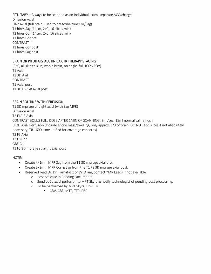
PITUITARY – Always to be scanned as an individual exam, separate ACC/charge. Diffusion Axial Flair Axial (full brain, used to prescribe true Cor/Sag) T1 hires Sag (14cm, 2x0, 16 slices min) T2 hires Cor (14cm, 2x0, 16 slices min) T1 hires Cor pre CONTRAST T1 hires Cor post T1 hires Sag post BRAIN OR PITUITARY AUSTIN CA CTR THERAPY STAGING (3X0, all skin to skin, whole brain, no angle, full 100% FOV) T1 Axial T2 3D Aial CONTRAST T1 Axial post T1 3D FSPGR Axial post
BRAIN ROUTINE WITH PERFUSION T1 3D mprage straight axial (with Sag MPR) Diffusion Axial T2 FLAIR Axial CONTRAST BOLUS FULL DOSE AFTER 1MIN OF SCANNING: 3ml/sec, 15ml normal saline flush EP2D Axial Perfusion (Include entire mass/swelling, only approx. 1/3 of brain, DO NOT add slices if not absolutely necessary, TR 1600, consult Rad for coverage concerns) T2 FS Axial T2 FS Cor GRE Cor T1 FS 3D mprage straight axial post NOTE:
Create 4x1mm MPR Sag from the T1 3D mprage axial pre.
Create 3x3mm MPR Cor & Sag from the T1 FS 3D mprage axial post.
Reserved read Dr. Dr. Farhataziz or Dr. Alam, contact *MR Leads if not available o Reserve case in Pending Documents o Send ep2d axial perfusion to MPT Skyra & notify technologist of pending post processing. o To be performed by MPT Skyra, How To
CBV, CBF, MTT, TTP, PBP
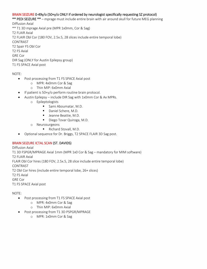
BRAIN SEIZURE 0-49y/o (50+y/o ONLY if ordered by neurologist specifically requesting SZ protocol) ** PEDI SEIZURE ** – mprage must include entire brain with air around skull for future MEG planning Diffusion Axial ** T1 3D mprage Axial pre (MPR 1x0mm, Cor & Sag) T2 FLAIR Axial T2 FLAIR Obl Cor (180 FOV, 2.5x.5, 28 slices include entire temporal lobe) CONTRAST T2 Spair FS Obl Cor T2 FS Axial GRE Cor DIR Sag (ONLY for Austin Epilepsy group) T1 FS SPACE Axial post NOTE:
Post processing from T1 FS SPACE Axial post o MPR: 4x0mm Cor & Sag o Thin MIP: 6x0mm Axial
If patient is 50+y/o perform routine brain protocol.
Austin Epilepsy – include DIR Sag with 1x0mm Cor & Ax MPRs. o Epileptologists
Sami Aboumatar, M.D. Daniel Schere, M.D. Jeanne Beattie, M.D. Diego Tovar Quiroga, M.D.
o Neurosurgeons Richard Stovall, M.D.
Optional sequence for Dr. Briggs, T2 SPACE FLAIR 3D Sag post. BRAIN SEIZURE ICTAL SCAN (ST. DAVIDS) Diffusion Axial T1 3D FSPGR/MPRAGE Axial 1mm (MPR 1x0 Cor & Sag – mandatory for MIM software) T2 FLAIR Axial FLAIR Obl Cor hires (180 FOV, 2.5x.5, 28 slice include entire temporal lobe) CONTRAST T2 Obl Cor hires (include entire temporal lobe, 26+ slices) T2 FS Axial GRE Cor T1 FS SPACE Axial post NOTE:
Post processing from T1 FS SPACE Axial post o MPR: 4x0mm Cor & Sag o Thin MIP: 6x0mm Axial
Post processing from T1 3D PSPGR/MPRAGE o MPR: 1x0mm Cor & Sag
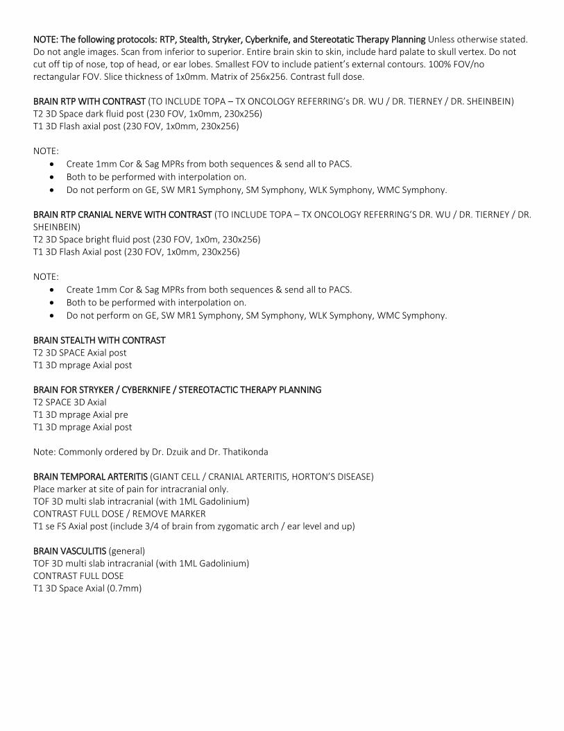
NOTE: The following protocols: RTP, Stealth, Stryker, Cyberknife, and Stereotatic Therapy Planning Unless otherwise stated. Do not angle images. Scan from inferior to superior. Entire brain skin to skin, include hard palate to skull vertex. Do not cut off tip of nose, top of head, or ear lobes. Smallest FOV to include patient’s external contours. 100% FOV/no rectangular FOV. Slice thickness of 1x0mm. Matrix of 256x256. Contrast full dose. BRAIN RTP WITH CONTRAST (TO INCLUDE TOPA – TX ONCOLOGY REFERRING’s DR. WU / DR. TIERNEY / DR. SHEINBEIN) T2 3D Space dark fluid post (230 FOV, 1x0mm, 230x256) T1 3D Flash axial post (230 FOV, 1x0mm, 230x256) NOTE:
Create 1mm Cor & Sag MPRs from both sequences & send all to PACS.
Both to be performed with interpolation on.
Do not perform on GE, SW MR1 Symphony, SM Symphony, WLK Symphony, WMC Symphony. BRAIN RTP CRANIAL NERVE WITH CONTRAST (TO INCLUDE TOPA – TX ONCOLOGY REFERRING’S DR. WU / DR. TIERNEY / DR. SHEINBEIN) T2 3D Space bright fluid post (230 FOV, 1x0m, 230x256) T1 3D Flash Axial post (230 FOV, 1x0mm, 230x256) NOTE:
Create 1mm Cor & Sag MPRs from both sequences & send all to PACS.
Both to be performed with interpolation on.
Do not perform on GE, SW MR1 Symphony, SM Symphony, WLK Symphony, WMC Symphony. BRAIN STEALTH WITH CONTRAST T2 3D SPACE Axial post T1 3D mprage Axial post BRAIN FOR STRYKER / CYBERKNIFE / STEREOTACTIC THERAPY PLANNING T2 SPACE 3D Axial T1 3D mprage Axial pre T1 3D mprage Axial post Note: Commonly ordered by Dr. Dzuik and Dr. Thatikonda BRAIN TEMPORAL ARTERITIS (GIANT CELL / CRANIAL ARTERITIS, HORTON’S DISEASE) Place marker at site of pain for intracranial only. TOF 3D multi slab intracranial (with 1ML Gadolinium) CONTRAST FULL DOSE / REMOVE MARKER T1 se FS Axial post (include 3/4 of brain from zygomatic arch / ear level and up) BRAIN VASCULITIS (general) TOF 3D multi slab intracranial (with 1ML Gadolinium) CONTRAST FULL DOSE T1 3D Space Axial (0.7mm)
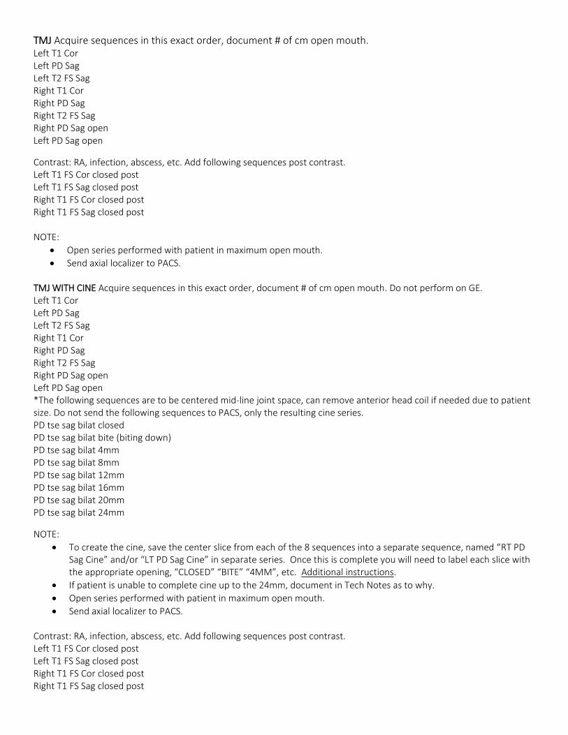
TMJ Acquire sequences in this exact order, document # of cm open mouth. Left T1 Cor Left PD Sag Left T2 FS Sag Right T1 Cor Right PD Sag Right T2 FS Sag Right PD Sag open Left PD Sag open
Contrast: RA, infection, abscess, etc. Add following sequences post contrast. Left T1 FS Cor closed post Left T1 FS Sag closed post Right T1 FS Cor closed post Right T1 FS Sag closed post NOTE:
Open series performed with patient in maximum open mouth.
Send axial localizer to PACS. TMJ WITH CINE Acquire sequences in this exact order, document # of cm open mouth. Do not perform on GE. Left T1 Cor Left PD Sag Left T2 FS Sag Right T1 Cor Right PD Sag Right T2 FS Sag Right PD Sag open Left PD Sag open *The following sequences are to be centered mid-line joint space, can remove anterior head coil if needed due to patient size. Do not send the following sequences to PACS, only the resulting cine series. PD tse sag bilat closed PD tse sag bilat bite (biting down) PD tse sag bilat 4mm PD tse sag bilat 8mm PD tse sag bilat 12mm PD tse sag bilat 16mm PD tse sag bilat 20mm PD tse sag bilat 24mm
NOTE:
To create the cine, save the center slice from each of the 8 sequences into a separate sequence, named “RT PD Sag Cine” and/or “LT PD Sag Cine” in separate series. Once this is complete you will need to label each slice with the appropriate opening, “CLOSED” “BITE” “4MM”, etc. Additional instructions.
If patient is unable to complete cine up to the 24mm, document in Tech Notes as to why.
Open series performed with patient in maximum open mouth.
Send axial localizer to PACS. Contrast: RA, infection, abscess, etc. Add following sequences post contrast. Left T1 FS Cor closed post Left T1 FS Sag closed post Right T1 FS Cor closed post Right T1 FS Sag closed post
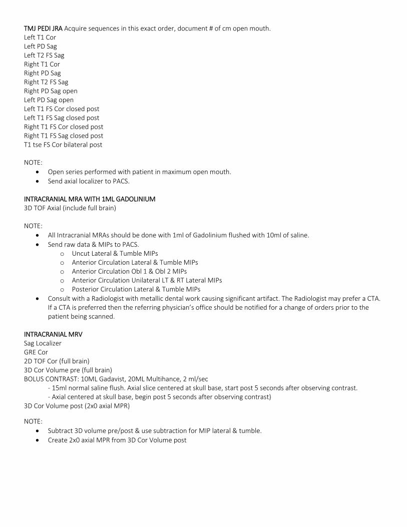
TMJ PEDI JRA Acquire sequences in this exact order, document # of cm open mouth. Left T1 Cor Left PD Sag Left T2 FS Sag Right T1 Cor Right PD Sag Right T2 FS Sag Right PD Sag open Left PD Sag open Left T1 FS Cor closed post Left T1 FS Sag closed post Right T1 FS Cor closed post Right T1 FS Sag closed post T1 tse FS Cor bilateral post NOTE:
Open series performed with patient in maximum open mouth.
Send axial localizer to PACS. INTRACRANIAL MRA WITH 1ML GADOLINIUM 3D TOF Axial (include full brain) NOTE:
All Intracranial MRAs should be done with 1ml of Gadolinium flushed with 10ml of saline.
Send raw data & MIPs to PACS. o Uncut Lateral & Tumble MIPs o Anterior Circulation Lateral & Tumble MIPs o Anterior Circulation Obl 1 & Obl 2 MIPs o Anterior Circulation Unilateral LT & RT Lateral MIPs o Posterior Circulation Lateral & Tumble MIPs
Consult with a Radiologist with metallic dental work causing significant artifact. The Radiologist may prefer a CTA. If a CTA is preferred then the referring physician’s office should be notified for a change of orders prior to the patient being scanned.
INTRACRANIAL MRV Sag Localizer GRE Cor 2D TOF Cor (full brain) 3D Cor Volume pre (full brain) BOLUS CONTRAST: 10ML Gadavist, 20ML Multihance, 2 ml/sec
- 15ml normal saline flush. Axial slice centered at skull base, start post 5 seconds after observing contrast. - Axial centered at skull base, begin post 5 seconds after observing contrast)
3D Cor Volume post (2x0 axial MPR)
NOTE:
Subtract 3D volume pre/post & use subtraction for MIP lateral & tumble.
Create 2x0 axial MPR from 3D Cor Volume post

INTRACRANIAL MRV WITHOUT CONTRAST GRE Cor 2D TOF Cor (full brain) 2D TOF Obl Sag (oblique 10 degrees from the midline brain to reduce the in-plane saturation effects) NOTE: Add T1 Sag full brain, if patient does not have a concurrent brain exam. CAROTID MRA WITH CONTRAST Sag Localizer 2D TOF Axial (7 mm slice thickness, 55 slices) T1 FS Axial 3D Cor Volume pre BOLUS FULL DOSE CONTRAST: 2ml/sec, 15ml normal saline flush 3D Cor Volume post NOTE:
Create R/L carotid & bilateral vertebral lateral MIP’s from pre/post subtraction. Do not send subtractions to PACS.
Axial 2x2mm MPR from 3D Cor Volume post.
CAROTID MRA NON-CONTRAST Sag Localizer 2D TOF Axial (3x1, -33% distance factor) 3D TOF Axial (centered at bifurcation) T1 tse FS dixon Axial NOTE:
Create R/L carotid & bilateral vertebral lateral MIP’s from the 3D TOF BRACHIAL PLEXUS AFFECTED SIDE T1 Axial (3X1) STIR Axial T2 FS Dixon Obl Cor (3x1) T1 Sag (5x1, FOV to include top of C1) STIR Cor (3x1, 400 FOV, bilateral)
NOTE: Contrast for diagnosis or history of tumor infection, radiation add following sequences post contrast. T1 FS dixon Axial post (3x1) T1 FS dixon Obl Cor post (3x1) CERVICAL SPINE TRAUMA – HOSPITALS ONLY T1 Sag STIR Sag T2 Sag T2* GRE Axial (16cm, mid T1 to inferior tip of clivus) T2 FS Axial (if hardware present, non-FS) PD 3D Axial (14cm, 1mm, craniocervical junction) NOTE:
Limit FOV, not including more than T1-T2 in sagittal planes.
Document presence or absence of radiculopathy with effected side.

CERVICAL SPINE T1 Sag (180 FOV, 3x.3, 15 slices) T2 Sag STIR Sag T2* GRE Axial (16cm, 3x.3, Mid T1 to inferior tip of clivus) T2 FS Axial (if hardware present, non-FS)
NOTE:
Limit FOV, not including more than T1-T2 in sagittal planes.
Document presence or absence of radiculopathy with effected side. CERVICAL SPINE WITH CONTRAST (myelopathy, MS, CA, etc.) T1 Sag (180 FOV) T2 Sag STIR Sag T2* GRE Axial (16cm, 3x.3, mid T1 to inferior tip of clivus) T2 FS Axial (if hardware present, non-FS) CONTRAST T1 FS Sag post T1 Sag post (if hardware present) T1 vibe Axial post NOTE:
Limit FOV, not including more than T1-T2 in sagittal planes.
If hardware limits the FS on post imaging, include additional non-FS series.
Document presence or absence of radiculopathy with effected side. SIALOGRAM Include orbits to mandible to ear lobes. T1 Sag (240 FOV, 5x1) T1 Axial (200 FOV, 3x.5) T2 FS Dixon Axial (200 FOV, 3x.5) T2 Space Axial (160 FOV, 1.2 mm, Cor MPR, to focus on salivary glands) STIR Cor (180 FOV, 3x.5) CONTRAST T1 FS Dixon Axial post (200 FOV, 2x.5) T1 FS Dixon Cor post (180 FOV, 2x.5) NOTE: Reserve read for Dr. Hassibi or Dr. Farhataziz. SKULL BASE / FACE T1 Sag (200 FOV, 3x.5, ~42 slices) T1 Axial (220 FOV, 3x.5, ~35 slices) T2 FS Dixon Axial STIR Cor (180 FOV, 3x1, ~50 slices) CONTRAST T1 FS Dixon Axial post T1 FS Dixon Cor post NOTE:
Confirm protocol with radiologist before performing.
Primarily used for facial lesions, include entire P>A / L>R diameter of skull / face, orbital roof through C1.

SOFT TISUE NECK T1 Sag (5x1) T1 Axial (4x1) T2 FS dixon Axial STIR Cor (5x1) CONTRAST T1 FS dixon Axial post immediate T1 FS dixon Cor post SOFT TISSUE NECK WITHOUT CONTRAST T1 Sag (5x1) T1 Axial (4x1) T2 FS dixon Axial STIR Cor (5x1) T1 tse Cor SOFT TISSUE NECK ACC THERAPY STAGING / RTP Include entire anatomy, skin to skin. T2 FS dixon Axial (3x0, 65 slices) T2 3D CISS Axial CONTRAST T1 fl2d Axial post T1 3D mprage Axial post SOFT TISSUE NECK MANDIBLE DR. PETER SCHOLL T1 Sag (5x1) T1 Axial (4x1) T2 FS dixon Axial STIR Cor (5x1) T1 hires Sag (2x0, 140 FOV, RT or LT affected side only, parallel to the long axis of the mandible) T1 hires Cor (3x0, 160 FOV, RT or LT affected side only, perpendicular to the long axis of the mandible) CONTRAST T1 FS Axial post immediate (4x1) T1 FS dixon Cor post (5x1) THORACIC SPINE T1 Sag Total Spine Localizer (3T auto-composing) T1 Sag (3x1, 14 slices) T2 Sag STIR Sag T2 Axial upper (16cm, 4x1, overlap at least 1 full vertebrae with lower axial, mid L1 through mid C7) T2 Axial lower Note: Document presence or absence of radiculopathy with effected side.
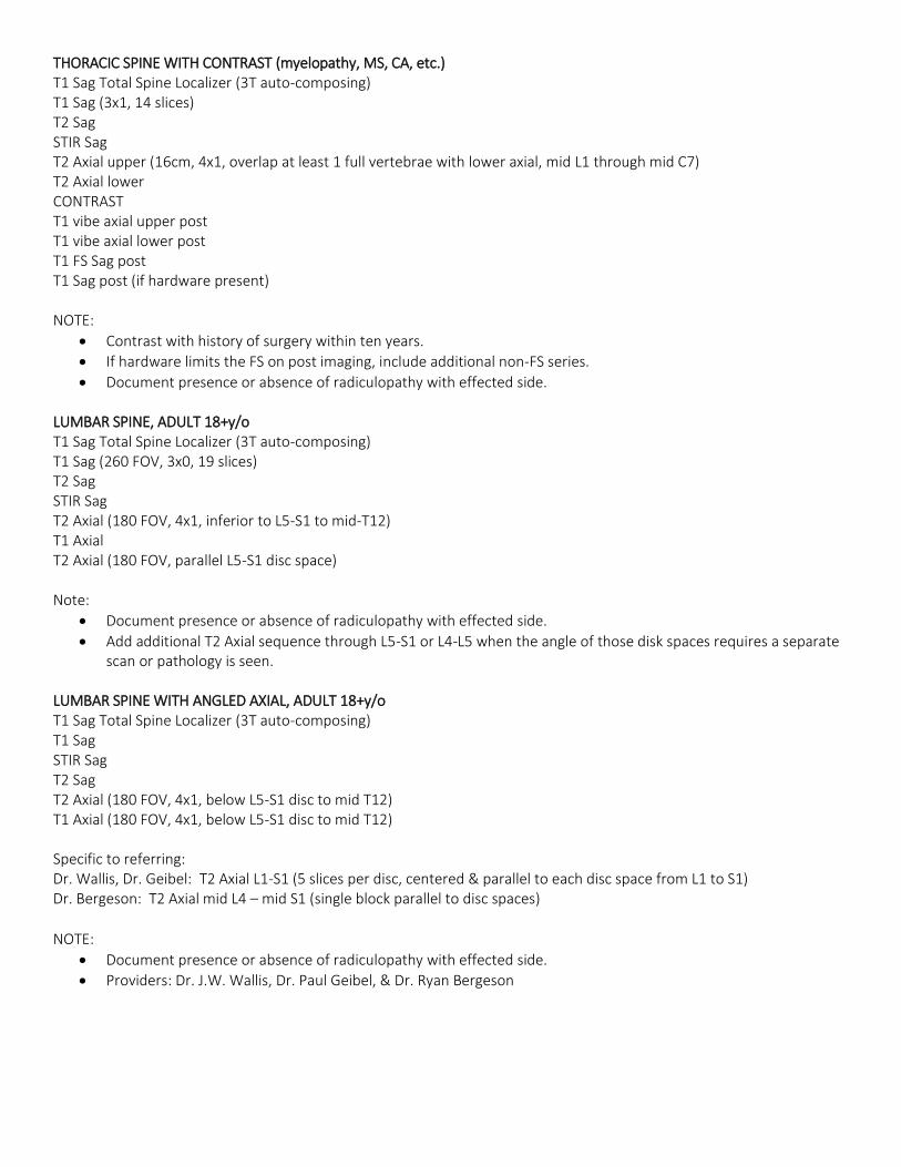
THORACIC SPINE WITH CONTRAST (myelopathy, MS, CA, etc.) T1 Sag Total Spine Localizer (3T auto-composing) T1 Sag (3x1, 14 slices) T2 Sag STIR Sag T2 Axial upper (16cm, 4x1, overlap at least 1 full vertebrae with lower axial, mid L1 through mid C7) T2 Axial lower CONTRAST T1 vibe axial upper post T1 vibe axial lower post T1 FS Sag post T1 Sag post (if hardware present) NOTE:
Contrast with history of surgery within ten years.
If hardware limits the FS on post imaging, include additional non-FS series.
Document presence or absence of radiculopathy with effected side. LUMBAR SPINE, ADULT 18+y/o T1 Sag Total Spine Localizer (3T auto-composing) T1 Sag (260 FOV, 3x0, 19 slices) T2 Sag STIR Sag T2 Axial (180 FOV, 4x1, inferior to L5-S1 to mid-T12) T1 Axial T2 Axial (180 FOV, parallel L5-S1 disc space) Note:
Document presence or absence of radiculopathy with effected side.
Add additional T2 Axial sequence through L5-S1 or L4-L5 when the angle of those disk spaces requires a separate scan or pathology is seen.
LUMBAR SPINE WITH ANGLED AXIAL, ADULT 18+y/o T1 Sag Total Spine Localizer (3T auto-composing) T1 Sag STIR Sag T2 Sag T2 Axial (180 FOV, 4x1, below L5-S1 disc to mid T12) T1 Axial (180 FOV, 4x1, below L5-S1 disc to mid T12) Specific to referring: Dr. Wallis, Dr. Geibel: T2 Axial L1-S1 (5 slices per disc, centered & parallel to each disc space from L1 to S1) Dr. Bergeson: T2 Axial mid L4 – mid S1 (single block parallel to disc spaces)
NOTE:
Document presence or absence of radiculopathy with effected side.
Providers: Dr. J.W. Wallis, Dr. Paul Geibel, & Dr. Ryan Bergeson
LUMBAR SPINE WITH CONTRAST, ADULT 18+y/o (myelopathy, MS, CA, etc.) T1 Sag Total Spine Localizer (3T auto-composing) T1 Sag (260 FOV, 3x0, 19 slices) T2 Sag STIR Sag T2 Axial (180 FOV, 4x1, inferior to L5-S1 to mid T12) T1 Axial pre T2 Axial (180 FOV, parallel L5-S1 disc space) CONTRAST T1 Axial post T1 FS Sag post T1 Sag post (if hardware present) NOTE:
Contrast with history of surgery within ten years.
If hardware limits the FS on post imaging, include additional non-FS series.
Document presence or absence of radiculopathy with effected side.
Add additional T2 Axial sequence through L5-S1 or L4-L5 when the angle of those disk spaces requires a separate scan or pathology is seen.
SPINE SCOLIOSIS Only if specifically ordered for scoliosis Routine Protocol Adults: T1 Cor Pedi: T2 Cor SPINE STEREOTACTIC THERAPHY PLANNING DR. DZIUK & DR. THATIKONDA T1 Sag Total Spine Localizer (3T auto-composing, not needed for cervical spine studies.) T2 3D Axial (180 FOV, 1-2 mm slice thickness depending on requested coverage. Do not angle.) T1 3D Axial pre T1 3D Axial post SPINE STRYKER T1 Sag Total Spine Localizer (3T auto-composing, not needed for cervical spine studies.) T2 Axial T1 Axial pre T1 Axial post NOTE: Do not angle images. Scan from inferior to superior. Include 1 vertebrae above and below area of interest. 3x0, 100% FOV, no rectangular FOV, matrix of 256x256. Position feet first, supine.
NEUROGRAPHY – SACRAL PLEXUS IR 3D SPC Cor (L2 down) T1 Cor PD Spair Cor T1 Axial (mid-L2 down) T2 Spair Axial (mid-L3 down) CONTRAST, if needed T1 FS Cor post T1 FS Ax post NOTE:
Create 1mm orthogonal Axial & Sag MPR’s from IR 3D SPC Cor.
All series are orthogonal, no angles.