Embed Size (px)
Citation preview

…..……………….………………..….…………………………… Results
-82-
4. RESULTS
The teratogenic effects of enrofloxacin and levofloxacin produced
in fetuses following administration to female pregnant rats from 6th
to 15th
day of gestation were compared with those of control fetuses from the
non-treated mother. Also, the histopathological changes, serum and tissue
concentrations of the drugs in mothers and fetuses were also studied.
4.1. Enrofloxacin.
4.1.1. Teratogenic effect: Oral administration of enrofloxacin in
therapeutic dose (10 mg / kg b.wt) and double therapeutic dose (20 mg /
kg b.wt) to pregnant female rats from 6th
to 15th
day of gestation period.
Highly significant decrease in number of fetuses per mother and highly
significant decrease in number of viable fetuses were recorded following
administration of both doses of enrofloxacin. The obtained results
revealed that enrofloxacin produced a highly significant increase in
number of resorbed fetuses per mother (Table 1 and Figure 1).
The effect of both given doses of enrofloxacin on fetal body weight
and length are tabulated in table (2) and shown in figure (2). Both fetal
body weight and length showed highly significant decrease when
compared with the data of control group. This indicated that therapeutic
dose of enrofloxacin (10 mg/kg b.wt) and double therapeutic dose (20 mg
/ kg b.wt) retard growth of developing fetuses during the period of
gestation.
The therapeutic dose of enrofloxacin (10 mg/kg b.wt) resulted in
diverticulum dilatation of the brain in 20%, thymus hypoplasia in 24.0%;
pulmonary hypoplasia in 52%, cardiac enlargement in 44%; hepatomegaly
in 56% ; kidney hypotrophy with dilatation of renal pelvis either

Results …………………..………………………………………….…...
-83-
unilateral or bilateral in 76% and decrease or small in size of suprarenal
gland in 12% (Table 3 and Figures 3, 4, 5, 6, 7 and 8).
Oral administration of double therapeutic dose of enrofloxacin (20
mg/kg b.wt) to pregnant rat from 6th
to 15th
day of gestation period
induced diverticulum dilatation of brain in 30%; thymus hypoplasia in
35.0% pulmonary hypoplasia in 75%; cardiac enlargement in 60.0%;
hepatomegaly in 65.0% atrophy or small in size of the kidney in 85% but
some cases showed complete absence of kidney with suprarenal gland
with unilateral or bilateral dilatation of renal pelvis and decrease or small
in size of suprarenal gland in 20% (Table 4 and Figures 9, 10 and 11).
Skeletal examinations of fetuses obtained from mothers given
orally enrofloxacin in therapeutic dose (10 mg/kg b.wt) from 6th
to 15th
day of gestation period showed impaired ossification of skull in 18.75%;
absence of sternebra in 62.5% as complete absence of 1st, 3
rd, 5
th and 6
th
sternebra or small in size of 2nd
, 4th
and 5th
. Other cases showed broken
dispersed sternebra. Absence of digit’s bone of fore and hind limb were
recorded 50.00 %; absence of some metatarsal bone in 25.00% and some
metacarpal bone in 31.25%, absence of some bone of caudal vertebrae in
56.25% of examined fetuses as shown in table (5) and Figures (12, 13 and
14).
Administration of double therapeutic dose of enrofloxacin (20
mg/kg b.wt) to pregnant rats resulted in impaired ossification of skull in
28.57%; absence of sternebra in 85.7%. Some cases showed complete
absence of all sternebra while others showed complete absence of 1st, 2
nd,
3rd
, 4th
, 5th
and 6th
with small in size of 2nd
, 3rd
, 4th
. Absence of digit’s
bone of fore and hind limb were recorded in 64.28%, absence of some
metatarsal bone in 71.4% and some of metacarpal bone in 57.14%,

…..……………….………………..….…………………………… Results
-84-
complete absence of caudal vertebrae with ischial bone in 92.85% of
examined fetuse as shown in table (6) and Figures (15 and 16).
4.1.2. Histopathological examination:
Histopathological examination of brain, lung, liver, kidney, spleen,
intestine, heart, uterus and placenta of the pregnant female rats
administered therapeutic dose of enrofloxacin (10 mg/kg b.wt) from 6th
to
15th
day of pregnancy revealed some pathological lesions in mothers. The
lung showed thickening in wall of blood vessels due to muscular
hypertrophy, other cases showed focal aggregation of mononuclear cell
with alveolar emphysema as shown in figure (17 and 18).The brain
showed odema with glaiosis (figure 19). The spleenic tissue showed
lymphoid deplation with excessive haemosiderosis (Figure 20).
Hyalinization of cardiac muscle was recorded also in figure (21).
Degenerative changes of hepatocytes with vacuolar and hydropic
degeneration of the hepatic tissue (Figure 22). Cloudy swelling of the
kidney, cattarhal enteritis, activation of goblet cells with inflammatory
cellular infiltration in the propria and submacosa of the lumen of intestine
were recorded in tissues of rats administered 10 mg / kg b.wt of
enrofloxacin orally and once daily from 6th
to 15th
day of pregnancy.
Administration of double therapeutic dose of enrofloxacin (20 mg /
kg b.wt) showed histopathological lesions represented by increase
incidence of pathological effects than recorded in therapeutic dose as
excessive degeneration with necrosis of hepatocytes, prevascular
lymphocytic aggregation of the kidney (Figure 23), excessive lymphoid
deplation with excessive haemosiderosis of spleen and hyalinization in
cardiac muscle. Excessive glaiosis of the brain were showed.
Histopathological changes in the uterus and placenta mainly occur with
double therapeutic dose as edematous hypertrophy of the chorion but

Results …………………..………………………………………….…...
-85-
some cases showed desuqmation of the endometerial gland and
epithelium of the uterus (Figures 24 and 25). The preceptation of calcium
salt (mineralization) in placenta was also reported as shown in figure
(26).
4.1.3. Standard curves of enrofloxacin:
Concentration of 0.025, 0.05, 0.1, 1.0, 1.25 (reference
concentration), 2.5, 5.0, 10.0 mg of enrofloxacin per milliliter phosphate
buffer (pH 7.2) or normal rat’s serum as well as their corresponding zone
of inhibition were illustrated in table (7) and shown in figure (27).
4.1.4. Serum and tissue concentration of enrofloxacin:
Following oral administration of therapeutic and double therapeutic
dose of enrofloxacin once daily. Serum and tissue concentrations of
therapeutic dose of enrofloxacin following (10 mg/kg b.wt) once daily
were represented in table (8) and shown in figure (29). These data
revealed distribution of the drug in tested tissue (brain, lung, heart, liver,
spleen, kidney, thigh and thoracic muscle, fat, skin and whole fetuses).
The data revealed that the liver and kidney contained the highest drug
concentrations (0.48 ± 0.0085 and 0.33 ± 0.0167 g/gm respectively) in
rats slaughtered at 10th
day of pregnancy. On other hand, the lowest
concentrations were recorded in fetuses and brain (0.115 ± 0.0043 and
0.088 ± 0.0018 g/gm respectively). The data reported for the rats
slaughtered at 16th
day of pregnancy (24 hours after last dose
administration) reported that the liver, kidney, lung and skin contained
the most highest concentrations as 0.79 ± 0.0191, 0.69 ± 0.0102, 0.53 ±
0.0116 and 0.44 ± 0.0058 g/gm respectively. The whole fetuses and the
brain contained the lowest concentrations of enrofloxacin (0.163 ± 0.0056
and 0.18 ± 0.0056 g/gm respectively). The data resulted from the rats

…..……………….………………..….…………………………… Results
-86-
slaughtered at 20th
day of pregnancy (5 days post drug administration, the
drug can not be assayed in all tissues except liver and kidney which
contained low concentrations (0.047 ± 0.0093 and 0.035 ± 0.0017 g/gm
respectively) as shown in table (8) and figure (28).
The serum and tissue concentrations following administration of
double therapeutic dose of enrofloxacin (20 mg/kg b.wt) were recorded in
table (9) and figure (29). The data revealed that high concentration of the
drug after double therapeutic administration were assayed in liver, kidney
and lung of slaughtered rats at 10th
day of pregnancy (0.54 ± 0.00 47,
0.449 ± 0.0052 and 0.35 ± 0.0091 g/gm respectively). Fetuses, brain and
spleen contained the lowest concentrations (0.169 ± 0.0048, 0.12 ±
0.0073 and 0.13 ± 0.0065ug/gm respectively). The results from the rats
slaughtered at 16th
day of pregnancy (24 hours post drug administration)
revealed that the liver, kidney, lung, skin, muscle (thigh and thoracic)
contained the highest concentrations (1.21 ± 0.0032, 0.84 ± 0.0085, 0.628
± 0.0061, 0.418 ± 0.0090, 0.44 ± 0.0063 and 0.39 ± 0.0070 mg/gm
respectively). Fetuses and brain contained the lowest concentrations
(0.237 ± 0.0062, 0.28 ± 0.0074 g/gm respectively). The data reported
from the rats slaughtered at 20th
day of pregnancy (5 day post drug
administration) recorded that the drug was disappeared from all tissues
except liver and kidney which contained (0.056 ± 0.0060 and 0.040 ±
0.0073 g/gm respectively)

Results …………………..………………………………………….…...
-87-

…..……………….………………..….…………………………… Results
-88-

Results …………………..………………………………………….…...
-89-

…..……………….………………..….…………………………… Results
-90-

Results …………………..………………………………………….…...
-91-

…..……………….………………..….…………………………… Results
-92-

Results …………………..………………………………………….…...
-93-

…..……………….………………..….…………………………… Results
-94-

Results …………………..………………………………………….…...
-95-

…..……………….………………..….…………………………… Results
-96-
Figure (1): Gravid rat’s uterus obtained from rat administered
therapeutic dose of enrofloxacin daily orally (10 mg/kg
b.wt) from 6th
to 15th
day of pregnancy showing early uterine
resorption.
Figure (2): Retardation of growth in a fetus obtained from rat
administered therapeutic dose of enrofloxacin daily orally
(10 mg/kg b.wt) from 6th
to 15th
day of pregnancy.
C: Control fetus T: treated fetus

Results …………………..………………………………………….…...
-97-
Figure (3): Diverticulum dilatation of brain in a fetus obtained from rat
administered therapeutic dose of enrofloxacin daily orally
(10 mg/kg b.wt) from 6th
to 15th
day of pregnancy.
C: Control fetus T: treated fetus
Figure (4): Thymus hypoplasia in fetus obtained from rat administered
therapeutic dose of enrofloxacin daily orally (10 mg/kg
b.wt) from 6th
to 15th
day of pregnancy.
C: Control fetus T: treated fetus

…..……………….………………..….…………………………… Results
-98-
Figure (5): Pulmonary hypoplasia with cardiac enlargement in a fetus
obtained from rat administered therapeutic dose of
enrofloxacin daily orally (10 mg/kg b.wt) from 6th
to 15th
day of pregnancy.
C: Control fetus T: treated fetus
Figure (6): Hepatomegaly in a fetus obtained from rat administered
therapeutic dose of enrofloxacin daily orally (10 mg/kg
b.wt) from 6th
to 15th
day of pregnancy.
C: Control fetus T: treated fetus

Results …………………..………………………………………….…...
-99-
Figure (7): Hypotrophy in left kidney in a fetus obtained from rat
administered therapeutic dose of enrofloxacin daily orally
(10 mg/kg b.wt) from 6th
to 15th
day of pregnancy.
C: Control fetus T: treated fetus
Figure (8): Bilateral dilatation of renal pelvis in a fetus obtained from rat
administered therapeutic dose of enrofloxacin daily orally
(10 mg/kg b.wt) from 6th
to 15th
day of pregnancy.
C: Control fetus T: treated fetus
…..……………….………………..….…………………………… Results
-100-
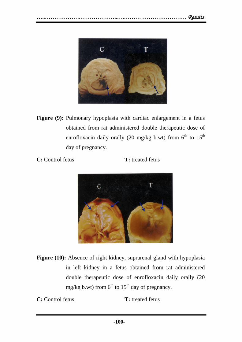
Figure (9): Pulmonary hypoplasia with cardiac enlargement in a fetus
obtained from rat administered double therapeutic dose of
enrofloxacin daily orally (20 mg/kg b.wt) from 6th
to 15th
day of pregnancy.
C: Control fetus T: treated fetus
Figure (10): Absence of right kidney, suprarenal gland with hypoplasia
in left kidney in a fetus obtained from rat administered
double therapeutic dose of enrofloxacin daily orally (20
mg/kg b.wt) from 6th
to 15th
day of pregnancy.
C: Control fetus T: treated fetus

Results …………………..………………………………………….…...
-101-
Figure (11): Bilateral dilatation of renal pelvis in kidney from fetus
obtained from rat administered double therapeutic dose of
enrofloxacin daily orally (20 mg/kg b.wt) from 6th
to 15th
day of pregnancy.
C: Control fetus T: treated fetus
Figure (12): Absence of 4th, 5th
and 6th
sternebra with small in size of 1st,
2nd
and 3rd
in a fetus obtained from rat administered
therapeutic dose of enrofloxacin daily orally (10 mg/kg
b.wt) from 6th
to 15th
day of pregnancy.
C: Control fetus T: treated fetus

…..……………….………………..….…………………………… Results
-102-
Figure (13): Absence of one ischum bone in a fetus obtained from rat
administered therapeutic dose of enrofloxacin daily orally
(10 mg/kg b.wt) from 6th
to 15th
day of pregnancy.
C: Control fetus T: treated fetus
Figure (14): Absence of digit’s bone in a fetus obtained from rat
administered therapeutic dose of enrofloxacin daily orally
(10 mg/kg b.wt) from 6th
to 15th
day of pregnancy.
C: Control fetus T: treated fetus

Results …………………..………………………………………….…...
-103-
Figure (15): Complete absence of all sternebra in fetus obtained from rat
administered double therapeutic dose of enrofloxacin daily
orally (20 mg/kg b.wt) from 6th
to 15th
day of pregnancy.
C: Control fetus T: treated fetus
Figure (16): Complete absence of all caudal vertebrae, ischum bone,
metatarsal bone and digital bones in fetus obtained from rat
administered double therapeutic dose of enrofloxacin daily
orally (20 mg/kg b.wt) from 6th
to 15th
day of pregnancy.
C: Control fetus T: treated fetus

…..……………….………………..….…………………………… Results
-104-
Figure (17): Lung of rat administered therapeutic dose of enrofloxacin
daily orally (10 mg/kg b.wt) from 6th
to 15th
day of
pregnancy showing thickening in wall of blood vessels of
lung due to muscular hypertrophy. H & E stain X 400.
Figure (18): Lung of rat administered therapeutic dose of enrofloxacin
daily orally (10 mg/kg b.wt) from 6th
to 15th
day of
pregnancy showing focal aggregation of mononuclear cell
with alveolar emphysema of lung. H & E stain X 400

Results …………………..………………………………………….…...
-105-
Figure (19): Brain of rat administered therapeutic dose of enrofloxacin
daily orally (10 mg/kg b.wt) from 6th
to 15th
day of
pregnancy showing odema and glaiosis. H & E stain X 400
Figure (20): Spleen from rat administered therapeutic dose of
enrofloxacin daily orally (10 mg/kg b.wt) from 6th
to 15th
day of pregnancy showing lymphoid deplation with
excessive haemosiderois. H & E stain X 400

…..……………….………………..….…………………………… Results
-106-
Figure (21): Heart from rat administered therapeutic dose of
enrofloxacin daily orally (10 mg/kg b.wt) from 6th
to 15th
day of pregnancy showing hyalanization of cardiac muscle.
H & E stain X 400
Figure (22): Liver from rat administered therapeutic dose of
enrofloxacin daily orally (10 mg/kg b.wt) from 6th
to 15th
day of pregnancy showing extensive vacuolar of
hepatocytes. H & E stain X 400

Results …………………..………………………………………….…...
-107-
Figure (23): Kidney of rat administered double therapeutic dose of
enrofloxacin daily orally (20 mg/kg b.wt) from 6th
to 15th
day of pregnancy showing perivascular lymphocytic
aggregation. H & E stain X 400
Figure (24): Uterus from rat administered double therapeutic dose of
enrofloxacin daily orally (20 mg/kg b.wt) from 6th
to 15th
day of pregnancy showing odematous hyperplasia of the
chorionic villi. H & E stain X 100

…..……………….………………..….…………………………… Results
-108-
Figure (25): Uterus of rat administered double therapeutic dose of
enrofloxacin daily orally (20 mg/kg b.wt) from 6th
to 15th
day of pregnancy showing desquamation in the endometerial
epithelium. H & E stain X 200
Figure (26): Placenta from rat administered double therapeutic dose of
enrofloxacin daily orally (20 mg/kg b.wt) from 6th
to 15th
day of pregnancy showing calcium deposition of nectoric
area. H & E stain X 400

Results …………………..………………………………………….…...
-109-
Dia
met
er o
f In
hib
ati
on
zon
e (m
m)
Concentration of enerofloxacin (g/ml)
Fig
ure
(27):
Sta
nd
ard
cu
rve
of
enro
floxa
cin
in
ph
osp
hate
bu
ffer
pH
(7.2
) an
d n
orm
al
rat’
s se
ru
m. (n
= 1
0).
Phosp
hat
e buff
er
Norm
al r
at’s
ser
um

…..……………….………………..….…………………………… Results
-110-

Results …………………..………………………………………….…...
-111-

…..……………….………………..….…………………………… Results
-112-
4.2. Levofloxacin
4.2.1. Teratogenic effect: Pregnant female rats were given
levofloxacin intravenously injection in therapeutic dose (25 mg/kg b.wt)
and double therapeutic dose (50 mg/kg b.wt) from 6th
to 15th
day of
gestation period. Significant decrease in number of fetuses per mother
and significant decrease in the number of viable fetus were recorded.
Both doses induced a significant increase in the number of resorbed
fetuses per mother (Table 10 and figure 30). Some cases showed early
uterine resorption but other showed late uterine resorption as shown in
figure (31).
Statistical analysis of the data in table (11) revealed that
levofloxacin exerted retardation in growth of all living fetuses following
administration of both doses to pregnant rats, this results indicated that
levofloxacin in both doses retarded the growth and length of developing
fetuses during gestation period as shown in figure (32).
Visceral examination of living fetuses obtained from mother given
therapeutic dose of levofloxacin in a dose of 25 mg/kg b.wt from 6th
to
15th
day of gestation period resulted some of visceral abnormalities such
as diverticulum dilatation of the brain in 25.92%, thymus hypoplasia in
18.52%, pulmonary hypoplasia in 59.25%, cardiac enlargement in
48.14% hepatomegaly in 40.74%, kidney hypoplasia in one or both
kidney with dilatation of renal pelvis either unilateral or bilateral in
77.77% and decrease or small in size of suprarenal gland in 29.62% (table
12 and Figure 33 and 34).
Intravenous administration of double therapeutic dose of
levofloxacin (50 mg/kg b.wt) to pregnant female rat from 6th
to 15th
day
of gestation period induced diverticulum dilatation of brain in 38.89%,

Results …………………..………………………………………….…...
-113-
thymus hypoplasia in 27.67%: pulmonary hypoplasia in 83.33%, cardiac
enlargement in 66.67%, hepatomegaly of the liver in 50.00% kidney
atrophy or hypoplasia in one or both kidney with unilateral or bilateral
dilatation of renal pelvis in 94.44% and decrease or small in size of supra
renal gland in 44.44% (Table 13 and Figure 35, 36, 37, 38 and 39).
Skeletal examination of fetuses obtained from mothers given
intravenous injection of levofloxacin in therapeutic dose (25 mg/kg b.wt)
from 6th
to 15th
day of gestation period showed impaired ossification of
skull in 31.58%; absence of sternebra in 52.63% as 1st, 2
nd, 5
th, 6
th with
very small in size of 2nd
, 3rd
and 4th
. Absence of digit’s bone in fore and
hind limb in 36.84%, absence of some metatarsal bone in 21.05%, and
some metacarpal bone in 26.32%; absence of caudal vertebrae either
complete absence of caudal vertebrae, small in size of ischial bone or
presence of one bone near to vertebral column in 47.37% as shown in
table 14 and figure 40 and 41).
Administration of double therapeutic dose of levofloxacin as
intravenous injection (50 mg/kg b.wt) to pregnant female rats from 6th
to
15th
day of gestation period resulted in impaired ossification of skull in
45.45%; absence of sternebra in 72.73%. Some cases showed small in
size of 1st, 2
nd, 5
th and 6
th with broken of 4
th and 5
th but other cases
showed presence of small dispersed bone with absence of other sternebra.
Absence of digital bones of fore and hind limb in 63.64%; absence of
some metatarsal bone in 54.55% and some metacarpal bone in 63.64%
complete absence of caudal vertebrae with ischial bone either absence or
small in size in 81.82% of examined fetuses as shown in table (15) and
figure (42 and 43).

…..……………….………………..….…………………………… Results
-114-
4.2.2. Histopathological examination:
Histopathological examination of brain, lung, liver, kidney, spleen,
intestine, heart, uterus and placenta of the pregnant female rats
administered therapeutic dose of levofloxacin (25 mg/b.wt) intravenously
from 6th
to 15th
day of pregnancy revealed some pathologic lesions in
mothers. The liver showed hydropic degeneration in hepatocytes with
infiltration of liver by inflammatory cell mainly lymphocytes, while
kidney showed necrosis (Figures 44 & 45), focal proliferation of glial cell
of brain with cerebral and cerebllum encephalomalesia (Figures 46, 47 &
48). The spleen showed lymphocyic deplation with haemosiderosis,
catharhal enteritis was also detected in the intestine.
Double therapeutic dose of levofloxacin (50 mg/kg b.wt) resulted
in many histopathological changes as prevascular lymphocytic
aggregation of the lung (Figure 49); degeneration of renal tubules with
lymphocytic aggregation of the kidney (Figure 50); hydropic
degeneration of the hepatocyte (Figure 51) and loss of striation of cardiac
muscle fibers with lymphocytic aggregation of the heart (figure 52). The
histopathological changes in the uterus and placenta mainly occur with
double therapeutic dose. Uterus revealed congestion of blood vessels with
lymphocytic infiltration in the lamina propria and submucosa but other
cases reported congestion of blood vessels, presence of patches of
lymphocytic infiltration in the proprea and around arterioles with
presence area of necrosis. Placenta revealed congestion of blood vessels
of surface of placenta with cellular desquamation in lining epithelium but
other cases showed area of necrosis in the villus surface (Figures 53, 54,
and 55).

Results …………………..………………………………………….…...
-115-
4.2.3. Standard curves of levofloxacin:
Concentration of 0.025, 0.05, 0.1, 1, 1.25 (reference concentration)
1, 2.5, 5, 10 mg of levofloxacin per milliliter phosphate buffer (pH 6.7) or
normal rat’s serum as well as their corresponding zone of inhibition were
illustrated in table (16) and show in Figure (56).
4.2.4. Serum and tissue concentrations of levofloxacin:
Following intravenous injection of therapeutic and double
therapeutic dose of levofloxacin once daily. Serum and tissue
concentrations of therapeutic dose of levofloxacin following (25 mg/kg
b.wt) once daily were represented in table (17) and show in Figure (57).
The data revealed distribution of the drug in tested tissues (brain, lung,
heart, liver, spleen, kidney, thigh and thoracic muscle, fat, skin and whole
fetuses). The data revealed that liver, kidney contained the highest drug
concentrations (0.41 ± 0.0043 and 0.45 ± 0.0061 g/gm respectively in
rats slaughtered at 10th
day of pregnancy. On other hand, the lowest
concentrations was recorded in brain, spleen, heart and whole fetuses
(0.11 ± 0.0043, 0.10 ± 0.0037, 0.12 ± 0.0029 and 0.126 ± 0.0048 g/gm
respectively). The data reported from rats slaughtered at 16th
day of
pregnancy (24 hours after last dose administration resulted in highest
concentrations of drug in liver, kidney, lung and skin (0.80 ± 0.0087, 0.87
± 0.0058, 0.63 ± 0.0049 and 0.46 ± 0.0049 g/gm respectively). The
whole fetus and spleen contained the lowest concentration of levofloxacin
(0.202 ± 0.0042 and 0.22 ± 0.0108 g/gm respectively). The data resulted
from the rats slaughtered at 20th
day of pregnancy (5 days post drug
administration, the drug can not be assayed in all tissues except liver and
kidney which contained low concentrations (0.022 ± 0.0092 and 0.028 ±
0.0092) g/gm respectively as shown in table (17) and Figure (58).

…..……………….………………..….…………………………… Results
-116-
The serum and tissue concentrations following administration of
double therapeutic dose of levofloxacin (50 mg/kg b.wt ) were recorded
in table (18) and figure (58). The data revealed that the highest
concentration of the drug after double therapeutic administration in liver,
kidney, lung of slaughtered rats at 10th
day of pregnancy (0.55 ± 0.0053,
0.65 ± 0.0040 and 0.42 ± 0.0074 g/gm respectively). Fetuses, heart and
spleen contained the lowest concentrations (0.183 ± 0.0029, 0.178 ±
0.0036 and 0.14 ± 0.0034 g/gm respectively). The results from the rats
slaughtered at 16th
day of pregnancy (24 hours post drug administration)
revealed that the liver, kidney, lung, skin contained, the highest
concentrations (0.91 ± 0.0138, 1.38 ± 0.0270, 0.73 ± 0.0049 and 0.608 ±
0.0141 g/gm respectively). Fetuses, heart and spleen contained the
lowest concentrations (0.247 ± 0.0060, 0.30 ± 0.0052 and 0.34 ± 0.0819
g/gm respectively). The data reported from the rats slaughtered at 20th
day of pregnancy (5 days post drug administration indicated that the drug
disappeared from all tissues except liver, kidney and lung (0.020 ±
0.0047, 0.036 ± 0.0071 and 0.046 ± 0.0060 g/gm respectively).

Results …………………..………………………………………….…...
-117-

…..……………….………………..….…………………………… Results
-118-

Results …………………..………………………………………….…...
-119-

…..……………….………………..….…………………………… Results
-120-

Results …………………..………………………………………….…...
-121-

…..……………….………………..….…………………………… Results
-122-

Results …………………..………………………………………….…...
-123-

…..……………….………………..….…………………………… Results
-124-

Results …………………..………………………………………….…...
-125-

…..……………….………………..….…………………………… Results
-126-
Figure (30): Gravid rat’s uterus in a fetus obtained from rat administered
therapeutic dose of levofloxacin daily intravenously (25
mg/kg b.wt) from 6th
to 15th
day of pregnancy.
Figure (31): Gravid rat’s uterus in a fetus obtained from rat administered
double therapeutic dose of levofloxacin daily intravenously
(50 mg/kg b.wt) from 6th
to 15th
day of pregnancy, it showed
late resorbed fetuses.

Results …………………..………………………………………….…...
-127-
Figure (32): Retardation of growth in a fetus obtained from rat
administered double therapeutic dose of levofloxacin daily
intravenously (50 mg/kg b.wt) from 6th
to 15th
day of
pregnancy.
C: Control fetus T: treated fetus
Figure (33): Diverticulum dilatation of the brain in a fetus obtained from
rat administered therapeutic dose of levofloxacin daily
intravenously (25 mg/kg b.wt) from 6th
to 15th
day of
pregnancy.
C: Control fetus T: treated fetus

…..……………….………………..….…………………………… Results
-128-
Figure (34): Bilateral dilatation of renal pelvis in a fetus obtained from
rat administered therapeutic dose of levofloxacin daily
intravenously (25 mg/kg b.wt) from 6th
to 15th
day of
pregnancy.
C: Control fetus T: treated fetus
Figure (35): Absence of thymus gland in a fetus obtained from rat
administered double therapeutic dose of levofloxacin daily
intravenously (50 mg/kg b.wt) from 6th
to 15th
day of
pregnancy.
C: Control fetus T: treated fetus

Results …………………..………………………………………….…...
-129-
Figure (36): Pulmonary hypoplasia with cardiac enlargment in a fetus
obtained from rat administered double therapeutic dose of
levofloxacin daily intravenously (50 mg/kg b.wt) from 6th
to
15th
day of pregnancy.
C: Control fetus T: treated fetus
Figure (37): Hepatomegaly in a fetus obtained from rat administered
double therapeutic dose of levofloxacin daily intravenously
(50 mg/kg b.wt) from 6th
to 15th
day of pregnancy.
C: Control fetus T: treated fetus

…..……………….………………..….…………………………… Results
-130-
Figure (38): Hypoplasia in both kidney with small in size of suprarenal
gland in a fetus obtained from rat administered double
therapeutic dose of levofloxacin daily intravenously (50
mg/kg b.wt) from 6th
to 15th
day of pregnancy.
C: Control fetus T: treated fetus
Figure (39): Atrophy in right kidney with small in size of suprarenal
gland in a fetus obtained from rat administered double
therapeutic dose of levofloxacin daily intravenously (50
mg/kg b.wt) from 6th
to 15th
day of pregnancy.
C: Control fetus T: treated fetus

Results …………………..………………………………………….…...
-131-
Figure (40): Impaired ossification of skull in a fetus obtained from rat
administered therapeutic dose of levofloxacin daily
intravenously (25 mg/kg b.wt) from 6th
to 15th
day of
pregnancy.
C: Control fetus T: treated fetus
Figure (41): Absence of digit’s bone in a fetus obtained from rat
administered therapeutic dose of levofloxacin daily
intravenously (25 mg/kg b.wt) from 6th
to 15th
day of
pregnancy.
C: Control fetus T: treated fetus

…..……………….………………..….…………………………… Results
-132-
Figure (42): Presence of small scattered three bone of strenbrae in a fetus
obtained from rat administered double therapeutic dose of
levofloxacin daily intravenously (50 mg/kg b.wt) from 6th
to
15th
day of pregnancy.
C: Control fetus T: treated fetus
Figure (43): Absence of one ischum bone and appear small in size and
near to the vertebral column in a fetus obtained from rat
administered double therapeutic dose of levofloxacin daily
intravenously (50 mg/kg b.wt) from 6th
to 15th
day of
pregnancy.
C: Control fetus T: treated fetus

Results …………………..………………………………………….…...
-133-
Figure (44): Liver of rat administered therapeutic dose of levofloxacin
daily intravenously (25 mg/kg b.wt) from 6th
to 15th
day of
pregnancy showing hydropic degeneration of hepatocytes.
H & E stain X 200
Figure (45): Kidney of rats administered therapeutic dose of levofloxacin
daily intravenously (25 mg/kg b.wt) from 6th
to 15th
day of
pregnancy showing coagulative necrosis of renal tubules.
H & E stain X 100.

…..……………….………………..….…………………………… Results
-134-
Figure (46): Brain of rats administered therapeutic dose of levofloxacin
daily intravenously (25 mg/kg b.wt) from 6th
to 15th
day of
pregnancy showing focal proliferation of glial cells. H & E
stain X 200.
Figure (47): Brain of rats administered therapeutic dose of levofloxacin
daily intravenously (25 mg/kg b.wt) from 6th
to 15th
day of
pregnancy showing encephalomalecia of the cerebllum. H &
E stain X 400

Results …………………..………………………………………….…...
-135-
Figure (48): Brain of rats administered therapeutic dose of levofloxacin
daily intravenously (25 mg/kg b.wt) from 6th
to 15th
day of
pregnancy showing cerebral encephalomalacia. H & E stain
X 400.
Figure (49): Lung of rats administered double therapeutic dose of
levofloxacin daily intravenously (50 mg/kg b.wt) from 6th
to
15th
day of pregnancy showing perivascular lymphocytic
cellular aggregation and alveolar emphysema. H & E stain X
200.
…..……………….………………..….…………………………… Results
-136-
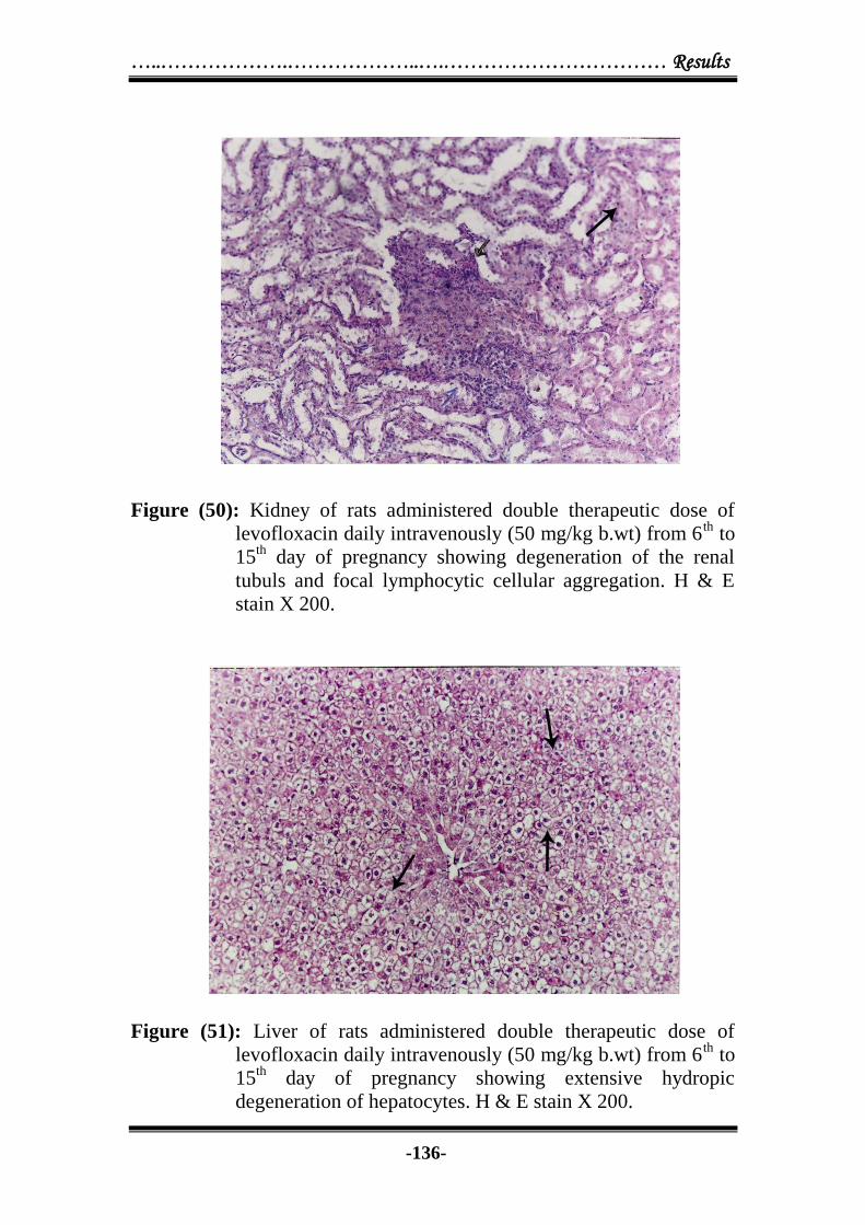
Figure (50): Kidney of rats administered double therapeutic dose of
levofloxacin daily intravenously (50 mg/kg b.wt) from 6th
to
15th
day of pregnancy showing degeneration of the renal
tubuls and focal lymphocytic cellular aggregation. H & E
stain X 200.
Figure (51): Liver of rats administered double therapeutic dose of
levofloxacin daily intravenously (50 mg/kg b.wt) from 6th
to
15th
day of pregnancy showing extensive hydropic
degeneration of hepatocytes. H & E stain X 200.

Results …………………..………………………………………….…...
-137-
Figure (52): Heart of rats administered double therapeutic dose of
levofloxacin daily intravenously (50 mg/kg b.wt) from 6th
to
15th
day of pregnancy showing hyalinization of some cardiac
muscles with aggregation of few lymphocytes. H & E stain
X 200.
Figure (53): Uterus of rats administered double therapeutic dose of
levofloxacin daily intravenously (50 mg/kg b.wt) from 6th
to
15th
day of pregnancy showing congested blood vessels,
focal lymphocytic cellular infiltration and necrosis. H & E
stain X 200.
…..……………….………………..….…………………………… Results
-138-
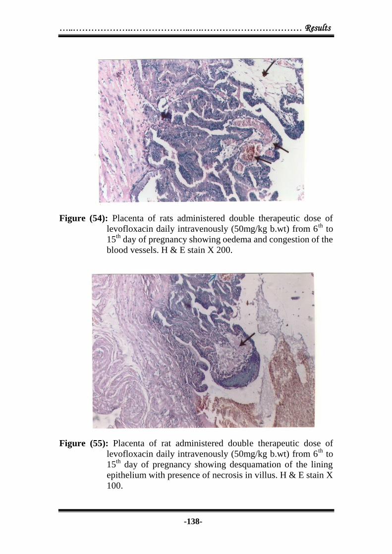
Figure (54): Placenta of rats administered double therapeutic dose of
levofloxacin daily intravenously (50mg/kg b.wt) from 6th
to
15th
day of pregnancy showing oedema and congestion of the
blood vessels. H & E stain X 200.
Figure (55): Placenta of rat administered double therapeutic dose of
levofloxacin daily intravenously (50mg/kg b.wt) from 6th
to
15th
day of pregnancy showing desquamation of the lining
epithelium with presence of necrosis in villus. H & E stain X
100.

Results …………………..………………………………………….…...
-139-
Dia
met
er o
f In
hib
ati
on
zon
e (m
m)
Fig
ure
(5
6):
Sta
nd
ard
curv
e of
lev
ofl
ox
acin
in
ph
osp
hat
e bu
ffer
pH
(6
.7)
and
no
rmal
rats
ser
um
. (n
= 1
0).
Concentration of levofloxacin (g/ml)
P
hosp
hat
e buff
er
Norm
al r
at’s
ser
um

…..……………….………………..….…………………………… Results
-140-

Results …………………..………………………………………….…...
-141-


![Indigenous Enhanced Mineralization Pyrene, Benzo[a]pyrene ...Indigenous soil microorganism mineralization experiments. All of the mineralization experiments were performed by using](https://img.pdfslide.net/doc/110x75/5e7c41b0b7c4ef64181e5e16/indigenous-enhanced-mineralization-pyrene-benzoapyrene-indigenous-soil-microorganism.jpg)