Embed Size (px)
Citation preview

Send Orders for Reprints to [email protected]
Current Pharmaceutical Design, 2015, 21, 000-000 1
1381-6128/15 $58.00+.00 © 2015 Bentham Science Publishers
Design of Multifunctional Nanocarriers for Delivery of Anti-Cancer Therapy
Neelesh Kumar Mehra*1,2, Keerti Jain1,3 and Narendra Kumar Jain1
1Pharmaceutical Nanotechnology Research Laboratory, Department of Pharmaceutics, ISF College of Pharmacy,
Moga (Panjab) India; 2Department of Pharmaceutical Sciences, Irma Lerma Rangel College of Pharmacy, Texas
A & M Health Science Centre, Kingsville, Texas, USA 783 63; 3National Institute of Pharmaceutical Education
and Research (NIPER Raebareli), Raebareli (U.P.) 229010, India
Abstract: Chemotherapy is the major and most widely used therapeutic strategy for the treatment of a wide vari-ety of cancers. The non-specific/non-targeted drug delivery in chemotherapy leads to undesired side effects in nor-mal and healthy tissues, and insufficient dosages to kill cancerous cells. Now-a-days, smart and intelligent multi-functional targeted nanomedicines based on various nanocarriers (dendrimers, carbon nanotubes, graphene, nanoparticles, quantum dots, self-emuslifying lipidic systems and carbon nanohorns etc) are being investigated promisingly in cancer treatment. In this article, we review the role of smart and intellegent multifucntional nanocar-riers in delivery of chemotherapeutic agents with the aim to develop promising treatment strategy to combat with one of the killer of man kind i.e. cancer.
Keywords: Chemotherapy, Multifunctional, Dendrimers, Carbon nanotubes, Drug Delivery, Drug Targeting.
INTRODUCTION
Nanotechnology, a contemporary discipline, involves utilization of nanosized biomaterials including liposomes, polymer-drug con-jugates, graphenes, quantum dots, dendrimers, nanoparticles, car-bon nanohorns, self emulsifying lipidic system and carbon nano-tubes not only in drug delivery, but also in biomedical research including bioimaging, biomedical-diagnosis and tissue engineering [1-4]. Nanotechnology is an applicable aspect in a broader area of nanoscience (nano means 10-9 m), which is one of the upcoming, highly challenging and rewarding key research area of the modern scientific set-up. The term nanotechnology includes the nanoscale phenomena, process, biomaterials, nano-devices, nano-systems and nano-manufacturing [5]. The in vivo use of nanotechnology in drug delivery, imaging and diagnosis is one of the rapidly expanding fields, which is necessary to meet the need of new delivery system with advantages, such as improved solubility and bioavailability of hydrophobic drugs, high drug payload, extended drug half-life, improved therapeutic index, controlled release of bioactive(s) along with reduced immunogenicity, and toxicity [6].
In the last two decades, outstanding progress has been made in using nanobiomaterials to (a) target biological moiety to cancerous cells through specific targeting ligands, and (b) increases localized delivery by increasing serum residence time [7]. Targeted drug delivery system is one of the most promising approach to expand the therapeutic window by improving specificity and targetibility to the target sites as well target-non-target tissue ratio, resulting mini-mum effective dose and toxicities of drugs [6,8].
Designing of Multifunctional Nanocarriers
Multifunctional nanocarriers are smart and intelligent delivery system, which could be designed and developed as per requirement. These multifunctional nanocarriers basically consist of three main design components: (i) a platform (core) material, (ii) encapsulated payload/biologically active agents, and (iii) targeting/surface-associated functional properties [9, 10, 11].
*Address correspondence to this author at the Department of Pharmaceuti-cal Sciences, Irma Lerma Rangel College of Pharmacy, Texas A & M Health Science Centre, Kingsville, Texas, USA 783 63; Tel: +1-361-720-3044; E-mails: [email protected]; [email protected]; [email protected]
Modification of surface properties of nanocarriers by chemical functional moieties including polymers and/or targeting ligands is known as functionalization. Surface functionalization has been achieved by incorporation, adsorption or covalent/non-covalent coupling of polymers, combination of polymers, carbohydrates, endogenous substances/ligands, peptides, proteins, nucleic acids and polysaccharides to the nanoparticles surface. The various multi-functional nanocarriers have been summarized in Fig. (1).
MULTIFUNCTIONAL NANOCARRIERS IN CANCER CHEMOTHERAPY
Multifunctional nanocarriers are the delivery systems that could deliver chemotherapeutic agent at desired/specific/target sites with improved therapeutic outcomes. In this review, the multifunctional nanocarriers used for chemotherapeutic delivery to cancerous cells are emphasized briefly.
Carbon nanotubes in Delivery of Chemotherapeutic Agents
Carbon nanotubes (CNTs) were first discovered in 1991 by Sumio Iijima (Japanese Microscopist) during his TEM observation [12]. CNTs are among the most interesting and attractive drug de-livery nanovectors currently under investigation. CNTs are three-dimensional sp2 hybridized, condensed benzene rings rolled up into seamless hollow tubular cylinder and classified into four categories based on the presence of number of wall: single-, double-, triple-, and multi-walled carbon nanotubes (SWCNTs, DWCNTs, TWCNTs and MWCNTs, respectively). Functionalized CNTs (f-CNTs) have shown great promise as drug delivery systems due to their easy penetration and ability to cross biological barriers. How-ever, specific translocation mechanisms (endocytosis or tiny nano-needle like penetration) of f-CNTs are not yet fully elucidated [8, 13-16]. Functionalization is a well known approach for surface alteration of nanomaterials [1].
f-CNTs have high surface area, which facilitates multiple at-tachment of chemical functional moieties as well high payload of drug molecules on to the surface or inside the nanotubes via various interaction mechanisms (hydrophobic, electrostatic, - stacking interactions). The degree and type of functionalization of CNTs also plays a pivotal role in delivery of chemotherapeutic agents [3, 13, 16]. CNTs have been functionalized using two main approaches based on (i) covalent, and (ii) non-covalent linkages between
Neelesh Kumar Mehra

2 Current Pharmaceutical Design, 2015, Vol. 21, No. 00 Mehra et al.
chemical functional moieties and surface of nanotubes [17-18]. f-CNTs have the ability to deliver water-insoluble (hydrophobic) drugs (taxol derivatives; paclitaxel and docetaxel) by conjugation onto the surface of the nanotubes [19]. The various transcellualr trafficking pathways known to be involved in transport of f-CNTs include endocytosis, pinocytosis, fluid-phase diffusion, carrier and receptor-mediated and facilitated transport mechanism. Interest-ingly, CNTs have the capacity to form supramolecular complexes with polycyclic aromatic molecules through - stacking interac-tions [20]. In this context, an anthracycline antibiotic (Doxorubicin hydrochloride; DOX) is most investigated chemotherapeutic agent till today. The significant contributions of f-CNTs in delivery of DOX for cancer treatment have been revealed in last one decade. High payload with controlled release was observed with f-CNTs for targeted delivery of Dox devoid any significant toxicity [20-21]. Block copolymers based MWCNTs aqueous dispersions are able to form supramolecular complexes with the aromatic chromophore and DOX via - stacking and enhance the cytotoxic activity [22].
Till date huge reports have been available on DOX delivery using multifunctional CNTs after decoration of targeting ligands like hyaluronic acid (HA) [23], hydroxybenzoic acid (HBA) [24], vitamin E (TPGS) [25], estrone (ES) [13], folic acid (FA) and chi-tosan (CHI) etc [26-27] for drug delivery and targeting purpose.
Our laboratory has explored the multifunctional CNTs as nanovectors for delivery of chemotherapeutics in cancer therapy employing dexamethasone [28], folate [21], vitamin E [25] and estrone [13, 29] as targeting moiety. We have also reported that the multifunctional CNTs showed the controlled and sustained release of DOX [21, 25] and gemcitabine [30].
Very recently, we have compared the targeting potential of ES and FA appended PEGylated MWCNTs [13] and TPGS appended PEGylated MWCNTs [25] on MCF-7 tumor bearing Balb/c mice for targeted delivery of DOX and shown in Fig. (2).
Collagen is the most abundant protein in mammals and has wide variety of application in regenerative medicine and tissue engineering owing to their regular helical structure, excellent bio-compatibility and immunogenicity. The uptake and intracellular distributions of well-dispersed, collagen-SWCNTs suspension was investigated [31] in bovine articular chondrocytes (BACs). The collagen-SWCNTs suspension showed no obvious negative cellular
effects on BACs and upto ten million SWCNTs were internalized and found most prevalent in the region of perinuclear. The cellular uptake and intracellular distribution of collagen functionalized SWCNTs are more suitable in bio-nanomedicine and cancer che-motherapy.
Various multifunctional CNTs have been designed, developed and evaluated for the nucleic acid delivery such as siRNA and dual delivery of anticancer agent and siRNA in cancer therapy [32-35]. Phospholipid-coated CNTs functionalized with amine-terminated polyethylene glycol (PL-PEG2000-NH2) were shown to be efficient in siRNA and DNA delivery in human T cells and primary cells [33].
Liposomes in Delivery of Chemotherapeutic Agents
Liposomes are the nano-particulate or colloidal carriers with nanometric size range. Liposomes are made up of aqueous core encapsulated within phospholipid bilayers which are formed spon-taneously. Surface functionalized liposomes are undergoing exten-sive investigation for targeted delivery of anticancer drugs in order to improve solubility and pharmacokinetic profile, increase thera-peutic index with increase in therapeutic efficacy and simultaneous reduction in side effects. They have been successfully exploited in cancer therapy, carrier for antigens, pulmonary delivery, leishmani-asis, ophthalmic drug delivery etc. Advanced variants of liposomes including multifunctional liposomes and modified liposomes i.e. ethosomes, transfersomes are also being explored for drug delivery applications. Various surface functionalization strategies to facili-tate recognition by cell surface receptors are used to design multi-functional liposomes. They could work in a very smart and intelli-gent way, and selectively act to target sites. Functionalized liposomes have shown good results ex vivo but in vivo efficacy and stability have shown some vague and controversial results. Multi-functional liposomes have shown promising propensity in therapeu-tic delivery including aptamers and small-interfering ribonucleic acid (siRNA) [1, 36-41]. Liposome was first discovered in 1965 by Bangham [42]. The DOXIL (Ben Venue Laboratories, Inc Bedford, OH) was the first United States of Food and Drug Administration (USFDA) approved liposomal pharmaceutical drug product for the treatment of chemotherapy refractory acquired immune deficiency syndrome (AIDS)-related Kaposi’s sarcoma. Currently there are about dozen liposomal based formulations that have been approved
Fig. (1). Various multifunctional nanocarriers for delivery of chemotherapeutic agents.

Design of Multifunctional Nanocarriers for Delivery of Anti-Cancer Therapy Current Pharmaceutical Design, 2015, Vol. 21, No. 00 3
for clinical use and more are in stages of clinical trials. Lipo-dox® (TTY Biopharm Company Ltd, Taipei, Taiwan) is a second generation of PEGylated liposomal doxorubicin formulation. Ther-moDox® (Celsion Corporation, Lawrenceville, New Jersey), a pro-prietary thermo-sensitive liposomes (TSL) encapsulated DOX, has recently entered in Phase III clinical trials for the treatment of hepa-tocellular carcinoma [28, 43-47].
Nanoparticles in Delivery of Chemotherapeutic Agents
Nanoparticles based chemotherapeutic drug delivery systems have created tremendous impact and enthusiasm in practically every branch of biomedicines (ophthalmology, cardiology, endocri-nology, pulmonology, immunology, and oncology). These nanopar-ticulate delivery systems have wide impact on highly specialized area, especially drug targeting to tumor. Nanoparticles have gained
more attention and importance in pharmaceutical and biomedical applications because they are made of biocompatible and biode-gradable polymers and serve as good nano-candidate in drug deliv-ery and targeting [47-48]. Importantly, very small particles (<200 nm) are not easily cleared out by mechanical clearance mechanism of reticulo endothelial system (RES), while larger particles (>200 nm) can be filtered and removed from the body [39, 48]. Nanoparticles could deliver chemotherapeutic agent to specific sites by size-dependent passive targeting approach. Nanoparticles have generally taken up by the liver within a few minutes after intravenous (i.v.) injection after proper optimization process [49]. Currently various modified forms of nanoparticles have been available like core shell-nanoparticles, self-assembled nanoparticles, super-paramagnetic iron oxide nanoparticles (SPIONs), gold nanoparticles (GNPs),
Fig. (2). DNA content and cell cycle analysis of the DOX and developed MWCNTs formulations on MCF-7 cell lines using flow cytometry. Cells were incu-bated with the formulation and analyzed by flow cytometry. Cell cycle results displayed as a histogram. Dip G1: proportion of cells in G0/G1 phase; Dip G2: proportion of cells in G2 phase; Dip S: proportion of cells in S phase. Peaks corresponding to G1/G0, G2/M, and S phases of the cell cycle were indicated (Above). Qualitative and Quantitative cellular uptake of the DOX in MCF-7 cell: (FA & A) Control, (FB & B) Free DOX solution, (FC & C) DOX/MWCNTs, and (FD & D) DOX/TPGS-MWCNTs formulations (Below).
(Reproduced with copyright permission from Mehra et al., 2014 [25]. Elsevier Pvt. Ltd).
4 Current Pharmaceutical Design, 2015, Vol. 21, No. 00 Mehra et al.
magnetic nanoparticles (MNPs), solid lipid nanoparticles (SLNs) etc and are used in drug delivery and targeting.
Dendrimers in Delivery of Chemotherapeutic Agents
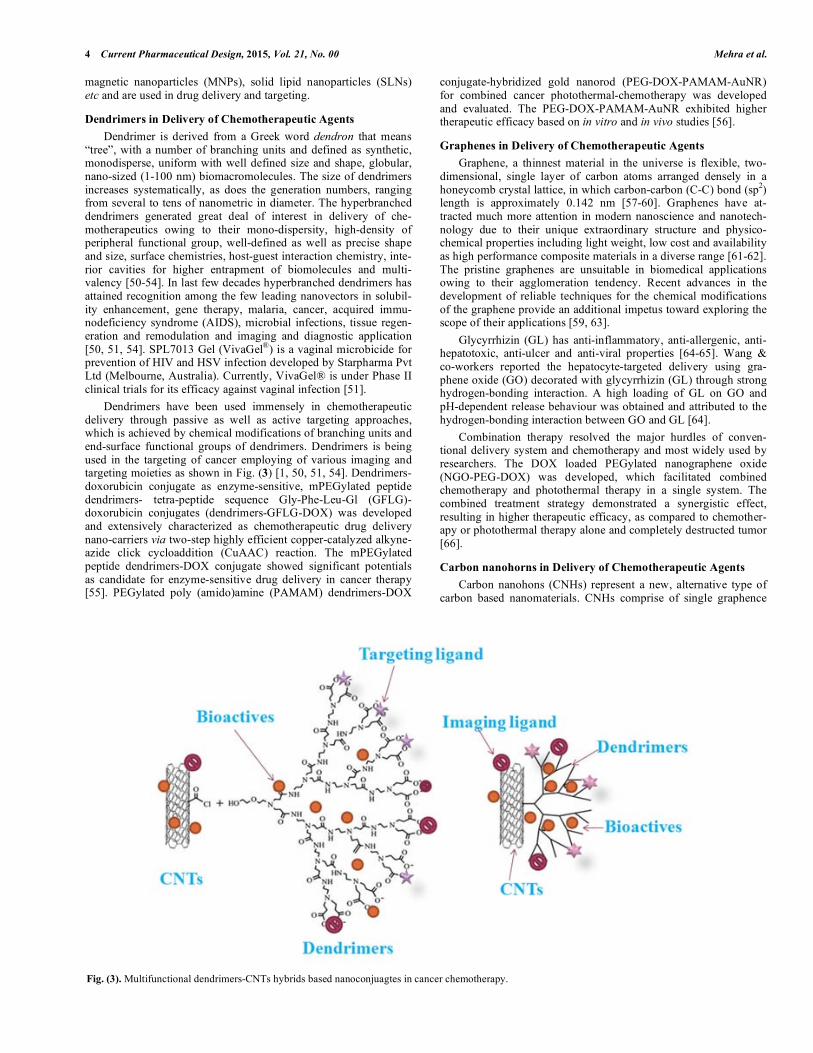
Dendrimer is derived from a Greek word dendron that means “tree”, with a number of branching units and defined as synthetic, monodisperse, uniform with well defined size and shape, globular, nano-sized (1-100 nm) biomacromolecules. The size of dendrimers increases systematically, as does the generation numbers, ranging from several to tens of nanometric in diameter. The hyperbranched dendrimers generated great deal of interest in delivery of che-motherapeutics owing to their mono-dispersity, high-density of peripheral functional group, well-defined as well as precise shape and size, surface chemistries, host-guest interaction chemistry, inte-rior cavities for higher entrapment of biomolecules and multi-valency [50-54]. In last few decades hyperbranched dendrimers has attained recognition among the few leading nanovectors in solubil-ity enhancement, gene therapy, malaria, cancer, acquired immu-nodeficiency syndrome (AIDS), microbial infections, tissue regen-eration and remodulation and imaging and diagnostic application [50, 51, 54]. SPL7013 Gel (VivaGel®) is a vaginal microbicide for prevention of HIV and HSV infection developed by Starpharma Pvt Ltd (Melbourne, Australia). Currently, VivaGel® is under Phase II clinical trials for its efficacy against vaginal infection [51].
Dendrimers have been used immensely in chemotherapeutic delivery through passive as well as active targeting approaches, which is achieved by chemical modifications of branching units and end-surface functional groups of dendrimers. Dendrimers is being used in the targeting of cancer employing of various imaging and targeting moieties as shown in Fig. (3) [1, 50, 51, 54]. Dendrimers-doxorubicin conjugate as enzyme-sensitive, mPEGylated peptide dendrimers- tetra-peptide sequence Gly-Phe-Leu-Gl (GFLG)-doxorubicin conjugates (dendrimers-GFLG-DOX) was developed and extensively characterized as chemotherapeutic drug delivery nano-carriers via two-step highly efficient copper-catalyzed alkyne-azide click cycloaddition (CuAAC) reaction. The mPEGylated peptide dendrimers-DOX conjugate showed significant potentials as candidate for enzyme-sensitive drug delivery in cancer therapy [55]. PEGylated poly (amido)amine (PAMAM) dendrimers-DOX
conjugate-hybridized gold nanorod (PEG-DOX-PAMAM-AuNR) for combined cancer photothermal-chemotherapy was developed and evaluated. The PEG-DOX-PAMAM-AuNR exhibited higher therapeutic efficacy based on in vitro and in vivo studies [56].
Graphenes in Delivery of Chemotherapeutic Agents
Graphene, a thinnest material in the universe is flexible, two-dimensional, single layer of carbon atoms arranged densely in a honeycomb crystal lattice, in which carbon-carbon (C-C) bond (sp2) length is approximately 0.142 nm [57-60]. Graphenes have at-tracted much more attention in modern nanoscience and nanotech-nology due to their unique extraordinary structure and physico-chemical properties including light weight, low cost and availability as high performance composite materials in a diverse range [61-62]. The pristine graphenes are unsuitable in biomedical applications owing to their agglomeration tendency. Recent advances in the development of reliable techniques for the chemical modifications of the graphene provide an additional impetus toward exploring the scope of their applications [59, 63].
Glycyrrhizin (GL) has anti-inflammatory, anti-allergenic, anti-hepatotoxic, anti-ulcer and anti-viral properties [64-65]. Wang & co-workers reported the hepatocyte-targeted delivery using gra-phene oxide (GO) decorated with glycyrrhizin (GL) through strong hydrogen-bonding interaction. A high loading of GL on GO and pH-dependent release behaviour was obtained and attributed to the hydrogen-bonding interaction between GO and GL [64].
Combination therapy resolved the major hurdles of conven-tional delivery system and chemotherapy and most widely used by researchers. The DOX loaded PEGylated nanographene oxide (NGO-PEG-DOX) was developed, which facilitated combined chemotherapy and photothermal therapy in a single system. The combined treatment strategy demonstrated a synergistic effect, resulting in higher therapeutic efficacy, as compared to chemother-apy or photothermal therapy alone and completely destructed tumor [66].
Carbon nanohorns in Delivery of Chemotherapeutic Agents
Carbon nanohons (CNHs) represent a new, alternative type of carbon based nanomaterials. CNHs comprise of single graphence
Fig. (3). Multifunctional dendrimers-CNTs hybrids based nanoconjuagtes in cancer chemotherapy.
Design of Multifunctional Nanocarriers for Delivery of Anti-Cancer Therapy Current Pharmaceutical Design, 2015, Vol. 21, No. 00 5
tubes with 205 nm in diameter and 40-50 nm in lengths. CNHs have an irregular, horn-like shape (conically-closed tip) [67-69] but are usually aggregate in assemblies that are reminiscent of dahlia flow-ers with a diameter that goes from 80 to 100 nm, although they can also form buds and seeds [69-70]. Single-walled carbon nanohorns (SWCNHs) are horn-shaped single-walled tubules with conical shape that have been widely studied for diverse applications includ-ing drug delivery, targeting and tissue engineering. SWCNHs do not exhibit cytotoxicity and hence may have potential application as drug carrier in delivery of chemotherapeutics [71]. CNHs are al-ready being used as delivery systems in nanomedicines. Sevral research reports demonstrated that the CNHs could easily deliver Cis-platin [72], Vincristine [73], and Doxorubicin [74] etc.
A recent report suggests that the targeted drug delivery system based on oxidized SWCNHs (ox-SWCNHs) were first modified non-covalently with sodium alginate (SA; natural polysaccharide) and then loaded with DOX, followed by binding with humanized anti-vascular endothelial growth factor (anti-VEGF) monoclonal antibody as targeting group (DOX@ox-SWCNHs/SA-mAb). The (DOX@ox-SWCNHs/SA-mAb) showed better anti-tumor activity on human breast adenocarcinoma (MCF-7) and human embryonic kidney 293 (HEK 293) cells devoid of any hepatotoxicity, car-diotoxicity and nephrotoxicity [75].
Very recently targeted killing of cancer cells using IGF-IR anti-body-directed oxidized carbon nanhorns based vincristine (VCR) delivery (VCR@ox-SWCNHs-PEG-mAb) was reported. The VCR@ox-SWCNHs-PEG-mAb showed higher antitumour efficacy with minimal side effecte to normal organs in in vivo tumor model in mice [72].
Quantum Dots (QDs) in Delivery of Chemotherapeutic Agents
QDs are also being explored as multifunctional nanomedicines for delivery of anticancer chemotherapeutic agents. In the last few decades, QDs have received considerable interest in the diagnosis, imaging and treatment of cancer and other diseases. QDs are the optical semiconducting in-organic nanomaterial comprising of ele-
ments from periodic group (II-VI) [76-78]. Their properties are ascribed due to their physical size, ranging from 10-100 Å in radius. QDs are 1-10 nm in size are credential to fluorescence under ligh source such as laser. QDs inherently possess numerous advantages over traditional fluorescent dyes such as increased photostability, higher brightness, and narrow fluorescence spectra [77, 79]. Till date various researchers and scientists have developed fluorescent nanoprobes and evaluated them on various animal models and rep-resent one of the fastest moving and exciting interfaces of nano-technology. QDs have unique optical properties, such as a broad absorption spectrum, narrow, size-tunable emission spectrum, high photostability, quantum efficiency, and strong non-linear response, from quantum confinement effects. The toxicity of QDs could be easily minimized by coating of PEG, polymers and other biomaterials, which make them more biocompatible and better candidate in cancer theragnostics [77, 79].
Ovarian cancer is one of the most common gynecological ma-lignancies in industrialized nations. Savla and co-workers [79] de-signed tumor-targeted pH-responsive QDs-mucin 1 aptamer-DOX (QD-MUC1-DOX) conjugate for chemotherapy of ovarian cancer, wherein DOX was attached to QDs via pH-sensitive hydrazone bond to provide greater stability in systemic circulation. The QDs-MUC1-DOX conjugate shows high potential in treatment of multidrug resistant ovarian cancer.
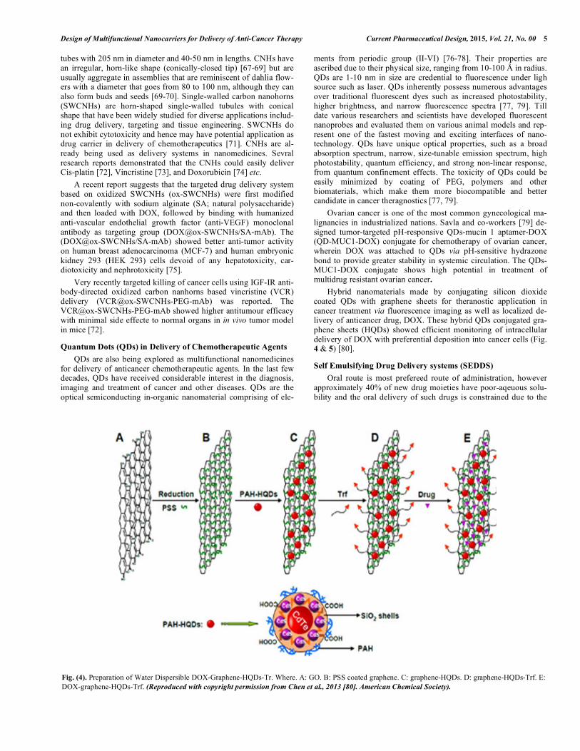
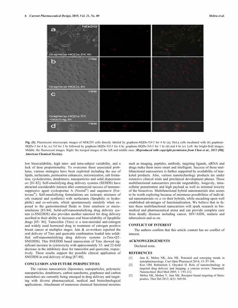
Hybrid nanomaterials made by conjugating silicon dioxide coated QDs with graphene sheets for theranostic application in cancer treatment via fluorescence imaging as well as localized de-livery of anticancer drug, DOX. These hybrid QDs conjugated gra-phene sheets (HQDs) showed efficient monitoring of intracellular delivery of DOX with preferential deposition into cancer cells (Fig. 4 & 5) [80].
Self Emulsifying Drug Delivery systems (SEDDS)
Oral route is most prefereed route of administration, however approximately 40% of new drug moieties have poor-aqeuous solu-bility and the oral delivery of such drugs is constrained due to the
Fig. (4). Preparation of Water Dispersible DOX-Graphene-HQDs-Tr. Where. A: GO. B: PSS coated graphene. C: graphene-HQDs. D: graphene-HQDs-Trf. E: DOX-graphene-HQDs-Trf. (Reproduced with copyright permission from Chen et al., 2013 [80]. American Chemical Society).
6 Current Pharmaceutical Design, 2015, Vol. 21, No. 00 Mehra et al.
low bioavailability, high inter- and intra-subject variability, and a lack of dose proportionality. To overcome these associated prob-lems, various strategies have been exploited including the use of lipids, surfactants, permeation enhancers, micronization, salt forma-tion, cyclodextrins, dendrimers, nanoparticles and solid dispersions etc [81-82]. Self-emulsifying drug delivery systems (SEDDS) have attracted considerable interest after commercial success of immuno-suppresive agent cyclosporine A (Neoral®) and saquinavir (For-tovase®). Self-emulsifiying formulations are isotropic mixtures of oils (natural and synthetic) with surfactants (lipophilic or hydro-philic) and co-solvents, which spontaneously emulsify when ex-posed to the gastrointestinal fluids to form emulsion or micro-emulsions [83-84]. Solid-self-nanoemulsifying drug delivery sys-tem (s-SNEDDS) also provides another nanotool for drug delivery ascribed to their ability to increases oral bioavailability of lipophilic drugs [85- 86]. Tamoxifen (Tmx) is a non-steroidal anti-estrogen and widely used hormonal drug in treatment of estrogen positive breast cancer at multiples stages. Jain & co-workers reported the oral delivery of Tmx and quercetin combination loaded into solidi-fied self-nanoemulsifying drug delivery systems (s-Tmx-QT-SNEDDS). This SNEDDS based nanosystem of Tmx showed sig-nificant increase in cytotoxicity with approximately 32- and 22-fold decrease in the inhibitory dose for tamoxifen and quercetin, respec-tively. These results support the possible clinical application of SNEDDS in oral delivery of drug [87-88].
CONCLUSION AND FUTURE PERSPECTIVES
The various nanocarriers (liposomes, nanoparticles, polymeric nanoparticles, dendrimers, carbon nanohorns, graphenes and carbon nanotubes) are currently being emerged in drug delivery and target-ing with diverse pharmaceutical, medical and biotechnological applications. Attachment of numerous chemical functional moieties
such as imaging, peptides, antibody, targeting ligands, siRNA and drugs make them more smart and intelligent. Success of these mul-tifunctional nanocarriers is further supported by availability of mar-keted products. Also, various nanotechnology products are under extensive clinical trials and preclinical development phases. These multifunctional nanocarriers provide targetability, longevity, intra-cellular penentration and high payload as well as minimal toxicity of the bioactives. Multifunctional hybrid nanomaterials also seems to be worth exploring because of enormous possibilities of individ-ual nanomaterials vis a vis their hybrids, while encashing upon well established advantages of functionalization. We believe that in fu-ture these multifunctional nanocarriers will spark research in bio-medical and pharmaceutical arena and can provide complete cure from deadly diseases including cancer, HIV/AIDS, malaria and tuberculosis and so on.
CONFLICT OF INTEREST
The authors confirm that this article content has no conflict of interest.
ACKNOWLEDGEMENTS
Declared none.
REFERENCES [1] Jain K, Mehra NK, Jain NK. Potential and emerging trends in
nanopharmacology. Curr Opin Pharmacol 2014; 15: 97-106. [2] Koo OM, Rubinstein I, Onyukel H. Role of nanotechnology in
targeted drug delivery and imaging: a concise review. Nanomed: Nanotechnol. Biol Med 2005; 1: 193-212.
[3] Mehra NK, Mishra V, Jain NK. Receptor based targeting of thera-peutics. Ther Del 2013; 4(3): 369-94.
Fig. (5). Fluorescent microscopic images of HEK293 cells directly labeled by graphene-HQDs-Trf-3 for 4 hr (a); HeLa cells incubated with (b) graphene-HQDs-3 for 4 hr, (c) Trf for 2 hr followed by graphene-HQDs-Trf-3 for 4 hr, graphene-HQDs-Trf-3 for 1 hr (d) and 4 hr (e). Left: the bright-field images. Middle: the fluorescent images. Right: the merged images of the left and middle ones. (Reproduced with copyright permission from Chen et al., 2013 [80],
American Chemical Society).
Design of Multifunctional Nanocarriers for Delivery of Anti-Cancer Therapy Current Pharmaceutical Design, 2015, Vol. 21, No. 00 7
[4] Mehra N K, Jain AK, Lodhi N, Dubey V, Mishra D, Jain NK. Challenges in the use of carbon nanotubes for biomedical application. Crit Rev Ther Drug Carr Sys 2008; 25(2): 169-206.
[5] Jain AK, Mehra NK, Lodhi N, et al. Carbon nanotubes and their toxicity. Nanotoxicology 2007; 1(3): 167-97.
[6] Jain NK, Mishra V, Mehra NK. Targeted drug delivery to macro-phages. Exp Opin Drug Deliv 2013; 10(3): 353-67.
[7] Steichen SD, Caldorera-Moore M, Peppas NA. A review of current nanoparticles and targeting moieties for the delivery of cancer therapeutics. Euro J Pharm Sci 2013; 48: 416-27.
[8] Mehra NK, Jain K, Jain NK. Pharmaceutical and biomedical appli-cation of surface engineered carbon nanotubes. Drug Discov Today 2015; 20(6): 750-9.
[9] Taratula O, Garbuzenko O, Savla R, Wang YA, He H, Minko T. Multifunctional nanomedicines platform for cancer specific deliv-ery of siRNA by superparamagnetic iron oxide nanoparticles-dendrimer complexes. Curr Drug Deliv 2011; 8(1): 59-69.
[10] Singh RK, Patel KD, Kim JJ, et al. Multifunctional hybrid nanocar-rier: magnetic CNTs ensheathed with mesoporous silica for drug delivery and imaging system. ACS Appl Mater Interfaces 2014; 6(4): 2201-8.
[11] Wen Y, Meng WS. Recent in vivo evidences of particle based delivery of small-interfering RNA (siRNA) into solid tumors. J Pharm Innov 2014; 9(2): 158-73.
[12] Iijima S. Helical microtubules of graphitic carbon. Nature 1991; 354: 56-8.
[13] Mehra NK, Jain NK. One platform comparison of estrone and folic acid anchored surface engineered MWCNTs for doxorubicin deliv-ery. Mol Pharm 2015 12(2): 630-43.
[14] Mehra NK, Mishra V, Jain NK. A review of ligand tethered surface engineered carbon nanotubes. Biomaterials 2014; 35(4): 1267-83.
[15] Lacerda L, Russier J, Pastorin G, et al. Translocation mechanisms of chemically functionalized carbon nanotubes across plasma membranes. Biomaterials 2012; 33: 3334-43.
[16] Fabbro C, Ali-Boucetta H, Da Ros T, Kostarelos K, Bianco A, Prato M. Targeting carbon nanotubes against cancer. Chem Comm 2012; 48: 3911-26.
[17] Mehra NK, Jain NK. Multifunctional hybrid-carbon nanotubes: new horizon in drug delivery and targeting. J Drug Target. 2015. (Epub ahead of print; doi: 10.3109/1061186X.2015.1055571).
[18] Mehra NK, Jain NK. Cancer targeting propensity of folate conjugated surface engineered multi-walled carbon nanotubes. Colloids and Surface B: Biointerfaces. 2015; 132: 17-26.
[19] Sobhani Z, Dinarvand R, Atyabi F, Ghahremani M, Adeli M. In-creased paclitaxel cytotoxicity against cancer cell lines using a novel functionalized carbon nanotubes. Int J Nanomed 2011; 6: 705-19.
[20] Liu Z, Sun X, Nakayama-Ratchford N, Dai H. Supramolecular chemistry on water-soluble carbon nanotubes for drug loading and delivery. ACS Nano 2007; 1(1): 50-6.
[21] Mehra NK, Jain NK. Development, characterization and cancer targeting potential of surface engineered carbon nanotubes. J Drug Target 2013; 21(8): 745-58.
[22] Al-Boucetta H, Al-Jamal KT, McCarthy D, Prato M, Bianco A, Kostarelos K. Multiwalled carbon nanotubes-doxorubicin su-pramolecular complexes for cancer therapeutics. Chem Commun 2008; 459-61.
[23] Datir SR, Das M, Singh RP, Jain S. Hyaluronate tethered smart multi walled carbon nanotubes for tumor-targeted delivery of doxorubicin. Bioconj Chem 2011; 23(11): 2201-13.
[24] Gu YJ, Cheng J, Jin J, Cheng SH, Wong WT. Development and evaluation of pH-responsive single-walled carbon nanotube-doxorubicin complexes in cancer cells. Int J Nanomed 2011; 6: 2889-98.
[25] Mehra NK, Verma AK, Mishra PR, Jain NK. The cancer targeting potential of D- -tocopheryl polyethylene glycol 1000 succinate tethered multi walled carbon nanotubes. Biomaterials 2014; 35: 4573-88.
[26] Huang H, Yuan Q, Shah JS, Misra RDK. A new family of folate-decorated and carbon nanotubes-mediated drug delivery system: synthesis and drug delivery response. Adv Drug Deliv Rev 2011; 63: 1332-9.
[27] Ji Z, Lin G, Lu Q, et al. Targeted therapy of SMMC-7721 liver cancer in vitro and in vivo with carbon nanotubes based drug deliv-ery systems. J Colloid and Interface Sci 2012; 365: 143-9.
[28] Lodhi N, Mehra NK, Jain NK. Development and characterization of dexamethasone mesylate anchored on multi walled carbon nano-tubes. J Drug Target 2013; 21(1): 67-76.
[29] Das M, Singh R P, Datir SR, Jain S. Intracellular drug delivery and effective in vivo cancer therapy via estradiol-PEGappended multi walled carbon nanotubes. Mol Pharm 2013; 10(9): 3404-16.
[30] Singh R, Mehra NK, Jain V, Jain NK. Folic acid conjugated carbon nanotubes for gemcitabine HCL delivery. J Drug Target 2013; 21(6): 581-92.
[31] Mao H, Kawazoe N, Chen G. Uptake and intracellular distribution of collagen-functionalized single-walled carbon nanotubes. Bioma-terials 2013; 34: 2472-9.
[32] Varkouhi AK, Foillard S, Lammers T, et al. siRNA delivery with functionalized carbon nanotubes. Int J Pharm 2011; 416: 419-25.
[33] Liu Z, Winters M, Holodniy M, Dai H. SiRNA delivery into human T cells and primary cells with carbon-nanotube transporters. Angew Chem Int Ed Engl 2007; 46: 2023-7.
[34] Al-Jamal KT, Toma FM, Yilmazer A, et al. Enhanced cellular internalization and gene silencing with a series of cationic Den-dron-multiwalled carbon nanotubes: siRNA complexes. FASEB J 2010; 24(11): 4354-65.
[35] Cheung W, Pntoriero F, Taratula O, Chen AM, He H. DNA and Carbon nanotubes as medicine. Adv Drug Deliv Rev 2010; 62(6): 633-49.
[36] Sousa S, Auriola S, Monkkonen J, Maatta J. Liposomes encapsu-lated zoledronate faours M1-like behaviour in murine macrophages cultured with soluble factors from breast cancer cells. BMC Cancer 2015; 15: 4
[37] Yuan A, Tang X, Qiu X, Jiang K, Wu J, Hu Y. Activatable pho-todynamic destruction of cancer cells by NIR dye/photosensitizer loaded liposomes. Chem Comm 2015; 51: 3340-2.
[38] Muthu MS, Kulkarni SA, Xiong J, Feng SS. Vitamin E TPGS coated liposomes enhanced cellular uptake and cytotoxicity of do-cetaxel in brain cancer cells. Int J Pharm 2011; 421: 332-40.
[39] Nahar M, Dutta T, Murugesan S, et al. Functional polymeric nanoparticles: an efficient and promising tool for active delivery of bioactives. Crit Rev Ther Drug Carr Sys 2008; 23(4): 259-318.
[40] Kaasgaard T, Andresen TL. Liposomal cancer therapy: exploiting tumor characeteristics. Exp Opin Drug Delivery 2010; 7(2): 225-43.
[41] Zhao L, Wei Y, Zhong X, et al. PK and tissue distribution of do-cetaxel in rabbits after i.v. administration of liposomal and in-jectable formulations. J Pharm Biomed Anal 2009; 49: 989-96.
[42] Bangham AD. Diffusion of univalent ions across unilamellar of swollen phospholipids. J Mol Biol 1965; 13: 238-52.
[43] Toh MR, Chiu GNC. Liposomes as sterile preparations and limita-tions of sterilization techniques in liposomal manufacturing. Asian J Pharm Sci 2013; 8(2): 88-95.
[44] Fan Y, Zhang Q. Development of liposomal formulations: from concept to clinical investigations. Asian J Pharm Sci 2013; 8(2): 81-7.
[45] Kaminskas LM, McLeoad VM, Kelly BD, et al. A comparison of changes to doxorubicin pharmacokinetics, antitumor activity, and toxicity mediated by PEGylated dendrimers and PEGylated liposomes drug delivery systems. Nanomed: Nanotechnol Biol Med 2012; 8(1): 103-11.
[46] Chang HI, Yeh MK. Clinical development of liposomes-based drugs: formulation, characterization, and therapeutic efficacy. Int J Nanomed 2012; 7: 49-60.
[47] Gao H, Zhang Q, Yu Z, He Q. Cell-penetrating peptide-based intelli-gent liposomal systems for enhanced drug delivery. Curr Pharm Biotechnol. 2014; 15: 210-9.
[48] Liu Y, Li K, Pan J, Liu B, Feng SS. Folic acid conjugated nanopar-ticles of mixed lipid monolayer shell and biodegradable polymer core for targeted delivery of docetaxel. Biomaterials 2010; 31: 330-8.
[49] Xu Z, Chen L, Gu W, et al. The performance of docetaxel-loaded solid lipid nanoparticles targeted to hepatocellualr carcinoma. Bio-materials 2009; 30: 226-32.
[50] Agrawal U, Mehra NK, Gupta U, Jain NK. Hyperbranched den-dritic nano-carriers for topical delivery of dithranol. J Drug Target 2013; 21(5): 497-506.
[51] Kesharwani P, Jain K, Jain NK. Dendrimer as nanocarrier for drug delivery. Prog Polym Sci 2014; 39: 268-307.

8 Current Pharmaceutical Design, 2015, Vol. 21, No. 00 Mehra et al.
[52] Kesharwani P, Tekade RK, Jain NK. Dendrimer generational no-menclature: the need to harmonize. Drug Discov Today 2015; 20(5): 497-9.
[53] Wu LP, Ficker M, Christensen JB, Trohopoulos PN, Moghimi SM. Dendrimers in medicine: Therapeutic concepts and pharmaceutical challenges. Biconj Chem 2015; 26(7): 1198-211.
[54] Jain K, Verma A, Mishra PR, Jain NK. Characterization and evaluation of amphotericin B loaded MDP conjugated poly(propylene imine) dendrimers. Nanomed: Nanotechnol Biol Med 2015; 11(3): 705-13.
[55] Zhang C, Pan D, Luo K, et al. Dendrimer-doxorubicin conjugate as enzyme-sensitive and polymeric nanoscale drug delivery vehicle of ovarian cancer therapy. Polym Chem 2014; 5: 5227-35.
[56] Li X, Takashima M, Yuba E, Harada A, Kono K. PEGylated PAMAM dendrimers-doxorubicin conjugate-hybridized gold nano-rod for combined photothermal-chemotherapy. Biomaterials 2014; 35(24): 6576-84.
[57] Ku SH, Park CB. Myoblast differentiation on graphene oxide. Biomaterials 2013; 34: 2017-23.
[58] Chowdhury SM, Lalwani G, Zhang K, Yang JY, Neville K, Sitharaman B. Cell specific cytotoxicity and uptake of graphene nanoribbons. Biomaterials 2013; 34: 283-93.
[59] Shen H, Zhang L, Liu M, Zhang Z. Biomedical applications of graphene. Theranostics 2012; 2(3): 283-94.
[60] Batzil M. The surface science of graphene: metal interfaces, CVD synthesis, nanoribbons, chemical modifications and defects. Surf Sci Reports 2012; 67: 83-115.
[61] Depan D, Shah J, Misra RDK. Controlled release of drug from folate-decorated and graphene mediated drug delivery system: syn-thesis, loading efficiency and drug release response. Mat Sci Eng C 2011; 31: 1305-12.
[62] Liu J, Guo S, Han L, Ren W, Liu Y, Wang E. Multiple pH-responsive graphene composites by non-covalent modification with chitosan. Talanta 2012; 101: 151-6.
[63] Tinchev SS, Surface modification of diamond-like carbon films to graphene under low energy ion beam irradiation. App Surf Sci 2012; 258: 2931-4.
[64] Wang Z, Gao Y, Xia J, Zhang F, Xia Y, Li Y. Synthesis and char-acterization of glycyrrhizin-decorated graphene oxide for hepato-cyte-targeted delivery. C R Chimie 2012; 15: 708-13.
[65] Chopdey PK, Tekade RK, Mehra NK, Mody N, Jain NK. Glycyrrhizin conjugated dendrimer and multi-walled carbon nanotubes for liver specific delivery of doxorubicin. J Nanosci Nanotechnol 2015; 15(2): 1088-100.
[66] Zhang W, Guo Z, Huang D, Liu Z, Guo X, Zhong H. Synergistic effect of chemo-photothermal therapy using PEGylated graphene oxide. Biomaterials 2011; 32: 8555-61.
[67] Zhang M, Yang M, Bussy C, Iijima S, Kostarelos K, Yudasaka M. Biodegradation of carbon nanohorns in macrophage cells. Nanoscale 2015; 7(7): 2834-40.
[68] Ajima K, Yudasaka M, Murakami T, Maigne A, Shiba K, Iijima S. Carbon nanohorns as anticancer drugs carriers. Mol Pharm 2005; 2(6): 475-80.
[69] Guerra J, Herrero MA, Carrion B, et al. Carbon nanohorns func-tionalized with polyamidoamine dendrimers as efficient biocarrier materials for gene therapy. Carbon 2012; 50: 2832-44.
[70] Whitney JR, Sarkar S, Zhang J, et al. Single walled carbon nano-horns as photothermal cancer agents. Lasers Surg Med 2011; 43(1): 43-51.
[71] Zhu S, Xu G. Single-walled carbon nanohorns and their applica-tions. Nanoscale 2010; 2: 2538-49.
[72] Dewitt MR, Pekkanen AM, Robertson J, RYlander CG, Nichole Rylander M. Influence of hyperthermia on efficacy and uptake of carbon nanohorns-cisplain conjugates. J Biomech Eng 2014; 136(2): 021003.
[73] Li N, Zhao Q, Shu C, et al. Targeted killing of cancer cells in vivo and in vitro with IGF-IR antibody-directed carbon nanohorns based drug delivery. Int J Pharm 2015; 478(2): 644-54.
[74] Murakami T, Fan J, Yudasaka M, Iijima S, Shiba K. Solubilization of single-wall carbon nanohorns using a PEG-doxorubicin conju-gates. Mol Pharm 2006; 3(4): 407-14.
[75] Ma X, Shu C, Guo J, et al. Targeted cancer therapy based on sin-gle-wall carbon nanohorns with doxorubicin in vitro and in vivo. J Nanopart Res 2014; 16: 2497.
[76] Mishra RDK. Quantum dots for tumor-targeted drug delivery and cell imaging. Nanomed 2008; 3(3): 271-4.
[77] Chakravarthy KV, Davidson BA, Helinski JD, D et al. Doxorub-cin-conjugated quantum dots to target alveolar macrophages and inflammation. Nanomed: nanotechnol. Biol Med 2011; 7: 88-96.
[78] Qi L, Gao X. Emerging application of quantum dots for drug deliv-ery and therapy. Exp Opin Drug Deliv 2008; 5(3): 263-7.
[79] Savla R, Taratula O, Garbuzenko O, Minko T. Tumor targeted quantum dot-mucin 1 aptamer-doxorubicin conjugate for imaging and treatment of cancer. J Control Rel 2011; 153: 16-22.
[80] Chen ML, He YJ, Chen XW, Wang JH. Quantum-Dot-conjugated graphene as a probe for simultaneous cancer-targeted fluorescent imaging, tracking and monitoring drug delivery. Bioconj Chem 2013; 24: 387-97.
[81] Zhang L, Zhu W, Yang C, et al. A novel folate-modified self-microemulsifying drug delivery system of curcumin for colon tar-geting. Int J Nanomed 2012; 7: 151-62.
[82] Nipun TS, Islam SMA. SEDDS of gliclazide: preparation and characterization by in vitro, ex-vivo and in-vivo techniques. Saudi Pharm J 2014; 22(4): 343-8.
[83] Kohli K, Chopra S, Dhar D, Arora S, Khar RK. Self-emulsifying drug delivery systems: an approach to enhance bioavailability. Drug Discovery Today 2010; 15(21-22): 958-65.
[84] Wei Y, Ye X, Shang X, et al. Enhanced oral bioavailability of silybin by a supersaturable self-emulsifying drug delivery system (s-SEDDS). Colloids and Surface A: Physicochemical and Eng As-pects 2012; 396: 22-8
[85] Gursoy RN, Benita S. Self-emulsifying drug delivery systes (SEDDS) for improved oral delivery of lipophilic drugs. Biomed & Pharmacotherpay 2004; 58(3): 173-82.
[86] Jain AK, Thanki K, Jain S. Novel self-nanoemulsifying formula-tion of quercetin: Implications of pro-oxidant activity on the anti-cancer efficacy. Nanomed: Nanotechnol Biol Med 2014; 10: 959-69.
[87] Jain AK, Thanki K, Jain S. Solidified self-nanoemulsifying formu-lation for oral delivery of combinatorial therapeutic regimen: part I formulation development, statistical optimization and in vitro char-acterization. Pharm Res 2014; 31: 923-45.
[88] Jain AK, Thanki K, Jain S. Solidified self-nanoemulsifying formu-lation for oral delivery of combinatorial therapeutic regimen: part II in vivo pharmacokinetcis, antitumor efficacy and hepatotoxicity. Pharm Res 2014; 31(4): 946-58.
Received: August 14, 2015 Accepted: October 26, 2015