Embed Size (px)
Citation preview
45 CyanosisH. L. SNIDER
Definition
Cyanosis is a bluish color of mucous membranes and/or skin .While this is most frequently attributable to increasedamounts of unoxygenated hemoglobin (deoxyhemoglobin)in the vasculature, there are other causes of bluish skincolor .
Technique
Daylight or artificial light sources simulating daylight's spec-tral composition are not necessarily optimal for detectingcyanosis . Nevertheless, consistency in observation makes itdesirable that artificial light sources be similar to sunlight,since most examining areas are lighted for at least a portionof the day by sunlight . Tungsten filament bulbs and certainfluorescent bulbs are satisfactory for this purpose . If one isuncertain as to the adequacy of an artificial light source,use of sunlight will obviate this potential problem . An in-tense light can make cyanosis less readily apparent . Onegroup has recommended using less than 20 footcandles ofillumination . As a point of reference, the standard sug-gested for patient rooms in Veterans Administration hos-pitals is 30 footcandles, so the recommended level of lightintensity to detect cyanosis is likely to be exceeded in at leastsome patient examination areas . When looking for cyanosis,one should inspect those body sites that contain minimalmelanotic pigment, that have a capillary bed close to theskin surface, and that are well perfused . Lips, ears, trunk,nailbed, hands, conjunctiva, and circumoral areas have beencompared in detecting cyanosis due to arterial hypoxemia ;the tongue is the most sensitive area, but the lips are morespecific .
Basic Science
Blue color can be perceived in a number of situations :(1) when the light source directly shined on the retina hasa predominant frequency in the upper (shorter) end of thevisual spectrum ; (2) when a light source with multiple fre-quencies (including high ones) is shined on an object, ab-sorbing all other frequencies except those at the blue endof the visual spectrum, which are reflected to the retina ;and (3) when a white light is scattered by particles, thefrequencies reflected are in the high end of the visual spec-trum (Tyndall effect)-the blue sky is an example of this .
The normal color of flesh is thought to result from thecombination of the pigments oxyhemoglobin, deoxyhemog
lobin, melanin, and carotene, and from the optical effectof scattering. The importance of the latter effect has beendisputed by at least one investigator, who attributes to col-lagen a major role in reflecting blue wavelengths . Blue skincoloration would result if the quantity of blue wavelengths
236
reflected disproportionately increased or if the quantity ofother wavelengths reflected disproportionately decreased .
Anyone who has observed a specimen of venous bloodin a tube can confirm that it is not blue . Thus the blue skincolor detected in individuals who have increased amountsof deoxyhemoglobin cannot be explained on the basis ofreflection of increased quantities of high-frequency wave-lengths from a "blue" pigment . One plausible theory toaccount for the observation of cyanosis under these circum-stances is that deoxyhemoglobin is less red than oxyhemog lobin and therefore absorbs more red spectrum. By
subtraction of red wavelengths, the blue spectrum is allowedto predominate in the reflected light (i .e., something thatis less red is more blue) . The bluish skin color observed withthe other pigments listed in Table 45 .1 is explained in asimilar fashion .
According to Lundsgaard and Van Slyke (1923), as wellas subsequent investigators, cyanosis generally becomes ap-parent when the subpapillary capillaries contain from 4 to6 gm/dl of deoxyhemoglobin . Since this measurement wasdifficult to obtain directly, they proposed estimating it byaveraging the amount of deoxyhemoglobin in arterial bloodwith that in venous blood . If one assumes a normal cardiacoutput, hemoglobin, and tissue extraction of 0 2 , an arterial02 saturation of approximately 80% would be required to
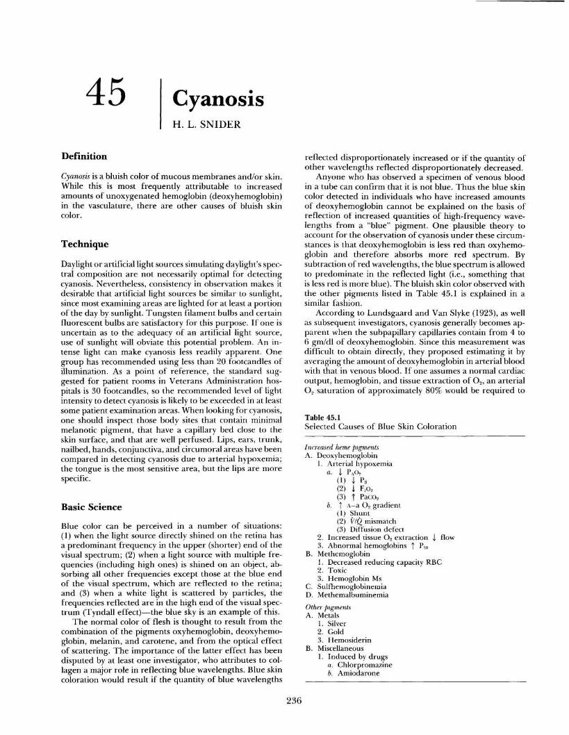
Table 45 .1Selected Causes of Blue Skin Coloration
IncreasedA. Deoxyhemoglobin
heme pigments
1 . Arterial hypoxemiaa . I
(1)P AO2
P B(2) j F,o 2(3) T Paco2
b. T(1)
A-a 0 2 gradientShunt
(2) V/Q mismatch(3) Diffusion defect
2 . Increased tissue 02 extraction .3 . Abnormal hemoglobins T Pyo
flow
B. Methemoglobin1 . Decreased reducing capacity RBC2 . Toxic3 . Hemoglobin Ms
C. SulfhemoglobinemiaD. MethemalbuminemiaOther pigmentsA. Metals
1 . Silver2. Gold3 . Hemosiderin
B. Miscellaneous1 . Induced by drugs
a . Chlorpromazineb. Amiodarone

45 . CYANOSIS
237
cause cyanosis . It should be noted that the conclusion ofLundsgaard and Van Slyke was based on measurements ofdeoxyhemoglobin in peripheral venous blood and did notinvolve sampling of arterial blood . Their proposal of 5 gm/dI deoxyhemoglobin in mean capillary blood as a thresholdfor detecting cyanosis has not been confirmed or refutedby more sophisticated techniques .
Reduced arterial oxygenation can result if the amountof oxygen in the alveoli is lowered or if the gradient betweenthe alveolar oxygen and the arterial oxygen is elevated . Onecan determine which of these is the explanation by meas-uring the arterial partial pressure of oxygen (Pao 2) andcalculating the alveolar partial pressure of oxygen (P Ao 2)and the A-a 02 gradient with the following formulas :
Even with normal arterial oxygenation, cyanosis can oc-cur when there is increased extraction of oxygen at thecapillary level because the average of arterial and venousoxygen saturation will be lower . Reduced flow through cap-illaries results in increased tissue extraction of oxygen (andtherefore increased amounts of deoxyhemoglobin), favor-ing the appearance of cyanosis .
In anemic patients, much more profound decreases intissue oxygen levels are required to produce 5 gm/dl ofdeoxyhemoglobin in capillary blood . For example, with ahemoglobin of 7 .5 gm/dl, capillary blood would have tohave a Poe of about 19 mm Hg (33% sat .), contrasted witha Po e of about 35 mm Hg (66% sat.) for a hemoglobin of15 gm/dl .
Hemoglobins that have an abnormally low affinity foroxygen (high P50 ) have decreased amounts of hemoglobinbound with oxygen at usual levels of Pao 2 . Cyanosis canresult on occasion .
A tube of blood containing excess methemoglobin is red-dish brown to brown in color and remains so even aftershaking in air or 100% 0 2. Methemoglobin is an oxidizedhemoglobin in which iron is in the ferric form . It does notbind oxygen. Some methemoglobin is normally formed inthe body, but this is usually reduced to deoxyhemoglobinby the NADH methemoglobin reductase system. If this en-zyme system is deficient or if it becomes overloaded byexcess amounts of methemoglobin, elevated blood levels ofmethemoglobin result. In some patients with congenitallyabnormal hemoglobins (Hgb Ms) the structure of the hemo-globin makes the heme unit susceptible to rapid oxidation .The level of methemoglobin capable of producing cyanosisis said to be about 1 .5 gm/dl, although this value seems tohave been less carefully scrutinized than that for deoxy-hemoglobin .
As with methemoglobin, a tube of blood containing suf-ficient sulfhemoglobin has a reddish brown color that doesnot change upon shaking in 100% 0 2. Sulfhemoglobin is a
pigment not normally formed in the body . Its chemicalcomposition is not well defined, although it has the spec-trophotometric characteristic of strongly absorbing light at620 nm in the presence of cyanide . The mechanism of for-mation is not known, although many of the same toxins thatresult in the oxidation of deoxyhemoglobin to methemo-globin can also produce sulfhemoglobin . The explanationfor the formation of sulfhemoglobin in one individual andmethemoglobin in another exposed to the same toxin is notknown. Once formed, the sulfhemoglobin molecule is stableand is not converted back to deoxyhemoglobin . Cyanosis isreported to be detectable at sulfhemoglobin levels as low as0.5 gm/dl .
Methemalbumin, which produces a brown plasma, is apigment formed by the union of albumin in the plasma withhemin. The pigment may be present in the blood whenexcessive breakdown of red cells results in saturation ofhaptoglobin with hemoglobin . Dissolution of the remainingfree hemoglobin into globin and heme can occur . Heme isimmediately oxidized to hematin and in the presence ofchloride forms hemin, which complexes with albumin . Theminimal amount of resulting methemalbumin required toproduce cyanosis is not stated in the literature .
Clinical Significance
Cyanosis as a tool for detecting arterial hypoxemia is neithersensitive nor specific . Comroe and Botelho (1947) studieda group of normal subjects breathing various concentrationsof oxygen . Definite cyanosis was not apparent to 25% ofobservers even at arterial oxygen saturations of 71 to 75%(Pao2 35 to 40 mm Hg) . In contrast, 6% and 17% of theobservers believed definite cyanosis to be present when ar-terial oxygen saturations were 96 to 100% and 91 to 95%,respectively. In the same study progressive hypoxemia wasinduced in a subject and one physician first noted definitecyanosis at arterial oxygen saturations of 84%, 77%, 94%,and 82% in consecutive trials on this same subject within aperiod of 40 minutes . The sensitivity of this sign is lessenedwhen examining deeply pigmented individuals . In blacks,3 to 6% more arterial oxygen desaturation may be requiredfor detection of cyanosis .
To confirm that arterial hypoxemia is responsible for cy-anosis, a blood specimen must be analyzed for Pao2. Whena reduction is found, one must consider the causes listed inTable 45.2. When cyanosis is due to arterial hypoxemia,other signs and symptoms are usually present. Peripheralchemoreceptors may be stimulated by a low Pao 2, causingincreased ventilation with dyspnea and tachypnea . Sym-pathetic nervous system stimulation produces restlessness,sweating, elevation of blood pressure, and tachycardia . Whenhypoxemia is severe and cerebral oxygenation is impaired,confusion or coma can occur. As demonstrated in a numberof studies, severe hypoxemia may be present at times whencyanosis is not readily detectable either because of observerinsensitivity or confounding factors in the patient, such asheavy melanin pigmentation or anemia . The importance ofarterial blood gas analysis in detecting hypoxemia cannotbe overemphasized .
The usual pattern of cyanosis noted in conditions ofreduced blood flow is for peripheral sites, in particular theextremities, to be affected preferentially (acrocyanosis) .Central portions of the body are typically spared . Low flowmay result from decreased arterial perfusion caused by poorcardiac output (as in cardiogenic shock), by fixed arterial
PAo2 = (PB - PH2o37°) F,o2 - PACO2 X RA-a 0 2 gradient = PAo2 - Pao2
wherePB = barometric pressure
PH 2o37° = partial pressure water vapor at 37 °C (47 mmHg)
F,o 2 = fraction of inspired air that is oxygenPaco2 = partial pressure of carbon dioxide in arterial
bloodR = respiratory quotient (Vco 2/Vo2, generally
about 0.8)
238
III . THE PULMONARY SYSTEM
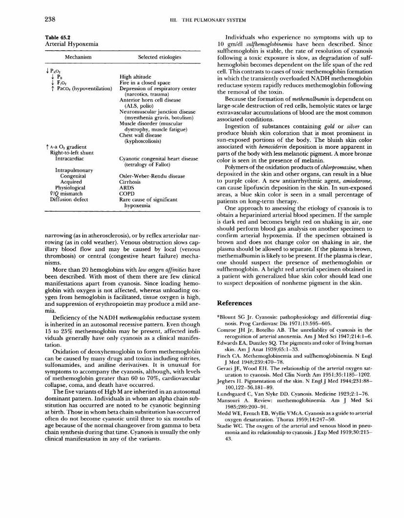
Table 45 .2Arterial Hypoxemia
narrowing (as in atherosclerosis), or by reflex arteriolar nar-rowing (as in cold weather) . Venous obstruction slows cap-illary blood flow and may be caused by local (venousthrombosis) or central (congestive heart failure) mecha-nisms .
More than 20 hemoglobins with low oxygen affinities havebeen described . With most of them there are few clinicalmanifestations apart from cyanosis . Since loading hemo-globin with oxygen is not affected, whereas unloading ox-ygen from hemoglobin is facilitated, tissue oxygen is high,and suppression of erythropoietin may produce a mild ane-mia .
Deficiency of the NADH methemoglobin reductase systemis inherited in an autosomal recessive pattern. Even though15 to 25% methemoglobin may be present, affected indi-viduals generally have only cyanosis as a clinical manifes-tation .
Oxidation of deoxyhemoglobin to form methemoglobincan be caused by many drugs and toxins including nitrites,sulfonamides, and aniline derivatives . It is unusual forsymptoms to accompany the cyanosis, although, with levelsof methemoglobin greater than 60 to 70%, cardiovascularcollapse, coma, and death have occurred .
The five variants of Hgb M are inherited in an autosomaldominant pattern. Individuals in whom an alpha chain sub-stitution has occurred are noted to be cyanotic beginningat birth. Those in whom beta chain substitution has occurredoften do not become cyanotic until three to six months ofage because of the normal changeover from gamma to betachain synthesis during that time . Cyanosis is usually the onlyclinical manifestation in any of the variants .
Individuals who experience no symptoms with up to10 gm/dl sulfhemoglobinemia have been described . Sincesulfhemoglobin is stable, the rate of resolution of cyanosisfollowing a toxic exposure is slow, as degradation of sulf-hemoglobin becomes dependent on the life span of the redcell . This contrasts to cases of toxic methemoglobin formationin which the transiently overloaded NADH methemoglobinreductase system rapidly reduces methemoglobin followingthe removal of the toxin .
Because the formation of methemalbumin is dependent onlarge-scale destruction of red cells, hemolytic states or largeextravascular accumulations of blood are the most commonassociated conditions .
Ingestion of substances containing gold or silver canproduce bluish skin coloration that is most prominent insun-exposed portions of the body . The bluish skin colorassociated with hemosiderin deposition is more apparent inparts of the body with less melanotic pigment . A more bronzecolor is seen in the presence of melanin .
Polymers of the oxidation products of chlorpromazine, whendeposited in the skin and other organs, can result in a blueto purple color. A new antiarrhythmic agent, amiodarone,can cause lipofuscin deposition in the skin. In sun-exposedareas, a blue skin color is seen in a small percentage ofpatients on long-term therapy .
One approach to assessing the etiology of cyanosis is toobtain a heparinized arterial blood specimen . If the sampleis dark red and becomes bright red on shaking in air, oneshould perform blood gas analysis on another specimen toconfirm arterial hypoxemia. If the specimen obtained isbrown and does not change color on shaking in air, theplasma should be allowed to separate. If the plasma is brown,methemalbumin is likely to be present . If the plasma is clear,one should suspect the presence of methemoglobin orsulfhemoglobin. A bright red arterial specimen obtained ina patient with generalized blue skin color should lead oneto suspect deposition of nonheme pigment in the skin .
References
*Blount SG Jr . Cyanosis : pathophysiology and differential diag-nosis. Prog Cardiovasc Dis 1971 ;13:595-605 .
Comroe JH Jr, Botelho AB . The unreliablity of cyanosis in therecognition of arterial anoxemia . Am J Med Sci 1947;214:1-6.
Edwards EA, Duntley SQ . The pigments and color of living humanskin. Am J Anat 1939;65 :1-33 .
Finch CA. Methemoglobinemia and sulfhemoglobinemia. N EnglJ Med 1948;239:470-78 .
Geraci JE, Wood EH . The relationship of the arterial oxygen sat-uration to cyanosis. Med Clin North Am 1951 ;35:1185-1202 .
Jeghers H . Pigmentation of the skin . N Engl J Med 1944;231 :88-100,122-36,181-89.
Lundsgaard C, Van Slyke DD. Cyanosis . Medicine 1923 ;2:1-76 .Mansouri A. Review: methemoglobinemia. Am J Med Sci
1985;289:200-91 .Medd WE, French EB, Wyllie VMcA . Cyanosis as a guide to arterial
oxygen desaturation . Thorax 1959;14:247-50 .Stadie WC . The oxygen of the arterial and venous blood in pneu-
monia and its relationship to cyanosis. J Exp Med 1919;30:215-43 .
Mechanism Selected etiologies
PAo2J PB High altitude
FIo2 Fire in a closed spaceT PaCO 2 (hypoventilation)
T A-a 02 gradientRight-to-left shunt
Depression of respiratory center(narcotics, trauma)
Anterior horn cell disease(ALS, polio)
Neuromuscular junction disease(myesthenia gravis, botulism)
Muscle disorder (musculardystrophy, muscle fatigue)
Chest wall disease(kyphoscoliosis)
Intracardiac
Intrapulmonary
Cyanotic congenital heart disease(tetralogy of Fallot)
Congenital Osler-Weber-Rendu diseaseAcquired Cirrhosis
Physiological ARDS//Q mismatch COPDDiffusion defect Rare cause of significant
hypoxemia


![A SIMPLE ACCURATE MULTI-COMPONENT … · The sulfhemoglobinemia is usually induced by various drugs such as sulphonamides, sulfasalazine and sumatriptan [19]. Also, it may occur due](https://img.pdfslide.net/doc/110x75/600deb57047e066e9c422afa/a-simple-accurate-multi-component-the-sulfhemoglobinemia-is-usually-induced-by-various.jpg)