Embed Size (px)
Citation preview
5/13/2018 47 Lecture Presentation PC 2012 (1) - slidepdf.com
http://slidepdf.com/reader/full/47-lecture-presentation-pc-2012-1 1/88
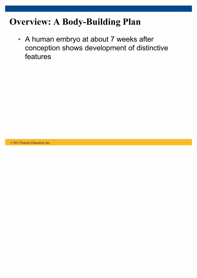
Overview: A Body-Building Plan
A human embryo at about 7 weeks after
conception shows development of distinctive
features
© 2011 Pearson Education, Inc.
5/13/2018 47 Lecture Presentation PC 2012 (1) - slidepdf.com
http://slidepdf.com/reader/full/47-lecture-presentation-pc-2012-1 2/88
Figure 47.1
1 mm
5/13/2018 47 Lecture Presentation PC 2012 (1) - slidepdf.com
http://slidepdf.com/reader/full/47-lecture-presentation-pc-2012-1 3/88
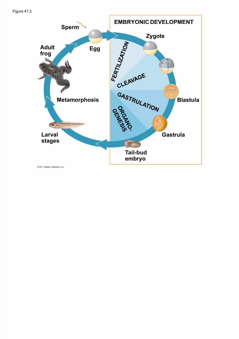
Development occurs at many points in the life
cycle of an animal
This includes metamorphosis and gamete
production, as well as embryonic development
© 2011 Pearson Education, Inc.
5/13/2018 47 Lecture Presentation PC 2012 (1) - slidepdf.com
http://slidepdf.com/reader/full/47-lecture-presentation-pc-2012-1 4/88
Figure 47.2
EMBRYONIC DEVELOPMENTSper m
Adultf rog
Egg
Metamorphosis
Larvalstages
Zygote
Blastula
Gastr ula
Tail-budembryo
5/13/2018 47 Lecture Presentation PC 2012 (1) - slidepdf.com
http://slidepdf.com/reader/full/47-lecture-presentation-pc-2012-1 5/88
Although animals display different body plans,
they share many basic mechanisms of
development and use a common set of
regulatory genes Biologists use model organisms to study
development, chosen for the ease with which
they can be studied in the laboratory
© 2011 Pearson Education, Inc.
5/13/2018 47 Lecture Presentation PC 2012 (1) - slidepdf.com
http://slidepdf.com/reader/full/47-lecture-presentation-pc-2012-1 6/88
Concept 47.1: Fertilization and cleavage
initiate embryonic development
Fer tilization is the formation of a diploid zygote
from a haploid egg and sperm
© 2011 Pearson Education, Inc.
5/13/2018 47 Lecture Presentation PC 2012 (1) - slidepdf.com
http://slidepdf.com/reader/full/47-lecture-presentation-pc-2012-1 7/88
Fertilization
Molecules and events at the egg surface play a
crucial role in each step of fertilization
± Sperm penetrate the protective layer around the
egg ± Receptors on the egg surface bind to molecules
on the sperm surface
± Changes at the egg surface prevent polyspermy,
the entry of multiple sperm nuclei into the egg
© 2011 Pearson Education, Inc.
5/13/2018 47 Lecture Presentation PC 2012 (1) - slidepdf.com
http://slidepdf.com/reader/full/47-lecture-presentation-pc-2012-1 8/88
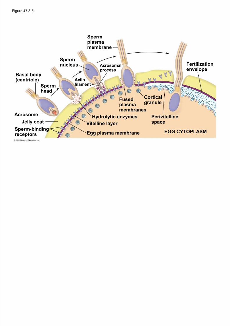
The Acrosomal Reaction
The acrosomal reaction is triggered when the
sperm meets the egg
The acrosome at the tip of the sperm releases
hydrolytic enzymes that digest materialsurrounding the egg
© 2011 Pearson Education, Inc.
5/13/2018 47 Lecture Presentation PC 2012 (1) - slidepdf.com
http://slidepdf.com/reader/full/47-lecture-presentation-pc-2012-1 9/88
Figure 47.3-5
Basal body
(centriole)
Sper mplasmamembr ane
Sper mnucleus
Sper mhead
Acrosome
Jelly coat
Sper m-bindingreceptors
Fer tilizationenvelope
Cor ticalgr anule
Fusedplasmamembr anes
Hydrolytic enzymes
Vitelline layer
Egg plasma membr ane
Perivitelline
space
EGG CYTOPLASM
Actinf ilament
Acrosomalprocess
5/13/2018 47 Lecture Presentation PC 2012 (1) - slidepdf.com
http://slidepdf.com/reader/full/47-lecture-presentation-pc-2012-1 10/88
Gamete contact and/or fusion depolarizes the egg
cell membrane and sets up a fast block to
polysper my
© 2011 Pearson Education, Inc.
5/13/2018 47 Lecture Presentation PC 2012 (1) - slidepdf.com
http://slidepdf.com/reader/full/47-lecture-presentation-pc-2012-1 11/88
The Cortical Reaction
Fusion of egg and sperm also initiates the cor tical
reaction
Seconds after the sperm binds to the egg, vesicles
just beneath the egg plasma membrane releasetheir contents and form a fertilization envelope
The fertilization envelope acts as the slow block
to polysper my
© 2011 Pearson Education, Inc.
5/13/2018 47 Lecture Presentation PC 2012 (1) - slidepdf.com
http://slidepdf.com/reader/full/47-lecture-presentation-pc-2012-1 12/88
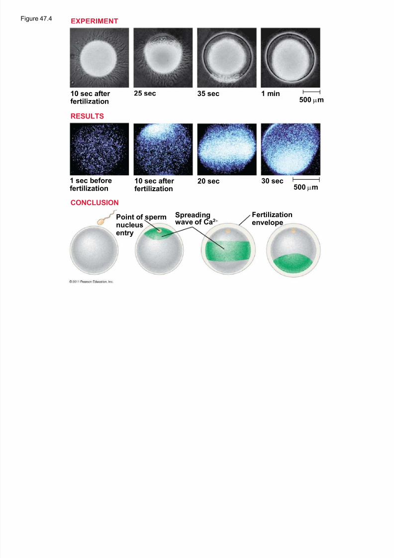
The cortical reaction requires a high concentration
of Ca2 ions in the egg
The reaction is triggered by a change in Ca2
concentration Ca2 spread across the egg correlates with the
appearance of the fertilization envelope
© 2011 Pearson Education, Inc.
5/13/2018 47 Lecture Presentation PC 2012 (1) - slidepdf.com
http://slidepdf.com/reader/full/47-lecture-presentation-pc-2012-1 13/88
Figure 47.4
10 sec after f er tilization
25 sec 35 sec 1 min500 Qm
500 Qm30 sec20 sec10 sec after
f er tilization1 sec bef oref er tilization
Point of sper mnucleusentry
Spreadingwave of Ca2
Fer tilizationenvelope
EXPERIMENT
RESULTS
CONCLUSION
5/13/2018 47 Lecture Presentation PC 2012 (1) - slidepdf.com
http://slidepdf.com/reader/full/47-lecture-presentation-pc-2012-1 14/88
Egg Activation
The rise in Ca2+ in the cytosol increases the rates
of cellular respiration and protein synthesis by the
egg cell
With these rapid changes in metabolism, the eggis said to be activated
The proteins and mRNAs needed for activation
are already present in the egg
The sperm nucleus merges with the egg nucleusand cell division begins
© 2011 Pearson Education, Inc.

5/13/2018 47 Lecture Presentation PC 2012 (1) - slidepdf.com
http://slidepdf.com/reader/full/47-lecture-presentation-pc-2012-1 15/88
F ertilization in Mammals
Fertilization in mammals and other terrestrial
animals is internal
Secretions in the mammalian female reproductive
tract alter sperm motility and structure This is called capacitation and must occur before
sperm are able to fertilize an egg
© 2011 Pearson Education, Inc.
5/13/2018 47 Lecture Presentation PC 2012 (1) - slidepdf.com
http://slidepdf.com/reader/full/47-lecture-presentation-pc-2012-1 16/88
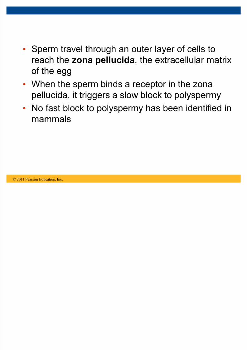
Sperm travel through an outer layer of cells to
reach the zona pellucida, the extracellular matrix
of the egg
When the sperm binds a receptor in the zonapellucida, it triggers a slow block to polyspermy
No fast block to polyspermy has been identified in
mammals
© 2011 Pearson Education, Inc.
5/13/2018 47 Lecture Presentation PC 2012 (1) - slidepdf.com
http://slidepdf.com/reader/full/47-lecture-presentation-pc-2012-1 17/88
Figure 47.5
Zona pellucida
Follicle cell
Sper mbasal body
Sper mnucleus
Cor ticalgr anules
5/13/2018 47 Lecture Presentation PC 2012 (1) - slidepdf.com
http://slidepdf.com/reader/full/47-lecture-presentation-pc-2012-1 18/88
In mammals the first cell division occurs 1236
hours after sperm binding
The diploid nucleus forms after this first division of
the zygote
© 2011 Pearson Education, Inc.

5/13/2018 47 Lecture Presentation PC 2012 (1) - slidepdf.com
http://slidepdf.com/reader/full/47-lecture-presentation-pc-2012-1 19/88
Cleavage
Fertilization is followed by cleavage, a period of
rapid cell division without growth
Cleavage partitions the cytoplasm of one large cell
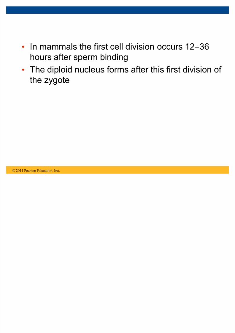
into many smaller cells called blastomeres The blastula is a ball of cells with a fluid-filled
cavity called a blastocoel
© 2011 Pearson Education, Inc.
5/13/2018 47 Lecture Presentation PC 2012 (1) - slidepdf.com
http://slidepdf.com/reader/full/47-lecture-presentation-pc-2012-1 20/88
Figure 47.6
(a) Fer tilized egg (b) Four -cell stage (c) Ear ly blastula (d) Later blastula
50 Qm
5/13/2018 47 Lecture Presentation PC 2012 (1) - slidepdf.com
http://slidepdf.com/reader/full/47-lecture-presentation-pc-2012-1 21/88
In frogs and many other animals, the distribution of
yolk (stored nutrients) is a key factor influencing
the pattern of cleavage
The vegetal pole has more yolk; the animal pole has less yolk
The difference in yolk distribution results in animal
and vegetal hemispheres that differ in appearance
© 2011 Pearson Education, Inc.
Cleava g e Patterns
5/13/2018 47 Lecture Presentation PC 2012 (1) - slidepdf.com
http://slidepdf.com/reader/full/47-lecture-presentation-pc-2012-1 22/88
The first two cleavage furrows in the frog form
four equally sized blastomeres
The third cleavage is asymmetric, forming
unequally sized blastomeres
© 2011 Pearson Education, Inc.
5/13/2018 47 Lecture Presentation PC 2012 (1) - slidepdf.com
http://slidepdf.com/reader/full/47-lecture-presentation-pc-2012-1 23/88
Holoblastic cleavage, complete division of the
egg, occurs in species whose eggs have little or
moderate amounts of yolk, such as sea urchins
and frogs
Meroblastic cleavage, incomplete division of the
egg, occurs in species with yolk-rich eggs, such as
reptiles and birds
© 2011 Pearson Education, Inc.
5/13/2018 47 Lecture Presentation PC 2012 (1) - slidepdf.com
http://slidepdf.com/reader/full/47-lecture-presentation-pc-2012-1 24/88
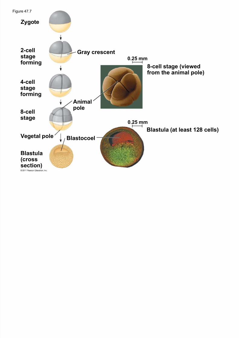
Zygote
2-cellstagef or ming
4-cellstagef or ming
8-cellstage
Vegetal pole
Blastula(cross
section)
Gr ay crescent
Animalpole
Blastocoel
0.25 mm
0.25 mm
8-cell stage (viewedf rom the animal pole)
Blastula (at least 128 cells)
Figure 47.7
5/13/2018 47 Lecture Presentation PC 2012 (1) - slidepdf.com
http://slidepdf.com/reader/full/47-lecture-presentation-pc-2012-1 25/88
Animal embryos complete cleavage when the ratio
of material in the nucleus relative to the cytoplasm
is sufficiently large
© 2011 Pearson Education, Inc.
Re gulation of Cleava g e
5/13/2018 47 Lecture Presentation PC 2012 (1) - slidepdf.com
http://slidepdf.com/reader/full/47-lecture-presentation-pc-2012-1 26/88
After cleavage, the rate of cell division slows and
the normal cell cycle is restored Morphogenesis, the process by which cells
occupy their appropriate locations, involves
± Gastr ulation, the movement of cells from the
blastula surface to the interior of the embryo
± Organogenesis, the formation of organs
© 2011 Pearson Education, Inc.
Concept 47.2: Morphogenesis in animals
involves specific changes in cell shape,position, and survival
5/13/2018 47 Lecture Presentation PC 2012 (1) - slidepdf.com
http://slidepdf.com/reader/full/47-lecture-presentation-pc-2012-1 27/88
Gastrulation
Gastr ulation rearranges the cells of a blastula
into a three-layered embryo, called a gastr ula
© 2011 Pearson Education, Inc.
5/13/2018 47 Lecture Presentation PC 2012 (1) - slidepdf.com
http://slidepdf.com/reader/full/47-lecture-presentation-pc-2012-1 28/88
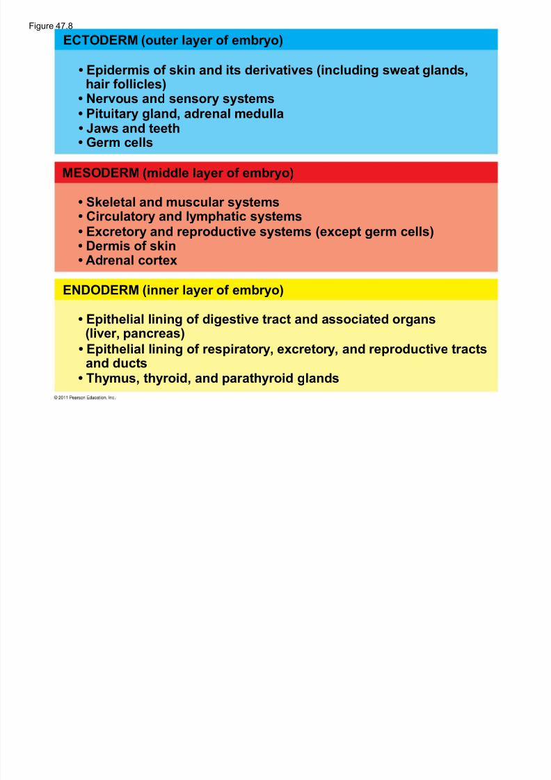
The three layers produced by gastrulation are
called embryonic ger m layers
± The ectoder m forms the outer layer
± The endoder m lines the digestive tract ± The mesoder m partly fills the space between the
endoderm and ectoderm
Each germ layer contributes to specific structures
in the adult animal
© 2011 Pearson Education, Inc.
5/13/2018 47 Lecture Presentation PC 2012 (1) - slidepdf.com
http://slidepdf.com/reader/full/47-lecture-presentation-pc-2012-1 29/88
ECTODERM (outer layer of embryo)
MESODERM (middle layer of embryo)
ENDODERM (inner layer of embryo)
Epider mis of skin and its derivatives (including sweat glands,hair f ollicles)
Epithelial lining of digestive tr act and associated organs(liver, pancreas)
Epithelial lining of respir atory, excretory, and reproductive tr actsand ducts
Ger m cells Jaws and teeth Pituitary gland, adrenal medulla Nervous and sensory systems
Skeletal and muscular systems Cir culatory and lymphatic systems Excretory and reproductive systems (except ger m cells) Der mis of skin Adrenal cor tex
Thymus, thyroid, and par athyroid glands
Figure 47.8
5/13/2018 47 Lecture Presentation PC 2012 (1) - slidepdf.com
http://slidepdf.com/reader/full/47-lecture-presentation-pc-2012-1 30/88
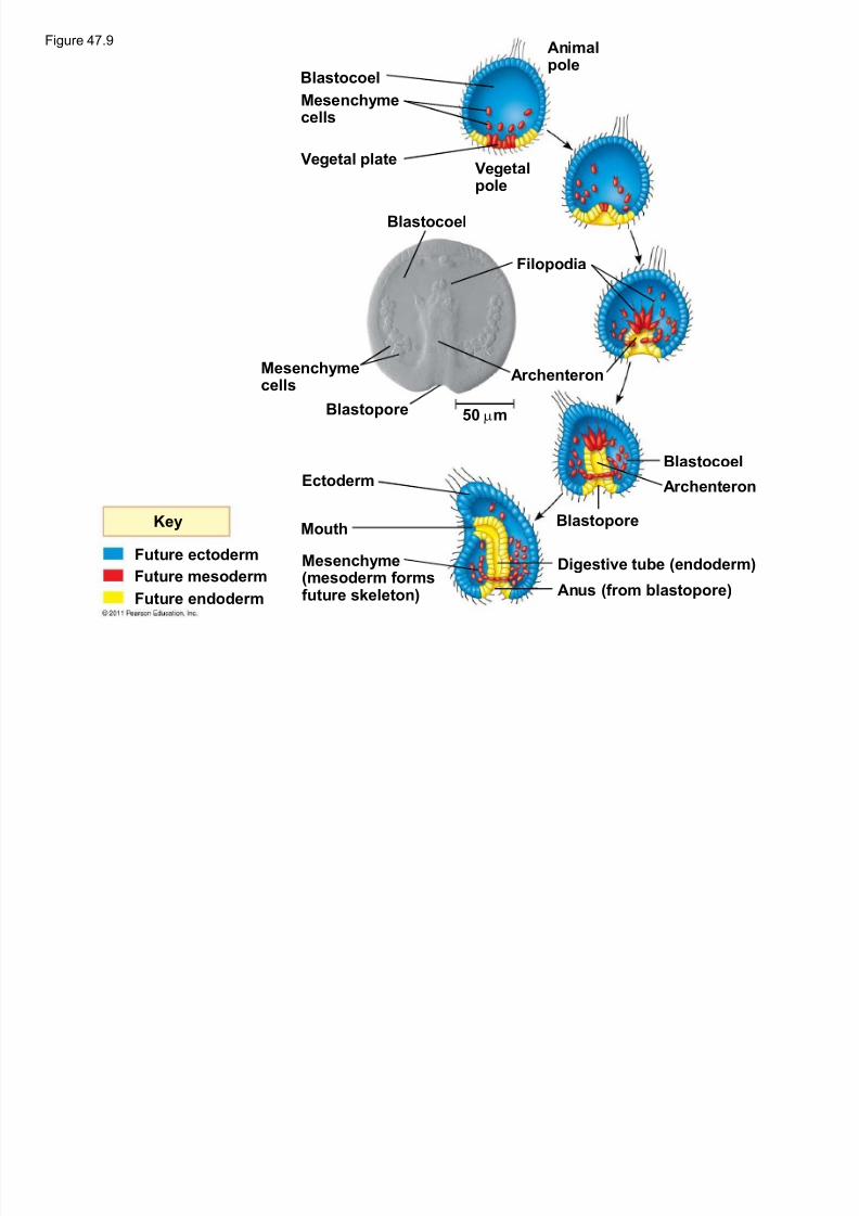
Gastrulation begins at the vegetal pole of the
blastula
Mesenchyme cells migrate into the blastocoel
The vegetal plate forms from the remaining cells of the vegetal pole and buckles inward through
invagination
© 2011 Pearson Education, Inc.
G astr ulation in Sea Urchins
5/13/2018 47 Lecture Presentation PC 2012 (1) - slidepdf.com
http://slidepdf.com/reader/full/47-lecture-presentation-pc-2012-1 31/88
The newly formed cavity is called the
ar chenteron
This opens through the blastopore, which will
become the anus
© 2011 Pearson Education, Inc.
5/13/2018 47 Lecture Presentation PC 2012 (1) - slidepdf.com
http://slidepdf.com/reader/full/47-lecture-presentation-pc-2012-1 32/88
Animalpole
Blastocoel
Mesenchymecells
Vegetal plate Vegetalpole
Blastocoel
Filopodia
Mesenchymecells
Blastopore
Ar chenteron
50 Qm
Ectoder m
Mouth
Mesenchyme(mesoder m f or msfuture skeleton)
Blastopore
Blastocoel
Ar chenteron
Digestive tube (endoder m)
Anus (f rom blastopore)
Key
Future ectoder m
Future mesoder m
Future endoder m
Figure 47.9
5/13/2018 47 Lecture Presentation PC 2012 (1) - slidepdf.com
http://slidepdf.com/reader/full/47-lecture-presentation-pc-2012-1 33/88
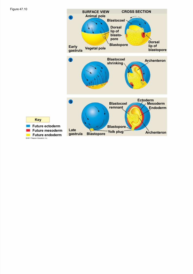
Frog gastrulation begins when a group of cells onthe dorsal side of the blastula begins toinvaginate
This forms a crease along the region where the
gray crescent formed
The part above the crease is called the dorsal lipof the blastopore
© 2011 Pearson Education, Inc.
G astr ulation in F ro g s
5/13/2018 47 Lecture Presentation PC 2012 (1) - slidepdf.com
http://slidepdf.com/reader/full/47-lecture-presentation-pc-2012-1 34/88
Cells continue to move from the embryo surface
into the embryo by involution
These cells become the endoderm and
mesoderm Cells on the embryo surface will form the
ectoderm
© 2011 Pearson Education, Inc.
5/13/2018 47 Lecture Presentation PC 2012 (1) - slidepdf.com
http://slidepdf.com/reader/full/47-lecture-presentation-pc-2012-1 35/88
Key
Future ectoder m
Future mesoder m
Future endoder m
SURFACE VIEW CROSS SECTION
Animal pole
Vegetal poleEar lygastr ula
Blastocoel
Dorsallip of
blasto-pore
BlastoporeDorsallip of blastopore
Blastocoelshrinking
Ar chenteron
Ar chenteron
Blastocoelremnant
Ectoder mMesoder m
Endoder m
Blastopore
Yolk plugBlastopore
Lategastr ula
3
2
1
Figure 47.10
5/13/2018 47 Lecture Presentation PC 2012 (1) - slidepdf.com
http://slidepdf.com/reader/full/47-lecture-presentation-pc-2012-1 36/88
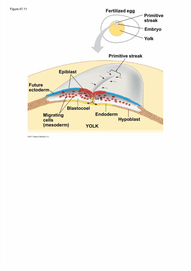
Prior to gastrulation, the embryo is composed of
an upper and lower layer, the epiblast and
hypoblast, respectively
During gastrulation, epiblast cells move toward themidline of the blastoderm and then into the
embryo toward the yolk
© 2011 Pearson Education, Inc.
G astr ulation in Chicks
5/13/2018 47 Lecture Presentation PC 2012 (1) - slidepdf.com
http://slidepdf.com/reader/full/47-lecture-presentation-pc-2012-1 37/88
The midline thickens and is called the primitive
streak
The hypoblast cells contribute to the sac that
surrounds the yolk and a connection between theyolk and the embryo, but do not contribute to the
embryo itself
© 2011 Pearson Education, Inc.
5/13/2018 47 Lecture Presentation PC 2012 (1) - slidepdf.com
http://slidepdf.com/reader/full/47-lecture-presentation-pc-2012-1 38/88
Future ectoder m
Migr atingcells(mesoder m)
Blastocoel
Epiblast
YOLK
Endoder mHypoblast
Primitive streak
Fer tilized eggPrimitivestreak
Embryo
Yolk
Figure 47.11
5/13/2018 47 Lecture Presentation PC 2012 (1) - slidepdf.com
http://slidepdf.com/reader/full/47-lecture-presentation-pc-2012-1 39/88
Human eggs have very little yolk
A blastocyst is the human equivalent of the
blastula
The inner cell mass is a cluster of cells at oneend of the blastocyst
The trophoblast is the outer epithelial layer of the
blastocyst and does not contribute to the embryo,
but instead initiates implantation
© 2011 Pearson Education, Inc.
G astr ulation in H umans
5/13/2018 47 Lecture Presentation PC 2012 (1) - slidepdf.com
http://slidepdf.com/reader/full/47-lecture-presentation-pc-2012-1 40/88
Following implantation, the trophoblast
continues to expand and a set of
extr aembryonic membr anes is formed
These enclose specialized structures outside of the embryo
Gastrulation involves the inward movement from
the epiblast, through a primitive streak, similar
to the chick embryo
© 2011 Pearson Education, Inc.
5/13/2018 47 Lecture Presentation PC 2012 (1) - slidepdf.com
http://slidepdf.com/reader/full/47-lecture-presentation-pc-2012-1 41/88
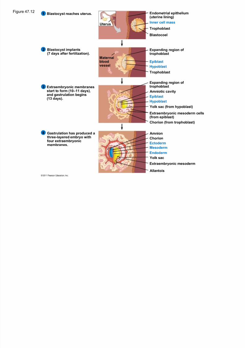
Blastocyst reaches uter us.1
2
3
4
Blastocyst implants(7 days after f er tilization).
Extr aembryonic membr anesstar t to f or m (10±11 days),and gastr ulation begins(13 days).
Gastr ulation has produced athree-layered embryo withf our extr aembryonicmembr anes.
Uter us
Mater nalbloodvessel
Endometrial epithelium(uterine lining)
Inner cell mass
Trophoblast
Blastocoel
Expanding region of trophoblast
Epiblast
Hypoblast
Trophoblast
Expanding region of
trophoblastAmniotic cavity
Epiblast
Hypoblast
Yolk sac (f rom hypoblast)
Extr aembryonic mesoder m cells(f rom epiblast)
Chorion (f rom trophoblast)
Amnion
Chorion
Ectoder m
Mesoder m
Endoder m
Yolk sac
Extr aembryonic mesoder m
Allantois
Figure 47.12
5/13/2018 47 Lecture Presentation PC 2012 (1) - slidepdf.com
http://slidepdf.com/reader/full/47-lecture-presentation-pc-2012-1 42/88
Developmental Adaptations of Amniotes
The colonization of land by vertebrates was made
possible only after the evolution of
± The shelled egg of birds and other reptiles as well
as monotremes (egg-laying mammals)
± The uterus of marsupial and eutherian mammals
© 2011 Pearson Education, Inc.
5/13/2018 47 Lecture Presentation PC 2012 (1) - slidepdf.com
http://slidepdf.com/reader/full/47-lecture-presentation-pc-2012-1 43/88
In both adaptations, embryos are surrounded
by fluid in a sac called the amnion
This protects the embryo from desiccation and
allows reproduction on dry land Mammals and reptiles including birds are
called amniotes for this reason
© 2011 Pearson Education, Inc.
5/13/2018 47 Lecture Presentation PC 2012 (1) - slidepdf.com
http://slidepdf.com/reader/full/47-lecture-presentation-pc-2012-1 44/88
The four extraembryonic membranes that formaround the embryo
± The chorion functions in gas exchange
± The amnion encloses the amniotic fluid ± The yolk sac encloses the yolk
± The allantois disposes of waste products and
contributes to gas exchange
© 2011 Pearson Education, Inc.
5/13/2018 47 Lecture Presentation PC 2012 (1) - slidepdf.com
http://slidepdf.com/reader/full/47-lecture-presentation-pc-2012-1 45/88
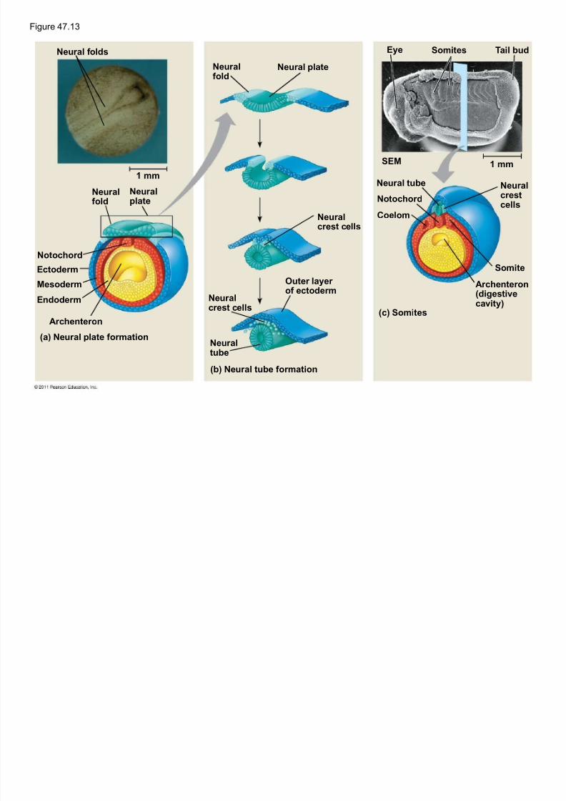
Organogenesis
During organogenesis, various regions of the
germ layers develop into rudimentary organs
Early in vertebrate organogenesis, the notochor d
forms from mesoderm, and the neural plate formsfrom ectoderm
© 2011 Pearson Education, Inc.
Fi 4 13
5/13/2018 47 Lecture Presentation PC 2012 (1) - slidepdf.com
http://slidepdf.com/reader/full/47-lecture-presentation-pc-2012-1 46/88
Figure 47.13
Neur al f olds
1 mm
Neur al
f old
Neur al
plate
Notochor d
Ectoder m
Mesoder m
Endoder m
Ar chenteron
(a) Neur al plate f or mation
(b) Neur al tube f or mation
(c) Somites
Neur alf old
Neur al plate
Neur alcrest cells
Outer layer of ectoder m
Neur alcrest cells
Neur altube
Eye Somites Tail bud
SEM
Neur al tube
Notochor dCoelom
Neur alcrest
cells
Somite
Ar chenteron(digestive
cavity)
1 mm
5/13/2018 47 Lecture Presentation PC 2012 (1) - slidepdf.com
http://slidepdf.com/reader/full/47-lecture-presentation-pc-2012-1 47/88
The neural plate soon curves inward, forming theneur al tube
The neural tube will become the central nervous
system (brain and spinal cord)
© 2011 Pearson Education, Inc.
5/13/2018 47 Lecture Presentation PC 2012 (1) - slidepdf.com
http://slidepdf.com/reader/full/47-lecture-presentation-pc-2012-1 48/88
Neur al crest cells develop along the neural tubeof vertebrates and form various parts of the
embryo (nerves, parts of teeth, skull bones, and so
on)
Mesoderm lateral to the notochord forms blocks
called somites
Lateral to the somites, the mesoderm splits to form
the coelom (body cavity)
© 2011 Pearson Education, Inc.
5/13/2018 47 Lecture Presentation PC 2012 (1) - slidepdf.com
http://slidepdf.com/reader/full/47-lecture-presentation-pc-2012-1 49/88
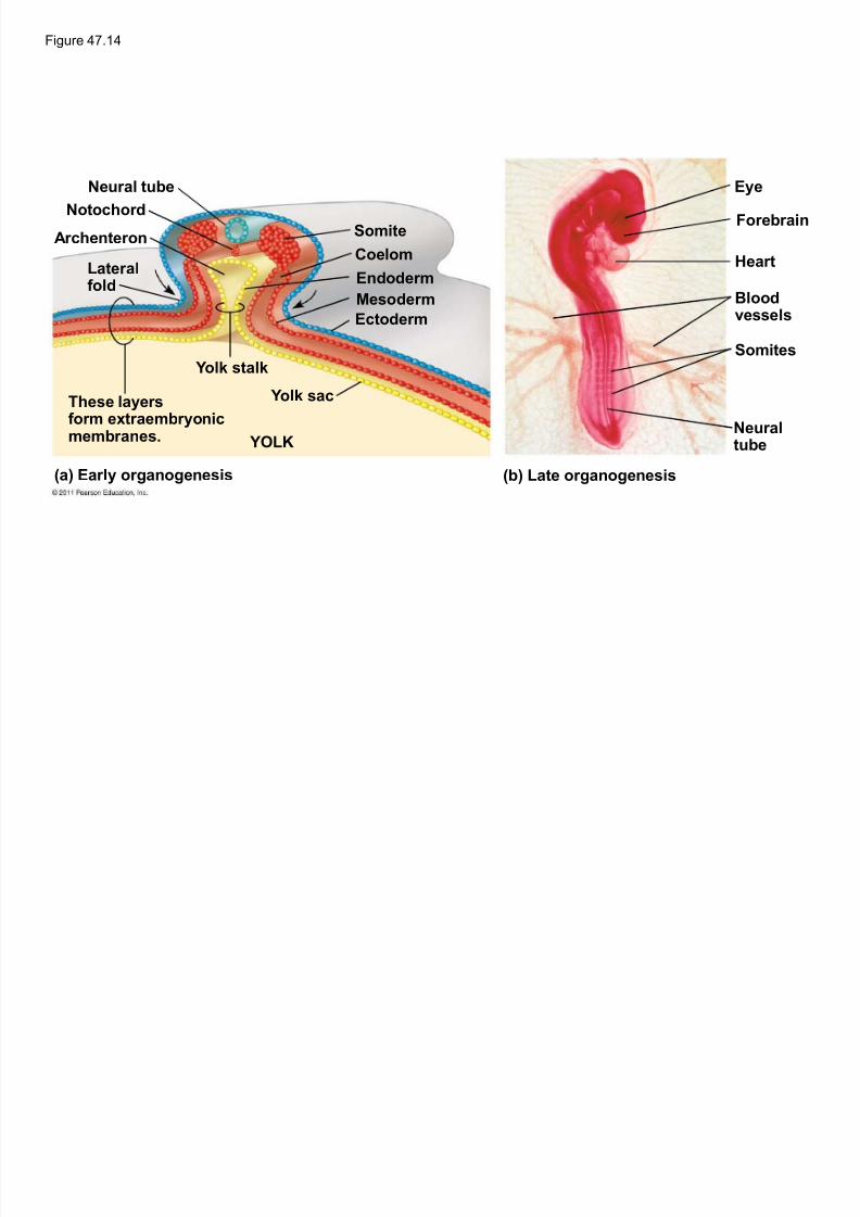
Organogenesis in the chick is quite similar to thatin the frog
© 2011 Pearson Education, Inc.
Figure 47 14
5/13/2018 47 Lecture Presentation PC 2012 (1) - slidepdf.com
http://slidepdf.com/reader/full/47-lecture-presentation-pc-2012-1 50/88
Figure 47.14
Neur al tube
Notochor d
Ar chenteron
Later alf old
These layersf or m extr aembryonicmembr anes.
Yolk stalk
YOLK
Yolk sac
Somite
Coelom
Endoder m
Mesoder m
Ectoder m
(a) Ear ly organogenesis (b) Late organogenesis
Eye
Forebr ain
Hear t
Bloodvessels
Somites
Neur altube
5/13/2018 47 Lecture Presentation PC 2012 (1) - slidepdf.com
http://slidepdf.com/reader/full/47-lecture-presentation-pc-2012-1 51/88
The mechanisms of organogenesis ininvertebrates are similar, but the body plan is very
different
For example, the neural tube develops along theventral side of the embryo in invertebrates, rather
than dorsally as occurs in vertebrates
© 2011 Pearson Education, Inc.
5/13/2018 47 Lecture Presentation PC 2012 (1) - slidepdf.com
http://slidepdf.com/reader/full/47-lecture-presentation-pc-2012-1 52/88
Mechanisms of Morphogenesis
Morphogenesis in animals but not plants involvesmovement of cells
© 2011 Pearson Education, Inc.
5/13/2018 47 Lecture Presentation PC 2012 (1) - slidepdf.com
http://slidepdf.com/reader/full/47-lecture-presentation-pc-2012-1 53/88
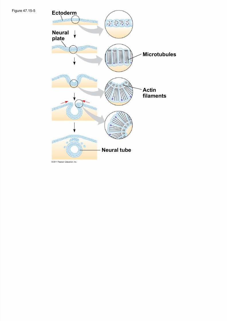
Reorganization of the cytoskeleton is a major forcein changing cell shape during development
For example, in neurulation, microtubules oriented
from dorsal to ventral in a sheet of ectodermalcells help lengthen the cells along that axis
© 2011 Pearson Education, Inc.© 2011 Pearson Education, Inc.
The Cytoskeleton in Morpho g enesis
Figure 47 15-5E t d
5/13/2018 47 Lecture Presentation PC 2012 (1) - slidepdf.com
http://slidepdf.com/reader/full/47-lecture-presentation-pc-2012-1 54/88
Figure 47.15 5Ectoder m
Neur alplate
Microtubules
Actinf ilaments
Neur al tube
5/13/2018 47 Lecture Presentation PC 2012 (1) - slidepdf.com
http://slidepdf.com/reader/full/47-lecture-presentation-pc-2012-1 55/88
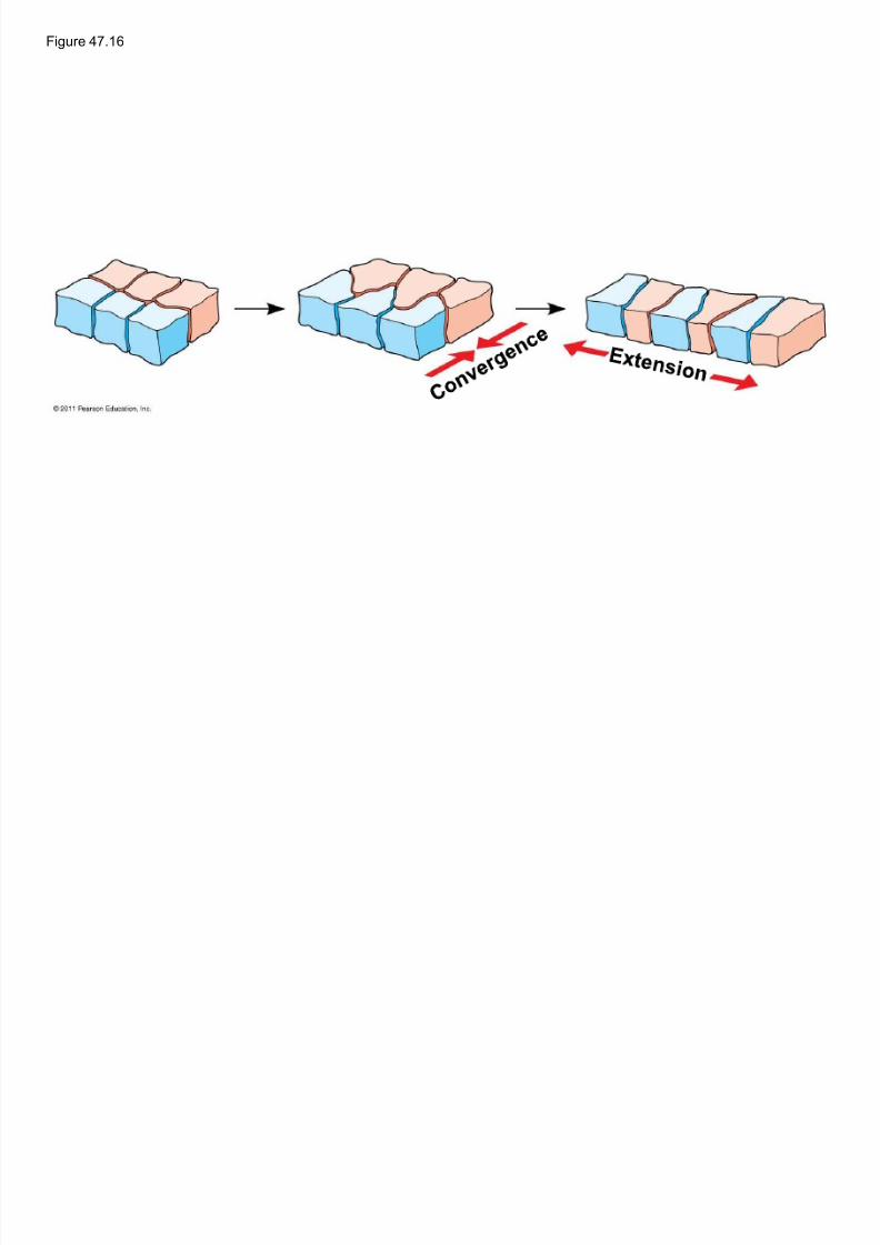
The cytoskeleton promotes elongation of thearchenteron in the sea urchin embryo
This is convergent extension, the
rearrangement of cells of a tissue that cause it tobecome narrower (converge) and longer (extend)
Convergent extension occurs in other developmental processes
The cytoskeleton also directs cell migration
© 2011 Pearson Education, Inc.
Figure 47 16
5/13/2018 47 Lecture Presentation PC 2012 (1) - slidepdf.com
http://slidepdf.com/reader/full/47-lecture-presentation-pc-2012-1 56/88
Figure 47.16
5/13/2018 47 Lecture Presentation PC 2012 (1) - slidepdf.com
http://slidepdf.com/reader/full/47-lecture-presentation-pc-2012-1 57/88
Programmed cell death is also called apoptosis
At various times during development, individualcells, sets of cells, or whole tissues stopdeveloping and are engulfed by neighboring cells
For example, many more neurons are produced indeveloping embryos than will be needed
Extra neurons are removed by apoptosis
© 2011 Pearson Education, Inc.
Pro g rammed Cell Death
5/13/2018 47 Lecture Presentation PC 2012 (1) - slidepdf.com
http://slidepdf.com/reader/full/47-lecture-presentation-pc-2012-1 58/88
Concept 47.3: Cytoplasmic determinants
and inductive signals contribute to cellfate specification
Deter mination is the term used to describe the
process by which a cell or group of cells becomescommitted to a particular fate
Diff erentiation refers to the resulting
specialization in structure and function
© 2011 Pearson Education, Inc.
5/13/2018 47 Lecture Presentation PC 2012 (1) - slidepdf.com
http://slidepdf.com/reader/full/47-lecture-presentation-pc-2012-1 59/88
Cells in a multicellular organism share the samegenome
Differences in cell types are the result of the
expression of different sets of genes
© 2011 Pearson Education, Inc.
5/13/2018 47 Lecture Presentation PC 2012 (1) - slidepdf.com
http://slidepdf.com/reader/full/47-lecture-presentation-pc-2012-1 60/88
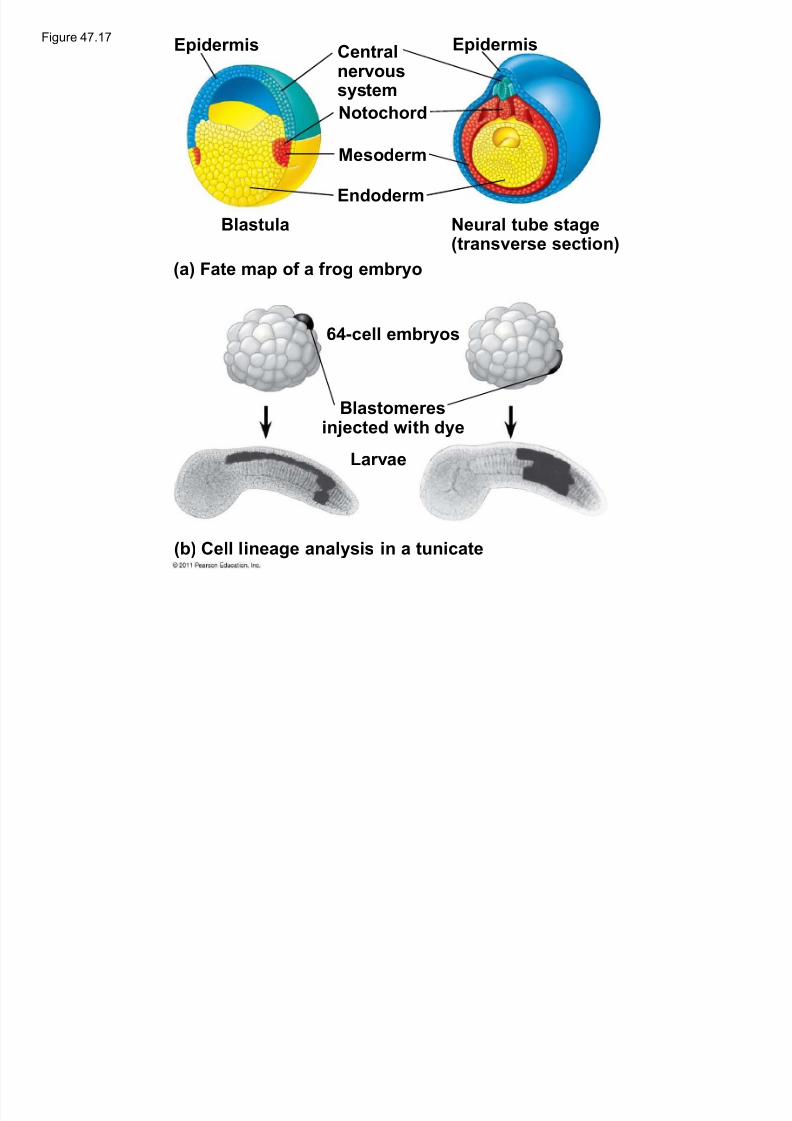
Fate Mapping
Fate maps are diagrams showing organs andother structures that arise from each region of an
embryo
Classic studies using frogs indicated that cell
lineage in germ layers is traceable to blastula cells
© 2011 Pearson Education, Inc.
Epidermis EpidermisFigure 47.17
5/13/2018 47 Lecture Presentation PC 2012 (1) - slidepdf.com
http://slidepdf.com/reader/full/47-lecture-presentation-pc-2012-1 61/88
Epider mis Epider misCentr alnervoussystem
Notochor d
Mesoder m
Endoder m
Blastula Neur al tube stage(tr ansverse section)
(a) Fate map of a f rog embryo
64-cell embryos
Blastomeres
injected with dye
Larvae
(b) Cell lineage analysis in a tunicate
5/13/2018 47 Lecture Presentation PC 2012 (1) - slidepdf.com
http://slidepdf.com/reader/full/47-lecture-presentation-pc-2012-1 62/88
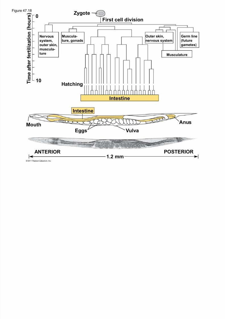
Later studies of C. elegans used the ablation(destruction) of single cells to determine the
structures that normally arise from each cell
The researchers were able to determine the
lineage of each of the 959 somatic cells in the
worm
© 2011 Pearson Education, Inc.
ZygoteFigure 47.18
5/13/2018 47 Lecture Presentation PC 2012 (1) - slidepdf.com
http://slidepdf.com/reader/full/47-lecture-presentation-pc-2012-1 63/88
First cell division
Zygote
Hatching T i m e
a f t e r f e r t i l i z a
t i o n ( h o u r s )
Intestine
Intestine
Mouth
Eggs Vulva
Anus
1.2 mmANTERIOR POSTERIOR
Nervous
system,outer skin,
muscula-
ture
Muscula-
ture, gonads
Outer skin,
nervous system
Ger m line
(futuregametes)
Musculature
10
0
5/13/2018 47 Lecture Presentation PC 2012 (1) - slidepdf.com
http://slidepdf.com/reader/full/47-lecture-presentation-pc-2012-1 64/88
Germ cells are the specialized cells that give riseto sperm or eggs
Complexes of RNA and protein are involved in the
specification of germ cell fate
In C. elegans, such complexes are called P
granules, persist throughout development, and
can be detected in germ cells of the adult worm
© 2011 Pearson Education, Inc.
Figure 47.19

5/13/2018 47 Lecture Presentation PC 2012 (1) - slidepdf.com
http://slidepdf.com/reader/full/47-lecture-presentation-pc-2012-1 65/88
100 Qm
5/13/2018 47 Lecture Presentation PC 2012 (1) - slidepdf.com
http://slidepdf.com/reader/full/47-lecture-presentation-pc-2012-1 66/88
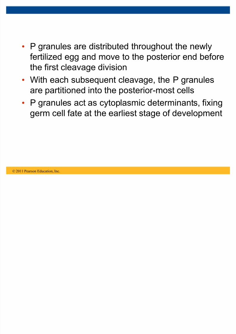
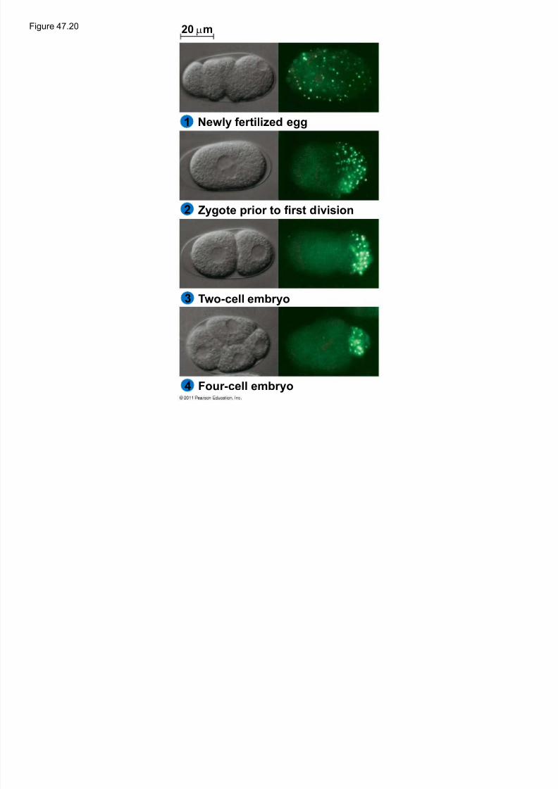
P granules are distributed throughout the newlyfertilized egg and move to the posterior end before
the first cleavage division
With each subsequent cleavage, the P granules
are partitioned into the posterior-most cells
P granules act as cytoplasmic determinants, fixing
germ cell fate at the earliest stage of development
© 2011 Pearson Education, Inc.
Figure 47.20 20 Qm
5/13/2018 47 Lecture Presentation PC 2012 (1) - slidepdf.com
http://slidepdf.com/reader/full/47-lecture-presentation-pc-2012-1 67/88
Newly f er tilized egg
Zygote prior to f irst division
Two-cell embryo
Four -cell embryo
Q
2
1
3
4
5/13/2018 47 Lecture Presentation PC 2012 (1) - slidepdf.com
http://slidepdf.com/reader/full/47-lecture-presentation-pc-2012-1 68/88
Axis F ormation
A body plan with bilateral symmetry is foundacross a range of animals
This body plan exhibits asymmetry across the
dorsal-ventral and anterior-posterior axes
The right-left axis is largely symmetrical
© 2011 Pearson Education, Inc.
5/13/2018 47 Lecture Presentation PC 2012 (1) - slidepdf.com
http://slidepdf.com/reader/full/47-lecture-presentation-pc-2012-1 69/88
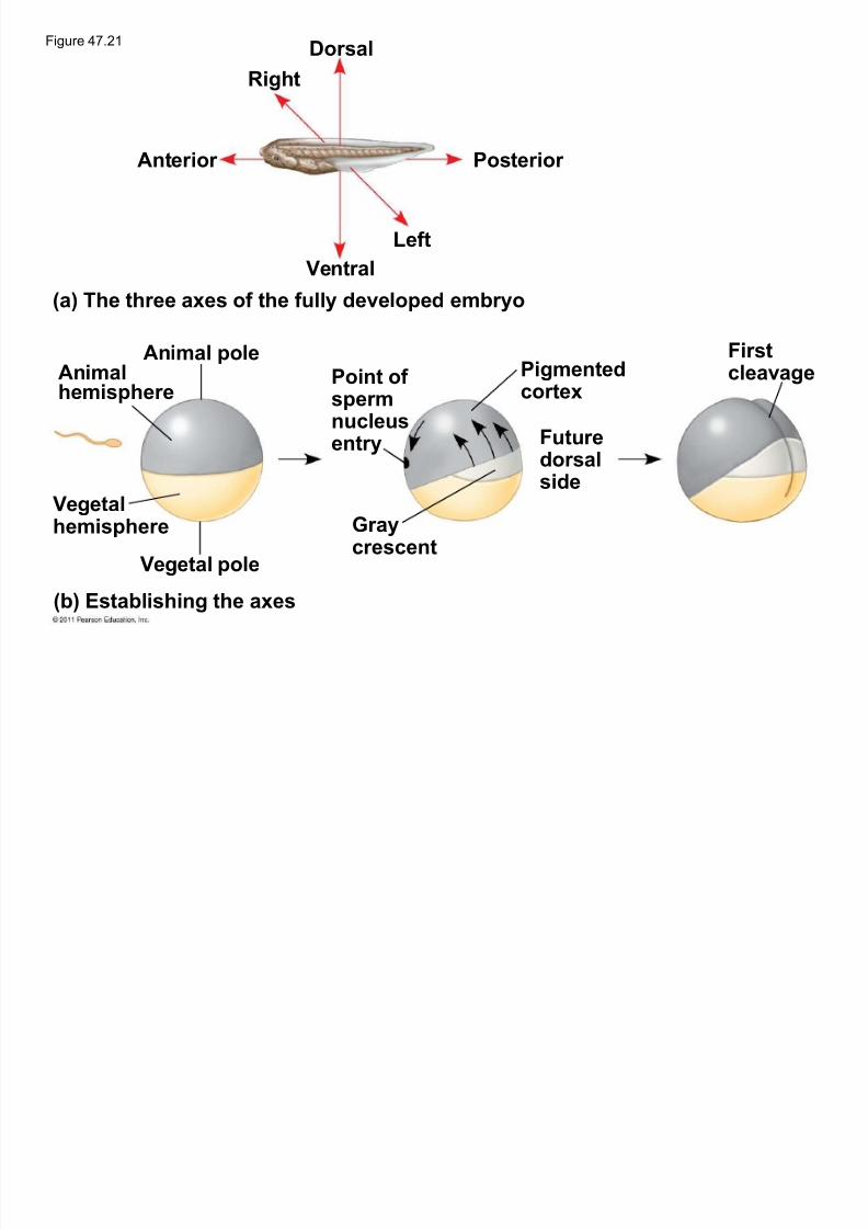
The anterior-posterior axis of the frog embryo isdetermined during oogenesis
The animal-vegetal asymmetry indicates where
the anterior-posterior axis forms
The dorsal-ventral axis is not determined until
fertilization
© 2011 Pearson Education, Inc.
5/13/2018 47 Lecture Presentation PC 2012 (1) - slidepdf.com
http://slidepdf.com/reader/full/47-lecture-presentation-pc-2012-1 70/88
Upon fusion of the egg and sperm, the eggsurface rotates with respect to the inner cytoplasm
This cortical rotation brings molecules from one
area of the inner cytoplasm of the animal
hemisphere to interact with molecules in the
vegetal cortex
This leads to expression of dorsal- and ventral-
specific gene expression
© 2011 Pearson Education, Inc.
DorsalFigure 47.21
5/13/2018 47 Lecture Presentation PC 2012 (1) - slidepdf.com
http://slidepdf.com/reader/full/47-lecture-presentation-pc-2012-1 71/88
Right
Anterior Posterior
Ventr al
Left
(a) The three axes of the fully developed embryo
(b) Establishing the axes
Animalhemisphere
Vegetalhemisphere
Animal pole
Vegetal pole
Point of sper mnucleusentry
Gr ay crescent
Pigmentedcor tex
Future
dorsalside
Firstcleavage
5/13/2018 47 Lecture Presentation PC 2012 (1) - slidepdf.com
http://slidepdf.com/reader/full/47-lecture-presentation-pc-2012-1 72/88
In chicks, gravity is involved in establishing theanterior-posterior axis
Later, pH differences between the two sides of the
blastoderm establish the dorsal-ventral axis
In mammals, experiments suggest that orientation
of the egg and sperm nuclei before fusion may
help establish embryonic axes
© 2011 Pearson Education, Inc.
5/13/2018 47 Lecture Presentation PC 2012 (1) - slidepdf.com
http://slidepdf.com/reader/full/47-lecture-presentation-pc-2012-1 73/88
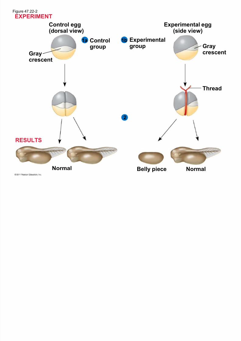
Restrictin g Developmental Potential
Hans Spemann performed experiments todetermine a cell¶s developmental potential (range
of structures to which it can give rise)
Embryonic fates are affected by distribution of
determinants and the pattern of cleavage
The first two blastomeres of the frog embryo are
totipotent (can develop into all the possible cell
types)
© 2011 Pearson Education, Inc.
EXPERIMENTFigure 47.22-2
5/13/2018 47 Lecture Presentation PC 2012 (1) - slidepdf.com
http://slidepdf.com/reader/full/47-lecture-presentation-pc-2012-1 74/88
Control egg(dorsal view)
2
1a 1b
Gr aycrescent
Control
group
Experimental
group
Experimental egg(side view)
Gr aycrescent
Thread
Nor malNor malBelly piece
EXPERIMENT
RESULTS
5/13/2018 47 Lecture Presentation PC 2012 (1) - slidepdf.com
http://slidepdf.com/reader/full/47-lecture-presentation-pc-2012-1 75/88
In mammals, embryonic cells remain totipotentuntil the 8-cell stage, much longer than other
organisms
Progressive restriction of developmental potential
is a general feature of development in all animals
In general tissue-specific fates of cells are fixed by
the late gastrula stage
© 2011 Pearson Education, Inc.
5/13/2018 47 Lecture Presentation PC 2012 (1) - slidepdf.com
http://slidepdf.com/reader/full/47-lecture-presentation-pc-2012-1 76/88
Cell Fate Determination and Pattern
Formation by Inductive Signals As embryonic cells acquire distinct fates, they
influence each other¶s fates by induction
© 2011 Pearson Education, Inc.
5/13/2018 47 Lecture Presentation PC 2012 (1) - slidepdf.com
http://slidepdf.com/reader/full/47-lecture-presentation-pc-2012-1 77/88
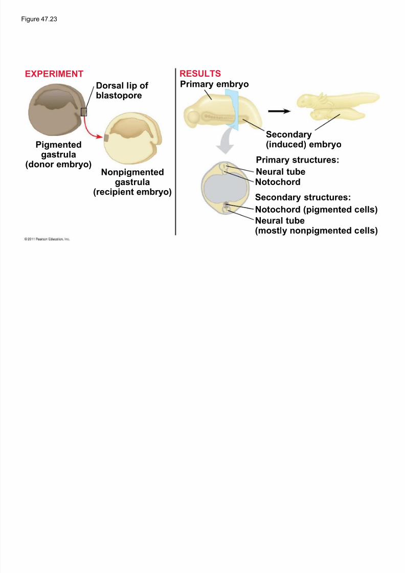
The ³Or g anizer´ of Spemann and Man g old
Spemann and Mangold transplanted tissuesbetween early gastrulas and found that the
transplanted dorsal lip triggered a second
gastrulation in the host
The dorsal lip functions as an organizer of the
embryo body plan, inducing changes in
surrounding tissues to form notochord, neural
tube, and so on
© 2011 Pearson Education, Inc.
Figure 47.23
5/13/2018 47 Lecture Presentation PC 2012 (1) - slidepdf.com
http://slidepdf.com/reader/full/47-lecture-presentation-pc-2012-1 78/88
Dorsal lip of blastopore
Pigmentedgastr ula(donor embryo)
Nonpigmentedgastr ula
(recipient embryo)
Primary embryo
Secondary
(induced) embryoPrimary str uctures:
Neur al tubeNotochor d
Secondary str uctures:
Notochor d (pigmented cells)
Neur al tube(mostly nonpigmented cells)
EXPERIMENT RESULTS
5/13/2018 47 Lecture Presentation PC 2012 (1) - slidepdf.com
http://slidepdf.com/reader/full/47-lecture-presentation-pc-2012-1 79/88
F ormation of the Vertebrate Limb
Inductive signals play a major role in patter nf or mation, development of spatial organization
The molecular cues that control pattern formation
are called positional inf or mation
This information tells a cell where it is with respect
to the body axes
It determines how the cell and its descendents
respond to future molecular signals
© 2011 Pearson Education, Inc.
5/13/2018 47 Lecture Presentation PC 2012 (1) - slidepdf.com
http://slidepdf.com/reader/full/47-lecture-presentation-pc-2012-1 80/88
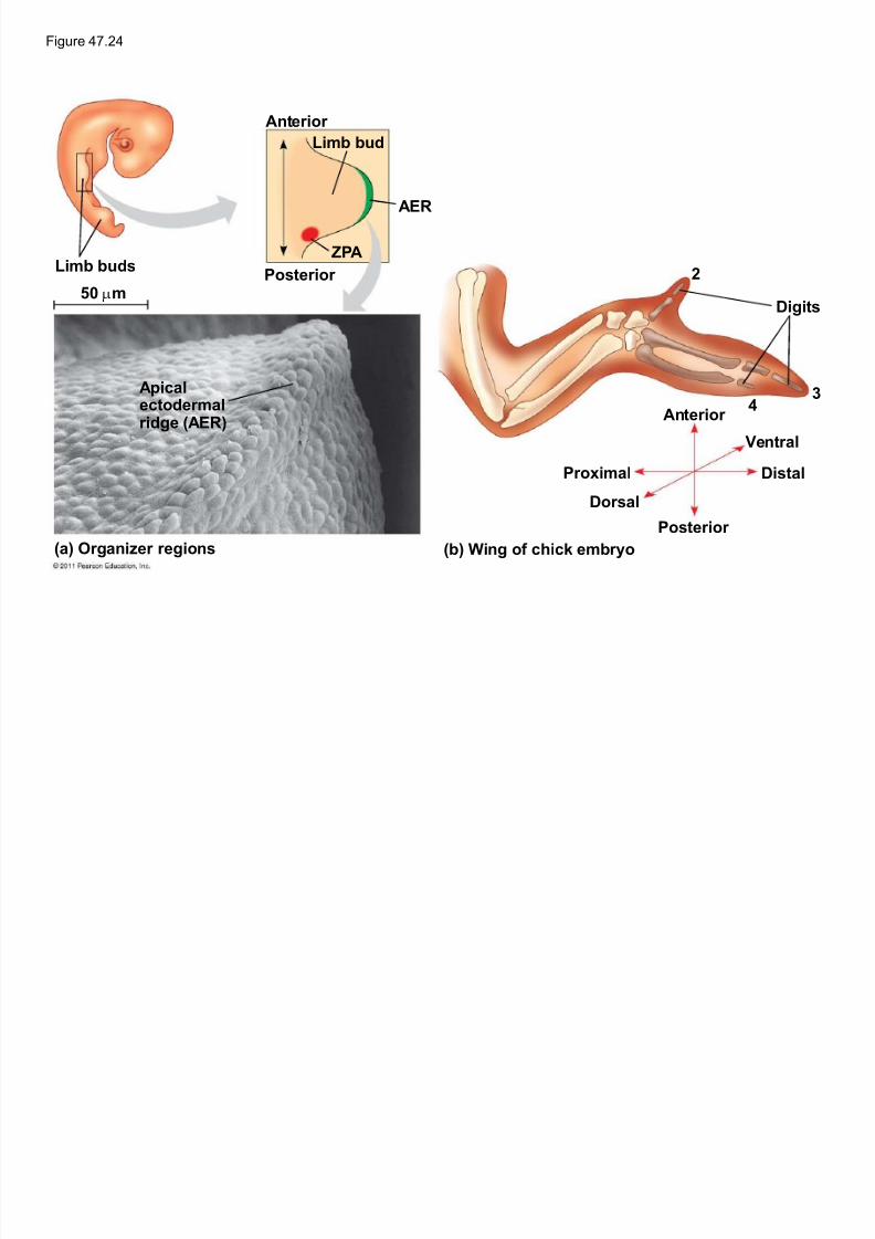
The wings and legs of chicks, like all vertebratelimbs, begin as bumps of tissue called limb buds
© 2011 Pearson Education, Inc.
Figure 47.24
5/13/2018 47 Lecture Presentation PC 2012 (1) - slidepdf.com
http://slidepdf.com/reader/full/47-lecture-presentation-pc-2012-1 81/88
Limb buds
50 Qm
Anterior
Limb bud
AER
ZPA
Posterior
Apicalectoder malridge (AER)
(a) Organizer regions (b) Wing of chick embryo
Digits
Anterior
Proximal
Dorsal
Posterior
Ventr al
Distal
2
34
5/13/2018 47 Lecture Presentation PC 2012 (1) - slidepdf.com
http://slidepdf.com/reader/full/47-lecture-presentation-pc-2012-1 82/88
The embryonic cells in a limb bud respond topositional information indicating location along
three axes
± Proximal-distal axis
± Anterior-posterior axis
± Dorsal-ventral axis
© 2011 Pearson Education, Inc.
5/13/2018 47 Lecture Presentation PC 2012 (1) - slidepdf.com
http://slidepdf.com/reader/full/47-lecture-presentation-pc-2012-1 83/88
One limb bud±regulating region is the apicalectoder mal ridge (AER)
The AER is thickened ectoderm at the bud¶s tip
The second region is the zone of polarizing activity (ZPA)
The ZP A is mesodermal tissue under the
ectoderm where the posterior side of the bud is
attached to the body
© 2011 Pearson Education, Inc.
5/13/2018 47 Lecture Presentation PC 2012 (1) - slidepdf.com
http://slidepdf.com/reader/full/47-lecture-presentation-pc-2012-1 84/88
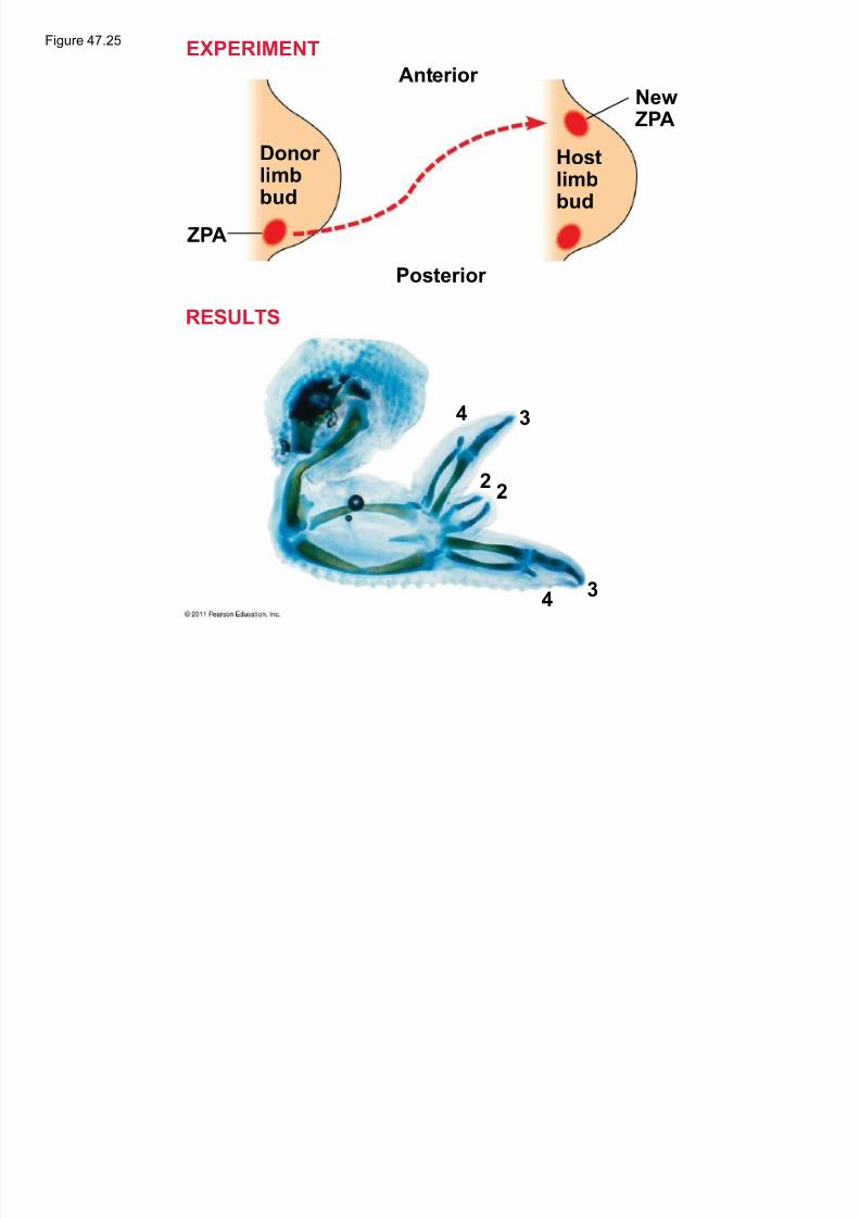
Tissue transplantation experiments support thehypothesis that the ZP A produces an inductive
signal that conveys positional information
indicating ³posterior´
© 2011 Pearson Education, Inc.
Figure 47.25
A t i
EXPERIMENT
5/13/2018 47 Lecture Presentation PC 2012 (1) - slidepdf.com
http://slidepdf.com/reader/full/47-lecture-presentation-pc-2012-1 85/88
Donor limbbud
Hostlimbbud
ZPA
Anterior
Posterior
New ZPA
4
4
3
3
22
RESULTS
5/13/2018 47 Lecture Presentation PC 2012 (1) - slidepdf.com
http://slidepdf.com/reader/full/47-lecture-presentation-pc-2012-1 86/88
Sonic hedgehog is an inductive signal for limbdevelopment
Hox genes also play roles during limb pattern
formation
© 2011 Pearson Education, Inc.© 2011 Pearson Education, Inc.
5/13/2018 47 Lecture Presentation PC 2012 (1) - slidepdf.com
http://slidepdf.com/reader/full/47-lecture-presentation-pc-2012-1 87/88
Cilia and Cell F ate
Ciliary function is essential for proper specificationof cell fate in the human embryo
Motile cilia play roles in left-right specification
Monocilia (nonmotile cilia) play roles in normalkidney development
© 2011 Pearson Education, Inc.
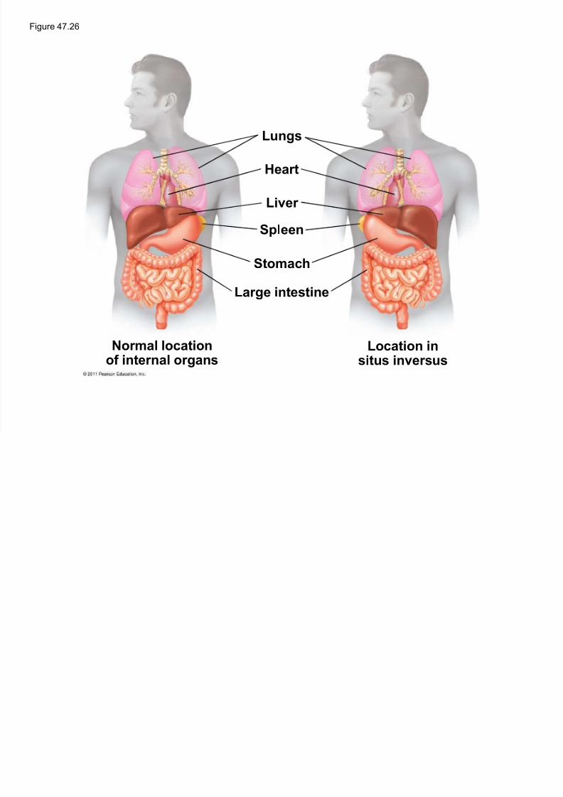
Figure 47.26
5/13/2018 47 Lecture Presentation PC 2012 (1) - slidepdf.com
http://slidepdf.com/reader/full/47-lecture-presentation-pc-2012-1 88/88
Lungs
Hear t
Liver
Spleen
Stomach
Large intestine
Nor mal locationof inter nal organs
Location insitus inversus
![Les processeurs à plusieurs niveaux de langagelsl · MDR ← M[PC] PC ← PC+1 IR ← MDR MAR ← PC MDR ← M[PC] PC ← PC+1 ... Pour faciliter la lecture des programmes en langages](https://img.pdfslide.net/doc/110x75/5f6a22012c4b7814127fdc30/les-processeurs-plusieurs-niveaux-de-mdr-a-mpc-pc-a-pc1-ir-a-mdr-mar.jpg)