Embed Size (px)
Citation preview

8/12/2019 49 Lecture Nervous System
http://slidepdf.com/reader/full/49-lecture-nervous-system 1/94
LECTURE PRESENTATIONS
For CAMPBELL BIOLOGY, NINTH EDITION
Jane B. Reece, Lisa A. Urry, Michael L. Cain, Steven A. Wasserman, Peter V. Minorsky, Robert B. Jackson
© 2011 Pearson Education, Inc.
Lectures by
Erin Barley
Kathleen Fitzpatrick
Nervous Systems
Chapter 49
8/12/2019 49 Lecture Nervous System
http://slidepdf.com/reader/full/49-lecture-nervous-system 2/94
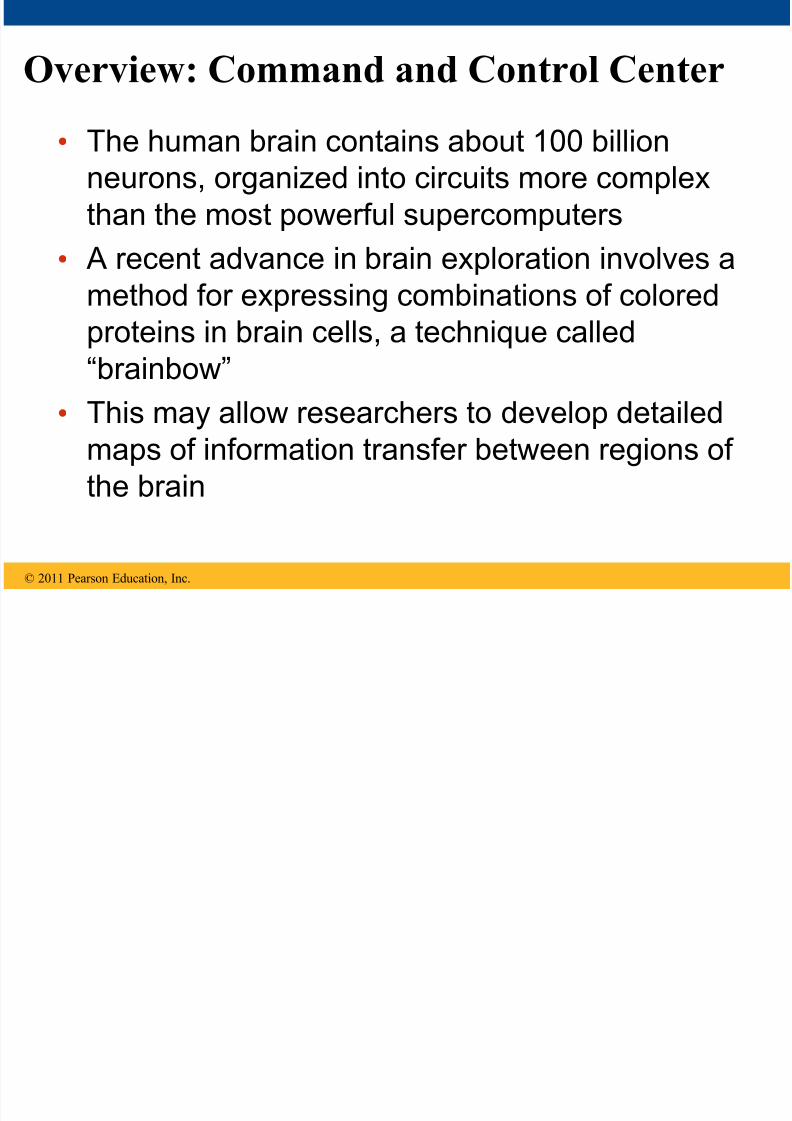
Overview: Command and Control Center
• The human brain contains about 100 billionneurons, organized into circuits more complex
than the most powerful supercomputers
• A recent advance in brain exploration involves a
method for expressing combinations of colored
proteins in brain cells, a technique called
“brainbow”
• This may allow researchers to develop detailedmaps of information transfer between regions of
the brain
© 2011 Pearson Education, Inc.

8/12/2019 49 Lecture Nervous System
http://slidepdf.com/reader/full/49-lecture-nervous-system 3/94
Figure 49.1

8/12/2019 49 Lecture Nervous System
http://slidepdf.com/reader/full/49-lecture-nervous-system 4/94
• Each single-celled organism can respond to
stimuli in its environment
•
Animals are multicellular and most groupsrespond to stimuli using systems of neurons
© 2011 Pearson Education, Inc.
Concept 49.1: Nervous systems consist of
circuits of neurons and supporting cells
8/12/2019 49 Lecture Nervous System
http://slidepdf.com/reader/full/49-lecture-nervous-system 5/94
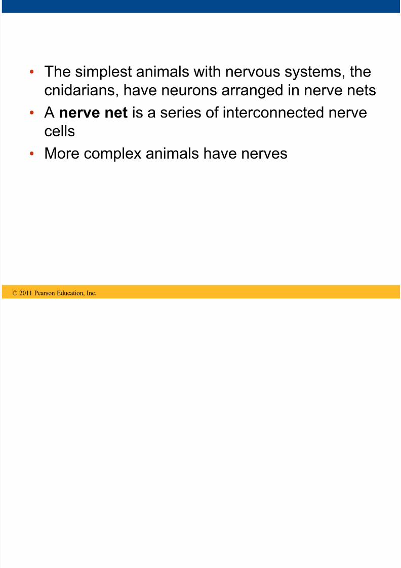
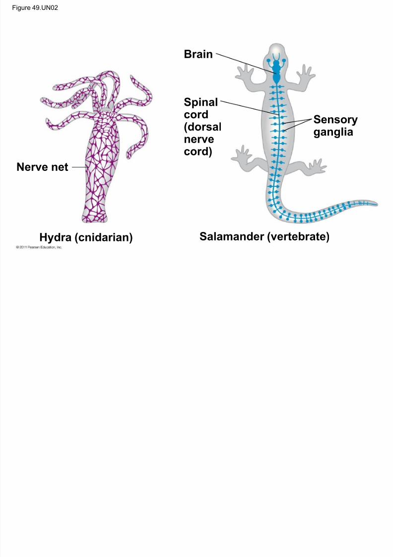
• The simplest animals with nervous systems, thecnidarians, have neurons arranged in nerve nets
• A nerve net is a series of interconnected nerve
cells
• More complex animals have nerves
© 2011 Pearson Education, Inc.

8/12/2019 49 Lecture Nervous System
http://slidepdf.com/reader/full/49-lecture-nervous-system 6/94
• Nerves are bundles that consist of the axons ofmultiple nerve cells
• Sea stars have a nerve net in each arm
connected by radial nerves to a central nerve
ring
© 2011 Pearson Education, Inc.

8/12/2019 49 Lecture Nervous System
http://slidepdf.com/reader/full/49-lecture-nervous-system 7/94

8/12/2019 49 Lecture Nervous System
http://slidepdf.com/reader/full/49-lecture-nervous-system 8/94
Figure 49.2a
Nerve net
(a) Hydra (cnidarian)
Radialnerve
Nerve
ring
(b) Sea star (echinoderm)

8/12/2019 49 Lecture Nervous System
http://slidepdf.com/reader/full/49-lecture-nervous-system 9/94
8/12/2019 49 Lecture Nervous System
http://slidepdf.com/reader/full/49-lecture-nervous-system 10/94
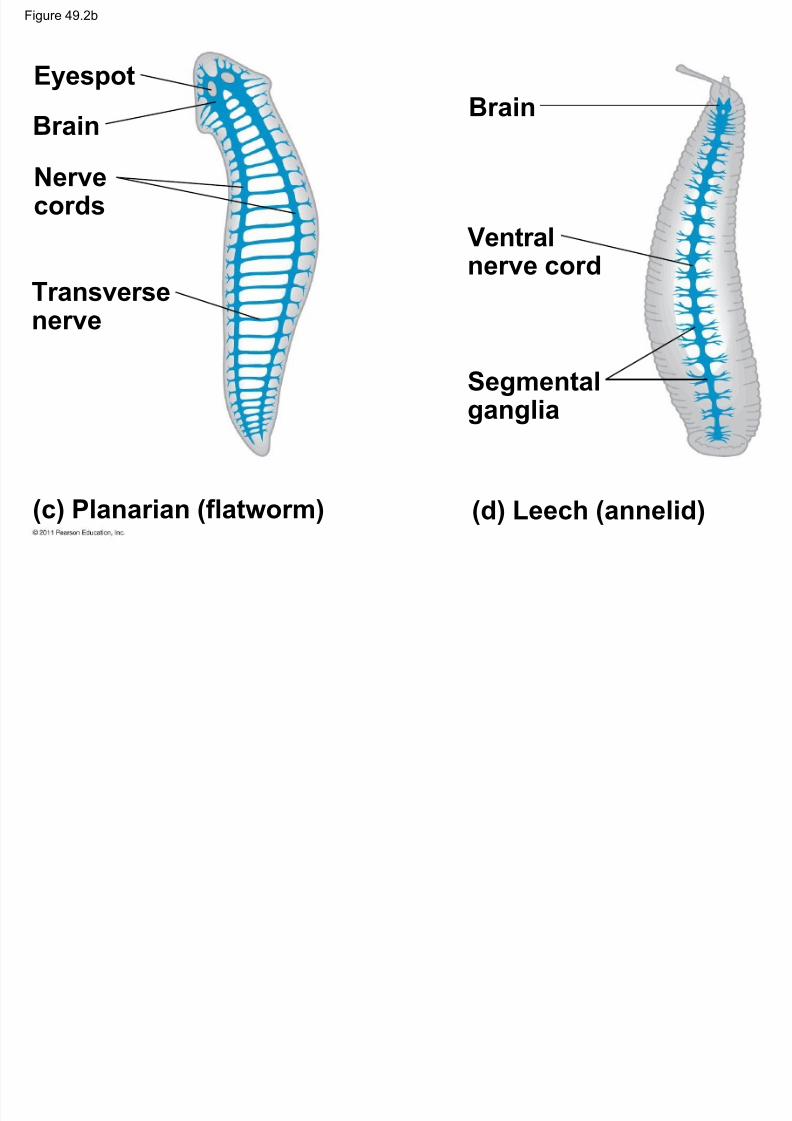
Figure 49.2b
Eyespot
BrainNervecords
Transversenerve
Brain
Ventral
nerve cord
Segmental
ganglia
(c) Planarian (flatworm) (d) Leech (annelid)
8/12/2019 49 Lecture Nervous System
http://slidepdf.com/reader/full/49-lecture-nervous-system 11/94
• Annelids and arthropods have segmentallyarranged clusters of neurons called ganglia
© 2011 Pearson Education, Inc.

8/12/2019 49 Lecture Nervous System
http://slidepdf.com/reader/full/49-lecture-nervous-system 12/94
Figure 49.2c
(e) Insect (arthropod) (f) Chiton (mollusc)
Brain
Ventralnerve cord
Segmentalganglia
Anteriornerve ring
Longitudinalnerve cords
Ganglia

8/12/2019 49 Lecture Nervous System
http://slidepdf.com/reader/full/49-lecture-nervous-system 13/94
• Nervous system organization usually correlateswith lifestyle
• Sessile molluscs (for example, clams and
chitons) have simple systems, whereas more
complex molluscs (for example, octopuses and
squids) have more sophisticated systems
© 2011 Pearson Education, Inc.

8/12/2019 49 Lecture Nervous System
http://slidepdf.com/reader/full/49-lecture-nervous-system 14/94
Figure 49.2d
(h) Salamander (vertebrate)(g) Squid (mollusc)
Brain
Brain
Ganglia
Spinalcord(dorsal
nervecord)
Sensoryganglia

8/12/2019 49 Lecture Nervous System
http://slidepdf.com/reader/full/49-lecture-nervous-system 15/94
• In vertebrates – The CNS is composed of the brain and spinal
cord
– The peripheral nervous system (PNS) is
composed of nerves and ganglia
© 2011 Pearson Education, Inc.

8/12/2019 49 Lecture Nervous System
http://slidepdf.com/reader/full/49-lecture-nervous-system 16/94
Organization of the Vertebrate Nervous
System
• The spinal cord conveys information from and
to the brain
•
The spinal cord also produces reflexesindependently of the brain
• A reflex is the body’s automatic response to a
stimulus
– For example, a doctor uses a mallet to triggera knee-jerk reflex
© 2011 Pearson Education, Inc.

8/12/2019 49 Lecture Nervous System
http://slidepdf.com/reader/full/49-lecture-nervous-system 17/94
Quadricepsmuscle
Cell body ofsensory neuron indorsal rootganglion
Graymatter
Whitematter
Hamstringmuscle
Spinal cord(cross section)
Sensory neuron
Motor neuron
Interneuron
Figure 49.3

8/12/2019 49 Lecture Nervous System
http://slidepdf.com/reader/full/49-lecture-nervous-system 18/94
• Invertebrates usually have a ventral nerve cordwhile vertebrates have a dorsal spinal cord
• The spinal cord and brain develop from the
embryonic nerve cord
• The nerve cord gives rise to the central canal
and ventricles of the brain
© 2011 Pearson Education, Inc.
Fi 49 4

8/12/2019 49 Lecture Nervous System
http://slidepdf.com/reader/full/49-lecture-nervous-system 19/94
Figure 49.4Central nervoussystem (CNS)
Brain
Spinal cord
Peripheral nervoussystem (PNS)
Cranial nerves
Ganglia outsideCNS
Spinal nerves
Fi 49 5

8/12/2019 49 Lecture Nervous System
http://slidepdf.com/reader/full/49-lecture-nervous-system 20/94
Figure 49.5
Gray matter
Whitematter
Ventricles
8/12/2019 49 Lecture Nervous System
http://slidepdf.com/reader/full/49-lecture-nervous-system 21/94
• The central canal of the spinal cord and theventricles of the brain are hollow and filled with
cerebrospinal fluid
• The cerebrospinal fluid is filtered from blood and
functions to cushion the brain and spinal cord as
well as to provide nutrients and remove wastes
© 2011 Pearson Education, Inc.

8/12/2019 49 Lecture Nervous System
http://slidepdf.com/reader/full/49-lecture-nervous-system 22/94
• The brain and spinal cord contain – Gray matter , which consists of neuron cell
bodies, dendrites, and unmyelinated axons
– White matter , which consists of bundles of
myelinated axons
© 2011 Pearson Education, Inc.

8/12/2019 49 Lecture Nervous System
http://slidepdf.com/reader/full/49-lecture-nervous-system 23/94
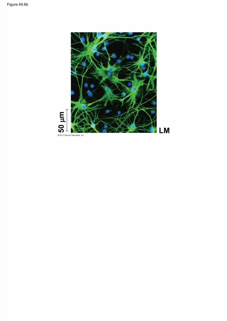
Glia
• Glia have numerous functions to nourish,support, and regulate neurons
– Embryonic radial glia form tracks along which
newly formed neurons migrate
– Astrocytes induce cells lining capillaries in the
CNS to form tight junctions, resulting in a
blood-brain barrier and restricting the entry of
most substances into the brain
© 2011 Pearson Education, Inc.
Figure 49 6

8/12/2019 49 Lecture Nervous System
http://slidepdf.com/reader/full/49-lecture-nervous-system 24/94
Figure 49.6CNS PNS
VENTRICLE
Cilia
Neuron Astrocyte
Oligodendrocyte
Capillary Ependymal cell
LM 5 0 m
Schwann cell
Microglial cell
Figure 49 6a

8/12/2019 49 Lecture Nervous System
http://slidepdf.com/reader/full/49-lecture-nervous-system 25/94
Figure 49.6a
CNS PNS
VENTRICLE
Cilia
Neuron Astrocyte
Oligodendrocyte
Capillary Ependymal cell
Schwann cell
Microglial cell
8/12/2019 49 Lecture Nervous System
http://slidepdf.com/reader/full/49-lecture-nervous-system 26/94

8/12/2019 49 Lecture Nervous System
http://slidepdf.com/reader/full/49-lecture-nervous-system 27/94
The Peripheral Nervous System
• The PNS transmits information to and from theCNS and regulates movement and the internalenvironment
• In the PNS, afferent neurons transmit informationto the CNS and efferent neurons transmitinformation away from the CNS
© 2011 Pearson Education, Inc.

8/12/2019 49 Lecture Nervous System
http://slidepdf.com/reader/full/49-lecture-nervous-system 28/94
• The PNS has two efferent components: themotor system and the autonomic nervous system
• The motor system carries signals to skeletal
muscles and is voluntary
• The autonomic nervous system regulates
smooth and cardiac muscles and is generally
involuntary
© 2011 Pearson Education, Inc.
Figure 49.7

8/12/2019 49 Lecture Nervous System
http://slidepdf.com/reader/full/49-lecture-nervous-system 29/94
Efferent neuronsAfferent neurons
Central NervousSystem
(information processing)
Peripheral NervousSystem
Sensoryreceptors
Internaland external
stimuli
Autonomic
nervous system
Motor
system
Control ofskeletal muscle
Sympatheticdivision
Parasympatheticdivision
Entericdivision
Control of smooth muscles,cardiac muscles, glands
Figure 49.7
8/12/2019 49 Lecture Nervous System
http://slidepdf.com/reader/full/49-lecture-nervous-system 30/94
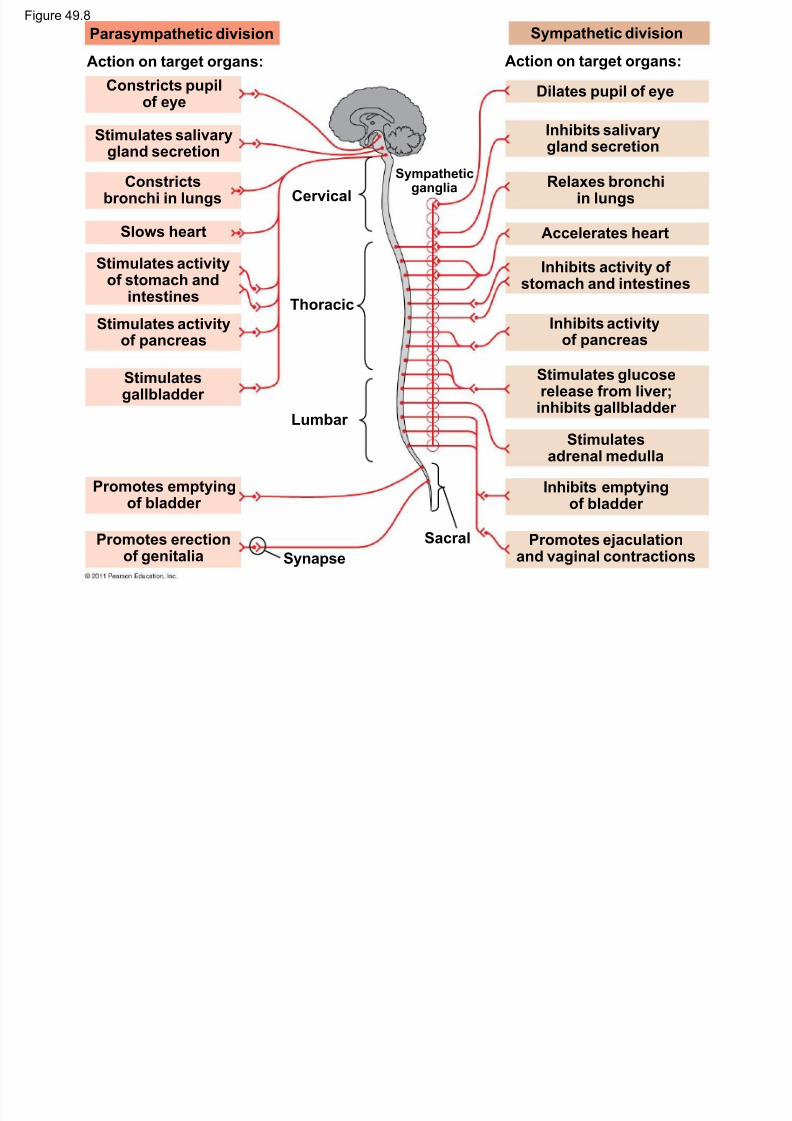
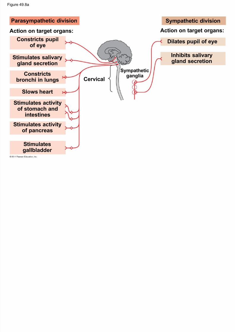
• The autonomic nervous system hassympathetic, parasympathetic, and enteric
divisions
• The sympathetic division regulates arousal
and energy generation (“fight-or-flight”
response)
• The parasympathetic division has
antagonistic effects on target organs andpromotes calming and a return to “rest and
digest” functions
© 2011 Pearson Education, Inc.

8/12/2019 49 Lecture Nervous System
http://slidepdf.com/reader/full/49-lecture-nervous-system 31/94
• The enteric division controls activity of thedigestive tract, pancreas, and gallbladder
© 2011 Pearson Education, Inc.
Figure 49.8
8/12/2019 49 Lecture Nervous System
http://slidepdf.com/reader/full/49-lecture-nervous-system 32/94
Parasympathetic division
Action on target organs:
Constricts pupilof eye
Stimulates salivarygland secretion
Constrictsbronchi in lungs
Slows heart
Stimulates activity
of stomach andintestines
Stimulates activityof pancreas
Stimulatesgallbladder
Promotes emptyingof bladder
Promotes erection
of genitalia
Cervical
Thoracic
Lumbar
Synapse
Sacral
Sympatheticganglia
Sympathetic division
Action on target organs:
Dilates pupil of eye
Accelerates heart
Inhibits salivary
gland secretion
Relaxes bronchiin lungs
Inhibits activity of
stomach and intestines
Inhibits activityof pancreas
Stimulates glucoserelease from liver;inhibits gallbladder
Stimulatesadrenal medulla
Inhibits emptyingof bladder
Promotes ejaculation
and vaginal contractions
g
Figure 49.8a
8/12/2019 49 Lecture Nervous System
http://slidepdf.com/reader/full/49-lecture-nervous-system 33/94
g
Parasympathetic division
Action on target organs:
Constricts pupilof eye
Stimulates salivarygland secretion
Constrictsbronchi in lungs
Slows heart
Stimulates activityof stomach and
intestines
Stimulates activityof pancreas
Stimulatesgallbladder
Cervical
Sympatheticganglia
Sympathetic division
Action on target organs:
Dilates pupil of eye
Inhibits salivarygland secretion
Figure 49.8b

8/12/2019 49 Lecture Nervous System
http://slidepdf.com/reader/full/49-lecture-nervous-system 34/94
Parasympathetic division
Promotes emptyingof bladder
Promotes erectionof genitalia
Thoracic
Lumbar
Synapse
Sacral
Sympathetic division
Accelerates heart
Relaxes bronchiin lungs
Inhibits activity ofstomach and intestines
Inhibits activity
of pancreas
Stimulates glucoserelease from liver;
inhibits gallbladder
Stimulates
adrenal medulla
Inhibits emptyingof bladder
Promotes ejaculationand vaginal contractions

8/12/2019 49 Lecture Nervous System
http://slidepdf.com/reader/full/49-lecture-nervous-system 35/94
Concept 49.2: The vertebrate brain is
regionally specialized
• Specific brain structures are particularly
specialized for diverse functions
•
These structures arise during embryonicdevelopment
© 2011 Pearson Education, Inc.
Figure 49.9a

8/12/2019 49 Lecture Nervous System
http://slidepdf.com/reader/full/49-lecture-nervous-system 36/94
Embryonic brain regions Brain structures in child and adultFigure 49.9b

8/12/2019 49 Lecture Nervous System
http://slidepdf.com/reader/full/49-lecture-nervous-system 37/94
Embryonic brain regions Brain structures in child and adult
Forebrain
Midbrain
Hindbrain
Telencephalon
Diencephalon
Mesencephalon
Metencephalon
Myelencephalon
Cerebrum (includes cerebral cortex, whitematter, basal nuclei)
Diencephalon (thalamus, hypothalamus,epithalamus)
Midbrain (part of brainstem)
Pons (part of brainstem), cerebellum
Medulla oblongata (part of brainstem)
Midbrain
Forebrain
Hindbrain
Telencephalon
Diencephalon
Mesencephalon
Metencephalon
Myelencephalon
Spinalcord
Cerebrum Diencephalon
Midbrain
Pons
Medullaoblongata
Cerebellum
Spinal cord
ChildEmbryo at 5 weeksEmbryo at 1 month
Figure 49.9ba

8/12/2019 49 Lecture Nervous System
http://slidepdf.com/reader/full/49-lecture-nervous-system 38/94
Midbrain
Forebrain
Hindbrain
Telencephalon
Diencephalon
Mesencephalon
Metencephalon
Myelencephalon
Spinalcord
Embryo at 5 weeksEmbryo at 1 month
Figure 49.9bb

8/12/2019 49 Lecture Nervous System
http://slidepdf.com/reader/full/49-lecture-nervous-system 39/94
Cerebrum Diencephalon
Midbrain
Pons
Medullaoblongata
Cerebellum
Spinal cord
Child
Figure 49.9c

8/12/2019 49 Lecture Nervous System
http://slidepdf.com/reader/full/49-lecture-nervous-system 40/94
Adult brain viewed from the rear
Cerebellum
Basal nucleiCerebrum
Left cerebralhemisphere
Right cerebralhemisphere
Cerebral cortex
Corpus callosum
Figure 49.9d

8/12/2019 49 Lecture Nervous System
http://slidepdf.com/reader/full/49-lecture-nervous-system 41/94
Diencephalon
Thalamus
Pineal gland
Hypothalamus
Pituitary gland
Spinal cord
Brainstem
Midbrain
Pons
Medulla
oblongata
8/12/2019 49 Lecture Nervous System
http://slidepdf.com/reader/full/49-lecture-nervous-system 42/94
Arousal and Sleep • The brainstem and cerebrum control arousal
and sleep
• The core of the brainstem has a diffuse networkof neurons called the reticular formation
• This regulates the amount and type ofinformation that reaches the cerebral cortexand affects alertness
•
The hormone melatonin is released by thepineal gland and plays a role in bird andmammal sleep cycles
© 2011 Pearson Education, Inc.
Figure 49.10

8/12/2019 49 Lecture Nervous System
http://slidepdf.com/reader/full/49-lecture-nervous-system 43/94
Eye
Reticular formation
Input from touch,pain, and temperaturereceptors
Input from nervesof ears
8/12/2019 49 Lecture Nervous System
http://slidepdf.com/reader/full/49-lecture-nervous-system 44/94
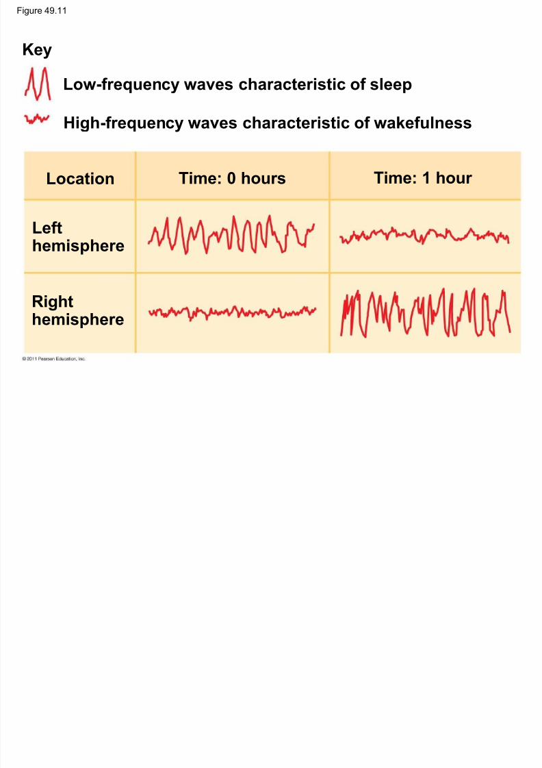
•Sleep is essential and may play a role in theconsolidation of learning and memory
• Dolphins sleep with one brain hemisphere at a
time and are therefore able to swim while
“asleep”
© 2011 Pearson Education, Inc.
Figure 49.11
8/12/2019 49 Lecture Nervous System
http://slidepdf.com/reader/full/49-lecture-nervous-system 45/94
Low-frequency waves characteristic of sleep
High-frequency waves characteristic of wakefulness
Key
Location Time: 0 hours Time: 1 hour
Lefthemisphere
Righthemisphere

8/12/2019 49 Lecture Nervous System
http://slidepdf.com/reader/full/49-lecture-nervous-system 46/94
Biological Clock Regulation •
Cycles of sleep and wakefulness are examplesof circadian rhythms, daily cycles of biological
activity
• Mammalian circadian rhythms rely on a
biological clock, molecular mechanism that
directs periodic gene expression
• Biological clocks are typically synchronized to
light and dark cycles
© 2011 Pearson Education, Inc.

8/12/2019 49 Lecture Nervous System
http://slidepdf.com/reader/full/49-lecture-nervous-system 47/94
Figure 49.12RESULTS

8/12/2019 49 Lecture Nervous System
http://slidepdf.com/reader/full/49-lecture-nervous-system 48/94
Wild-type hamster
Wild-type hamster withSCN from hamster
hamster
hamster with SCNfrom wild-type hamster
Beforeprocedures
After surgeryand transplant
C i r c a d i a n c y c l e p e r i o d ( h o u r s )
24
23
22
21
20
19
8/12/2019 49 Lecture Nervous System
http://slidepdf.com/reader/full/49-lecture-nervous-system 49/94
Emotions
•
Generation and experience of emotions involvemany brain structures including the amygdala,
hippocampus, and parts of the thalamus
• These structures are grouped as the limbic
system
• The limbic system also functions in motivation,
olfaction, behavior, and memory
© 2011 Pearson Education, Inc.

8/12/2019 49 Lecture Nervous System
http://slidepdf.com/reader/full/49-lecture-nervous-system 50/94

8/12/2019 49 Lecture Nervous System
http://slidepdf.com/reader/full/49-lecture-nervous-system 51/94
•
Generation and experience of emotion alsorequire interaction between the limbic systemand sensory areas of the cerebrum
• The structure most important to the storage of
emotion in the memory is the amygdala, a massof nuclei near the base of the cerebrum
© 2011 Pearson Education, Inc.
Figure 49.14

8/12/2019 49 Lecture Nervous System
http://slidepdf.com/reader/full/49-lecture-nervous-system 52/94
Nucleus accumbens Amygdala
Happy music Sad music

8/12/2019 49 Lecture Nervous System
http://slidepdf.com/reader/full/49-lecture-nervous-system 53/94
Figure 49.14b

8/12/2019 49 Lecture Nervous System
http://slidepdf.com/reader/full/49-lecture-nervous-system 54/94
Amygdala
Sad music

8/12/2019 49 Lecture Nervous System
http://slidepdf.com/reader/full/49-lecture-nervous-system 55/94
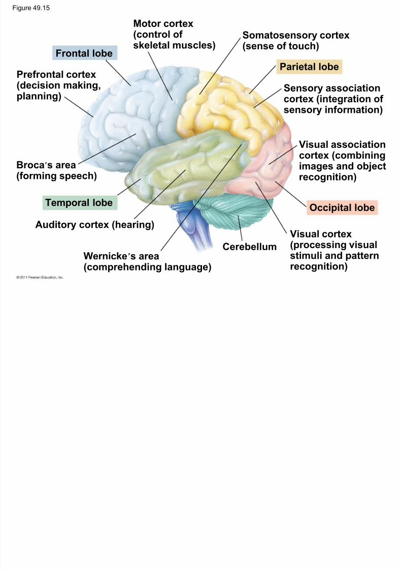
Concept 49.3: The cerebral cortex controls
voluntary movement and cognitive functions
• The cerebrum, the largest structure in the
human brain, is essential for awareness,
language, cognition, memory, and
consciousness
• Four regions, or lobes (frontal, temporal,
occipital, and parietal), are landmarks for
particular functions
© 2011 Pearson Education, Inc.
Figure 49.15
M t t
8/12/2019 49 Lecture Nervous System
http://slidepdf.com/reader/full/49-lecture-nervous-system 56/94
Motor cortex(control ofskeletal muscles)
Frontal lobe
Prefrontal cortex(decision making,planning)
Broca’s area(forming speech)
Temporal lobe
Auditory cortex (hearing)
Wernicke’s area(comprehending language)
Somatosensory cortex(sense of touch)
Parietal lobe
Sensory associationcortex (integration ofsensory information)
Visual associationcortex (combiningimages and objectrecognition)
Occipital lobe
Cerebellum
Visual cortex(processing visualstimuli and patternrecognition)
8/12/2019 49 Lecture Nervous System
http://slidepdf.com/reader/full/49-lecture-nervous-system 57/94
Language and Speech
•
Studies of brain activity have mapped areasresponsible for language and speech
• Broca’s area in the frontal lobe is active when
speech is generated
• Wernicke’s area in the temporal lobe is active
when speech is heard
• These areas belong to a larger network of
regions involved in language
© 2011 Pearson Education, Inc.
Figure 49.16

8/12/2019 49 Lecture Nervous System
http://slidepdf.com/reader/full/49-lecture-nervous-system 58/94
Hearingwords
Speakingwords
Seeingwords
Generatingwords
Max
Min
8/12/2019 49 Lecture Nervous System
http://slidepdf.com/reader/full/49-lecture-nervous-system 59/94
Lateralization of Cortical Function
•
The two hemispheres make distinct contributionsto brain function
• The left hemisphere is more adept at language,
math, logic, and processing of serial sequences
• The right hemisphere is stronger at pattern
recognition, nonverbal thinking, and emotional
processing
© 2011 Pearson Education, Inc.
8/12/2019 49 Lecture Nervous System
http://slidepdf.com/reader/full/49-lecture-nervous-system 60/94
•
The differences in hemisphere function arecalled lateralization
• Lateralization is partly linked to handedness
• The two hemispheres work together by
communicating through the fibers of the corpus
callosum
© 2011 Pearson Education, Inc.

8/12/2019 49 Lecture Nervous System
http://slidepdf.com/reader/full/49-lecture-nervous-system 61/94
Information Processing
•
The cerebral cortex receives input from sensoryorgans and somatosensory receptors
• Somatosensory receptors provide information
about touch, pain, pressure, temperature, and
the position of muscles and limbs
• The thalamus directs different types of input to
distinct locations
© 2011 Pearson Education, Inc.

8/12/2019 49 Lecture Nervous System
http://slidepdf.com/reader/full/49-lecture-nervous-system 62/94
•
Adjacent areas process features in the sensoryinput and integrate information from different
sensory areas
• Integrated sensory information passes to the
prefrontal cortex, which helps plan actions andmovements
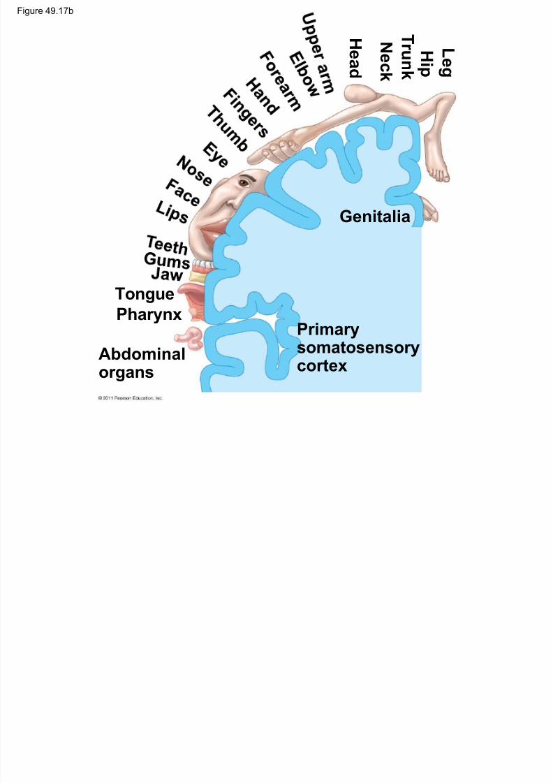
• In the somatosensory cortex and motor cortex,
neurons are arranged according to the part ofthe body that generates input or receives
commands
© 2011 Pearson Education, Inc.
Figure 49.17

8/12/2019 49 Lecture Nervous System
http://slidepdf.com/reader/full/49-lecture-nervous-system 63/94
Frontal lobe Parietal lobe
Primarymotor cortex
Primarysomatosensorycortex
GenitaliaToes
Abdominalorgans
Tongue
Jaw
Hi p
Kn e e
Tongue
Pharynx
H e a d
N e ck
T r unk
Hi p
L e g
Figure 49.17a

8/12/2019 49 Lecture Nervous System
http://slidepdf.com/reader/full/49-lecture-nervous-system 64/94
Primarymotor cortex
Toes
Tongue
Jaw
Hi p
Kn e e
Figure 49.17b
8/12/2019 49 Lecture Nervous System
http://slidepdf.com/reader/full/49-lecture-nervous-system 65/94
Primarysomatosensorycortex
Genitalia
Abdominalorgans
TonguePharynx
H e a d
N e ck
T r unk
Hi p
L e g

8/12/2019 49 Lecture Nervous System
http://slidepdf.com/reader/full/49-lecture-nervous-system 66/94
Frontal Lobe Function
•
Frontal lobe damage may impair decisionmaking and emotional responses but leave
intellect and memory intact
• The frontal lobes have a substantial effect on
“executive functions”
© 2011 Pearson Education, Inc.
Figure 49.UN01

8/12/2019 49 Lecture Nervous System
http://slidepdf.com/reader/full/49-lecture-nervous-system 67/94
8/12/2019 49 Lecture Nervous System
http://slidepdf.com/reader/full/49-lecture-nervous-system 68/94
Evolution of Cognition in Vertebrates
•
Previous ideas that a highly convolutedneocortex is required for advanced cognition
may be incorrect
• The anatomical basis for sophisticated
information processing in birds (without a highlyconvoluted neocortex) appears to be the
clustering of nuclei in the top or outer portion of
the brain (pallium)
© 2011 Pearson Education, Inc.
Human brain Cerebrum (includingcerebral cortex)
Figure 49.18

8/12/2019 49 Lecture Nervous System
http://slidepdf.com/reader/full/49-lecture-nervous-system 69/94
Avian brain
Thalamus
Midbrain
Hindbrain Cerebellum
Avian brain
to scale
Thalamus
Midbrain
Hindbrain
Cerebellum
cerebral cortex)
Cerebrum(including pallium)

8/12/2019 49 Lecture Nervous System
http://slidepdf.com/reader/full/49-lecture-nervous-system 70/94
Concept 49.4 Changes in synaptic
connections underlie memory and learning
• Two processes dominate embryonic
development of the nervous system
– Neurons compete for growth-supporting factors
in order to survive
– Only half the synapses that form during embryo
development survive into adulthood
© 2011 Pearson Education, Inc.

8/12/2019 49 Lecture Nervous System
http://slidepdf.com/reader/full/49-lecture-nervous-system 71/94
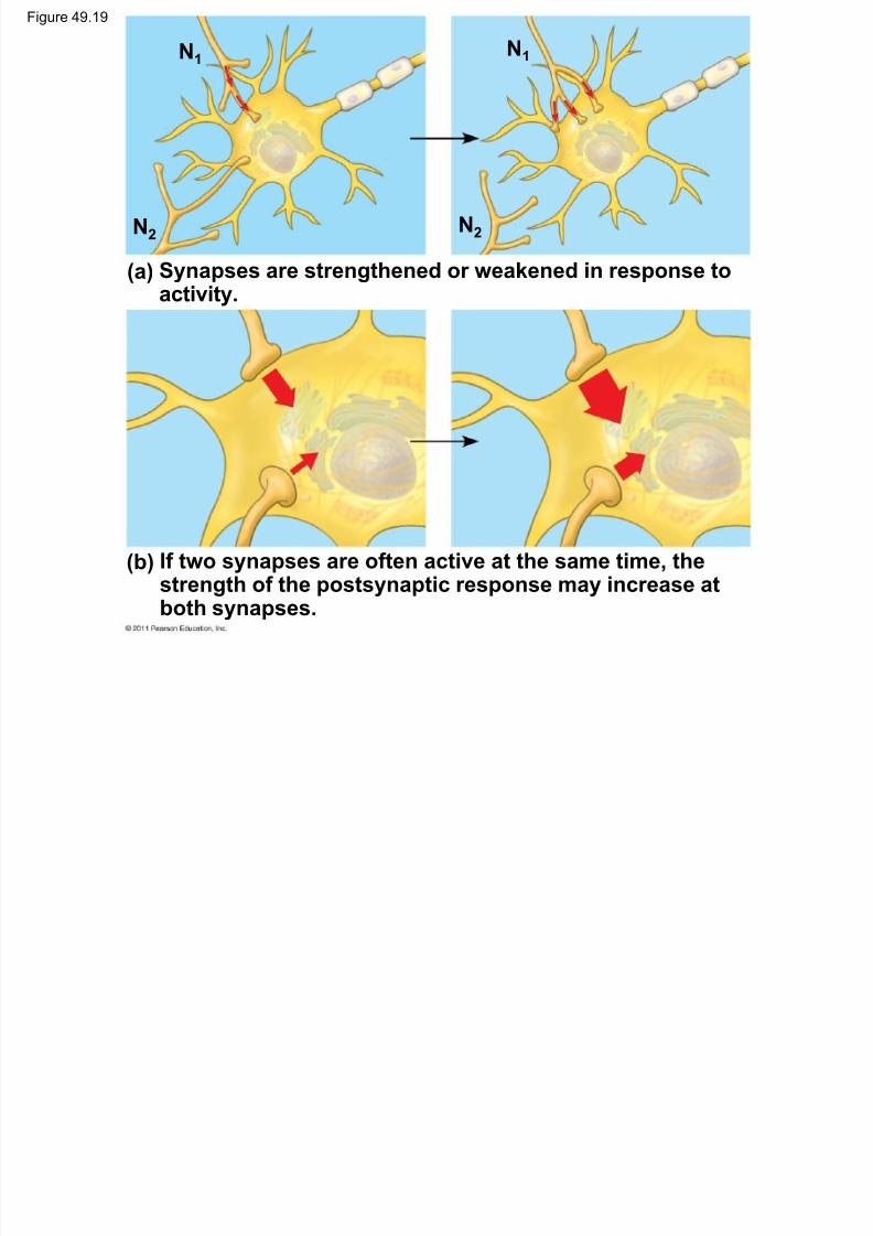
Neural Plasticity
•
Neural plasticity describes the ability of thenervous system to be modified after birth
• Changes can strengthen or weaken signaling at
a synapse
© 2011 Pearson Education, Inc.
Figure 49.19
N1N1
8/12/2019 49 Lecture Nervous System
http://slidepdf.com/reader/full/49-lecture-nervous-system 72/94
N2
N1
N2
1
(a) Synapses are strengthened or weakened in response toactivity.
(b) If two synapses are often active at the same time, thestrength of the postsynaptic response may increase atboth synapses.

8/12/2019 49 Lecture Nervous System
http://slidepdf.com/reader/full/49-lecture-nervous-system 73/94
Memory and Learning
•
The formation of memories is an example ofneural plasticity
• Short-term memory is accessed via the
hippocampus
• The hippocampus also plays a role in forming
long-term memory, which is stored in the
cerebral cortex
•
Some consolidation of memory is thought tooccur during sleep
© 2011 Pearson Education, Inc.

8/12/2019 49 Lecture Nervous System
http://slidepdf.com/reader/full/49-lecture-nervous-system 74/94
Long-Term Potentiation
•
In the vertebrate brain, a form of learning calledlong-term potentiation (LTP) involves an
increase in the strength of synaptic transmission
• LTP involves glutamate receptors
• If the presynaptic and postsynaptic neurons are
stimulated at the same time, the set of receptors
present on the postsynaptic membranes
changes
© 2011 Pearson Education, Inc.
Figure 49.20 PRESYNAPTICNEURON
Ca2
Na

8/12/2019 49 Lecture Nervous System
http://slidepdf.com/reader/full/49-lecture-nervous-system 75/94
GlutamateMg2
NMDA
receptor(closed)
Stored
AMPAreceptor
NMDA receptor (open)
POSTSYNAPTICNEURON
(a) Synapse prior to long-term potentiation (LTP)
(b) Establishing LTP
(c) Synapse exhibiting LTP
Depolarization
Actionpotential
2
1
3
1
2
3
4
Figure 49.20a

8/12/2019 49 Lecture Nervous System
http://slidepdf.com/reader/full/49-lecture-nervous-system 76/94
PRESYNAPTICNEURON
Glutamate Mg2
Ca2
Na
NMDAreceptor
(closed)
Stored
AMPAreceptor
NMDA receptor (open)
POSTSYNAPTICNEURON
(a) Synapse prior to long-term potentiation (LTP)

8/12/2019 49 Lecture Nervous System
http://slidepdf.com/reader/full/49-lecture-nervous-system 77/94
Figure 49.20c

8/12/2019 49 Lecture Nervous System
http://slidepdf.com/reader/full/49-lecture-nervous-system 78/94
(c) Synapse exhibiting LTP
Depolarization
Actionpotential
AMPAreceptor
NMDA receptor
1 3
42
St C ll i th B i

8/12/2019 49 Lecture Nervous System
http://slidepdf.com/reader/full/49-lecture-nervous-system 79/94
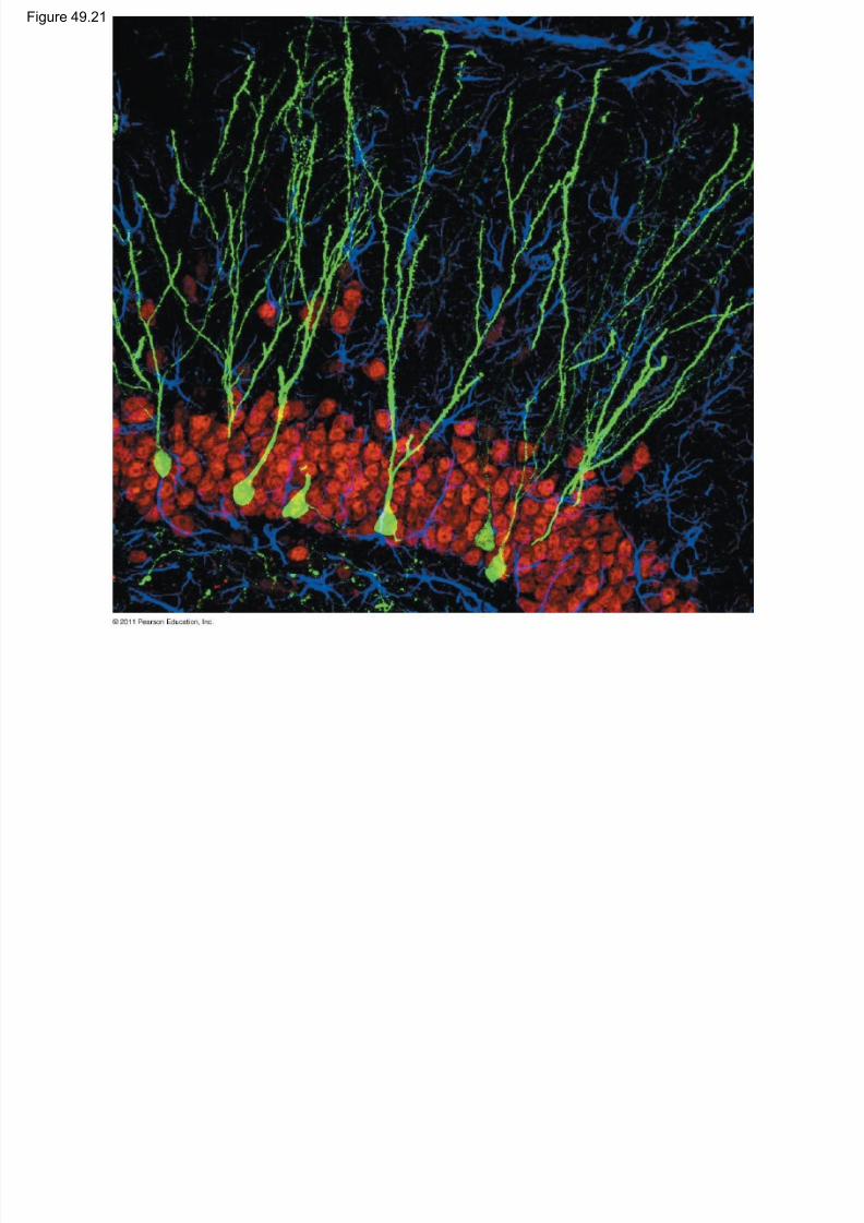
Stem Cells in the Brain
•
The adult human brain contains neural stemcells
• In mice, stem cells in the brain can give rise to
neurons that mature and become incorporated
into the adult nervous system
• Such neurons play an essential role in learning
and memory
© 2011 Pearson Education, Inc.
Figure 49.21
8/12/2019 49 Lecture Nervous System
http://slidepdf.com/reader/full/49-lecture-nervous-system 80/94
C t 49 5 N t di d

8/12/2019 49 Lecture Nervous System
http://slidepdf.com/reader/full/49-lecture-nervous-system 81/94
Concept 49.5: Nervous system disorders can
be explained in molecular terms
• Disorders of the nervous system include
schizophrenia, depression, drug addiction,
Alzheimer ’s disease, and Parkinson’s disease
• Genetic and environmental factors contribute to
diseases of the nervous system
© 2011 Pearson Education, Inc.
Figure 49.22
Genes shared with relatives ofperson ith schi ophrenia
50

8/12/2019 49 Lecture Nervous System
http://slidepdf.com/reader/full/49-lecture-nervous-system 82/94
person with schizophrenia
12.5% (3rd-degree relative)
25% (2nd-degree relative)
50% (1st-degree relative)
100%
40
30
20
10
0
Relationship to person with schizophrenia
R i s k o f d e v e
l o p i n g s c h i z o p h r e n i a ( % )
I n d i v i d u a l ,
g e n e r a l
p o p u l a t i o n
F i r s t c o u s i n
U n c l e / a u n t
N e p h e w /
n i e c e
F r a t e r n a l
t w i n
I d e n t i c a l
t w i n
G r a n d c h i l d
H a l f s i b l i n g
P a r e n t
F u l l s i b l i n g
C h i l d
S hi h i

8/12/2019 49 Lecture Nervous System
http://slidepdf.com/reader/full/49-lecture-nervous-system 83/94
Schizophrenia
•
About 1% of the world’s population suffers fromschizophrenia
• Schizophrenia is characterized by hallucinations,
delusions, and other symptoms
• Available treatments focus on brain pathways
that use dopamine as a neurotransmitter
© 2011 Pearson Education, Inc.
D i
8/12/2019 49 Lecture Nervous System
http://slidepdf.com/reader/full/49-lecture-nervous-system 84/94
Depression
•
Two broad forms of depressive illness areknown: major depressive disorder and bipolar
disorder
• In major depressive disorder , patients have a
persistent lack of interest or pleasure in mostactivities
• Bipolar disorder is characterized by manic
(high-mood) and depressive (low-mood) phases• Treatments for these types of depression include
drugs such as Prozac
© 2011 Pearson Education, Inc.
D Addi ti d th B i ’ R d

8/12/2019 49 Lecture Nervous System
http://slidepdf.com/reader/full/49-lecture-nervous-system 85/94
Drug Addiction and the Brain’s Reward
System
• The brain’s reward system rewards motivation
with pleasure
• Some drugs are addictive because they
increase activity of the brain’s reward system
• These drugs include cocaine, amphetamine,
heroin, alcohol, and tobacco
• Drug addiction is characterized by compulsiveconsumption and an inability to control intake
© 2011 Pearson Education, Inc.

8/12/2019 49 Lecture Nervous System
http://slidepdf.com/reader/full/49-lecture-nervous-system 86/94
•
Addictive drugs enhance the activity of thedopamine pathway
• Drug addiction leads to long-lasting changes in
the reward circuitry that cause craving for the
drug
© 2011 Pearson Education, Inc.
Figure 49.23 Nicotinestimulatesdopamine
Inhibitory neuron

8/12/2019 49 Lecture Nervous System
http://slidepdf.com/reader/full/49-lecture-nervous-system 87/94
dopamine-releasingVTA neuron.
Dopamine-releasingVTA neuron
Cerebralneuron ofrewardpathway
Opium and heroindecrease activityof inhibitoryneuron.
Cocaine andamphetaminesblock removalof dopaminefrom synaptic
cleft.
Rewardsystemresponse
Al h i ’ Di

8/12/2019 49 Lecture Nervous System
http://slidepdf.com/reader/full/49-lecture-nervous-system 88/94
Alzheimer’s Disease
•
Alzheimer ’s disease is a mental deteriorationcharacterized by confusion and memory loss
• Alzheimer ’s disease is caused by the formationof neurofibrillary tangles and amyloid plaques in
the brain• There is no cure for this disease though some
drugs are effective at relieving symptoms
© 2011 Pearson Education, Inc.

8/12/2019 49 Lecture Nervous System
http://slidepdf.com/reader/full/49-lecture-nervous-system 89/94
Parkinson’s Disease

8/12/2019 49 Lecture Nervous System
http://slidepdf.com/reader/full/49-lecture-nervous-system 90/94
Parkinson s Disease
•
Parkinson’s disease is a motor disordercaused by death of dopamine-secreting
neurons in the midbrain
• It is characterized by muscle tremors, flexed
posture, and a shuffling gait• There is no cure, although drugs and various
other approaches are used to manage
symptoms
© 2011 Pearson Education, Inc.
Figure 49.UN02
8/12/2019 49 Lecture Nervous System
http://slidepdf.com/reader/full/49-lecture-nervous-system 91/94
Nerve net
Hydra (cnidarian) Salamander (vertebrate)
Sensoryganglia
Spinalcord(dorsal
nervecord)
Brain
Figure 49.UN03

8/12/2019 49 Lecture Nervous System
http://slidepdf.com/reader/full/49-lecture-nervous-system 92/94
Capillary Neuron Microglial cell
Schwanncells
Oligodendrocyte
Astrocyte
PNSCNS
Cilia
VENTRICLEEpendy-malcell

8/12/2019 49 Lecture Nervous System
http://slidepdf.com/reader/full/49-lecture-nervous-system 93/94
Figure 49.UN05

8/12/2019 49 Lecture Nervous System
http://slidepdf.com/reader/full/49-lecture-nervous-system 94/94