Embed Size (px)
Citation preview

OPEN
ARTICLE
5-Aminoimidazole-4-carboxamide ribonucleoside-inducedautophagy flux during differentiation of monocyticleukemia cellsVilma Dembitz, Hrvoje Lalic and Dora Visnjic
Pharmacological modulators of AMP-dependent kinase (AMPK) have been suggested in treatment of cancer. The biguanidemetformin and 5-aminoimidazole-4-carboxamide ribonucleoside (AICAR) have been reported to inhibit proliferation of solid tumorsand hematological malignancies, but their role in differentiation is less explored. Our previous study demonstrated that AICARalone induced AMPK-independent expression of differentiation markers in monocytic U937 leukemia cells, and no such effectswere observed in response to metformin. The aim of this study was to determine the mechanism of AICAR-mediated effects and totest for the possible role of autophagy in differentiation of leukemia cells. The results showed that AICAR-mediated effects on theexpression of differentiation markers were not mimicked by A769662, a more specific direct AMPK activator. Long-term incubationof U937 cells with AICAR and other differentiation agents, all-trans-retinoic acid (ATRA) and phorbol 12-myristate 13-acetate,increased the expression of the autophagy marker LC3B-II, and these effects were not observed in response to metformin. Westernblot and immunofluorescence analyses of U937 cells treated with bafilomycin A1 or transfected with mRFP-GFP-LC3 proved thatthe increase in the expression of LC3B-II was due to an increase in autophagy flux, and not to a decrease in lysosomal degradation.3-Methyladenine inhibited the expression of differentiation markers in response to all inducers, but had stimulatory effects onautophagy flux at dose that effectively inhibited the production of phosphatidylinositol 3-phosphate. The small inhibitory RNA-mediated down-modulation of Beclin 1 and hVPS34 had no effects on AICAR and ATRA-mediated increase in the expression ofdifferentiation markers. These results show that AICAR and other differentiation agents induce autophagy flux in U937 cells andthat the effects of AICAR and ATRA on the expression of differentiation markers do not depend on the normal levels of key proteinsof the classical or canonical autophagy pathway.
Cell Death Discovery (2017) 3, 17066; doi:10.1038/cddiscovery.2017.66; published online 2 October 2017
INTRODUCTIONSeveral studies have suggested that drugs that modulate activityof AMP-activated kinase (AMPK) have potential for the treatmentand prevention of cancer. The biguanide metformin, a widely useddrug for the treatment of type 2 diabetes, and 5-aminoimidazole-4-carboxamide ribonucleoside (AICAR, acadesine) have beenreported to exert antiproliferative effects in various solid tumorsand hematological malignancies, but their role in differentiationhas been less explored.1,2 Although both drugs activate AMPK, anevolutionary conserved serine/threonine kinase that is activatedwhenever the energy level in the cell is low and the ratio of AMPto ATP increased, an increasing number of studies demonstratethat majority of beneficial effects of metformin and AICAR areactually AMPK-independent.3,4 Our recent study demonstratedthat AICAR alone induced the expression of cell surface markersassociated with mature monocytes and macrophages in mono-cytic U937 cells.5 However, no significant increase in theexpression of differentiation markers was observed in U937 cellstreated with metformin alone, although the effects on prolifera-tion and survival were similar to the ones observed in thepresence of AICAR.It is still unknown which are the mechanisms responsible for
AICAR-mediated effects in acute myeloid leukemia (AML) cell lines.
Within the cell, AICA-ribonucleoside could be phosphorylated byadenosine kinase into AICA-ribonucleotide or ZMP, which is ananalog of 5′-AMP; ZMP then binds to γ regulatory subunit andactivates AMPK in a similar manner to AMP.6 Although wedetected time- and dose-dependent increase in the level of Thr-phosphorylated AMPK, a significant decrease in AMPK expressionthat was achieved by using commercially available siRNAsequences in U937 cells had no significant effects on the AICAR-mediated effects on the number of viable cells or the expression ofdifferentiation markers.5 Therefore, present studies are aimed todetermine the mechanism responsible for beneficial effects of AICARin AML cells and to further elucidate signaling mechanisms responsiblefor differentiation of U937 cells in response to other inducers.In chronic myelogenous leukemia (CML) cell lines, AMPK-
independent cell death induced by AICAR involved autophagy,the major intracellular pathway for the selective degradation ofcytoplasmic organelles and long-lived proteins.7 Although initiallydescribed as a protective mechanism that allowed cells to survivein the absence of nutrients, the autophagy has been recentlyimplicated in several other conditions, including cell death,neurodegeneration, immunity, cancer and differentiation.8,9
Autophagy is mediated by autophagy-related (ATG) genes thatare evolutionary conserved from yeasts to mammals; a key
Department of Physiology and Croatian Institute for Brain Research, School of Medicine, University of Zagreb, Salata 12, Zagreb 10 000, Croatia.Correspondence: D Visnjic ([email protected])Received 14 June 2017; revised 29 July 2017; accepted 18 August 2017; Edited by N Barlev
Citation: Cell Death Discovery (2017) 3, 17066; doi:10.1038/cddiscovery.2017.66Official journal of the Cell Death Differentiation Association
www.nature.com/cddiscovery

initiation signal is provided by a complex consisting of Beclin 1 (orAtg6), class III phosphatidylinositol 3-kinase (PI3KC3 or Vps34),Vps15 and Atg14. In unstressed cells, autophagy is inhibited bymammalian target of rapamycin complex 1 (mTORC1). Therefore,inhibition of mTOR by either rapamycin or AMPK activationpromotes autophagy.8 Recent studies pointed to the role of
autophagy in differentiation of some leukemia cell lines, includingall-trans-retinoic acid (ATRA)-induced differentiation of acutepromyelocytic leukemia NB4 cells,10–12 vitamin D3-mediateddifferentiation of myeloblastic HL-60 cells13 and megakaryocyticdifferentiation of CML K562 cells in response to phorbol 12-myristate 13-acetate (PMA) or lapatinib.14,15
Autophagy during differentiation of U937 cellsV Dembitz et al
2
Cell Death Discovery (2017) 17066 Official journal of the Cell Death Differentiation Association
In the present study, we tested for the possible role ofautophagy in AICAR-mediated differentiation of monocytic U937leukemia cells. The results of the study showed that long-termincubation of U937 with AICAR and other inducers of differentia-tion induced autophagy flux that was not observed in cells treatedwith metformin. However, AICAR-mediated increase in theexpression of differentiation markers did not depend on thepresence of key proteins of canonical autophagy pathway.
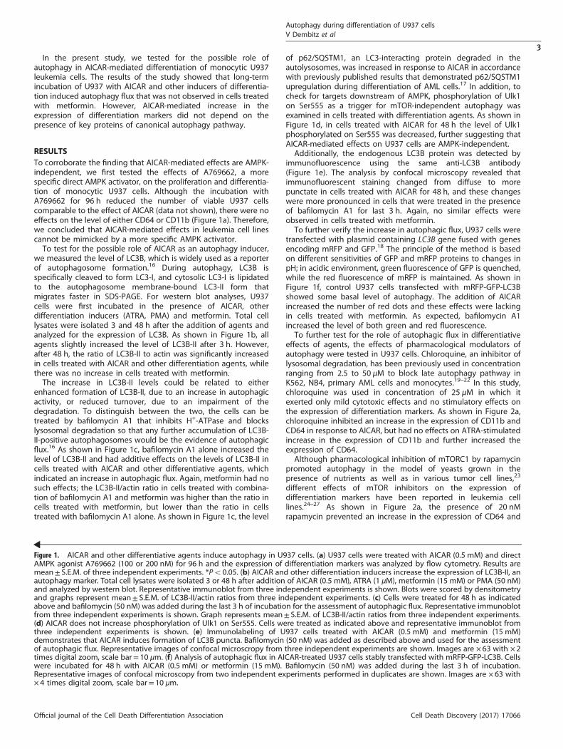
RESULTSTo corroborate the finding that AICAR-mediated effects are AMPK-independent, we first tested the effects of A769662, a morespecific direct AMPK activator, on the proliferation and differentia-tion of monocytic U937 cells. Although the incubation withA769662 for 96 h reduced the number of viable U937 cellscomparable to the effect of AICAR (data not shown), there were noeffects on the level of either CD64 or CD11b (Figure 1a). Therefore,we concluded that AICAR-mediated effects in leukemia cell linescannot be mimicked by a more specific AMPK activator.To test for the possible role of AICAR as an autophagy inducer,
we measured the level of LC3B, which is widely used as a reporterof autophagosome formation.16 During autophagy, LC3B isspecifically cleaved to form LC3-I, and cytosolic LC3-I is lipidatedto the autophagosome membrane-bound LC3-II form thatmigrates faster in SDS-PAGE. For western blot analyses, U937cells were first incubated in the presence of AICAR, otherdifferentiation inducers (ATRA, PMA) and metformin. Total celllysates were isolated 3 and 48 h after the addition of agents andanalyzed for the expression of LC3B. As shown in Figure 1b, allagents slightly increased the level of LC3B-II after 3 h. However,after 48 h, the ratio of LC3B-II to actin was significantly increasedin cells treated with AICAR and other differentiation agents, whilethere was no increase in cells treated with metformin.The increase in LC3B-II levels could be related to either
enhanced formation of LC3B-II, due to an increase in autophagicactivity, or reduced turnover, due to an impairment of thedegradation. To distinguish between the two, the cells can betreated by bafilomycin A1 that inhibits H+-ATPase and blockslysosomal degradation so that any further accumulation of LC3B-II-positive autophagosomes would be the evidence of autophagicflux.16 As shown in Figure 1c, bafilomycin A1 alone increased thelevel of LC3B-II and had additive effects on the levels of LC3B-II incells treated with AICAR and other differentiative agents, whichindicated an increase in autophagic flux. Again, metformin had nosuch effects; the LC3B-II/actin ratio in cells treated with combina-tion of bafilomycin A1 and metformin was higher than the ratio incells treated with metformin, but lower than the ratio in cellstreated with bafilomycin A1 alone. As shown in Figure 1c, the level
of p62/SQSTM1, an LC3-interacting protein degraded in theautolysosomes, was increased in response to AICAR in accordancewith previously published results that demonstrated p62/SQSTM1upregulation during differentiation of AML cells.17 In addition, tocheck for targets downstream of AMPK, phosphorylation of Ulk1on Ser555 as a trigger for mTOR-independent autophagy wasexamined in cells treated with differentiation agents. As shown inFigure 1d, in cells treated with AICAR for 48 h the level of Ulk1phosphorylated on Ser555 was decreased, further suggesting thatAICAR-mediated effects on U937 cells are AMPK-independent.Additionally, the endogenous LC3B protein was detected by
immunofluorescence using the same anti-LC3B antibody(Figure 1e). The analysis by confocal microscopy revealed thatimmunofluorescent staining changed from diffuse to morepunctate in cells treated with AICAR for 48 h, and these changeswere more pronounced in cells that were treated in the presenceof bafilomycin A1 for last 3 h. Again, no similar effects wereobserved in cells treated with metformin.To further verify the increase in autophagic flux, U937 cells were
transfected with plasmid containing LC3B gene fused with genesencoding mRFP and GFP.18 The principle of the method is basedon different sensitivities of GFP and mRFP proteins to changes inpH; in acidic environment, green fluorescence of GFP is quenched,while the red fluorescence of mRFP is maintained. As shown inFigure 1f, control U937 cells transfected with mRFP-GFP-LC3Bshowed some basal level of autophagy. The addition of AICARincreased the number of red dots and these effects were lackingin cells treated with metformin. As expected, bafilomycin A1increased the level of both green and red fluorescence.To further test for the role of autophagic flux in differentiative
effects of agents, the effects of pharmacological modulators ofautophagy were tested in U937 cells. Chloroquine, an inhibitor oflysosomal degradation, has been previously used in concentrationranging from 2.5 to 50 μM to block late autophagy pathway inK562, NB4, primary AML cells and monocytes.19–22 In this study,chloroquine was used in concentration of 25 μM in which itexerted only mild cytotoxic effects and no stimulatory effects onthe expression of differentiation markers. As shown in Figure 2a,chloroquine inhibited an increase in the expression of CD11b andCD64 in response to AICAR, but had no effects on ATRA-stimulatedincrease in the expression of CD11b and further increased theexpression of CD64.Although pharmacological inhibition of mTORC1 by rapamycin
promoted autophagy in the model of yeasts grown in thepresence of nutrients as well as in various tumor cell lines,23
different effects of mTOR inhibitors on the expression ofdifferentiation markers have been reported in leukemia celllines.24–27 As shown in Figure 2a, the presence of 20 nMrapamycin prevented an increase in the expression of CD64 and
Figure 1. AICAR and other differentiative agents induce autophagy in U937 cells. (a) U937 cells were treated with AICAR (0.5 mM) and directAMPK agonist A769662 (100 or 200 nM) for 96 h and the expression of differentiation markers was analyzed by flow cytometry. Results aremean± S.E.M. of three independent experiments. *Po0.05. (b) AICAR and other differentiation inducers increase the expression of LC3B-II, anautophagy marker. Total cell lysates were isolated 3 or 48 h after addition of AICAR (0.5 mM), ATRA (1 μM), metformin (15 mM) or PMA (50 nM)and analyzed by western blot. Representative immunoblot from three independent experiments is shown. Blots were scored by densitometryand graphs represent mean± S.E.M. of LC3B-II/actin ratios from three independent experiments. (c) Cells were treated for 48 h as indicatedabove and bafilomycin (50 nM) was added during the last 3 h of incubation for the assessment of autophagic flux. Representative immunoblotfrom three independent experiments is shown. Graph represents mean± S.E.M. of LC3B-II/actin ratios from three independent experiments.(d) AICAR does not increase phosphorylation of Ulk1 on Ser555. Cells were treated as indicated above and representative immunoblot fromthree independent experiments is shown. (e) Immunolabeling of U937 cells treated with AICAR (0.5 mM) and metformin (15 mM)demonstrates that AICAR induces formation of LC3B puncta. Bafilomycin (50 nM) was added as described above and used for the assessmentof autophagic flux. Representative images of confocal microscropy from three independent experiments are shown. Images are × 63 with × 2times digital zoom, scale bar= 10 μm. (f) Analysis of autophagic flux in AICAR-treated U937 cells stably transfected with mRFP-GFP-LC3B. Cellswere incubated for 48 h with AICAR (0.5 mM) or metformin (15 mM). Bafilomycin (50 nM) was added during the last 3 h of incubation.Representative images of confocal microscopy from two independent experiments performed in duplicates are shown. Images are × 63 with× 4 times digital zoom, scale bar= 10 μm.
Autophagy during differentiation of U937 cellsV Dembitz et al
3
Official journal of the Cell Death Differentiation Association Cell Death Discovery (2017) 17066
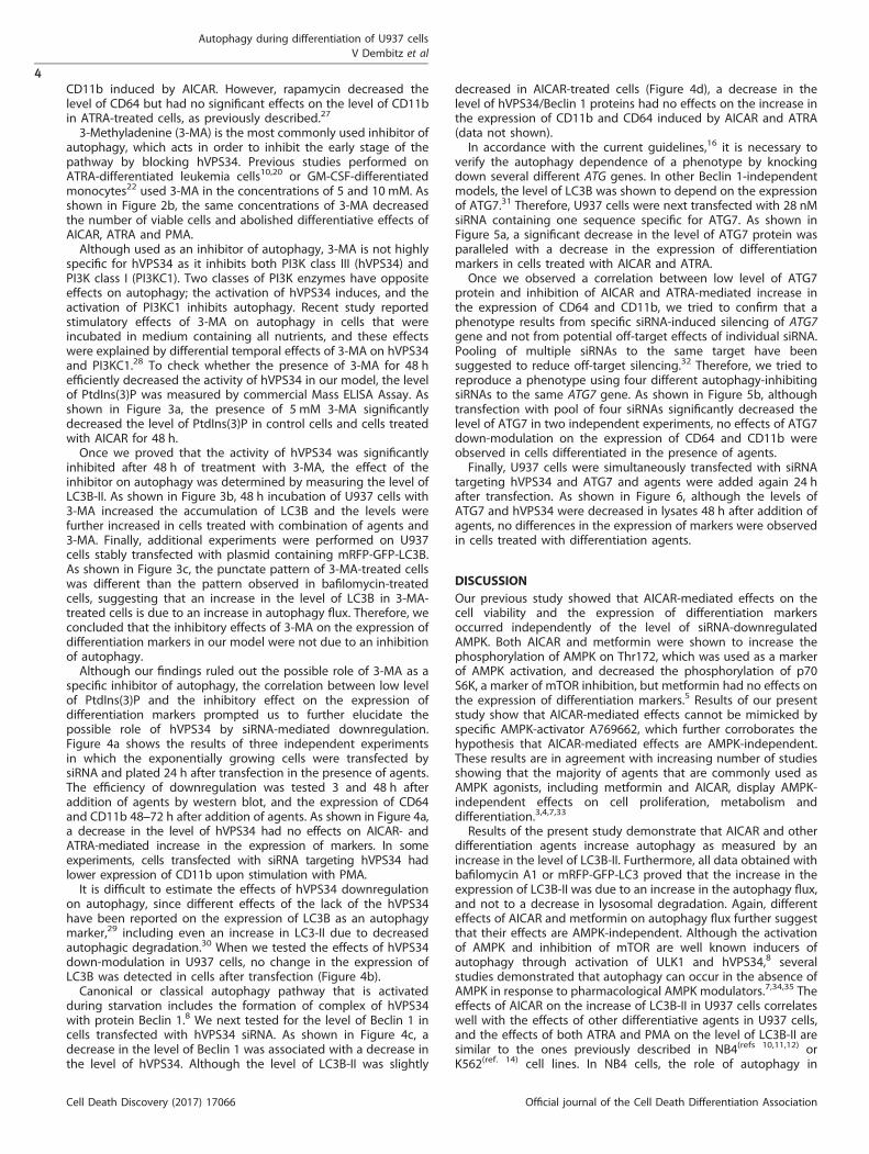
CD11b induced by AICAR. However, rapamycin decreased thelevel of CD64 but had no significant effects on the level of CD11bin ATRA-treated cells, as previously described.27
3-Methyladenine (3-MA) is the most commonly used inhibitor ofautophagy, which acts in order to inhibit the early stage of thepathway by blocking hVPS34. Previous studies performed onATRA-differentiated leukemia cells10,20 or GM-CSF-differentiatedmonocytes22 used 3-MA in the concentrations of 5 and 10 mM. Asshown in Figure 2b, the same concentrations of 3-MA decreasedthe number of viable cells and abolished differentiative effects ofAICAR, ATRA and PMA.Although used as an inhibitor of autophagy, 3-MA is not highly
specific for hVPS34 as it inhibits both PI3K class III (hVPS34) andPI3K class I (PI3KC1). Two classes of PI3K enzymes have oppositeeffects on autophagy; the activation of hVPS34 induces, and theactivation of PI3KC1 inhibits autophagy. Recent study reportedstimulatory effects of 3-MA on autophagy in cells that wereincubated in medium containing all nutrients, and these effectswere explained by differential temporal effects of 3-MA on hVPS34and PI3KC1.28 To check whether the presence of 3-MA for 48 hefficiently decreased the activity of hVPS34 in our model, the levelof PtdIns(3)P was measured by commercial Mass ELISA Assay. Asshown in Figure 3a, the presence of 5 mM 3-MA significantlydecreased the level of PtdIns(3)P in control cells and cells treatedwith AICAR for 48 h.Once we proved that the activity of hVPS34 was significantly
inhibited after 48 h of treatment with 3-MA, the effect of theinhibitor on autophagy was determined by measuring the level ofLC3B-II. As shown in Figure 3b, 48 h incubation of U937 cells with3-MA increased the accumulation of LC3B and the levels werefurther increased in cells treated with combination of agents and3-MA. Finally, additional experiments were performed on U937cells stably transfected with plasmid containing mRFP-GFP-LC3B.As shown in Figure 3c, the punctate pattern of 3-MA-treated cellswas different than the pattern observed in bafilomycin-treatedcells, suggesting that an increase in the level of LC3B in 3-MA-treated cells is due to an increase in autophagy flux. Therefore, weconcluded that the inhibitory effects of 3-MA on the expression ofdifferentiation markers in our model were not due to an inhibitionof autophagy.Although our findings ruled out the possible role of 3-MA as a
specific inhibitor of autophagy, the correlation between low levelof PtdIns(3)P and the inhibitory effect on the expression ofdifferentiation markers prompted us to further elucidate thepossible role of hVPS34 by siRNA-mediated downregulation.Figure 4a shows the results of three independent experimentsin which the exponentially growing cells were transfected bysiRNA and plated 24 h after transfection in the presence of agents.The efficiency of downregulation was tested 3 and 48 h afteraddition of agents by western blot, and the expression of CD64and CD11b 48–72 h after addition of agents. As shown in Figure 4a,a decrease in the level of hVPS34 had no effects on AICAR- andATRA-mediated increase in the expression of markers. In someexperiments, cells transfected with siRNA targeting hVPS34 hadlower expression of CD11b upon stimulation with PMA.It is difficult to estimate the effects of hVPS34 downregulation
on autophagy, since different effects of the lack of the hVPS34have been reported on the expression of LC3B as an autophagymarker,29 including even an increase in LC3-II due to decreasedautophagic degradation.30 When we tested the effects of hVPS34down-modulation in U937 cells, no change in the expression ofLC3B was detected in cells after transfection (Figure 4b).Canonical or classical autophagy pathway that is activated
during starvation includes the formation of complex of hVPS34with protein Beclin 1.8 We next tested for the level of Beclin 1 incells transfected with hVPS34 siRNA. As shown in Figure 4c, adecrease in the level of Beclin 1 was associated with a decrease inthe level of hVPS34. Although the level of LC3B-II was slightly
decreased in AICAR-treated cells (Figure 4d), a decrease in thelevel of hVPS34/Beclin 1 proteins had no effects on the increase inthe expression of CD11b and CD64 induced by AICAR and ATRA(data not shown).In accordance with the current guidelines,16 it is necessary to
verify the autophagy dependence of a phenotype by knockingdown several different ATG genes. In other Beclin 1-independentmodels, the level of LC3B was shown to depend on the expressionof ATG7.31 Therefore, U937 cells were next transfected with 28 nMsiRNA containing one sequence specific for ATG7. As shown inFigure 5a, a significant decrease in the level of ATG7 protein wasparalleled with a decrease in the expression of differentiationmarkers in cells treated with AICAR and ATRA.Once we observed a correlation between low level of ATG7
protein and inhibition of AICAR and ATRA-mediated increase inthe expression of CD64 and CD11b, we tried to confirm that aphenotype results from specific siRNA-induced silencing of ATG7gene and not from potential off-target effects of individual siRNA.Pooling of multiple siRNAs to the same target have beensuggested to reduce off-target silencing.32 Therefore, we tried toreproduce a phenotype using four different autophagy-inhibitingsiRNAs to the same ATG7 gene. As shown in Figure 5b, althoughtransfection with pool of four siRNAs significantly decreased thelevel of ATG7 in two independent experiments, no effects of ATG7down-modulation on the expression of CD64 and CD11b wereobserved in cells differentiated in the presence of agents.Finally, U937 cells were simultaneously transfected with siRNA
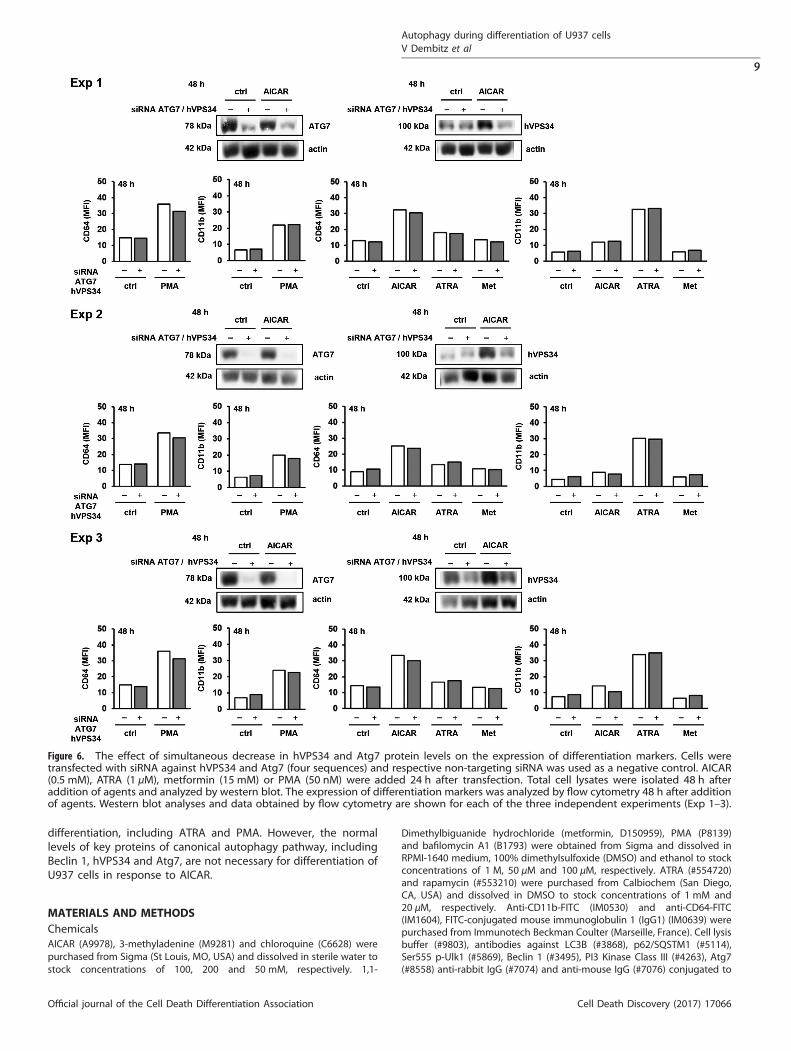
targeting hVPS34 and ATG7 and agents were added again 24 hafter transfection. As shown in Figure 6, although the levels ofATG7 and hVPS34 were decreased in lysates 48 h after addition ofagents, no differences in the expression of markers were observedin cells treated with differentiation agents.
DISCUSSIONOur previous study showed that AICAR-mediated effects on thecell viability and the expression of differentiation markersoccurred independently of the level of siRNA-downregulatedAMPK. Both AICAR and metformin were shown to increase thephosphorylation of AMPK on Thr172, which was used as a markerof AMPK activation, and decreased the phosphorylation of p70S6K, a marker of mTOR inhibition, but metformin had no effects onthe expression of differentiation markers.5 Results of our presentstudy show that AICAR-mediated effects cannot be mimicked byspecific AMPK-activator A769662, which further corroborates thehypothesis that AICAR-mediated effects are AMPK-independent.These results are in agreement with increasing number of studiesshowing that the majority of agents that are commonly used asAMPK agonists, including metformin and AICAR, display AMPK-independent effects on cell proliferation, metabolism anddifferentiation.3,4,7,33
Results of the present study demonstrate that AICAR and otherdifferentiation agents increase autophagy as measured by anincrease in the level of LC3B-II. Furthermore, all data obtained withbafilomycin A1 or mRFP-GFP-LC3 proved that the increase in theexpression of LC3B-II was due to an increase in the autophagy flux,and not to a decrease in lysosomal degradation. Again, differenteffects of AICAR and metformin on autophagy flux further suggestthat their effects are AMPK-independent. Although the activationof AMPK and inhibition of mTOR are well known inducers ofautophagy through activation of ULK1 and hVPS34,8 severalstudies demonstrated that autophagy can occur in the absence ofAMPK in response to pharmacological AMPK modulators.7,34,35 Theeffects of AICAR on the increase of LC3B-II in U937 cells correlateswell with the effects of other differentiative agents in U937 cells,and the effects of both ATRA and PMA on the level of LC3B-II aresimilar to the ones previously described in NB4(refs 10,11,12) orK562(ref. 14) cell lines. In NB4 cells, the role of autophagy in
Autophagy during differentiation of U937 cellsV Dembitz et al
4
Cell Death Discovery (2017) 17066 Official journal of the Cell Death Differentiation Association
differentiative effects of ATRA was proposed based on theinhibitory effects of 3-MA on the expression of CD11b.10 Althoughboth chloroquine and 3-methyladenine prevented AICAR-mediated increase in the expression of CD11b and CD64 in ourmodel, we cannot conclude that autophagy is necessary fordifferentiative effects. Our additional studies showed that 3-MAactually increased the level of LC3B-II, even when applied atconcentration and time interval that efficiently inhibited theactivity of hVPS34. Therefore, the present study further corrobo-rate the hypothesis that 3-MA cannot be used as a specificpharmacological inhibitor of autophagy, at least not undernutrient-rich conditions.28
In the classical autophagy pathway, the key initiation signal isprovided by hVPS34/Beclin 1 complex, and the elongation ofautophagosome is dependent on ATG7.8 The literature searchreveals many studies in which the selection of various autophagyknockdown targets in leukemia cell lines has different effects ondifferentiation, and the simplest explanation for these findingscould be that the role of autophagy in leukemia cell line may becell type and/or agonist dependent. In K562 cells, shRNA-mediated knockdown of ATG7 increased CD71 and glycophorinas markers of erythroid differentiation,19 but in NB4 cells the lackof ATG7 protein inhibited ATRA-mediated increase in CD11b.20
The downregulation of Beclin 1 by siRNA moderately, butsignificantly, inhibited vitamin D3-mediated increase in CD14 inHL-60 cells.13 In NB4 cells, shRNA-mediated downregulation ofhVPS34 inhibited the expression of CD11b,12 but downregulationof Beclin 1 had no effects on ATRA-mediated increase in CD11b12
or CD11c.11 The effects of the downregulation of the same proteincorrelate better with the marker tested than with the mode ofdownregulation (shRNA versus siRNA) since downregulation ofp62/SQSTM1 by shRNA36,37 inhibited ATRA-mediated increase inCD11b, but had no effects on ATRA-mediated increase in CD11c.17
There are no reports regarding the role of autophagy indifferentiation of U937 cells, but the role of autophagy wasinvestigated in differentiation of primary monocytes into macro-phages. In CSF-1-stimulated monocytes, siRNA for Beclin 1, ATG7and ATG5 decreased both the level of LC3B and CSF-1-mediatedincrease in CD71 and CD163.38 However, in contrast to AICAR-mediated effects in U937 cells, CSF-1-mediated effects ondifferentiation and autophagy were AMPK-dependent.39 Currentguidelines in autophagy research suggest focusing on in vivomodels instead of cell lines.40 The analyses of Atg7f/f;Vav-Cre micelacking Atg7 in hemopoietic system revealed severe anemia andlymphopenia and self-renewal defects in hemopoietic stem cells(HSC), but the number of CD11b-positive cells was actuallyincreased resembling myelodysplastic syndrome.41,42 An extensiveanalysis of macrophages of Atg7f/f;Vav-Cre mice revealed amodified response to LPS and IFN-γ-stimulation, but no differ-ences in the expression of CD11b were detected, and the level ofCD64 was not investigated.43 The analysis of Atg7f/f;Lyz-Cre micein which the Atg7 deletion was limited to myeloid cell lineagerevealed normal number of myeloid cells and normal physiologicinduction of monocyte to macrophage differentiation. However, incontrast to HSC from Atg7f/f;Vav-Cre mice in which Atg7 deletionimpairs autophagy, Atg7-deficient cells of Atg7f/f;Lyz-Cre micemaintain an active Atg7-independent alternative autophagy thatdepended on Rab9.44
Although results of our study suggest that canonical autophagyis not necessary for differentiation induced by AICAR and ATRA inU937 cells, we cannot completely rule out the possibility thatminimal levels of Atg7, Vps34 or Beclin 1 in siRNA-treated cells aresufficient to allow differentiation to proceed. In comparison toother AML cell lines, U937 cells have higher activity of Akt andmTOR, probably due to heterozygous deletion of phosphataseand tensin homolog (PTEN),45,46 and loss of PTEN has beenreported to inhibit autophagy without affecting LC3 lipidation,47
which raises a possibility that even low level of autophagy in U937
Figure 2. The effect of pharmacological modulators of autophagyon the expression of differentiation markers. (a) U937 cells wereincubated with AICAR (0.5 mM) and ATRA (1 μM) for 96 h.Chloroquine (25 μM) and rapamycin (20 nM) were added 15 minbefore addition of differentiation agents. (b) U937 cells wereincubated with AICAR (0.5 mM), ATRA (1 μM), metformin (15 mM)and PMA (50 nM) for 48 h. 3-MA (5 or 10 mM) was added 15 minbefore addition of agents tested. The number of viable cells wasdetermined by trypan blue exclusion and the expression ofdifferentiation markers was analyzed by flow cytometry. Resultsare mean± S.E.M. of at least three independent experiments.
Autophagy during differentiation of U937 cellsV Dembitz et al
5
Official journal of the Cell Death Differentiation Association Cell Death Discovery (2017) 17066

cells may be sufficient for differentiation to occur. Anotherpossibility is that an increase in lipidated LC3B is not necessaryfor the expression of surface markers, but occurs simultaneouslyduring differentiation since LC3B has been reported in both LC3B-associated phagocytosis and antigen presentation.48 Models ofoncogene-transformed cell lines grown under nutrient-rich con-ditions and exposed to different agents are obviously differentfrom a simple model of starving yeast cells in which principal
members of canonical autophagy pathway, were initiallydescribed. In last years, several studies have reported that manyinducers of autophagy flux act independently from Beclin 1/Vps34complex, especially inducers of either differentiation orapoptosis.11,12,31,49
In conclusion, results of our study show that AMPK-independenteffects of AICAR-mediated differentiation include induction ofautophagy flux that is common to other inducers of
Figure 3. PI3K class III (hVPS34) inhibitor 3-MA reduces PtdIns(3)P but increases the levels of LC3B-II and autophagic flux. (a) U937 cells weretreated with AICAR (0.5 mM) or metformin (15 mM) for 48 h and lipids were extracted by chloroform–methanol procedure. The levels of PtdIns(3)P were determined using Mass ELISA assay. Results are mean± S.E.M. of three independent experiments performed. (b) U937 cells wereincubated with AICAR (0.5 mM), ATRA (1 μM), metformin (15 mM) and PMA (50 nM). 3-MA (5 or 10 mM) was added 15 min before addition ofagents tested. Total cell lysates were isolated 48 h after addition of agents and analyzed by western blot. Representative immunoblot fromthree independent experiments is shown. Blots were scored by densitometry and graph represents mean± S.E.M. of LC3B-II/actin ratios fromthree independent experiments. (c) U937 cells stably transfected with mRFP-GFP-LC3B were incubated with 3-MA (5mM) for 48 h orbafilomycin (50 nM) for 3 h. Representative images of confocal microscropy from two independent experiments performed in duplicates areshown. Images are × 63 with × 4 times digital zoom, scale bar= 10 μm.
Autophagy during differentiation of U937 cellsV Dembitz et al
6
Cell Death Discovery (2017) 17066 Official journal of the Cell Death Differentiation Association
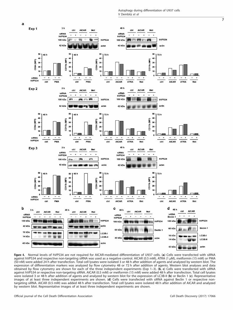
Figure 4. Normal levels of hVPS34 are not required for AICAR-mediated differentiation of U937 cells. (a) Cells were transfected with siRNAagainst hVPS34 and respective non-targeting siRNA was used as a negative control. AICAR (0.5 mM), ATRA (1 μM), metformin (15 mM) or PMA(50 nM) were added 24 h after transfection. Total cell lysates were isolated 3 or 48 h after addition of agents and analyzed by western blot. Theexpression of differentiation markers was analyzed by flow cytometry 48 or 72 h after addition of agents. Western blot analyses and dataobtained by flow cytometry are shown for each of the three independent experiments (Exp 1–3). (b, c) Cells were transfected with siRNAagainst hVPS34 or respective non-targeting siRNA. AICAR (0.5 mM) or metformin (15 mM) were added 48 h after transfection. Total cell lysateswere isolated 3 or 48 h after addition of agents and analyzed by western blot for the expression of LC3B-II (b) or Beclin 1 (c). Representativeimages of at least three independent experiments are shown. (d) Cells were transfected with siRNA against Beclin 1 or respective non-targeting siRNA. AICAR (0.5 mM) was added 48 h after transfection. Total cell lysates were isolated 48 h after addition of AICAR and analyzedby western blot. Representative images of at least three independent experiments are shown.
Autophagy during differentiation of U937 cellsV Dembitz et al
7
Official journal of the Cell Death Differentiation Association Cell Death Discovery (2017) 17066
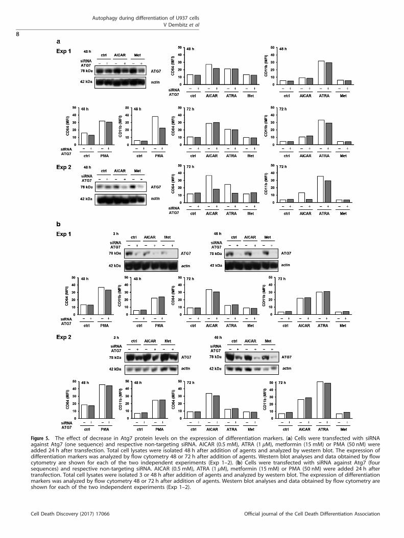
Figure 5. The effect of decrease in Atg7 protein levels on the expression of differentiation markers. (a) Cells were transfected with siRNAagainst Atg7 (one sequence) and respective non-targeting siRNA. AICAR (0.5 mM), ATRA (1 μM), metformin (15 mM) or PMA (50 nM) wereadded 24 h after transfection. Total cell lysates were isolated 48 h after addition of agents and analyzed by western blot. The expression ofdifferentiation markers was analyzed by flow cytometry 48 or 72 h after addition of agents. Western blot analyses and data obtained by flowcytometry are shown for each of the two independent experiments (Exp 1–2). (b) Cells were transfected with siRNA against Atg7 (foursequences) and respective non-targeting siRNA. AICAR (0.5 mM), ATRA (1 μM), metformin (15 mM) or PMA (50 nM) were added 24 h aftertransfection. Total cell lysates were isolated 3 or 48 h after addition of agents and analyzed by western blot. The expression of differentiationmarkers was analyzed by flow cytometry 48 or 72 h after addition of agents. Western blot analyses and data obtained by flow cytometry areshown for each of the two independent experiments (Exp 1–2).
Autophagy during differentiation of U937 cellsV Dembitz et al
8
Cell Death Discovery (2017) 17066 Official journal of the Cell Death Differentiation Association
differentiation, including ATRA and PMA. However, the normallevels of key proteins of canonical autophagy pathway, includingBeclin 1, hVPS34 and Atg7, are not necessary for differentiation ofU937 cells in response to AICAR.
MATERIALS AND METHODSChemicalsAICAR (A9978), 3-methyladenine (M9281) and chloroquine (C6628) werepurchased from Sigma (St Louis, MO, USA) and dissolved in sterile water tostock concentrations of 100, 200 and 50 mM, respectively. 1,1-
Dimethylbiguanide hydrochloride (metformin, D150959), PMA (P8139)and bafilomycin A1 (B1793) were obtained from Sigma and dissolved inRPMI-1640 medium, 100% dimethylsulfoxide (DMSO) and ethanol to stockconcentrations of 1 M, 50 μM and 100 μM, respectively. ATRA (#554720)and rapamycin (#553210) were purchased from Calbiochem (San Diego,CA, USA) and dissolved in DMSO to stock concentrations of 1 mM and20 μM, respectively. Anti-CD11b-FITC (IM0530) and anti-CD64-FITC(IM1604), FITC-conjugated mouse immunoglobulin 1 (IgG1) (IM0639) werepurchased from Immunotech Beckman Coulter (Marseille, France). Cell lysisbuffer (#9803), antibodies against LC3B (#3868), p62/SQSTM1 (#5114),Ser555 p-Ulk1 (#5869), Beclin 1 (#3495), PI3 Kinase Class III (#4263), Atg7(#8558) anti-rabbit IgG (#7074) and anti-mouse IgG (#7076) conjugated to
Figure 6. The effect of simultaneous decrease in hVPS34 and Atg7 protein levels on the expression of differentiation markers. Cells weretransfected with siRNA against hVPS34 and Atg7 (four sequences) and respective non-targeting siRNA was used as a negative control. AICAR(0.5 mM), ATRA (1 μM), metformin (15 mM) or PMA (50 nM) were added 24 h after transfection. Total cell lysates were isolated 48 h afteraddition of agents and analyzed by western blot. The expression of differentiation markers was analyzed by flow cytometry 48 h after additionof agents. Western blot analyses and data obtained by flow cytometry are shown for each of the three independent experiments (Exp 1–3).
Autophagy during differentiation of U937 cellsV Dembitz et al
9
Official journal of the Cell Death Differentiation Association Cell Death Discovery (2017) 17066
horseradish peroxidase were purchased from Cell Signaling Technology(Beverly, MA, USA). Enhanced chemiluminescence (ECL) substrate wasobtained from Thermo Fisher Scientific (Waltham, MA, USA), and proteinassay from Bio-Rad Laboratories (Hercules, CA, USA; #500-0006) or Sigma(B6916). A Neon Transfection System 100 μl Kit (#MPK10096) and anti-rabbit IgG secondary antibody conjugated to Alexa Fluor 488 (A11034)were purchased from Invitrogen (Carlsbad, CA, USA). Small interfering RNA(siRNA) SignalSilence Unconjugated Control (#6568), SignalSilence Beclin 1siRNA I (#6222) and II (#6246), SignalSilence Atg7 siRNA I (#6604) werepurchased from Cell Signaling Technology. ON-TARGETplus SMARTpoolHuman ATG7 (L-020112-00), PIK3C3 (L-005250-00) and negative control(D-001810-10) siRNA were obtained from Dharmacon (Lafayette, CO, USA).Monoclonal anti-β-actin antibody (#A5441), propidium iodide (PI), RNaseA,Igepal, color markers, bovine serum albumin, Triton X-100, sodium dodecylsulfate (SDS), leupeptin and phenylmethylsulfonyl fluoride (PMSF) werefrom Sigma, RPMI-1640, fetal bovine serum (FBS), penicillin/streptomycinwere from Gibco/Invitrogen (Grand Island, NY, USA) and G418 was fromRoche (Basel, Switzerland). Vectastain normal goat serum (FI-1200) andVectashield mounting medium (H-1000) were from Vector Laboratories Inc.(Burlingame, CA, USA). Deoxyribonuclease I (D5025) was from Sigma.PtdIns(3)P Mass ELISA Kit (K-3300) was from Echelon Biosciences Inc. (SaltLake City, UT, USA).
Cell cultureU937 cells were obtained from two sources: one was a kind gift from Dr.Mirna Golemovic (Clinical Hospital Centre, Croatia) and another wasbought from European Collection of Animal Cell Cultures (ECACC no.88112501; Porton, Salisbury, UK). The cells were maintained in RPMI-1640supplemented with 10% heat-inactivated FBS, 2 mM L-glutamine, 50 U/mlpenicillin and 50 μg/ml streptomycin at 37 °C in a humidified atmospherecontaining 5% CO2.For the experiments, cells were harvested, resuspended in fresh medium
containing FBS, L-glutamine and penicillin/streptomycin and seeded at aconcentration of 0.2 × 10⁶/ml in six-well plates. The cells were incubatedfor various time intervals in the presence of 0.5 mM AICAR, 1 μM ATRA,15 mM metformin and 50 nM PMA. Autophagy modulators, chloroquine(25 μM), 3-MA (5 or 10 mM) and rapamycin (20 nM), were added 15 minprior to other agents. Bafilomycin was added for the last 3 h of incubationat a final concentration of 50 nM. At the end of incubation, the number ofviable cells was determined by trypan blue staining and hemocytometry.
Expression of surface markersThe expression of surface markers was determined by flow cytometricanalysis, as previously described.50 Briefly, cells were collected, washed andincubated with FITC-conjugated monoclonal antibodies against CD11band CD64, or with their isotypic control for 20 min, washed and analyzedusing the FACSCalibur system and Cell Quest software (Becton DickinsonImmunocytometry Systems, San Jose, CA, USA). Live cells were gatedbased upon forward and side scatter patterns. A total of 15 000 eventswere collected for each marker from the gated area detecting viable cells.On a single fluorescence histogram of the sample stained with isotypiccontrol, a cursor was set to include up to 1.0% of events as positive. Todetermine the mean fluorescence intensity (MFI) of the sample, MFI levelsof isotypic controls were deducted from MFI levels of the cells stained withCD-specific antibodies.
Isolation of total cell lysates and western blot analysisAt the end of incubation, cells were collected by centrifugation, washed inice-cold PBS and incubated in 1 × cell lysis buffer containing freshly added1 μM microcystin and 1 mM PMSF on ice. After 10 min, cells were furtherdisrupted by seven passages through a 23-gauge needle, incubated on icefor 10 min and centrifugated at 14 000× g for 10 min. The supernatantswere collected and stored at − 80 °C. The protein concentration of eachsample was determined using Bradford protein assay (Bio-Rad or Sigma).Western blot analysis was performed as previously described.46 Briefly,
equal amounts of proteins (35–50 μg) in each sample were loaded ontotwo parallel 8 or 12% SDS-polyacrilamide gels. Electrophoresis was carriedout using the Bio-Rad mini-Protean apparatus, and proteins weretransferred to nitrocellulose membranes (Whatman, Dassel, Germany)using the Bio-Rad mini Trans-Blot system. After blocking for 30 min in TBS-Tween buffer containing 5% (w/v) non-fat dried milk, membranes wereincubated with primary antibodies (1:20 000 for actin; 1:1000 for otherantibodies) overnight at 4 °C, and then with secondary antibodies (1:2000)
for 120 min at room temperature. Bands were visualized using the ECL kit.Relative densitometric values for autoradiography signals were analyzedwith Adobe Photoshop CS version 8.0 software (San Jose, CA, USA).
Isolation of acidic lipids and competitive PtdIns(3)P Mass ELISAMeasurement of total cellular PtdIns(3)P levels was carried out using PtdIns(3)P Mass ELISA Kit (Echelon) following the manufacturer’s instructions.Briefly, after 48 h incubation with AICAR (0.5 mM) and metformin (15 mM),viable cells were counted and the volume adjusted so that each samplecontains the same number of cells (13–15 × 106). Neutral lipids wereisolated with methanol:chloroform (2:1) extraction and acidic lipids weresubsequentially isolated using methanol:chloroform: 12 N HCl (80:40:1).Dried acidic lipids were stored at − 20 °C. For PtdIns(3)P measurement withMass ELISA assay, dried lipids were reconstituted in PBS with 0.05% Tween-20 and 3% Protein Stabilizer and incubated for 30 min in a water bathsonicator at room temperature. The quantities of PtdIns(3)P in each samplewere calculated by comparison with a standard curve derived frommeasurements of PtdIns(3)P standards supplied by the manufacturer usingnonlinear regression analysis (GraphPad Prism Software, La Jolla, CA, USA).
siRNA transfectionTransfection with controls and indicated siRNAs targeting proteins ofinterest was performed using the Neon transfection system (Invitrogen) aspreviously described.5 Briefly, the cells were collected in their exponentialgrowth, resuspended in transfection buffer at a concentration of 22 × 106
cells/ml and siRNAs were added at recommended concentrations. Theelectroporation was carried out in a 100 μl tip, with single pulse, at avoltage of 1050 V and pulse width of 50 ms. Following the electroporation,100 μl of cell suspension was resuspended in 200 μl of RPMI-1640 with10% FBS and 2 mM L-glutamine without penicillin/streptomycin, incubatedfor 15 min at 37 °C and resuspended in the total volume of 5 ml RPMIwithout antibiotics. Final concentrations of 28 nM siRNA against Beclin 1and ATG7 (Cell Signaling Technologies) or 45–140 nM siRNA againsthVPS34 and ATG7 (Dharmacon) were used. Transfected cells wereincubated for 24 or 48 h, collected, resuspended in fresh medium andplated in six-well plates for the differentiation experiments. An aliquot ofcells was used for the preparation of total cell lysates and the level ofdownmodulated proteins was determined by western blot analysis.
Immunostaining for confocal microscopyU937 cells were stained for LC3B in round bottom 5 ml tubes according tothe manufacturer’s instructions. Briefly, cells were fixed with ice-cold 100%methanol at − 20 °C, blocked in 5% normal goat serum with 0.3% TritonX-100, washed in PBS with addition of 0.1 mg/ml DNAse to preventclumping, stained with primary antibody at +4 °C overnight (1 : 200) andwith secondary antibody (1:600) at room temperature for 120 min. Thepellet of immunostained cells was resuspended in 20 μl Vectashieldmounting medium and mounted on slides. Images were taken using aZeiss LSM 510 Meta confocal microscope with a plan-apochromat × 63 1.4NA oil immersion and analyzed with Zeiss LSM software (Jena, Germany).
Generation of stably transfected U937 cell line expressing mRFP-GFP-LC3BptfLC3 (mRFP-GFP-LC3) plasmid was a gift from Tamotsu Yoshimori(Addgene plasmid # 21074, Cambridge, MA, USA). U937 cells (2 × 10⁶) weretransfected with 10 μg of linearized and purified plasmid using the NeonTransfection system as described above. Cells stably expressing mRFP-GFP-LC3 were selected in medium supplemented with 400 μg/ml G418 during2 weeks. After selection, cells were expanded and maintained in mediumcontaining 250 μg/ml G418.For fluorescence microscopy, cells were seeded on chambered cover-
slips (Nunc Lab-Tek II) with pharmacological agents of interest. Imageswere acquired on a Leica TCS SP2 AOBS confocal microscope using a plan-apochromat × 63 1.4 NA oil immersion with × 4 digital zoom and imageswere analyzed with ImageJ software (Bethesda, MD, USA) and AdobePhotoshop CS version 8.0 software.
Statistical analysisData are presented as mean± standard error of the mean (S.E.M.) from thenumber of experiment indicated. Difference between two groups was
Autophagy during differentiation of U937 cellsV Dembitz et al
10
Cell Death Discovery (2017) 17066 Official journal of the Cell Death Differentiation Association

determined by Student’s t-test and P-values o0.05 were consideredstatistically significant.
ACKNOWLEDGEMENTSWe thank Ms Marijana Andrijašević and Dunja Tankovic for valuable technical helpand assistance. This work has been supported by Croatian Science Foundation underthe project IP-2016-06-4581 (to DV), the University of Zagreb Grant BM-2016 (to DV)and European Social Fund and Ministry of Science Grant HR.3.2.01-0105 (to HL).
COMPETING INTERESTSThe authors declare no conflict of interest.
PUBLISHER’S NOTESpringer Nature remains neutral with regard to jurisdictional claims in publishedmaps and institutional affiliations.
REFERENCES1 Bost F, Decoux-Poullot AG, Tanti JF, Clavel S. Energy disruptors: rising stars in
anticancer therapy? Oncogenesis 2016; 5: e188.2 Hauge M, Bruserud Ø, Hatfield KJ. Targeting of cell metabolism in human acute
myeloid leukemia – more than targeting of isocitrate dehydrogenase mutationsand PI3K/AKT/mTOR signaling? Eur J Hematol 2015; 96: 211–221.
3 Vincent EE, Coelho PP, Blagih J, Griss T, Viollet B, Jones RG. Differential effectsof AMPK agonists on cell growth and metabolism. Oncogene 2015; 34:3627–3639.
4 Liu X, Chhipa RR, Pooya S, Wortman M, Yachyshin S, Chow LM et al. Discretemechanisms of mTOR and cell cycle regulation by AMPK agonists independentof AMPK. Proc Natl Acad Sci USA 2014; 111: E435–E444.
5 Lalic H, Dembitz V, Lukinovic-Skudar V, Banfic H, Visnjic D. 5-Aminoimidazole-4-carboxamide ribonucleoside induces differentiation of acute myeloidleukemia cells. Leuk Lymphoma 2014; 55: 2375–2383.
6 Hardie DG. AMP-activated protein kinase: an energy sensor that regulates allaspects of cell function. Genes Dev 2011; 25: 1895–1908.
7 Robert G, Ben Sahra I, Puissant A, Colosetti P, Belhacene N, Gounon P et al.Acadesine kills chronic myelogenous leukemia (CML) cells through PKC-dependent induction of autophagic cell death. PLoS One 2009; 4: e7889.
8 Rubinsztein DC, Codogno P, Levine B. Autophagy modulation as a potentialtherapeutic target for diverse diseases. Nat Rev Drug Discov 2012; 11: 709–730.
9 Riffelmacher T, Simon AK. Mechanistic roles of autophagy in hematopoietic dif-ferentiation. FEBS J 2017; 284: 1008–1020.
10 Isakson P, Bjørås M, Bøe SO, Simonsen A. Autophagy contributes to therapy-induced degradation of the PML/RARα oncoprotein. Blood 2010; 116: 2324–2331.
11 Trocoli A, Mathieu J, Priault M, Reiffers J, Souquère S, Pierron G et al. ATRA-induced upregulation of Beclin 1 prolongs the life span of differentiated acutepromyelocytic leukemia cells. Autophagy 2011; 7: 1108–1114.
12 Brigger D, Proikas-Cezanne T, Tschan MP. WIPI-dependent autophagy duringneutrophil differentiation of NB4 acute promyelocytic leukemia cells. Cell DeathDis 2014; 5: e1315.
13 Wang J, Lian H, Zhao Y, Kauss MA, Spindel S. Vitamin D3 induces autophagy ofhuman myeloid leukemia cells. J Biol Chem 2008; 283: 25596–25605.
14 Colosetti P, Puissant A, Robert G, Luciano F, Jacquel A, Gounon P et al. Autophagyis an important event for megakaryocytic differentiation of the chronic myelo-genous leukemia K562 cell line. Autophagy 2009; 5: 1092–1098.
15 Huang HL, Chen YC, Huang YC, Yang KC, Pan Hy, Shih SP et al. Lapatinib inducesautophagy, apoptosis and megakaryocytic differentiation in chronic myelogenousleukemia K562 cells. PLoS One 2011; 6: e29014.
16 Klionsky DJ, Abdelmohsen K, Abe A, Abedin MJ, Abeliovich H, Acevedo Arozena Aet al. Guidelines for the use and interpretation of assays for monitoring autophagy(3rd edition). Autophagy 2016; 12: 1–222.
17 Trocoli A, Bensadoun P, Richard E, Labrunie G, Merhi F, Schläfli AM et al. p62/SQSTM1 upregulation constitutes a survival mechanism that occurs duringgranulocytic differentiation of acute myeloid leukemia cells. Cell Death Differ 2014;21: 1852–1861.
18 Kimura S, Noda T, Yoshimori T. Dissection of the autophagosome maturationprocess by a novel reporter protein, tandem fluorescent-tagged LC3. Autophagy2007; 3: 452–460.
19 Karvela M, Baquero P, Kuntz EM, Mukhopadhyay A, Mitchell R, Allan EK et al. ATG7regulates energy metabolism, differentiation and survival of Philadelphia-chromosome-positive cells. Autophagy 2016; 12: 936–948.
20 Orfali N, O'Donovan TR, Nyhan MJ, Britschgi A, Tschan MP, Cahill MR et al.Induction of autophagy is a key component of all-trans-retinoic acid-induceddifferentiation in leukemia cells and a potential target for pharmacologic mod-ulation. Exp Hematol 2015; 43: 781–793.
21 Goussetis DJ, Altman JK, Glaser H, McNeer JL, Tallman MS, Platanias LC. Autop-hagy is a critical mechanism for the induction of the antileukemic effects ofarsenic trioxide. J Biol Chem 2010; 285: 29989–29997.
22 Zhang Y, Morgan MJ, Chen K, Choksi S, Liu ZG. Induction of autophagy is essentialfor monocyte-macrophage differentiation. Blood 2012; 119: 2895–2905.
23 Kim YC, Guan KL. mTOR: a pharmacologic target for autophagy regulation. J ClinInvest 2015; 125: 25–32.
24 Nishioka C, Ikezoe T, Yang J, Nishioka C, Ikezoe T, Yang J et al. Inhibition ofmammalian target of rapamycin signaling potentiates the effects of all-transretinoic acid to induce growth arrest and differentiation of human acute mye-logenous leukemia cells. Int J Cancer 2009; 125: 1710–1720.
25 Yang J, Ikezoe T, Nishioka C, Ni L, Koeffler HP, Yokoyama A. Inhibition of mTORC1by RAD001 (everolimus) potentiates the effect of 1,25-dihydroxyvitamin D3 toinduce growth arrest and differentiation of AML cells in vitro and in vivo. ExpHematol 2010; 38: 666–676.
26 Mise J, Dembitz V, Banfic H, Visnjic D. Combined inhibition of PI3K and mTORexerts synergistic antiproliferative effect, but diminishes differentiative propertiesof rapamycin in acute myeloid leukemia cells. Pathol Oncol Res 2011; 17: 645–656.
27 Dembitz V, Lalic H, Ostojic A, Vrhovac R, Banfic H, Visnjic D. The mechanism ofsynergistic effects of arsenic trioxide and rapamycin in acute myeloid leukemiacell lines lacking typical t(15;17) translocation. Int J Hematol 2015; 102: 12–24.
28 Wu YT, Tan HL, Shui G, Bauvy C, Huang Q, Wenk MR et al. Dual role of3-methyladenine in modulation of autophagy via different temporal patterns ofinhibition on class I and III phosphoinositide 3-kinase. J Biol Chem 2010; 285:10850–10861.
29 Staskiewicz L, Thorburn J, Morgan MJ, Thorburn A. Inhibiting autophagy by shRNAknockdown. Autophagy 2013; 9: 1449–1450.
30 Jaber N, Dou Z, Chen JS, Catanzaro J, Jiang YP, Ballou LM et al. Class III PI3K Vps34plays an essential role in autophagy and in heart and liver function. Proc Natl AcadSci USA 2012; 109: 2003–2008.
31 McCoy F, Hurwitz J, McTavish N, Paul I, Barnes C, O'Hagan B et al. Obatoclaxinduces Atg7-dependent autophagy independent of beclin-1 and BAX/BAK. CellDeath Dis 2010; 1: e108.
32 Jackson AL, Linsley PS. Recognizing and avoiding siRNA off-target effects fortarget identification and therapeutic application. Nat Rev Drug Discov 2010; 9:57–67.
33 Zang Y, Yu LF, Pang T, Fang LP, Feng X, Wen TQ et al. AICAR induces astro-glial differentiation of neural stem cells via activating the JAK/STAT3 pathwayindependently of AMP-activated protein kinase. J Biol Chem 2008; 283:6201–6208.
34 Williams T, Forsberg LJ, Viollet B, Brenman JE. Basal autophagy induction withoutAMP-activated protein kinase under low glucose conditions. Autophagy 2009; 5:1155–1165.
35 Song YM, Lee YH, Kim JW, Ham DS, Kang ES, Cha BS et al. Metformin alleviateshepatosteatosis by restoring SIRT1-mediated autophagy induction via an AMP-activated protein kinase-independent pathway. Autophagy 2015; 11: 46–59.
36 Yang L, Chai W, Wang Y, Cao L, Xie M, Yang M et al. Reactive oxygen speciesregulate the differentiation of acute promyelocytic leukemia cells throughHMGB1-mediated autophagy. Am J Cancer Res 2015; 5: 714–725.
37 Wang Z, Cao L, Kang R, Yang M, Liu L, Zhao Y et al. Autophagy regulates myeloidcell differentiation by p62/SQSTM1-mediated degradation of PML-RARα onco-protein. Autophagy 2011; 7: 401–411.
38 Jacquel A, Obba S, Boyer L, Dufies M, Robert G, Gounon P et al. Autophagy isrequired for CSF-1-induced macrophagic differentiation and acquisition of pha-gocytic functions. Blood 2012; 119: 4527–4531.
39 Obba S, Hizir Z, Boyer L, Selimoglu-Buet D, Pfeifer A, Michel G et al. The PRKAA1/AMPKα1 pathway triggers autophagy during CSF1-induced human monocytedifferentiation and is a potential target in CMML. Autophagy 2015; 11: 1114–1129.
40 Lindqvist LM, Simon AK, Baehrecke EH. Current questions and possible con-troversies in autophagy. Cell Death Discov 2015; 1, pii:15036.
41 Mortensen M, Ferguson DJ, Edelmann M, Kessler B, Morten KJ, Komatsu M et al.Loss of autophagy in erythroid cells leads to defective removal of mitochondriaand severe anemia in vivo. Proc Natl Acad Sci USA 2010; 107: 832–837.
42 Mortensen M, Soilleux EJ, Djordjevic G, Tripp R, Lutteropp M, Sadighi-Akha E et al.The autophagy protein Atg7 is essential for hematopoietic stem cell maintenance.J Exp Med 2011; 208: 455–467.
43 Stranks AJ, Hansen AL, Panse I, Mortensen M, Ferguson DJ, Puleston DJ et al.Autophagy controls acquisition of aging features in macrophages. J Innate Immun2015; 7: 375–391.
44 Cao Y, Zhang S, Yuan N, Wang J, Li X, Xu F et al. Hierarchal autophagic divergenceof hematopoietic system. J Biol Chem 2015; 290: 23050–23063.
Autophagy during differentiation of U937 cellsV Dembitz et al
11
Official journal of the Cell Death Differentiation Association Cell Death Discovery (2017) 17066

45 Dahia PL, Aguiar RC, Alberta J, Kum JB, Caron S, Sill H et al. PTEN is inverselycorrelated with the cell survival factor Akt/PKB and is inactivated via multiplemechanisms in haematological malignancies. Hum Mol Genet 1999; 8: 185–193.
46 Lalic H, Lukinovic-Skudar V, Banfic H, Visnjic D. Rapamycin enhances dimethylsulfoxide-mediated growth arrest in human myelogenous leukemia cells. LeukLymphoma 2012; 53: 2253–2261.
47 Ueno T, Sato W, Horie Y, Komatsu M, Tanida I, Yoshida M et al. Loss of Pten,a tumor suppressor, causes the strong inhibition of autophagy without affectingLC3 lipidation. Autophagy 2008; 4: 692–700.
48 Münz C. The macroautophagy machinery in endo- and exocytosis. J Mol Biol 2017;429: 473–485.
49 Scarlatti F, Maffei R, Beau I, Codogno P, Ghidoni R. Role of non-canonical Beclin1-independent autophagy in cell death induced by resveratrol in human breastcancer cells. Cell Death Differ 2008; 15: 1318–1329.
50 Matkovic K, Brugnoli F, Bertagnolo V, Banfic H, Visnjic D. The role of thenuclear Akt activation and Akt inhibitors in all-trans-retinoic acid-differentiatedHL-60 cells. Leukemia 2006; 20: 941–951.
This work is licensed under a Creative Commons Attribution 4.0International License. The images or other third party material in this
article are included in the article’s Creative Commons license, unless indicatedotherwise in the credit line; if the material is not included under the Creative Commonslicense, users will need to obtain permission from the license holder to reproduce thematerial. To view a copy of this license, visit http://creativecommons.org/licenses/by/4.0/
© The Author(s) 2017
Autophagy during differentiation of U937 cellsV Dembitz et al
12
Cell Death Discovery (2017) 17066 Official journal of the Cell Death Differentiation Association


![Furine and Pyrimidine Ribonucleoside Monophosphate ...cancerres.aacrjournals.org/content/canres/42/4/1326.full.pdf · [CANCER RESEARCH 42, 1326-1330, April1982] 0008-5472/82/0042-0000$02.00](https://img.pdfslide.net/doc/110x75/5a7e72ec7f8b9ae9398e75ca/furine-and-pyrimidine-ribonucleoside-monophosphate-cancer-research-42-1326-1330.jpg)