Embed Size (px)
Citation preview
Ultrasonics 50 (2010) 777–781
Contents lists available at ScienceDirect
Ultrasonics
journal homepage: www.elsevier .com/locate /ul t ras
5-Aminolaevulinic acid enhances ultrasound-induced mitochondrial damagein K562 cells
Yong He a,c, Xinshu Xia a, Chuanshan Xu b,c,*, Heping Yu a, Dingqun Bai a, Junyan Xiang a,Albert Wingnang Leung b,**
a Department of Rehabilitation Medicine, The First Affiliated Hospital, Chongqing Medical University, Chongqing, Chinab School of Chinese Medicine, Chinese University of Hong Kong, Hong Kong, Chinac Department of Photodynamic and Sonodynamic Therapy, The Second Affiliated Hospital, Chongqing Medical University, Chongqing, China
a r t i c l e i n f o a b s t r a c t
Article history:Received 14 March 2009Received in revised form 5 February 2010Accepted 17 March 2010Available online 21 March 2010
Keywords:Sonodynamic therapy5-Aminolaevulinic acidApoptosisMitochondrial membrane potentialMitochondrial damage
0041-624X/$ - see front matter � 2010 Elsevier B.V.doi:10.1016/j.ultras.2010.03.004
* Corresponding author at: Department of PhotoTherapy, The Second Affiliated Hospital, Chongqing MChina.
** Corresponding author.E-mail addresses: [email protected] (C. Xu), aw
Leung).
Background: Ultrasound therapy is a new modality in the control of malignant cancers. The aim of thepresent study was to investigate the effect of 5-aminolaevulinic acid on the ultrasonic killing action inthe cancer cells.Materials/methods: The K562 cells as a cancer cell model were subjected to investigate the effect of 5-aminolaevulinic acid (5-ALA) on the ultrasonic killing action, in which the 5-ALA concentration was2 mM and the ultrasound exposure was 15 s at the intensity of 0.46 W/cm2 and the frequency of1.7 MHz. Cytotoxicity was investigated 24 h after ultrasound exposure using the trypan blue exclusiontest. Ultrastructural cell morphology and mitochondrial changes were observed using transmission elec-tron microscopy (TEM). Mitochondrial membrane potential (DWm) was evaluated using Rhodamine 123assay.Results: The death rates of the K562 cells in the controls including sham radiation and 5-ALA treatmentalone were 1.81 ± 0.13%, 1.27 ± 0.20%, respectively. Those in ultrasound radiation alone and 5-ALA-ultra-sound treatment were 12.61 ± 2.63%, 46.87 ± 4.09%, respectively. There were significant differencesbetween 5-ALA-ultrasound treatment, ultrasound radiation alone and the controls (P < 0.05). TEMshowed that the mitochondria expanding and some vacuoles were found in the ultrasound-treated cells.After the treatment of ultrasound and 5-ALA together some cells presented typical characteristics ofapoptotic cells, such as nuclear condensation and crescent formation. Mitochondria of the cells weredamaged more seriously than those treated by ultrasound alone, there were obvious swollen mitochon-dria and mitochondria in which cristae were almost perfectly disappeared, and more vacuolar mitochon-dria were founded. Mitochondrial membrane potential (DWm) was more significantly collapsed whenthe K562 cells were exposed to 2 mM 5-ALA for 4 h and then 0.46 W/cm2 irradiation of ultrasound thanultrasound radiation alone.Conclusion: 5-ALA pretreatment significantly enhanced the cytotoxicity of ultrasound radiation in K562cells. The damage of mitochondria structure and function might be an important cause of cell death inK562 cells induced by the treatment of ultrasound radiation and 5-ALA together.
� 2010 Elsevier B.V. All rights reserved.
1. Introduction
Malignant tumor is still one of refractory diseases threateningthe lives and health of human being worldwide. The current ther-apeutic modalities commonly have limited success with serious
All rights reserved.
dynamic and Sonodynamicedical University, Chongqing,
[email protected] (A.W.
side-effect. Developing novel efficient method to treat malignanttumor is being pursued.
Recent experimental studies have revealed that sonodynamictherapy (SDT) is a new and effective approach to treat malignanttumor. SDT deactivates the malignant cells depending on cytotoxicreactive oxygen species (ROS) produced by ultrasound-activatingsensitizer (sonosensitizer) [1–3]. Recent studies from Wang, et al.showed that the treatment with ultrasound and protoporphyrinIX (PPIX) together increased the cell damage rate of S180 cells to50.91%, while the treatment with ultrasound alone gave a celldamage rate of 24.24%, and PPIX alone kept this rate unchanged,which demonstrated that PPIX was a potential sonosensitizer [4].
778 Y. He et al. / Ultrasonics 50 (2010) 777–781
5-Aminolaevulinic acid (5-ALA), a precursor of exogenous sensi-tizer porphyrins, could be changed into tissue PPIX by biosynthesis[5,6]. The present study aims to investigate whether 5-ALA couldenhance the ultrasonic killing action in vitro.
2. Materials and methods
2.1. Sensitizer
5-Aminolaevulinic acid (5-ALA) was supplied by Sigma–Aldrich.A stock solution was made in Roswell Park Memorials Institute(RPMI)-1640 medium at a concentration of 100 mM and kept indark at �20 �C.
2.2. Cell culture
Chronic myelogenous leukemia cell line K562 cells as cancermodel cells were provided by Laboratory Center, Southwest Hospi-tal, Third Military Medical University. Cells were grown in suspen-sion cultures employing RPMI-1640 medium supplemented with10% fetal calf serum (FCS, Gibco), 50 lg/ml penicillin, 50 lg/mlstreptomycin and 10 lg/ml neomycin. Cells were incubated at37 �C in a humidified CO2 (5%) incubator.
2.3. Ultrasound treatment
The K562 cells were exposed to ultrasound at 4 h after the incu-bation of 5-ALA (2 mM). For ultrasound exposure, the K562 cells(1 � 106 cells/ml) in a test tube which the bottom was replacedby a latex film from the condom were put on the platform contain-ing a ultrasound transducer as the same as described in our pervi-ous paper [7,8]. The intensity was set at 0.46 W/cm2 for theexposure of 15 s. The test tube containing 1 ml cell suspensionand the plane transducer was placed in a water tank filled with de-gassed water during ultrasonic exposure. All experiments wererandomly divided into four groups: 5-ALA-ultrasound treatment,ultrasound radiation alone and the controls including 5-ALA treat-ment alone and sham radiation. In the 5-ALA-ultrasound treatmentthe cells were pretreated by 5-ALA incubation before ultrasoundradiation. The cells in ultrasound radiation alone were only radi-ated by ultrasound, but not incubated by 5-ALA. Those in 5-ALAtreatment alone were only pretreated by 5-ALA incubation withoutultrasound radiation and sham radiation without both ultrasoundradiation and 5-ALA incubation. Other conditions of sham radia-tion were the same as the 5-ALA-ultrasound treatment, ultrasoundradiation alone and 5-ALA treatment alone.
2.4. Cytotoxicity
The cytotoxicity of the K562 cells was assessed using the trypanblue (0.4%) exclusion test following the reports from Buldakov etal. [9] and Wang et al. [10]. The cells unstained by trypan blue werecounted under light microscopy. The cell death rate was calculatedusing the following equation:
cytotoxicity ð%Þ ¼ ðthe unstained cell number of the controlgroup� the unstained cell number of thetreatment groupÞ=the unstained cell numberof the control group � 100%:
Fig. 1. The cytotoxicity of the K562 cells after different treatments. The controlsinclude sham radiation and ALA alone. Sham control: the group with no treatment;ALA alone: the group with 2 mM 5-ALA alone; ultrasound alone: the group withultrasound alone; ALA + ultrasound: ultrasound radiation plus 2 mM 5-ALAtreatment.
2.5. Mitochondrial membrane potential (DWm)
Mitochondrial membrane potential (DWm) were monitoredusing a Confocal Laser Scanning Microscopy (CLSM) with Rhoda-mine 123 staining. Briefly, the K562 cells were sensitized with
5-ALA (2 mM) for 4 h. The cells were then irradiated by ultrasoundat the intensity of 0.46 W/cm2 for the exposure of 15 s, and furtherincubated for 14 h. The Rhodamine 123 (dissolved in DMSO to pro-duce a 1 mg/ml stock solution) (5 lg/ml) was added 30 min beforecell harvesting. Washed cells were resuspended in PBS and ob-served using a CLSM (LSM 510, Zeiss, Esslingen, Germany) withthe excitation setting at 488 nm, the fluorescence intensity of Rho-damine 123 were analyzed.
2.6. Mitochondrial morphological changes
Transmission electron microscopy (TEM) was used to observemitochondrial morphological changes of the K562 cells 14 h afterultrasound exposure. The fixed cells were postfixed in 2% OsO4,
dehydrated in graded alcohol, and flat embedded in Epon 812(Electron Microscopy Sciences, Fort Washington, PA). Ultrathinsections (100 nm) were prepared, stained with uranyl acetateand lead citrate, and examined under an electron microscopy (H-600; Hitche, Japan).
2.7. Statistical analysis
All values were the average of three experiments expressedas mean ± SD. Differences between data from two groups wereassessed by t test and P < 0.05 was considered to besignificant.
3. Results
3.1. Cytotoxicity of 5-ALA and ultrasound in the K562 cells
To assess the cytotoxicity of 5-ALA and ultrasound in chronicmyelogenous leukemia cell line K562 cells, the treated cells wereincubated for 24 h. The cells stained by trypan blue were counted.The cytotoxicity of the K562 cells in the presence of 2 mM 5-ALAafter 15 s of exposure is shown in Fig. 1. The cell death of theK562 cells in sham radiation is 1.81 ± 0.13%. The cell death rate in-duced by ultrasound and 5-ALA treatment is 46.87 ± 4.09%, and12.61 ± 2.63% with ultrasound radiation and no 5-ALA. The celldeath rate induced by ultrasound and 5-ALA treatment was moresignificantly increased than that of ultrasound exposure alone(P < 0.05) and no effect on K562 cells following 2 mM 5-ALA treat-ment alone (1.27 ± 0.20%) (P > 0.05).
Y. He et al. / Ultrasonics 50 (2010) 777–781 779
3.2. Ultrastructural cell morphology and mitochondrial morphologicalchanges
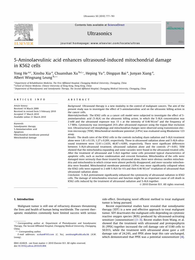
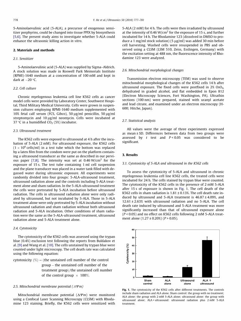
Transmission electron microscopy (TEM) showed the integrityof cell membrane of the K562 cells with many normal mitochon-dria with well developed cristae and a variety of cell nuclei wasobserved in the controls including sham radiation and 5-ALAalone (Figs. 2A and 3A). After ultrasound treatment alone the cellsmaintained intact cell membrane, but enlarge mitochondria withcristae and some vacuoles were found in the ultrasound-treatedcells (Figs. 2B and 3B). After the treatment of ultrasound and 5-ALA together some cells presented typical characteristics of apop-totic cells, such as nuclear condensation and crescent formation.Mitochondria in the cells were damaged more seriously, therewere obvious swollen mitochondria and mitochondria in whichcristae were almost perfectly disappeared, and more vacuolarmitochondria were found, which demonstrated that the 5-ALA-ultrasound treatment significantly affected the mitochondrialstructure.
3.3. Mitochondrial membrane potential (DWm)
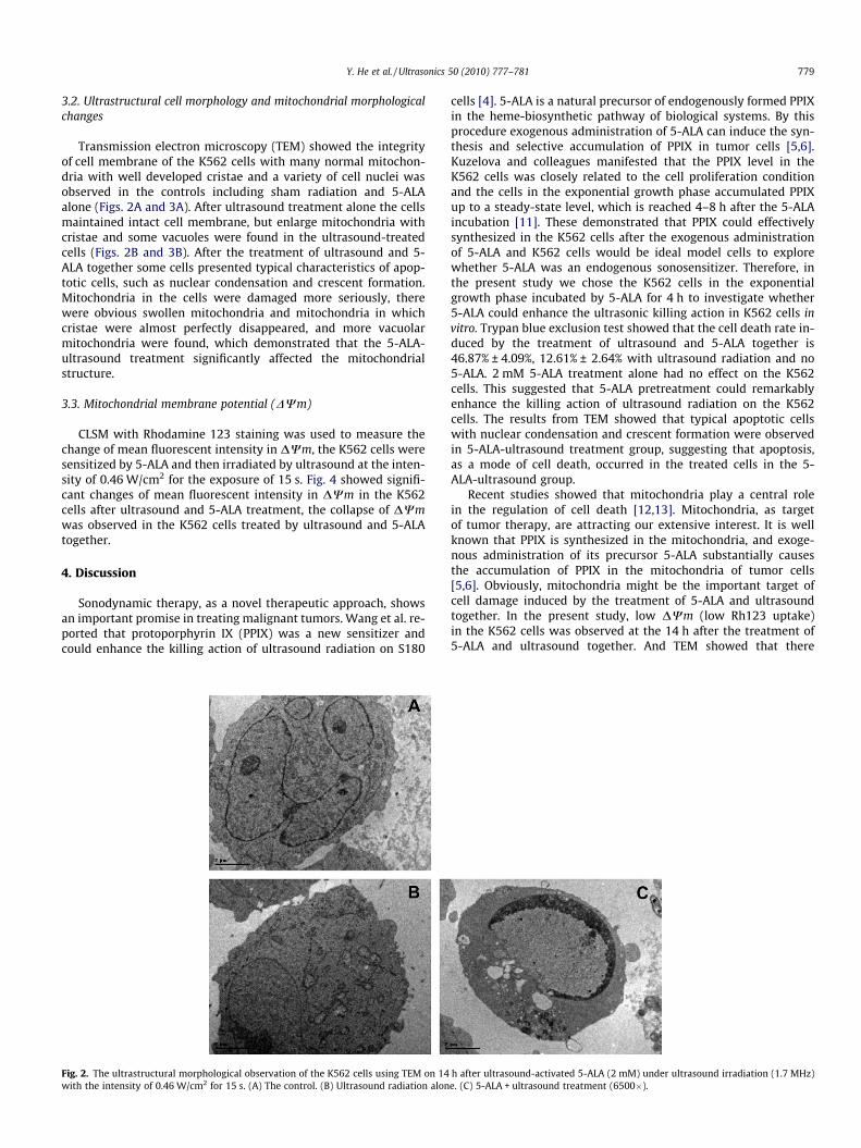
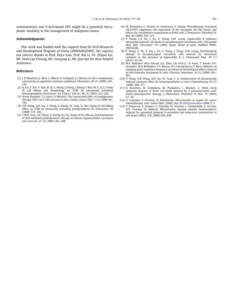
CLSM with Rhodamine 123 staining was used to measure thechange of mean fluorescent intensity in DWm, the K562 cells weresensitized by 5-ALA and then irradiated by ultrasound at the inten-sity of 0.46 W/cm2 for the exposure of 15 s. Fig. 4 showed signifi-cant changes of mean fluorescent intensity in DWm in the K562cells after ultrasound and 5-ALA treatment, the collapse of DWmwas observed in the K562 cells treated by ultrasound and 5-ALAtogether.
4. Discussion
Sonodynamic therapy, as a novel therapeutic approach, showsan important promise in treating malignant tumors. Wang et al. re-ported that protoporphyrin IX (PPIX) was a new sensitizer andcould enhance the killing action of ultrasound radiation on S180
Fig. 2. The ultrastructural morphological observation of the K562 cells using TEM on 14with the intensity of 0.46 W/cm2 for 15 s. (A) The control. (B) Ultrasound radiation alon
cells [4]. 5-ALA is a natural precursor of endogenously formed PPIXin the heme-biosynthetic pathway of biological systems. By thisprocedure exogenous administration of 5-ALA can induce the syn-thesis and selective accumulation of PPIX in tumor cells [5,6].Kuzelova and colleagues manifested that the PPIX level in theK562 cells was closely related to the cell proliferation conditionand the cells in the exponential growth phase accumulated PPIXup to a steady-state level, which is reached 4–8 h after the 5-ALAincubation [11]. These demonstrated that PPIX could effectivelysynthesized in the K562 cells after the exogenous administrationof 5-ALA and K562 cells would be ideal model cells to explorewhether 5-ALA was an endogenous sonosensitizer. Therefore, inthe present study we chose the K562 cells in the exponentialgrowth phase incubated by 5-ALA for 4 h to investigate whether5-ALA could enhance the ultrasonic killing action in K562 cells invitro. Trypan blue exclusion test showed that the cell death rate in-duced by the treatment of ultrasound and 5-ALA together is46.87% ± 4.09%, 12.61% ± 2.64% with ultrasound radiation and no5-ALA. 2 mM 5-ALA treatment alone had no effect on the K562cells. This suggested that 5-ALA pretreatment could remarkablyenhance the killing action of ultrasound radiation on the K562cells. The results from TEM showed that typical apoptotic cellswith nuclear condensation and crescent formation were observedin 5-ALA-ultrasound treatment group, suggesting that apoptosis,as a mode of cell death, occurred in the treated cells in the 5-ALA-ultrasound group.
Recent studies showed that mitochondria play a central rolein the regulation of cell death [12,13]. Mitochondria, as targetof tumor therapy, are attracting our extensive interest. It is wellknown that PPIX is synthesized in the mitochondria, and exoge-nous administration of its precursor 5-ALA substantially causesthe accumulation of PPIX in the mitochondria of tumor cells[5,6]. Obviously, mitochondria might be the important target ofcell damage induced by the treatment of 5-ALA and ultrasoundtogether. In the present study, low DWm (low Rh123 uptake)in the K562 cells was observed at the 14 h after the treatment of5-ALA and ultrasound together. And TEM showed that there
h after ultrasound-activated 5-ALA (2 mM) under ultrasound irradiation (1.7 MHz)e. (C) 5-ALA + ultrasound treatment (6500�).
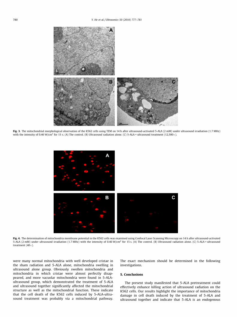
Fig. 3. The mitochondrial morphological observation of the K562 cells using TEM on 14 h after ultrasound-activated 5-ALA (2 mM) under ultrasound irradiation (1.7 MHz)with the intensity of 0.46 W/cm2 for 15 s. (A) The control. (B) Ultrasound radiation alone. (C) 5-ALA + ultrasound treatment (12,500�).
Fig. 4. The determination of mitochondria membrane potential in the K562 cells was examined using Confocal Laser Scanning Microscopy on 14 h after ultrasound-activated5-ALA (2 mM) under ultrasound irradiation (1.7 MHz) with the intensity of 0.46 W/cm2 for 15 s. (A) The control. (B) Ultrasound radiation alone. (C) 5-ALA + ultrasoundtreatment (40�).
780 Y. He et al. / Ultrasonics 50 (2010) 777–781
were many normal mitochondria with well developed cristae inthe sham radiation and 5-ALA alone, mitochondria swelling inultrasound alone group. Obviously swollen mitochondria andmitochondria in which cristae were almost perfectly disap-peared, and more vacuolar mitochondria were found in 5-ALA-ultrasound group, which demonstrated the treatment of 5-ALAand ultrasound together significantly affected the mitochondrialstructure as well as the mitochondrial function. These indicatethat the cell death of the K562 cells induced by 5-ALA-ultra-sound treatment was probably via a mitochondrial pathway.
The exact mechanism should be determined in the followinginvestigations.
5. Conclusions
The present study manifested that 5-ALA pretreatment couldeffectively enhance killing action of ultrasound radiation on theK562 cells. Our results highlight the importance of mitochondriadamage in cell death induced by the treatment of 5-ALA andultrasound together and indicate that 5-ALA is an endogenous
Y. He et al. / Ultrasonics 50 (2010) 777–781 781
sonosensitizer and 5-ALA-based SDT might be a potential thera-peutic modality in the management of malignant tumor.
Acknowledgments
This work was funded with the support from Hi-Tech Researchand Development Program of China (2006AA02Z4F0). We expressour sincere thanks to Prof. Deyu Guo, Prof. Xin Li, Dr. Zhijun Liu,Mr. Wah Lap Cheung, Mr. Guoyang Li, Mr. Jiasi Bai for their helpfulassistance.
References
[1] J. El Maalouf, J.C. Béra, L. Alberti, D. Cathignol, J.L. Mestas, In vitro sonodynamiccytotoxicity in regulated cavitation conditions, Ultrasonics 49 (2) (2009) 238–243.
[2] Q. Liu, S. Sun, Y. Xiao, H. Qi, Z. Shang, J. Zhang, J. Zhang, Y. Ren, M. Li, Q. Li, Studyof cell killing and morphology on S180 by ultrasound activatinghematoporphyrin derivatives, Sci. China C Life Sci. 46 (3) (2003) 253–262.
[3] Wang Xiaohuai, T.J. Lewis, D. Mitchell, The tumoricidal effect of sonodynamictherapy (SDT) on S-180 sarcoma in mice, Integr. Cancer Ther. 7 (2) (2008) 96–102.
[4] X.B. Wang, Q.H. Liu, P. Wang, K. Zhang, W. Tang, Q. Hao, Study of cell killingeffect on S180 by ultrasound activating protoporphyrin IX, Ultrasonics 48(2008) 135–140.
[5] C.M.N. Yow, C.K. Wong, Z. Huang, R.J. Ho, Study of the efficacy and mechanismof ALA-mediated photodynamic therapy on human hepatocellular carcinomacell, Liver Int. 27 (2) (2007) 201–208.
[6] M. Pluskalora, G. Peslova, D. Grebenova, P. Halada, Photodynamic treatment(ALA-PDT) suppresses the expression of the oncogenic Bcr-Abl kinase andeffects the cytoskeleton organization in K562 cells, J. Photochem. Photobiol. B:Biol. 83 (2006) 205–212.
[7] P. Wang, C.S. Xu, J. Xu, X. Wang, A.W. Leung, Hypocrellin B enhancesultrasound-induced cell death of nasopharyngeal carcinoma cells, UltrasoundMed. Biol. (December 15) (2009) [Epub ahead of print, PubMed PMID:20018428].
[8] P. Wang, C. Xu, X. Xia, J. Xu, X. Wang, J. Xiang, A.W. Leung, Mitochondrialdamage in nasopharyngeal carcinoma cells induced by ultrasoundradiation in the presence of hypocrellin B, J. Ultrasound Med. 29 (1)(2010) 43–50.
[9] M.A. Buldakov, M.A. Hassan, Q.L. Zhao, L.B. Feril Jr., N. Kudo, T. Kondo, N.V.Litvyakov, M.A. Bolshakov, V.V. Rostov, N.V. Cherdyntseva, P. Riesz, Influence ofchanging pulse repetition frequency on chemical and biological effects inducedby low-intensity ultrasound in vitro, Ultrason. Sonochem. 16 (3) (2009) 392–397.
[10] P. Wang, X.B. Wang, Q.H. Liu, W. Tang, T. Li, Enhancement of ultrasonicallyinduced cytotoxic effect by hematoporphyrin in vitro, Chemotherapy 54 (5)(2008) 364–371.
[11] K. Kuzelova, D. Grebenova, M. Pluskalova, I. Marinov, Z. Hrkal, Earlyapoptosis features of K562 cell death induced by 5-aminolaevulinic acid-based photodynamic therapy, J. Photochem. Photobiol. B: Biol. 73 (2004)67–78.
[12] V. Gogradze, S. Orrenius, B. Zhivotovsky, Mitochondria as targets for cancerchemotherapy, Sem. Cancer Biol. (2009), doi:10.1016/j.semcancer.2008.11.7.
[13] P. Matarrese, E. Straface, G. Palambo, M. Anselmi, L. Gambardella, B. Ascione,D.D. Principe, W. Malorni, Mitochondria regulate platelet metamorphosisinduced by opsonized zymosan A-activation and long-term commitment tocell death, FEBS J. 276 (2009) 845–856.