Embed Size (px)
Citation preview

© 2007 by Taylor & Francis Group, LLC
© 2007 by Taylor & Francis Group, LLC
5 Multifunctional Nanoparticles
for Cancer Therapy
Todd J. Harris, Geoffrey von Maltzahn,and Sangeeta N. Bhatia
CONTENTS
5.1 Introduction ........................................................................................................................... 59
5.2 Modular Functionalities at the Biosynthetic Interface ......................................................... 60
5.2.1 Targeting.................................................................................................................... 60
5.2.2 Imaging Agents ......................................................................................................... 61
5.2.3 Sensing ...................................................................................................................... 64
5.2.4 Therapeutic Payloads ................................................................................................ 66
5.2.5 Remote Actuation...................................................................................................... 69
5.3 Challenges in Integrating Multiple Functionalities and Future Directions ......................... 70
References ...................................................................................................................................... 70
5.1 INTRODUCTION
The use of nanoparticles in cancer therapy is attractive for several reasons: they exhibit unique
pharmacokinetics, including minimal renal filtration; they have high surface-to-volume ratios
enabling modification with various surface functional groups that home, internalize, or stabilize;
and they may be constructed from a wide range of materials used to encapsulate or solubilize
therapeutic agents for drug delivery or to provide unique optical, magnetic, and electrical properties
for imaging and remote actuation. The topology of a nanoparticle—core, coating, and surface
functional groups—makes it particularly amenable to modular design, whereby features and
functional moieties may be interchanged or combined. Although many functionalities of nanopar-
ticles have been demonstrated, including some clinically approved drug formulations and imaging
agents,3,8 the consolidation of these into multifunctional nanoparticles capable of targeting,
imaging, and delivering therapeutics is an exciting area of research that holds great promise for
cancer therapy in the future.
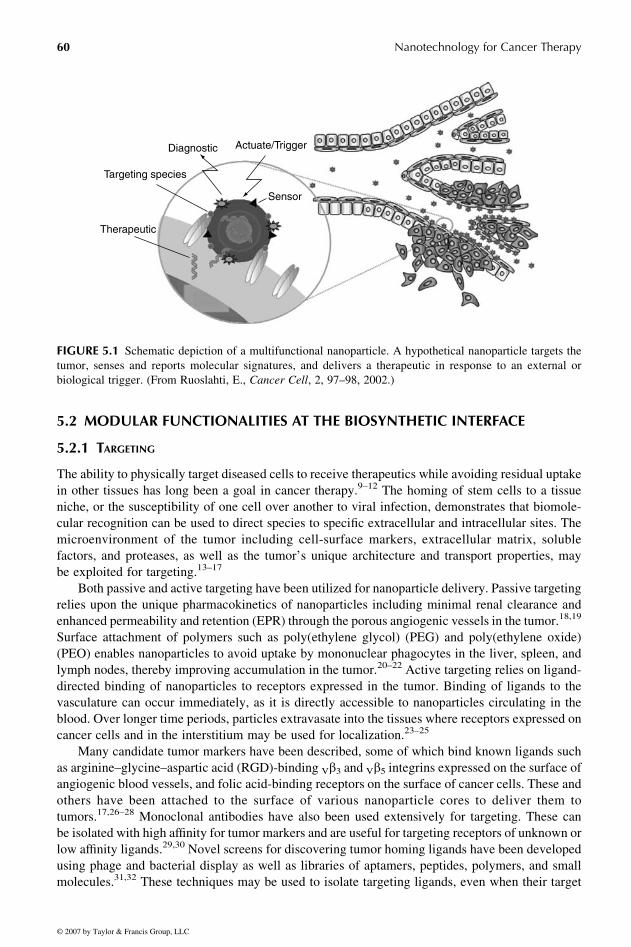
Figure 5.11 schematically depicts a hypothetical multifunctional particle that has been engine-
ered to include many features such as the ability to target tumors, evade uptake by the
reticuloendothelial system (RES), protect therapeutics that can be released on demand, act as
sensors of tumor responsiveness, and provide image contrast to visualize sites of disease and
monitor disease progression. Some of these features, such as targeting, leverage biological
machinery. Others are derived synthetically and enable external probing or manipulation that is
otherwise not feasible in biological systems. In this chapter, we review both bio-inspired and
synthetic nanoparticle functionalities that have been used in cancer therapy and address both
current efforts and future opportunities to combine these into multifunctional devices.
Cat-7194—CHAPTER 5—8/11/2006—11:59—MUGUNTHAN—14557—XML MODEL C – pp. 59–75
59
© 2007 by Taylor & Francis Group, LLC
© 2007 by Taylor & Francis Group, LLC
5.2 MODULAR FUNCTIONALITIES AT THE BIOSYNTHETIC INTERFACE
5.2.1 TARGETING
The ability to physically target diseased cells to receive therapeutics while avoiding residual uptake
in other tissues has long been a goal in cancer therapy.9–12 The homing of stem cells to a tissue
niche, or the susceptibility of one cell over another to viral infection, demonstrates that biomole-
cular recognition can be used to direct species to specific extracellular and intracellular sites. The
microenvironment of the tumor including cell-surface markers, extracellular matrix, soluble
factors, and proteases, as well as the tumor’s unique architecture and transport properties, may
be exploited for targeting.13–17
Both passive and active targeting have been utilized for nanoparticle delivery. Passive targeting
relies upon the unique pharmacokinetics of nanoparticles including minimal renal clearance and
enhanced permeability and retention (EPR) through the porous angiogenic vessels in the tumor.18,19
Surface attachment of polymers such as poly(ethylene glycol) (PEG) and poly(ethylene oxide)
(PEO) enables nanoparticles to avoid uptake by mononuclear phagocytes in the liver, spleen, and
lymph nodes, thereby improving accumulation in the tumor.20–22 Active targeting relies on ligand-
directed binding of nanoparticles to receptors expressed in the tumor. Binding of ligands to the
vasculature can occur immediately, as it is directly accessible to nanoparticles circulating in the
blood. Over longer time periods, particles extravasate into the tissues where receptors expressed on
cancer cells and in the interstitium may be used for localization.23–25
Many candidate tumor markers have been described, some of which bind known ligands such
as arginine–glycine–aspartic acid (RGD)-binding Vb3 and Vb5 integrins expressed on the surface ofangiogenic blood vessels, and folic acid-binding receptors on the surface of cancer cells. These and
others have been attached to the surface of various nanoparticle cores to deliver them to
tumors.17,26–28 Monoclonal antibodies have also been used extensively for targeting. These can
be isolated with high affinity for tumor markers and are useful for targeting receptors of unknown or
low affinity ligands.29,30 Novel screens for discovering tumor homing ligands have been developed
using phage and bacterial display as well as libraries of aptamers, peptides, polymers, and small
molecules.31,32 These techniques may be used to isolate targeting ligands, even when their target
Diagnostic Actuate/Trigger
Sensor
Targeting species
Therapeutic
FIGURE 5.1 Schematic depiction of a multifunctional nanoparticle. A hypothetical nanoparticle targets the
tumor, senses and reports molecular signatures, and delivers a therapeutic in response to an external or
biological trigger. (From Ruoslahti, E., Cancer Cell, 2, 97–98, 2002.)
Cat-7194—CHAPTER 5—8/11/2006—11:59—MUGUNTHAN—14557—XML MODEL C – pp. 59–75
Nanotechnology for Cancer Therapy60

© 2007 by Taylor & Francis Group, LLC
© 2007 by Taylor & Francis Group, LLC
receptor is unknown. For example, the 34 amino acid, cationic peptide F3, which has been used to
deliver quantum dots to tumor endothelium, was uncovered initially by a blind-page display screen
in a breast cancer xenograft model and later found to bind cell surface nucleolin expressed on tumor
endothelium and cancer cells.6,33,34
Although extracellular targeting to the tumor is sufficient for many modes of imaging and drug
delivery, intracellular delivery of nanoparticles into the cytosol is essential for some applications.
For example, nanoparticles carrying membrane-impermeable cargo that perform their function in
the cytosol, such as siRNA, antisense DNA, peptides, and other drugs, are minimally effective if
delivered extracellularly or sequestered in the endosome.35 Protein and peptide motifs capable of
translocating nanoparticles into the cytoplasm have been borrowed from mechanisms of viral
transfection. Two important classes of translocating domains include polycationic sequences and
membrane fusion domains. Attaching the short polycationic sequence of HIV’s TAT protein, amino
acid residues 48–57, to a nanoparticle facilitates its adsorption on a cell surface and subsequent
internalization into the cell.36,37 This peptide has been used to internalize dextran coated iron-oxide
nanoparticles into T-cells in vitro, which were subsequently used to monitor T-cell trafficking in
tumors with MRI.38 Use of this peptide for intracellular delivery in vivo is limited by the adverse
effect that polycationic sequences have on nanoparticle circulation time and RES uptake.39 The
amphiphilic domain derived from the N-terminus of the influenza protein hemagluttinin (HA2) is a
membrane fusion peptide that destabilizes the endosome at low pH and facilitates viral escape into
the cytosol.40 Variations of this peptide with improved infectivity have also been synthesized.41
Influenza-derived peptides have been used to enhance the delivery of liposomes as well as 100 nm
poly-L-lysine particles. Although the peptide modification of these particles improves endosomal
escape over unmodified particles, the transfection efficiency still remains well below that of intact
viruses.42,43
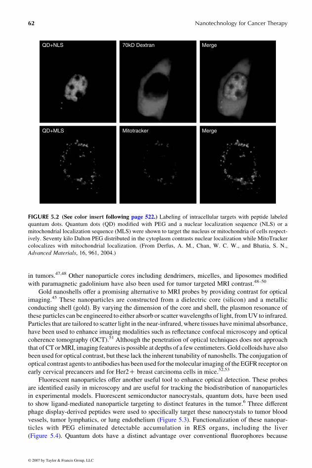
Another level of targeting can occur after translocation of nanoparticles into the cytosol to
direct nanoparticles to specific sub-cellular structures. Using peptide localization sequences, fluor-
escent quantum dots have been targeted to the nucleus and the mitochondria (Figure 5.2).2 Several
other localization sequences exist and could be used to traffic nanoparticles to the endoplasmic
reticulum, golgi apparatus, or peroxisomes. Although work in this area has been focused on
organelle labeling, the potential for delivering therapeutic nanoparticles to sub-cellular structures
is possible. Such nanoparticles could sense sub-cellular aspects of disease or specifically intervene
for more potent treatment or eradication of cancer cells (i.e., free-radical-mediated mitochondrial
damage to induce apoptosis).
5.2.2 IMAGING AGENTS
Imaging cancer is crucial for guiding decisions about treatment and for monitoring the efficacy of
administered therapies. The use of nanoparticles for image contrast and enhancement has enabled
improvements in cancer imaging by conventional modalities, such as magnetic resonance imaging
(MRI) and ultrasound, and has also established new techniques such as optical-based imaging for
cancer detection.39,44,45 Targeted imaging agents that can identify specific biomarkers have the
potential to improve detection, classification, and treatment of cancer with minimal invasiveness
and reduced costs.
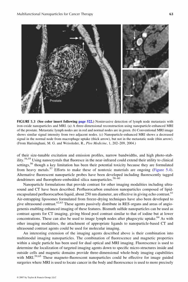
The use of nanoparticles in cancer imaging has already demonstrated clinical efficacy in
detecting liver cancer and staging lymph node metastasis noninvasively.3,46 Superparamagnetic
iron-oxide nanoparticles disrupt local magnetic field gradients in tissues, causing a detectable signal
void in MRI. Dextran coated iron-oxide nanoparticles administered intravenously get phagocytosed
by normal macrophages of the liver and lymph and the failure of these tissues to darken after iron-
oxide administration identifies invading cancer cells. Directly targeting these magnetic nanoparti-
cles to cancer cells has also been demonstrated. For example, herceptin mAb and folic acid on the
surface of iron-oxide nanoparticles enable MRI-based molecular imaging of their respective targets
Cat-7194—CHAPTER 5—8/11/2006—11:59—MUGUNTHAN—14557—XML MODEL C – pp. 59–75
Multifunctional Nanoparticles for Cancer Therapy 61
© 2007 by Taylor & Francis Group, LLC
© 2007 by Taylor & Francis Group, LLC
in tumors.47,48 Other nanoparticle cores including dendrimers, micelles, and liposomes modified
with paramagnetic gadolinium have also been used for tumor targeted MRI contrast.48–50
Gold nanoshells offer a promising alternative to MRI probes by providing contrast for optical
imaging.45 These nanoparticles are constructed from a dielectric core (silicon) and a metallic
conducting shell (gold). By varying the dimension of the core and shell, the plasmon resonance of
these particles can be engineered to either absorb or scatter wavelengths of light, fromUV to infrared.
Particles that are tailored to scatter light in the near-infrared, where tissues haveminimal absorbance,
have been used to enhance imaging modalities such as reflectance confocal microscopy and optical
coherence tomography (OCT).51 Although the penetration of optical techniques does not approach
that ofCTorMRI, imaging features is possible at depths of a few centimeters. Gold colloids have also
been used for optical contrast, but these lack the inherent tunability of nanoshells. The conjugation of
optical contrast agents to antibodies has been used for themolecular imaging of the EGFR receptor on
early cervical precancers and for Her2C breast carcinoma cells in mice.52,53
Fluorescent nanoparticles offer another useful tool to enhance optical detection. These probes
are identified easily in microscopy and are useful for tracking the biodistribution of nanoparticles
in experimental models. Fluorescent semiconductor nanocrystals, quantum dots, have been used
to show ligand-mediated nanoparticle targeting to distinct features in the tumor.6 Three different
phage display-derived peptides were used to specifically target these nanocrystals to tumor blood
vessels, tumor lymphatics, or lung endothelium (Figure 5.3). Functionalization of these nanopar-
ticles with PEG eliminated detectable accumulation in RES organs, including the liver
(Figure 5.4). Quantum dots have a distinct advantage over conventional fluorophores because
QD+NLS
QD+MLS MergeMitotracker
70kD Dextran Merge
FIGURE 5.2 (See color insert following page 522.) Labeling of intracellular targets with peptide labeled
quantum dots. Quantum dots (QD) modified with PEG and a nuclear localization sequence (NLS) or a
mitochondrial localization sequence (MLS) were shown to target the nucleus or mitochondria of cells respect-
ively. Seventy kilo Dalton PEG distributed in the cytoplasm contrasts nuclear localization while MitoTracker
colocalizes with mitochondrial localization. (From Derfus, A. M., Chan, W. C. W., and Bhatia, S. N.,
Advanced Materials, 16, 961, 2004.)
Cat-7194—CHAPTER 5—8/11/2006—11:59—MUGUNTHAN—14557—XML MODEL C – pp. 59–75
Nanotechnology for Cancer Therapy62
© 2007 by Taylor & Francis Group, LLC
© 2007 by Taylor & Francis Group, LLC
of their size-tunable excitation and emission profiles, narrow bandwidths, and high photo-stab-
ility.54,55 Using nanocrystals that fluoresce in the near-infrared could extend their utility to clinical
settings,56 though a key limitation has been their potential toxicity because they are formulated
from heavy metals.57 Efforts to make these of nontoxic materials are ongoing (Figure 5.4).
Alternative fluorescent nanoparticle probes have been developed including fluorescently tagged
dendrimers and fluorophore-embedded silica nanoparticles.58–60
Nanoparticle formulations that provide contrast for other imaging modalities including ultra-
sound and CT have been described. Perfluorocarbon emulsion nanoparticles composed of lipid-
encapsulated perfluorocarbon liquid, about 250 nm diameter, are effective in giving echo contrast.61
Air-entrapping liposomes formulated from freeze-drying techniques have also been developed to
give ultrasound contrast.62,63 These agents passively distribute in RES organs and areas of angio-
genenis enabling enhanced imaging of these features. Bismuth sulfide nanoparticles can be used as
contrast agents for CT imaging, giving blood pool contrast similar to that of iodine but at lower
concentrations. These can also be used to image lymph nodes after phagocytic uptake.64 As with
other imaging modalities, the attachment of appropriate ligands to nanoparticle-based CT and
ultrasound contrast agents could be used for molecular imaging.
An interesting extension of the imaging agents described above is their combination into
multimodal imaging nanoparticles. The combination of fluorescence and magnetic properties
within a single particle has been used for dual optical and MRI imaging. Fluorescence is used to
determine the localization of targeted imaging agents down to specific micro-structures inside and
outside cells and magnetic domains provide three-dimensional whole-body imaging capabilities
with MRI.59,65 These magneto-fluorescent nanoparticles could be effective for image guided
surgeries where MRI is used to locate cancer in the body and fluorescence is used to more precisely
(a)
(b)
(c)
Prostate
FIGURE 5.3 (See color insert following page 522.) Noninvasive detection of lymph node metastasis with
iron-oxide nanoparticles and MRI. (a) A three-dimensional reconstruction using nanoparticle-enhanced MRI
of the prostate. Metastatic lymph nodes are in red and normal nodes are in green. (b) Conventional MRI image
shows similar signal intensity from two adjacent nodes. (c) Nanoparticle-enhanced MRI shows a decreased
signal in the normal node from macrophage uptake (thick arrow), but not in the metastatic node (thin arrow).
(From Harisinghani, M. G. and Weissleder, R., Plos Medicine, 1, 202–209, 2004.)
Cat-7194—CHAPTER 5—8/11/2006—11:59—MUGUNTHAN—14557—XML MODEL C – pp. 59–75
Multifunctional Nanoparticles for Cancer Therapy 63

© 2007 by Taylor & Francis Group, LLC
© 2007 by Taylor & Francis Group, LLC
delineate tumor borders during resection. Other dual-imaging probes have been described
including: perfluorocarbon emulsions tagged with gadolinium for combined ultrasound and MRI.66
5.2.3 SENSING
Functionalities that undergo chemical alterations in response to enzymatic activity or other
properties such as pH or oxygen could be used as sensors to report information about the status of
the tumor or efficacy of treatment. Many nanoparticle-based sensors that respond to biological
triggers including proteases, DNAses, proteins, peroxidase, pH, and others have been demonstrated
in vitro.67–72 These generally rely on assembly or disassembly of inorganic nanocrystals
including: gold nanoparticles, nanoshells, or nanorods, which undergo a shift in their plasmon
resonance when aggregated; iron-oxide nanoparticles have enhanced T2 relaxivity when clustered;
and fluorescent quencher-based nanoparticle systems that dequench after triggered release.
A system using cleavable polymeric shielding of self-assembling nanoparticles has been
proposed as a mechanism for translating nanoparticle-based enzyme sensors to in vivo use
(Figure 5.5).73 Self-assembling, complementary iron-oxide nanoparticles are rendered latent with
PEG polymers linked to the nanoparticle surface by protease-cleavable substrates that serve to both
inhibit assembly and stabilize the particles in serum. Upon proteolytic removal of PEG polymers by
MMP-2 expressing cancer cells, nanoparticles assemble and acquire amplified magnetic properties
that can be detected with MRI. In the future, similar to thrombin-driven self-assembly of fibrin and
platelets at sites of endothelial injury, this systemmay allow the hyper-active proteolytic processes of
cancers to drive the self-assembly of nanoparticles in regions of cancer angiogenesis, invasion, and
metastasis in vivo. Due to its modular design, this system can easily be modified for a number of
detection schemes by substituting the complementary binding pairs, cleavable substrates
(a)(c)
(b)
Pep
tide
Peptide Peptid
e
Peptide
Peptide
PeptidePeptide
Peptide
CdSe
ZnS
(d)
PE
G
Peptide Peptid
e
PE
G
PeptidePEG
PEG PeptideCdSe
ZnS
FIGURE 5.4 (See color insert following page 522.) Targeting quantum dots (QDs) to site-specific endo-
thelium with phage display-derived peptides. (a) Schematic representation of co-injected red and green
quantum dots that home to tumor and lung vasculature respectively after intravenous injection. (b) Schematic
representation of peptide-coated QDs and peptide-coated PEG-QDs. (c) QDs labeled with the tumor endo-
thelium homing peptide F3 co-localize with a blood vessel marker. (d) QDs labeled with the tumor lymphatic
homing peptide Lyp-1 highlight the endothelium but do not colocalize with a blood vessel marker. (From
Akerman, M. E., Chan, W. C. W., Laakkonen, P., Bhatia, S. N., and Ruoslahti, E., Proceedings of the National
Academy of Sciences of the United States of America, 99, 12617–12621, 2002.)
Cat-7194—CHAPTER 5—8/11/2006—11:59—MUGUNTHAN—14557—XML MODEL C – pp. 59–75
Nanotechnology for Cancer Therapy64

© 2007 by T
aylor & Francis G
roup, LL
C
(a)
(b)
'Latent' nanoparticles Proteolytically actuated self-assembly
(c) (d)
0
0.00
0.01
0.02
0.03
30 60
No MMP-2Scmbl cntrlMMP-2
90 120 150 180
MMP-2
No MMP-2
10 kDa PEG
Nanoassemblies exhibit:
MMP-2
Magnetic susceptibility
T2 relaxativity
Diffusivity
t /min
∆e
0
3.2
10
32
85 170 340 680
MMP-2/ng/ml
particleconcn./pM
1360
400
100
200
300T2/ms
FIGURE 5.5 (See color insert following page 522.) Protease-triggered self-assembling nanoparticles with polymer-shielded coatings. (a) Schematic representation of
nanoparticles that self-assemble after protease-mediated cleavage of PEG chains reveals complementary moieties (neutravidin and biotin). (b) Iron-oxide nanoparticles
with cleavable linkers assemble in the presence of MMP-2, as measured by changes their light extinction, while particles with noncleavable scrambled peptides do not. (c)
Atomic force micrographs of particles incubated with MMP-2 shows detectable aggregation (scale bars are 500 nm). (d) T2 maps of particles in solution using a 4.7 T
MRI demonstrate enhanced T2 relaxivity for increasing concentrations of MMP-2.
Cat-7
194—CHAPTER5—8/11/2006—11:59—MUGUNTHAN—14557—XMLMODELC–pp.59–75
Multifu
nctio
nalNanoparticles
forCancer
Therap
y65
© 2007 by Taylor & Francis Group, LLC

© 2007 by Taylor & Francis Group, LLC
© 2007 by Taylor & Francis Group, LLC
(e.g., glycans, lipids, oligonucleotides), or multivalent nanoparticle cores (e.g., gold, quantum dot,
dendrimer).
5.2.4 THERAPEUTIC PAYLOADS
The use of nanoparticulate drug carriers can address many critical challenges in drug delivery
including: improving drug solubility and stability; extending drug half-lives in the blood; reducing
adverse effects in nontarget organs; and concentrating drugs at the disease site.74 Drugs may be
dispersed in a matrix, encapsulated in a vesicle, dissolved in a hydrophobic core, or attached to the
surface of a nanoparticle. Several nanoparticle-based drug delivery systems including liposomes,
polymeric nanoparticles, dendrimers, ceramic based carriers, micelles, and others have been used to
carry small molecule, peptide, and oligonucleotide therapeutic agents.24,75 Many promising
anti-cancer drugs fail to make it to the clinic because of poor solubility or high collateral toxicity
at therapeutic levels, thus motivating the need for these carriers in cancer therapy.
Liposomes have been the most extensively utilized nanoparticle-based carriers for delivering
anti-cancer drugs. First described decades ago, these submicron-sized carriers consist of amphiphilic
lipids assembled to form vesicles that can encapsulate drugs.76 Liposome-encapsulated doxorubicin
is a clinically approved nanoparticle formulation used for chemotherapy.77 The surface of this
nanocarrier is PEGylated to reduce rapid uptake by phagocytic cells and extend the drug circulation
time for better therapeutic efficacy. Several other liposome-encapsulated chemotherapeutic drugs
have been described, with many in clinical trials.78 Active targeting of these liposomes through the
attachment of antibodies and various ligands has also been demonstrated.30,79Drug loaded liposomes
with encapsulated or surface-functionalized gadolinium or fluorophores have been used to simul-
taneously image tumors during nanoparticle-targeted drug delivery.80–82
Biodegradable polymer nanocarriers have also been investigated as a means of encapsulating
drugs and releasing themover time. Both poly dl-lactide co-glycolide (PLGA) and polylactide (PLA)
nanoparticles have been formed that immobilize drugs dispersed in their matrix and release them
upon degradation.24,83 Other polymers, including polyethyleneimine (PEI), polylysine, and cyclo-
dextrin-containing polymers, are used to condense DNA or RNA into nanoparticle carriers that can
be targeted to cancer cells for gene or siRNA delivery.35 Polymeric micelles consist of amphiphilic
block copolymers that self-assemble into a water-soluble nanoparticle with a hydrophobic core.
These can be used to encapsulate water-insoluble drugs such as doxorubicin and adriamycin and
targeted to tumors.84–86 Polymersomes are another variation of polymer-based nanoparticulate
vesicles that self-assemble from amphiphilic block copolymers.87 These have been used to encap-
sulate doxorubicin with well-controlled release over several days.88,89
Another class of nanoparticle-based drug carriers are dendrimers. These consist of a network of
branching chemical bonds around an inner core. One of the more popular dendrimers, polyami-
doamine dendrimers (PAMAMs), are nonimmunogenic, water-soluble, and possess terminal amine
functional groups for conjugation of a variety of surface moieties.90 Their inner core can been used
to encapsulate anti-cancer drugs such as doxorubicin and methotrexate.91 Drugs may also be
conjugated to the dendrimer surface along with ligands for targeting.92,93 A dendrimer functiona-
lized with FITC, folic acid, and methoxetrate has been synthesized to have imaging, targeting, and
drug delivery capabilities (Figure 5.6).4,7,94 The synthesis of these conjugates in a scalable and
reproducible manner has been described for potential clinical applications.4
Other nanoparticulate carriers, including nanoemulsions, drug nanocrystals, and polyelectro-
lyte carriers, have been developed. Nanoemulsions are formed by dissolving a drug in a lipid,
cooling the solution under high pressure, and using homogenization to form solid nanoparticle lipid
carriers at body temperature. Homogenization techniques can also be used to form crystalline
nanosuspensions of drugs.74 These formulations increase drug solubility and control release
kinetics of the drug in the blood and at the tumor site. Polyelectrolyte carriers formed by the
Cat-7194—CHAPTER 5—8/11/2006—11:59—MUGUNTHAN—14557—XML MODEL C – pp. 59–75
Nanotechnology for Cancer Therapy66

© 2007 by Taylor & Francis Group, LLC

© 2007 by Taylor & Francis Group, LLC
© 2007 by Taylor & Francis Group, LLC
layer-by-layer absorption of polycationic and polyanionic moieties can be used to encapsulate
therapeutic cargo, particularly larger agents such as peptides and oligonucleotides.95
A clever combination of drug-release modalities was recently demonstrated by the creation of
a dual drug-release nanoparticle having a PLGA polymer core encapsulating doxorubicin and a
PEG-lipid block copolymer shell loaded with the combretastatin (Figure 5.7).5 The lipophilic
anti-angiogenesis drug, combretastatin, intercalates in the nanoparticle membrane and releases
rapidly upon association with tumor endothelial cells, while the slower-releasing doxorubicin
increases cytotoxic killing of tumor cells for a prolonged time after the vasculature shuts down.
This novel system demonstrates the feasibility of integrating multiple functionalities of drug
delivery on a single nanoparticle to enhance therapeutic efficacy.
8
6
4
2
00 20 40 60
7500
5000
2500
09 10 11 12 13
Days
Tum
orvo
lum
e
14
80 100 120
Time (hours)
Tota
ldru
gre
leas
edCombretastatin (×102 μg)
Doxorubicin (μg)
140 160
Veh
C
N
NC
N + C
(a) (c)
(b) (d)
FIGURE 5.7 Dual drug-release nanoparticle for combined anti-angiogenisis and anti-cancer treatment. (a)
Scanning electron micrograph showing nanocores prepared from doxorubicin-coupled PLGA. The nanocores
are encapsulated inside a lipid coat, which is also loaded with an anti-angiogenesis agent. (b) Cross section of a
nanocell with the dark nanocore. The lipid coat is surface-modified through pegylation, which confers stealth
characteristics to the nanocell from the RES. (c) The composition of the nanocell enables a spatiotemporal
release of the two agents in an acidic pH mimicking the tumor environment, as shown in the graph. (d) In vivo
studies using F10 melanoma clearly show that the spatiotemporal release from the nanocells (NC) achieves
better outcome than the doxorubicin-loaded nanocore or the lipid-entrapped combretastatin (C) alone, or
combinations of both (N+C).5
Cat-7194—CHAPTER 5—8/11/2006—11:59—MUGUNTHAN—14557—XML MODEL C – pp. 59–75
Nanotechnology for Cancer Therapy68
© 2007 by Taylor & Francis Group, LLC
© 2007 by Taylor & Francis Group, LLC
5.2.5 REMOTE ACTUATION
Temporal and spatial control of therapeutic administration is important for eliminating off-target
toxicity and achieving optimal delivery. Temporally controlled release profiles may be designed into
nanoparticle carriers mentioned previously and spatial control can be improved with targeting.
However, off-target effects, including eventual accumulation of nanoparticles in RES organs, limit
many aspects of these methods of control. The ability to trigger the therapeutic activity of adminis-
tered nanoparticles remotely could be a valuable tool for localizing treatments to a diseased site.
Many inorganic nanocrystals and nanoemulsions used for imaging contrast absorb electromagnetic
or ultrasonic energy that can also be used to remotely heat or trigger drug delivery.
Thermal ablation of tumors by nanoparticles that absorb external energy has been demonstrated
both with iron-oxide nanoparticles and gold nanoshells. Superparamagnetic iron-oxide nanoparti-
cles under the influence of an alternating electromagnetic (EM) field heat by Brownian relaxation,
where heat is generated by the rotation of particles in the field, and Neel relaxation, where the
magnetic domains are moved away from their easy axis with the resultant energy being deposited as
heat in the solution.96,97 Nanoparticle concentrations of 0.1–1% are required to achieve critical
temperatures for tumor ablation.98,99 Ongoing work to increase the absorption of magnetic nano-
particles using clinically safe RF frequencies and to increase the concentration of particles that can
be targeted to the tumor may extend the utility of this technique. Alternatively, near-infrared-
absorbing gold nanoshells targeted to the tumor can be used to thermally ablate the cancer
cells upon illumination with a high intensity laser.100,101 This technique can be applied to solid
tumors in close proximity to the skin, but cannot be applied to deeper lesions because of tissue
absorbance.53,100,101 By synthesizing nanoshells with a plasmon resonance that has both absorp-
tion and scattering profiles, these nanoparticles may be capable of both heating and imaging
tumors.53
Remotely-triggered release of a therapy by heating is a promising extension of the use of
nanoparticles that can absorb external energy. An example of this has been demonstrated with a
model drug linked to an iron-oxide nanoparticle via a heat-labile tether that is released and diffuses
into the peripheral tissue after irradiation with RF energy.98 By modifying the susceptibility of the
linker, it is possible to tune the release profile over a range of temperatures and to enable repeated
administrations. The iron core of these drug-releasing nanoparticles can be used simultaneously for
imaging with MRI. Additionally, the magnetic properties of these nanoparticles can be manipulated
by magnetic field gradients to target sites near externally- or internally-placed magnets.102
Drug activation using EM energy has been explored extensively with photodynamic therapy
(PDT). PDT agents, when irradiated by light, produce reactive oxygen species that are toxic to cells.
Agents such as porphyrins have been conjugated to various nanoparticle cores including dendri-
mers, liposomes, and polymers.103,104 When excited by light, these nanoparticles can produce
enough reactive oxygen species to kill tumor cells.60 The inherent fluorescent properties of
many PDT agents enable simultaneous imaging with therapeutic delivery. A multifunctional nano-
particle platform combining MRI contrast and photodynamic therapy has been used to target,
image, and treat brain cancer in a rat model.105 In the future, integrating these nanoparticles
with peptides capable of targeting tumors and subcellularly localizing them to the nuclei or mito-
chondria of tumor cells may enhance the therapeutic efficacy of these treatments.
Other forms of externally applied energy such as ultrasound and x-ray radiation provide
alternative mechanisms to achieve remote actuation. Acoustic energy has been shown to
enhance the delivery of lipid drugs from a perfluorocarbon emulsion targeted to cell membranes
and from doxorubicin-loaded polymeric micelles.106,107 Atomically dense nanoparticles have been
shown to increase the absorption of x-ray radiation, enhancing their destructive effect in
surrounding tissue.108 There is potential for simultaneous imaging and therapeutic delivery with
these particles also.
Cat-7194—CHAPTER 5—8/11/2006—11:59—MUGUNTHAN—14557—XML MODEL C – pp. 59–75
Multifunctional Nanoparticles for Cancer Therapy 69

© 2007 by Taylor & Francis Group, LLC
© 2007 by Taylor & Francis Group, LLC
5.3 CHALLENGES IN INTEGRATING MULTIPLE FUNCTIONALITIES AND
FUTURE DIRECTIONS
Although remotely actuated nanoparticle cores such as iron-oxide and metal nanoshells naturally
lend themselves to dual-imaging and therapeutic applications, the combination of imaging and
other functionalities using other nanoparticle cores can be challenging. There are inherent trade-offs
when combining many functional groups into one nanoparticle. In many cases, a limited number of
attachment sites are available on the particle surface, making it difficult to couple several functional
groups in sufficient concentration for each to function. Moreover, some groups may interact to
sterically shield or alter the activity of one another when combined in close proximity. Multiple
functional moieties on a nanoparticle may also reduce colloidal stability or adversely affect its
in vivo pharmacokinetics. With significant characterization and fine tuning, dendrimers that
combine targeting, imaging, and therapeutic moieties on their surface have been synthesized
successfully.4 Similar efforts will be necessary to achieve other multifunctional nanoparticles
with decorated-surface moieties.
An alternative strategy to consolidate multiple functionalities onto a single particle is to use
core-shell architecture. In this case, an outer shell with one functionality, such as targeting, may be
unveiled to reveal an inner core that performs a secondary function such as endosomal escape or
drug release. This has been demonstrated with the conjugation of targeting moieties or protective
PEG groups on the surface of dendrimers or polymers via acid-labile chemistries that degrade in the
lower pH of the endosome and unveil endosomal escape mechanisms on the particle core.109,110
This has also been demonstrated with protease-cleavable linkers that release protective polymers on
the surface of complementary nanoparticles to initiate their self-assembly.73
The synthesis of nanoparticles with polar domains is another strategy that could be used to
incorporate multiple functionalities on a single particle. Janus nanoparticles—named for Janus, the
Roman God of doorways typically depicted with faces on the front and back of his head—have been
engineered with two chemically distinct hemispheres or surfaces. These nanoparticles may be
spherical (with opposing faces of unique composition), dumbbell-shaped (with two equal-sized
spheres linked together), snowman-shaped, and may have other morphologies as well.95,111 The
creation of nanoparticles with spatially separated chemical domains is a step towards replicating the
controlled polarity exhibited in nature across many length scales. Separate hemispheres may be
used to isolate and organize functional domains on nanoparticles such that they may simultaneously
carry targeting molecules, endosomal escape domains, sensing moieties, hydrophilic and hydro-
phobic therapeutics, or contrast agents that otherwise might be mutually inhibitory if randomly
incorporated. Moreover, there may be specific applications for which the polarity and anisotropy of
Janus nanoparticles have benefit, such as real-time detection of oriented binding events, targeted
bridging of multiple components at a tumor cell, directed drug delivery, or guided self-assembly.
Although there have been many exciting advances in the application of nanoparticles for cancer
imaging and treatment, the true power of these materials will be in their ability to interact with
disease processes intelligently. The modular design of functionalities that target, sense, signal, and
treat and the ongoing efforts to consolidate these into single nanoparticle platforms is one way in
which such ‘smart’ materials are being developed. The further elucidation of complex biological
processes in tumorogenesis, the discovery of nanomaterials with other novel properties, and the
consolidation of biological and synthetic machinery in these materials in new and elegant ways are
key factors that will determine their future success in cancer therapy.
REFERENCES
1. Ruoslahti, E., Antiangiogenics meet nanotechnology, Cancer Cell, 2, 97–98, 2002.
2. Derfus, A. M., Chan, W. C. W., and Bhatia, S. N., Intracellular delivery of quantum dots for live cell
labeling and organelle tracking, Advanced Materials, 16, 961–966, 2004.
Cat-7194—CHAPTER 5—8/11/2006—11:59—MUGUNTHAN—14557—XML MODEL C – pp. 59–75
Nanotechnology for Cancer Therapy70

© 2007 by Taylor & Francis Group, LLC
© 2007 by Taylor & Francis Group, LLC
3. Harisinghani, M. G. and Weissleder, R., Sensitive, noninvasive detection of lymph node metastases,
Plos Medicine, 1, 202–209, 2004.
4. Majoros, I. J., Thomas, T. P., Mehta, C. B., and Baker, J. R., Poly(amidoamine) dendrimer-based
multifunctional engineered nanodevice for cancer therapy, Journal of Medicinal Chemistry, 48,
5892–5899, 2005.
5. Sengupta, S. et al., Temporal targeting of tumour cells and neovasculature with a nanoscale delivery
system, Nature, 436, 568–572, 2005.
6. Akerman, M. E., Chan, W. C. W., Laakkonen, P., Bhatia, S. N., and Ruoslahti, E., Nanocrystal
targeting in vivo, Proceedings of the National Academy of Sciences of the United States of America,
99, 12617–12621, 2002.
7. Thomas, T. P. et al., In vitro targeting of synthesized antibody-conjugated dendrimer nanoparticles,
Biomacromolecules, 5, 2269–2274, 2004.
8. Gordon, A. N. et al., Recurrent epithelial ovarian carcinoma: A randomized phase III study of pegy-
lated liposomal doxorubicin versus topotecan, Journal of Clinical Oncology, 19, 3312–3322, 2001.
9. Ruoslahti, E., Drug targeting to specific vascular sites, Drug Discovery Today, 7, 1138–1143, 2002.
10. Allen, T. M., Charrois, G. J. R., and Sapra, P., Recent advances in passively and actively targeted
liposomal drug delivery systems for the treatment of cancer, Abstracts of Papers of the American
Chemical Society, 226, U458, 2003.
11. Allen, T. M. and Cullis, P. R., Drug delivery systems: Entering the mainstream, Science, 303,
1818–1822, 2004.
12. Wickham, T. J., Targeting adenovirus, Gene Therapy, 7, 110–114, 2000.
13. Ruoslahti, E. and Rajotte, D., An address system in the vasculature of normal tissues and tumors,
Annual Review of Immunology, 18, 813–827, 2000.
14. Jain, R. K., Delivery of molecular and cellular medicine to solid tumors, Advanced Drug Delivery
Reviews, 46, 149–168, 2001.
15. Jain, R. K., Delivery of molecular and cellular medicine to solid tumors, Journal of Controlled
Release, 53, 49–67, 1998.
16. Jain, R. K., Delivery of molecular and cellular medicine to solid tumors,Microcirculation—London,
4, 3–23, 1997.
17. Satchi-Fainaro, R. et al., Targeting angiogenesis with a conjugate of HPMA copolymer and
TNP-470, Nature Medicine, 10, 255–261, 2004.
18. Matsumura, Y. and Maeda, H., A new concept for macromolecular therapeutics in cancer-
chemotherapy—mechanism of tumoritropic accumulation of proteins and the antitumor agent
Smancs, Cancer Research, 46, 6387–6392, 1986.
19. Tabata, T., Murakami, Y., and Ikada, Y., Tumor accumulation of poly(vinyl alcohol) of different
sizes after intravenous injection, Journal of Controlled Release, 50, 123–133, 1998.
20. Moghimi, S. M., Hunter, A. C., and Murray, J. C., Long-circulating and target-specific nanoparticles:
Theory to practice, Pharmacological Reviews, 53, 283–318, 2001.
21. Moghimi, S. M. and Hunter, A. C., Recognition by macrophages and liver cells of opsonized
phospholipid vesicles and phospholipid headgroups, Pharmaceutical Research, 18, 1–8, 2001.
22. Nicolazzi, C. et al., Anionic polyethyleneglycol lipids added to cationic lipoplexes increase their
plasmatic circulation time, Journal of Controlled Release, 88, 429–443, 2003.
23. Ruoslahti, E., Specialization of tumour vasculature, Nature Reviews Cancer, 2, 83–90, 2002.
24. Sahoo, S. K. and Labhasetwar, V., Nanotech approaches to delivery and imaging drug, Drug
Discovery Today, 8, 1112–1120, 2003.
25. Wickline, S. A. and Lanza, G. M., Nanotechnology for molecular imaging and targeted therapy,
Circulation, 107, 1092–1095, 2003.
26. Stella, B. et al., Design of folic acid-conjugated nanoparticles for drug targeting, Journal of Pharma-
ceutical Sciences, 89, 1452–1464, 2000.
27. Lockman, P. R. et al., Brain uptake of thiamine-coated nanoparticles, Journal of Controlled Release,
93, 271–282, 2003.
28. Dubey, P. K., Mishra, V., Jain, S., Mahor, S., and Vyas, S. P., Liposomes modified with cyclic RGD
peptide for tumor targeting, Journal of Drug Targeting, 12, 257–264, 2004.
29. Kirpotin, D. et al., Sterically stabilized Anti-HER2 immunoliposomes: Design and targeting to
human breast cancer cells in vitro, Biochemistry, 36, 66–75, 1997.
Cat-7194—CHAPTER 5—8/11/2006—11:59—MUGUNTHAN—14557—XML MODEL C – pp. 59–75
Multifunctional Nanoparticles for Cancer Therapy 71

© 2007 by Taylor & Francis Group, LLC
© 2007 by Taylor & Francis Group, LLC
30. Li, L. Y. et al., A novel antiangiogenesis therapy using an integrin antagonist or anti-FLK-1 antibody
coated Y-90-labeled nanoparticles, International Journal of Radiation Oncology Biology Physics,
58, 1215–1227, 2004.
31. Farokhzad, O. C. et al., Nanopartide-aptamer bioconjugates: A new approach for targeting prostate
cancer cells, Cancer Research, 64, 7668–7672, 2004.
32. Allen, T. M., Sapra, P., Moase, E., Moreira, J., and Iden, D., Adventures in targeting, Journal of
Liposome Research, 12, 5–12, 2002.
33. Christian, S. et al., Nucleolin expressed at the cell surface is a marker of endothelial cells in
angiogenic blood vessels, Journal of Cell Biology, 163, 871–878, 2003.
34. Porkka, K., Laakkonen, P., Hoffman, J. A., Bernasconi, M., and Ruoslahti, E., A fragment of the
HMGN2 protein homes to the nuclei of tumor cells and tumor endothelial cells in vivo, Proceedings
of the National Academy of Sciences of the United States of America, 99, 7444–7449, 2002.
35. Pack, D. W., Hoffman, A. S., Pun, S., and Stayton, P. S., Design and development of polymers for
gene delivery, Nature Reviews Drug Discovery, 4, 581–593, 2005.
36. Frankel, A. D. and Pabo, C. O., Cellular uptake of the tat protein from human immunodeficiency
virus, Cell, 55, 1189–1193, 1988.
37. Wadia, J. S., Stan, R.V., andDowdy, S. F., Transducible TAT-HA fusogenic peptide enhances escape of
TAT-fusion proteins after lipid raft macropinocytosis, Nature Medicine, 10, 310–315, 2004.
38. Kircher, M. F. et al., In vivo high resolution three-dimensional imaging of antigen-specific cytotoxic
T-lymphocyte trafficking to tumors, Cancer Research, 63, 6838–6846, 2003.
39. Weissleder, R., Bogdanov, A., Neuwelt, E. A., and Papisov, M., Long-circulating iron-oxides for
Mr-imaging, Advanced Drug Delivery Reviews, 16, 321–334, 1995.
40. Wagner, E., Application of membrane-active peptides for nonviral gene delivery, Advanced Drug
Delivery Reviews, 38, 279–289, 1999.
41. Plank, C., Oberhauser, B., Mechtler, K., Koch, C., and Wagner, E., The influence of endosome-
disruptive peptides on gene-transfer using synthetic virus-like gene-transfer systems, Journal of
Biological Chemistry, 269, 12918–12924, 1994.
42. Mastrobattista, E., Crommelin, D. J. A., Wilschut, J., and Storm, G., Targeted liposomes for delivery of
protein-based drugs into the cytoplasm of tumor cells, Journal of Liposome Research, 12, 57–65, 2002.
43. Kakudo, T. et al., Transferrin-modified liposomes equipped with a pH-sensitive fusogenic peptide:
An artificial viral-like delivery system, Biochemistry, 43, 5618–5628, 2004.
44. Lanza, G. M. et al., Novel paramagnetic contrast agents for molecular imaging and targeted drug
delivery, Current Pharmaceutical Biotechnology, 5, 495–507, 2004.
45. West, J. L. and Halas, N. J., Engineered nanomaterials for biophotonics applications: Improving
sensing, imaging, and therapeutics, Annual Review of Biomedical Engineering, 5, 285–292, 2003.
46. Stark, D. D. et al., Superparamagnetic iron-oxide—clinical-application as a contrast agent for MR
imaging of the liver, Radiology, 168, 297–301, 1988.
47. Choi, H., Choi, S. R., Zhou, R., Kung, H. F., and Chen, I. W., Iron oxide nanoparticles as magnetic
resonance contrast agent for tumor imaging via folate receptor-targeted delivery, Academic
Radiology, 11, 996–1004, 2004.
48. Huh, Y. M. et al., In vivo magnetic resonance detection of cancer by using multifunctional magnetic
nanocrystals, Journal of the American Chemical Society, 127, 12387–12391, 2005.
49. Wang, S. J., Brechbiel, M., and Wiener, E. C., Characteristics of a new MRI contrast agent
prepared from polypropyleneimine dendrimers, generation 2, Investigative Radiology, 38,
662–668, 2003.
50. Lanza, G. M. et al., Molecular imaging and targeted drug delivery with a novel, ligand-directed
paramagnetic nanoparticle technology, Academic Radiology, 9, S330–S331, 2002.
51. Loo, C. et al., Nanoshell-enabled photonics-based imaging and therapy of cancer, Technology in
Cancer Research & Treatment, 3, 33–40, 2004.
52. Sokolov, K. et al., Real-time vital optical imaging of precancer using anti-epidermal growth factor
receptor antibodies conjugated to gold nanoparticles, Cancer Research, 63, 1999–2004, 2003.
53. Loo, C., Lowery, A., Halas, N., West, J., and Drezek, R., Immunotargeted nanoshells for integrated
cancer imaging and therapy, Nano Letters, 5, 709–711, 2005.
54. Parak, W. J. et al., Biological applications of colloidal nanocrystals, Nanotechnology, 14, R15–R27,
2003.
Cat-7194—CHAPTER 5—8/11/2006—11:59—MUGUNTHAN—14557—XML MODEL C – pp. 59–75
Nanotechnology for Cancer Therapy72

© 2007 by Taylor & Francis Group, LLC
© 2007 by Taylor & Francis Group, LLC
55. Gao, X. H., Cui, Y. Y., Levenson, R. M., Chung, L. W. K., and Nie, S. M., In vivo cancer targeting
and imaging with semiconductor quantum dots, Nature Biotechnology, 22, 969–976, 2004.
56. Kim, S. et al., Near-infrared fluorescent type II quantum dots for sentinel lymph node mapping,
Nature Biotechnology, 22, 93–97, 2004.
57. Derfus, A. M., Chan, W. C. W., and Bhatia, S. N., Probing the cytotoxicity of semiconductor
quantum dots, Nano Letters, 4, 11–18, 2004.
58. Choi, Y. and Baker, J. R., Targeting cancer cells with DNA-assembled dendrimers—a mix and
match strategy for cancer, Cell Cycle, 4, 669–671, 2005.
59. Lu, Y., Yin, Y. D., Mayers, B. T., and Xia, Y. N., Modifying the surface properties of superpar-
amagnetic iron oxide nanoparticles through a sol–gel approach, Nano Letters, 2, 183–186, 2002.
60. Roy, I. et al., Ceramic-based nanoparticles entrapping water-insoluble photosensitizing anti-cancer
drugs: A novel drug–carrier system for photodynamic therapy, Journal of the American Chemical
Society, 125, 7860–7865, 2003.
61. Lanza, G. M. et al., A novel site-targeted ultrasonic contrast agent with broad biomedical appli-
cation, Circulation, 94, 3334–3340, 1996.
62. Alkan-Onyuksel, H. et al., Development of inherently echogenic liposomes as an ultrasonic contrast
agent, Journal of Pharmaceutical Sciences, 85, 486–490, 1996.
63. Huang, S. L. et al., Improving ultrasound reflectivity and stability of echogenic liposomal dispersions
for use as targeted ultrasound contrast agents, Journal of Pharmaceutical Sciences, 90, 1917–1926,
2001.
64. Rabin, O., Perez, J. M., Grimm, J., Wojtkiewicz, G., and Weissleder, R., An x-ray computed
tomography imaging agent based on long-circulating bismuth sulphide nanoparticles, Nature
Materials, 5, 118–122, 2006.
65. Kelly, K. A. et al., Detection of vascular adhesion molecule-1 expression using a novel multimodal
nanoparticle, Circulation Research, 96, 327–336, 2005.
66. Morawski, A. M., Lanza, G. A., and Wickline, S. A., Targeted contrast agents for magnetic reso-
nance imaging and ultrasound, Current Opinion in Biotechnology, 16, 89–92, 2005.
67. Perez, J. M., Josephson, L., O’Loughlin, T., Hogemann, D., and Weissleder, R., Magnetic relaxation
switches capable of sensing molecular interactions, Nature Biotechnology, 20, 816–820, 2002.
68. Perez, J. M., Simeone, F. J., Saeki, Y., Josephson, L., and Weissleder, R., Viral-induced self-
assembly of magnetic nanoparticles allows the detection of viral particles in biological media,
Journal of the American Chemical Society, 125, 10192–10193, 2003.
69. Perez, J. M., Simeone, F. J., Tsourkas, A., Josephson, L., and Weissleder, R., Peroxidase substrate
nanosensors for MR imaging, Nano Letters, 4, 119–122, 2004.
70. Georganopoulou, D. G. et al., Nanoparticle-based detection in cerebral spinal fluid of a soluble
pathogenic biomarker for Alzheimer’s disease, Proceedings of the National Academy of Sciences
of the United States of America, 102, 2273–2276, 2005.
71. Mirkin, C. A., Letsinger, R. L., Mucic, R. C., and Storhoff, J. J., A DNA-based method for rationally
assembling nanoparticles into macroscopic materials, Nature, 382, 607–609, 1996.
72. Stevens, M. M., Flynn, N. T., Wang, C., Tirrell, D. A., and Langer, R., Coiled-coil peptide-based
assembly of gold nanoparticles, Advanced Materials, 16, 915–918, 2004.
73. Harris, T. J., von Maltzahn, G., Derfus, A. M., Ruoslahti, E., and Bhatia, S. N., Proteolytic actuation
of nanoparticle self-assembly, Angewandte Chemie-International Edition, In press.
74. Muller, R. H. and Keck, C. M., Challenges and solutions for the delivery of biotech drugs—a review
of drug nanocrystal technology and lipid nanoparticles, Journal of Biotechnology, 113, 151–170,
2004.
75. Duncan, R., The dawning era of polymer therapeutics, Nature Reviews Drug Discovery, 2, 347–360,
2003.
76. Sessa, G. and Weissman, G., Phospholipid spherules (liposomes) as a model for biological
membranes, Journal of Lipid Research, 9(3): 310–318, 1968.
77. Gabizon, A. and Martin, F., Polyethylene glycol coated (pegylated) liposomal doxorubicin—
rationale for use in solid tumours, Drugs, 54, 15–21, 1997.
78. Campbell, R. B., Balasubramanian, S. V., and Straubinger, R. M., Influence of cationic lipids on the
stability and membrane properties of paclitaxel-containing liposomes, Journal of Pharmaceutical
Sciences, 90, 1091–1105, 2001.
Cat-7194—CHAPTER 5—8/11/2006—11:59—MUGUNTHAN—14557—XML MODEL C – pp. 59–75
Multifunctional Nanoparticles for Cancer Therapy 73
© 2007 by Taylor & Francis Group, LLC
© 2007 by Taylor & Francis Group, LLC
79. Sapra, P. and Allen, T. M., Improved outcome when B-cell lymphoma is treated with combinations
of immunoliposomal anti-cancer drugs targeted to both the CD19 and CD20 epitopes, Clinical
Cancer Research, 10, 4893, 2004.
80. Torchilin, V. P. et al., p-nitrophenylcarbonyl-PEG-PE-liposomes: Fast and simple attachment of
specific ligands, including monoclonal antibodies, to distal ends of PEG chains via p-nitrophenyl-
carbonyl groups, Biochimica et Biophysica Acta-Biomembranes, 1511, 397–411, 2001.
81. Park, J. W. et al., Tumor targeting using anti-her2 immunoliposomes, Journal of Controlled Release,
74, 95–113, 2001.
82. Miyamoto, M. et al., Preparation of gadolinium-containing emulsions stabilized with phosphatidyl-
choline-surfactant mixtures for neutron-capture therapy, Chemical & Pharmaceutical Bulletin, 47,
203–208, 1999.
83. Panyam, J. and Labhasetwar, V., Biodegradable nanoparticles for drug and gene delivery to cells and
tissue, Advanced Drug Delivery Reviews, 55, 329–347, 2003.
84. Nakanishi, T. et al., Development of the polymer micelle carrier system for doxorubicin, Journal of
Controlled Release, 74, 295–302, 2001.
85. Yokoyama, M. et al., Selective delivery of adiramycin to a solid tumor using a polymeric micelle
carrier system, Journal of Drug Targeting, 7, 171–186, 1999.
86. Torchilin, V. P., Structure and design of polymeric surfactant-based drug delivery systems, Journal
of Controlled Release, 73, 137–172, 2001.
87. Discher, B. M. et al., Polymersomes: Tough vesicles made from diblock copolymers, Science, 284,
1143–1146, 1999.
88. Ahmed, F. and Discher, D. E., Self-porating polymersomes of PEG-PLA and PEG-PCL: Hydrolysis-
triggered controlled release vesicles, Journal of Controlled Release, 96, 37–53, 2004.
89. Xu, J. P., Ji, J., Chen, W. D., and Shen, J. C., Novel biomimetic polymersomes as polymer thera-
peutics for drug delivery, Journal of Controlled Release, 107, 502–512, 2005.
90. Patri, A. K., Majoros, I. J., and Baker, J. R., Dendritic polymer macromolecular carriers for drug
delivery, Current Opinion in Chemical Biology, 6, 466–471, 2002.
91. Kojima, C., Kono, K., Maruyama, K., and Takagishi, T., Synthesis of polyamidoamine dendrimers
having poly(ethylene glycol) grafts and their ability to encapsulate anti-cancer drugs, Bioconjugate
Chemistry, 11, 910–917, 2000.
92. Patri, A. K. et al., Synthesis and in vitro testing of J591 antibody-dendrimer conjugates for targeted
prostate cancer therapy, Bioconjugate Chemistry, 15, 1174–1181, 2004.
93. Tripathi, P. K. et al., Dendrimer grafts for delivery of 5-flurouracil, Pharmazie, 57, 261–264, 2002.
94. Quintana, A. et al., Design and function of a dendrimer-based therapeutic nanodevice targeted to
tumor cells through the folate receptor, Pharmaceutical Research, 19, 1310–1316, 2002.
95. Vinogradov, S. V., Batrakova, E. V., and Kabanov, A. V., Nanogels for oligonucleotide delivery to
the brain, Bioconjugate Chemistry, 15, 50–60, 2004.
96. Jordan, A., Scholz, R., Wust, P., Fahling, H., and Felix, R., Magnetic fluid hyperthermia (MFH):
Cancer treatment with AC magnetic field induced excitation of biocompatible superparamagnetic
nanoparticles, Journal of Magnetism and Magnetic Materials, 201, 413–419, 1999.
97. Pankhurst, Q. A., Connolly, J., Jones, S. K., and Dobson, J., Applications of magnetic nanoparticles
in biomedicine, Journal of Physics D—Applied Physics, 36, R167–R181, 2003.
98. Derfus, A. M. and Bhatia, S. N., Unpublished data.
99. Rabin, Y., Is intracellular hyperthermia superior to extracellular hyperthermia in the thermal sense?
International Journal of Hyperthermia, 18, 194–202, 2002.
100. Hirsch, L. R. et al., Nanoshell-mediated near-infrared thermal therapy of tumors under magnetic
resonance guidance, Proceedings of the National Academy of Sciences of the United States of
America, 100, 13549–13554, 2003.
101. O’Neal, D. P., Hirsch, L. R., Halas, N. J., Payne, J. D., and West, J. L., Photo-thermal tumor ablation
in mice using near infrared-absorbing nanoparticles, Cancer Letters, 209, 171–176, 2004.
102. Plank, C. et al., The magnetofection method: Using magnetic force to enhance gene delivery,
Biological Chemistry, 384, 737–747, 2003.
103. Nishiyama, N. et al., Light-harvesting ionic dendrimer porphyrins as new photosensitizers for
photodynamic therapy, Bioconjugate Chemistry, 14, 58–66, 2003.
Cat-7194—CHAPTER 5—8/11/2006—11:59—MUGUNTHAN—14557—XML MODEL C – pp. 59–75
Nanotechnology for Cancer Therapy74

© 2007 by Taylor & Francis Group, LLC
© 2007 by Taylor & Francis Group, LLC
104. Konan, Y. N., Gurny, R., and Allemann, E., State of the art in the delivery of photosensitizers for
photodynamic therapy, Journal of Photochemistry and Photobiology B—Biology, 66, 89–106, 2002.
105. Kopelman, R. et al., Multifunctional nanoparticle platforms for in vivo MRI enhancement and
photodynamic therapy of a rat brain cancer, Journal of Magnetism and Magnetic Materials, 293,
404–410, 2005.
106. Crowder, K. C. et al., Sonic activation of molecularly-targeted nanoparticles accelerates trans-
membrane lipid delivery to cancer cells through contact-mediated mechanisms: Implications for
enhanced local drug delivery, Ultrasound in Medicine and Biology, 31, 1693–1700, 2005.
107. Gao, Z. G., Fain, H. D., and Rapoport, N., Controlled and targeted tumor chemotherapy by micellar-
encapsulated drug and ultrasound, Journal of Controlled Release, 102, 203–222, 2005.
108. Hainfeld, J. F., Slatkin, D. N., and Smilowitz, H. M., The use of gold nanoparticles to enhance
radiotherapy in mice, Physics in Medicine and Biology, 49, N309–N315, 2004.
109. Haag, R., Supramolecular drug-delivery systems based on polymeric core-shell architectures, Ange-
wandte Chemie-International Edition, 43, 278–282, 2004.
110. Murthy, N., Campbell, J., Fausto, N., Hoffman, A. S., and Stayton, P. S., Design and synthesis of
pH-responsive polymeric carriers that target uptake and enhance the intracellular delivery of oligo-
nucleotides, Journal of Controlled Release, 89, 365–374, 2003.
111. Forster, S., Abetz, V., and Muller, A. H. E., Polyelectrolyte block copolymer micelles, Polyelec-
trolytes with Defined Molecular Architecture Ii, 166, 173–210, 2004.
Multifunctional Nanoparticles for Cancer Therapy 75
Cat-7194—CHAPTER 5—8/11/2006—11:59—MUGUNTHAN—14557—XML MODEL C – pp. 59–75


![Engineered Multifunctional Nanocarriers for Cancer Diagnosis ...homepages.uc.edu/~shid/publications/PDFfiles/Engineered...[55–59 ] Magnetic nanoparticles (MNPs) or magnetic nanospheres](https://img.pdfslide.net/doc/110x75/603a819c2af00a55936733e1/engineered-multifunctional-nanocarriers-for-cancer-diagnosis-shidpublicationspdffilesengineered.jpg)