Embed Size (px)
Citation preview
5�-triphosphate RNA requires base-paired structuresto activate antiviral signaling via RIG-IAndreas Schmidta,1, Tobias Schwerda,1, Wolfgang Hamma,1, Johannes C. Hellmutha, Sheng Cuib, Michael Wenzela,Franziska S. Hoffmanna, Marie-Cecile Michalletc, Robert Beschd, Karl-Peter Hopfnerb, Stefan Endresa,and Simon Rothenfussera,e,2
aDivision of Clinical Pharmacology, Department of Medicine, Ludwig-Maximilian University Munich, 80336 Munich, Germany; bCenter for Integrated ProteinSciences, Department of Chemistry and Biochemistry, Gene Center, Ludwig-Maximilian University Munich, 81377 Munich, Germany; dDepartment ofDermatology and Allergology and eSection Gastroenterology and Endocrinology, Medizinische Klinik Innenstadt, Ludwig-Maximilian University Munich,80337 Munich, Germany; and cDepartment of Biochemistry, University of Lausanne, CH-1066 Epalinges, Switzerland
Edited by Charles A. Dinarello, University of Colorado Health Sciences Center, Denver, CO, and approved May 26, 2009 (received for reviewJanuary 29, 2009)
The ATPase retinoid acid-inducible gene (RIG)-I senses viral RNA in thecytoplasm of infected cells and subsequently activates cellular anti-viral defense mechanisms. RIG-I recognizes molecular structures thatdiscriminate viral from host RNA. Here, we show that RIG-I ligandsrequire base-paired structures in conjunction with a free 5�-triphos-phate to trigger antiviral signaling. Hitherto unavailable chemicallysynthesized 5�-triphosphate RNA ligands do not trigger RIG-I-depen-dent IFN production in cells, and they are unable to trigger the ATPaseactivity of RIG-I without a base-paired stretch. Consistently, immu-nostimulatory RNA from cells infected with a virus recognized by RIG-Iis sensitive to double-strand, but not single-strand, specific RNases. Invitro, base-paired stretches and the 5�-triphosphate bind to distinctsites of RIG-I and synergize to trigger the induction of signalingcompetent RIG-I multimers. Strengthening our model of a bipartitemolecular pattern for RIG-I activation, we show that the activity ofsupposedly ‘‘single-stranded’’ 5�-triphosphate RNAs generated by invitro transcription depends on extended and base-paired by-productsinadvertently, but commonly, produced by this method. Together,our findings accurately define a minimal molecular pattern sufficientto activate RIG-I that can be found in viral genomes or transcripts.
immunostimulatory RNA � melanoma differentiation-associated protein 5 �retinoid acid-inducible gene-I-like helicases � virus infection �interferon production
V iral infections are sensed by pattern-recognition receptors(PRRs) of the innate immune system that recognize pathogen-
associated molecular patterns (PAMPs), and trigger antiviral geneprograms, including the production of IFN type-I (1). Viral RNAserves as a PAMP and can be recognized by toll-like receptor(TLR)-3, TLR-7/8, double-stranded (ds)RNA-activated proteinkinase (PKR), and the retinoid acid-inducible gene (RIG)-I-likehelicase (RLH) family members RIG-I, melanoma differentiation-associated protein 5 (MDA-5), and laboratory of genetics andphysiology (Lgp)2 (2–4). There is evidence that RIG-I signals oninfection by many RNA viruses, including important human patho-gens (5, 6). After ligand-mediated activation critically involvingATPase activity and the C-terminal regulatory domain (RD)RIG-I binds via its amino-terminal caspase-activation and recruit-ment domain (CARD) to the adaptor protein Cardif (MAVS,VISA, Ips-1) that then triggers the �F�� and IRF signalingpathways (7). The exact nature of the PAMP that allows RIG-I todiscriminate viral from host RNA in the cytosol is highly contro-versial. Kim et al. (8) have shown that RNA produced by in vitrotranscription (IVT) bearing a 5�-triphosphate end is able to triggerIFN production in cells. Thereafter, our laboratory and others havereported that an essential feature of the viral RNA ligand of RIG-Iis a free 5�-triphosphate that is absent from host cytoplasmic RNAdue to eukaryotic RNA metabolism (9, 10). Using 5�-triphosphateRNAs produced by IVT, these studies concluded that both single-stranded (ss) and dsRNAs activate RIG-I as long as they carry the5�-triphosphate (8–10). The RD of RIG-I has subsequently been
characterized as the structural entity that binds 5�-triphosphate and,thus, aids in defining ligand specificity (11, 12). However, theconcept that the 5�-triphosphate modification in cytosolic RNArepresents the complete PAMP recognized by RIG-I was chal-lenged recently by several prominent studies, suggesting that (i)3�-monophosphate RNAs, as produced by RNase L, might beRIG-I ligands (13); (ii) a 5�-triphosphate end is dispensable if theRNA ligand is double stranded and carries either 5�-monophos-phates or is long enough (12, 14); and (iii) RIG-I ligands requireuridine- or adenosine-rich sequences (15). These reports raise thequestion whether the 5�-modification with (tri)phosphate is suffi-cient, merely required, or in some cases dispensable for physiolog-ical RIG-I ligands. For this report, we have investigated thestructural requirements to activate RIG-I-mediated antiviral sig-naling using defined ligands including synthetic 5�-triphosphateRNA.
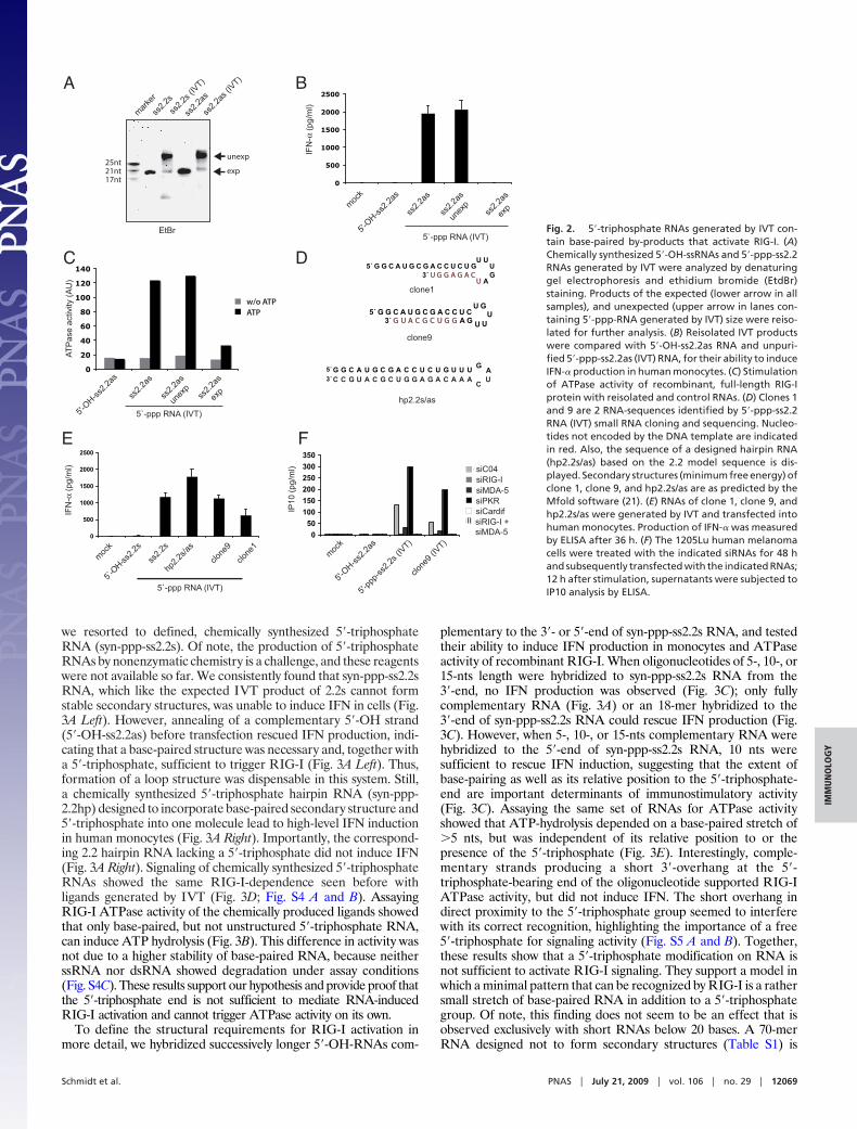
ResultsLigand-Induced ATPase Activity Is Triggered by a Feature Other thanthe 5�-Triphosphate Moiety. Previous studies have shown that short5�-triphosphate RNAs of 19 to 21 bases generated by IVT arepotent RIG-I ligands that induce IFN in human monocytes inde-pendently of TLRs (8–10). To identify a minimal pattern sufficientto trigger RIG-I signaling, we analyzed different versions of a19-mer model-RNA named 2.2 (Table S1). The chemically synthe-sized 5�-OH version of ss2.2 sense (s) RNA, its complementaryantisense (as) strand, and their annealed base-paired version (ds2.2)failed to induce IFN-� when transfected into human monocytes(Fig. 1A). However, as expected from previous studies, IVT (5�-triphosphate) 2.2 RNA induced strong IFN production either asssRNA (ss2.2s) or as dsRNA (ds2.2) (Fig. 1A). Of note, in ourhands, chemically synthesized ds or ss 3�- or 5�-monophosphory-lated RNAs did not show significant immunostimulatory activitywhen we transfected them into human monocytes using the 2.2sequence or a 25-mer sequence published to be active in mouse cellsby Takahasi et al. (Fig. S1A) (12). However, they were active whentransfected into human PBMCs containing plasmacytoid dendriticcells, indicating that these RNAs can be recognized by a different
Author contributions: A.S., T.S., W.H., J.C.H., S.C., K.-P.H., S.E., and S.R. designed research;A.S., T.S., W.H., J.C.H., S.C., M.W., and F.S.H. performed research; M.-C.M., R.B., and K.-P.H.contributed new reagents/analytic tools; A.S., T.S., W.H., J.C.H., S.C., M.W., F.S.H., R.B., S.E.,and S.R. analyzed data; and A.S., S.E., and S.R. wrote the paper.
The authors declare no conflict of interest.
This article is a PNAS Direct Submission.
Freely available online through the PNAS open access option.
1A.S., T.S., and W.H. contributed equally to this work.
2To whom correspondence should be addressed. E-mail: [email protected].
This article contains supporting information online at www.pnas.org/cgi/content/full/0900971106/DCSupplemental.
www.pnas.org�cgi�doi�10.1073�pnas.0900971106 PNAS � July 21, 2009 � vol. 106 � no. 29 � 12067–12072
IMM
UN
OLO
GY
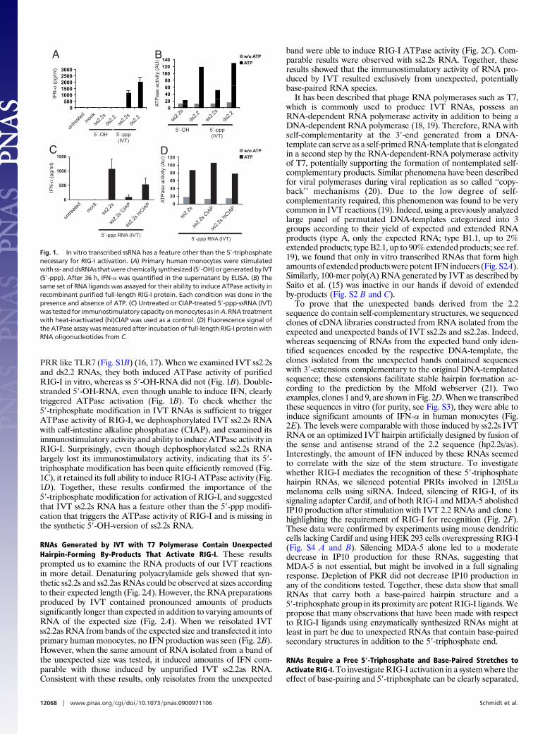
PRR like TLR7 (Fig. S1B) (16, 17). When we examined IVT ss2.2sand ds2.2 RNAs, they both induced ATPase activity of purifiedRIG-I in vitro, whereas ss 5�-OH-RNA did not (Fig. 1B). Double-stranded 5�-OH-RNA, even though unable to induce IFN, clearlytriggered ATPase activation (Fig. 1B). To check whether the5�-triphosphate modification in IVT RNAs is sufficient to triggerATPase activity of RIG-I, we dephosphorylated IVT ss2.2s RNAwith calf-intestine alkaline phosphatase (CIAP), and examined itsimmunostimulatory activity and ability to induce ATPase activity inRIG-I. Surprisingly, even though dephosphorylated ss2.2s RNAlargely lost its immunostimulatory activity, indicating that its 5�-triphosphate modification has been quite efficiently removed (Fig.1C), it retained its full ability to induce RIG-I ATPase activity (Fig.1D). Together, these results confirmed the importance of the5�-triphosphate modification for activation of RIG-I, and suggestedthat IVT ss2.2s RNA has a feature other than the 5�-ppp modifi-cation that triggers the ATPase activity of RIG-I and is missing inthe synthetic 5�-OH-version of ss2.2s RNA.
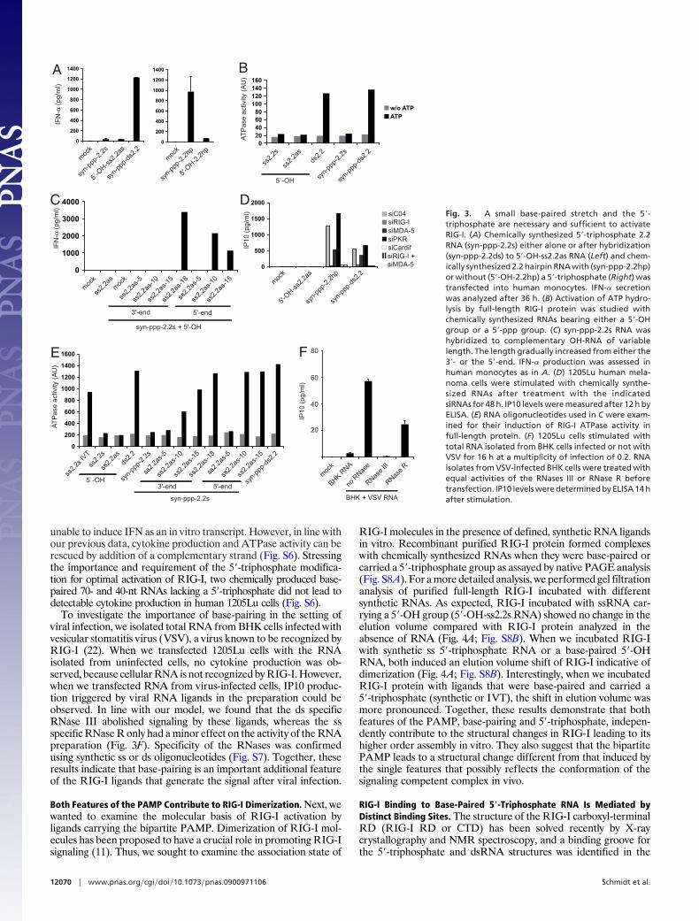
RNAs Generated by IVT with T7 Polymerase Contain UnexpectedHairpin-Forming By-Products That Activate RIG-I. These resultsprompted us to examine the RNA products of our IVT reactionsin more detail. Denaturing polyacrylamide gels showed that syn-thetic ss2.2s and ss2.2as RNAs could be observed at sizes accordingto their expected length (Fig. 2A). However, the RNA preparationsproduced by IVT contained pronounced amounts of productssignificantly longer than expected in addition to varying amounts ofRNA of the expected size (Fig. 2A). When we reisolated IVTss2.2as RNA from bands of the expected size and transfected it intoprimary human monocytes, no IFN production was seen (Fig. 2B).However, when the same amount of RNA isolated from a band ofthe unexpected size was tested, it induced amounts of IFN com-parable with those induced by unpurified IVT ss2.2as RNA.Consistent with these results, only reisolates from the unexpected
band were able to induce RIG-I ATPase activity (Fig. 2C). Com-parable results were observed with ss2.2s RNA. Together, theseresults showed that the immunostimulatory activity of RNA pro-duced by IVT resulted exclusively from unexpected, potentiallybase-paired RNA species.
It has been described that phage RNA polymerases such as T7,which is commonly used to produce IVT RNAs, possess anRNA-dependent RNA polymerase activity in addition to being aDNA-dependent RNA polymerase (18, 19). Therefore, RNA withself-complementarity at the 3�-end generated from a DNA-template can serve as a self-primed RNA-template that is elongatedin a second step by the RNA-dependent-RNA polymerase activityof T7, potentially supporting the formation of nontemplated self-complementary products. Similar phenomena have been describedfor viral polymerases during viral replication as so called ‘‘copy-back’’ mechanisms (20). Due to the low degree of self-complementarity required, this phenomenon was found to be verycommon in IVT reactions (19). Indeed, using a previously analyzedlarge panel of permutated DNA-templates categorized into 3groups according to their yield of expected and extended RNAproducts (type A, only the expected RNA; type B1.1, up to 2%extended products; type B2.1, up to 90% extended products; see ref.19), we found that only in vitro transcribed RNAs that form highamounts of extended products were potent IFN inducers (Fig. S2A).Similarly, 100-mer poly(A) RNA generated by IVT as described bySaito et al. (15) was inactive in our hands if devoid of extendedby-products (Fig. S2 B and C).
To prove that the unexpected bands derived from the 2.2sequence do contain self-complementary structures, we sequencedclones of cDNA libraries constructed from RNA isolated from theexpected and unexpected bands of IVT ss2.2s and ss2.2as. Indeed,whereas sequencing of RNAs from the expected band only iden-tified sequences encoded by the respective DNA-template, theclones isolated from the unexpected bands contained sequenceswith 3�-extensions complementary to the original DNA-templatedsequence; these extensions facilitate stable hairpin formation ac-cording to the prediction by the Mfold webserver (21). Twoexamples, clones 1 and 9, are shown in Fig. 2D. When we transcribedthese sequences in vitro (for purity, see Fig. S3), they were able toinduce significant amounts of IFN-� in human monocytes (Fig.2E). The levels were comparable with those induced by ss2.2s IVTRNA or an optimized IVT hairpin artificially designed by fusion ofthe sense and antisense strand of the 2.2 sequence (hp2.2s/as).Interestingly, the amount of IFN induced by these RNAs seemedto correlate with the size of the stem structure. To investigatewhether RIG-I mediates the recognition of these 5�-triphosphatehairpin RNAs, we silenced potential PRRs involved in 1205Lumelanoma cells using siRNA. Indeed, silencing of RIG-I, of itssignaling adapter Cardif, and of both RIG-I and MDA-5 abolishedIP10 production after stimulation with IVT 2.2 RNAs and clone 1highlighting the requirement of RIG-I for recognition (Fig. 2F).These data were confirmed by experiments using mouse dendriticcells lacking Cardif and using HEK 293 cells overexpressing RIG-I(Fig. S4 A and B). Silencing MDA-5 alone led to a moderatedecrease in IP10 production for these RNAs, suggesting thatMDA-5 is not essential, but might be involved in a full signalingresponse. Depletion of PKR did not decrease IP10 production inany of the conditions tested. Together, these data show that smallRNAs that carry both a base-paired hairpin structure and a5�-triphosphate group in its proximity are potent RIG-I ligands. Wepropose that many observations that have been made with respectto RIG-I ligands using enzymatically synthesized RNAs might atleast in part be due to unexpected RNAs that contain base-pairedsecondary structures in addition to the 5�-triphosphate end.
RNAs Require a Free 5�-Triphosphate and Base-Paired Stretches toActivate RIG-I. To investigate RIG-I activation in a system where theeffect of base-pairing and 5�-triphosphate can be clearly separated,
0500
10001500200025003000
A
untre
ated
moc
k
ss2.
2s
ds2.
2ds
2.2
ss2.
2s
5`-OH 5`-ppp (IVT)
BIF
N-α
(pg/
ml)
0
20
40
60
80
100
120
140w/o ATP
ATP
ss2.
2s
ds2.
2ds
2.2
ss2.
2s
5`-OH 5`-ppp (IVT)
C
0
500
1000
1500
leer Lipo 2.2s unb. 2.2s C(4u) 2.2s iC(4u)
untre
ated
moc
k
IFN
-α (p
g/m
l)
ss2.
2s
ss2.
2s C
IAP
5`-ppp RNA (IVT)
0
20
40
60
80
100
120
2.2s .2s iC
w/o ATPATP
5`-ppp RNA (IVT)
D
ss2.
2s h
iCIA
P
ss2.
2s
ss2.
2s C
IAP
ss2.
2s h
iCIA
P
AT
Pas
e ac
tivity
(A
U)
AT
Pas
e ac
tivity
(A
U)
Fig. 1. In vitro transcribed ssRNA has a feature other than the 5�-triphosphatenecessary for RIG-I activation. (A) Primary human monocytes were stimulatedwith ss-anddsRNAsthatwerechemically synthesized (5�-OH)orgeneratedby IVT(5�-ppp). After 36 h, IFN-� was quantified in the supernatant by ELISA. (B) Thesame set of RNA ligands was assayed for their ability to induce ATPase activity inrecombinant purified full-length RIG-I protein. Each condition was done in thepresence and absence of ATP. (C) Untreated or CIAP-treated 5�-ppp-ssRNA (IVT)was tested for immunostimulatory capacity on monocytes as in A. RNA treatmentwith heat-inactivated (hi)CIAP was used as a control. (D) Fluorescence signal ofthe ATPase assay was measured after incubation of full-length RIG-I protein withRNA oligonucleotides from C.
12068 � www.pnas.org�cgi�doi�10.1073�pnas.0900971106 Schmidt et al.
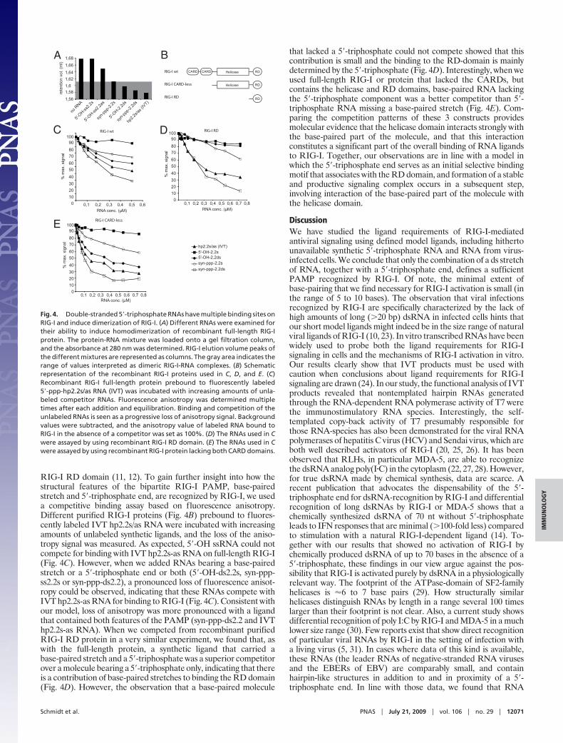
we resorted to defined, chemically synthesized 5�-triphosphateRNA (syn-ppp-ss2.2s). Of note, the production of 5�-triphosphateRNAs by nonenzymatic chemistry is a challenge, and these reagentswere not available so far. We consistently found that syn-ppp-ss2.2sRNA, which like the expected IVT product of 2.2s cannot formstable secondary structures, was unable to induce IFN in cells (Fig.3A Left). However, annealing of a complementary 5�-OH strand(5�-OH-ss2.2as) before transfection rescued IFN production, indi-cating that a base-paired structure was necessary and, together witha 5�-triphosphate, sufficient to trigger RIG-I (Fig. 3A Left). Thus,formation of a loop structure was dispensable in this system. Still,a chemically synthesized 5�-triphosphate hairpin RNA (syn-ppp-2.2hp) designed to incorporate base-paired secondary structure and5�-triphosphate into one molecule lead to high-level IFN inductionin human monocytes (Fig. 3A Right). Importantly, the correspond-ing 2.2 hairpin RNA lacking a 5�-triphosphate did not induce IFN(Fig. 3A Right). Signaling of chemically synthesized 5�-triphosphateRNAs showed the same RIG-I-dependence seen before withligands generated by IVT (Fig. 3D; Fig. S4 A and B). AssayingRIG-I ATPase activity of the chemically produced ligands showedthat only base-paired, but not unstructured 5�-triphosphate RNA,can induce ATP hydrolysis (Fig. 3B). This difference in activity wasnot due to a higher stability of base-paired RNA, because neitherssRNA nor dsRNA showed degradation under assay conditions(Fig. S4C). These results support our hypothesis and provide proof thatthe 5�-triphosphate end is not sufficient to mediate RNA-inducedRIG-I activation and cannot trigger ATPase activity on its own.
To define the structural requirements for RIG-I activation inmore detail, we hybridized successively longer 5�-OH-RNAs com-
plementary to the 3�- or 5�-end of syn-ppp-ss2.2s RNA, and testedtheir ability to induce IFN production in monocytes and ATPaseactivity of recombinant RIG-I. When oligonucleotides of 5-, 10-, or15-nts length were hybridized to syn-ppp-ss2.2s RNA from the3�-end, no IFN production was observed (Fig. 3C); only fullycomplementary RNA (Fig. 3A) or an 18-mer hybridized to the3�-end of syn-ppp-ss2.2s RNA could rescue IFN production (Fig.3C). However, when 5-, 10-, or 15-nts complementary RNA werehybridized to the 5�-end of syn-ppp-ss2.2s RNA, 10 nts weresufficient to rescue IFN induction, suggesting that the extent ofbase-pairing as well as its relative position to the 5�-triphosphate-end are important determinants of immunostimulatory activity(Fig. 3C). Assaying the same set of RNAs for ATPase activityshowed that ATP-hydrolysis depended on a base-paired stretch of�5 nts, but was independent of its relative position to or thepresence of the 5�-triphosphate (Fig. 3E). Interestingly, comple-mentary strands producing a short 3�-overhang at the 5�-triphosphate-bearing end of the oligonucleotide supported RIG-IATPase activity, but did not induce IFN. The short overhang indirect proximity to the 5�-triphosphate group seemed to interferewith its correct recognition, highlighting the importance of a free5�-triphosphate for signaling activity (Fig. S5 A and B). Together,these results show that a 5�-triphosphate modification on RNA isnot sufficient to activate RIG-I signaling. They support a model inwhich a minimal pattern that can be recognized by RIG-I is a rathersmall stretch of base-paired RNA in addition to a 5�-triphosphategroup. Of note, this finding does not seem to be an effect that isobserved exclusively with short RNAs below 20 bases. A 70-merRNA designed not to form secondary structures (Table S1) is
0
500
1000
1500
2000
2500
mock 2.2as syn 2.2as IVT as o unbeh. as u
A
IFN
-α (p
g/m
l)
moc
k
5'-OH-s
s2.2
as
ss2.
2as
ss2.
2as
unex
pss
2.2a
s
exp
5`-ppp RNA (IVT)
IFN
-α (p
g/m
l)
0
500
1000
1500
2000
2500
1 2 3 4 5 6
5`-O
H-ss2.
2sm
ock
ss2.
2s
hp2.
2s/a
s
clone
9
clone
1
5`-ppp RNA (IVT)
0
50
100
150
200
250
300
350
RNAiMax 2.2as-OH 2.2s-IVT Clone9
CO4RIG-IMDA-5PKRIPS-1RIG-I & MDA-5
IP10
(pg/
ml)
5'-OH-s
s2.2
asm
ock
5'-pp
p-ss
2.2s
(IVT)
clone
9 (IV
T)
siRIG-IsiMDA-5siPKRsiCardifsiRIG-I + siMDA-5
B
C D
ss2.
2s
ss2.
2s (I
VT)
ss2.
2as
ss2.
2as (
IVT)
mar
ker
17nt21nt25nt
unexp
exp
EtBr
siC04
G G C A U G GC A CC U C U GCAGAGGU
UUU
GAU
5´3´
G G C A U G GC A CC U CU G
C AGA GGUU
UUGU5´
3´ G C
clone1
clone9
G G G GG CC CC UU U UUCA AG G G G GC UUC CCA A AAA C
GU AA
CU
5´
3´
hp2.2s/as
0
20
40
60
80
100
120
140
as as complete as upper band as lower band
w/o ATPATP
w/o ATPATP
5'-OH-s
s2.2
as
ss2.
2as
ss2.
2as
unex
pss
2.2a
s
exp
5`-ppp RNA (IVT)
AT
Pas
e ac
tivity
(A
U)
E F
Fig. 2. 5�-triphosphate RNAs generated by IVT con-tain base-paired by-products that activate RIG-I. (A)Chemically synthesized 5�-OH-ssRNAs and 5�-ppp-ss2.2RNAs generated by IVT were analyzed by denaturinggel electrophoresis and ethidium bromide (EtdBr)staining. Products of the expected (lower arrow in allsamples), and unexpected (upper arrow in lanes con-taining 5�-ppp-RNA generated by IVT) size were reiso-lated for further analysis. (B) Reisolated IVT productswere compared with 5�-OH-ss2.2as RNA and unpuri-fied 5�-ppp-ss2.2as (IVT) RNA, for their ability to induceIFN-� production in human monocytes. (C) Stimulationof ATPase activity of recombinant, full-length RIG-Iprotein with reisolated and control RNAs. (D) Clones 1and 9 are 2 RNA-sequences identified by 5�-ppp-ss2.2RNA (IVT) small RNA cloning and sequencing. Nucleo-tides not encoded by the DNA template are indicatedin red. Also, the sequence of a designed hairpin RNA(hp2.2s/as) based on the 2.2 model sequence is dis-played. Secondary structures (minimum free energy) ofclone 1, clone 9, and hp2.2s/as are as predicted by theMfold software (21). (E) RNAs of clone 1, clone 9, andhp2.2s/as were generated by IVT and transfected intohuman monocytes. Production of IFN-� was measuredby ELISA after 36 h. (F) The 1205Lu human melanomacells were treated with the indicated siRNAs for 48 hand subsequently transfected with the indicated RNAs;12 h after stimulation, supernatants were subjected toIP10 analysis by ELISA.
Schmidt et al. PNAS � July 21, 2009 � vol. 106 � no. 29 � 12069
IMM
UN
OLO
GY
unable to induce IFN as an in vitro transcript. However, in line withour previous data, cytokine production and ATPase activity can berescued by addition of a complementary strand (Fig. S6). Stressingthe importance and requirement of the 5�-triphosphate modifica-tion for optimal activation of RIG-I, two chemically produced base-paired 70- and 40-nt RNAs lacking a 5�-triphosphate did not lead todetectable cytokine production in human 1205Lu cells (Fig. S6).
To investigate the importance of base-pairing in the setting ofviral infection, we isolated total RNA from BHK cells infected withvesicular stomatitis virus (VSV), a virus known to be recognized byRIG-I (22). When we transfected 1205Lu cells with the RNAisolated from uninfected cells, no cytokine production was ob-served, because cellular RNA is not recognized by RIG-I. However,when we transfected RNA from virus-infected cells, IP10 produc-tion triggered by viral RNA ligands in the preparation could beobserved. In line with our model, we found that the ds specificRNase III abolished signaling by these ligands, whereas the ssspecific RNase R only had a minor effect on the activity of the RNApreparation (Fig. 3F). Specificity of the RNases was confirmedusing synthetic ss or ds oligonucleotides (Fig. S7). Together, theseresults indicate that base-pairing is an important additional featureof the RIG-I ligands that generate the signal after viral infection.
Both Features of the PAMP Contribute to RIG-I Dimerization. Next, wewanted to examine the molecular basis of RIG-I activation byligands carrying the bipartite PAMP. Dimerization of RIG-I mol-ecules has been proposed to have a crucial role in promoting RIG-Isignaling (11). Thus, we sought to examine the association state of
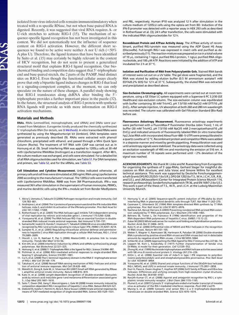
RIG-I molecules in the presence of defined, synthetic RNA ligandsin vitro. Recombinant purified RIG-I protein formed complexeswith chemically synthesized RNAs when they were base-paired orcarried a 5�-triphosphate group as assayed by native PAGE analysis(Fig. S8A). For a more detailed analysis, we performed gel filtrationanalysis of purified full-length RIG-I incubated with differentsynthetic RNAs. As expected, RIG-I incubated with ssRNA car-rying a 5�-OH group (5�-OH-ss2.2s RNA) showed no change in theelution volume compared with RIG-I protein analyzed in theabsence of RNA (Fig. 4A; Fig. S8B). When we incubated RIG-Iwith synthetic ss 5�-triphosphate RNA or a base-paired 5�-OHRNA, both induced an elution volume shift of RIG-I indicative ofdimerization (Fig. 4A; Fig. S8B). Interestingly, when we incubatedRIG-I protein with ligands that were base-paired and carried a5�-triphosphate (synthetic or IVT), the shift in elution volume wasmore pronounced. Together, these results demonstrate that bothfeatures of the PAMP, base-pairing and 5�-triphosphate, indepen-dently contribute to the structural changes in RIG-I leading to itshigher order assembly in vitro. They also suggest that the bipartitePAMP leads to a structural change different from that induced bythe single features that possibly reflects the conformation of thesignaling competent complex in vivo.
RIG-I Binding to Base-Paired 5�-Triphosphate RNA Is Mediated byDistinct Binding Sites. The structure of the RIG-I carboxyl-terminalRD (RIG-I RD or CTD) has been solved recently by X-raycrystallography and NMR spectroscopy, and a binding groove forthe 5�-triphosphate and dsRNA structures was identified in the
0
1000
2000
3000
4000
0
200
400
600
800
1000
1200
1400
1600
1 2 3 4 5 6 7 8 9 10 11 12 13
A B
AT
Pas
e ac
tivity
(A
U)
5`-OH
020406080
100120140160
1 2 3 4 5
w/o ATPATP
ss2.
2as
ss2.
2s
syn-
ppp-
2.2s
syn-
ppp-
ds2.
2
C
moc
k
ss2.
2as
syn-ppp-2.2s + 5'-OH
ss2.
2as-
5
ss2.
2as-
10
ss2.
2as-
15
ss2.
2as-
18
ss2.
2as-
5
ss2.
2as-
10
ss2.
2as-
15m
ock
3'-end 5'-end
ds2.
2
ss2.
2s IV
T
ss2.
2s
ss2.
2as
ds2.
2
5`-OH syn-
ppp-
2.2s
ss2.
2as-
5
ss2.
2as-
10
ss2.
2as-
15
ss2.
2as-
18
3'-endsy
n-pp
p-ds
2.2
ss2.
2as-
5
ss2.
2as-
10
ss2.
2as-
15
5'-end
syn-ppp-2.2s
AT
Pas
e ac
tivity
(A
U)
0
500
1000
1500
2000
RNAiMax 2.2as-OH syn3p hp syn3p ds
CO4RIG-IMDA-5PKRIPS-1RIG-I & MDA-5
siC04siRIG-IsiMDA-5siPKRsiCardifsiRIG-I + siMDA-5
moc
k
5'-OH-s
s2.2
as
syn-
ppp-
ds2.
2
syn-
ppp-
2.2h
p
IP10
(pg/
ml)
D
E
IFN
-α (p
g/m
l)
0
200
400
600
800
1000
1200
1400
Lipo as-s hp syn3p as-s hp synOH0
200
400
600
800
1000
1200
1400
1 2 3 4
IFN
-α (p
g/m
l)
moc
k
5´-O
H-ss2
.2as
syn-
ppp-
2.2s
syn-
ppp-
ds2.
2m
ock
syn-
ppp-
2.2h
p
5'-OH-2
.2hp
0
20
40
60
80
mock BHK VSV VSV III VSV R
20
40
60
80
IP10
(pg/
ml)
moc
k
BHK RNA
no R
Nase
RNase
III
RNase
R
BHK + VSV RNA
F
Fig. 3. A small base-paired stretch and the 5�-triphosphate are necessary and sufficient to activateRIG-I. (A) Chemically synthesized 5�-triphosphate 2.2RNA (syn-ppp-2.2s) either alone or after hybridization(syn-ppp-2.2ds) to 5�-OH-ss2.2as RNA (Left) and chem-ically synthesized 2.2 hairpin RNA with (syn-ppp-2.2hp)or without (5�-OH-2.2hp) a 5�-triphosphate (Right) wastransfected into human monocytes. IFN-� secretionwas analyzed after 36 h. (B) Activation of ATP hydro-lysis by full-length RIG-I protein was studied withchemically synthesized RNAs bearing either a 5�-OHgroup or a 5�-ppp group. (C) syn-ppp-2.2s RNA washybridized to complementary OH-RNA of variablelength. The length gradually increased from either the3�- or the 5�-end. IFN-� production was assessed inhuman monocytes as in A. (D) 1205Lu human mela-noma cells were stimulated with chemically synthe-sized RNAs after treatment with the indicatedsiRNAs for 48 h. IP10 levels were measured after 12 h byELISA. (E) RNA oligonucleotides used in C were exam-ined for their induction of RIG-I ATPase activity infull-length protein. (F) 1205Lu cells stimulated withtotal RNA isolated from BHK cells infected or not withVSV for 16 h at a multiplicity of infection of 0.2. RNAisolates from VSV-infected BHK cells were treated withequal activities of the RNases III or RNase R beforetransfection. IP10 levels were determined by ELISA 14 hafter stimulation.
12070 � www.pnas.org�cgi�doi�10.1073�pnas.0900971106 Schmidt et al.
RIG-I RD domain (11, 12). To gain further insight into how thestructural features of the bipartite RIG-I PAMP, base-pairedstretch and 5�-triphosphate end, are recognized by RIG-I, we useda competitive binding assay based on fluorescence anisotropy.Different purified RIG-I proteins (Fig. 4B) prebound to fluores-cently labeled IVT hp2.2s/as RNA were incubated with increasingamounts of unlabeled synthetic ligands, and the loss of the aniso-tropy signal was measured. As expected, 5�-OH ssRNA could notcompete for binding with IVT hp2.2s-as RNA on full-length RIG-I(Fig. 4C). However, when we added RNAs bearing a base-pairedstretch or a 5�-triphosphate end or both (5�-OH-ds2.2s, syn-ppp-ss2.2s or syn-ppp-ds2.2), a pronounced loss of fluorescence anisot-ropy could be observed, indicating that these RNAs compete withIVT hp2.2s-as RNA for binding to RIG-I (Fig. 4C). Consistent withour model, loss of anisotropy was more pronounced with a ligandthat contained both features of the PAMP (syn-ppp-ds2.2 and IVThp2.2s-as RNA). When we competed from recombinant purifiedRIG-I RD protein in a very similar experiment, we found that, aswith the full-length protein, a synthetic ligand that carried abase-paired stretch and a 5�-triphosphate was a superior competitorover a molecule bearing a 5�-triphosphate only, indicating that thereis a contribution of base-paired stretches to binding the RD domain(Fig. 4D). However, the observation that a base-paired molecule
that lacked a 5�-triphosphate could not compete showed that thiscontribution is small and the binding to the RD-domain is mainlydetermined by the 5�-triphosphate (Fig. 4D). Interestingly, when weused full-length RIG-I or protein that lacked the CARDs, butcontains the helicase and RD domains, base-paired RNA lackingthe 5�-triphosphate component was a better competitor than 5�-triphosphate RNA missing a base-paired stretch (Fig. 4E). Com-paring the competition patterns of these 3 constructs providesmolecular evidence that the helicase domain interacts strongly withthe base-paired part of the molecule, and that this interactionconstitutes a significant part of the overall binding of RNA ligandsto RIG-I. Together, our observations are in line with a model inwhich the 5�-triphosphate end serves as an initial selective bindingmotif that associates with the RD domain, and formation of a stableand productive signaling complex occurs in a subsequent step,involving interaction of the base-paired part of the molecule withthe helicase domain.
DiscussionWe have studied the ligand requirements of RIG-I-mediatedantiviral signaling using defined model ligands, including hithertounavailable synthetic 5�-triphosphate RNA and RNA from virus-infected cells. We conclude that only the combination of a ds stretchof RNA, together with a 5�-triphosphate end, defines a sufficientPAMP recognized by RIG-I. Of note, the minimal extent ofbase-pairing that we find necessary for RIG-I activation is small (inthe range of 5 to 10 bases). The observation that viral infectionsrecognized by RIG-I are specifically characterized by the lack ofhigh amounts of long (�20 bp) dsRNA in infected cells hints thatour short model ligands might indeed be in the size range of naturalviral ligands of RIG-I (10, 23). In vitro transcribed RNAs have beenwidely used to probe both the ligand requirements for RIG-Isignaling in cells and the mechanisms of RIG-I activation in vitro.Our results clearly show that IVT products must be used withcaution when conclusions about ligand requirements for RIG-Isignaling are drawn (24). In our study, the functional analysis of IVTproducts revealed that nontemplated hairpin RNAs generatedthrough the RNA-dependent RNA polymerase activity of T7 werethe immunostimulatory RNA species. Interestingly, the self-templated copy-back activity of T7 presumably responsible forthose RNA-species has also been demonstrated for the viral RNApolymerases of hepatitis C virus (HCV) and Sendai virus, which areboth well described activators of RIG-I (20, 25, 26). It has beenobserved that RLHs, in particular MDA-5, are able to recognizethe dsRNA analog poly(I�C) in the cytoplasm (22, 27, 28). However,for true dsRNA made by chemical synthesis, data are scarce. Arecent publication that advocates the dispensability of the 5�-triphosphate end for dsRNA-recognition by RIG-I and differentialrecognition of long dsRNAs by RIG-I or MDA-5 shows that achemically synthesized dsRNA of 70 nt without 5�-triphosphateleads to IFN responses that are minimal (�100-fold less) comparedto stimulation with a natural RIG-I-dependent ligand (14). To-gether with our results that showed no activation of RIG-I bychemically produced dsRNA of up to 70 bases in the absence of a5�-triphosphate, these findings in our view argue against the pos-sibility that RIG-I is activated purely by dsRNA in a physiologicallyrelevant way. The footprint of the ATPase-domain of SF2-familyhelicases is �6 to 7 base pairs (29). How structurally similarhelicases distinguish RNAs by length in a range several 100 timeslarger than their footprint is not clear. Also, a current study showsdifferential recognition of poly I:C by RIG-I and MDA-5 in a muchlower size range (30). Few reports exist that show direct recognitionof particular viral RNAs by RIG-I in the setting of infection witha living virus (5, 31). In cases where data of this kind is available,these RNAs (the leader RNAs of negative-stranded RNA virusesand the EBERs of EBV) are comparably small, and containhairpin-like structures in addition to and in proximity of a 5�-triphosphate end. In line with those data, we found that RNA
1,56
1,58
1,6
1,62
1,64
1,66
1,68
0 1 8 7 5 9 6
A
0
10
20
30
40
50
60
70
80
90
100
0 100 200 300 400 500 6000
10
30
20
40
50
60
70
90
80
100
0,1 0,2 0,3 0,4 0,5 0,6
% m
ax. s
igna
lB
RNA conc. (µM)
RIG-I wt
0
10
20
30
40
50
60
70
80
90
100
0 100 200 300 400 500 600 700 8000
10
30
20
40
50
60
70
90
80
100
% m
ax. s
igna
l
0,1 0,2 0,3 0,4 0,5 0,6 0,7 0,8RNA conc. (µM)
RIG-I RDC
0
10
20
30
40
50
60
70
80
90
100
0 100 200 300 400 500 600 700 8000
10
30
20
40
50
60
70
90
80
100
0,1 0,2 0,3 0,4 0,5 0,6 0,7 0,8
% m
ax. s
igna
l
RNA conc. (µM)
RIG-I CARD-less
D
no R
NA
5'-OH-s
s2.2
s
5'-OH-2
.2ds
hp2.
2s/a
s (IV
T)
syn-
ppp-
2.2s
5'-OH-s
s2.2
as
syn-
ppp-
2.2d
s1,56
1,58
1,6
1,62
1,64
1,66
1,68re
tent
ion
vol.
(ml)
CARD CARD Helicase RDRIG-I wt
RIG-I CARD-less Helicase RD
RIG-I RD RD
Reihe1
Reihe2
Reihe3
Reihe4
Reihe5syn-ppp-2.2dssyn-ppp-2.2s5'-OH-2.2ds5'-OH-2.2shp2.2s/as (IVT)
E
Fig. 4. Double-stranded 5�-triphosphate RNAs have multiple binding sites onRIG-I and induce dimerization of RIG-I. (A) Different RNAs were examined fortheir ability to induce homodimerization of recombinant full-length RIG-Iprotein. The protein-RNA mixture was loaded onto a gel filtration column,and the absorbance at 280 nm was determined. RIG-I elution volume peaks ofthe different mixtures are represented as columns. The gray area indicates therange of values interpreted as dimeric RIG-I-RNA complexes. (B) Schematicrepresentation of the recombinant RIG-I proteins used in C, D, and E. (C)Recombinant RIG-I full-length protein prebound to fluorescently labeled5�-ppp-hp2.2s/as RNA (IVT) was incubated with increasing amounts of unla-beled competitor RNAs. Fluorescence anisotropy was determined multipletimes after each addition and equilibration. Binding and competition of theunlabeled RNAs is seen as a progressive loss of anisotropy signal. Backgroundvalues were subtracted, and the anisotropy value of labeled RNA bound toRIG-I in the absence of a competitor was set as 100%. (D) The RNAs used in Cwere assayed by using recombinant RIG-I RD domain. (E) The RNAs used in Cwere assayed by using recombinant RIG-I protein lacking both CARD domains.
Schmidt et al. PNAS � July 21, 2009 � vol. 106 � no. 29 � 12071
IMM
UN
OLO
GY
isolated from virus-infected cells remains immunostimulatory whentreated with a ss specific RNase, but not when base-paired RNA isdigested. Recently, it was suggested that viral RNA requires A- orU-rich stretches to activate RIG-I (15). The mechanism of se-quence-specific ligand recognition has not been investigated in thiscontext. We did not systematically test the influence of sequencecontent on RIG-I activation. However, the different short se-quences we found to be active were neither A nor U rich (�50%A plus U). Therefore, the ligand features that have been identifiedby Saito et al. (15) may certainly be highly relevant in the contextof HCV recognition, but do not seem to present a generalizedstructural motif that explains RIG-I ligand recognition. Our bio-chemical binding and competition data suggest that 5�-triphosphateend and base-paired stretch, the 2 parts of the PAMP, bind distinctsites on RIG-I. Even though the functional cellular assays clearlyprove that only a bipartite ligand induces changes in RIG-I that leadto a signaling-competent complex, at the moment, we can onlyspeculate on the nature of these changes. A parallel study showingthat RIG-I translocates along dsRNA after binding the 5�-triphosphate provides a further important piece in the puzzle (32).In the future, the structural analysis of RIG-I protein with syntheticRNA ligands will provide us with more information on RIG-Iactivation mechanisms.
Materials and MethodsRNAs. RNAs (unmodified, monophosphate, and siRNA) and DNAs were pur-chased from Metabion. Eurogentec kindly produced the chemically synthesized5�-triphosphate RNA (for details, see SI Methods). In vitro transcribed RNAs weresynthesized by using the Megashortscript kit (Ambion). DNA templates weregenerated as previously described (9). RNAs were extracted with phenol/chloroform, precipitated with ethanol, and passed through a Mini Quick SpinColumn (Roche). The treatment of IVT RNA with CIAP was carried out as inHornung et al. (9). Small interfering RNA was applied to 1205Lu cells at 30 nMwith Lipofectamine RNAiMax (Invitrogen) as a transfection reagent. After 48 h,the culture medium was exchanged, and cells were stimulated. For a detailed listof all RNA oligonucleotides used for stimulation, see Table S1. For DNA templatesand primers, see Table S2, and for the siRNAs, see Table S3.
Cell Stimulation and Cytokine Measurement. Unless indicated otherwise, allprimarycellsandcell lineswerestimulatedat200ng/mLRNAusingLipofectamine2000 according to the manufacturer’s manual. The 1205LU cells were transfectedwith Lipofectamine RNAiMax. R848 was from 3M Pharmaceuticals. IFN-� wasmeasured36hafter stimulation in thesupernatantofhumanmonocytes, PBMCs,and murine dendritic cells using the IFN-� module set from Bender MedSystems
and PBL, respectively. Human IP10 was analyzed 12 h after stimulation in theculture medium of 1205LU cells using the opteia set from BD. Induction of theIFN-ß promoter was detected with a reporter assay in HEK 293 cells as describedin Rothenfusser et al. (3); 24 h after transfection, the cells were stimulated withthe indicated RNA oligonucleotides for 12 h.
Recombinant Protein and ATPase Activity Assay. The ATPase activity of recom-binant, purified RIG-I-protein was measured using the ADP Quest HS Assay(DiscoveRx). Full-length RIG-I was expressed in insect cells and purified as de-scribedpreviously (11).Thereactionmixturewaspreparedonice inatotalvolumeof 10 �L, containing 1 ng/�L purified RIG-I-protein, 1 ng/�L purified RNA oligo-nucleotide, and 100 �M ATP. Reactions were initiated by the addition of ATP andincubated for 2 h at 37 °C.
Isolation and Purification of RNA from Polyacrylamide Gels. Stained RNA bandsof interest were cut out on a UV table. The gel slices were fragmented, and theRNA was eluted by adding elution buffer (0.5 M ammonium acetate/1 mMEDTA/0.2% SDS) for 12 h at 37 °C. Subsequently, the eluted RNA was extractedand precipitated as described above.
Size-Exclusion Chromatography. All experiments were carried out at room tem-perature, using a GE Ettan LC system equipped with a Superose 6 PC 3.2/30 (GEHealthcare) size-exclusion column. The size-exclusion column was equilibratedwith buffer containing 30 mM Tris�HCl, pH 7.5/150 mM NaCl/2 mM DTT/10 �MZnCl2. After sample injection, UV absorption at both 260 and 280 nm wavelengthwas recorded. The column was calibrated with Gel Filtration Standard (Bio-Rad)before use.
Fluorescence Anisotropy Measurement. Fluorescence anisotropy experimentswere performed with a FluoroMax-P fluorimeter (Horiba Jobin Yvon); 1 mL ofbuffer (30 mM Tris�HCl, pH 7.5/150 mM NaCl/5 mM 2-mercaptoethanol/10 �MZnCl2) and indicated amounts of fluorescently labeled RNA (in vitro transcribedhp2.2s/asRNAwith incorporatedAlexaFluor488–5-UTP)werepreequilibrated inaquartzcuvetteat12 °C.RecombinantproteinandcompetitorRNAswereaddedin a stepwise manner and mixed by gentle pippetting. Preequilibration was useduntil anisotropy signals were stabilized. The anisotropy data were collected usingan excitation wavelength of 495 nm and monitoring the emission at 516 nm. Amaximum number of 10 repeats were performed until �2% deviation of thesignal was reached.
ACKNOWLEDGMENTS. We thank M. Linke and M. Ruesenberg from Eurogentecfor organizing the synthesis of 5�-ppp-RNAs, Gerhard Steger for insightful dis-cussions on RNA structure, and Julia Vorac and Simone Willms for excellenttechnical assistance. This work was supported by Deutsche Forschungsgemein-schaftGrantsDFGRO2525/3-1 (toS.R.),DFGGK1202(toT.S.,W.H,J.C.H.,S.R.,R.B.,and S.E.), and LMUexcellent (Center for Integrated Protein Science Munich 114,research professorship), Sonderforschungsbereich-TR 36, and EN 169/7-2 (to S.E.).This work is part of the thesis of T.S., W.H., and J.C.H. at the Ludwig-MaximilianUniversity Munich.
1. Akira S, Uematsu S, Takeuchi O (2006) Pathogen recognition and innate immunity. Cell124:783–801.
2. Andrejeva J, et al. (2004) The V proteins of paramyxoviruses bind the IFN-inducible RNAhelicase, mda-5, and inhibit its activation of the IFN-beta promoter. Proc Natl Acad SciUSA 101:17264–17269.
3. Rothenfusser S, et al. (2005) The RNA helicase Lgp2 inhibits TLR-independent sensingof viral replication by retinoic acid-inducible gene-I. J Immunol 175:5260–5268.
4. Yoneyama M, et al. (2004) The RNA helicase RIG-I has an essential function in double-stranded RNA-induced innate antiviral responses. Nat Immunol 5:730–737.
5. Samanta M, Iwakiri D, Kanda T, Imaizumi T, Takada K (2006) EB virus-encoded RNAs arerecognized by RIG-I and activate signaling to induce type I IFN. EMBO J 25:4207–4214.
6. Sumpter R, Jr, et al. (2005) Regulating intracellular antiviral defense and permissive-ness to hepatitis C virus RNA replication through a cellular RNA helicase, RIG-I. J Virol79:2689–2699.
7. Hiscott J, Lin R, Nakhaei P, Paz S (2006) MasterCARD: A priceless link to innateimmunity. Trends Mol Med 12:53–56.
8. Kim DH, et al. (2004) Interferon induction by siRNAs and ssRNAs synthesized by phagepolymerase. Nat Biotechnol 22:321–325.
9. Hornung V, et al. (2006) 5�-Triphosphate RNA is the ligand for RIG-I. Science 314:994–997.10. Pichlmair A, et al. (2006) RIG-I-mediated antiviral responses to single-stranded RNA
bearing 5�-phosphates. Science 314:997–1001.11. Cui S, et al. (2008) The C-terminal regulatory domain is the RNA 5�-triphosphate sensor
of RIG-I. Mol Cell 29:169–179.12. Takahasi K, et al. (2008) Nonself RNA-sensing mechanism of RIG-I helicase and activa-
tion of antiviral immune responses. Mol Cell 29:428–440.13. Malathi K, Dong B, Gale M, Jr, Silverman RH (2007) Small self-RNA generated by RNase
L amplifies antiviral innate immunity. Nature 448:816–819.14. Kato H, et al. (2008) Length-dependent recognition of double-stranded ribonucleic
acids by retinoic acid-inducible gene-I and melanoma differentiation-associated gene5. J Exp Med 205:1601–1610.
15. Saito T, Owen DM, Jiang F, Marcotrigiano J, Gale M (2008) Innate immunity induced bycomposition-dependent RIG-I recognition of hepatitis C virus RNA. Nature 454:523–527.
16. Diebold SS, Kaisho T, Hemmi H, Akira S, Reis e Sousa C (2004) Innate antiviral responses bymeans of TLR7-mediated recognition of single-stranded RNA. Science 303:1529–1531.
17. Hornung V, et al. (2005) Sequence-specific potent induction of IFN-alpha by shortinterfering RNA in plasmacytoid dendritic cells through TLR7. Nat Med 11:263–270.
18. Cazenave C, Uhlenbeck OC (1994) RNA template-directed RNA synthesis by T7 RNApolymerase. Proc Natl Acad Sci USA 91:6972–6976.
19. Nacheva GA, Berzal-Herranz A (2003) Preventing nondesired RNA-primed RNA exten-sion catalyzed by T7 RNA polymerase. Eur J Biochem 270:1458–1465.
20. Behrens SE, Tomei L, De Francesco R (1996) Identification and properties of theRNA-dependent RNA polymerase of hepatitis C virus. EMBO J 15:12–22.
21. Zuker M (2003) Mfold web server for nucleic acid folding and hybridization prediction.Nucleic Acids Res 31:3406–3415.
22. Kato H, et al. (2006) Differential roles of MDA5 and RIG-I helicases in the recognitionof RNA viruses. Nature 441:101–105.
23. Weber F, Wagner V, Rasmussen SB, Hartmann R, Paludan SR (2006) Double-strandedRNA is produced by positive-strand RNA viruses and DNA viruses but not in detectableamounts by negative-strand RNA viruses. J Virol 80:5059–5064.
24. Schlee M, et al. (2009) Approaching the RNA ligand for RIG-I? Immunol Rev 227:66–74.25. Leppert M, Kort L, Kolakofsky D (1977) Further characterization of Sendai virus
DI-RNAs: A model for their generation. Cell 12:539–552.26. Zhong W, et al. (1999) Nucleoside triphosphatase and RNA helicase activities associated
with GB virus B nonstructural protein 3. Virology 261:216–226.27. Gitlin L, et al. (2006) Essential role of mda-5 in type I IFN responses to polyriboi-
nosinic:polyribocytidylic acid and encephalomyocarditis picornavirus. Proc Natl AcadSci USA 103:8459–8464.
28. Yoneyama M, et al. (2005) Shared and unique functions of the DExD/H-box helicasesRIG-I, MDA5, and LGP2 in antiviral innate immunity. J Immunol 175:2851–2858.
29. Durr H, Flaus A, Owen-Hughes T, Hopfner KP (2006) Snf2 family ATPases and DExx boxhelicases: Differences and unifying concepts from high-resolution crystal structures.Nucleic Acids Res 34:4160–4167.
30. Ranjith-Kumar CT, et al. (2008) Agonist and antagonist recognition by RIG-I, a cyto-plasmic innate immunity receptor. J Biol Chem 284:1155–1165.
31. Plumet S, et al. (2007) Cytosolic 5�-triphosphate ended viral leader transcript of measlesvirus as activator of the RIG I-mediated interferon response. PLoS ONE 2:e279.
32. Myong S, et al. (2009) Cytosolic viral sensor RIG-I is a 5�-triphosphate-dependenttranslocase on double-stranded RNA. Science 323:1070–1074.
12072 � www.pnas.org�cgi�doi�10.1073�pnas.0900971106 Schmidt et al.
LETTERS
STING regulates intracellular DNA-mediated, type Iinterferon-dependent innate immunityHiroki Ishikawa1, Zhe Ma1 & Glen N. Barber1
The innate immune system is critical for the early detection ofinvading pathogens and for initiating cellular host defence counter-measures, which include the production of type I interferon(IFN)1–3. However, little is known about how the innate immunesystem is galvanized to respond to DNA-based microbes. Here weshow that STING (stimulator of interferon genes) is critical for theinduction of IFN by non-CpG intracellular DNA species producedby various DNA pathogens after infection4. Murine embryonicfibroblasts, as well as antigen presenting cells such as macrophagesand dendritic cells (exposed to intracellular B-form DNA, the DNAvirus herpes simplex virus 1 (HSV-1) or bacteria Listeria mono-cytogenes), were found to require STING to initiate effective IFNproduction. Accordingly, Sting-knockout mice were susceptibleto lethal infection after exposure to HSV-1. The importance ofSTING in facilitating DNA-mediated innate immune responseswas further evident because cytotoxic T-cell responses induced byplasmid DNA vaccination were reduced in Sting-deficient animals.In the presence of intracellular DNA, STING relocalized withTANK-binding kinase 1 (TBK1) from the endoplasmic reticulumto perinuclear vesicles containing the exocyst component Sec5 (alsoknown as EXOC2). Collectively, our studies indicate that STING isessential for host defence against DNA pathogens such as HSV-1and facilitates the adjuvant activity of DNA-based vaccines.
Nucleic acid species inadvertently generated by microbes afterinfection are potent inducers of cellular innate immune defencesimportant for protection of the host1–3. Although considerable pro-gress has been made into unravelling how RNA viruses induce type IIFN, required for triggering the production of anti-viral genes, little isknown at the molecular level about the induction of IFN by DNApathogens such as herpes simplex virus I (HSV-1) or by intracellularbacteria or parasites5–10. Toll-like receptor 9 (TLR9) is known torecognize CpG DNA to trigger IFN production in plasmacytoiddendritic cells (pDCs), and Z-DNA binding protein 1 (ZBP1, alsoknown as DAI) was recently shown to be able to stimulate IFN tran-scription, but was found to be largely redundant in studies usingDAI-deficient cells and mice11–13. Recently, a DNA receptor AIM2was found to be important for ASC (also known as PYCARD)-dependent inflammasome mediated production of IL1b, but wasnot required for type I IFN production14–18. Thus, other innate sign-alling pathways that recognize intracellular non-CpG DNA speciesmust exist to facilitate type I IFN production.
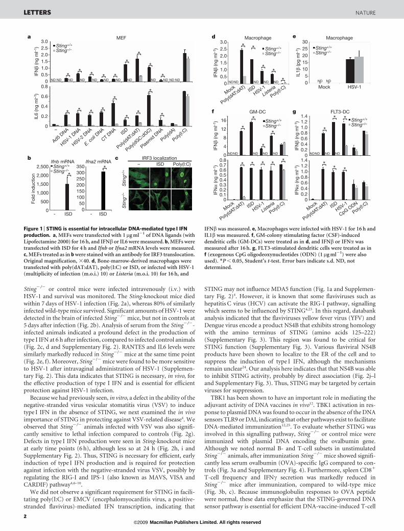
We previously demonstrated for the first time a role for STING(also referred to as TMEM173, MPYS and MITA), an endoplasmicreticulum (ER) resident transmembrane protein, in facilitating theproduction of type I IFN4,19,20. To evaluate the importance of STINGin mediating DNA-induced innate immune responses, we used wildtype (1/1) or Sting2/2 low passage number mouse embryonic fibro-blasts (MEFs) and compared the induction of type I IFN (IFNb) inresponse to a variety of DNA ligands. Our results indicated that
STING was essential for inducing IFNb in response to transfectedviral DNA (adenovirus, Ad5; herpes simplex virus, HSV-1 and -2),purified Escherichia coli DNA, calf thymus (CT) DNA, and interferonstimulatory DNA (ISD; double-stranded 45-base-pair oligonucleo-tides lacking CpG sequences) (Fig. 1a). Complete abrogation of IFNbproduction was also observed after transfection of synthetic double-stranded DNA (poly(dG-dC).poly(dC-dG), hereafter referred to aspoly(dGC:dGC)) in Sting2/2 MEFs, and slight IFNb production wasobserved using poly(dAT:dAT), probably due to STING-independent,RIG-I (also known as DDX58)-dependent signalling21,22. The loss ofSTING did not significantly affect poly(I:C)-mediated type I IFN pro-duction, which is largely governed by MDA5 (ref. 5). Concomitantanalysis further indicated a marked reduction in IL6 production inSting2/2 MEFs compared to controls after similar DNA transfections(Fig. 1a). ISD-mediated production of Ifnb and Ifn2a messengerRNA was not detectable in Sting2/2 MEFs compared to controls(Fig. 1b). Translocation of IRF3 or IRF7 was thus not observed in ISD-transfected Sting2/2 MEFs, indicating that STING probably functionsin mediating intracellular-DNA-triggered IFN production upstreamof TBK1 (Fig. 1c and Supplementary Fig. 1). NF-kB signalling was alsodefective in Sting2/2 MEFs after exposure to transfected ISD (Sup-plementary Fig. 1). Given this, we next examined the importance ofSTING in facilitating intracellular-DNA-mediated production oftype I IFN in antigen presenting cells. This analysis indicated thatSting2/2 macrophages transfected with ISD, or infected with theDNA pathogens HSV-1 or Listeria monocytogenes, were greatly defec-tive in their ability to manufacture type I IFN (Fig. 1d). However, thecleavage of pro-caspase 1 and production of active IL1b, which isAIM2-dependent, was unaffected by the loss of STING (Fig. 1e andSupplementary Fig. 1). Thus, STING functions independently of theAIM2 ‘inflammasome’ pathway. Further analysis also indicated thatSTING was required for efficient DNA-mediated production of type IIFN in granulocyte–macrophage dendritic cells (GM-DCs), as well aspDCs (FLT3-ligand-induced dendritic cells, FLT3-DCs) (Fig. 1f, g).However, exogenous CpG DNA remained able to induce type I IFN inSting2/2 FLT3-DCs compared to controls, indicating that TLR9 func-tions independently of the STING pathway (Fig. 1g). The induction ofIL6 in response to intracellular DNA was also reduced in Sting2/2
macrophages (Supplementary Fig. 1). However, HSV-1 and CpGDNA remained able to induce IL6 in Sting2/2 macrophages, probablythrough TLR9-dependent signalling (Supplementary Fig. 1)11.Furthermore, we noted that STING seemed to be essential for theproduction of type I IFN by cytomegalovirus (CMV), vaccinia virus(VVDE3L) and baculovirus (Supplementary Fig. 1). STING thereforeseems critical for intracellular-DNA-mediated production of type IIFN in fibroblasts, macrophages, conventional dendritic cells as wellas pDCs.
We next evaluated the in vivo importance of STING in facilitatingeffective host defence against select virus infection. Principally,
1Department of Medicine and Sylvester Comprehensive Cancer Center, University of Miami Miller School of Medicine, Miami, Florida 33136, USA.
doi:10.1038/nature08476
1 Macmillan Publishers Limited. All rights reserved©2009
Sting2/2 or control mice were infected intravenously (i.v.) withHSV-1 and survival was monitored. The Sting-knockout mice diedwithin 7 days of HSV-1 infection (Fig. 2a), whereas 80% of similarlyinfected wild-type mice survived. Significant amounts of HSV-1 weredetected in the brain of infected Sting2/2 mice, but not in controls at5 days after infection (Fig. 2b). Analysis of serum from the Sting2/2-infected animals indicated a profound defect in the production oftype I IFN at 6 h after infection, compared to infected control animals(Fig. 2c, d and Supplementary Fig. 2). RANTES and IL6 levels weresimilarly markedly reduced in Sting2/2 mice at the same time point(Fig. 2e, f). Moreover, Sting2/2 mice were found to be more sensitiveto HSV-1 after intravaginal administration of HSV-1 (Supplemen-tary Fig. 2). This data indicates that STING is necessary, in vivo, forthe effective production of type I IFN and is essential for efficientprotection against HSV-1 infection.
Because we had previously seen, in vitro, a defect in the ability of thenegative-stranded virus vesicular stomatitis virus (VSV) to inducetype I IFN in the absence of STING, we next examined the in vivoimportance of STING in protecting against VSV-related disease4. Weobserved that Sting2/2 animals infected with VSV was also signifi-cantly sensitive to lethal infection compared to controls (Fig. 2g).Defects in type I IFN production were seen in Sting-knockout miceat early time points (6 h), although less so at 24 h (Fig. 2h, i andSupplementary Fig. 2). Thus, STING is necessary for efficient, earlyinduction of type I IFN production and is required for protectionagainst infection with the negative-stranded virus VSV, possibly byregulating the RIG-I and IPS-1 (also known as MAVS, VISA andCARDIF) pathway4,6–10.
We did not observe a significant requirement for STING in facili-tating poly(I:C) or EMCV (encephalomyocarditis virus, a positive-stranded flavivirus)-mediated IFN transcription, indicating that
STING may not influence MDA5 function (Fig. 1a and Supplemen-tary Fig. 2)4. However, it is known that some flaviviruses such ashepatitis C virus (HCV) can activate the RIG-I pathway, signallingwhich seems to be influenced by STING4,23. In this regard, databankanalysis indicated that the flaviviruses yellow fever virus (YFV) andDengue virus encode a product NS4B that exhibits strong homologywith the amino terminus of STING (amino acids 125–222)(Supplementary Fig. 3). This region was found to be critical forSTING function (Supplementary Fig. 3). Various flaviviral NS4Bproducts have been shown to localize to the ER of the cell and tosuppress the induction of type I IFN, although the mechanismsremain unclear24. Our analysis here indicates that that NS4B was ableto inhibit STING activity, probably by direct association (Fig. 2j–land Supplementary Fig. 3). Thus, STING may be targeted by certainviruses for suppression.
TBK1 has been shown to have an important role in mediating theadjuvant activity of DNA vaccines in vivo12. TBK1 activation in res-ponse to plasmid DNA was found to occur in the absence of the DNAsensors TLR9 or DAI, indicating that other pathways exist to facilitateDNA-mediated immunization12,25. To evaluate whether STING wasinvolved in this signalling pathway, Sting2/2 or control mice wereimmunized with plasmid DNA encoding the ovalbumin gene.Although we noted normal B- and T-cell subsets in unstimulatedSting2/2 animals, after immunization Sting2/2 mice showed signifi-cantly less serum ovalbumin (OVA)-specific IgG compared to con-trols (Fig. 3a and Supplementary Fig. 4). Furthermore, spleen CD81
T-cell frequency and IFNc secretion was markedly reduced inSting2/2 mice after immunization, compared to wild-type mice(Fig. 3b, c). Because immunoglobulin responses to OVA peptidewere normal, these data emphasize that the STING-governed DNAsensor pathway is essential for efficient DNA-vaccine-induced T-cell
aIF
Nβ
(ng
ml–1
)
IFN
β (n
g m
l–1)
IL1β
(pg
ml–1
)
IFN
β (n
g m
l–1)
IFN
α (n
g m
l–1)
IFN
β (n
g m
l–1)
IFN
α (n
g m
l–1)
MEF
Ad5 DNA
HSV-1 D
NA
HSV-2 D
NA
E. coli D
NA
CT DNA
ISD
Poly(dAT:d
AT)
Poly(dGC:dGC)
Plasmid D
NA
Poly(dA)
Poly(I:C
)
c
30
Sting–/–Sting+/+
Sting–/–Sting+/+
Sting–/–Sting+/+
Sting–/–Sting+/+
Sting–/–Sting+/+
Stin
g–/–
Stin
g+/+Sting–/–
Sting+/+
f
b
FLT3-DC1.4
1.0
0.6
0
0.4
0.8
1.2
0.2
- ISD
g
Fold
ind
uctio
n
0
500
2,500
2,000
1,500
1,000
– ISD Poly(I:C)
- ISD
Ifnb mRNA Ifna2 mRNA
0
150
350300250200
10050
d
05
10152025
Macrophage
Mock HSV-1
e
–
ND ND ND ND ND ND ND ND ND ND ND ND
*
**
***** *
* * * *
*
**
*
*
**
1.4
1.0
0.6
0
0.4
0.8
1.2
0.2
** *
*
* *
ND ND
ND NDNDND
GM-DC
0.8
0.6
0.4
0.2
0
0.7
0.5
0.3
0.1
**
*
*
* * * *NDND ND ND ND
12
0
4
8
16
*
Macrophage3.0
2.0
1.0
00.5
1.5
2.5 * *
**
*NDND ND ND ND
IRF3 localization
0.2
0.4
0.6
00.51.01.52.02.53.0
IL6
(ng
ml–1
)
0
0.8Mock
ISDHSV-1
Listeria
Poly(I:C)
Poly(dAT:d
AT)
MockISD
HSV-1
Listeria
Poly(I:C)
Poly(dAT:d
AT)Mock
ISDHSV-1
CpG ODN
Poly(I:C)
Poly(dAT:d
AT)
Figure 1 | STING is essential for intracellular DNA-mediated type I IFNproduction. a, MEFs were transfected with 1 mg ml21 of DNA ligands (withLipofectamine 2000) for 16 h, and IFNb or IL6 were measured. b, MEFs weretransfected with ISD for 4 h and Ifnb or Ifna2 mRNA levels were measured.c, MEFs treated as in b were stained with an antibody for IRF3 translocation.Original magnification, 340. d, Bone-marrow-derived macrophages weretransfected with poly(dAT:dAT), poly(I:C) or ISD, or infected with HSV-1(multiplicity of infection (m.o.i.) 10) or Listeria (m.o.i. 10) for 16 h, and
IFNb was measured. e, Macrophages were infected with HSV-1 for 16 h andIL1b was measured. f, GM-colony stimulating factor (CSF)-induceddendritic cells (GM-DCs) were treated as in d, and IFNb or IFNa wasmeasured after 16 h. g, FLT3-stimulated dendritic cells were treated as inf (exogenous CpG oligodeoxynucleotides (ODN) (1mg ml21) were alsoused). *P , 0.05, Student’s t-test. Error bars indicate s.d. ND, notdetermined.
LETTERS NATURE
2 Macmillan Publishers Limited. All rights reserved©2009

responses to antigen (Fig. 3 and Supplementary Fig. 4). Similarstudies also indicated that STING had a key role in facilitatingT-cell responses to the DNA virus vaccinia expressing ovalbumin(VV-OVA). Our data emphasizes the importance of STING in innateimmune signalling processes required for DNA adjuvant activity(Fig. 3d).
We previously demonstrated that STING is an ER resident proteinand member of the TRAP (translocon associated protein) complex thatcan associate with RIG-I and the mitochondrial innate immune sig-nalling adaptor IPS-1 (refs 4, 26). Physical association of mitochondria
and the ER, referred to as mitochondria-associated ER membrane(MAM), is important for transmission of Ca21 to the mitochondriaand for oxidative metabolism27. We thus examined whether STINGcould associate with MAMs. First, we reconstituted haemagglutinin(HA)-tagged STING into Sting2/2 MEFs to follow endogenous STINGlocalization using a haemagglutinin antibody. This analysis confirmedthat STING is predominantly associated with the ER as determined bycalreticulin marker co-staining (Fig. 4a). Mitotracker co-staining alsoindicated that STING may co-localize with mitochondria associatedwith the ER (Fig. 1b). The association of endogenous STING withthe ER was also confirmed using anti-STING serum (SupplementaryFig. 5). Fractionation analysis subsequently demonstrated thatSTING is associated with microsomes, a complex of continuous mem-branes that comprise the ER, Golgi and transport vesicles (Fig. 4c).Endogenous STING was found to fractionate with MAMs and mito-chondria fractions under non-stimulated conditions in MEFs (Fig. 4c).Calreticulin, known to be a chaperone involved in regulating the asso-ciation of the ER and mitochondria, was observed to fractionatesimilarly27. This data may indicate that STING could associate withIPS-1 by MAM interaction4. Interestingly, after HSV-1 infection,STING was shown to become predominantly associated only withmicrosome fractions (Fig. 4c). To clarify these observations, weinfected STING–HA MEFs with HSV-1, or transfected these cells withstimulatory ISD or negative-control single-stranded DNA (ssDNA).These results indicated that in response to HSV-1 infection or ISDtransfection, STING translocated from the ER and predominantlycongregated to perinuclear, non-ER microsome compartments inthe cell (Fig. 4d and Supplementary Figs 5 and 6). Brefeldin A, butnot chloroquine, blocked STING trafficking, indicating that STINGlocates from the ER via the Golgi to vesicles in the perinuclear region(Supplementary Fig. 5). This trafficking, in response to intracellularDNA, was similarly observed for TBK1, which we have previouslyshown to associate with STING4 (Fig. 4e). Notably, in the absence ofSTING, TBK1 failed to relocate to perinuclear regions in response toISD transfection (Supplementary Fig. 7).
We further observed that in the presence of DNA, STING mostlylocalized with the early endosome marker protein EEA1 and recycling
VSV
HSV-1
Sting+/+
Sting–/–
Sting+/+ Sting–/–0 2 4 6 8 10 12 14
8
Day
0
20
40
60
80
100S
urvi
val (
%)
0
20
40
60
80
100
Sur
viva
l (%
)
0 1 2 3 4 5 6 7Day
+/+ –/–Sting
+/+ –/– +/+ –/– +/+ –/–Sting Sting
+/+ –/–Sting
+/+ –/–Sting
Sting
HSV-1 HSV-1
0
0.2
0.6
0.8
1.0
IFN
β (n
g m
l–1)
IFN
α (n
g m
l–1)
0.4
0
0.1
0.3
0.4
0.5
0.2
VSV
0
0.5
1.5
2.0
2.5
IFN
β (n
g m
l–1)
1.0
a
c d
g
Fold
ind
uctio
n
j IFNβ-Luc
YFV NS4B:
hSTING– ––
VectorΔRIG-I
HSV-1/Brain
2
3
5
4
Vira
l titr
e (lo
g p
.f.u.
g–1
)
e f
b
h i
k
IFN
α (n
g m
l–1)
0
1.2
1.6
2.0
0.8
0.4
VSV
HSV-1
RA
NTE
S (p
g m
l–1)
HSV-1
IL6
(pg
ml–1
)400
300
200
0
100
240200
80120160
400
** * *
**
**
**
*
ND ND
ND
P = 0.001
P = 0.017
lNS4B–HA: – +
WB: STING
WB: HA
LysatesWB: STING
IP: H
A
VectorNS4B
IFNβ production
Mock VSVΔM0
100150200250300
50IFN
β (p
g m
l–1) *
Sting+/+
Sting–/–
350300250200150100
400
050
Figure 2 | STING is required for effective in vivo host defence. a, Sting-deficient animals (Sting2/2) or littermate controls (Sting1/1) (n 5 7;approximately 8-weeks-of-age) were infected with HSV-1 (1 3 107 i.v.) andsurvival was monitored. b, Sting2/2 or control mice were infected with HSV-1 as in a and brains were retrieved after 5 days for HSV-1 plaque assays.p.f.u., plaque-forming units. c, d, Serum from animals (n 5 3) infected withHSV-1 (1 3 107 i.v.) was analysed for IFNb (c) or IFNa (d) production after6 h. e, f, Serum from animals infected as in c was analysed for RANTES(e) and IL6 (f) production. g, Sting2/2 or control mice (n 5 6) were infectedwith VSV (5 3 107 i.v.) and survival was monitored. h, i, Mice (n 5 3) weretreated as in g and IFNb (h) or IFNa (i) was measured after 6 h. j, Increasingamounts of YFV NS4B were co-transfected into 293T cells with humanSTING or the amino terminus of RIG-I (DRIG-I, residues 1–284) andtransfected IFNb promoter-driven luciferase (IFNb-Luc) was measuredafter 36 h. k, Immortalized MEFs were transfected with YFV NS4B for 24 h,infected with VSVDM4 (m.o.i. 1) for 16 h, and IFNb was measured. l, 293cells were transfected with NS4B–HA for 36 h and afterimmunoprecipitation (IP) with anti-haemagglutinin antibody, wereanalysed by western blot (WB) using anti-STING serum. *P , 0.05,Student’s t-test. Error bars indicate s.d.
b
CD
8+ IF
Nγ +
cel
ls (%
)
02468
1012
c d
a1416
123456789
0
VV-OVA
*
*
Naive Sting+/+ Sting–/–
OVA plasmid
Naive Sting+/+ Sting–/–
OVA plasmid
Naive Sting+/+ Sting–/– Naive Sting+/+ Sting–/–
OVA plasmid
Ant
i-O
VA t
itre
(×10
5 )
0
1
2
3
4
5
6
*
IFN
γ (n
g m
l–1)
IFN
γ (n
g m
l–1)
0
0.4
0.6
0.8
1.0
1.2
0.2
*
Control SIINFEKL
Control SIINFEKL
Control SIINFEKL
Figure 3 | STING is required for effective DNA-mediated adaptive immuneresponses. a, Sting2/2 or control (Sting1/1) mice (n 5 5; approximately8-weeks-of-age) were immunized twice (100mg i.m.) by electroporation witha DNA vaccine encoding ovalbumin. Serum was measured for anti-OVAIgG. b, c, Mice were treated as in a and spleen CD81 IFNc1 cells weremeasured by fluorescence-activated cell sorting (FACS; b), and anti-OVA-specific IFNc production was measured by ELISA after stimulation ofsplenocytes using SIINFEKL peptide (c). d, Sting2/2 mice or controls (n 5 4;approximately 8-weeks-of-age) were infected with vaccinia expressingovalbumin (VV-OVA; 5 3 106 i.v.) and spleen anti-OVA-specific IFNcproduction was measured by ELISA. *P , 0.05, Student’s t-test. Error barsindicate s.d. All experiments were repeated twice.
NATURE LETTERS
3 Macmillan Publishers Limited. All rights reserved©2009

endosome marker transferrin receptor (TFR; Fig. 4f and Supplemen-tary Fig. 6). TBK1 has also been demonstrated to associate with Sec5, acomponent of the excocyst 8 subunit complex that facilitates vesi-cular transport processes28. After intracellular DNA stimulation,STING was found to strongly colocalize with Sec5, which has alsobeen demonstrated to associate in perinuclear endosome compart-ments (Fig. 4g)29. The RALB and Sec5 pathway has been previouslyshown to be required for efficient Sendai-virus-mediated type I IFNproduction28. However, our data here indicates that STING and TBK1complexes may traffic to endosome compartments to associate withSec5/exocyst components and facilitate the production of type I IFNin response to intracellular DNA. To evaluate whether Sec5 alsomodulates the production of IFNb in response to ISD, we suppressedSec5 production in normal MEFs using RNA interference (RNAi).This study indicated that in the absence of Sec5, ISD-mediated IFNproduction was significantly impaired (Fig. 4h, i). A similar effectwas observed after knockdown of Trapb (also known as Ssr2) andSec61b, components of the TRAP complex (Fig. 4h, i and Sup-plementary Fig. 8). Our data thus indicates that intracellular DNAmay induce STING to complex with TBK1 and traffic to Sec5-containing endosome compartments—events that facilitate the pro-duction of type I IFN.
In conclusion, we demonstrate that STING is essential for therecognition of intracellular DNA and efficient production of type IIFN in all cell types examined. Loss of STING renders mice suscep-tible to lethal DNA virus infection (HSV-1). However STING alsofacilitates host defence responses to negative-stranded viruses such asVSV, plausibly through RIG-I and IPS-1–MAM translocon interac-tions. Although STING-independent, VSV-mediated type I IFN-induction pathways clearly exist, they do not seem to be sufficienton their own to protect mice against lethal VSV infection. We con-clude that in response to intracellular DNA, STING and TBK1 com-plexes traffic to endosomal compartments to associate with exocystcomponents including Sec5, resulting in the induction of type I IFN.
METHODS SUMMARY
Details of mice, cells, viruses, plasmids, antibodies and reagents are given in the
Methods. ELISA kits were obtained from following sources: murine IFNb and
IFNa (PBL), murine IL6 (R&D systems or Quansys Biosciences), murine IL1band IFNc (R&D systems), active NF-kB p65 (Active Motif) murine RANTES
(Quansys Biosciences).
DNA vaccine. Mice were immunized with a plasmid encoding OVA by intra-
muscular (i.m.) electroporation (100 mg per mouse). The booster immunization
was given within 4 weeks of the primary immunization.
No
trea
tmen
t+
ISD
d mSTING–HA Calreticulin
Calreticulin
Mitotracker
mS
TIN
G–H
A
No treatment ISD HSV-1 ssDNA
a e
No treatment ISD
mSTING–HA TFRf g mSTING–HA Sec5
No treatment ISD
Sigma1R
COXIV
STINGM
AM
Mito
chon
dria
Mic
roso
me
Calreticulin
MA
M
Mito
chon
dria
Mic
roso
me
HSV-1
Tota
l
Tota
l
Ifnb mRNA
No treatment ISDNo treatment ISD
160
0
8060
100120140
Fold
ind
uctio
n
h
4020
Control siRNATrapb siRNASec5 siRNASting siRNA
Control siRNATrapb siRNASec5 siRNASting siRNA
* * *IFNβ production
IFN
β (n
g m
l–1)
1.8
1.00.8
1.21.41.6
0
0.40.2
0.6
i
* * *
mSTING–HA TBK1 Zoom0 Merge
mSTING–HA TBK1 Merge Zoom
Calreticulin
kDa
mSTING–HA Calreticulin Merge Zoom
49
3764
261915
b c Mock
Figure 4 | STING translocates from the ER to Sec5-containing vesicles.a, Sting2/2 MEFs, stably reconstituted with haemagglutinin-tagged mouseSTING (mSTING–HA) were stained using haemagglutinin (green) and acalreticulin (red) antibody. b, STING–HA MEFs were stained forSTING–HA (green), calreticulin (blue) or Mitotracker (red) and three-dimensional reconstruction images were taken. c, Immunoblot analysis offractionation experiments of uninfected or HSV-1-infected (m.o.i. 10; 4 h)MEFs. Endogenous STING was detected using an anti-STING antibody.Calreticulin detects ER, Sigma1R detects MAM, and COXIV detectsmitochondria. d, Haemagglutinin (green) or calreticulin (red) staining ofmSTING–HA MEFs after treatment with transfected ISD (1 mg ml21),
transfected ssDNA (1 mg ml21) or HSV-1 infection as in c. e, mSTING–HAMEFs were transfected with or without ISD and cells were stained withhaemagglutinin (green), calreticulin (blue) and a TBK1 (red) antibody.f, mSTING–HA MEFs were transfected as in e and stained withhaemagglutinin (green) and a TFR (red) antibody. g, mSTING–HA MEFswere transfected as in e and stained with haemagglutinin (green) and a Sec5antibody (red). h, i, MEFs were treated with RNAi to Trapb, Sting or Sec5 for72 h and transfected with ISD. IFNb mRNA and protein were measured at 4and 16 h, respectively. *P , 0.05, Student’s t-test. Error bars indicate s.d.Scale bars, 10 mm.
LETTERS NATURE
4 Macmillan Publishers Limited. All rights reserved©2009
Measurement of OVA-specific immune response. Spleen cells were extracted2 weeks after the second immunization and stimulated with synthetic peptide for
OVA (H-2Kb SIINFEKL, Proimmune) at 10 mg ml21. After 3 days, the cell cul-
ture supernatants were collected and analysed for the IFNc titre by ELISA (R&D
systems). For intracellular IFNc staining, stimulated splenocytes were stained
using FITC-labelled anti-CD8 antibody (BD). The serum anti-OVA antibody
titre was measured by ELISA. Further details are given in the Methods.
Confocal microscopy. For localization of Sec5 and LAMP1, cells grown on
coverslips were fixed in 80%/20% methanol/acetone at 220 uC for 5 min. For
EEA1 staining, cells were fixed with 4% paraformaldehyde in PBS for 15 min at
37 uC, and were permeabilized in 0.2% Triton X-100. For staining of other
proteins, cells were fixed with 4% formaldehyde in DMEM for 15 min at
37 uC, and were permeabilized in 0.2% Triton X-100. For mitochondria staining,
living cells were incubated with 300 nM of Mito Tracker Red (Invitrogen) for
45 min at 37 uC.
RNA interference. Chemically synthesized 21-nucleotide short interfering RNA
(siRNA) duplexes were obtained from Dharmacon, Inc. The sequences of each
siRNA oligonucleotide used in this study are given in the Methods. MEFs were
transfected using an Amaxa nucleofector apparatus (program A-023) andAmaxa MEF nucleofector kit 1 according to the manufacturer’s instructions.
Full Methods and any associated references are available in the online version ofthe paper at www.nature.com/nature.
Received 2 August; accepted 3 September 2009.Published online 23 September 2009.
1. Palm, N. W. & Medzhitov, R. Pattern recognition receptors and control of adaptiveimmunity. Immunol. Rev. 227, 221–233 (2009).
2. Takeuchi, O. & Akira, S. Innate immunity to virus infection. Immunol. Rev. 227,75–86 (2009).
3. Beutler, B. A. TLRs and innate immunity. Blood 113, 1399–1407 (2009).4. Ishikawa, H. & Barber, G. N. STING is an endoplasmic reticulum adaptor that
facilitates innate immune signalling. Nature 455, 674–678 (2008).5. Kato, H. et al. Differential roles of MDA5 and RIG-I helicases in the recognition of
RNA viruses. Nature 441, 101–105 (2006).6. Yoneyama, M. et al. The RNA helicase RIG-I has an essential function in double-
stranded RNA-induced innate antiviral responses. Nature Immunol. 5, 730–737(2004).
7. Kawai, T. et al. IPS-1, an adaptor triggering RIG-I- and Mda5-mediated type Iinterferon induction. Nature Immunol. 6, 981–988 (2005).
8. Seth, R. B., Sun, L., Ea, C. K. & Chen, Z. J. Identification and characterization ofMAVS, a mitochondrial antiviral signaling protein that activates NF-kB and IRF 3.Cell 122, 669–682 (2005).
9. Meylan, E. et al. Cardif is an adaptor protein in the RIG-I antiviral pathway and istargeted by hepatitis C virus. Nature 437, 1167–1172 (2005).
10. Xu, L. G. et al. VISA is an adapter protein required for virus-triggered IFN-bsignaling. Mol. Cell 19, 727–740 (2005).
11. Bauer, S., Pigisch, S., Hangel, D., Kaufmann, A. & Hamm, S. Recognition of nucleicacid and nucleic acid analogs by Toll-like receptors 7, 8 and 9. Immunobiology 213,315–328 (2008).
12. Ishii, K. J. et al. TANK-binding kinase-1 delineates innate and adaptive immuneresponses to DNA vaccines. Nature 451, 725–729 (2008).
13. Takaoka, A. et al. DAI (DLM-1/ZBP1) is a cytosolic DNA sensor and an activator ofinnate immune response. Nature 448, 501–505 (2007).
14. Muruve, D. A. et al. The inflammasome recognizes cytosolic microbial and hostDNA and triggers an innate immune response. Nature 452, 103–107 (2008).
15. Roberts, T. L. et al. HIN-200 proteins regulate caspase activation in response toforeign cytoplasmic DNA. Science 323, 1057–1060 (2009).
16. Hornung, V. et al. AIM2 recognizes cytosolic dsDNA and forms a caspase-1-activating inflammasome with ASC. Nature 458, 514–518 (2009).
17. Fernandes-Alnemri, T., Yu, J. W., Datta, P., Wu, J. & Alnemri, E. S. AIM2 activatesthe inflammasome and cell death in response to cytoplasmic DNA. Nature 458,509–513 (2009).
18. Burckstuummer, T. et al. An orthogonal proteomic-genomic screen identifiesAIM2 as a cytoplasmic DNA sensor for the inflammasome. Nature Immunol. 10,266–272 (2009).
19. Jin, L. et al. MPYS, a novel membrane tetraspanner, is associated with majorhistocompatibility complex class II and mediates transduction of apoptoticsignals. Mol. Cell. Biol. 28, 5014–5026 (2008).
20. Zhong, B. et al. The adaptor protein MITA links virus-sensing receptors to IRF3transcription factor activation. Immunity 29, 538–550 (2008).
21. Ablasser, A. et al. RIG-I-dependent sensing of poly(dA:dT) through the inductionof an RNA polymerase III-transcribed RNA intermediate. Nature Immunol.doi:10.1038/ni.1779 (16 July 2009).
22. Chiu, Y. H., Macmillan, J. B. & Chen, Z. J. RNA polymerase III detects cytosolic dnaand induces type I interferons through the RIG-I pathway. Cell 138, 576–591(2009).
23. Saito, T., Owen, D. M., Jiang, F., Marcotrigiano, J. & Gale, M. Jr. Innate immunityinduced by composition-dependent RIG-I recognition of hepatitis C virus RNA.Nature 454, 523–527 (2008).
24. Munoz-Jordan, J. L. et al. Inhibition ofa/b interferon signaling by the NS4B proteinof flaviviruses. J. Virol. 79, 8004–8013 (2005).
25. Spies, B. et al. Vaccination with plasmid DNA activates dendritic cells via Toll-likereceptor 9 (TLR9) but functions in TLR9-deficient mice. J. Immunol. 171,5908–5912 (2003).
26. Menetret, J. F. et al. Single copies of Sec61 and TRAP associate with anontranslating mammalian ribosome. Structure 16, 1126–1137 (2008).
27. Hayashi, T., Rizzuto, R., Hajnoczky, G. & Su, T. P. MAM: more than just ahousekeeper. Trends Cell Biol. 19, 81–88 (2009).
28. Chien, Y. et al. RalB GTPase-mediated activation of the IkB family kinase TBK1couples innate immune signaling to tumor cell survival. Cell 127, 157–170 (2006).
29. Spiczka, K. S. & Yeaman, C. Ral-regulated interaction between Sec5 and paxillintargets Exocyst to focal complexes during cell migration. J. Cell Sci. 121,2880–2891 (2008).
Supplementary Information is linked to the online version of the paper atwww.nature.com/nature.
Acknowledgements We thank J. Yewdell for VV-OVA, B. Jacobs for VVDE3L,K. Frueh for HCMV, M. Kobayashi for baculovirus, H. Horiuchi for the Sec5antibody, Y. C. Weh for Tbk1-knockout MEFs, and S. Nagata, T. Maniatis, J. Hiscottand N. Reich for plasmid constructs. This work was supported by NIH grantAI079336.
Author Contributions H.I. and G.N.B. designed the research and analysed the data.H.I. performed most experiments. Z.M. performed experiments related to YFVNS4B, carried out exocyst RNAi studies and helped with experiments. G.N.B. wrotethe paper.
Author Information Reprints and permissions information is available atwww.nature.com/reprints. Correspondence and requests for materials should beaddressed to G.N.B. ([email protected]).
NATURE LETTERS
5 Macmillan Publishers Limited. All rights reserved©2009
METHODSMice, cells, viruses and reagents. Sting-knockout mice on a 129SvEv 3 C57BL/6J
background have been described previously4. MEFs, bone-marrow-derived macro-
phages and GM-DCs were prepared as described previously4. To prepare FLT3-DCs,
bone marrow cells were cultured in RPMI 1640 medium supplemented with 10%
FBS, 50mM 2-mercaptoethanol, 10 mM HEPES, pH 7.4, and 100 ng ml21 human
FLT3 ligand (Peprotech) for 8 days. 293T cells were obtained from the American
Type Culture Collection (ATCC) and were maintained in DMEM medium supple-
mented with 10% FBS. VSV (Indiana strain), VSVDM and EMCV were described
previously4. HSV-1 (KOS strain) and Listeria monocytogenes (10403 serotype) wereobtained from ATCC. Vaccinia virus encoding chicken ovalbumin (VV-OVA),
vaccinia virus E3L deletion mutant (VVDE3L), human cytomegalovirus (AD169
strain), and baculovirus (Autographa californica M nucleopolyhedrovirus) were gifts
from J. Yewdell, B. Jacobs, K. Frueh and M. Kobayashi, respectively. Genomic DNA
was obtained from following sources: HSV-1, HSV-2, adenovirus type 5 (ATCC);
E. coli, and calf thymus (Sigma). Poly(dAT:dAT) and poly(I:C) were obtained from
Amersham Biosciences. Poly(dGC:dGC) and poly(dA) were obtained from Sigma.
CpG ODN (ODN 1585) was obtained from Invivogen. For stimulation of cells,
genomic DNA, polydeoxynucleotides or poly(I:C) were mixed with Lipofectamine
2000 (Invitrogen) at a ratio of 1:1 (v/w), and then added to cells at a final concen-
tration of 1mg ml21. LPS was obtained from Invivogen. Brefeldin A and chloroquine
were obtained from Sigma.
Plasmids. YFV NS4B sequence was amplified by PCR using pYFM5.2 encoding
the complete YFV-17D sequence as a template, and was cloned into a pcDNA3
(Invitrogen) plasmid to generate carboxy-terminally haemagglutinin-tagged
expression construct. C-terminally haemagglutinin-tagged STINGDSP (D1–36
amino acids) and STINGDTM5 (D153–173 amino acids) were amplified by PCR
and cloned into a pcDNA3 plasmid. The expression plasmid containing chickenovalbumin (OVA) complementary DNA was constructed by cloning of PCR-
amplified OVA cDNA into pCDNA3. Expression plasmids encoding haemag-
glutinin-tagged murine STING (mSTING–HA), Flag-tagged DRIG-I (amino
acids 1–284), DMDA5 (amino acids 1–349) and IRF-7 were described previ-
ously4. p110-Luc (IFNb-Luc) was obtained from T. Maniatis. pUNO-hsaIRF3
(IRF3SA) and pUNO-hsaIRF7D (IRF7SA) were obtained from Invivogen.
pCMV-SPORT6 containing murine DAI was obtained from Open Biosystems.
Primers. The following primers were used for cloning: YFV NS4B forward,
59- GGGGTACCATGAACGAGCTAGGCATGCTGGAG-39; YFV NS4B reverse,
59- CCGCTCGAGCCGGCGTCCAGTTTTCATCTTC-39; STINGDSP forward,
59-CCCAAGCTTGCCGCCACCATGCTAGGAGAGCCACCAGAGCAC-39;STINGDSP
reverse, 59- CCGCTCGAGAGAGAAATCCGTGCGGAGAG-39; OVA forward, 59-ATGG
GCTCCATCGGCGCAGCAA-39; OVA reverse, 59-TTAAGGGGAAACACATCTGCC-39.
Antibodies and ELISA. Rabbit polyclonal antibody against STING was described
previously4. The antibody against STING-C was generated by immunizing rabbit
with recombinant glutathione S-transferase (GST)–hSTING-C (amino acids
173–379) produced in E. coli. Rabbit polyclonal antibody against Sec5 was a gift
from H. Horiuchi. Other antibodies were obtained from following sources:caspase-1 p10 (Santa Cruz Biotechnology), calreticulin (ab14234; Abcam),
Sigma1 receptor (ab53852; Abcam), TBK1 (EP611Y, Abcam), COXIV
(ab16056, Abcam), rabbit polyclonal HA (ab9110; Abcam), transferrin receptor
(H68.4; Invitrogen), mouse monoclonal haemagglutinin (Sigma), Flag (M2;
Sigma), IRF3 (ZM3; Zymed), TGN46 (ab16059; Abcam), giantin (ab24586;
Abcam), EEA1 (no.2441; Cell Signaling), LAMP1 (NB120; Novus Biologicals)
and Sec61b (Upstate). ELISA kits were obtained from following sources: murine
IFNb and IFNa (PBL), murine IL6 (R&D systems or Quansys Biosciences),
murine IL1b and IFNc (R&D systems), active NF-kB p65 (Active Motif), and
murine RANTES (Quansys Biosciences).
Real-time PCR. Fluorescence real-time PCR analysis was performed using a
LightCycler 2.0 instrument (Roche Molecular Biochemicals) and the following
TaqMan Gene Expression Assays (Applied Biosystems): IFNb (Mm00439546_s1),
IFNa2 (Mm00833961_s1) and TRAPb (Mm00481383_m1). Relative amounts of
mRNA were normalized to the 18S ribosomal RNA levels in each sample.
Reporter analysis. 293T cells seeded on 24-well plates were transiently trans-
fected with 50 ng of the luciferase reporter plasmid together with a total of 600 ng
of various expression plasmids or empty control plasmids. As an internal con-trol, 10 ng pRL-TK was transfected simultaneously. Then, 24 or 36 h later, the
luciferase activity in the total cell lysate was measured.
DNA vaccine. Mice were immunized with a plasmid encoding OVA by i.m.
electroporation (100 mg per mouse). The booster immunization was given
within 4 weeks of the primary immunization.
Measurement of OVA-specific immune response. Spleens were extracted
2 weeks after the second immunization and 5 3 105 spleen cells were seeded on
96-well plates and then stimulated with synthetic peptide for OVA (H-2Kb
SIINFEKL, Proimmune) at 10mg ml21. After 3 days, the cell culture supernatants
were collected and analysed for the IFNc titre by ELISA (R&D systems). For intra-
cellular IFNc staining, stimulated splenocytes were stained using FITC-labelled
anti-CD8 antibody (BD). After washing, cells were fixed and permeabilized.
Then cells were stained using phycoerythrin (PE)-labelled anti-IFNc antibody
(BD). Flow cytometric analysis was performed on a FACScaliber instrument
(BD). The serum anti-OVA antibody titre was measured by ELISA. In brief,
96-well plates were coated with an OVA protein at 1mg ml21 and then blocked
with PBS containing 5% skimmed milk. Plates were washed and overlaid with
serially diluted serum for 1 h at room temperature. After washing, antibodies were
detected using goat anti-mouse IgG conjugated to horseradish peroxidase (Jackson
Immuno Research). After further washing, the plates were stained using 3,39,5,59-
tetramethylbenzidine (TMB, Sigma) as a substrate. The reaction was stopped with
1 M H2SO4 and the absorbance was measured. Antibody titres were expressed as the
reciprocal of the endpoint dilution after background subtraction.
Fractionation. MAM, mitochondria and microsomes were isolated from
Sting2/2 MEFs stably transfected with mSTING–HA plasmid as previously
described30. In brief, cells were washed in PBS and pelleted by centrifugation
at 1,000g for 10 min. The pellet was resuspended in sucrose homogenization
buffer (0.25 M sucrose, 10 mM HEPES, pH 7.4), and cells were lysed by using
a dounce homogenizer. Lysed cells were centrifuged at 500g for 10 min, and the
supernatant was collected. The supernatant was then centrifuged at 10,300g for
10 min to separate the crude microsomal (microsome and cytosol) from the
crude mitochondrial (MAM and mitochondria) fraction, and the crude micro-
somal fraction (supernatant) was subjected to ultracentrifugation at 100,000g for
60 min. The crude mitochondrial fraction (pellet) was resuspended in ice-cold
mannitol buffer A (0.25 M mannitol, 5 mM HEPES, 0.5 mM EDTA) and layered
on top of a 30% Percoll in mannitol buffer B (0.225 M mannitol, 25 mM HEPES,
1 mM EDTA). Mitochondria and MAM fractions were separated by ultracen-
trifugation at 95,000g for 65 min. Both isolated fractions were diluted with
mannitol buffer B and centrifuged at 6,300g for 10 min. The supernatant of
MAM centrifugation was further separated by centrifugation at 100,000g for
60 min and the pellet was used for the MAM fraction, whereas the pellet of the
mitochondria centrifugation was used as the mitochondria fraction. All of the
fractions were resuspended in mannitol buffer B.
Confocal microscopy. For localization of Sec5 and LAMP1, cells grown on
coverslips were fixed in 80%/20% methanol/acetone at 220 uC for 5 min. For
EEA1 staining, cells were fixed with 4% paraformaldehyde in PBS for 15 min at
37 uC, and were permeabilized in 0.2% Triton X-100. For staining of other
proteins, cells were fixed by 4% formaldehyde in DMEM for 15 min at 37 uC,
and were permeabilized by 0.2% Triton X-100. For mitochondria staining, living
cells were incubated with 300 nM of Mito Tracker Red (Invitrogen) for 45 min at
37 uC. Fixed and permeabilized cells were pre-incubated with 0.1% BSA in PBS
and were incubated with primary antibodies. Cells were then incubated with
secondary antibodies conjugated with FITC, Cy3 or Cy5 (Sigma).
RNA interference. Chemically synthesized 21-nucleotide siRNA duplexes were
obtained from Dharmacon, Inc. The sequences of each siRNA oligonucleotide
used in this study are follows: murine Trapb siRNA, 59-UGAAAGAGAGGAC
GGGUUAUU-39; murine Sec5 siRNA, 59-AGAAGUAUUAGGUCGGAAA-39,
59-UCAACGUACUUCAGCGAUU-39, 59-CAGCAGAGAUUACACGUCA-39,
59-GUGAGUGGCUUGCGCAGUA-39; murine Sting siRNA, 59-CCAACAGC
GUCUACGAGA-39; murine Sec61b siRNA, 59-GCAAGUACACGCGAUCA
UA-39, 59-CAUCGCUGCUGUAUUUAUG-39, 59-CCACUGUUCGGCAGAGA
AA-39, 59-GGCGAUUCUACACGGAAGA-39. Control siRNA was obtained
from Dharmacon (D-001206–01-80). MEFs were transfected by using an
Amaxa nucleofector apparatus (program A-023) and Amaxa MEF nucleofector
kit 1 according to the manufacturer’s instructions. L929 cells were transfected
using Lipofectamine RNAiMAX (Invitrogen). At 72 h after transfection, cells
were used for further experiments.
Statistics. Student’s t-test was used to analyse data.
30. Mavinakere, M. S., Williamson, C. D., Goldmacher, V. S. & Colberg-Poley, A. M.Processing of human cytomegalovirus UL37 mutant glycoproteins in theendoplasmic reticulum lumen prior to mitochondrial importation. J. Virol. 80,6771–6783 (2006).
doi:10.1038/nature08476
Macmillan Publishers Limited. All rights reserved©2009
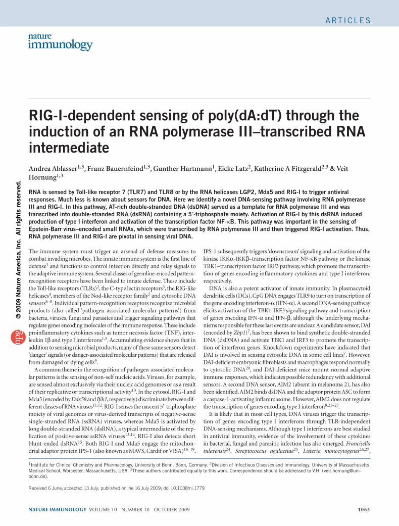
RIG-I-dependent sensing of poly(dA:dT) through the induction of an RNA polymerase III–transcribed RNA intermediateAndrea Ablasser1,3, Franz Bauernfeind1,3, Gunther Hartmann1, Eicke Latz2, Katherine A Fitzgerald2,3 & Veit Hornung1,3
RNA is sensed by Toll-like receptor 7 (TLR7) and TLR8 or by the RNA helicases LGP2, Mda5 and RIG-I to trigger antiviral responses. Much less is known about sensors for DNA. Here we identify a novel DNA-sensing pathway involving RNA polymerase III and RIG-I. In this pathway, AT-rich double-stranded DNA (dsDNA) served as a template for RNA polymerase III and was transcribed into double-stranded RNA (dsRNA) containing a 5′-triphosphate moiety. Activation of RIG-I by this dsRNA induced production of type I interferon and activation of the transcription factor NF-κB. This pathway was important in the sensing of Epstein-Barr virus–encoded small RNAs, which were transcribed by RNA polymerase III and then triggered RIG-I activation. Thus, RNA polymerase III and RIG-I are pivotal in sensing viral DNA.
The immune system must trigger an arsenal of defense measures to combat invading microbes. The innate immune system is the first line of defense1 and functions to control infection directly and relay signals to the adaptive immune system. Several classes of germline-encoded pattern-recognition receptors have been linked to innate defense. These include the Toll-like receptors (TLRs)2, the C-type lectin receptors3, the RIG-like helicases4, members of the Nod-like receptor family5 and cytosolic DNA sensors6–8. Individual pattern-recognition receptors recognize microbial products (also called ‘pathogen-associated molecular patterns’) from bacteria, viruses, fungi and parasites and trigger signaling pathways that regulate genes encoding molecules of the immune response. These include proinflammatory cytokines such as tumor necrosis factor (TNF), inter-leukin 1β and type I interferons1,5. Accumulating evidence shows that in addition to sensing microbial products, many of these same sensors detect ‘danger’ signals (or danger-associated molecular patterns) that are released from damaged or dying cells9.
A common theme in the recognition of pathogen-associated molecu-lar patterns is the sensing of non-self nucleic acids. Viruses, for example, are sensed almost exclusively via their nucleic acid genomes or as a result of their replicative or transcriptional activity10. In the cytosol, RIG-I and Mda5 (encoded by Ddx58 and Ifih1, respectively) discriminate between dif-ferent classes of RNA viruses11,12. RIG-I senses the nascent 5′-triphosphate moiety of viral genomes or virus-derived transcripts of negative-sense single-stranded RNA (ssRNA) viruses, whereas Mda5 is activated by long double-stranded RNA (dsRNA), a typical intermediate of the rep-lication of positive-sense ssRNA viruses13,14. RIG-I also detects short blunt-ended dsRNA15. Both RIG-I and Mda5 engage the mitochon-drial adaptor protein IPS-1 (also known as MAVS, Cardif or VISA)16–19.
IPS-1 subsequently triggers ‘downstream’ signaling and activation of the kinase IKKα-IKKβ–transcription factor NF-κB pathway or the kinase TBK1–transcription factor IRF3 pathway, which promote the transcrip-tion of genes encoding inflammatory cytokines and type I interferon, respectively.
DNA is also a potent activator of innate immunity. In plasmacytoid dendritic cells (DCs), CpG DNA engages TLR9 to turn on transcription of the gene encoding interferon-α (IFN-α). A second DNA-sensing pathway elicits activation of the TBK1-IRF3 signaling pathway and transcription of genes encoding IFN-α and IFN-β, although the underlying mecha-nisms responsible for these last events are unclear. A candidate sensor, DAI (encoded by Zbp1)7, has been shown to bind synthetic double-stranded DNA (dsDNA) and activate TBK1 and IRF3 to promote the transcrip-tion of interferon genes. Knockdown experiments have indicated that DAI is involved in sensing cytosolic DNA in some cell lines7. However, DAI-deficient embryonic fibroblasts and macrophages respond normally to cytosolic DNA20, and DAI-deficient mice mount normal adaptive immune responses, which indicates possible redundancy with additional sensors. A second DNA sensor, AIM2 (absent in melanoma 2), has also been identified. AIM2 binds dsDNA and the adaptor protein ASC to form a caspase-1-activating inflammasome. However, AIM2 does not regulate the transcription of genes encoding type I interferon8,21–23.
It is likely that in most cell types, DNA viruses trigger the transcrip-tion of genes encoding type I interferons through TLR-independent DNA-sensing mechanisms. Although type I interferons are best studied in antiviral immunity, evidence of the involvement of these cytokines in bacterial, fungal and parasitic infection has also emerged. Francisella tularensis24, Streptococcus agalactiae25, Listeria monocytogenes26,27,
1Institute for Clinical Chemistry and Pharmacology, University of Bonn, Bonn, Germany. 2Division of Infectious Diseases and Immunology, University of Massachusetts Medical School, Worcester, Massachusetts, USA. 3These authors contributed equally to this work. Correspondence should be addressed to V.H. ([email protected]).
Received 6 June; accepted 13 July; published online 16 July 2009; doi:10.1038/ni.1779
nature immunology volume 10 number 10 october 2009 1065
ArT IC le S
©20
09 N
atu
re A
mer
ica,
Inc.
All
rig
hts
res
erve
d.
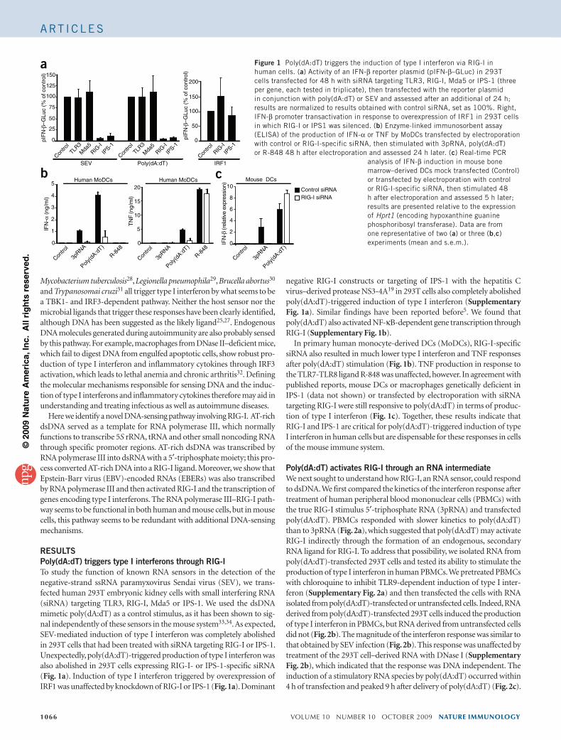
Mycobacterium tuberculosis28, Legionella pneumophila29, Brucella abortus30 and Trypanosomai cruzi31 all trigger type I interferon by what seems to be a TBK1- and IRF3-dependent pathway. Neither the host sensor nor the microbial ligands that trigger these responses have been clearly identified, although DNA has been suggested as the likely ligand25,27. Endogenous DNA molecules generated during autoimmunity are also probably sensed by this pathway. For example, macrophages from DNase II–deficient mice, which fail to digest DNA from engulfed apoptotic cells, show robust pro-duction of type I interferon and inflammatory cytokines through IRF3 activation, which leads to lethal anemia and chronic arthritis32. Defining the molecular mechanisms responsible for sensing DNA and the induc-tion of type I interferons and inflammatory cytokines therefore may aid in understanding and treating infectious as well as autoimmune diseases.
Here we identify a novel DNA-sensing pathway involving RIG-I. AT-rich dsDNA served as a template for RNA polymerase III, which normally functions to transcribe 5S rRNA, tRNA and other small noncoding RNA through specific promoter regions. AT-rich dsDNA was transcribed by RNA polymerase III into dsRNA with a 5′-triphosphate moiety; this pro-cess converted AT-rich DNA into a RIG-I ligand. Moreover, we show that Epstein-Barr virus (EBV)-encoded RNAs (EBERs) was also transcribed by RNA polymerase III and then activated RIG-I and the transcription of genes encoding type I interferons. The RNA polymerase III–RIG-I path-way seems to be functional in both human and mouse cells, but in mouse cells, this pathway seems to be redundant with additional DNA-sensing mechanisms.
RESULTSPoly(dA:dT) triggers type I interferons through RIG-ITo study the function of known RNA sensors in the detection of the negative-strand ssRNA paramyxovirus Sendai virus (SEV), we trans-fected human 293T embryonic kidney cells with small interfering RNA (siRNA) targeting TLR3, RIG-I, Mda5 or IPS-1. We used the dsDNA mimetic poly(dA:dT) as a control stimulus, as it has been shown to sig-nal independently of these sensors in the mouse system33,34. As expected, SEV-mediated induction of type I interferon was completely abolished in 293T cells that had been treated with siRNA targeting RIG-I or IPS-1. Unexpectedly, poly(dA:dT)-triggered production of type I interferon was also abolished in 293T cells expressing RIG-I- or IPS-1-specific siRNA (Fig. 1a). Induction of type I interferon triggered by overexpression of IRF1 was unaffected by knockdown of RIG-I or IPS-1 (Fig. 1a). Dominant
negative RIG-I constructs or targeting of IPS-1 with the hepatitis C virus–derived protease NS3-4A19 in 293T cells also completely abolished poly(dA:dT)-triggered induction of type I interferon (Supplementary Fig. 1a). Similar findings have been reported before5. We found that poly(dA:dT) also activated NF-κB-dependent gene transcription through RIG-I (Supplementary Fig. 1b).
In primary human monocyte-derived DCs (MoDCs), RIG-I-specific siRNA also resulted in much lower type I interferon and TNF responses after poly(dA:dT) stimulation (Fig. 1b). TNF production in response to the TLR7-TLR8 ligand R-848 was unaffected, however. In agreement with published reports, mouse DCs or macrophages genetically deficient in IPS-1 (data not shown) or transfected by electroporation with siRNA targeting RIG-I were still responsive to poly(dA:dT) in terms of produc-tion of type I interferon (Fig. 1c). Together, these results indicate that RIG-I and IPS-1 are critical for poly(dA:dT)-triggered induction of type I interferon in human cells but are dispensable for these responses in cells of the mouse immune system.
Poly(dA:dT) activates RIG-I through an RNA intermediateWe next sought to understand how RIG-I, an RNA sensor, could respond to dsDNA. We first compared the kinetics of the interferon response after treatment of human peripheral blood mononuclear cells (PBMCs) with the true RIG-I stimulus 5′-triphosphate RNA (3pRNA) and transfected poly(dA:dT). PBMCs responded with slower kinetics to poly(dA:dT) than to 3pRNA (Fig. 2a), which suggested that poly(dA:dT) may activate RIG-I indirectly through the formation of an endogenous, secondary RNA ligand for RIG-I. To address that possibility, we isolated RNA from poly(dA:dT)-transfected 293T cells and tested its ability to stimulate the production of type I interferon in human PBMCs. We pretreated PBMCs with chloroquine to inhibit TLR9-dependent induction of type I inter-feron (Supplementary Fig. 2a) and then transfected the cells with RNA isolated from poly(dA:dT)-transfected or untransfected cells. Indeed, RNA derived from poly(dA:dT)-transfected 293T cells induced the production of type I interferon in PBMCs, but RNA derived from untransfected cells did not (Fig. 2b). The magnitude of the interferon response was similar to that obtained by SEV infection (Fig. 2b). This response was unaffected by treatment of the 293T cell–derived RNA with DNase I (Supplementary Fig. 2b), which indicated that the response was DNA independent. The induction of a stimulatory RNA species by poly(dA:dT) occurred within 4 h of transfection and peaked 9 h after delivery of poly(dA:dT) (Fig. 2c).
Poly(dA:dT)
pIF
N-β
–GLu
c (%
of c
ontr
ol)
SEV
TLR3M
da5
IPS-1
IPS-1
IPS-1
Contro
l
RIG-I
TLR3
Mda
5
Contro
l
RIG-I
Contro
l
RIG-I
150
125
100
75
50
25
0
a
b c
Poly(d
A:dT)
3pRNA
R-848
Contro
l
Poly(d
A:dT)
3pRNA
R-848
Contro
l
Poly(d
A:dT)
3pRNA
Contro
l
10
8
6
4
2
0
5
4
3
2
1
0
20
15
10
5
0
(rel
ativ
e ex
pres
sion
)IF
N-β
Mouse DCsHuman MoDCs Human MoDCs
Control siRNA RIG-I siRNA
pIF
N-β
–GLu
c (%
of c
ontr
ol)
150
100
50
200
0
IFN
-α (
ng/m
l)
TN
F (n
g/m
l)
IRF1
Figure 1 Poly(dA:dT) triggers the induction of type I interferon via rIG-I in human cells. (a) Activity of an IFN-β reporter plasmid (pIFN-β–Gluc) in 293T cells transfected for 48 h with sirNA targeting Tlr3, rIG-I, Mda5 or IPS-1 (three per gene, each tested in triplicate), then transfected with the reporter plasmid in conjunction with poly(dA:dT) or SeV and assessed after an additional of 24 h; results are normalized to results obtained with control sirNA, set as 100%. right, IFN-β promoter transactivation in response to overexpression of IrF1 in 293T cells in which rIG-I or IPS1 was silenced. (b) enzyme-linked immunosorbent assay (elISA) of the production of IFN-α or TNF by MoDCs transfected by electroporation with control or rIG-I-specific sirNA, then stimulated with 3prNA, poly(dA:dT) or r-848 48 h after electroporation and assessed 24 h later. (c) real-time PCr
analysis of IFN-β induction in mouse bone marrow–derived DCs mock transfected (Control) or transfected by electroporation with control or rIG-I-specific sirNA, then stimulated 48 h after electroporation and assessed 5 h later; results are presented relative to the expression of Hprt1 (encoding hypoxanthine guanine phosphoribosyl transferase). Data are from one representative of two (a) or three (b,c) experiments (mean and s.e.m.).
1066 volume 10 number 10 october 2009 nature immunology
ArT IC le S
©20
09 N
atu
re A
mer
ica,
Inc.
All
rig
hts
res
erve
d.
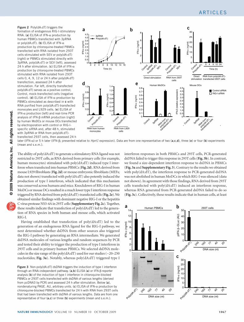
The ability of poly(dA:dT) to generate a stimulatory RNA ligand was not restricted to 293T cells, as RNA derived from primary cells (for example, human monocytes) stimulated with poly(dA:dT) induced type I inter-feron when transfected into human PBMCs (Fig. 2d). RNA derived from mouse L929 fibroblasts (Fig. 2d) or mouse embryonic fibroblasts (MEFs; data not shown) transfected with poly(dA:dT) also potently induced the production of type I interferon, which indicated that this mechanism was conserved across humans and mice. Knockdown of RIG-I in human MoDCs or mouse DCs resulted in a much lower type I interferon response triggered by RNA derived from poly(dA:dT)-transfected cells (Fig. 2e). We obtained similar findings with dominant negative RIG-I or the hepatitis C virus protease NS3-4A in 293T cells (Supplementary Fig. 2c). Together, these results indicate that transfection of poly(dA:dT) led to the genera-tion of RNA species in both human and mouse cells, which activated RIG-I.
Having established that transfection of poly(dA:dT) led to the generation of an endogenous RNA ligand for the RIG-I pathway, we next determined whether dsDNA from other sources also triggered the RIG-I pathway by generating an RNA intermediate. We generated dsDNA molecules of various lengths and random sequences by PCR and tested their ability to trigger the production of type I interferon in 293T cells and in primary human PBMCs. We selected dsDNA mole-cules in the size range of the poly(dA:dT) used for our studies (~20–250 nucleotides; Fig. 3a). Notably, whereas poly(dA:dT) triggered type I
interferon responses in both PBMCs and 293T cells, PCR-generated dsDNA failed to trigger this response in 293T cells (Fig. 3b). In contrast, we found a size-dependent interferon response to dsDNA in PBMCs (Fig. 3a and Supplementary Fig. 3). Contrary to the results we obtained with poly(dA:dT), the interferon response to PCR-generated dsDNA was not abolished in human MoDCs in which RIG-I was silenced (data not shown). In agreement with those findings, RNA derived from 293T cells transfected with poly(dA:dT) induced an interferon response, whereas RNA generated from PCR-generated dsDNA failed to do so (Fig. 3c). Collectively, these results indicate that in human cells, at least
50
40
30
20
10
0
30
20
25
15
5
10
0
Poly(d
A:dT)
245
163 77 40 20
Poly(d
A:dT)
245
163 77 40 20
Poly(d
A:dT)
245
163 77 40 20
a b
c
Human PBMCs
DNA size (nt)
293T cells
pIF
N- β
–GLu
c (A
U)
14
12
10
8
6
4
2
0
DNA size (nt)
DNA size (nt)
IFN
-α (
ng/m
l)
IFN
-α (
ng/m
l)Figure 3 Non-poly(dA:dT) dsDNA triggers the induction of type I interferon through an rNA-independent pathway. (a,b) elISA (a) or IFN-β reporter analysis (b) of the induction of type I interferon in chloroquine-blocked PBMCs or 293T cells transfected with dsDNA of various lengths (derived from pcDNA3 by PCr) and assessed 24 h after stimulation. Below (a), nondenaturing PAGe. AU, arbitrary units. (c) elISA of IFN-α production by chloroquine-blocked PBMCs transfected for 24 h with rNA from 293T cells that had been transfected with dsDNA of various lengths. Data are from one representative of four (a,c) or three (b) experiments (mean and s.e.m.).
nature immunology volume 10 number 10 october 2009 1067
e
Poly(d
A:dT)
Poly(d
A:dT)
Contro
l
Contro
l
Contro
l
Mouse DCsHuman MoDCs
3pRNA
RNA from
poly(
dA:d
T)
trans
fecte
d ce
lls
RNA from
poly(
dA:d
T)
trans
fecte
d ce
lls
Contro
l
3pRNA
Control siRNARIG-I siRNA
0.8
0.7
0.6
0.5
0.4
0.3
0.2
0.1
0
0.8
1.0
0.6
0.4
0.2
0
0.8
1.0
1.2
1.4
3.02.52.0
4.54.03.5
1.51.00.5
0
0.6
0.4
0.2
0
a c
d
b
Poly(dA:dT)3pRNA
30
25
20
15
10
5
00 2 4 6 8 10
Time (h)Time (h)
0 4 9 12 2412 14 16
Poly(d
A:dT)
Poly(d
A:dT)
3pRNA
SEVSEV
Contro
l
Poly(d
A:dT)
Contro
l
Contro
l
RNA from 293T cells
RNA from humanmonocytes
RNA from L929 cells
IFN
-α (
ng/m
l)
IFN
-α (
ng/m
l)
IFN
-α (
ng/m
l)
IFN
-α (
ng/m
l)
IFN
-α (
ng/m
l)
IFN
-α (
ng/m
l)
Direct stimulationRNA from poly(dA:dT)-transfected 293T cells
Directstimulation
4.54.03.53.02.52.01.51.00.5
0
6
8
4
2
0
(rel
ativ
e ex
pres
sion
)IF
N-β
Figure 2 Poly(dA:dT) triggers the formation of endogenous rIG-I-stimulatory rNA. (a) elISA of IFN-α production by human PBMCs transfected with 3prNA or poly(dA:dT). (b) elISA of IFN-α production by chloroquine-treated PBMCs transfected with rNA isolated from 293T cells stimulated with SeV or poly(dA:dT) (right) or PBMCs stimulated directly with 3prNA, poly(dA:dT) or SeV (left), assessed 24 h after stimulation. (c) elISA of IFN-α production by chloroquine-treated PBMCs stimulated with rNA isolated from 293T cells 0, 4, 9, 12 or 24 h after poly(dA:dT) transfection, assessed 24 h after stimulation. Far left, directly transfected poly(dA:dT) serves as a positive control; Control, mock-transfected cells (negative control). (d) elISA of IFN-α production by PBMCs stimulated as described in c with rNA purified from poly(dA:dT)-transfected monocytes and l929 cells. (e) elISA of IFN-α production (left) and real-time PCr analysis of IFN-β mrNA production (right) by human MoDCs or mouse DCs transfected by electroporation with control or rIG-I-specific sirNA and, after 48 h, stimulated with 3prNA or rNA from poly(dA:dT)-transfected 293T cells, then assessed 24 h later (IFN-α) or 5 h later (IFN-β; presented relative to Hprt1 expression). Data are from one representative of two (a,c,d), three (e) or four (b) experiments (mean and s.e.m.).
ArT IC le S
©20
09 N
atu
re A
mer
ica,
Inc.
All
rig
hts
res
erve
d.
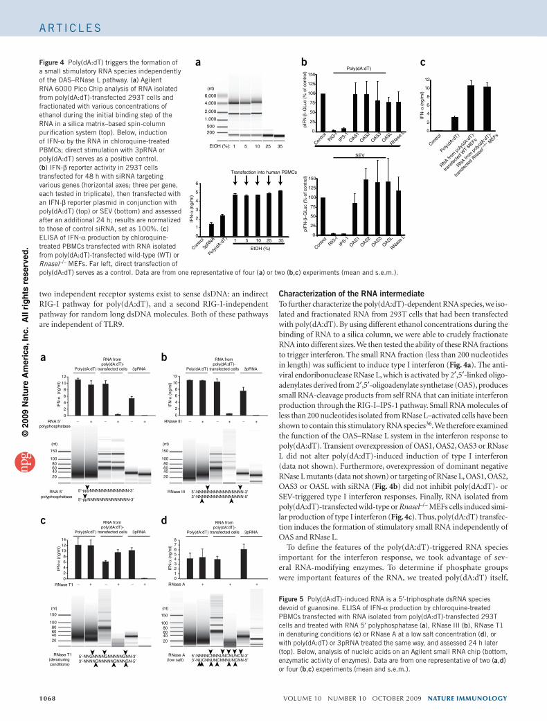
two independent receptor systems exist to sense dsDNA: an indirect RIG-I pathway for poly(dA:dT), and a second RIG-I-independent pathway for random long dsDNA molecules. Both of these pathways are independent of TLR9.
Characterization of the RNA intermediateTo further characterize the poly(dA:dT)-dependent RNA species, we iso-lated and fractionated RNA from 293T cells that had been transfected with poly(dA:dT). By using different ethanol concentrations during the binding of RNA to a silica column, we were able to crudely fractionate RNA into different sizes. We then tested the ability of these RNA fractions to trigger interferon. The small RNA fraction (less than 200 nucleotides in length) was sufficient to induce type I interferon (Fig. 4a). The anti-viral endoribonuclease RNase L, which is activated by 2′,5′-linked oligo-adenylates derived from 2′,5′-oligoadenylate synthetase (OAS), produces small RNA-cleavage products from self RNA that can initiate interferon production through the RIG-I–IPS-1 pathway. Small RNA molecules of less than 200 nucleotides isolated from RNase L–activated cells have been shown to contain this stimulatory RNA species36. We therefore examined the function of the OAS–RNase L system in the interferon response to poly(dA:dT). Transient overexpression of OAS1, OAS2, OAS3 or RNase L did not alter poly(dA:dT)-induced induction of type I interferon (data not shown). Furthermore, overexpression of dominant negative RNase L mutants (data not shown) or targeting of RNase L, OAS1, OAS2, OAS3 or OASL with siRNA (Fig. 4b) did not inhibit poly(dA:dT)- or SEV-triggered type I interferon responses. Finally, RNA isolated from poly(dA:dT)-transfected wild-type or Rnasel–/– MEFs cells induced simi-lar production of type I interferon (Fig. 4c). Thus, poly(dA:dT) transfec-tion induces the formation of stimulatory small RNA independently of OAS and RNase L.
To define the features of the poly(dA:dT)-triggered RNA species important for the interferon response, we took advantage of sev-eral RNA-modifying enzymes. To determine if phosphate groups were important features of the RNA, we treated poly(dA:dT) itself,
1068 volume 10 number 10 october 2009 nature immunology
RNA 5′ polyphosphatase
3pRNA
12
10
8
6
4
2
0
RNA frompoly(dA:dT)-
transfected cellsPoly(dA:dT)
+ __ + _ + + __ + _ +
+ + ++ __ + _ +
3pRNA
RNA frompoly(dA:dT)-
transfected cellsPoly(dA:dT)
3pRNA
RNA frompoly(dA:dT)-
transfected cellsPoly(dA:dT)
RNase III
5′-pppNNNNNNNNNNNNN-3′
5′-ppNNNNNNNNNNNNNN-3′
RNase III
(nt) (nt)
(nt)(nt)
2040
150
1008060
2040
150
1008060
12
10
8
6
4
2
0
5′-NNNNNNNNNNNNNNNN-3′3′-NNNNNNNNNNNNNNNN-5′
a b
c d
RNase T1(denaturingconditions)
1214
1086420
RNase T1
2040
150
1008060
2040
150
1008060
5′-NNGNNNNGNNNNNGNN-3′3′-NNNNGNNNNNGNNNGN-5′
5′-NNNNCNNNUNCNUNCN-3′3′-NUCNNUNCNNNUNCNN-5′
876543210
3pRNA
RNA frompoly(dA:dT)-
transfected cellsPoly(dA:dT)
RNase A
RNase A(low salt)
IFN
-α (
ng/m
l)
IFN
-α (
ng/m
l)IF
N-α
(ng
/ml)
IFN
-α (
ng/m
l)
RNA 5′ polyphosphatase
Figure 5 Poly(dA:dT)-induced rNA is a 5′-triphosphate dsrNA species devoid of guanosine. elISA of IFN-α production by chloroquine-treated PBMCs transfected with rNA isolated from poly(dA:dT)-transfected 293T cells and treated with rNA 5′ polyphosphatase (a), rNase III (b), rNase T1 in denaturing conditions (c) or rNase A at a low salt concentration (d), or with poly(dA:dT) or 3prNA treated the same way, and assessed 24 h later (top). Below, analysis of nucleic acids on an Agilent small rNA chip (bottom, enzymatic activity of enzymes). Data are from one representative of two (a,d) or four (b,c) experiments (mean and s.e.m.).
a b c
EtOH (%):
EtOH (%)
(nt)
6
200500
6,000
4,000
2,000
1,000
5
4
3
2
1
035251051
35251051
Poly(d
A:dT)
Contro
l
3pRNA
Transfection into human PBMCs
12
10
8
6
4
2
0
pIF
N-β
−G
Luc
(% o
f con
trol
)pI
FN
-β−
GLu
c (%
of c
ontr
ol)
150
125
100
75
50
25
0
150
125
100
75
50
25
0
IPS-1
RNase
L
OAS1
OAS2
OAS3
OASL
Contro
lRIG
-I
IPS-1
RNase
L
OAS1
OAS2
OAS3
OASL
Contro
lRIG
-I
Poly(dA:dT)
SEVPoly
(dA:d
T)
Contro
l
RNA from
poly
(dA:d
T)-
trans
fecte
d W
T MEFs
RNA from
poly
(dA:d
T)-
trans
fecte
d Rna
sel –/– M
EFs
IFN
-α (
ng/m
l)
IFN
-α (
ng/m
l)
Figure 4 Poly(dA:dT) triggers the formation of a small stimulatory rNA species independently of the OAS–rNase l pathway. (a) Agilent rNA 6000 Pico Chip analysis of rNA isolated from poly(dA:dT)-transfected 293T cells and fractionated with various concentrations of ethanol during the initial binding step of the rNA in a silica matrix–based spin-column purification system (top). Below, induction of IFN-α by the rNA in chloroquine-treated PBMCs; direct stimulation with 3prNA or poly(dA:dT) serves as a positive control. (b) IFN-β reporter activity in 293T cells transfected for 48 h with sirNA targeting various genes (horizontal axes; three per gene, each tested in triplicate), then transfected with an IFN-β reporter plasmid in conjunction with poly(dA:dT) (top) or SeV (bottom) and assessed after an additional 24 h; results are normalized to those of control sirNA, set as 100%. (c) elISA of IFN-α production by chloroquine-treated PBMCs transfected with rNA isolated from poly(dA:dT)-transfected wild-type (WT) or Rnasel–/– MeFs. Far left, direct transfection of poly(dA:dT) serves as a control. Data are from one representative of four (a) or two (b,c) experiments (mean and s.e.m.).
ArT IC le S
©20
09 N
atu
re A
mer
ica,
Inc.
All
rig
hts
res
erve
d.
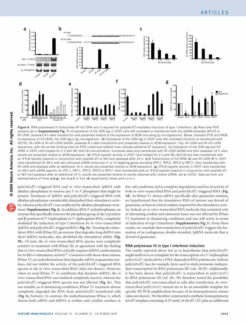
poly(dA:dT)-triggered RNA and in vitro–transcribed 3pRNA with alkaline phosphatase to remove any 5′ or 3′ phosphates that might be present. Treatment of 3pRNA and poly(dA:dT)-triggered RNA with alkaline phosphatase considerably diminished their stimulatory activ-ity, whereas poly(dA:dT) was unaffected by alkaline phosphatase treat-ment (Supplementary Fig. 4). In addition, RNA 5′-polyphosphatase, an enzyme that specifically removes the phosphate group in the γ-position and β-position of 5′-triphosphate or 5′-diphosphate RNA, completely abolished the induction of type I interferon by in vitro–transcribed 3pRNA and poly(dA:dT)-triggered RNA (Fig. 5a). Treating the stimu-latory RNA with RNase III, an enzyme that degrades long dsRNA into short dsRNA molecules, also abolished the stimulatory ability (Fig. 5b). Of note, the in vitro–transcribed RNA species were completely sensitive to treatment with RNase III, in agreement with the finding that in vitro–transcribed RNA critically requires dsRNA conformation for its RIG-I-stimulatory activity37. Consistent with those observations, RNase T1, an endoribonuclease that degrades ssRNA at guanosine resi-dues, did not inhibit the activity of the poly(dA:dT)-triggered RNA species or the in vitro–transcribed RNA (data not shown). However, when we used RNase T1 in conditions that denature dsRNA, the in vitro–transcribed RNA was rendered completely inactive, whereas the poly(dA:dT)-triggered RNA species was not affected (Fig. 5c). This was notable, as in denaturing conditions, RNase T1 treatment almost completely degraded the RNA from poly(dA:dT)-transfected cells (Fig. 5c, bottom). In contrast, the endoribonuclease RNase A, which cleaves both ssRNA and dsRNA at uridine and cytidine residues in
low-salt conditions, led to complete degradation and loss of activity of both in vitro–transcribed RNA and poly(dA:dT)-triggered RNA (Fig. 5d). As RNase T1 cleaves ssRNA specifically after guanosine residues, we hypothesized that the stimulatory RNA of interest was devoid of guanosine, at least at critical residues required for the stimulatory activ-ity. Indeed, an in vitro–transcribed RNA molecule that consisted only of alternating uridine and adenosine bases was not affected by RNase T1 treatment in denaturing conditions and was still active in terms of induction of type I interferon (Supplementary Fig. 5). From these results, we conclude that transfection of poly(dA:dT) triggers the for-mation of an endogenous, double-stranded, 3pRNA molecule that is devoid of guanosine.
RNA polymerase III in type I interferon inductionThe results reported above led us to hypothesize that poly(dA:dT) might itself serve as a template for the transcription of a 5′-triphosphate poly(rA:rU) molecule by a DNA-dependent RNA polymerase. Indeed, poly(dA:dT) has, for example, been used to study promoter-indepen-dent transcription by RNA polymerase III (refs. 38,39). Additionally, it has been shown that poly(dA:dT) is transcribed to poly(rA:rU) by RNA polymerase III (ref. 40). We therefore tested the possibility that poly(dA:dT) was transcribed in cells after transfection. In vitro–transcribed poly(rA:rU) turned out to be an unsuitable template for specific RT-PCR amplification because of its homopolymeric nature (data not shown). We therefore constructed a synthetic homopolymeric dA:dT template consisting of 35 ‘units’ of dA:dT (AT) plus an additional
nature immunology volume 10 number 10 october 2009 1069
a b
100
150
50
0
200
250
100
150
50
0IPS-1 RPC1 RPC2 RPC3 RPC7Control030 20 10 5 030 20 10 5
200
250 0.06
0.05
0.04
0.03
0.02
0.01
0
SEVPoly(dA:dT)
SEV Poly(dA:dT) RPC1
1 2 1 2 1 2 1 2
RPC2 RPC3 RPC7Con
trol
pIF
N-β
–GLu
c (%
of c
ontr
ol)
pIF
N-β
–GLu
c (%
of c
ontr
ol)
d e
c
f g
ML-60218 (µM)0 6.25 12.5 25
ML-60218 (µM)
ML-60218(µM)
0 6.25 12.5 25
AT+30N(AT)35
Control AT+30N
AT+6T+30N
(AT)35
Controlβ2-microglobulin
5S rRNA + 3′ RACE
30N + 3′ RACE
0.30
0.25
0.20
0.15
0.10
0.05
0
0.6
0.7
0.5
0.4
0.3
0.2
0.1
0
3.0
2.5
2.0
1.5
1.0
0.5
0
400
350
300
250
200
150
100
50
0
AT+30N:
AT+6T+30N:
5′-ATATATATATA ..................GAGCGGCCGCGAAGATCAGAGCACCGCAAC-3′
5′-ATATATATATA ....TTTTTTGAGCGGCCGCGAAGATCAGAGCACCGCAAC-3′
30N
(re
lativ
e ex
pres
sion
)
30N
(re
lativ
e ex
pres
sion
)
30N
(re
lativ
e ex
pres
sion
)
30N
(re
lativ
e ex
pres
sion
)
5S r
RN
A (
rela
tive
expr
essi
on)
5S r
RN
A (
102 r
elat
ive
expr
essi
on)
30
25
20
15
10
5
0
RPC1
1 2 1 2 1 2 1 2
RPC2 RPC3 RPC7Con
trol
Figure 6 rNA polymerase III transcribes AT-rich DNA and is required for poly(dA:dT)-mediated induction of type I interferon. (a) real-time PCr analysis (as in Supplementary Fig. 7) of expression of the 30N tag in 293T cells left untreated or transfected with the dsDNA template (AT)35 or AT+30N, assessed 8 h after transfection and presented relative to the expression of B2M (encoding β2-microglobulin). Below, standard PCr and PAGe of expression of 5S rrNA, the 30N tag or β2-microglobulin. (b) expression of the 30N tag in 293T cells left untreated (Control) or transfected with (AT)35, AT+30N or AT+6T+30N dsDNA, assessed 8 h after transfection and presented relative to B2M expression. Top, AT+30N and AT+6T+30N sequences, with the primer-binding sites for PCr underlined (dotted lines indicate additonal AT sequence). (c) expression of the 30N tag and 5S rrNA in 293T cells treated for 2 h with Ml-60218 (concentration, horizontal axes) and transfected with AT+30N dsDNA and then assessed 16 h later; results are presented relative to B2M expression. (d) IFN-β reporter activity in 293T cells treated for 2 h with Ml-60218 and then transfected with an IFN-β reporter plasmid in conjunction with poly(dA:dT) or SeV and assessed after 24 h. (e,f) Transcription of 5S rrNA (e) and AT+30N (f) in 293T cells transfected for 48 h with two individual sirNA molecules (1 or 2) targeting genes encoding rPC1, rPC2, rPC3 or rPC7, then transfected with AT+30N and assessed after an additional 16 h; results are presented relative to B2M expression. (g) IFN-β reporter activity in 293T cells transfected for 48 h with sirNA specific for IPS-1, rPC1, rPC2, rPC3 or rPC7, then transfected with an IFN-β reporter plasmid in conjunction with poly(dA:dT) or SeV and assessed after an additional 24 h; results are presented relative to results obtained with control sirNA, set as 100%. Data are from one representative of three (a,b,g), two (c,e,f) or four (d) experiments (mean and s.e.m.).
ArT IC le S
©20
09 N
atu
re A
mer
ica,
Inc.
All
rig
hts
res
erve
d.
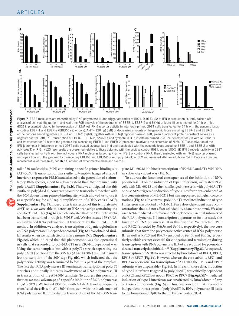
tail of 30 nucleotides (30N) containing a specific primer-binding site (AT+30N). Transfection of this synthetic template triggered a type I interferon response in PBMCs and also led to the generation of a stimu-latory RNA species, albeit to a lower extent than that obtained with poly(dA:dT) (Supplementary Fig. 6a,b). Thus, we anticipated that this synthetic poly(dA:dT) construct would be transcribed together with the 30N downstream primer-binding site, which allowed us to use it as a specific tag for a 3′ rapid amplification of cDNA ends (RACE; Supplementary Fig. 7). Indeed, after transfection of this template into 293T cells, we were able to detect an RNA transcript containing the specific 3′ RACE tag (Fig. 6a), which indicated that the AT+30N dsDNA had been transcribed through its 30N 3′ end. We also assessed 5S rRNA, an established RNA polymerase III transcript, by the 3′ RACE PCR method. In addition, we analyzed transcription of β2-microglobulin as an RNA polymerase II–dependent control (Fig. 6a). We obtained simi-lar results when we transfected primary mouse DCs (Supplementary Fig. 6c), which indicated that this phenomenon was also operational in cells that responded to poly(dA:dT) in a RIG-I-independent way. Using the same template but with a poly(T) stretch separating the poly(dA:dT) portion from the 30N tag (AT+6T+30N) resulted in much less transcription of the 30N tag (Fig. 6b), which indicated that the polymerase activity was terminated before this part of the template. The fact that RNA polymerase III terminates transcription at poly(T) stretches additionally indicates involvement of RNA polymerase III in transcription of the AT+30N template. To address this possibility further, we took advantage of a specific inhibitor of RNA polymerase III, ML-60218. We treated 293T cells with ML-60218 and subsequently transfected the cells with AT+30N. Consistent with the involvement of RNA polymerase III in mediating transcription of the AT+30N tem-
plate, ML-60218 inhibited transcription of 5S rRNA and AT+30N DNA in a dose-dependent way (Fig. 6c).
To address the functional consequences of the inhibition of RNA polymerase III on the induction of type I interferon, we treated 293T cells with ML-60218 and then challenged these cells with poly(dA:dT) or SEV. SEV-triggered induction of type I interferon was enhanced at low concentrations of ML-60218 but was suppressed at higher concen-trations (Fig. 6d). In contrast, poly(dA:dT)-mediated induction of type I interferon was blocked by ML-60218 in a dose-dependent way at con-centrations that did not affect cell viability (data not shown). We also used RNA-mediated interference to ‘knock down’ essential subunits of the RNA polymerase III transcription apparatus to further study the function of RNA polymerase III in this response. We targeted RPC1 and RPC2 (encoded by Polr3a and Polr3b, respectively), the two core subunits that form the polymerase active center of RNA polymerase III, as well as RPC3 and RPC7 (encoded by Polr3c and Polr3g, respec-tively), which are not essential for elongation and termination during transcription with RNA polymerase III but are required for promoter-directed transcription initiation41 (Supplementary Fig. 8). As expected, transcription of 5S rRNA was affected by knockdown of RPC1, RPC2, RPC3 or RPC7 (Fig. 6e). However, whereas the core subunits RPC1 and RPC2 were essential for transcription of AT+30N, the RPC3 and RPC7 subunits were dispensable (Fig. 6f). In line with those data, induction of type I interferon triggered by poly(dA:dT) was critically dependent on RPC1 and RPC2 but not on RPC3 or RPC7 (Fig. 6g). SEV-mediated induction of type I interferon was unaffected by knockdown of any of these components (Fig. 6g). Thus, we conclude that promoter- independent transcription of poly(dA:dT) by RNA polymerase III leads to the formation of 3pRNA that in turn activates RIG-I.
1070 volume 10 number 10 october 2009 nature immunology
100
120
80
60
40
20
0SEV Poly(dA:dT)
pIF
N-β
–GLu
c (%
of c
ontr
ol)
pIF
N-β
–GLu
c (%
of c
ontr
ol)16
18
20
14
12
10
8
6
4
2
0
350
400
450
300
100
80
60
40
20
0
250
200
150
100
50
0
20
25
30
15
10
5
0
Control siRNA
RIG-I siRNA
IPS-1 siRNA
5s
5
6
7 0.4
0.3
0.2
0.1
0
4
3
2
1
0
EBER-1+2
EBER-1+2
Poly(dA:dT)
Control
35
30
25
20
15
10
5
0
20
30
10
1,000
1,200
1,400
1,600
0
EBER-1
120 ng
90 ng
60 ng
30 ng
0 ng
EBER-2
ML-60218 (µM) ML-60218 (µM)
0 10 20 30
ML-60218 (µM) ML-60218 (µM) ML-60218 (µM)0 20 40 0 20 40
ML-60218 (µM)
0 10 20 30
ML-60218 (µM)
0 10 20 30
ML-60218 (µM)
0 10 20 30
a b
d
c
f
IFN
-
Cel
l via
bilit
y (%
of c
ontr
ol)
α (
pg/m
l)
90
70
50
30
10
20
40
60
80
0
100
80
60
40
20
00 20 40
ML-60218 (µM)0 20 40
ML-60218 (µM)0 20 40
700
500
300
400
600
200
100
0
EB
ER
-1 (
rela
tive
expr
essi
on)
EB
ER
-2 (
rela
tive
expr
essi
on)
5S r
RN
A (
103 re
lativ
e ex
pres
sion
)
Cyc
loph
ilin
B (
rela
tive
expr
essi
on)
pIF
N-β
–GLu
c (A
U)
pIF
N-β
–GLu
c (A
U)
EB
ER
-1 (
rela
tive
expr
essi
on)
EB
ER
-2 (
rela
tive
expr
essi
on)
5S r
RN
A (
rela
tive
expr
essi
on)
100
120
80
60
40
20
0
RIG-I
Poly(dA:dT)
EBER-1+2
EBER-1+20 10 20 30
e
Figure 7 eBer moleucles are transcribed by rNA polymerase III and trigger activation of rIG-I. (a,b) elISA of IFN-α production (a, left), calcein AM analysis of cell viability (a, right) and real-time PCr analysis of the production of eBer-1, eBer-2 and 5S (b) of Mutu III cells treated for 24 h with Ml-60218, presented relative to the expression of B2M. (c) IFN-β reporter activity in interferon-primed 293T cells transfected for 24 h with the genomic locus encoding eBer-1 and eBer-2 (eBer-1+2) or poly(dA:dT) (120 ng) (left) or decreasing amounts of the genomic locus encoding eBer-1 and eBer-2 or the portions encoding either eBer-1 or eBer-2 (right), together with an IFN-β reporter plasmid. left, green fluorescent protein construct serves as a negative control (left). (d) Transcription of eBer-1, eBer-2, 5S rrNA and cyclophilin B in interferon-primed 293T cells treated for 2 h with Ml-60218 and transfected for 24 h with the genomic locus encoding eBer-1 and eBer-2, presented relative to the expression of B2M. (e) Transactivation of the IFN-β promoter in interferon-primed 293T cells treated as described in d and transfected with the genomic locus encoding eBer-1 and eBer-2 or with poly(dA:dT) or rIG-I (120 ng); results are presented relative to those obtained with the positive control rIG-I, set as 100%. (f) IFN-β reporter activity in 293T cells transfected for 48 h with two individual sirNA molecules targeting rIG-I or IPS-1 or control sirNA, then transfected with an IFN-β reporter plasmid in conjunction with the genomic locus encoding eBer-1 and eBer-2 or with poly(dA:dT) or SeV and assessed after an additional 24 h. Data are from one representative of three (a,e), two (b,d,f) or four (c) experiments (mean and s.e.m.).
ArT IC le S
©20
09 N
atu
re A
mer
ica,
Inc.
All
rig
hts
res
erve
d.
RNA polymerase III–transcribed EBER RNA activates RIG-ITo address the physiological relevance of the RNA polymerase III path-way in antiviral host defenses, we examined the function of RNA poly-merase III in the regulation of type I interferon responses to a DNA virus. We chose EBV because it encodes small EBER molecules that are transcribed by RNA polymerase III in very large amounts. EBER molecules are nonpolyadenylated, untranslated RNA molecules 167 nucleotides (EBER-1) or 172 nucleotides (EBER-2) in length42 and are the most abundant viral transcripts in cells with latent EBV infection43. Notably, EBV-immortalized lymphoblastoid cell lines and EBV-positive Burkitt lymphoma cell lines produce type I interferons in resting condi-tions44,45. To determine if pattern-recognition receptors are activated constitutively in Burkitt lymphoma cells and to define the involve-ment of RNA polymerase III in controlling the constitutive produc-tion of type I interferons, we treated the EBV-positive human Burkitt lymphoma Mutu III cell line with the RNA polymerase III inhibitor ML-60218. Inhibition of RNA polymerase III blocked IFN-α produc-tion in Mutu III cells in a dose-dependent way (Fig. 7a). The defect in interferon production correlated with repression of the expression of EBER-1, EBER-2 and 5S rRNA in these cells (Fig. 7b). To directly examine the ability of EBER to activate the RIG-I pathway, we cloned the entire EBER gene locus from EBV and transiently overexpressed this construct in interferon-primed 293T cells. Overexpression of EBER molecules led to substantial induction of type I interferon, albeit to a lower extent than did poly(dA:dT) (Fig. 7c). Expression of the loci for EBER-1 and EBER-2 separately showed that each EBER molecule could trigger type I interferon responses independently, although EBER-1 was slightly more active at high concentrations (Fig. 7c). Notably, when we expressed a dsDNA template encoding a blunt-ended version of EBER-1 RNA, we found more induction of type I interferon (Supplementary Fig. 9). This is in line with the finding that RIG-I favors fully blunt-ended 5′-triphosphate dsRNA37. As expected, expression of EBER RNA in 293T cells was suppressed by inhibition of RNA polymerase III activ-ity, whereas RNA polymerase II–driven transcription of cyclophilin B was not affected (Fig. 7d). In addition, inhibition of RNA polymerase III or expression of RIG-I- or IPS-1-specific siRNA led to less EBER-induced type I interferon (Fig. 7e,f). Collectively, these results indi-cate that EBER molecules are transcribed by RNA polymerase III and trigger the induction of type I interferon through RIG-I. In line with those findings, we found that certain synthetic RNA polymerase III genes that encode blunt-ended dsRNA, such as short hairpin RNA, were potent triggers of RIG-I activation (Supplementary Fig. 10 and data not shown). These findings furthermore establish RIG-I as an important checkpoint in the detection of non-self RNA polymerase III transcripts.
DISCUSSIONHere we have demonstrated a novel mechanism for the sensing of AT-rich dsDNA. The synthetic dsDNA mimetic poly(dA:dT) was converted by host RNA polymerase III into a 3pRNA intermediate, which was in turn recognized by RIG-I. RNA polymerase III–mediated conversion of poly(dA:dT) occurred in mouse and human cells, but at least one additional as-yet-undefined sensing mechanism oper-ates in cells of the mouse immune system; this pathway responds to poly(dA:dT) in an RNA-independent way, presumably as a result of sensing DNA directly. These observations are consistent with pub-lished reports showing that mouse cells lacking RIG-I or IPS-1 still produce type I interferons in response to poly(dA:dT)33,34. This matter is further complicated by the fact that in the human system, at least one additional DNA-sensing mechanism exists to recognize dsDNA independently of a RIG-I-stimulatory RNA intermediate. Poly(dA:dT)
induced the formation of the RIG-I-activating RNA species, but other dsDNA moleules that were not poly(dA:dT)-homopolymeric in nature failed to do so, despite being active when directly transfected into PBMCs. Nevertheless, our data clearly demonstrate that AT-rich DNA transcribed by RNA polymerase III is sensed by RIG-I in the human system in a nonredundant way.
Poly(dA:dT) has been used to study RNA polymerase III–mediated promoter-independent transcription38–40. This nonspecific transcrip-tional activity requires the core polymerase complex of RNA polymerase III, which includes RPC1 and RPC2, yet is independent of subunits used by RNA polymerase III for recruitment to specific promoter sites (RPC3, RPC6 and RPC7)41. It is unclear at present if RNA polymerase III has transcriptional activity independent of its characterized pro-moter sites in vivo, as no studies have addressed the RNA polymerase III transcriptome at a global cellular level in an unbiased way. It is plausible that RNA polymerase III also transcribes DNA templates from endogenous sources in a promoter-independent way. Such a mecha-nism could function as an innate defense strategy by tagging DNA as an RNA intermediate to be detected by RIG-I. Poly(dA:dT) or AT-rich DNA is a ‘peculiar’ template for RNA polymerase III and thereby indi-rectly for RIG-I for two main reasons. First, AT-rich sequences might be particularly suited for promoter-independent transcription by RNA polymerase III because of their propensity to form dsDNA regions with low helix stability, which may be particularly accessible as initiation sites for polymerases. However, other DNA templates have also been used to study promoter-independent transcription by RNA polymerase III and thus it is possible that non-AT DNA is also transcribed by RNA poly-merase III in a promoter-independent way. Second, and probably more importantly, homopolymeric AT-rich DNA is also transcribed into a homopolymeric RNA molecule that has the propensity to form a com-plete RNA duplex. RIG-I strongly favors blunt-ended dsRNA over ssRNA or incompletely annealed dsRNA for binding and subsequent activ-ity37. Although 5′-triphosphate ssRNA that is not self-complementary is equally active in terms of RIG-I activation when annealed to a complementary ssRNA strand, it shows little or no activity as a single-stranded molecule. Poly(rA:rU) RNA, in contrast, is completely self-complementary and thus has a strong propensity to form dsRNA with complete blunt-end formation, thereby making it ideally suited to be recognized by RIG-I.
Studying the function of host RNA polymerase III–dependent tran-scription of pathogen-derived DNA in innate immune defense will be important yet technically challenging. During acute infection with DNA viruses, it is likely that several DNA-sensing pathways are trig-gered in the nucleus or in the cytosol simultaneously, which makes it difficult to study the contribution of a single sensor. Here we focused on cells that were latently infected with EBV and thus contained viral DNA in the nucleus in steady-state conditions. EBV EBER molecules are transcribed in large amounts by RNA polymerase III in latently infected, EBV-positive cells43. In these cells, we found an RNA poly-merase III–dependent activation of the induction of type I interferon that coincided with EBER RNA expression. Ectopic expression of the EBER locus also induced type I interferon by a mechanism dependent on RIG-I and IPS-1. Published work has also indicated EBER RNAs are endogenous triggers for RIG-I (ref. 46). We speculate that other herpes viruses also encode RNA polymerase III transcripts that can trigger RIG-I activation. For example, the genomes of some γ-herpesviruses contain variable numbers of internal repeats that are highly conserved across different viruses. In this context, it is noteworthy that a highly repetitive region in the genome of murine γ-herpesviruses 68 triggers type I interferon responses47.
Notably, published work has linked the RIG-like helicase–IPS-1
nature immunology volume 10 number 10 october 2009 1071
ArT IC le SArT IC le S
©20
09 N
atu
re A
mer
ica,
Inc.
All
rig
hts
res
erve
d.
1072 volume 10 number 10 october 2009 nature immunology
pathway to the detection of pathogens that replicate in the cytosol but are not RNA viruses. For example, induction of type I interferon trig-gered by L. pneumophila29 or vaccinia virus48 is strongly attenuated in cells devoid of IPS-1. It is unclear whether the activation of RIG-like helicases in these cases is due to pathogen-dependent RNA poly-merase activity or host-derived RNA generated by an RNA polymerase III–dependent mechanism. Future studies that allow discrimination between host-derived and pathogen-derived RIG-I ligands could help clarify these issues.
METHODS
Methods and any associated references are available in the online version of the paper at http://www.nature.com/natureimmunology/.
Note: Supplementary information is available on the Nature Immunology website.
ACKNOWLEDGMENTSWe thank R. Silverman (Lerner Research Institute, Cleveland Clinic) for Rnasel–/– MEFs and their respective controls; and G. Bornkamm and M. Schlee for discussions. Supported by the Deutsche Forschungsgemeinschaft (Ho2783/2-1 V.H.) and the US National Institutes of Health (AI-065483 to E.L.; AI-067497 to K.A.F.; AI-083713 to K.A.F. and E.L.).
AUTHOR CONTRIBUTIONSA.A., F.B. and V.H. did the experiments; V.H. conceived of and K.A.F. and V.H. supervised the research; G.H. and E.L. provided technical support; E.L., K.A.F. and V.H. sponsored the research; and K.A.F. and V.H. prepared the manuscript.
Published online at http://www.nature.com/natureimmunology/.Reprints and permissions information is available online at http://npg.nature.com/reprintsandpermissions/.
1. Kawai, T. & Akira, S. Toll-like receptor and rIG-I-like receptor signaling. Ann. NY Acad. Sci. 1143, 1–20 (2008).
2. Takeda, K. & Akira, S. Toll-like receptors in innate immunity. Int. Immunol. 17, 1–14 (2005).
3. Huysamen, C. & Brown, G.D. The fungal pattern recognition receptor, Dectin-1, and the associated cluster of C-type lectin-like receptors. FEMS Microbiol. Lett. 290, 121–128 (2009).
4. Yoneyama, M. & Fujita, T. rIG-I family rNA helicases: cytoplasmic sensor for antiviral innate immunity. Cytokine Growth Factor Rev. 18, 545–551 (2007).
5. Meylan, e., Tschopp, J. & Karin, M. Intracellular pattern recognition receptors in the host response. Nature 442, 39–44 (2006).
6. Takaoka, A. & Taniguchi, T. Cytosolic DNA recognition for triggering innate immune responses. Adv. Drug Deliv. Rev. 60, 847–857 (2008).
7. Takaoka, A. et al. DAI (DlM-1/ZBP1) is a cytosolic DNA sensor and an activator of innate immune response. Nature 448, 501–505 (2007).
8. Hornung, V. et al. AIM2 recognizes cytosolic dsDNA and forms a caspase-1-activating inflammasome with ASC. Nature 458, 514–518 (2009).
9. Kono, H. & rock, K.l. How dying cells alert the immune system to danger. Nat. Rev. Immunol. 8, 279–289 (2008).
10. Takeuchi, O. & Akira, S. Innate immunity to virus infection. Immunol. Rev. 227, 75–86 (2009).
11. Takeuchi, O. & Akira, S. MDA5/rIG-I and virus recognition. Curr. Opin. Immunol. 20, 17–22 (2008).
12. Kato, H. et al. Differential roles of MDA5 and rIG-I helicases in the recognition of rNA viruses. Nature 441, 101–105 (2006).
13. Pichlmair, A. et al. rIG-I-mediated antiviral responses to single-stranded rNA bearing 5′-phosphates. Science 314, 997–1001 (2006).
14. Hornung, V. et al. 5′-Triphosphate rNA is the ligand for rIG-I. Science 314, 994–997 (2006).
15. Kato, H. et al. length-dependent recognition of double-stranded ribonucleic acids by retinoic acid-inducible gene-I and melanoma differentiation-associated gene 5. J. Exp. Med. 205, 1601–1610 (2008).
16. Kawai, T. et al. IPS-1, an adaptor triggering rIG-I- and Mda5-mediated type I interferon induction. Nat. Immunol. 6, 981–988 (2005).
17. Seth, r.B., Sun, l., ea, C.K. & Chen, Z.J. Identification and characterization of MAVS, a mitochondrial antiviral signaling protein that activates NF-κB and IrF 3. Cell 122, 669–682 (2005).
18. Xu, l.G. et al. VISA is an adapter protein required for virus-triggered IFN-beta signaling. Mol. Cell 19, 727–740 (2005).
19. Meylan, e. et al. Cardif is an adaptor protein in the rIG-I antiviral pathway and is targeted by hepatitis C virus. Nature 437, 1167–1172 (2005).
20. Ishii, K.J. et al. TANK-binding kinase-1 delineates innate and adaptive immune responses to DNA vaccines. Nature 451, 725–729 (2008).
21. Burckstummer, T. et al. An orthogonal proteomic-genomic screen identifies AIM2 as a cytoplasmic DNA sensor for the inflammasome. Nat. Immunol. 10, 266–272 (2009).
22. Fernandes-Alnemri, T., Yu, J.W., Datta, P., Wu, J. & Alnemri, e.S. AIM2 activates the inflammasome and cell death in response to cytoplasmic DNA. Nature 458, 509–513 (2009).
23. roberts, T.l. et al. HIN-200 proteins regulate caspase activation in response to foreign cytoplasmic DNA. Science 323, 1057–1060 (2009).
24. Henry, T., Brotcke, A., Weiss, D.S., Thompson, l.J. & Monack, D.M. Type I interferon signaling is required for activation of the inflammasome during Francisella infection. J. Exp. Med. 204, 987–994 (2007).
25. Charrel-Dennis, M. et al. Tlr-independent type I interferon induction in response to an extracellular bacterial pathogen via intracellular recognition of its DNA. Cell Host Microbe 4, 543–554 (2008).
26. O’riordan, M., Yi, C.H., Gonzales, r., lee, K.D. & Portnoy, D.A. Innate recognition of bacteria by a macrophage cytosolic surveillance pathway. Proc. Natl. Acad. Sci. USA 99, 13861–13866 (2002).
27. Stetson, D.B. & Medzhitov, r. recognition of cytosolic DNA activates an IrF3-dependent innate immune response. Immunity 24, 93–103 (2006).
28. Stanley, S.A., Johndrow, J.e., Manzanillo, P. & Cox, J.S. The Type I IFN response to infection with Mycobacterium tuberculosis requires eSX-1-mediated secretion and contributes to pathogenesis. J. Immunol. 178, 3143–3152 (2007).
29. Opitz, B. et al. Legionella pneumophila induces IFNβ in lung epithelial cells via IPS-1 and IrF3, which also control bacterial replication. J. Biol. Chem. 281, 36173–36179 (2006).
30. roux, C.M. et al. Brucella requires a functional type IV secretion system to elicit innate immune responses in mice. Cell. Microbiol. 9, 1851–1869 (2007).
31. Chessler, A.D., Ferreira, l.r., Chang, T.H., Fitzgerald, K.A. & Burleigh, B.A. A novel IFN regulatory factor 3-dependent pathway activated by trypanosomes triggers IFN-β in macrophages and fibroblasts. J. Immunol. 181, 7917–7924 (2008).
32. Yoshida, H., Okabe, Y., Kawane, K., Fukuyama, H. & Nagata, S. lethal anemia caused by interferon-β produced in mouse embryos carrying undigested DNA. Nat. Immunol. 6, 49–56 (2005).
33. Sun, Q. et al. The specific and essential role of MAVS in antiviral innate immune responses. Immunity 24, 633–642 (2006).
34. Kumar, H. et al. essential role of IPS-1 in innate immune responses against rNA viruses. J. Exp. Med. 203, 1795–1803 (2006).
35. Cheng, G., Zhong, J., Chung, J. & Chisari, F.V. Double-stranded DNA and double-stranded rNA induce a common antiviral signaling pathway in human cells. Proc. Natl. Acad. Sci. USA 104, 9035–9040 (2007).
36. Malathi, K., Dong, B., Gale, M., Jr & Silverman, r.H. Small self-rNA generated by rNase l amplifies antiviral innate immunity. Nature 448, 816–819 (2007).
37. Schlee, M. et al. recognition of 5′ triphosphate by rIG-I helicase requires short blunt double-stranded rNA as contained in panhandle of negative-strand virus. Immunity published online, doi:10.1016/j.immuni.2009.05.008 (2 July 2009).
38. Huet, J., riva, M., Sentenac, A. & Fromageot, P. Yeast rNA polymerase C and its subunits. Specific antibodies as structural and functional probes. J. Biol. Chem. 260, 15304–15310 (1985).
39. Zaros, C. & Thuriaux, P. rpc25, a conserved rNA polymerase III subunit, is critical for transcription initiation. Mol. Microbiol. 55, 104–114 (2005).
40. Thuillier, V., Brun, I., Sentenac, A. & Werner, M. Mutations in the α-amanitin conserved domain of the largest subunit of yeast rNA polymerase III affect pausing, rNA cleavage and transcriptional transitions. EMBO J. 15, 618–629 (1996).
41. Wang, Z. & roeder, r.G. Three human rNA polymerase III-specific subunits form a subcomplex with a selective function in specific transcription initiation. Genes Dev. 11, 1315–1326 (1997).
42. rosa, M.D., Gottlieb, e., lerner, M.r. & Steitz, J.A. Striking similarities are exhibited by two small epstein-Barr virus-encoded ribonucleic acids and the adenovirus-associated ribonucleic acids VAI and VAII. Mol. Cell. Biol. 1, 785–796 (1981).
43. lerner, M.r., Andrews, N.C., Miller, G. & Steitz, J.A. Two small rNAs encoded by epstein-Barr virus and complexed with protein are precipitated by antibodies from patients with systemic lupus erythematosus. Proc. Natl. Acad. Sci. USA 78, 805–809 (1981).
44. Schlee, M., Schuhmacher, M., Holzel, M., laux, G. & Bornkamm, G.W. c-Myc impairs immunogenicity of human B cells. Adv. Cancer Res. 97, 167–188 (2007).
45. Schlee, M. et al. C-myc activation impairs the NF-κB and the interferon response: implications for the pathogenesis of Burkitt’s lymphoma. Int. J. Cancer 120, 1387–1395 (2007).
46. Samanta, M., Iwakiri, D., Kanda, T., Imaizumi, T. & Takada, K. eB virus-encoded rNAs are recognized by rIG-I and activate signaling to induce type I IFN. EMBO J. 25, 4207–4214 (2006).
47. Sanchez, D.J. et al. A repetitive region of γ-herpesvirus genomic DNA is a ligand for induction of type I interferon. J. Virol. 82, 2208–2217 (2008).
48. Deng, l. et al. Vaccinia virus subverts a mitochondrial antiviral signaling protein-depen-dent innate immune response in keratinocytes through its double-stranded rNA binding protein, e3. J. Virol. 82, 10735–10746 (2008).
ArT IC le S ArT IC le S
©20
09 N
atu
re A
mer
ica,
Inc.
All
rig
hts
res
erve
d.
nature immunology doi :10.1038/ni .1779
ONLINE METHODSReagents. Poly(dA:dT) and chloroquine were from Sigma-Aldrich. ML-60218 was from Calbiochem. Human interleukin 4 and granulocyte-macrophage colony-stimulating factor were from ImmunoTools. RNase T1, alkaline phosphatase and DNase I were from Fermentas. RNase III, RNase A, RNA 5′ polyphosphatase and poly(A) polymerase were from Epicentre. The synthetic imidazoquinoline resiquimod (R-848) and CpG oligonucleotide 2216 were from Invivogen.
Cell isolation and culture. Human PBMCs were isolated from whole blood of healthy volunteers by density-gradient centrifugation (Biochrom). Red blood cells were lysed with red blood cell lysis buffer (Sigma). Human monocytes were isolated from PBMCs with anti-CD14 paramagnetic beads (Miltenyi Biotec) and were differentiated for 6 d into MoDCs in the presence of interleukin 4 (800 U/ml) and granulocyte-macrophage colony-stimulating factor (800 U/ml). Mouse bone marrow was cultured for 6 d with 5% (vol/vol) superna-tant from J558L cells containing granulocyte-macrophage colony-stimulating factor for the generation of bone marrow–derived DCs. Primary cells were cultured in RPMI medium supplemented with l-glutamine, sodium pyruvate, 10% (vol/vol) FCS (all from Invitrogen) and ciprofloxacin (Bayer Schering Pharma). The 293T cells were cultured in DMEM (Invitrogen) with the same additives described above. Rnasel–/– MEFs and their respective controls were provided by R. Silverman. Cell viability was assessed by staining with the fluo-rescent cell viability indicator calcein AM as described8. Experiments involving human and mouse materials were approved by the institutional review board of the University Hospital of the University of Bonn and the University of Massachusetts Medical School.
RNA preparation. If not indicated otherwise, DNA-transfected or SEV-infected 293T cells (2.4 × 10⁵ cells per 12 wells) were collected 16 h after stimulation. The Mini RNA Isolation II kit (Zymo Research) was used for RNA isolation with the following modifications. Cells were lysed with TRIzol (Invitrogen), followed by one round of chloroform extraction. For total RNA extraction, the solution obtained was mixed with 0.7 volumes of ethanol and applied to the separation column and, after washing, column-bound RNA was eluted. For enrichment of small RNA molecules, 0.2 volumes of ethanol were added to the chloroform-extracted solution and applied to the separation column and the flow-through was collected. That fraction was then mixed with 1.0 volume of ethanol and ‘passed’ to a second column. After washing, column-bound RNA was eluted. For some experiments, the amount of ethanol used in the first binding step was varied to obtain flow-through fractions of different sizes.
Enzymatic reactions. RNA or poly(dA:dT) (2 µg) was treated with alkaline phosphatase (100 U/ml), RNA 5′-polyphosphatase (1,000 U/ml), RNase A (50 µg/ml), RNase III (100 U/ml) or DNase I (1,000 U/ml to 100 U/ml) in the cor-responding buffer solution in a volume of 10 µl for 30 min (RNA 5′ polyphos-phatase) or 1 h (alkaline phosphatase, RNase A, RNase III or DNase I) at 37 °C. Samples were digested for 30 min at 55 °C with RNase T1 in denaturing condi-tions in 6 M urea and 17.8 mM sodium citrate, pH 3.5, and 0.9 mM EDTA.
Cell stimulation. For reporter studies, if not indicated otherwise, 293T cells (2 × 10⁴ cells per 96 wells) were transfected with 50 ng IFN-β promoter reporter plasmid pIFN-β–GLuc in conjunction with 50 ng poly(dA:dT) or other nucleic
acids using Lipofectamine 2000 (Invitrogen). In some cases, 50 ng expression plasmid (pCMV-hsIRF1, pCMV-hsTBK1, pEF-BOS-RIG-I, pEF-BOS-RIG-I∆Card, pME18-NS3-4A WT or pME18-NS3-4A S139A) was transfected while the total amount of DNA was kept at 200 ng with pcDNA3 as a ‘stuffer’ plasmid. In some experiments, 293T cells were primed overnight with IFN-β (10 U/ml). Transactivation of the IFN-β promoter was measured 24 h after stimulation in an EnVision 2104 Multilabel Reader (Perkin Elmer). Human PBMCs, human MoDCs and mouse bone marrow–derived DCs (2 × 10⁵ cells per 96 wells) were preincubated with chloroquine (2000 ng/ml), followed by transfection with 200 ng of DNA or RNA using Lipofectamine 2000. Sendai Virus (Cantell strain; Charles River Laboratories) was used at a concentration of 300 hemagglutinating units per ml.
3′ RACE. For 3′ RACE analysis, isolated RNA was ‘tailed’ with poly(A) polymerase and subsequently reverse-transcribed with the RevertAid First Strand cDNA Synthesis kit (Fermentas) with the 3-RACE-RT primer (5 ′-CTATAGGCGCGCCACCGGTGTTTTTTTTTTTT TTTTTTTTTTTTTTTTTTTTVN-3′). The cDNA was subsequently amplified by conventional or real-time PCR with a transcript-specific forward primer and the 3-RT-PCR reverse primer (Supplementary Table 1).
Quantitative real-time PCR. After cDNA was synthesized from total RNA with the RevertAid First Strand cDNA Synthesis kit (Fermentas), a Roche LC480 with Maxima SYBR Green qPCR Master Mix (Fermentas) was used for quantitative RT-PCR analysis. The specificity of amplification was assessed for each sample by melting-curve and gel analysis. Standard curve analysis was used for rela-tive quantification. Mouse quantification data are presented relative to Hprt1 expression; human data are presented relative to B2M expression. A detailed list of all primer sequences is in Supplementary Table 1.
RNA analysis. Enriched small cellular RNA molecules and in vitro–transcribed RNA molecules were monitored with the Small RNA kit or RNA 6000 Pico Chip kit with an Agilent Bioanalyzer 2100 (Agilent Technologies).
ELISA. Human IFN-α (Bender Med Systems) and TNF (BD Biosciences) were assessed with commercial ELISA kits according to the manufacturer’s instruc-tions.
In vitro–transcribed RNA and PCR-generated dsDNA. In vitro transcription was done as described14 (templates, Supplementary Table 2). The 20-, 40-, 77-, 163-, 245-nucleotide dsDNA molecules and EBER dsDNA templates were generated by PCR with pCDNA3 or EBV genomic DNA as the template (prim-ers, Supplementary Table 3).
RNA-mediated interference. First, siRNA was reverse-transfected at a con-centration of 25 nM into 293T cells (1 × 10⁴ cells per 96 wells) with 0.5 µl Lipofectamine. Then, 48 h after transfection, cells were stimulated and, after an additional period of 24 h, luciferase activity was assessed. Electroporation of human MoDCs and mouse bone marrow–derived DCs was done as described49 (siRNA sequences, Supplementary Table 4).
49. Ablasser, A. et al. Selection of molecular structure and delivery of rNA oligonucle-otides to activate Tlr7 versus Tlr8 and to induce high amounts of Il-12p70 in primary human monocytes. J. Immunol. 182, 6824–6833 (2009).
ArT IC le S
©20
09 N
atu
re A
mer
ica,
Inc.
All
rig
hts
res
erve
d.
RNA Polymerase III Detects CytosolicDNA and Induces Type I Interferonsthrough the RIG-I PathwayYu-Hsin Chiu,1 John B. MacMillan,2 and Zhijian J. Chen1,3,*1Department of Molecular Biology2Department of Biochemistry3Howard Hughes Medical Institute
University of Texas Southwestern Medical Center, Dallas, TX 75390-9148, USA*Correspondence: [email protected]
DOI 10.1016/j.cell.2009.06.015
SUMMARY
Type I interferons (IFNs) are important for antiviral andautoimmune responses. Retinoic acid-induced geneI (RIG-I) and mitochondrial antiviral signaling (MAVS)proteins mediate IFN production in response tocytosolic double-stranded RNA or single-strandedRNA containing 50-triphosphate (50-ppp). CytosolicB form double-stranded DNA, such as poly(dA-dT)dpoly(dA-dT) [poly(dA-dT)], can also induce IFN-b, but the underlying mechanism is unknown. Here,we show that the cytosolic poly(dA-dT) DNA is con-verted into 50-ppp RNA to induce IFN-b through theRIG-I pathway. Biochemical purification led to theidentification of DNA-dependent RNA polymerase III(Pol-III) as the enzyme responsible for synthesizing50-ppp RNA from the poly(dA-dT) template. Inhibitionof RNA Pol-III prevents IFN-b induction by transfec-tion of DNA or infection with DNA viruses. Further-more, Pol-III inhibition abrogates IFN-b induction bythe intracellular bacterium Legionella pneumophilaand promotes the bacterial growth. These resultssuggest that RNA Pol-III is a cytosolic DNA sensorinvolved in innate immune responses.
INTRODUCTION
Innate immunity is the first line of host defense against microbial
pathogens. Host cells utilize their pattern recognition receptors
(PRRs) to detect pathogen-associated molecular patterns
(PAMPs). The Toll-like receptor family (TLRs) is one class of
PRRs that recognize PAMPs including lipoproteins, lipopolysac-
charides (LPS), double-stranded RNA (dsRNA), single-stranded
RNA (ssRNA), and unmethylated CpG DNA (reviewed by Akira
et al., 2006). Ligand-engaged TLRs recruit the adaptor proteins
MyD88 or TRIF to activate downstream kinases including IkB
kinase complex (IKK) and IKK-related kinases (TBK1 and IKK3),
which activate the transcription factors nuclear factor-kappa B
(NF-kB) and interferon regulatory factors (IRFs), respectively.
576 Cell 138, 576–591, August 7, 2009 ª2009 Elsevier Inc.
NF-kB and IRFs function together in the nucleus to induce type
I interferons (IFNs; e.g., IFN-a and IFN-b) and other cytokines.
In another PRR pathway, cytosolic RNAs are recognized by
RIG-I-like receptors (RLRs), which include RIG-I, MDA5, and
LGP2 (Yoneyama et al., 2004). RLRs contain RNA helicase
domains that recognize viral double-stranded RNA. In addition,
RIG-I and LGP2 contain a C-terminal regulatory domain that
recognizes single-stranded RNA containing 50-triphosphate,
which distinguishes foreign (e.g., viral) RNAs from self-RNAs
that normally contain 50 modification (e.g., capped mRNA).
RIG-I and MDA5 also contain N-terminal tandem caspase acti-
vation and recruitment domains (CARD), which interact with
the CARD domain of mitochondrial antiviral signaling protein
(MAVS, also known as IPS-1, VISA, and CARDIF) (Kawai et al.,
2005; Meylan et al., 2005; Seth et al., 2005; Xu et al., 2005).
MAVS localizes on the mitochondrial outer membrane through
its C-terminal transmembrane domain, and this localization is
important for MAVS to activate the cytosolic kinases IKK and
TBK1 to induce IFNs (Seth et al., 2005).
Like RNA, accumulation of foreign or self-DNA in the cytosol
also triggers potent innate immune responses. DNA can be intro-
duced into the cytosol of mammalian cells after infection with
DNA viruses or bacteria, and the detection of cytosolic DNA is
important for mounting an immune response against these path-
ogens. Under certain conditions, self-DNA is inappropriately
delivered to the cytosol, resulting in autoimmune responses.
For example, DNase II-deficient macrophages lack the ability
to digest self-DNA from engulfed apoptotic cells, leading to
IFN-b production (Okabe et al., 2005; Yoshida et al., 2005).
However, the mechanism by which cytosolic DNA induces
IFNs is not well understood. In particular, the sensor that detects
cytosolic DNA and triggers IFN production has remained largely
unknown. Although DNA-dependent activator of IFN-regulatory
factors (DAI) has been proposed to be a potential cytosolic
DNA sensor (Takaoka et al., 2007), DAI-deficient mice still
produce interferons in response to B form DNA and have similar
innate and adaptive immune responses to those of wild-type
mice (Ishii et al., 2008). Recent studies identify AIM2 as a cyto-
solic DNA sensor that activates the inflammasome and cas-
pase-1 (reviewed by Schroder et al., 2009). However, AIM2 is
not involved in type I interferon induction by cytosolic DNA.
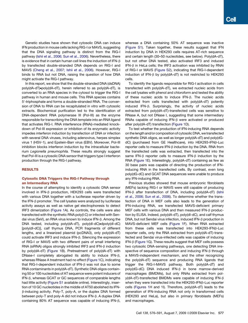
Genetic studies have shown that cytosolic DNA can induce
IFN production in mouse cells lacking RIG-I or MAVS, suggesting
that the DNA signaling pathway is distinct from the RIG-I
pathway (Ishii et al., 2006; Sun et al., 2006). Nevertheless, there
is evidence that in certain human cell lines the induction of IFN-b
by transfected double-stranded DNA depends on RIG-I and
MAVS (Cheng et al., 2007; Ishii et al., 2006). However, RIG-I
binds to RNA but not DNA, raising the question of how DNA
might activate the RIG-I pathway.
In this report, we show that the double-stranded DNA (dsDNA)
poly(dA-dT)dpoly(dA-dT), herein referred to as poly(dA-dT), is
converted to an RNA species in the cytosol to trigger the RIG-I
pathway in human and mouse cells. This RNA species contains
50-triphosphate and forms a double-stranded RNA. The conver-
sion of DNA to RNA can be recapitulated in vitro with cytosolic
extracts. Biochemical purification led to the identification of
DNA-dependent RNA polymerase III (Pol-III) as the enzyme
responsible for transcribing the DNA template into an RNA ligand
that activates RIG-I. RNA interference (RNAi)-mediated knock-
down of Pol-III expression or inhibition of its enzymatic activity
impedes interferon induction by transfection of DNA or infection
with several DNA viruses, including adenovirus, herpes simplex
virus 1 (HSV-1), and Epstein-Barr virus (EBV). Moreover, Pol-III
inhibition blocks interferon induction by the intracellular bacte-
rium Legionella pneumophila. These results strongly suggest
that Pol-III is a cytosolic DNA sensor that triggers type I interferon
production through the RIG-I pathway.
RESULTS
Cytosolic DNA Triggers the RIG-I Pathway throughan Intermediary RNAIn the course of attempting to identify a cytosolic DNA sensor
involved in IFN-b production, HEK293 cells were transfected
with various DNA together with a luciferase reporter driven by
the IFN-b promoter. The cell lysates were analyzed by luciferase
activity assays as well as native gel electrophoresis to detect
IRF3 dimerization (Figure 1A). As controls, the cells were also
transfected with the synthetic RNA poly(I:C) or infected with Sen-
dai virus (SeV), an RNA virus known to induce IFN-b. Among the
DNA tested, including poly(dA-dT), poly(dI-dC)ddpoly(dI-dC)
[poly(dI-dC)], calf thymus DNA, PCR fragments of different
lengths, and a linearized plasmid (pcDNA3), only poly(dA-dT)
could activate IRF3 and induce IFN-b. Silencing the expression
of RIG-I or MAVS with two different pairs of small interfering
RNA (siRNA) oligos strongly inhibited IRF3 and IFN-b induction
by poly(dA-dT) (Figure 1B). Pretreatment of poly(dA-dT) with
DNase-I completely abrogated its ability to induce IFN-b,
whereas RNase A treatment had no effect (Figure 1C), indicating
that RIG-I-dependent induction of IFN-b was not due to some
RNA contaminants in poly(dA-dT). Synthetic DNA oligos contain-
ing 50 or 100 nucleotides of AT sequence were potent inducers of
IFN-b, whereas GCAT or GC sequences of comparable lengths
had little activity (Figure S1 available online). Interestingly, inser-
tion of 10 GC nucleotides in the middle of AT50 abolished its IFN-
b-inducing activity. Poly-T, poly-A, or the duplex DNA formed
between poly-T and poly-A did not induce IFN-b. A duplex DNA
containing 80% AT sequence was capable of inducing IFN-b,
whereas a DNA containing 50% AT sequence was inactive
(Figure S1). Taken together, these results suggest that IFN
induction by DNA in HEK293 cells requires AT-rich sequence
and certain length (30–50 nucleotides, see below). Poly(dA-dT),
but not other DNA tested, also activated IRF3 and induced
IFN-b in HeLa cells; the IRF3 activation was inhibited by RNAi
of RIG-I or MAVS (Figure S2), indicating that RIG-I-dependent
induction of IFN-b by poly(dA-dT) is not restricted to HEK293
cells.
To identify the ligands responsible for RIG-I activation in cells
transfected with poly(dA-dT), we extracted nucleic acids from
the cell lysates with phenol and chloroform and tested the ability
of these nucleic acids to induce IFN-b. The nucleic acids
extracted from cells transfected with poly(dA-dT) potently
induced IFN-b. Surprisingly, the activity of nucleic acids
extracted from poly(dA-dT)-transfected cells was sensitive to
RNase A, but not DNase I, suggesting that some intermediary
RNAs capable of inducing IFN-b were activated or produced
after poly(dA-dT) transfection (Figure 1D).
To test whether the production of IFN-inducing RNA depends
on the lengthand/or composition ofcytosolicDNA,we transfected
synthetic DNA oligos, as well as longer poly(dA-dT) and poly(dG-
dC) (purchased from GE Healthcare), into HEK293-IFNb-Luc
reporter cells to measure IFN-b induction by the DNA. RNA from
the transfected cells was extracted, then transfected into the
same IFN-b reporter cells to measure IFN-b induction by the
RNA (Figure 1E). Interestingly, poly(dA-dT) containing as few as
30 base pairs was capable of directing the production of IFN-
inducing RNA in the transfected cells. By contrast, even long
poly(dG-dC) and GCAT DNA sequences were unable to produce
any IFN-inducing RNA.
Previous studies showed that mouse embryonic fibroblasts
(MEFs) lacking RIG-I or MAVS were still capable of producing
IFN-b after transfection of DNA, including poly(dA-dT) (Ishii
et al., 2006; Sun et al., 2006). To determine whether the trans-
fection of DNA in MEF cells also leads to the generation of
IFN-inducing RNA, we transfected MAVS-deficient primary
MEF cells with various DNA and then measured IFN-b produc-
tion by ELISA. Indeed, poly(dA-dT), poly(dI-dC), and calf thymus
DNA, but not Sendai virus infection, induced IFN-b production in
MAVS-deficient MEF cells (Figure 1F). When RNA extracted
from these cells was transfected into HEK293-IFNb-Luc
reporter cells, only the RNA extracted from poly(dA-dT)-trans-
fected and Sendai virus-infected cells was capable of inducing
IFN-b (Figure 1G). These results suggest that MEF cells possess
two cytosolic DNA-sensing pathways, one detecting DNA irre-
spective of sequence composition and inducing IFN-b through
a MAVS-independent mechanism, and the other recognizing
the poly(dA-dT) sequence and producing RNA ligands that
trigger the RIG-I-MAVS pathway. Both poly(dA-dT) and
poly(dG-dC) DNA induced IFN-b in bone marrow-derived
macrophages (BMDMs), but only RNAs extracted from pol-
y(dA-dT)-transfected BMDMs were capable of inducing IFN-b
when they were transfected into the HEK293-IFNb-Luc reporter
cells (Figures 1H and 1I). Therefore, poly(dA-dT) leads to the
generation of IFN-inducing RNA not only in transformed cells
(HEK293 and HeLa), but also in primary fibroblasts (MEFs)
and macrophages.
Cell 138, 576–591, August 7, 2009 ª2009 Elsevier Inc. 577
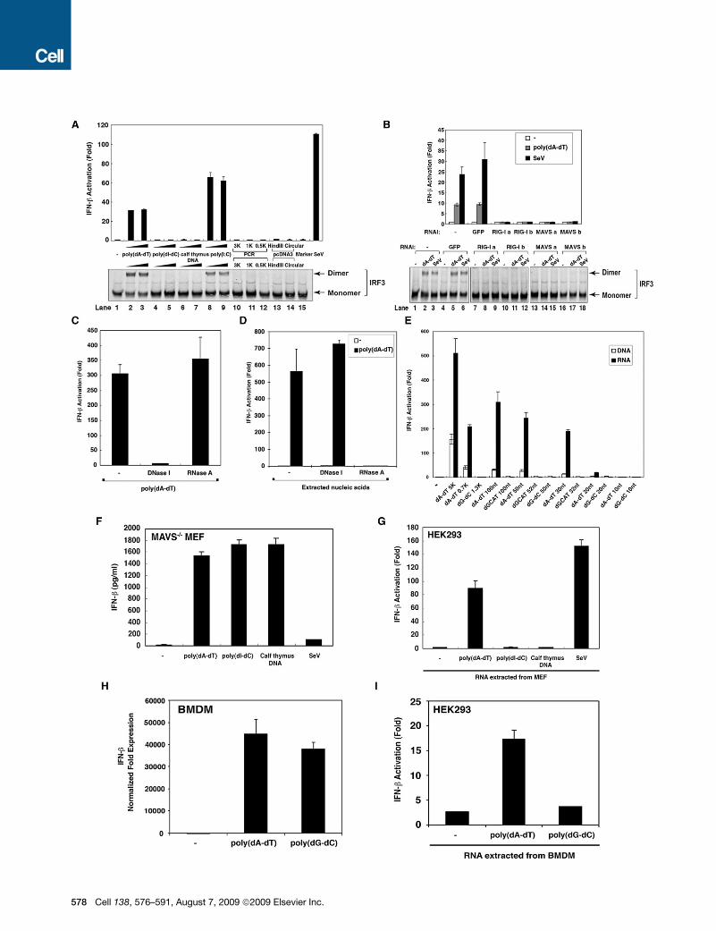
578 Cell 138, 576–591, August 7, 2009 ª2009 Elsevier Inc.
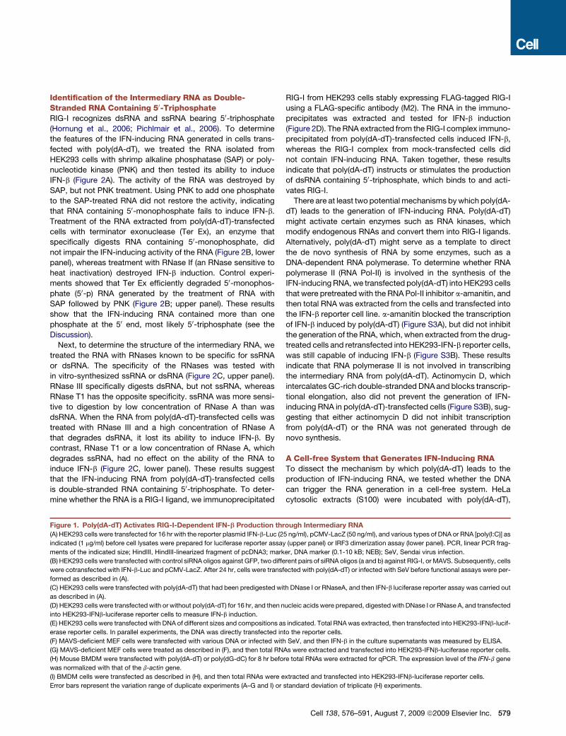
Identification of the Intermediary RNA as Double-Stranded RNA Containing 50-TriphosphateRIG-I recognizes dsRNA and ssRNA bearing 50-triphosphate
(Hornung et al., 2006; Pichlmair et al., 2006). To determine
the features of the IFN-inducing RNA generated in cells trans-
fected with poly(dA-dT), we treated the RNA isolated from
HEK293 cells with shrimp alkaline phosphatase (SAP) or poly-
nucleotide kinase (PNK) and then tested its ability to induce
IFN-b (Figure 2A). The activity of the RNA was destroyed by
SAP, but not PNK treatment. Using PNK to add one phosphate
to the SAP-treated RNA did not restore the activity, indicating
that RNA containing 50-monophosphate fails to induce IFN-b.
Treatment of the RNA extracted from poly(dA-dT)-transfected
cells with terminator exonuclease (Ter Ex), an enzyme that
specifically digests RNA containing 50-monophosphate, did
not impair the IFN-inducing activity of the RNA (Figure 2B, lower
panel), whereas treatment with RNase If (an RNase sensitive to
heat inactivation) destroyed IFN-b induction. Control experi-
ments showed that Ter Ex efficiently degraded 50-monophos-
phate (50-p) RNA generated by the treatment of RNA with
SAP followed by PNK (Figure 2B; upper panel). These results
show that the IFN-inducing RNA contained more than one
phosphate at the 50 end, most likely 50-triphosphate (see the
Discussion).
Next, to determine the structure of the intermediary RNA, we
treated the RNA with RNases known to be specific for ssRNA
or dsRNA. The specificity of the RNases was tested with
in vitro-synthesized ssRNA or dsRNA (Figure 2C, upper panel).
RNase III specifically digests dsRNA, but not ssRNA, whereas
RNase T1 has the opposite specificity. ssRNA was more sensi-
tive to digestion by low concentration of RNase A than was
dsRNA. When the RNA from poly(dA-dT)-transfected cells was
treated with RNase III and a high concentration of RNase A
that degrades dsRNA, it lost its ability to induce IFN-b. By
contrast, RNase T1 or a low concentration of RNase A, which
degrades ssRNA, had no effect on the ability of the RNA to
induce IFN-b (Figure 2C, lower panel). These results suggest
that the IFN-inducing RNA from poly(dA-dT)-transfected cells
is double-stranded RNA containing 50-triphosphate. To deter-
mine whether the RNA is a RIG-I ligand, we immunoprecipitated
RIG-I from HEK293 cells stably expressing FLAG-tagged RIG-I
using a FLAG-specific antibody (M2). The RNA in the immuno-
precipitates was extracted and tested for IFN-b induction
(Figure 2D). The RNA extracted from the RIG-I complex immuno-
precipitated from poly(dA-dT)-transfected cells induced IFN-b,
whereas the RIG-I complex from mock-transfected cells did
not contain IFN-inducing RNA. Taken together, these results
indicate that poly(dA-dT) instructs or stimulates the production
of dsRNA containing 50-triphosphate, which binds to and acti-
vates RIG-I.
There are at least two potential mechanisms by which poly(dA-
dT) leads to the generation of IFN-inducing RNA. Poly(dA-dT)
might activate certain enzymes such as RNA kinases, which
modify endogenous RNAs and convert them into RIG-I ligands.
Alternatively, poly(dA-dT) might serve as a template to direct
the de novo synthesis of RNA by some enzymes, such as a
DNA-dependent RNA polymerase. To determine whether RNA
polymerase II (RNA Pol-II) is involved in the synthesis of the
IFN-inducing RNA, we transfected poly(dA-dT) into HEK293 cells
that were pretreated with the RNA Pol-II inhibitor a-amanitin, and
then total RNA was extracted from the cells and transfected into
the IFN-b reporter cell line. a-amanitin blocked the transcription
of IFN-b induced by poly(dA-dT) (Figure S3A), but did not inhibit
the generation of the RNA, which, when extracted from the drug-
treated cells and retransfected into HEK293-IFN-b reporter cells,
was still capable of inducing IFN-b (Figure S3B). These results
indicate that RNA polymerase II is not involved in transcribing
the intermediary RNA from poly(dA-dT). Actinomycin D, which
intercalates GC-rich double-stranded DNA and blocks transcrip-
tional elongation, also did not prevent the generation of IFN-
inducing RNA in poly(dA-dT)-transfected cells (Figure S3B), sug-
gesting that either actinomycin D did not inhibit transcription
from poly(dA-dT) or the RNA was not generated through de
novo synthesis.
A Cell-free System that Generates IFN-Inducing RNATo dissect the mechanism by which poly(dA-dT) leads to the
production of IFN-inducing RNA, we tested whether the DNA
can trigger the RNA generation in a cell-free system. HeLa
cytosolic extracts (S100) were incubated with poly(dA-dT),
Figure 1. Poly(dA-dT) Activates RIG-I-Dependent IFN-b Production through Intermediary RNA
(A) HEK293 cells were transfected for 16 hr with the reporter plasmid IFN-b-Luc (25 ng/ml), pCMV-LacZ (50 ng/ml), and various types of DNA or RNA [poly(I:C)] as
indicated (1 mg/ml) before cell lysates were prepared for luciferase reporter assay (upper panel) or IRF3 dimerization assay (lower panel). PCR, linear PCR frag-
ments of the indicated size; HindIII, HindIII-linearized fragment of pcDNA3; marker, DNA marker (0.1-10 kB; NEB); SeV, Sendai virus infection.
(B) HEK293 cells were transfected with control siRNA oligos against GFP, two different pairs of siRNA oligos (a and b) against RIG-I, or MAVS. Subsequently, cells
were cotransfected with IFN-b-Luc and pCMV-LacZ. After 24 hr, cells were transfected with poly(dA-dT) or infected with SeV before functional assays were per-
formed as described in (A).
(C) HEK293 cells were transfected with poly(dA-dT) that had been predigested with DNase I or RNaseA, and then IFN-b luciferase reporter assay was carried out
as described in (A).
(D) HEK293 cells were transfected with or without poly(dA-dT) for 16 hr, and then nucleic acids were prepared, digested with DNase I or RNase A, and transfected
into HEK293-IFNb-luciferase reporter cells to measure IFN-b induction.
(E) HEK293 cells were transfected with DNA of different sizes and compositions as indicated. Total RNA was extracted, then transfected into HEK293-IFNb-lucif-
erase reporter cells. In parallel experiments, the DNA was directly transfected into the reporter cells.
(F) MAVS-deficient MEF cells were transfected with various DNA or infected with SeV, and then IFN-b in the culture supernatants was measured by ELISA.
(G) MAVS-deficient MEF cells were treated as described in (F), and then total RNAs were extracted and transfected into HEK293-IFNb-luciferase reporter cells.
(H) Mouse BMDM were transfected with poly(dA-dT) or poly(dG-dC) for 8 hr before total RNAs were extracted for qPCR. The expression level of the IFN-b gene
was normalized with that of the b-actin gene.
(I) BMDM cells were transfected as described in (H), and then total RNAs were extracted and transfected into HEK293-IFNb-luciferase reporter cells.
Error bars represent the variation range of duplicate experiments (A–G and I) or standard deviation of triplicate (H) experiments.
Cell 138, 576–591, August 7, 2009 ª2009 Elsevier Inc. 579
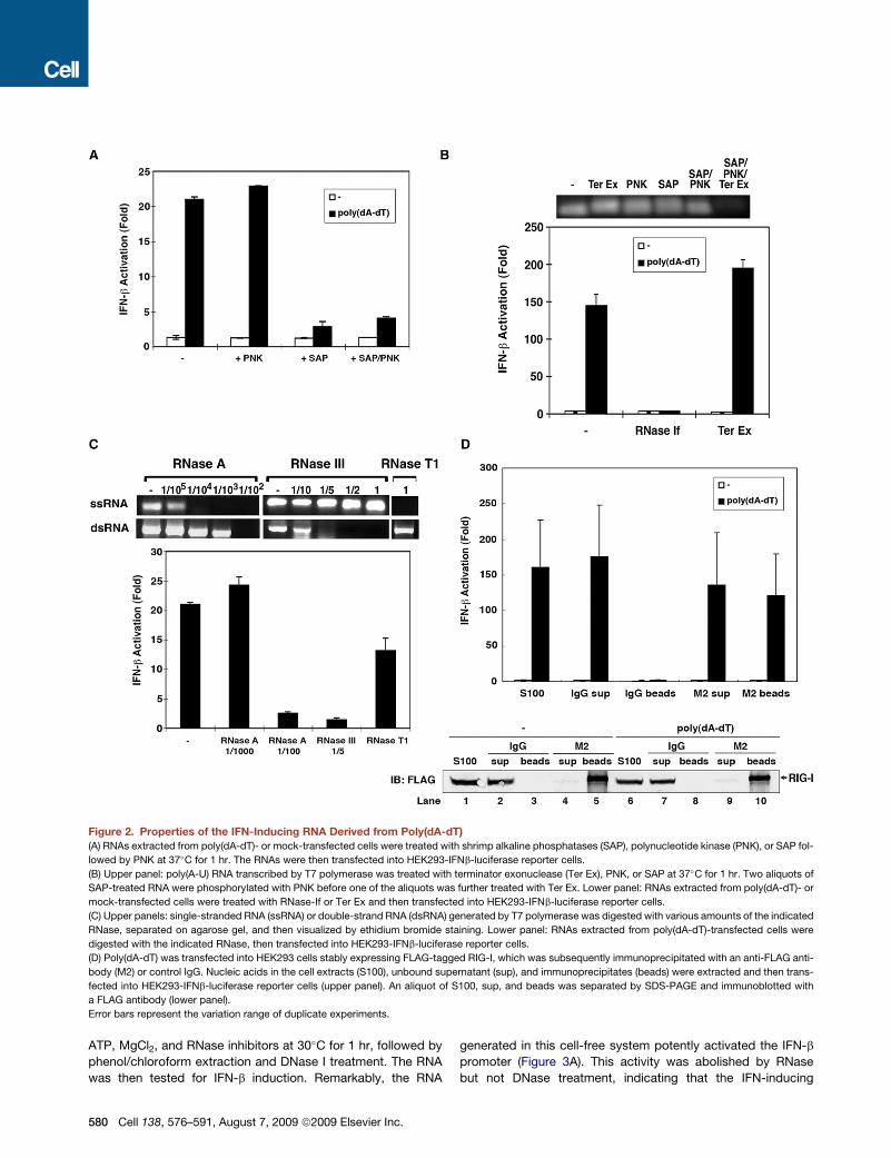
Figure 2. Properties of the IFN-Inducing RNA Derived from Poly(dA-dT)
(A) RNAs extracted from poly(dA-dT)- or mock-transfected cells were treated with shrimp alkaline phosphatases (SAP), polynucleotide kinase (PNK), or SAP fol-
lowed by PNK at 37�C for 1 hr. The RNAs were then transfected into HEK293-IFNb-luciferase reporter cells.
(B) Upper panel: poly(A-U) RNA transcribed by T7 polymerase was treated with terminator exonuclease (Ter Ex), PNK, or SAP at 37�C for 1 hr. Two aliquots of
SAP-treated RNA were phosphorylated with PNK before one of the aliquots was further treated with Ter Ex. Lower panel: RNAs extracted from poly(dA-dT)- or
mock-transfected cells were treated with RNase-If or Ter Ex and then transfected into HEK293-IFNb-luciferase reporter cells.
(C) Upper panels: single-stranded RNA (ssRNA) or double-strand RNA (dsRNA) generated by T7 polymerase was digested with various amounts of the indicated
RNase, separated on agarose gel, and then visualized by ethidium bromide staining. Lower panel: RNAs extracted from poly(dA-dT)-transfected cells were
digested with the indicated RNase, then transfected into HEK293-IFNb-luciferase reporter cells.
(D) Poly(dA-dT) was transfected into HEK293 cells stably expressing FLAG-tagged RIG-I, which was subsequently immunoprecipitated with an anti-FLAG anti-
body (M2) or control IgG. Nucleic acids in the cell extracts (S100), unbound supernatant (sup), and immunoprecipitates (beads) were extracted and then trans-
fected into HEK293-IFNb-luciferase reporter cells (upper panel). An aliquot of S100, sup, and beads was separated by SDS-PAGE and immunoblotted with
a FLAG antibody (lower panel).
Error bars represent the variation range of duplicate experiments.
ATP, MgCl2, and RNase inhibitors at 30�C for 1 hr, followed by
phenol/chloroform extraction and DNase I treatment. The RNA
was then tested for IFN-b induction. Remarkably, the RNA
580 Cell 138, 576–591, August 7, 2009 ª2009 Elsevier Inc.
generated in this cell-free system potently activated the IFN-b
promoter (Figure 3A). This activity was abolished by RNase
but not DNase treatment, indicating that the IFN-inducing
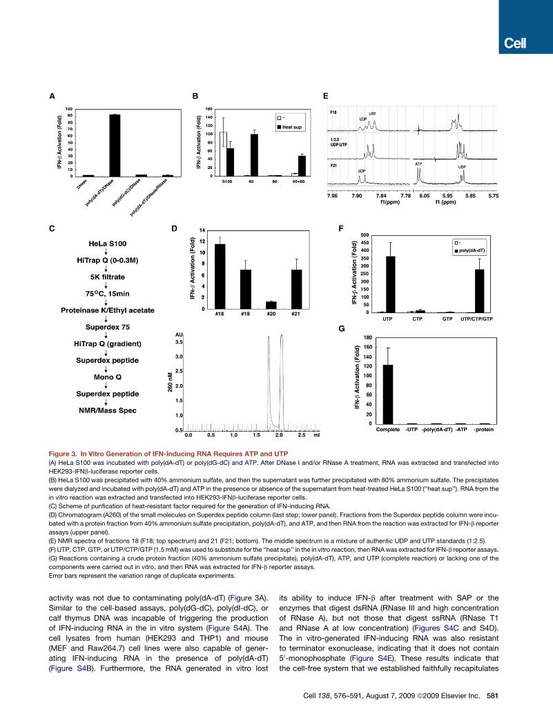
Figure 3. In Vitro Generation of IFN-Inducing RNA Requires ATP and UTP
(A) HeLa S100 was incubated with poly(dA-dT) or poly(dG-dC) and ATP. After DNase I and/or RNase A treatment, RNA was extracted and transfected into
HEK293-IFNb-luciferase reporter cells.
(B) HeLa S100 was precipitated with 40% ammonium sulfate, and then the supernatant was further precipitated with 80% ammonium sulfate. The precipitates
were dialyzed and incubated with poly(dA-dT) and ATP in the presence or absence of the supernatant from heat-treated HeLa S100 (‘‘heat sup’’). RNA from the
in vitro reaction was extracted and transfected into HEK293-IFNb-luciferase reporter cells.
(C) Scheme of purification of heat-resistant factor required for the generation of IFN-inducing RNA.
(D) Chromatogram (A260) of the small molecules on Superdex peptide column (last step; lower panel). Fractions from the Superdex peptide column were incu-
bated with a protein fraction from 40% ammonium sulfate precipitation, poly(dA-dT), and ATP, and then RNA from the reaction was extracted for IFN-b reporter
assays (upper panel).
(E) NMR spectra of fractions 18 (F18; top spectrum) and 21 (F21; bottom). The middle spectrum is a mixture of authentic UDP and UTP standards (1:2.5).
(F) UTP, CTP, GTP, or UTP/CTP/GTP (1.5 mM) was used to substitute for the ‘‘heat sup’’ in the in vitro reaction, then RNA was extracted for IFN-b reporter assays.
(G) Reactions containing a crude protein fraction (40% ammonium sulfate precipitate), poly(dA-dT), ATP, and UTP (complete reaction) or lacking one of the
components were carried out in vitro, and then RNA was extracted for IFN-b reporter assays.
Error bars represent the variation range of duplicate experiments.
activity was not due to contaminating poly(dA-dT) (Figure 3A).
Similar to the cell-based assays, poly(dG-dC), poly(dI-dC), or
calf thymus DNA was incapable of triggering the production
of IFN-inducing RNA in the in vitro system (Figure S4A). The
cell lysates from human (HEK293 and THP1) and mouse
(MEF and Raw264.7) cell lines were also capable of gener-
ating IFN-inducing RNA in the presence of poly(dA-dT)
(Figure S4B). Furthermore, the RNA generated in vitro lost
its ability to induce IFN-b after treatment with SAP or the
enzymes that digest dsRNA (RNase III and high concentration
of RNase A), but not those that digest ssRNA (RNase T1
and RNase A at low concentration) (Figures S4C and S4D).
The in vitro-generated IFN-inducing RNA was also resistant
to terminator exonuclease, indicating that it does not contain
50-monophosphate (Figure S4E). These results indicate that
the cell-free system that we established faithfully recapitulates
Cell 138, 576–591, August 7, 2009 ª2009 Elsevier Inc. 581
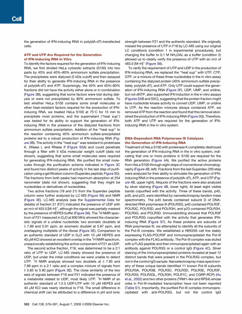
the generation of IFN-inducing RNA in poly(dA-dT)-transfected
cells.
ATP and UTP Are Required for the Generationof IFN-Inducing RNA In VitroTo identify the factors required for the generation of IFN-inducing
RNA, we first divided HeLa cytosolic extracts (S100) into two
parts by 40% and 40%–80% ammonium sulfate precipitation.
The precipitates were dialyzed (5 kDa cutoff) and then assayed
for their ability to generate IFN-inducing RNA in the presence
of poly(dA-dT) and ATP. Surprisingly, the 40% and 40%–80%
fractions did not have the activity either alone or in combination
(Figure 3B), suggesting that some factors were lost during dial-
ysis or were not precipitated by 80% ammonium sulfate. To
test whether HeLa S100 contains some small molecules or
other heat-resistant factors required for the production of IFN-
inducing RNA, we treated HeLa S100 at 75�C for 15 min to
precipitate most proteins, and the supernatant (‘‘heat sup’’)
was tested for its ability to support the generation of IFN-
inducing RNA in the presence of the dialyzed fractions from
ammonium sulfate precipitation. Addition of the ‘‘heat sup’’ to
the reaction containing 40% ammonium sulfate-precipitated
proteins led to a robust production of IFN-inducing RNA (Fig-
ure 3B). The activity in the ‘‘heat sup’’ was resistant to proteinase
K, DNase I, and RNase If (Figure S5A) and could penetrate
through a filter with 5 kDa molecular weight cutoff (data not
shown), suggesting that some small molecules were required
for generating IFN-inducing RNA. We purified the small mole-
cules through the purification scheme indicated in Figure 3C
and found two distinct peaks of activity in the last step of purifi-
cation using a gel filtration column (Superdex peptide; Figure 3D).
The fractions from both peaks had maximum absorption at 254
nanometer (data not shown), suggesting that they might be
nucleotides or derivatives of nucleotides.
Two active fractions (18 and 21) from the Superdex peptide
column were further analyzed by LC-MS (Figure S6) and NMR
(Figure 3E). LC-MS analysis (see the Supplemental Data for
details) of fraction 21 (F21) indicated the presence of UDP with
an m/z of 403.0 [M–H]+, although the signal was partially masked
by the presence of HEPES buffer (Figure S6). The 1H NMR spec-
trum of F21 measured in D2O at 600 MHz showed the character-
istic signals of a uridine nucleotide: two aromatic doublets at
d 7.89 and 5.91 ppm, an anomeric doublet at 5.87 ppm, and
overlapping multiplets of the ribose (Figure 3E). Comparison to
an authentic standard of UDP in D2O with 10 mM HEPES and
40 mM KCl showed an excellent overlap in the 1H NMR spectrum,
unequivocally establishing the active component of F21 as UDP.
The second active fraction, F18, was determined to be a 2:1
ratio of UTP to UDP. LC-MS clearly showed the presence of
UDP, but under the initial conditions we were unable to detect
UTP. 1H NMR analysis showed two doublets at d 7.85 and
7.89 ppm in a 2:1 ratio and a complex mixture of signals from
d 5.85 to 5.90 ppm (Figure 3E). The close similarity of the two
sets of signals between F18 and F21 indicated the presence of
a metabolite related to UDP, most likely UTP. 1H NMR of an
authentic standard of 1:2.5 UDP:UTP with 10 mM HEPES and
40 mM KCl was nearly identical to F18. The small difference in
chemical shift can be attributed to a difference in pH and ionic
582 Cell 138, 576–591, August 7, 2009 ª2009 Elsevier Inc.
strength between F21 and the authentic standard. We originally
missed the presence of UTP in F18 by LC-MS using our original
LC conditions (condition 1 in experimental procedures), but
changing the buffer to 0.1 M NH4OAc as a buffer (condition 2)
allowed us to clearly verify the presence of UTP with an m/z of
483.0 [M–H]+ (Figure S6).
To verify the requirement of UTP and UDP in the production of
IFN-inducing RNA, we replaced the ‘‘heat sup’’ with UTP, CTP,
GTP, or a mixture of these three nucleotides in the in vitro assay
containing the dialyzed protein (40% ammonium sulfate precip-
itate), poly(dA-dT), and ATP. Only UTP could support the gener-
ation of IFN-inducing RNA (Figure 3F). UDP, UMP, and uridine,
but not dNTP, also supported IFN induction in the in vitro assays
(Figures S5B and S5C), suggesting that the protein fraction might
have nucleotide kinase activity to convert UDP, UMP, or uridine
to UTP. As the reaction mixtures always contained ATP, we
removed ATP from the reaction and found that this removal abol-
ished the production of IFN-inducing RNA (Figure 3G). Therefore,
both ATP and UTP are required for the generation of IFN-
inducing RNA in the in vitro system.
DNA-Dependent RNA Polymerase III Catalyzesthe Generation of IFN-Inducing RNATreatment of HeLa S100 with proteinase K completely destroyed
the generation of IFN-inducing RNA in the in vitro system, indi-
cating that one or more proteins in S100 are required for the
RNA generation (Figure 4A). We purified the active proteins
from HeLa S100 through eight steps of conventional chromatog-
raphy (Figure 4B, left). Fractions from the last Superdex-200 step
were analyzed for their ability to stimulate the generation of IFN-
inducing RNA in the presence of poly(dA-dT), ATP, and UTP (Fig-
ure 4B, upper right). Aliquots of the fractions were also analyzed
by silver staining (Figure 4B, lower right). At least eight visible
bands copurified with the activity. Three of these bands, p45,
p40, and p23, were identified by nanoelectrospray tandem mass
spectrometry. The p45 bands contained subunit D of DNA-
directed RNA polymerase III (POLR3D), p40 contained POLR3F,
POLR3C, POLR3D, and POLR3H, and p23 contained POLR3H,
POLR3G, and POLR3D. Immunoblotting showed that POLR3F
and POLR3G copurified with the activity that generates IFN-
inducing RNA (Figure S7). As these proteins are subunits of
RNA polymerase III, we attempted to identify all the subunits of
the Pol-III complex. We established a HEK293 cell line stably
expressing FLAG-POLR3F and immunoprecipitated the Pol-III
complex with the FLAG antibody. The Pol-III complex was eluted
with a FLAG peptide and then immunoprecipitated again with an
antibody against POLR3G or a control IgG (Figure 4C). Silver
staining of the immunoprecipitated proteins revealed at least 12
distinct bands that were present in the POLR3G complex, but
not in the control IgG sample. Nanoelectrospray mass spectrom-
etry of these unique bands identified 11 known Pol-III subunits
(POLR3A, POLR3B, POLR3C, POLR3D, POLR3E, POLR3F,
POLR3G, POLR3GL, POLR3H, POLR1C, and CGRP-RCP) (Hu
et al., 2002) and two other proteins (TRM1-like and RPS4) whose
roles in Pol-III-mediated transcription have not been reported
(Table S1). Importantly, the purified Pol-III complex immunopre-
cipitated with anti-POLR3G, but not the control IgG
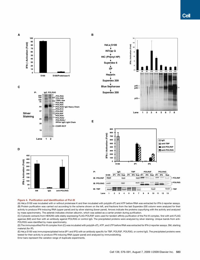
Figure 4. Purification and Identification of Pol-III
(A) HeLa S100 was incubated with or without proteinase K and then incubated with poly(dA-dT) and ATP before RNA was extracted for IFN-b reporter assays.
(B) Protein purification was carried out according to the scheme shown on the left, and fractions from the last Superdex-200 column were analyzed for their
activity to produce IFN-inducing RNA (upper panel) and by silver staining (lower panel). Arrows indicate the proteins copurifying with the activity and analyzed
by mass spectrometry. The asterisk indicates chicken albumin, which was added as a carrier protein during purification.
(C) Cytosolic extracts from HEK293 cells stably expressing FLAG-POLR3F were used for tandem affinity purification of the Pol-III complex, first with anti-FLAG
agarose (M2) and then with an antibody against POLR3G or control IgG. The precipitated proteins were analyzed by silver staining. Unique bands from anti-
POLR3G were identified by mass spectrometry.
(D) The immunopurified Pol-III complex from (C) was incubated with poly(dA-dT), ATP, and UTP before RNA was extracted for IFN-b reporter assays. SM, starting
material (for IP).
(E) HeLa S100 was immunoprecipitated twice (IP1 and IP2) with an antibody specific for TBP, POLR3F, POLR3G, or control IgG. The precipitated proteins were
tested for their activity to produce IFN-inducing RNA (upper panel) and analyzed by immunoblotting.
Error bars represent the variation range of duplicate experiments.
Cell 138, 576–591, August 7, 2009 ª2009 Elsevier Inc. 583
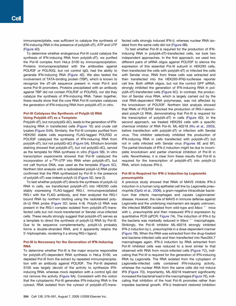
immunoprecipitate, was sufficient to catalyze the synthesis of
IFN-inducing RNA in the presence of poly(dA-dT), ATP and UTP
(Figure 4D).
To determine whether endogenous Pol-III could catalyze the
synthesis of IFN-inducing RNA from poly(dA-dT), we purified
the Pol-III complex from HeLa S100 by immunoprecipitation.
Proteins immunoprecipitated with the antibodies against
POLR3F or POLR3G, but not control IgG, had the ability to
generate IFN-inducing RNA (Figure 4E). We also tested the
involvement of TATA-binding protein (TBP), which is known to
recognize the dT-dA sequence present in most Pol-II and
some Pol-III promoters. Proteins precipitated with an antibody
against TBP did not contain POLR3F or POLR3G, nor did they
catalyze the synthesis of IFN-inducing RNA. Taken together,
these results show that the core RNA Pol-III complex catalyzes
the generation of IFN-inducing RNA from poly(dA-dT) in vitro.
Pol-III Catalyzes the Synthesis of Poly(A-U) RNAUsing Poly(dA-dT) as a TemplatePoly(dA-dT), but not poly(dG-dC), leads to the generation of IFN-
inducing RNA in transfected cells (Figure 1E) and crude cell
lysates (Figure S4A). Similarly, the Pol-III complex purified from
HEK293 stable cells expressing FLAG-tagged POLR3D or
POLR3F catalyzed the synthesis of IFN-inducing RNA from
poly(dA-dT), but not poly(dG-dC) (Figure 5A). Ethidium bromide
staining showed that poly(dA-dT), but not poly(dG-dC), served
as the template for RNA synthesis in vitro (Figure 5B). In vitro
transcription experiments showed that Pol-III catalyzed the
incorporation of a-32P-UTP into RNA when poly(dA-dT), but
not calf thymus DNA, was used as the template (Figure 5C).
Northern blot analysis with a radiolabeled poly(A-U) RNA probe
confirmed that the RNA synthesized by Pol-III in the presence
of poly(dA-dT) was indeed poly(A-U) (Figure 5D, lane 2).
To test whether poly(dA-dT) directs the synthesis of poly(A-U)
RNA in cells, we transfected poly(dA-dT) into HEK293 cells
stably expressing FLAG-tagged RIG-I, immunoprecipitated
RIG-I with the FLAG antibody, and then analyzed the RIG-I
bound RNA by northern blotting using the radiolabeled poly-
(A-U) RNA probe (Figure 5D; lanes 4–9). Poly(A-U) RNA was
present in the RIG-I complex isolated from poly(dA-dT)-trans-
fected cells but not mock-transfected or Sendai virus-infected
cells. These results strongly suggest that poly(dA-dT) serves as
a template to direct the synthesis of poly(A-U) RNA by Pol-III.
Due to its sequence complementarity, poly(A-U) probably
forms a double-stranded RNA, and it apparently retains the
50-triphosphate, rendering it a strong RIG-I ligand.
Pol-III Is Necessary for the Generation of IFN-InducingRNATo determine whether Pol-III is the major enzyme responsible
for poly(dA-dT)-dependent RNA synthesis in HeLa S100, we
depleted Pol-III from the extract by repeated immunoprecipita-
tion with an antibody against POLR3F. The Pol-III depleted
S100 was largely devoid of the activity to synthesize IFN-
inducing RNA, whereas mock depletion with a control IgG did
not remove the activity (Figure 6A). Consistent with the notion
that the cytoplasmic Pol-III generates IFN-inducing RNA in the
cytosol, RNA isolated from the cytosol of poly(dA-dT)-trans-
584 Cell 138, 576–591, August 7, 2009 ª2009 Elsevier Inc.
fected cells strongly induced IFN-b, whereas nuclear RNA iso-
lated from the same cells did not (Figure 6B).
To test whether Pol-III is required for the production of IFN-
inducing RNA in poly(dA-dT)-transfected cells, we took two
independent approaches. In the first approach, we used two
different pairs of siRNA oligos against POLR3F to silence the
expression of this essential Pol-III subunit in HEK293 cells,
then transfected the cells with poly(dA-dT) or infected the cells
with Sendai virus. RNA from these cells was extracted and
then transfected into the HEK293-IFNb-luciferase reporter
cell line. Both siRNA oligos, but not the control GFP siRNA,
strongly inhibited the generation of IFN-inducing RNA in pol-
y(dA-dT)-transfected cells (Figure 6C). In contrast, the produc-
tion of Sendai virus RNA, which is largely carried out by the
viral RNA-dependent RNA polymerase, was not affected by
the knockdown of POLR3F. Northern blot analysis showed
that RNAi of POLR3F blocked the production of RIG-I-associ-
ated poly(A-U) RNA, demonstrating that Pol-III is required for
the transcription of poly(dA-dT) in cells (Figure 6D). In the
second approach, we treated HEK293 cells with a specific
chemical inhibitor of RNA Pol-III, ML-60218 (Wu et al., 2003),
before transfection with poly(dA-dT) or infection with Sendai
virus. This inhibitor selectively inhibited the production of
IFN-inducing RNA in cells transfected with poly(dA-dT) but
not in cells infected with Sendai virus (Figures 6E and 6F).
The partial blockade of IFN-b induction might be due to incom-
plete knockdown and partial chemical inhibition of Pol-III in
cells. Nevertheless, it is clear from these results that Pol-III is
required for the transcription of poly(dA-dT) into poly(A-U)
RNA, which induces IFN-b.
Pol-III Is Required for IFN-b Induction by Legionella
pneumophila
A previous study showed that RNAi of MAVS inhibits IFN-b
induction in a human lung epithelial cell line by Legionella pneu-
mophila (Opitz et al., 2006), a gram-negative intracellular bacte-
rium that infects macrophages and causes Legionnaires’
disease. However, the role of MAVS in immune defense against
Legionella and the underlying mechanism are largely unknown.
We infected BMDM isolated from wild-type and Mavs�/� mice
with L. pneumophila and then measured IFN-b expression by
quantitative PCR (qPCR; Figure 7A). The induction of IFN-b by
the bacteria was markedly reduced in Mavs�/� macrophages.
Strikingly, the Pol-III inhibitor ML-60218 strongly inhibited
IFN-b induction by L. pneumophila in a dose-dependent manner
(Figure 7B). When the RNA was extracted from the drug-treated
and bacteria-infected cells and then transfected into Raw264.7
macrophages again, IFN-b induction by RNA extracted from
Pol-III inhibited cells was reduced to a level similar to that
observed with RNA from mock-infected cells (Figure 7C), indi-
cating that Pol-III is required for the generation of IFN-inducing
RNA by Legionella. The RNA isolated from the cytoplasm of
Legionella-infected cells contained IFN-inducing activity,
whereas the nuclear RNA from the same cells did not induce
IFN (Figure 7D). Importantly, ML-60218 treatment significantly
increased the bacterial load in the macrophages (Figure 7E), indi-
cating that inhibition of the host Pol-III promotes rather than
impedes bacterial growth. IFN-b treatment restored inhibition
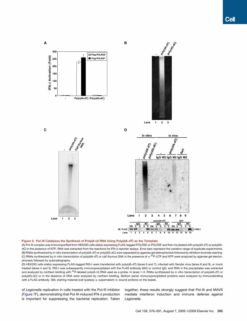
Figure 5. Pol-III Catalyzes the Synthesis of Poly(A-U) RNA Using Poly(dA-dT) as the Template
(A) Pol-III complex was immunopurified from HEK293 cells stably expressing FLAG-tagged POLR3D or POLR3F and then incubated with poly(dA-dT) or poly(dG-
dC) in the presence of NTP. RNA was extracted from the reactions for IFN-b reporter assays. Error bars represent the variation range of duplicate experiments.
(B) RNAs synthesized by in vitro transcription of poly(dA-dT) or poly(dG-dC) were separated by agarose gel eletrophoresis followed by ethidium bromide staining.
(C) RNAs synthesized by in vitro transcription of poly(dA-dT) or calf thymus DNA in the presence of a-32P-UTP and NTP were analyzed by agarose gel electro-
phoresis followed by autoradiography.
(D) HEK293 cells stably expressing FLAG-tagged RIG-I were transfected with poly(dA-dT) (lanes 6 and 7), infected with Sendai virus (lanes 8 and 9), or mock
treated (lanes 4 and 5). RIG-I was subsequently immunoprecipitated with the FLAG antibody (M2) or control IgG, and RNA in the precipitates was extracted
and analyzed by northern blotting with 32P-labeled poly(A-U) RNA used as a probe. In lanes 1–3, RNAs synthesized by in vitro transcription of poly(dA-dT) or
poly(dG-dC) or in the absence of DNA were analyzed by northern blotting. Bottom panel: Immunoprecipitated proteins were analyzed by immunoblotting
with a FLAG antibody. SM, starting material (cell lysates); s, supernatant; b, bound proteins on the beads.
of Legionella replication in cells treated with the Pol-III inhibitor
(Figure 7F), demonstrating that Pol-III-induced IFN-b production
is important for suppressing the bacterial replication. Taken
together, these results strongly suggest that Pol-III and MAVS
mediate interferon induction and immune defense against
Legionella.
Cell 138, 576–591, August 7, 2009 ª2009 Elsevier Inc. 585
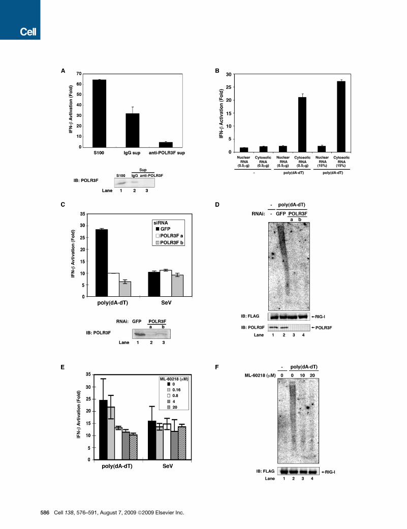
586 Cell 138, 576–591, August 7, 2009 ª2009 Elsevier Inc.
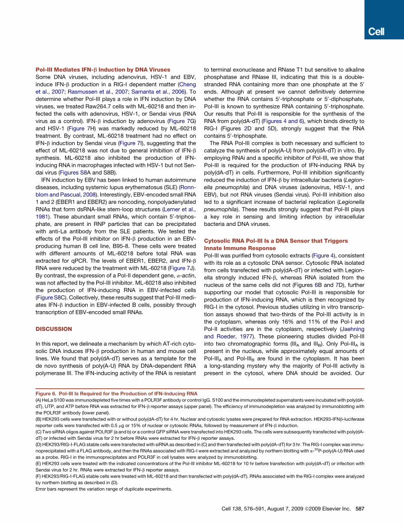
Pol-III Mediates IFN-b Induction by DNA VirusesSome DNA viruses, including adenovirus, HSV-1 and EBV,
induce IFN-b production in a RIG-I dependent matter (Cheng
et al., 2007; Rasmussen et al., 2007; Samanta et al., 2006). To
determine whether Pol-III plays a role in IFN induction by DNA
viruses, we treated Raw264.7 cells with ML-60218 and then in-
fected the cells with adenovirus, HSV-1, or Sendai virus (RNA
virus as a control). IFN-b induction by adenovirus (Figure 7G)
and HSV-1 (Figure 7H) was markedly reduced by ML-60218
treatment. By contrast, ML-60218 treatment had no effect on
IFN-b induction by Sendai virus (Figure 7I), suggesting that the
effect of ML-60218 was not due to general inhibition of IFN-b
synthesis. ML-60218 also inhibited the production of IFN-
inducing RNA in macrophages infected with HSV-1 but not Sen-
dai virus (Figures S8A and S8B).
IFN induction by EBV has been linked to human autoimmune
diseases, including systemic lupus erythematosus (SLE) (Ronn-
blom and Pascual, 2008). Interestingly, EBV-encoded small RNA
1 and 2 (EBER1 and EBER2) are noncoding, nonpolyadenylated
RNAs that form dsRNA-like stem-loop structures (Lerner et al.,
1981). These abundant small RNAs, which contain 50-triphos-
phate, are present in RNP particles that can be precipitated
with anti-La antibody from the SLE patients. We tested the
effects of the Pol-III inhibitor on IFN-b production in an EBV-
producing human B cell line, B95-8. These cells were treated
with different amounts of ML-60218 before total RNA was
extracted for qPCR. The levels of EBER1, EBER2, and IFN-b
RNA were reduced by the treatment with ML-60218 (Figure 7J).
By contrast, the expression of a Pol-II dependent gene, a-actin,
was not affected by the Pol-III inhibitor. ML-60218 also inhibited
the production of IFN-inducing RNA in EBV-infected cells
(Figure S8C). Collectively, these results suggest that Pol-III medi-
ates IFN-b induction in EBV-infected B cells, possibly through
transcription of EBV-encoded small RNAs.
DISCUSSION
In this report, we delineate a mechanism by which AT-rich cyto-
solic DNA induces IFN-b production in human and mouse cell
lines. We found that poly(dA-dT) serves as a template for the
de novo synthesis of poly(A-U) RNA by DNA-dependent RNA
polymerase III. The IFN-inducing activity of the RNA is resistant
to terminal exonuclease and RNase T1 but sensitive to alkaline
phosphatase and RNase III, indicating that this is a double-
stranded RNA containing more than one phosphate at the 50
ends. Although at present we cannot definitively determine
whether the RNA contains 50-triphosphate or 50-diphosphate,
Pol-III is known to synthesize RNA containing 50-triphosphate.
Our results that Pol-III is responsible for the synthesis of the
RNA from poly(dA-dT) (Figures 4 and 6), which binds directly to
RIG-I (Figures 2D and 5D), strongly suggest that the RNA
contains 50-triphosphate.
The RNA Pol-III complex is both necessary and sufficient to
catalyze the synthesis of poly(A-U) from poly(dA-dT) in vitro. By
employing RNAi and a specific inhibitor of Pol-III, we show that
Pol-III is required for the production of IFN-inducing RNA by
poly(dA-dT) in cells. Furthermore, Pol-III inhibition significantly
reduced the induction of IFN-b by intracellular bacteria (Legion-
ella pneumophila) and DNA viruses (adenovirus, HSV-1, and
EBV), but not RNA viruses (Sendai virus). Pol-III inhibition also
led to a significant increase of bacterial replication (Legionella
pneumophila). These results strongly suggest that Pol-III plays
a key role in sensing and limiting infection by intracellular
bacteria and DNA viruses.
Cytosolic RNA Pol-III Is a DNA Sensor that TriggersInnate Immune ResponsePol-III was purified from cytosolic extracts (Figure 4), consistent
with its role as a cytosolic DNA sensor. Cytosolic RNA isolated
from cells transfected with poly(dA-dT) or infected with Legion-
ella strongly induced IFN-b, whereas RNA isolated from the
nucleus of the same cells did not (Figures 6B and 7D), further
supporting our model that cytosolic Pol-III is responsible for
production of IFN-inducing RNA, which is then recognized by
RIG-I in the cytosol. Previous studies utilizing in vitro transcrip-
tion assays showed that two-thirds of the Pol-III activity is in
the cytoplasm, whereas only 16% and 11% of the Pol-I and
Pol-II activities are in the cytoplasm, respectively (Jaehning
and Roeder, 1977). These pioneering studies divided Pol-III
into two chromatographic forms (IIIA and IIIB). Only Pol-IIIA is
present in the nucleus, while approximately equal amounts of
Pol-IIIA and Pol-IIIB are found in the cytoplasm. It has been
a long-standing mystery why the majority of Pol-III activity is
present in the cytosol, where DNA should be avoided. Our
Figure 6. Pol-III Is Required for the Production of IFN-Inducing RNA
(A) HeLa S100 was immunodepleted five times with a POLR3F antibody or control IgG. S100 and the immunodepleted supernatants were incubated with poly(dA-
dT), UTP, and ATP before RNA was extracted for IFN-b reporter assays (upper panel). The efficiency of immunodepletion was analyzed by immunoblotting with
the POLR3F antibody (lower panel).
(B) HEK293 cells were transfected with or without poly(dA-dT) for 4 hr. Nuclear and cytosolic lysates were prepared for RNA extraction. HEK293-IFNb-luciferase
reporter cells were transfected with 0.5 mg or 15% of nuclear or cytosolic RNAs, followed by measurement of IFN-b induction.
(C) Two siRNA oligos against POLR3F (a and b) or a control GFP siRNA were transfected into HEK293 cells. The cells were subsequently transfected with poly(dA-
dT) or infected with Sendai virus for 2 hr before RNAs were extracted for IFN-b reporter assays.
(D) HEK293/RIG-I-FLAG stable cells were transfected with siRNA as described in (C) and then transfected with poly(dA-dT) for 3 hr. The RIG-I complex was immu-
noprecipitated with a FLAG antibody, and then the RNAs associated with RIG-I were extracted and analyzed by northern blotting with a-32P-poly(A-U) RNA used
as a probe. RIG-I in the immunoprecipitates and POLR3F in cell lysates were analyzed by immunoblotting.
(E) HEK293 cells were treated with the indicated concentrations of the Pol-III inhibitor ML-60218 for 10 hr before transfection with poly(dA-dT) or infection with
Sendai virus for 2 hr. RNAs were extracted for IFN-b reporter assays.
(F) HEK293/RIG-I-FLAG stable cells were treated with ML-60218 and then transfected with poly(dA-dT). RNAs associated with the RIG-I complex were analyzed
by northern blotting as described in (D).
Error bars represent the variation range of duplicate experiments.
Cell 138, 576–591, August 7, 2009 ª2009 Elsevier Inc. 587
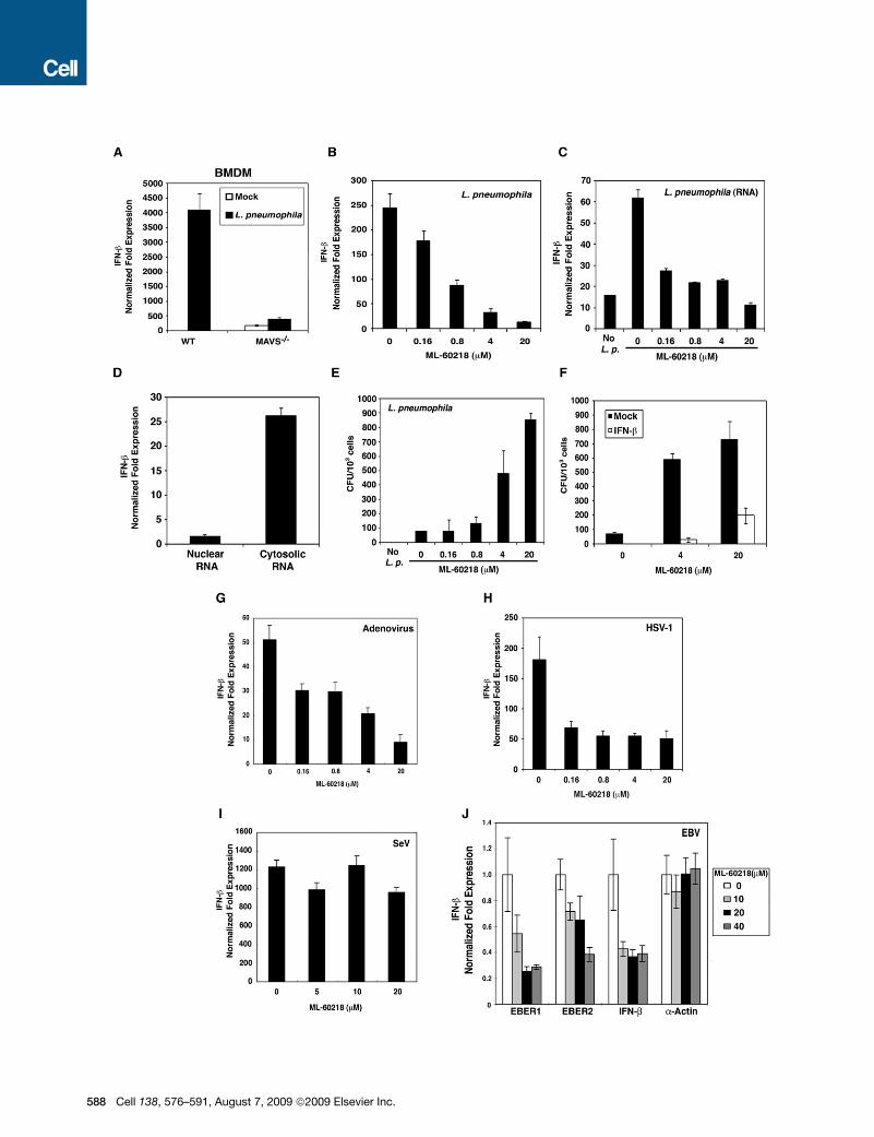
588 Cell 138, 576–591, August 7, 2009 ª2009 Elsevier Inc.

finding that cytosolic Pol-III is a DNA sensor that mediates innate
immune response provides a potential answer to this conun-
drum. The cytosol is a strategic location for Pol-III to function
in immune defense, because RNA synthesized in the cytosol
contains 50-triphosphate that can be detected by RIG-I, whereas
RNA synthesized in the nucleus is likely to be modified by
enzymes such as the capping enzymes (e.g., mRNA) or pro-
cessed to generate 50-monophosphate (e.g., tRNA), which
escapes detection by RIG-I. Therefore, we propose that nuclear
and cytoplasmic RNA polymerases serve opposing functions:
while the nuclear polymerases produce the host RNA that should
be modified to avoid triggering autoimmune response through
the RIG-I pathway, the cytosolic Pol-III is specialized to detect
foreign and aberrant DNA to mount an immune response to
defend the integrity of the host cytoplasm.
Recognition of AT-Rich DNA Sequences by RNA Pol-IIIAt least three different complexes have been identified as tran-
scription factors for polymerase III (TFIIIA, TFIIIB, and TFIIIC)
(Schramm and Hernandez, 2002). TBP is one of the subunits in
TFIIIB, and it binds to the TATA box. The nature of the poly(dA-
dT) sequence might provide the TATA motifs for TBP binding.
However, TBP is not present in the highly purified Pol-III complex
that catalyzes in vitro transcription of poly(dA-dT), and TBP
immunoprecipitated from cytosolic extracts does not have the
transcription activity (Figures 4C and 4E). Therefore, TBP may
not be responsible for the preferential recognition of dA-dT-
rich sequences by Pol-III, although our results cannot rule out
the possibility that TBP might play a role in DNA sequence recog-
nition by cytosolic Pol-III in vivo. The Pol-III complex we purified
contains at least 13 subunits; some of these subunits may deter-
mine the optimal sequences that permit transcription by Pol-III.
A recent study suggests that homopolymeric ribonucleotide
composition, such as A-U rich sequence, is required for RNA
to bind and activate RIG-I (Saito et al., 2008). Thus, it is possible
that a GC-rich DNA could still be transcribed into an RNA, but the
RNA lacks the ability to activate RIG-I. However, we found that
Pol-III could not transcribe poly(dG-dC) or poly(dG-dC-dA-dT)
DNA (Figure 5). Moreover, if we used T7 RNA polymerase to tran-
scribe an RNA containing a GCAU sequence or a random
sequence with 50% GC composition, this RNA could efficiently
induce IFN-b (Figure S9), indicating that the failure of GC-rich
DNA to induce IFN-b was due to the lack of the production of
the RNA rather than the inability of the GC-rich RNA to induce
IFN-b.
Detection of Viral and Bacterial DNA by Cytosolic RNAPol-IIIAlthough many DNA viruses encode DNA-dependent RNA poly-
merases and some use the cellular RNA polymerases for viral
gene transcription, the majority of the viral RNA is modified by
cellular and virus-encoded modification enzymes such as the
capping enzymes. However, some viral RNAs, such as the
VA-I and VA-II RNAs from adenovirus and EBER1 and EBER2
RNAs from EBV, are noncoding small RNAs containing 50-
triphosphate (Lerner et al., 1981; Rosa et al., 1981). Our results
and those of previous studies suggest that these viral RNAs
are transcribed by cellular Pol-III. EBER genes contain internal
control sequence motifs (box A and B) that are recognized by
TFIIIB and TFIIIC, which direct Pol-III transcription (Schramm
and Hernandez, 2002). In addition, the upstream promoters of
EBER genes contain a TATA-like sequence that specifies Pol-III,
but not Pol-II, transcription (Howe and Shu, 1993). Thus, the tran-
scription of cytosolic DNA, some of which is derived from
bacteria and viruses, is controlled not only by recognition of
dA-dT rich sequence by Pol-III, but also by other transcription
factors associated with Pol-III, such as TFIIIA, TFIIIB, and TFIIIC.
While the VA and EBER small RNAs are abundant Pol-III
transcripts that contain 50-triphosphate, they are normally in
complex with cellular proteins to form RNP particles that
predominantly reside in the nucleus (Lerner et al., 1981). There-
fore, these viral RNAs are segregated from RIG-I in the cytosol,
providing an explanation for why cells containing these abundant
viral RNAs do not produce copious amounts of IFN. This may be
a mechanism by which the virus evades the host immune
system, and it may also be a mechanism by which the host
avoids excessive autoimmune responses. However, the VA
and EBER RNP particles react with antibodies from SLE patients
(Lerner et al., 1981; Rosa et al., 1981). Both the sera from SLE
patients and cells chronically infected with EBV have elevated
Figure 7. Pol-III Is Required for IFN-b Induction by Intracellular Bacteria and DNA Viruses
(A) Wild-type or MAVS-deficient BMDM cells were infected with Legionella pneumophila for 8 hr. Total RNAs were extracted for qPCR. The expression level of
IFN-b was normalized with that of b-actin.
(B) Raw264.7 cells were treated with the indicated concentrations of ML-60218 for 10 hr and then infected with L. pneumophila. Total RNAs were extracted after
8 hr of infection for qPCR of IFN-b and b-actin.
(C) RNAs extracted from ML-60218-treated and L. pneumophila-infected Raw264.7 cells as described in (B) were transfected into Raw264.7 cells to induce
IFN-b.
(D) Raw264.7 cells were infected with L. pneumophila for 8 hr before nuclear and cytosolic lysates were prepared to extract RNAs. The RNAs were transfected
into Raw264.7 cells to induce IFN-b.
(E) Raw264.7 cells were treated with ML-60218 for 10 hr and infected with L. pneumophila for 24 hr. The bacterial colonies were counted and shown as CFU per
1000 cells.
(F) Raw264.7 cells were treated with ML-60218 in the presence or absence of mouse IFN-b and subsequently infected with L. pneumophila. Bacterial colonies
were determined as described in (E).
(G–I) Raw264.7 cells were treated with ML-60218 for 10 hr and then infected with adenovirus (AdV; G), Herpes Simplex Virus-1 (HSV-1; H) or Sendai virus (SeV; I).
Total RNAs were extracted for qPCR of IFN-b and b-actin.
(J) The Epstein-Bar virus (EBV)-producing B95-8 cells were treated with ML-60218 for 24 hr, and then total RNAs were prepared for qPCR. The expression levels
of EBER1, EBER2, IFN-b, and a-actin genes were normalized with that of the b-actin RNA.
Error bars represent standard deviations of triplicate experiments.
Cell 138, 576–591, August 7, 2009 ª2009 Elsevier Inc. 589
levels of type I interferons, and epidemiology studies have sug-
gested a link between EBV infection and SLE (Ronnblom and
Pascual, 2008). It is possible that under certain pathological
conditions, some of the viral RNA gains access to RIG-I, trig-
gering IFN production that contributes to autoimmune diseases.
Like viruses, intracellular bacteria are potent inducers of type I
interferons. However, the mechanisms by which bacteria induce
interferons are largely unknown. Interestingly, Mavs-deficient
macrophages are severely defective in producing IFN-b in
response to infection by Legionella pneumophila. Our finding
that Pol-III is required for IFN-b induction by L. pneumophila
provides a mechanism for the Mavs-dependent innate immune
response against this bacterium. Our results suggest that Pol-III
detects some segments of the bacterial DNA and transcribes
them into RNA ligands that induce IFN-b through the RIG-I-
MAVS pathway. The utilization of Pol-III as a DNA sensor for
bacteria and DNA viruses may allow the host to take advantage
of the RIG-I-MAVS pathway to defend against a much larger
spectrum of microbial pathogens.
MAVS-Dependent and -Independent PathwaysTriggered by Cytosolic DNAOur results that deletion of MAVS or inhibition of Pol-III blocked
IFN-b production in response to Legionella infection suggest that
the Pol-III-MAVS pathway is the major pathway responsible for
interferon induction by this bacterium (Figures 7A and 7B).
Future research should reveal whether the MAVS-dependent
DNA-sensing pathway is the major pathway for immune defense
against other pathogens, including bacteria, DNA viruses, or
even parasites such as malaria. However, the Pol-III-MAVS
pathway is unlikely to be the only cytosolic DNA-sensing
pathway that induces type I IFNs. While the human cell lines
HEK293 and HeLa solely rely on the Pol-III-MAVS pathway to
induce IFN in response to poly(dA-dT), primary MEF cells
possess both MAVS-dependent and -independent pathways
to detect cytosolic DNA. Interestingly, we noticed that sponta-
neous immortalization of MEF cells by repeated passages led
to a loss of the MAVS-independent pathway (Y.C. and Z.J.C.,
unpublished data). It is not clear why immortalized or trans-
formed cells lose the MAVS-independent pathway, but it is inter-
esting to note that the loss of IFN induction by DNA transfection
in some transformed cells such as HEK293 and HeLa may allow
the widespread use of these cells in molecular cell biology. The
MAVS-independent pathway appears to recognize DNA struc-
ture rather than its sequence. Although the DNA-binding protein
DAI (ZBP1) has been proposed as a DNA sensor that induces
type I IFNs, DAI-deficient mice and cells from these mice do
not have apparent defects in producing type I IFNs in response
to DNA stimulation (Ishii et al., 2008; Takaoka et al., 2007). The
recently discovered DNA-binding protein AIM2 is clearly a cyto-
solic DNA sensor that activates the inflammasome and caspase-
1, but it does not induce type I IFNs (Schroder et al., 2009). It is
possible that multiple sensors exist to detect the invasion of
cytosol by foreign or aberrant self DNA. The identification of
these DNA sensors, including Pol-III, should provide critical
insights into the mechanism of innate immune responses against
a large variety of microbial pathogens including bacteria and
viruses. Furthermore, this line of research will lead to a deeper
590 Cell 138, 576–591, August 7, 2009 ª2009 Elsevier Inc.
understanding of the mechanism underlying debilitating auto-
immune diseases such as lupus.
EXPERIMENTAL PROCEDURES
Nucleic Acid Extraction
Total RNAs were extracted by TRIzol (Invitrogen) from cells according to the
manufacturer’s instructions. For preparation of nuclear and cytoplasmic
RNA, cells were lysed in buffer A (10 mM HEPES [pH 7.5], 10 mM KCl, and
1.5 mM MgCl2) by douncing and centrifuged at 100 3 g for 5 min to remove
cell debris. The supernatant was centrifuged at 500 3 g for 5 min to collect
the nuclear pellet. The supernatant was further centrifuged at 20,000 3 g for
10 min to collect the cytosolic supernatant. Nucleic acids were extracted
from the nuclear and cytosolic lysates with phenol and chloroform, followed
by isopropanol precipitation.
In Vitro Reconstitution Assay
Cell extracts (S100) were prepared as described previously (Deng et al., 2000).
S100 (2 mg/ml) was incubated with poly(dA-dT) or poly(dG-dC) (20 mg/ml)
together with ATP (2 mM), MgCl2 (5 mM), and RNase inhibitor (0.4U/ml, Invitro-
gen) in a 50 ml reaction mixture at 30�C for 1 hr. After incubation, 50 ml of buffer
A was added, and then nucleic acids were extracted by phenol/chloroform,
followed by isopropanol precipitation and DNase I digestion. The extracted
RNA was tested for IFN-b induction. For reconstitution experiments, dialyzed
proteins from ammonium sulfate precipitation or column fractions and heat
supernatant (heat sup) were used in the reactions in lieu of S100. Heat sup
was obtained by incubating HeLa S100 at 75�C for 15 min followed by centri-
fugation at 20,000 3 g to collect the supernatant. For purification of small
molecules from the heat sup, the reaction mixtures contain 40% dialyzed
ammonium sulfate precipitates from S100, column fractions from heat sup,
poly(dA-dT), ATP, MgCl2, and RNase inhibitor. For purification of Pol-III,
the reaction mixtures contain column fractions from S100, UTP (0.2 mM),
poly(dA-dT), ATP, MgCl2, and RNase inhibitor.
Purification of the Enzyme that Converts DNA into IFN-Inducing RNA
HeLa S100 (440 mg) was applied to a HiTrap Q column (10 ml bed volume, GE
Healthcare) pre-equilibrated with buffer B (10 mM HEPES [pH 8.0], 10 mM KCl,
and 1.5 mM MgCl2), and eluted with a 100 ml linear gradient KCl (0–500 mM).
Fractions (380–500 mM) were dialyzed against buffer C (10 mM HEPES
[pH 8.0], 1 M (NH4)2SO4, 10 mM KCl, and 1.5 mM MgCl2) and loaded onto
a hydrophobic interaction chromatography (HIC) column (Phenyl-Sepharose
High Performance, 1 ml bed volume, GE Healthcare) pre-equilibrated with
buffer C. Bound proteins were eluted with a 10 ml linear reverse gradient of
(NH4)2SO4 (1.0–0 M). The active fractions (700–300 mM) were applied to
a Superose-6 gel filtration column (24 ml bed volume, GE Healthcare) pre-
equilibrated with buffer D (10 mM HEPES [pH 6.5], 10 mM KCl, 1.5 mM
MgCl2, and 0.02% CHAPS). After elution with buffer D, fractions containing
the activity were pooled and loaded onto a SP cation exchange column
(1 ml bed volume, GE Healthcare) pre-equilibrated with buffer D. Bound
proteins were eluted with a 10 ml linear gradient of KCl (0–500 mM). The active
fractions (100–250 mM KCl) were dialyzed against buffer E (10 mM HEPES
[pH 8.0], 10 mM KCl, 1.5 mM MgCl2, and 0.02% CHAPS) and applied to
a Heparin column with chicken ovalbumin (10 mg) as a carrier protein (1 ml
bed volume, GE Healthcare) pre-equilibrated with buffer E. The active fractions
(300–450 mM KCl) were eluted with a 10 ml linear gradient of KCl (0–500 mM)
and loaded onto a Superdex-200 gel filtration column (0.1 ml bed volume). The
SMART system was used for this and subsequent steps of purification, and
ovalbumin was used as a carrier. After elution with buffer E, the fractions
were applied to a Blue-Sepharose column (0.1 ml bed volume, GE Healthcare).
The column was washed with 1M KCl, and the fraction containing the activity
was eluted with 2 M KCl. This fraction was further separated on a Superdex-
200 gel filtration column (0.1 ml bed volume). The fractions corresponding to
the peak of the activity were analyzed by 12% SDS-PAGE and silver staining.
The bands that copurified with the activity were excised for in-gel digestion
with trypsin and identified by nano-high-pressure liquid chromatography/
tandem mass spectrometry.
Viral and Bacterial Infection
Raw264.7 cells were pretreated with the Pol-III inhibitor, ML-60218 (Calbio-
chem), for 10 hr and then infected with an engineered adenovirus type 5
(Ad-CMV-GFP from Vector Biolabs; this virus lacks the E1 and E3 genes) at
100 multiplicity of infection (MOI), HSV-1 at 100 MOI, or Legionella pneumo-
phila (serogroup 1 strain AA100, from ATCC) at 50 MOI for 8 hr. Cells were
rinsed with PBS and lysed in Trizol for total RNA extraction.
SUPPLEMENTAL DATA
Supplemental Data include Supplemental Experimental Procedures, nine
figures, and one table and can be found with this article online at http://
www.cell.com/supplemental/S0092-8674(09)00714-4.
ACKNOWLEDGMENTS
We thank Xiang Chen and Xiao-Dong Li for assistance in preparing BMDM,
Hon-Ren Huang (Harvard University) for technical advice, and Brian Skaug
for critical reading of the manuscript. This work was supported by grants
from the National Institutes of Health and the Welch Foundation (Z.J.C.) and
by the Chilton Foundation (J.M.). Z.J.C. is an Investigator of Howard Hughes
Medical Institute.
Received: February 8, 2009
Revised: April 13, 2009
Accepted: June 3, 2009
Published online: July 23, 2009
REFERENCES
Akira, S., Uematsu, S., and Takeuchi, O. (2006). Pathogen recognition and
innate immunity. Cell 124, 783–801.
Cheng, G., Zhong, J., Chung, J., and Chisari, F.V. (2007). Double-stranded
DNA and double-stranded RNA induce a common antiviral signaling pathway
in human cells. Proc. Natl. Acad. Sci. USA 104, 9035–9040.
Deng, L., Wang, C., Spencer, E., Yang, L., Braun, A., You, J., Slaughter, C.,
Pickart, C., and Chen, Z.J. (2000). Activation of the IkappaB kinase complex
by TRAF6 requires a dimeric ubiquitin-conjugating enzyme complex and
a unique polyubiquitin chain. Cell 103, 351–361.
Hornung, V., Ellegast, J., Kim, S., Brzozka, K., Jung, A., Kato, H., Poeck, H.,
Akira, S., Conzelmann, K.K., Schlee, M., et al. (2006). 50-Triphosphate RNA
is the ligand for RIG-I. Science 314, 994–997.
Howe, J.G., and Shu, M.D. (1993). Upstream basal promoter element impor-
tant for exclusive RNA polymerase III transcription of the EBER 2 gene. Mol.
Cell. Biol. 13, 2655–2665.
Hu, P., Wu, S., Sun, Y., Yuan, C.C., Kobayashi, R., Myers, M.P., and Hernan-
dez, N. (2002). Characterization of human RNA polymerase III identifies ortho-
logues for Saccharomyces cerevisiae RNA polymerase III subunits. Mol. Cell.
Biol. 22, 8044–8055.
Ishii, K.J., Coban, C., Kato, H., Takahashi, K., Torii, Y., Takeshita, F., Ludwig,
H., Sutter, G., Suzuki, K., Hemmi, H., et al. (2006). A Toll-like receptor-indepen-
dent antiviral response induced by double-stranded B-form DNA. Nat. Immu-
nol. 7, 40–48.
Ishii, K.J., Kawagoe, T., Koyama, S., Matsui, K., Kumar, H., Kawai, T.,
Uematsu, S., Takeuchi, O., Takeshita, F., Coban, C., et al. (2008). TANK-
binding kinase-1 delineates innate and adaptive immune responses to DNA
vaccines. Nature 451, 725–729.
Jaehning, J.A., and Roeder, R.G. (1977). Transcription of specific adenovirus
genes in isolated nuclei by exogenous RNA polymerases. J. Biol. Chem.
252, 8753–8761.
Kawai, T., Takahashi, K., Sato, S., Coban, C., Kumar, H., Kato, H., Ishii, K.J.,
Takeuchi, O., and Akira, S. (2005). IPS-1, an adaptor triggering RIG-I- and
Mda5-mediated type I interferon induction. Nat. Immunol. 6, 981–988.
Lerner, M.R., Andrews, N.C., Miller, G., and Steitz, J.A. (1981). Two small RNAs
encoded by Epstein-Barr virus and complexed with protein are precipitated by
antibodies from patients with systemic lupus erythematosus. Proc. Natl. Acad.
Sci. USA 78, 805–809.
Meylan, E., Curran, J., Hofmann, K., Moradpour, D., Binder, M., Bartenschl-
ager, R., and Tschopp, J. (2005). Cardif is an adaptor protein in the RIG-I anti-
viral pathway and is targeted by hepatitis C virus. Nature 437, 1167–1172.
Okabe, Y., Kawane, K., Akira, S., Taniguchi, T., and Nagata, S. (2005). Toll-like
receptor-independent gene induction program activated by mammalian DNA
escaped from apoptotic DNA degradation. J. Exp. Med. 202, 1333–1339.
Opitz, B., Vinzing, M., van Laak, V., Schmeck, B., Heine, G., Gunther, S., Pre-
issner, R., Slevogt, H., N’Guessan, P.D., Eitel, J., et al. (2006). Legionella
pneumophila induces IFNbeta in lung epithelial cells via IPS-1 and IRF3, which
also control bacterial replication. J. Biol. Chem. 281, 36173–36179.
Pichlmair, A., Schulz, O., Tan, C.P., Naslund, T.I., Liljestrom, P., Weber, F., and
Reis e Sousa, C. (2006). RIG-I-mediated antiviral responses to single-stranded
RNA bearing 50-phosphates. Science 314, 997–1001.
Rasmussen, S.B., Sorensen, L.N., Malmgaard, L., Ank, N., Baines, J.D., Chen,
Z.J., and Paludan, S.R. (2007). Type I interferon production during herpes
simplex virus infection is controlled by cell-type-specific viral recognition
through Toll-like receptor 9, the mitochondrial antiviral signaling protein
pathway, and novel recognition systems. J. Virol. 81, 13315–13324.
Ronnblom, L., and Pascual, V. (2008). The innate immune system in SLE: type I
interferons and dendritic cells. Lupus 17, 394–399.
Rosa, M.D., Gottlieb, E., Lerner, M.R., and Steitz, J.A. (1981). Striking similar-
ities are exhibited by two small Epstein-Barr virus-encoded ribonucleic acids
and the adenovirus-associated ribonucleic acids VAI and VAII. Mol. Cell.
Biol. 1, 785–796.
Saito, T., Owen, D.M., Jiang, F., Marcotrigiano, J., and Gale, M., Jr. (2008).
Innate immunity induced by composition-dependent RIG-I recognition of
hepatitis C virus RNA. Nature 454, 523–527.
Samanta, M., Iwakiri, D., Kanda, T., Imaizumi, T., and Takada, K. (2006). EB
virus-encoded RNAs are recognized by RIG-I and activate signaling to induce
type I IFN. EMBO J. 25, 4207–4214.
Schramm, L., and Hernandez, N. (2002). Recruitment of RNA polymerase III to
its target promoters. Genes Dev. 16, 2593–2620.
Schroder, K., Muruve, D.A., and Tschopp, J. (2009). Innate immunity: cyto-
plasmic DNA sensing by the AIM2 inflammasome. Curr. Biol. 19, R262–R265.
Seth, R.B., Sun, L., Ea, C.K., and Chen, Z.J. (2005). Identification and charac-
terization of MAVS, a mitochondrial antiviral signaling protein that activates
NF-kappaB and IRF 3. Cell 122, 669–682.
Sun, Q., Sun, L., Liu, H.H., Chen, X., Seth, R.B., Forman, J., and Chen, Z.J.
(2006). The specific and essential role of MAVS in antiviral innate immune
responses. Immunity 24, 633–642.
Takaoka, A., Wang, Z., Choi, M.K., Yanai, H., Negishi, H., Ban, T., Lu, Y., Miya-
gishi, M., Kodama, T., Honda, K., et al. (2007). DAI (DLM-1/ZBP1) is a cytosolic
DNA sensor and an activator of innate immune response. Nature 448,
501–505.
Wu, L., Pan, J., Thoroddsen, V., Wysong, D.R., Blackman, R.K., Bulawa, C.E.,
Gould, A.E., Ocain, T.D., Dick, L.R., Errada, P., et al. (2003). Novel small-mole-
cule inhibitors of RNA polymerase III. Eukaryot. Cell 2, 256–264.
Xu, L.G., Wang, Y.Y., Han, K.J., Li, L.Y., Zhai, Z., and Shu, H.B. (2005). VISA is
an adapter protein required for virus-triggered IFN-beta signaling. Mol. Cell 19,
727–740.
Yoneyama, M., Kikuchi, M., Natsukawa, T., Shinobu, N., Imaizumi, T., Miya-
gishi, M., Taira, K., Akira, S., and Fujita, T. (2004). The RNA helicase RIG-I
has an essential function in double-stranded RNA-induced innate antiviral
responses. Nat. Immunol. 5, 730–737.
Yoshida, H., Okabe, Y., Kawane, K., Fukuyama, H., and Nagata, S. (2005).
Lethal anemia caused by interferon-beta produced in mouse embryos carrying
undigested DNA. Nat. Immunol. 6, 49–56.
Cell 138, 576–591, August 7, 2009 ª2009 Elsevier Inc. 591