Embed Size (px)
Citation preview
53rd Symposium of International Society for Clinical Electrophysiology of Vision
24–27 June 2015 Ljubljana, Slovenia
http://www.iscev2015.org/
Programme and Book of Abstracts
Organised by:International Society for Clinical Electrophysiology of Vision – ISCEV
Slovenian Society of Ophthalmology
Editors: Marko Hawlina, Jelka Brecelj (Chairs), Maja Šuštar, Martina Jarc Vidmar, Manca Tekavčič Pompe
Design: Brane Žalar
Published by: International Society for Clinical Electrophysiology of Vision - ISCEV
Slovenian Society of Ophthalmology
Printing:
CIP - Kataložni zapis o publikaciji Narodna in univerzitetna knjižnica, Ljubljana 612.84.014.42(082) 617.7(082) INTERNATIONAL Society for Clinical Electrophysiology of Vision. Symposium (53 ; 2015 ; Ljubljana) Programme and book of abstracts / 53rd Symposium of International Society for Clinical Electrophysiology of Vision, 24-27 June 2015, Ljubljana, Slovenia ; [organised by International Society for Clinical Electrophysiology of Vision - ISCEV [and] Slovenian Society of Ophthalmology ; editors Marko Hawlina ... et al.]. - Europe : International Society for Clinical Electrophysiology of Vision - ISCEV ; Ljubljana : Slovenian Society of Ophthalmology, 2015 ISBN 978-961-91886-4-4 (Slovenian Society of Ophthalmology) 1. Hawlina, Marko 2. Slovensko združenje oftalmologov 279868672
CONTENTS
Welcome Addresses .. .. .. .. .. .. .. .. .. .. .. .. .. .. .. .. .. .. .. .. .. .. .. .. .. .. .. .. .. .. .. ..iii
Committees and Organisation .. .. .. .. .. .. .. .. .. .. .. .. .. .. .. .. .. .. .. .. .. .. .. .. .. .. .. ..iv
Programme at a Glance.. .. .. .. .. .. .. .. .. .. .. .. .. .. .. .. .. .. .. .. .. .. .. .. .. .. .. .. .. .. ..vi
Scientific Programme . .. .. .. .. .. .. .. .. .. .. .. .. .. .. .. .. .. .. .. .. .. .. .. .. .. .. .. .. .. .. .vii
Information for Speakers and Poster Presenters. .. .. .. .. .. .. .. .. .. .. .. .. .. .. .. .. .. .. xix
Registration and Fees . .. .. .. .. .. .. .. .. .. .. .. .. .. .. .. .. .. .. .. .. .. .. .. .. .. .. .. .. .. .. xix
Social Programme .. .. .. .. .. .. .. .. .. .. .. .. .. .. .. .. .. .. .. .. .. .. .. .. .. .. .. .. .. .. .. .. .xx
General Information .. .. .. .. .. .. .. .. .. .. .. .. .. .. .. .. .. .. .. .. .. .. .. .. .. .. .. .. .. .. .. xxi
ISCEV Conference Abstracts .. .. .. .. .. .. .. .. .. .. .. .. .. .. .. .. .. .. .. .. .. .. .. .. .. .. .. .. .. 1
Index of Authors .. .. .. .. .. .. .. .. .. .. .. .. .. .. .. .. .. .. .. .. .. .. .. .. .. .. .. .. .. .. .. .. .. 93
iii
WELCOME ADDRESS Dear members, friends, newcomers!
You are warmly invited to the 53rd ISCEV Symposium to be held in Ljubljana, the capital of Slovenia, on 24–27 June 2015. We are very honoured to host the distinguished annual meeting of the Society for
Clinical Electrophysiology of Vision.
We have attended many ISCEV symposia, from our first – in Budapest in 1983 – to the most recent one, in
Boston last year, which gave us the opportunity to learn visual electrophysiology from the experts. This
experience enabled us to transfer the latest knowledge into our own routine work with the patients, as
well as encouraged our research endeavours. We sincerely hope that the mission of ISCEV 2015, which
is to present studies on ‘new findings in basic and clinical visual electrophysiology’, will attract many old
members and newcomers to share their experience and gain new knowledge. Throughout the years,
visual electrophysiology has become an important medical field, providing valuable diagnostic tools.
Nowadays, it is dealt with in ophthalmological textbooks and is relevant to many basic physiological,
pharmacological and veterinary aspects. Furthermore, electroretinography has been included in
neurological recommendations of the International Federation of Clinical Neurophysiology (IFCN). We
plan to devote one day of our Symposium, Wednesday, 24 June, to a joint meeting with the European
Neuroophthalmological Society (EUNOS) and thus create new opportunity for presentation of topics
that are relevant to both societies.
We shall do our best to assure the Ljubljana ISCEV Symposium provides you with new scientific and
practical knowledge, enrich you with a variety of new social contacts, and, as much as possible, also
with the beauties of Slovenian culture and country. We do hope you will leave Ljubljana with the same
good feelings and memories as we have cherished after attending the previous Symposia.
We look forward to welcoming you!
Marko Hawlina and Jelka Brecelj
iv
COMMITTEES AND ORGANISATIONHonorary Committee
Tine S Prevec
Martin Štrucl
Hisako Ikeda
Colin Barber
Günter Niemeyer
Eberhart Zrenner
Organising Committee
Marko Hawlina (Chair)
Jelka Brecelj (Chair)
Martina Jarc-Vidmar
Manca Tekavčič-Pompe
Maja Šuštar
Zoran Rodi
Scientific Committee:
Jelka Brecelj
Scott Brodie
Marko Hawlina
Karen Holopigian
Zoran Rodi
Branka Stirn-Kranjc
Invited Speakers
Adachi Lecture: Richard Weleber, USA
Metabolic Disease with Choroidal Atrophy: Gyrate Atrophy and LCHADD
Dawson Lecture: Graham E. Holder, United Kingdom
The Role of Visual Electrophysiology in Neuro-ophthalmology.
Congress Secretariat
CANKARJEV DOM, Cultural and Congress Centre
Mrs. Alenka Kregar
Prešernova 10, SI-1000 Ljubljana, Slovenia
Phone: +386 1 24 17 133 Fax: +386 1 24 17 296 E-mail: [email protected]
Venue
Cankarjev dom, Cultural and Congress Centre
Prešernova 10, SI-1000 Ljubljana, Slovenia
Phone: +386 1 241 7100 Fax: +386 1 241 7296
v
ACKNOWLEDGEMENTSThe Organising Committee is deeply appreciative of the sponsorship generously provided by the following companies:
General Sponsor
Golden Sponsor
Silver Sponsors
Bronze Sponsor
Exhibitor
vi
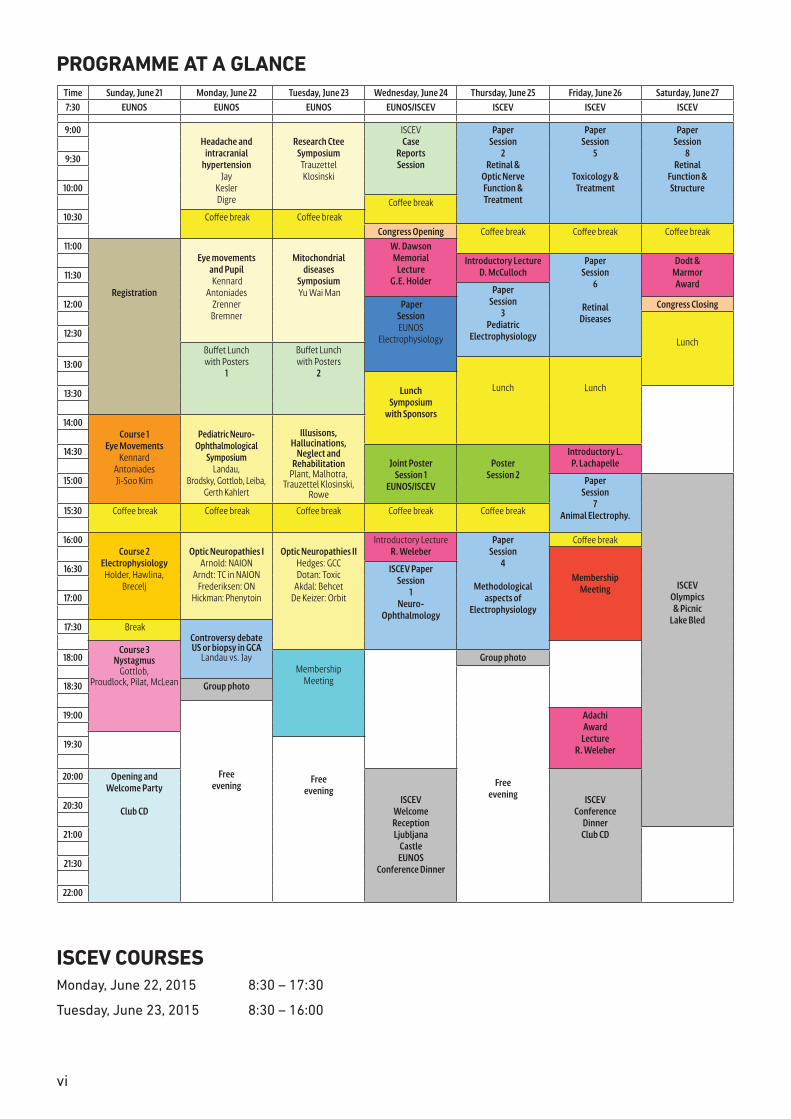
PROGRAMME AT A GLANCETime Sunday, June 21 Monday, June 22 Tuesday, June 23 Wednesday, June 24 Thursday, June 25 Friday, June 26 Saturday, June 277:30 EUNOS EUNOS EUNOS EUNOS/ISCEV ISCEV ISCEV ISCEV
9:00
Headache and
intracranial hypertension
JayKeslerDigre
Research Ctee
Symposium Trauzettel Klosinski
ISCEVCase
ReportsSession
PaperSession
2Retinal &
Optic NerveFunction &Treatment
PaperSession
5
Toxicology &Treatment
PaperSession
8Retinal
Function &Structure
9:30
10:00
Coffee break 10:30 Coffee break Coffee break
Congress Opening Coffee break
Coffee break Coffee break11:00
Registration
Eye movements
and Pupil Kennard
AntoniadesZrennerBremner
Mitochondrial
diseasesSymposium Yu Wai Man
W. DawsonMemorial
LectureG.E. Holder
Introductory Lecture
D. McCullochPaper
Session6
RetinalDiseases
Dodt &MarmorAward
11:30 Paper
Session3
PediatricElectrophysiology
12:00 PaperSessionEUNOS
Electrophysiology
Congress Closing
Lunch
12:30
Buffet Lunchwith Posters
1
Buffet Lunchwith Posters
213:00
Lunch
Lunch
Lunch
Symposiumwith Sponsors
13:30
14:00
Course 1Eye Movements
KennardAntoniadesJi-Soo Kim
Pediatric Neuro-Ophthalmological
SymposiumLandau,
Brodsky, Gottlob, Leiba, Gerth Kahlert
Illusisons,Hallucinations,
Neglect andRehabilitation
Plant, Malhotra,Trauzettel Klosinski,
Rowe
14:30
Joint PosterSession 1
EUNOS/ISCEV
Poster
Session 2
Introductory L.P. Lachapelle
15:00 PaperSession
7Animal Electrophy.
ISCEVOlympics& Picnic
Lake Bled
15:30 Coffee break Coffee break
Coffee break Coffee break
Coffee break
16:00 Course 2
ElectrophysiologyHolder, Hawlina,
Brecelj
Optic Neuropathies IArnold: NAION
Arndt: TC in NAIONFrederiksen: ON
Hickman: Phenytoin
Optic Neuropathies IIHedges: GCCDotan: Toxic
Akdal: BehcetDe Keizer: Orbit
Introductory LectureR. Weleber
PaperSession
4
Methodologicalaspects of
Electrophysiology
Coffee break
Membership
Meeting
16:30 ISCEV PaperSession
1Neuro-
Ophthalmology
17:00
17:30 Break
Controversy debateUS or biopsy in GCA
Landau vs. Jay
Course 3
NystagmusGottlob,
Proudlock, Pilat, McLean
18:00Membership
Meeting
Group photo
Freeevening
18:30 Group photo
Freeevening
19:00 AdachiAward
LectureR. Weleber
19:30
Freeevening
20:00 Opening and
Welcome Party
Club CD
ISCEVWelcomeReceptionLjubljana
CastleEUNOS
Conference Dinner
ISCEVConference
DinnerClub CD
20:30
21:00
21:30
22:00
ISCEV COURSESMonday, June 22, 2015 8:30 – 17:30
Tuesday, June 23, 2015 8:30 – 16:00
vii
ISCEV COURSES TIMETABLE
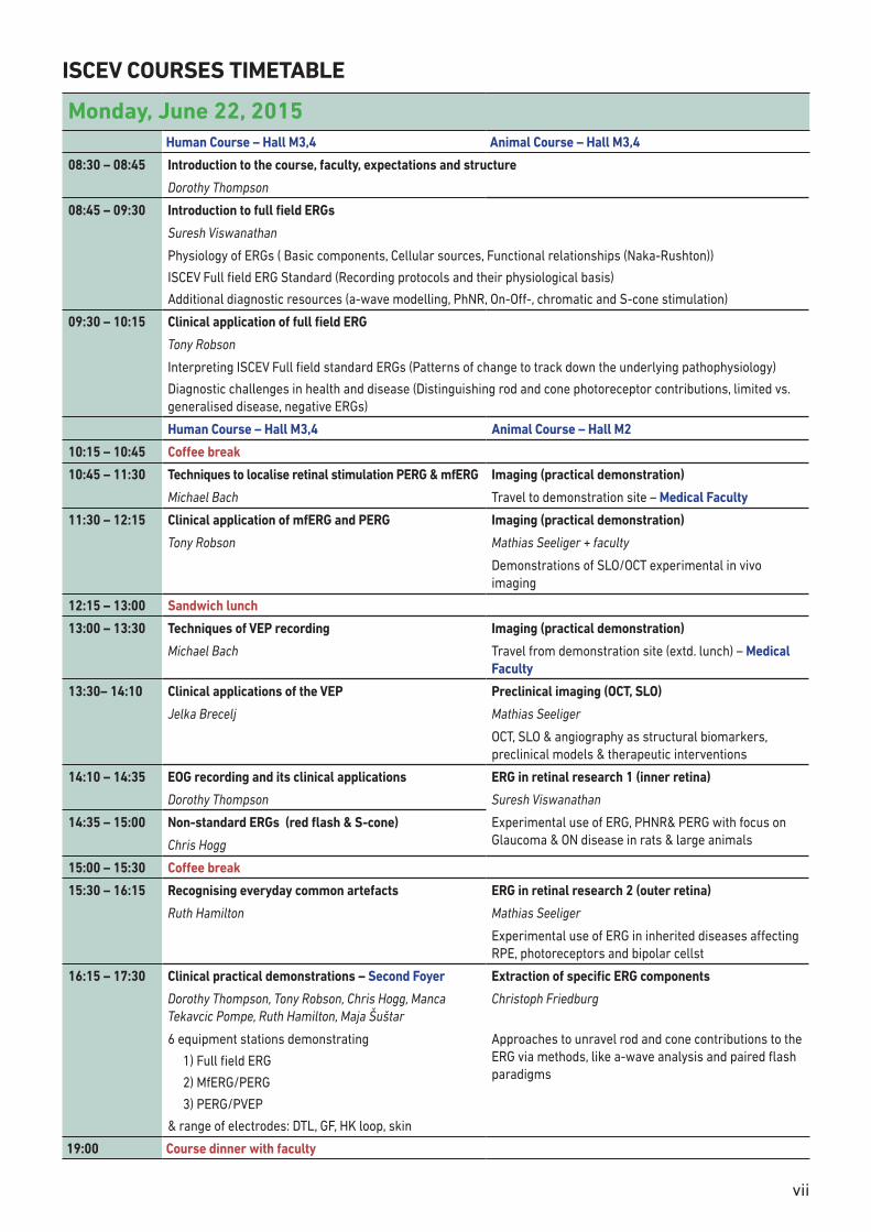
Monday, June 22, 2015Human Course – Hall M3,4 Animal Course – Hall M3,4
08:30 – 08:45 Introduction to the course, faculty, expectations and structure
Dorothy Thompson
08:45 – 09:30 Introduction to full field ERGs
Suresh Viswanathan
Physiology of ERGs ( Basic components, Cellular sources, Functional relationships (Naka-Rushton))
ISCEV Full field ERG Standard (Recording protocols and their physiological basis)
Additional diagnostic resources (a-wave modelling, PhNR, On-Off-, chromatic and S-cone stimulation)
09:30 – 10:15 Clinical application of full field ERG
Tony Robson
Interpreting ISCEV Full field standard ERGs (Patterns of change to track down the underlying pathophysiology)
Diagnostic challenges in health and disease (Distinguishing rod and cone photoreceptor contributions, limited vs. generalised disease, negative ERGs)
Human Course – Hall M3,4 Animal Course – Hall M2
10:15 – 10:45 Coffee break
10:45 – 11:30 Techniques to localise retinal stimulation PERG & mfERG Imaging (practical demonstration)
Michael Bach Travel to demonstration site – Medical Faculty
11:30 – 12:15 Clinical application of mfERG and PERG Imaging (practical demonstration)
Tony Robson Mathias Seeliger + faculty
Demonstrations of SLO/OCT experimental in vivo imaging
12:15 – 13:00 Sandwich lunch
13:00 – 13:30 Techniques of VEP recording Imaging (practical demonstration)
Michael Bach Travel from demonstration site (extd. lunch) – Medical Faculty
13:30– 14:10 Clinical applications of the VEP Preclinical imaging (OCT, SLO)
Jelka Brecelj Mathias Seeliger
OCT, SLO & angiography as structural biomarkers, preclinical models & therapeutic interventions
14:10 – 14:35 EOG recording and its clinical applications ERG in retinal research 1 (inner retina)
Dorothy Thompson Suresh Viswanathan
14:35 – 15:00 Non-standard ERGs (red flash & S-cone) Experimental use of ERG, PHNR& PERG with focus on Glaucoma & ON disease in rats & large animals Chris Hogg
15:00 – 15:30 Coffee break
15:30 – 16:15 Recognising everyday common artefacts ERG in retinal research 2 (outer retina)
Ruth Hamilton Mathias Seeliger
Experimental use of ERG in inherited diseases affecting RPE, photoreceptors and bipolar cellst
16:15 – 17:30 Clinical practical demonstrations – Second Foyer Extraction of specific ERG components
Dorothy Thompson, Tony Robson, Chris Hogg, Manca Tekavcic Pompe, Ruth Hamilton, Maja Šuštar
Christoph Friedburg
6 equipment stations demonstrating
1) Full field ERG
2) MfERG/PERG
3) PERG/PVEP
& range of electrodes: DTL, GF, HK loop, skin
Approaches to unravel rod and cone contributions to the ERG via methods, like a-wave analysis and paired flash paradigms
19:00 Course dinner with faculty
viii
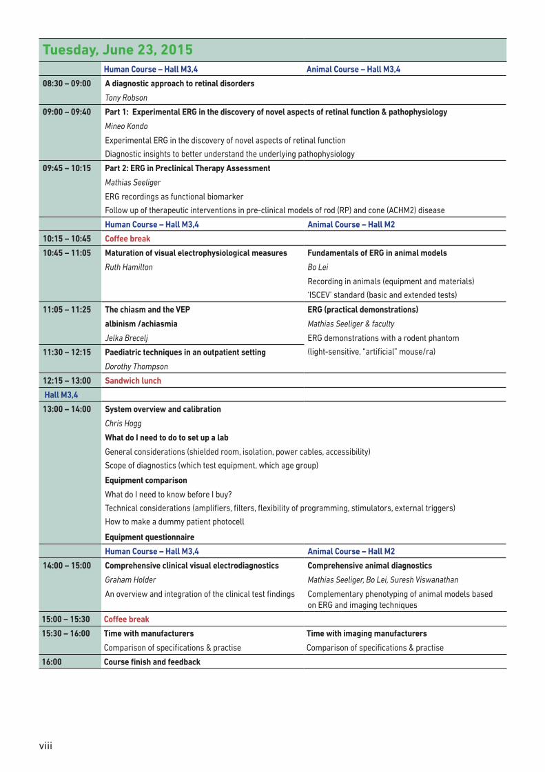
Tuesday, June 23, 2015Human Course – Hall M3,4 Animal Course – Hall M3,4
08:30 – 09:00 A diagnostic approach to retinal disorders
Tony Robson
09:00 – 09:40 Part 1: Experimental ERG in the discovery of novel aspects of retinal function & pathophysiology
Mineo Kondo
Experimental ERG in the discovery of novel aspects of retinal function
Diagnostic insights to better understand the underlying pathophysiology
09:45 – 10:15 Part 2: ERG in Preclinical Therapy Assessment
Mathias Seeliger
ERG recordings as functional biomarker
Follow up of therapeutic interventions in pre-clinical models of rod (RP) and cone (ACHM2) disease
Human Course – Hall M3,4 Animal Course – Hall M2
10:15 – 10:45 Coffee break
10:45 – 11:05 Maturation of visual electrophysiological measures Fundamentals of ERG in animal models
Ruth Hamilton Bo Lei
Recording in animals (equipment and materials)
‘ISCEV’ standard (basic and extended tests)
11:05 – 11:25 The chiasm and the VEP ERG (practical demonstrations)
albinism /achiasmia Mathias Seeliger & faculty
Jelka Brecelj ERG demonstrations with a rodent phantom
(light-sensitive, “artificial” mouse/ra)11:30 – 12:15 Paediatric techniques in an outpatient setting
Dorothy Thompson
12:15 – 13:00 Sandwich lunch
Hall M3,4
13:00 – 14:00 System overview and calibration
Chris Hogg
What do I need to do to set up a lab
General considerations (shielded room, isolation, power cables, accessibility)
Scope of diagnostics (which test equipment, which age group)
Equipment comparison
What do I need to know before I buy?
Technical considerations (amplifiers, filters, flexibility of programming, stimulators, external triggers)
How to make a dummy patient photocell
Equipment questionnaire
Human Course – Hall M3,4 Animal Course – Hall M2
14:00 – 15:00 Comprehensive clinical visual electrodiagnostics Comprehensive animal diagnostics
Graham Holder Mathias Seeliger, Bo Lei, Suresh Viswanathan
An overview and integration of the clinical test findings Complementary phenotyping of animal models based on ERG and imaging techniques
15:00 – 15:30 Coffee break
15:30 – 16:00 Time with manufacturers Time with imaging manufacturers
Comparison of specifications & practise Comparison of specifications & practise
16:00 Course finish and feedback
ix
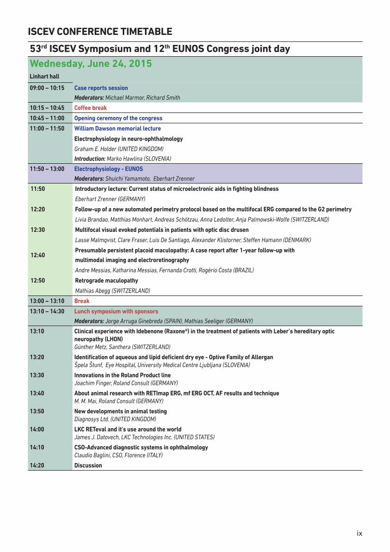
ISCEV CONFERENCE TIMETABLE
53rd ISCEV Symposium and 12th EUNOS Congress joint dayWednesday, June 24, 2015Linhart hall
09:00 – 10:15 Case reports session
Moderators: Michael Marmor, Richard Smith
10:15 – 10:45 Coffee break
10:45 – 11:00 Opening ceremony of the congress
11:00 – 11:50 William Dawson memorial lecture
Electrophysiology in neuro-ophthalmology
Graham E. Holder (UNITED KINGDOM)
Introduction: Marko Hawlina (SLOVENIA)
11:50 – 13:00 Electrophysiology - EUNOS
Moderators: Shuichi Yamamoto, Eberhart Zrenner
11:50 Introductory lecture: Current status of microelectronic aids in fighting blindness
Eberhart Zrenner (GERMANY)
12:20 Follow-up of a new automated perimetry protocol based on the multifocal ERG compared to the G2 perimetry
Livia Brandao, Matthias Monhart, Andreas Schötzau, Anna Ledolter, Anja Palmowski-Wolfe (SWITZERLAND)
12:30 Multifocal visual evoked potentials in patients with optic disc drusen
Lasse Malmqvist, Clare Fraser, Luis De Santiago, Alexander Klistorner, Steffen Hamann (DENMARK)
12:40 Presumable persistent placoid maculopathy: A case report after 1-year follow-up with
multimodal imaging and electroretinography
Andre Messias, Katharina Messias, Fernanda Crotti, Rogério Costa (BRAZIL)
12:50 Retrograde maculopathy
Mathias Abegg (SWITZERLAND)
13:00 – 13:10 Break
13:10 – 14:30 Lunch symposium with sponsors
Moderators: Jorge Arruga Ginebreda (SPAIN), Mathias Seeliger (GERMANY)
13:10 Clinical experience with Idebenone (Raxone®) in the treatment of patients with Leber’s hereditary optic neuropathy (LHON) Günther Metz, Santhera (SWITZERLAND)
13:20 Identification of aqueous and lipid deficient dry eye - Optive Family of Allergan Špela Štunf, Eye Hospital, University Medical Centre Ljubljana (SLOVENIA)
13:30 Innovations in the Roland Product line Joachim Finger, Roland Consult (GERMANY)
13:40 About animal research with RETImap ERG, mf ERG OCT, AF results and technique M. M. Mai, Roland Consult (GERMANY)
13:50 New developments in animal testing Diagnosys Ltd. (UNITED KINGDOM)
14:00 LKC RETeval and it's use around the world James J. Datovech, LKC Technologies Inc. (UNITED STATES)
14:10 CSO-Advanced diagnostic systems in ophthalmology Claudio Baglini, CSO, Florence (ITALY)
14:20 Discussion
x
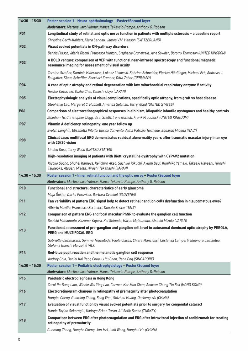
14:30 – 15:30 Poster session 1 - Neuro-ophthalmology - Poster/Second foyer
Moderators: Martina Jarc-Vidmar, Manca Tekavcic-Pompe, Anthony G. Robson
P01 Longitudinal study of retinal and optic nerve function in patients with multiple sclerosis – a baseline report
Christina Gerth-Kahlert, Klara Landau, James V.M. Hanson (SWITZERLAND)
P02 Visual evoked potentials in ON-pathway disorders
Dennis Fritsch, Valeria Ricotti, Francesco Muntoni, Stephanie Grunewald, Jane Sowden, Dorothy Thompson (UNITED KINGDOM)
P03A BOLD venture: comparison of VEP with functional near-infrared spectroscopy and functional magnetic resonance imaging for assessment of visual acuity
Torsten Straßer, Dominic Hillerkuss, Lukasz Lisowski, Sabrina Schneider, Florian Häußinger, Michael Erb, Andreas J. Fallgatter, Klaus Scheffler, Eberhart Zrenner, Ditta Zobor (GERMANY)
P04 A case of optic atrophy and retinal degeneration with low mitochondrial respiratory enzyme V activity
Hiroko Yamazaki, Yuzhu Chai, Yasushi Ooya (JAPAN)
P05 Electrophysiologic analysis of visual complications, specifically optic atrophy, from graft vs host disease
Stephanie Lao, Margaret C. Hubbell, Amanda Selchau, Terry Wood (UNITED STATES)
P06 Comparison of electroretinographical responses in albinism, idiopathic infantile nystagmus and healthy controls
Zhanhan Tu, Christopher Degg, Viral Sheth, Irene Gottlob, Frank Proudlock (UNITED KINGDOM)
P07 Vitamin A deficiency retinopathy: one year follow up
Evelyn Longhin, Elisabetta Pilotto, Enrica Convento, Alma Patrizia Tormene, Edoardo Midena (ITALY)
P08Clinical case: multifocal ERG demonstrates residual abnormality years after traumatic macular injury in an eye with 20/20 vision
Linden Doss, Terry Wood (UNITED STATES)
P09 High-resolution imaging of patients with Bietti crystalline dystrophy with CYP4V2 mutation
Kiyoko Gocho, Shuhei Kameya, Keiichiro Akeo, Sachiko Kikuchi, Ayumi Usui, Kunihiko Yamaki, Takaaki Hayashi, Hiroshi Tsuneoka, Atsushi Mizota, Hiroshi Takahashi (JAPAN)
14:30 – 15:30 Poster session 1 - Inner retinal function and the optic nerve – Poster/Second foyer
Moderators: Martina Jarc-Vidmar, Manca Tekavcic-Pompe, Anthony G. Robson
P10 Functional and structural characteristics of early glaucoma
Maja Šuštar, Darko Perovšek, Barbara Cvenkel (SLOVENIA)
P11 Can variability of pattern ERG signal help to detect retinal ganglion cells dysfunction in glaucomatous eyes?
Alberto Mavilio, Francesca Scrimieri, Donato Errico (ITALY)
P12 Comparison of pattern ERG and focal macular PhNR to evaluate the ganglion cell function
Souichi Matsumoto, Kazuma Yagura, Kei Shinoda, Harue Matsumoto, Atsushi Mizota (JAPAN)
P13Functional assessment of pre-ganglion and ganglion cell level in autosomal dominant optic atrophy by PERGLA, PERG and MULTIFOCAL ERG
Gabriella Cammarata, Gemma Tremolada, Paola Ciasca, Chiara Manclossi, Costanza Lamperti, Eleonora Lamantea, Stefania Bianchi Marzoli (ITALY)
P14 Red-blue pupil reaction and the melanotic ganglion cell response
Audrey Chia, Daniel Kai Peng Chua, Li Yu Chen, Rena Png (SINGAPORE)
14:30 – 15:30 Poster session 1 – Pediatric electrophysiology – Poster/Second foyer
Moderators: Martina Jarc-Vidmar, Manca Tekavcic-Pompe, Anthony G. Robson
P15 Paediatric electrodiagnosis in Hong Kong
Carol Po-Sang Lam, Winnie Wai Ying Lau, Carmen Kar Mun Chan, Andrew Chung Tin Fok (HONG KONG)
P16 Electroretinogram changes in retinopathy of prematurity after photocoagulation
Hongbo Cheng, Guoming Zhang, Feng Wen, Shizhou Huang, Dezheng Wu (CHINA)
P17 Evaluation of visual function by visual evoked potentials prior to surgery for congenital cataract
Hande Taylan Sekeroglu, Kadriye Erkan Turan, Ali Sefik Sanac (TURKEY)
P18Comparison between ERG after photocoagulation and ERG after intravitreal injection of ranibizumab for treating retinopathy of prematurity
Guoming Zhang, Hongbo Cheng, Jun Mei, Linli Wang, Honghui He (CHINA)
xi
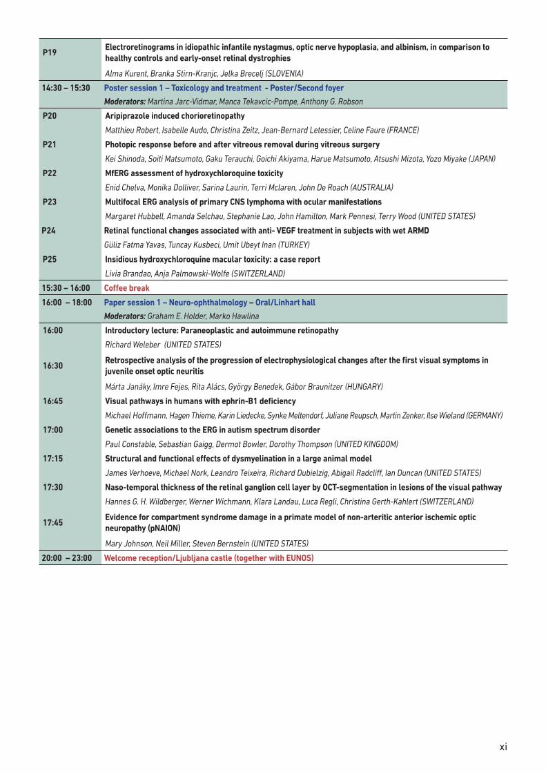
P19Electroretinograms in idiopathic infantile nystagmus, optic nerve hypoplasia, and albinism, in comparison to healthy controls and early-onset retinal dystrophies
Alma Kurent, Branka Stirn-Kranjc, Jelka Brecelj (SLOVENIA)
14:30 – 15:30 Poster session 1 – Toxicology and treatment - Poster/Second foyer
Moderators: Martina Jarc-Vidmar, Manca Tekavcic-Pompe, Anthony G. Robson
P20 Aripiprazole induced chorioretinopathy
Matthieu Robert, Isabelle Audo, Christina Zeitz, Jean-Bernard Letessier, Celine Faure (FRANCE)
P21 Photopic response before and after vitreous removal during vitreous surgery
Kei Shinoda, Soiti Matsumoto, Gaku Terauchi, Goichi Akiyama, Harue Matsumoto, Atsushi Mizota, Yozo Miyake (JAPAN)
P22 MfERG assessment of hydroxychloroquine toxicity
Enid Chelva, Monika Dolliver, Sarina Laurin, Terri Mclaren, John De Roach (AUSTRALIA)
P23 Multifocal ERG analysis of primary CNS lymphoma with ocular manifestations
Margaret Hubbell, Amanda Selchau, Stephanie Lao, John Hamilton, Mark Pennesi, Terry Wood (UNITED STATES)
P24 Retinal functional changes associated with anti- VEGF treatment in subjects with wet ARMD
Güliz Fatma Yavas, Tuncay Kusbeci, Umit Ubeyt Inan (TURKEY)
P25 Insidious hydroxychloroquine macular toxicity: a case report
Livia Brandao, Anja Palmowski-Wolfe (SWITZERLAND)
15:30 – 16:00 Coffee break
16:00 – 18:00 Paper session 1 – Neuro-ophthalmology – Oral/Linhart hall
Moderators: Graham E. Holder, Marko Hawlina
16:00 Introductory lecture: Paraneoplastic and autoimmune retinopathy
Richard Weleber (UNITED STATES)
16:30Retrospective analysis of the progression of electrophysiological changes after the first visual symptoms in juvenile onset optic neuritis
Márta Janáky, Imre Fejes, Rita Alács, György Benedek, Gábor Braunitzer (HUNGARY)
16:45 Visual pathways in humans with ephrin-B1 deficiency
Michael Hoffmann, Hagen Thieme, Karin Liedecke, Synke Meltendorf, Juliane Reupsch, Martin Zenker, Ilse Wieland (GERMANY)
17:00 Genetic associations to the ERG in autism spectrum disorder
Paul Constable, Sebastian Gaigg, Dermot Bowler, Dorothy Thompson (UNITED KINGDOM)
17:15 Structural and functional effects of dysmyelination in a large animal model
James Verhoeve, Michael Nork, Leandro Teixeira, Richard Dubielzig, Abigail Radcliff, Ian Duncan (UNITED STATES)
17:30 Naso-temporal thickness of the retinal ganglion cell layer by OCT-segmentation in lesions of the visual pathway
Hannes G. H. Wildberger, Werner Wichmann, Klara Landau, Luca Regli, Christina Gerth-Kahlert (SWITZERLAND)
17:45Evidence for compartment syndrome damage in a primate model of non-arteritic anterior ischemic optic neuropathy (pNAION)
Mary Johnson, Neil Miller, Steven Bernstein (UNITED STATES)
20:00 – 23:00 Welcome reception/Ljubljana castle (together with EUNOS)

xii
EUNOS Poster session 3Electrophysiology, toxic states, methods (Second foyer) Moderators: Gabriella Szatmary (UNITED STATES), Jan Willem Pott (NETHERLANDS), Anja Palmowski-Wolfe (SWITZERLAND)
PE094 Comparison of electroretinographical responses in albinism, idiopathic infantile nystagmus and healthy controls Zhanhan Tu, Christopher Degg, Viral Sheth, Irene Gottlob, Frank Proudlock (UNITED KINGDOM)
PE095 Retinal ganglion cell volume correlates with pattern electroretinogram in acute optic neuritis Justin Mckee, Charles Cottriall, John Elston, Lars Fugger, Chris Kennard, Nikos Evangelou, Jacqueline Palace, Matthew Craner (UNITED KINGDOM)
PE96 Methanol toxicity: Bangladesh perspective Sarwat Rahman (BANGLADESH)
PE97 Ketamine affects single vesicle exocytosis in cultured rat astrocytes Eva Lasič, Matjaž Stenovec, Mićo Božić, Saša Trkov, Marko Kreft, Vladimir Grubišić, Vladimir Parpura, Robert Zorec (SLOVENIA)
PE98 Tamoxifen-induced optic neuropathy or NAION? Idit Maharshak (ISRAEL)
PE99 Intracellular cAMP and Ca2+ signalling in cultured rat astrocytes Anemari Horvat, Nina Vardjan, Robert Zorec (SLOVENIA)
PE100 Low vision in multiple sclerosis Andreia Soares, João Cerqueira, Cristina Almeida, Fernando Vaz (PORTUGAL)
PE101 Novel update 2015: It is more than a library Kathleen Digre, Nancy Lombardo, Chrisy Jarvis, Valerie Biousse, Aki Kawasaki (UNITED STATES)
PE102 Use of non-mydriatic wide-field retinography for periphlebitis detection in patients with multiple sclerosis Ruben Torres-Torres, Elena Fraga-Pumar, Elena H. Martínez-Lapiscina, Pablo Villoslada, Bernardo F. Sanchez-Dalmau (SPAIN)
PE103 Visual dysfunction induced by cobalt toxicity Bernardo Sanchez Dalmau, Amélia Maria De Carvalho, Cristina Nieto, Christian Fontecilla, Ruben Torres Torres, Santiago Nogués, Jenaro Angel Fernández-Valencia (SPAIN)
PE104 Quantification of retinal ganglion cell loss detected by spectral domain optical coherence tomography in patients with stroke Stefania Bianchi Marzoli, Lisa Melzi, Ciasca Paola, Gabriella Cammarata, Carlotta Casati, Giuseppe Vallar, Nadia Bolognini (ITALY)
PE105 Abnormal optic nerve head morphology in albinism imaged using high resolution spectral domain optical coherence tomography Frank Proudlock, Sarim Mohammad, Viral Sheth, Anastasia Pilat, Helena Lee, Ellen Pollheimer, Irene Gottlob (UNITED KINGDOM)
PE106 High resolution imaging of the optic nerve and retina in optic nerve hypoplasia Anastasia Pilat, Daniel Sibley, Rebecca McLean, Frank Proudlock, Irene Gottlob (UNITED KINGDOM)
PE107 A prospective study on the prevalence of microcystic macular changes on optical coherence tomography of the macular region in optic nerve atrophy of non-neuritis origin Jan Willem Pott, Willemien De Vries, Axel Petzold (NETHERLANDS)
PE108 Analysis of the macular ganglion cell layer of patients with vascular lesions of the posterior pathway Rita Anjos, André Vicente, Livio Costa, Arnaldo Santos, Duarte Amado, Joana Ferreira, João Paulo Cunha (PORTUGAL)
PE109 Unusual acquired temporal sectoral scotomas M.J. Gilhooley, Clare Fraser, Gordon T. Plant (UNITED KINGDOM)
PE110 The role of B-cells in early onset acute optic neuritis Ruth Huna-Baron, Anna Feldman, Michael Gurevich, Anat Achiron (ISRAEL)
PE111 Unilateral optic neuritis and venous sinus thrombosis related to myelin-oligodendrocyte glycoprotein antibodies: A case report Maribel Acuna Salles, Pedro Pascual Ruiz, Laura Cabrejas Martínez, Miguel Ángel Alonso Peralta, Viviana Lezcano Carduz, Cristina Molero Langa (SPAIN)
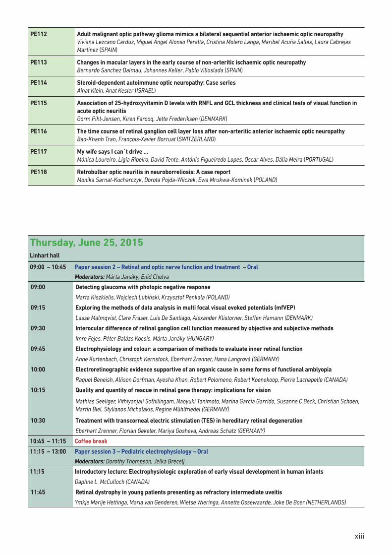
xiii
PE112 Adult malignant optic pathway glioma mimics a bilateral sequential anterior ischaemic optic neuropathy Viviana Lezcano Carduz, Miguel Ángel Alonso Peralta, Cristina Molero Langa, Maribel Acuña Salles, Laura Cabrejas Martinez (SPAIN)
PE113 Changes in macular layers in the early course of non-arteritic ischaemic optic neuropathy Bernardo Sanchez Dalmau, Johannes Keller, Pablo Villoslada (SPAIN)
PE114 Steroid-dependent autoimmune optic neuropathy: Case series Ainat Klein, Anat Kesler (ISRAEL)
PE115 Association of 25-hydroxyvitamin D levels with RNFL and GCL thickness and clinical tests of visual function in acute optic neuritis Gorm Pihl-Jensen, Kiren Farooq, Jette Frederiksen (DENMARK)
PE116 The time course of retinal ganglion cell layer loss after non-arteritic anterior ischaemic optic neuropathy Bao-Khanh Tran, François-Xavier Borruat (SWITZERLAND)
PE117 My wife says I can´t drive … Mónica Loureiro, Lígia Ribeiro, David Tente, António Figueiredo Lopes, Óscar Alves, Dália Meira (PORTUGAL)
PE118 Retrobulbar optic neuritis in neuroborreliosis: A case report Monika Sarnat-Kucharczyk, Dorota Pojda-Wilczek, Ewa Mrukwa-Kominek (POLAND)
Thursday, June 25, 2015Linhart hall
09:00 – 10:45 Paper session 2 – Retinal and optic nerve function and treatment – Oral
Moderators: Márta Janáky, Enid Chelva
09:00 Detecting glaucoma with photopic negative response
Marta Kiszkielis, Wojciech Lubiński, Krzysztof Penkala (POLAND)
09:15 Exploring the methods of data analysis in multi focal visual evoked potentials (mfVEP)
Lasse Malmqvist, Clare Fraser, Luis De Santiago, Alexander Klistorner, Steffen Hamann (DENMARK)
09:30 Interocular difference of retinal ganglion cell function measured by objective and subjective methods
Imre Fejes, Péter Balázs Kocsis, Márta Janáky (HUNGARY)
09:45 Electrophysiology and colour: a comparison of methods to evaluate inner retinal function
Anne Kurtenbach, Christoph Kernstock, Eberhart Zrenner, Hana Langrová (GERMANY)
10:00 Electroretinographic evidence supportive of an organic cause in some forms of functional amblyopia
Raquel Beneish, Allison Dorfman, Ayesha Khan, Robert Polomeno, Robert Koenekoop, Pierre Lachapelle (CANADA)
10:15 Quality and quantity of rescue in retinal gene therapy: implications for vision
Mathias Seeliger, Vithiyanjali Sothilingam, Naoyuki Tanimoto, Marina Garcia Garrido, Susanne C Beck, Christian Schoen, Martin Biel, Stylianos Michalakis, Regine Mühlfriedel (GERMANY)
10:30 Treatment with transcorneal electric stimulation (TES) in hereditary retinal degeneration
Eberhart Zrenner, Florian Gekeler, Mariya Gosheva, Andreas Schatz (GERMANY)
10:45 – 11:15 Coffee break
11:15 – 13:00 Paper session 3 – Pediatric electrophysiology – Oral
Moderators: Dorothy Thompson, Jelka Brecelj
11:15 Introductory lecture: Electrophysiologic exploration of early visual development in human infants
Daphne L. McCulloch (CANADA)
11:45 Retinal dystrophy in young patients presenting as refractory intermediate uveitis
Ymkje Marije Hettinga, Maria van Genderen, Wietse Wieringa, Annette Ossewaarde, Joke De Boer (NETHERLANDS)
xiv
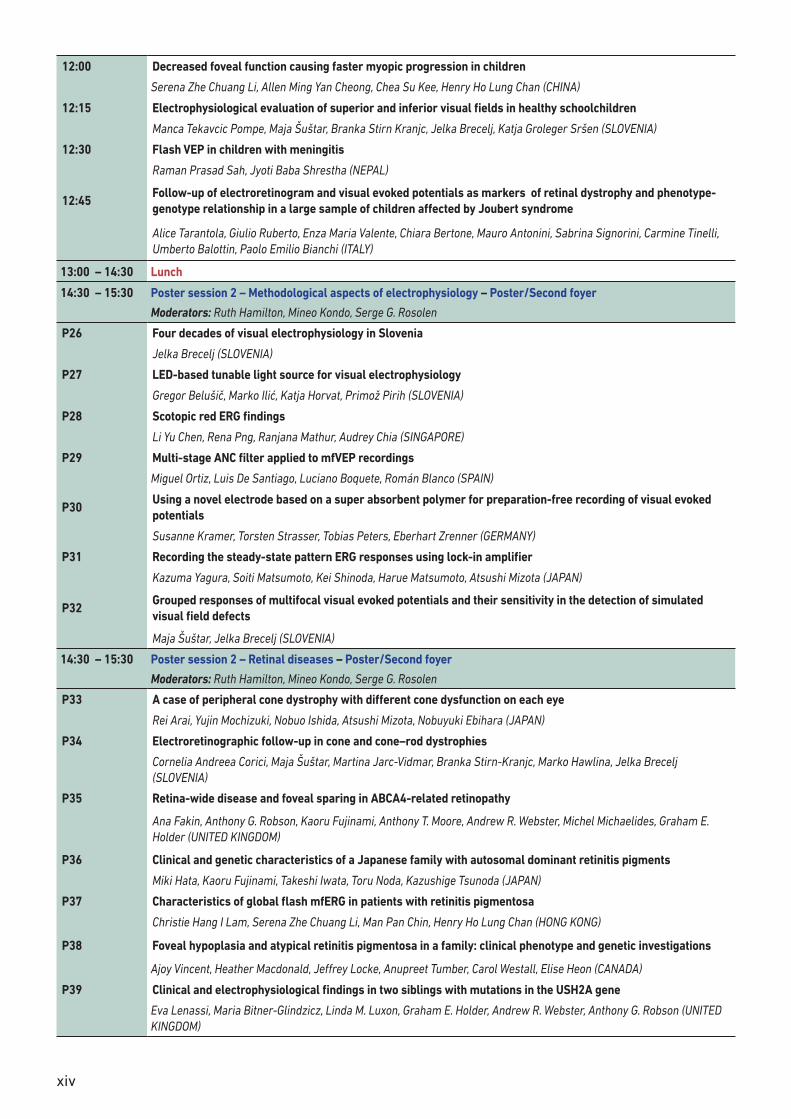
12:00 Decreased foveal function causing faster myopic progression in children
Serena Zhe Chuang Li, Allen Ming Yan Cheong, Chea Su Kee, Henry Ho Lung Chan (CHINA)
12:15 Electrophysiological evaluation of superior and inferior visual fields in healthy schoolchildren
Manca Tekavcic Pompe, Maja Šuštar, Branka Stirn Kranjc, Jelka Brecelj, Katja Groleger Sršen (SLOVENIA)
12:30 Flash VEP in children with meningitis
Raman Prasad Sah, Jyoti Baba Shrestha (NEPAL)
12:45Follow-up of electroretinogram and visual evoked potentials as markers of retinal dystrophy and phenotype-genotype relationship in a large sample of children affected by Joubert syndrome
Alice Tarantola, Giulio Ruberto, Enza Maria Valente, Chiara Bertone, Mauro Antonini, Sabrina Signorini, Carmine Tinelli, Umberto Balottin, Paolo Emilio Bianchi (ITALY)
13:00 – 14:30 Lunch
14:30 – 15:30 Poster session 2 – Methodological aspects of electrophysiology – Poster/Second foyer
Moderators: Ruth Hamilton, Mineo Kondo, Serge G. Rosolen
P26 Four decades of visual electrophysiology in Slovenia
Jelka Brecelj (SLOVENIA)
P27 LED-based tunable light source for visual electrophysiology
Gregor Belušič, Marko Ilić, Katja Horvat, Primož Pirih (SLOVENIA)
P28 Scotopic red ERG findings
Li Yu Chen, Rena Png, Ranjana Mathur, Audrey Chia (SINGAPORE)
P29 Multi-stage ANC filter applied to mfVEP recordings
Miguel Ortiz, Luis De Santiago, Luciano Boquete, Román Blanco (SPAIN)
P30Using a novel electrode based on a super absorbent polymer for preparation-free recording of visual evoked potentials
Susanne Kramer, Torsten Strasser, Tobias Peters, Eberhart Zrenner (GERMANY)
P31 Recording the steady-state pattern ERG responses using lock-in amplifier
Kazuma Yagura, Soiti Matsumoto, Kei Shinoda, Harue Matsumoto, Atsushi Mizota (JAPAN)
P32Grouped responses of multifocal visual evoked potentials and their sensitivity in the detection of simulated visual field defects
Maja Šuštar, Jelka Brecelj (SLOVENIA)
14:30 – 15:30 Poster session 2 – Retinal diseases – Poster/Second foyer
Moderators: Ruth Hamilton, Mineo Kondo, Serge G. Rosolen
P33 A case of peripheral cone dystrophy with different cone dysfunction on each eye
Rei Arai, Yujin Mochizuki, Nobuo Ishida, Atsushi Mizota, Nobuyuki Ebihara (JAPAN)
P34 Electroretinographic follow-up in cone and cone–rod dystrophies
Cornelia Andreea Corici, Maja Šuštar, Martina Jarc-Vidmar, Branka Stirn-Kranjc, Marko Hawlina, Jelka Brecelj (SLOVENIA)
P35 Retina-wide disease and foveal sparing in ABCA4-related retinopathy
Ana Fakin, Anthony G. Robson, Kaoru Fujinami, Anthony T. Moore, Andrew R. Webster, Michel Michaelides, Graham E. Holder (UNITED KINGDOM)
P36 Clinical and genetic characteristics of a Japanese family with autosomal dominant retinitis pigments
Miki Hata, Kaoru Fujinami, Takeshi Iwata, Toru Noda, Kazushige Tsunoda (JAPAN)
P37 Characteristics of global flash mfERG in patients with retinitis pigmentosa
Christie Hang I Lam, Serena Zhe Chuang Li, Man Pan Chin, Henry Ho Lung Chan (HONG KONG)
P38 Foveal hypoplasia and atypical retinitis pigmentosa in a family: clinical phenotype and genetic investigations
Ajoy Vincent, Heather Macdonald, Jeffrey Locke, Anupreet Tumber, Carol Westall, Elise Heon (CANADA)
P39 Clinical and electrophysiological findings in two siblings with mutations in the USH2A gene
Eva Lenassi, Maria Bitner-Glindzicz, Linda M. Luxon, Graham E. Holder, Andrew R. Webster, Anthony G. Robson (UNITED KINGDOM)
xv
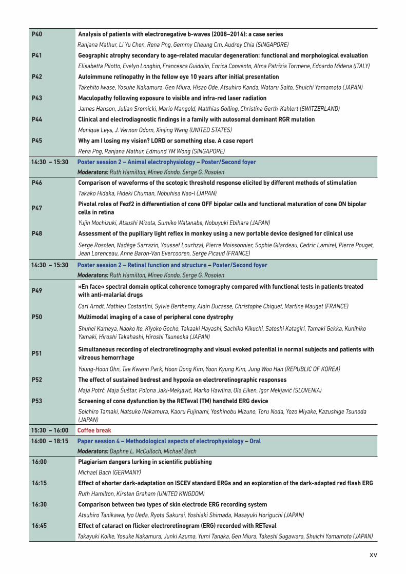
P40 Analysis of patients with electronegative b-waves (2008–2014): a case series
Ranjana Mathur, Li Yu Chen, Rena Png, Gemmy Cheung Cm, Audrey Chia (SINGAPORE)
P41 Geographic atrophy secondary to age-related macular degeneration: functional and morphological evaluation
Elisabetta Pilotto, Evelyn Longhin, Francesca Guidolin, Enrica Convento, Alma Patrizia Tormene, Edoardo Midena (ITALY)
P42 Autoimmune retinopathy in the fellow eye 10 years after initial presentation
Takehito Iwase, Yosuhe Nakamura, Gen Miura, Hisao Ode, Atsuhiro Kanda, Wataru Saito, Shuichi Yamamoto (JAPAN)
P43 Maculopathy following exposure to visible and infra-red laser radiation
James Hanson, Julian Sromicki, Mario Mangold, Matthias Golling, Christina Gerth-Kahlert (SWITZERLAND)
P44 Clinical and electrodiagnostic findings in a family with autosomal dominant RGR mutation
Monique Leys, J. Vernon Odom, Xinjing Wang (UNITED STATES)
P45 Why am I losing my vision? LORD or something else. A case report
Rena Png, Ranjana Mathur, Edmund YM Wong (SINGAPORE)
14:30 – 15:30 Poster session 2 – Animal electrophysiology – Poster/Second foyer
Moderators: Ruth Hamilton, Mineo Kondo, Serge G. Rosolen
P46 Comparison of waveforms of the scotopic threshold response elicited by different methods of stimulation
Takako Hidaka, Hideki Chuman, Nobuhisa Nao-I (JAPAN)
P47Pivotal roles of Fezf2 in differentiation of cone OFF bipolar cells and functional maturation of cone ON bipolar cells in retina
Yujin Mochizuki, Atsushi Mizota, Sumiko Watanabe, Nobuyuki Ebihara (JAPAN)
P48 Assessment of the pupillary light reflex in monkey using a new portable device designed for clinical use
Serge Rosolen, Nadège Sarrazin, Youssef Lourhzal, Pierre Moissonnier, Sophie Gilardeau, Cedric Lamirel, Pierre Pouget, Jean Lorenceau, Anne Baron-Van Evercooren, Serge Picaud (FRANCE)
14:30 – 15:30 Poster session 2 – Retinal function and structure – Poster/Second foyer
Moderators: Ruth Hamilton, Mineo Kondo, Serge G. Rosolen
P49»En face« spectral domain optical coherence tomography compared with functional tests in patients treated with anti-malarial drugs
Carl Arndt, Mathieu Costantini, Sylvie Berthemy, Alain Ducasse, Christophe Chiquet, Martine Mauget (FRANCE)
P50 Multimodal imaging of a case of peripheral cone dystrophy
Shuhei Kameya, Naoko Ito, Kiyoko Gocho, Takaaki Hayashi, Sachiko Kikuchi, Satoshi Katagiri, Tamaki Gekka, Kunihiko Yamaki, Hiroshi Takahashi, Hiroshi Tsuneoka (JAPAN)
P51Simultaneous recording of electroretinography and visual evoked potential in normal subjects and patients with vitreous hemorrhage
Young-Hoon Ohn, Tae Kwann Park, Hoon Dong Kim, Yoon Kyung Kim, Jung Woo Han (REPUBLIC OF KOREA)
P52 The effect of sustained bedrest and hypoxia on electroretinographic responses
Maja Potrč, Maja Šuštar, Polona Jaki-Mekjavić, Marko Hawlina, Ola Eiken, Igor Mekjavić (SLOVENIA)
P53 Screening of cone dysfunction by the RETeval (TM) handheld ERG device
Soichiro Tamaki, Natsuko Nakamura, Kaoru Fujinami, Yoshinobu Mizuno, Toru Noda, Yozo Miyake, Kazushige Tsunoda (JAPAN)
15:30 – 16:00 Coffee break
16:00 – 18:15 Paper session 4 – Methodological aspects of electrophysiology – Oral
Moderators: Daphne L. McCulloch, Michael Bach
16:00 Plagiarism dangers lurking in scientific publishing
Michael Bach (GERMANY)
16:15 Effect of shorter dark-adaptation on ISCEV standard ERGs and an exploration of the dark-adapted red flash ERG
Ruth Hamilton, Kirsten Graham (UNITED KINGDOM)
16:30 Comparison between two types of skin electrode ERG recording system
Atsuhiro Tanikawa, Iyo Ueda, Ryota Sakurai, Yoshiaki Shimada, Masayuki Horiguchi (JAPAN)
16:45 Effect of cataract on flicker electroretinogram (ERG) recorded with RETeval
Takayuki Koike, Yosuke Nakamura, Junki Azuma, Yumi Tanaka, Gen Miura, Takeshi Sugawara, Shuichi Yamamoto (JAPAN)
xvi
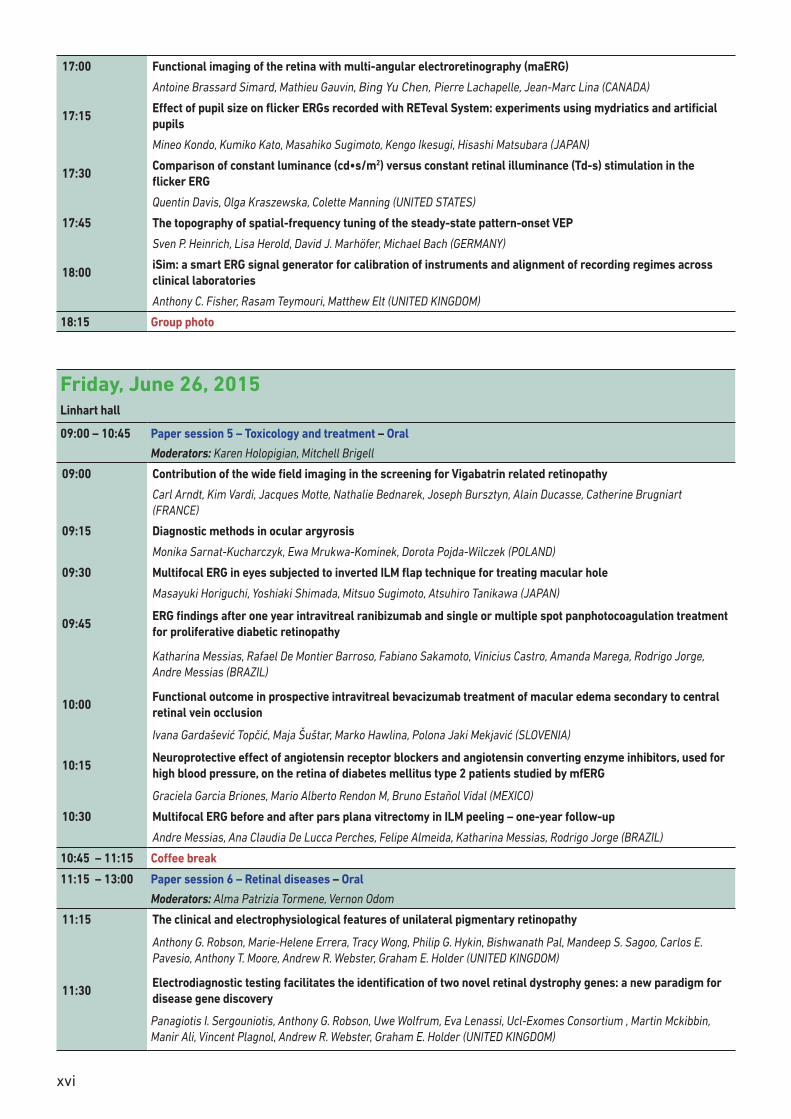
17:00 Functional imaging of the retina with multi-angular electroretinography (maERG)
Antoine Brassard Simard, Mathieu Gauvin, Bing Yu Chen, Pierre Lachapelle, Jean-Marc Lina (CANADA)
17:15Effect of pupil size on flicker ERGs recorded with RETeval System: experiments using mydriatics and artificial pupils
Mineo Kondo, Kumiko Kato, Masahiko Sugimoto, Kengo Ikesugi, Hisashi Matsubara (JAPAN)
17:30Comparison of constant luminance (cd•s/m2) versus constant retinal illuminance (Td-s) stimulation in the flicker ERG
Quentin Davis, Olga Kraszewska, Colette Manning (UNITED STATES)
17:45 The topography of spatial-frequency tuning of the steady-state pattern-onset VEP
Sven P. Heinrich, Lisa Herold, David J. Marhöfer, Michael Bach (GERMANY)
18:00iSim: a smart ERG signal generator for calibration of instruments and alignment of recording regimes across clinical laboratories
Anthony C. Fisher, Rasam Teymouri, Matthew Elt (UNITED KINGDOM)
18:15 Group photo
Friday, June 26, 2015Linhart hall
09:00 – 10:45 Paper session 5 – Toxicology and treatment – Oral
Moderators: Karen Holopigian, Mitchell Brigell
09:00 Contribution of the wide field imaging in the screening for Vigabatrin related retinopathy
Carl Arndt, Kim Vardi, Jacques Motte, Nathalie Bednarek, Joseph Bursztyn, Alain Ducasse, Catherine Brugniart (FRANCE)
09:15 Diagnostic methods in ocular argyrosis
Monika Sarnat-Kucharczyk, Ewa Mrukwa-Kominek, Dorota Pojda-Wilczek (POLAND)
09:30 Multifocal ERG in eyes subjected to inverted ILM flap technique for treating macular hole
Masayuki Horiguchi, Yoshiaki Shimada, Mitsuo Sugimoto, Atsuhiro Tanikawa (JAPAN)
09:45ERG findings after one year intravitreal ranibizumab and single or multiple spot panphotocoagulation treatment for proliferative diabetic retinopathy
Katharina Messias, Rafael De Montier Barroso, Fabiano Sakamoto, Vinicius Castro, Amanda Marega, Rodrigo Jorge, Andre Messias (BRAZIL)
10:00Functional outcome in prospective intravitreal bevacizumab treatment of macular edema secondary to central retinal vein occlusion
Ivana Gardašević Topčić, Maja Šuštar, Marko Hawlina, Polona Jaki Mekjavić (SLOVENIA)
10:15Neuroprotective effect of angiotensin receptor blockers and angiotensin converting enzyme inhibitors, used for high blood pressure, on the retina of diabetes mellitus type 2 patients studied by mfERG
Graciela Garcia Briones, Mario Alberto Rendon M, Bruno Estañol Vidal (MEXICO)
10:30 Multifocal ERG before and after pars plana vitrectomy in ILM peeling – one-year follow-up
Andre Messias, Ana Claudia De Lucca Perches, Felipe Almeida, Katharina Messias, Rodrigo Jorge (BRAZIL)
10:45 – 11:15 Coffee break
11:15 – 13:00 Paper session 6 – Retinal diseases – Oral
Moderators: Alma Patrizia Tormene, Vernon Odom
11:15 The clinical and electrophysiological features of unilateral pigmentary retinopathy
Anthony G. Robson, Marie-Helene Errera, Tracy Wong, Philip G. Hykin, Bishwanath Pal, Mandeep S. Sagoo, Carlos E. Pavesio, Anthony T. Moore, Andrew R. Webster, Graham E. Holder (UNITED KINGDOM)
11:30Electrodiagnostic testing facilitates the identification of two novel retinal dystrophy genes: a new paradigm for disease gene discovery
Panagiotis I. Sergouniotis, Anthony G. Robson, Uwe Wolfrum, Eva Lenassi, Ucl-Exomes Consortium , Martin Mckibbin, Manir Ali, Vincent Plagnol, Andrew R. Webster, Graham E. Holder (UNITED KINGDOM)
xvii
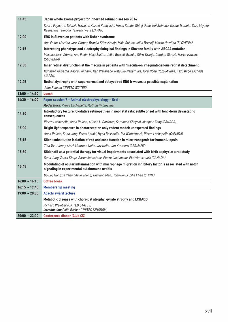
11:45 Japan whole exome project for inherited retinal diseases 2014
Kaoru Fujinami, Takaaki Hayashi, Kazuki Kuniyoshi, Mineo Kondo, Shinji Ueno, Kei Shinoda, Kazuo Tsubota, Yozo Miyake, Kazushige Tsunoda, Takeshi Iwata (JAPAN)
12:00 ERG in Slovenian patients with Usher syndrome
Ana Fakin, Martina Jarc-Vidmar, Branka Stirn Kranjc, Maja Šuštar, Jelka Brecelj, Marko Hawlina (SLOVENIA)
12:15 Interesting phenotype and electrophysiological findings in Slovene family with ABCA4 mutation
Martina Jarc-Vidmar, Ana Fakin, Maja Šuštar, Jelka Brecelj, Branka Stirn-Kranjc, Damjan Glavač, Marko Hawlina (SLOVENIA)
12:30 Inner retinal dysfunction at the macula in patients with ‘macula-on’ rhegmatogenous retinal detachment
Kunihiko Akiyama, Kaoru Fujinami, Ken Watanabe, Natsuko Nakamura, Toru Noda, Yozo Miyake, Kazushige Tsunoda (JAPAN)
12:45 Retinal dystrophy with supernormal and delayed rod ERG b-waves: a possible explanation
John Robson (UNITED STATES)
13:00 – 14:30 Lunch
14:30 – 16:00 Paper session 7 – Animal electrophysiology – Oral
Moderators: Pierre Lachapelle, Mathias W. Seeliger
14:30Introductory lecture: Oxidative retinopathies in neonatal rats: subtle onset with long-term devastating consequences
Pierre Lachapelle, Anna Polosa, Allison L. Dorfman, Samaneh Chaychi, Xiaojuan Yang (CANADA)
15:00 Bright light exposure in photoreceptor-only rodent model: unexpected findings
Anna Polosa, Suna Jung, Fares Antaki, Hyba Bessaklia, Pia Wintermark, Pierre Lachapelle (CANADA)
15:15 Silent substitution isolation of rod and cone function in mice transgenic for human L-opsin
Tina Tsai, Jenny Atorf, Maureen Neitz, Jay Neitz, Jan Kremers (GERMANY)
15:30 Sildenafil as a potential therapy for visual impairments associated with birth asphyxia: a rat study
Suna Jung, Zehra Khoja, Aaron Johnstone, Pierre Lachapelle, Pia Wintermark (CANADA)
15:45Modulating of ocular inflammation with macrophage migration inhibitory factor is associated with notch signaling in experimental autoimmune uveitis
Bo Lei, Hongxia Yang, Shijie Zheng, Yingying Mao, Hongwei Li, Zihe Chen (CHINA)
16:00 – 16:15 Coffee break
16:15 – 17:45 Membership meeting
19:00 – 20:00 Adachi award lecture
Metabolic disease with choroidal atrophy: gyrate atrophy and LCHADD
Richard Weleber (UNITED STATES) Introduction: Colin Barber (UNITED KINGDOM)
20:00 – 23:00 Conference dinner (Club CD)
xviii
Saturday, June 27, 2015Linhart hall
09:00 – 10:45 Paper session 8 – Retinal function and structure – Oral
Moderators: Mary Johnson, Audrey Chia Wei Lin
09:00 Evidence for an asymmetrical effect of type 1 diabetes on retinal structure and function
Thomas Wright, Alan Poon, Ahmed Salem, Carol Westall (CANADA)
09:15 Comparing the retinal structure, function and biochemistry in a animal model of spontaneous diabetes
Serge Rosolen, Sylvie Lavillegrand, Stephanie Lafarge-Beurlet, Pierre-Francois Isard, Charles Cassagnes, Thomas Dulaurent, Serge Picaud, Patricia Crissanti, Marianne Berdugo (FRANCE)
09:30Assessment of macular function and structure in patients with idiopathic epiretinal membrane treated by pars plana vitrectomy
Wojciech Lubiński, Wojciech Gosławski, Leszek Kuprjanowicz, Karol Krzystolik, Maciej Mularczyk (POLAND)
09:45 Unmasking ERG ON-OFF interactions with the discrete wavelet transform (DWT)
Mathieu Gauvin, Maja Šuštar, Jean-Marc Lina, John M. Little, Robert K. Koenekoop, Pierre Lachapelle (CANADA)
10:00 Does flicker ERG depend on stimulus color?
Olga Kraszewska, Quentin Davis, Colette Manning (UNITED STATES)
10:15 Correlation between macular cone density and focal macular electroretinogram in normal eyes
Hiroko Terasaki, Kennichi Kawano, Yasuki Ito, Taro Kominami, Shinji Ueno (JAPAN)
10:30 When function, structure and electrophysiology don’t agree
Michael Marmor (UNITED STATES)
10:45 – 11:15 Coffee break
11:15 – 12:00 Dodt and Marmor award
12:00 – 12:10 Closing ceremony
12:15 – 13:30 Lunch
15:00 – 21:00 ISCEV Olympics and picnic at lake Bled
xix
INFORMATION FOR SPEAKERS AND POSTER PRESENTERSPoster presentationsPosters will be displayed in Foyer II. Display boards will be numbered. Presenting authors are requested to be available for discussion next to their posters during the scheduled time. Posters will be displayed all the time during the congress. Cankarjev dom is not responsible for any poster left behind at the end of the day.
Speakers’ CentreThe technical organizer will give you additional instructions concerning your session and the presentation of your paper in the Speakers’ Centre. The Congress staff will ensure that your presentation is downloaded on the computer in your specific session room. Please make sure that your computer presentation is fully operational before your talk. Only Power Point presentations, CDROM, ZIP disk USB1 Memory cards will be accepted. Version MS PowerPoint 2007 is recommended. We recommend installing and testing your computer presentation at least two hours before your talk. A technician and a room attendant will be in every room to provide assistance when needed. The Speakers’ Centre will have the same opening hours as the registration desk.
InternetWireless internet connection is available in Foyer II. The name of the network is CDWLAN. No login or password is required.
REGISTRATION AND FEESParticipant’s registration fee includes:• Participation in all lectures
• Congress bag including programme and abstract book
• Electronic Abstract Book
• Certificate of Attendance
• Welcome Reception
• Coffee during breaks
• Lunches
• Conference Dinner
• Sports Game & Outdoor Party at Bled
The registration fee for Accompanying Person’s includes:• Welcome Reception / Wednesday, June 24
• Sightseeing of Ljubljana / Thursday, June 25, 10.00–12.00
• Excursion to Postojna and Predjama Castle - half day/ Friday, June 26, 9.00–14.30
• Conference Dinner / Friday, June 26, 20.00–24.00
• Sports Game & Outdoor Party at Bled / Saturday, June 27, 15.00–19.00
Registration and Information Desk The ISCEV 2015 Registration Desk is located in Foyer II of Cankarjev dom and is open as follows:
Wednesday, June 24 17.00–20.00
Thursday, June 25 8.00–19.00
Friday, June 26 8.00–19.00
Saturday, June 27 8.00–14.00
xx
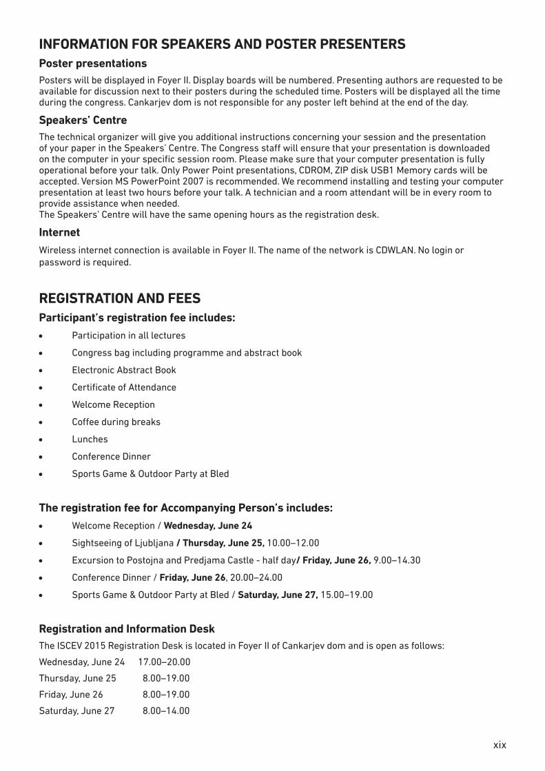
SOCIAL PROGRAMMEWEDNESDAY, JUNE 24, 201520.00–23.00 Welcome Reception/Ljubljana Castle Included in the fee for participants and accompanying persons.
Meeting point: 19.45 at Cankarjev dom, Erjavčeva Street (by bus), returning at 23.00
Please note that you can take a funicular train to the Ljubljana Castle.
Price:
• Return ticket à EUR 4.00
• Single ticket à EUR 2.20
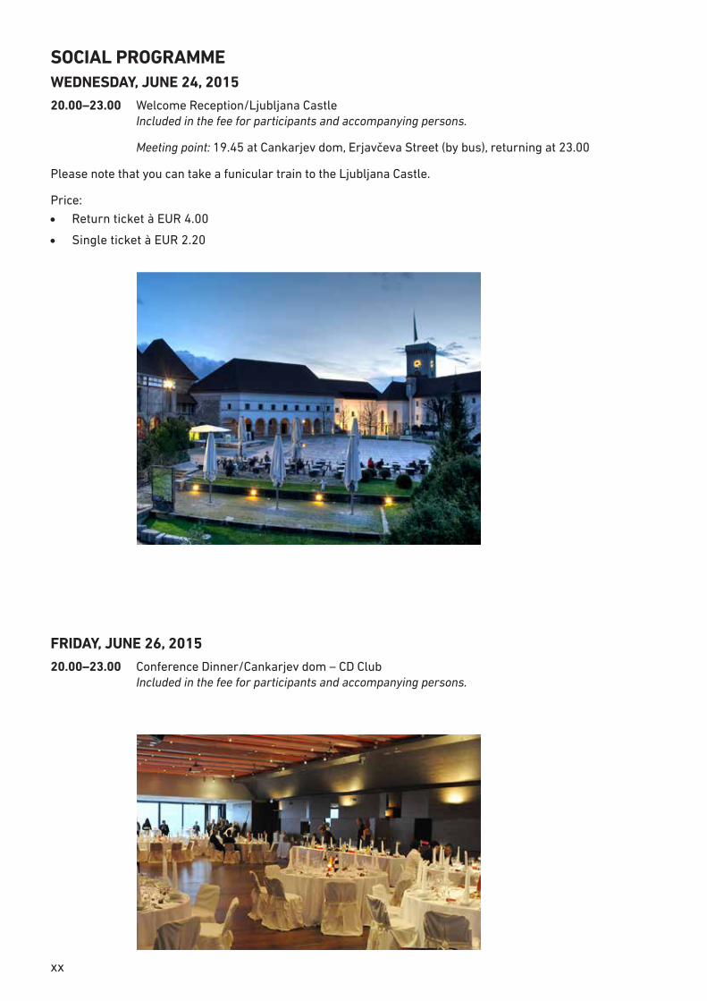
FRIDAY, JUNE 26, 201520.00–23.00 Conference Dinner/Cankarjev dom – CD Club Included in the fee for participants and accompanying persons.
xxi
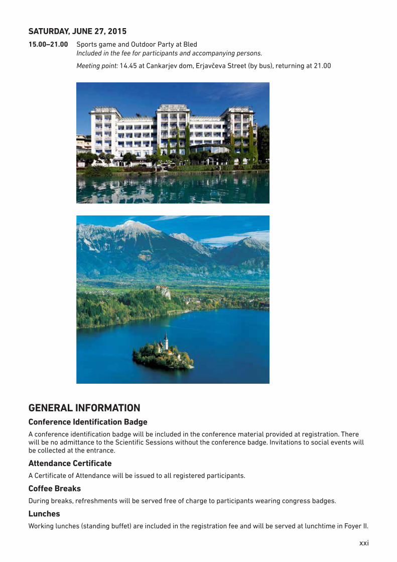
SATURDAY, JUNE 27, 201515.00–21.00 Sports game and Outdoor Party at Bled Included in the fee for participants and accompanying persons.
Meeting point: 14.45 at Cankarjev dom, Erjavčeva Street (by bus), returning at 21.00
GENERAL INFORMATIONConference Identification Badge A conference identification badge will be included in the conference material provided at registration. There will be no admittance to the Scientific Sessions without the conference badge. Invitations to social events will be collected at the entrance.
Attendance CertificateA Certificate of Attendance will be issued to all registered participants.
Coffee BreaksDuring breaks, refreshments will be served free of charge to participants wearing congress badges.
Lunches Working lunches (standing buffet) are included in the registration fee and will be served at lunchtime in Foyer II.
xxiv
ABSTRACTS……………
3
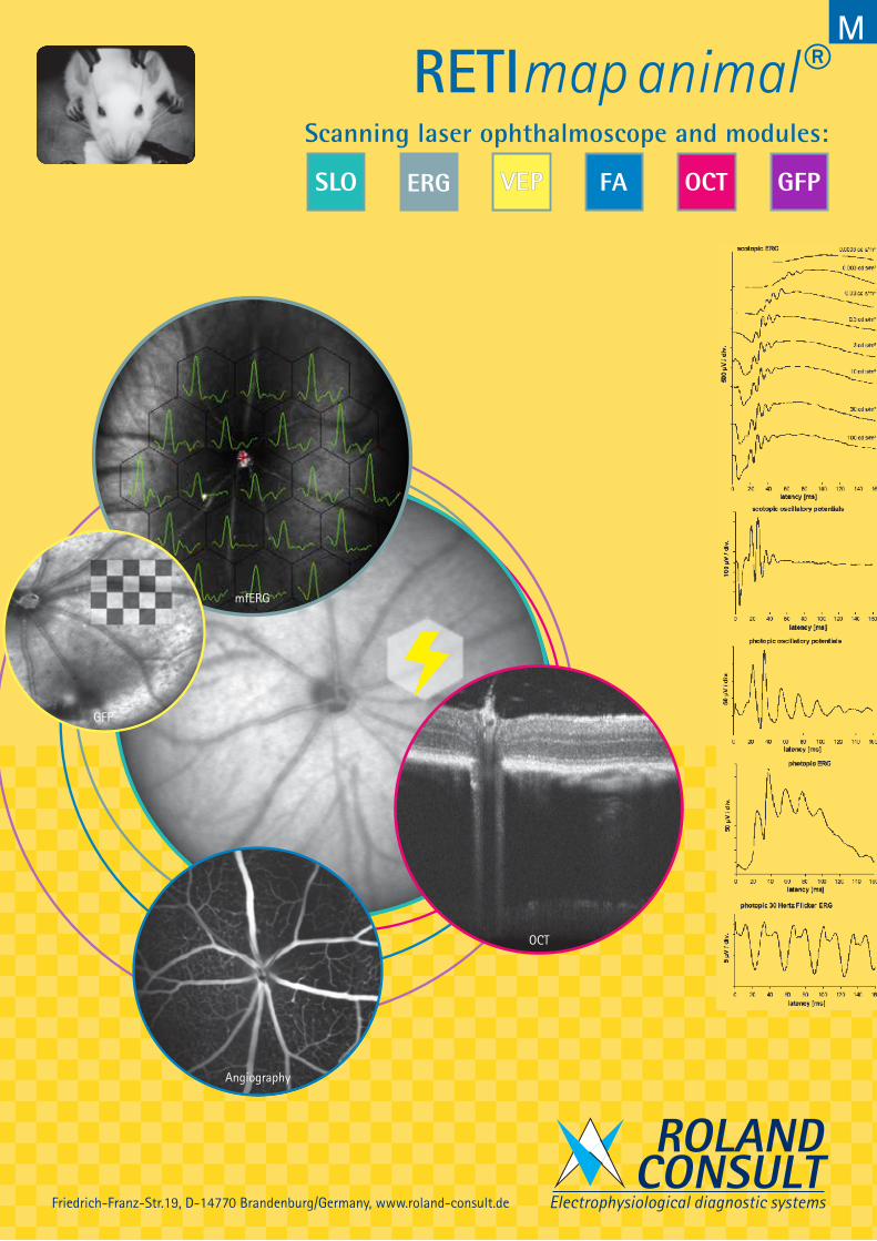
Scanning laser ophthalmoscope and modules:
ROLANDCONSULT
Electrophysiological diagnostic systems
M
VEP
Angiography
mfERG
OCT
GFP
RETImap animal®
Friedrich-Franz-Str.19, D-14770 Brandenburg/Germany, www.roland-consult.de
VEP
Angiography
mfERG
OCT
cSLO
VEP
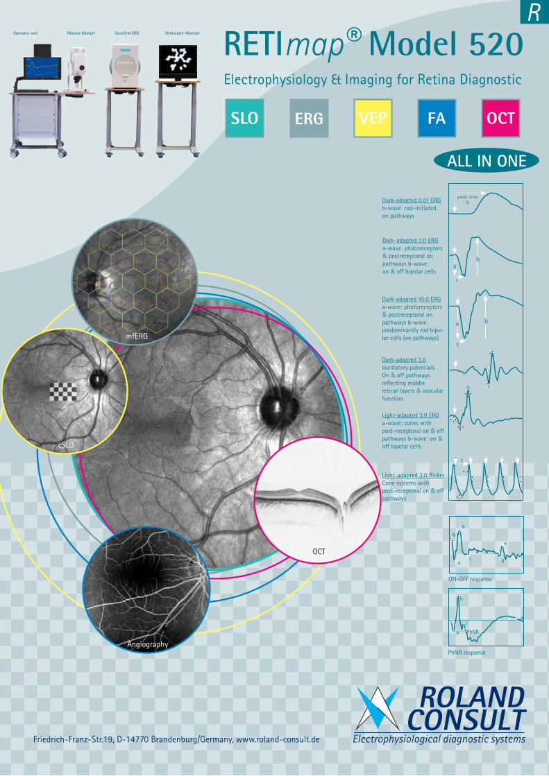
Electrophysiology & Imaging for Retina Diagnostic
RETImap® Model 520
ROLANDCONSULT
Electrophysiological diagnostic systems
2,5µV/div
25 ms/div
Ganzfeld ERG Stimulator MonitorMacula ModuleOperator unit
ON-OFF response
ac d
b
e
N
PhNR response
b
i
PhNRa
4
Light-adapted 3.0 ERG a-wave: cones with post-receptoral on & off pathways b-wave: on & off bipolar cells
Dark-adapted 3.0 ERG a-wave: photoreceptors & postreceptoral on pathways b-wave: on & off bipolar cells
Dark-adapted 3.0 oscillatory potentialsOn & off pathways reflecting middle retinal layers & vascular function
Dark-adapted 0.01 ERG b-wave: rod-initiated on pathways
Light-adapted 3.0 flickerCone systems with post-receptoral on & off pathways
Dark-adapted 10.0 ERGa-wave: photoreceptors & postreceptoral on pathways b-wave: predominantly rod bipo-lar cells (on pathways)
a
b
ba
t
t
ab
peak time (t)
t
ALL IN ONE
Friedrich-Franz-Str.19, D-14770 Brandenburg/Germany, www.roland-consult.de
R
xxv3
Scanning laser ophthalmoscope and modules:
ROLANDCONSULT
Electrophysiological diagnostic systems
M
VEP
Angiography
mfERG
OCT
GFP
RETImap animal®
Friedrich-Franz-Str.19, D-14770 Brandenburg/Germany, www.roland-consult.de
AUTHOR’S INDEX
VEP
Angiography
mfERG
OCT
cSLO
VEP
Electrophysiology & Imaging for Retina Diagnostic
RETImap® Model 520
ROLANDCONSULT
Electrophysiological diagnostic systems
2,5µV/div
25 ms/div
Ganzfeld ERG Stimulator MonitorMacula ModuleOperator unit
ON-OFF response
ac d
b
e
N
PhNR response
b
i
PhNRa
4
Light-adapted 3.0 ERG a-wave: cones with post-receptoral on & off pathways b-wave: on & off bipolar cells
Dark-adapted 3.0 ERG a-wave: photoreceptors & postreceptoral on pathways b-wave: on & off bipolar cells
Dark-adapted 3.0 oscillatory potentialsOn & off pathways reflecting middle retinal layers & vascular function
Dark-adapted 0.01 ERG b-wave: rod-initiated on pathways
Light-adapted 3.0 flickerCone systems with post-receptoral on & off pathways
Dark-adapted 10.0 ERGa-wave: photoreceptors & postreceptoral on pathways b-wave: predominantly rod bipo-lar cells (on pathways)
a
b
ba
t
t
ab
peak time (t)
t
ALL IN ONE
Friedrich-Franz-Str.19, D-14770 Brandenburg/Germany, www.roland-consult.de
R
xxvi
OGLASI RETIcompact II®
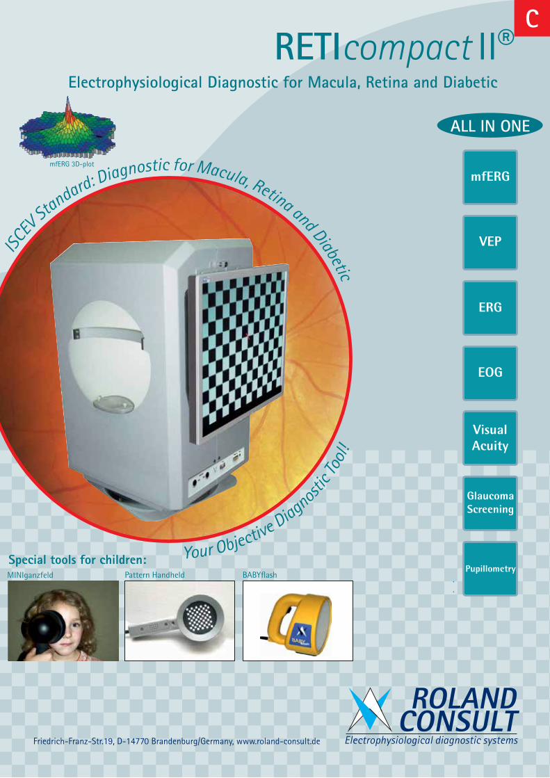
ALL IN ONE
mfERG
VEP
ERG
EOG
Visual Acuity
Glaucoma Screening
Pupillometry
mfERG 3D-plot
Pattern Handheld BABYflashMINIganzfeld
ISCEV Standard: Diagnostic for Macula, Retina and Diabetic
Your Objective Diagnostic T
ool!
Electrophysiological Diagnostic for Macula, Retina and Diabetic
Special tools for children:
ROLANDCONSULT
Electrophysiological diagnostic systemsFriedrich-Franz-Str.19, D-14770 Brandenburg/Germany, www.roland-consult.de
C D
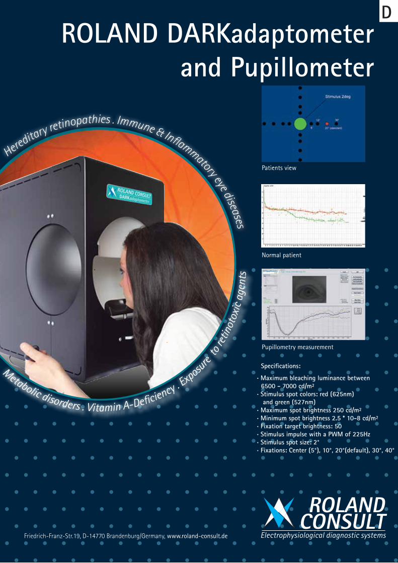
Normal patient
Patients view
ROLAND DARKadaptometer and Pupillometer
ROLANDCONSULT
Electrophysiological diagnostic systems
Pupillometry measurement
Specifications:
. Maximum bleaching luminance between 6500 - 7000 cd/m². Stimulus spot colors: red (625nm) and green (527nm). Maximum spot brightness 250 cd/m². Minimum spot brightness 2.5 * 10-8 cd/m². Fixation target brightness: 50. Stimulus impulse with a PWM of 225Hz. Stimulus spot size: 2°. Fixations: Center (5°), 10°, 20°(default), 30°, 40°
Metabolic disorders . Vitamin A-Def ciency . Exposu
re to
retin
otox
ic a
gent
s
Hereditary retinopathies . Immune & Inf ammatory eye diseases
Friedrich-Franz-Str.19, D-14770 Brandenburg/Germany, www.roland-consult.de
xxvii
RETIcompact II®
ALL IN ONE
mfERG
VEP
ERG
EOG
Visual Acuity
Glaucoma Screening
Pupillometry
mfERG 3D-plot
Pattern Handheld BABYflashMINIganzfeld
ISCEV Standard: Diagnostic for Macula, Retina and Diabetic
Your Objective Diagnostic T
ool!
Electrophysiological Diagnostic for Macula, Retina and Diabetic
Special tools for children:
ROLANDCONSULT
Electrophysiological diagnostic systemsFriedrich-Franz-Str.19, D-14770 Brandenburg/Germany, www.roland-consult.de
C D
Normal patient
Patients view
ROLAND DARKadaptometer and Pupillometer
ROLANDCONSULT
Electrophysiological diagnostic systems
Pupillometry measurement
Specifications:
. Maximum bleaching luminance between 6500 - 7000 cd/m². Stimulus spot colors: red (625nm) and green (527nm). Maximum spot brightness 250 cd/m². Minimum spot brightness 2.5 * 10-8 cd/m². Fixation target brightness: 50. Stimulus impulse with a PWM of 225Hz. Stimulus spot size: 2°. Fixations: Center (5°), 10°, 20°(default), 30°, 40°
Metabolic disorders . Vitamin A-Def ciency . Exposu
re to
retin
otox
ic a
gent
s
Hereditary retinopathies . Immune & Inf ammatory eye diseases
Friedrich-Franz-Str.19, D-14770 Brandenburg/Germany, www.roland-consult.de
xxviii
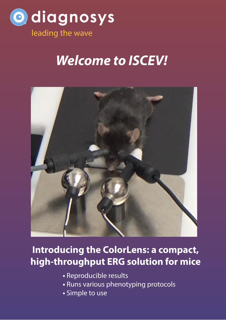
leading the wave
Introducing the ColorLens: a compact, high-throughput ERG solution for mice
Welcome to ISCEV!
• Reproducible results• Runs various phenotyping protocols• Simple to use
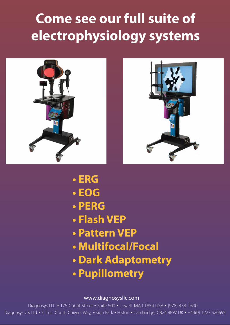
Come see our full suite ofelectrophysiology systems
• ERG• EOG• PERG• Flash VEP• Pattern VEP• Multifocal/Focal• Dark Adaptometry• Pupillometry
xxix
leading the wave
Introducing the ColorLens: a compact, high-throughput ERG solution for mice
Welcome to ISCEV!
• Reproducible results• Runs various phenotyping protocols• Simple to use
Come see our full suite ofelectrophysiology systems
• ERG• EOG• PERG• Flash VEP• Pattern VEP• Multifocal/Focal• Dark Adaptometry• Pupillometry
xxx
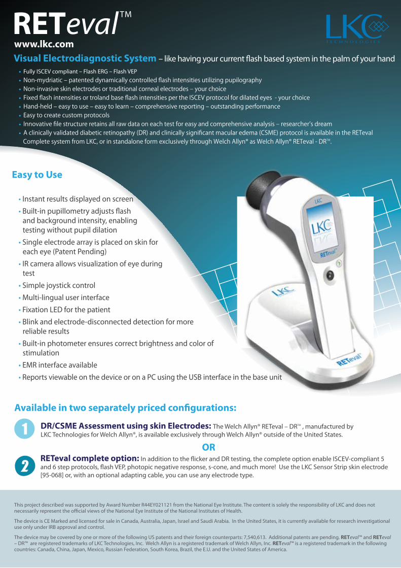
RETeval TM
Visual Electrodiagnostic System – like having your current flash based system in the palm of your hand
Easy to Use
• Instant results displayed on screen
• Built-in pupillometry adjusts flash and background intensity, enabling testing without pupil dilation
• Single electrode array is placed on skin for each eye (Patent Pending)
• IR camera allows visualization of eye during test
• Simple joystick control
• Multi-lingual user interface
• Fixation LED for the patient
• Blink and electrode-disconnected detection for more reliable results
• Built-in photometer ensures correct brightness and color of stimulation
• EMR interface available
• Reports viewable on the device or on a PC using the USB interface in the base unit
Available in two separately priced configurations:
DR/CSME Assessment using skin Electrodes: The Welch Allyn® RETeval – DRTM , manufactured by LKC Technologies for Welch Allyn®, is available exclusively through Welch Allyn® outside of the United States.
ORRETeval complete option: In addition to the flicker and DR testing, the complete option enable ISCEV-compliant 5 and 6 step protocols, flash VEP, photopic negative response, s-cone, and much more! Use the LKC Sensor Strip skin electrode [95-068] or, with an optional adapting cable, you can use any electrode type.
This project described was supported by Award Number R44EY021121 from the National Eye Institute. The content is solely the responsibility of LKC and does not necessarily represent the official views of the National Eye Institute of the National Institutes of Health.
The device is CE Marked and licensed for sale in Canada, Australia, Japan, Israel and Saudi Arabia. In the United States, it is currently available for research investigational use only under IRB approval and control.
The device may be covered by one or more of the following US patents and their foreign counterparts: 7,540,613. Additional patents are pending. RETeval™ and RETeval – DR™ are registered trademarks of LKC Technologies, Inc. Welch Allyn is a registered trademark of Welch Allyn, Inc. RETeval™ is a registered trademark in the following countries: Canada, China, Japan, Mexico, Russian Federation, South Korea, Brazil, the E.U. and the United States of America.
• Fully ISCEV compliant – Flash ERG – Flash VEP
• Non-mydriatic ‒ patented dynamically controlled flash intensities utilizing pupilography• Non-invasive skin electrodes or traditional corneal electrodes ‒ your choice• Fixed flash intensities or troland base flash intensities per the ISCEV protocol for dilated eyes - your choice• Hand-held ‒ easy to use ‒ easy to learn ‒ comprehensive reporting ‒ outstanding performance• Easy to create custom protocols• Innovative file structure retains all raw data on each test for easy and comprehensive analysis ‒ researcher’s dream• A clinically validated diabetic retinopathy (DR) and clinically significant macular edema (CSME) protocol is available in the RETeval Complete system from LKC, or in standalone form exclusively through Welch Allyn® as Welch Allyn® RETeval - DRTM.
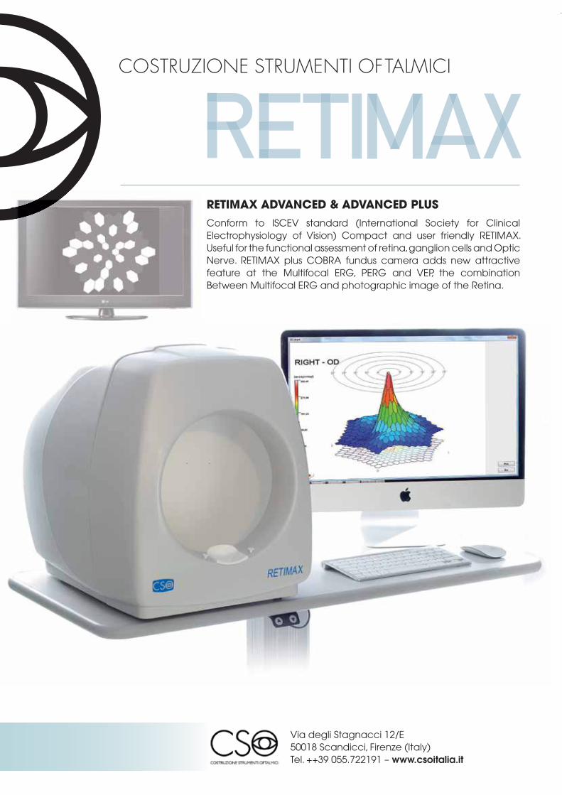
www.lkc.com COSTRUZIONE STRUMENTI OFTALMICI
RETIMAX BASIC, ADVANCED E ADVANCED PLUS
Glaucoma PERG Hemifield TestDiagnosi precoce del Glaucoma.Database normativo correlato all’età per itest ERG, PERG, VEP, EOG.Vision Trainer Riabilitazione visivaBiofeedback
RETIMAX ADVANCED & ADVANCED PLUSConform to ISCEV standard (International Society for Clinical Electrophysiology of Vision) Compact and user friendly RETIMAX. Useful for the functional assessment of retina, ganglion cells and Optic Nerve. RETIMAX plus COBRA fundus camera adds new attractive feature at the Multifocal ERG, PERG and VEP, the combination Between Multifocal ERG and photographic image of the Retina.
Via degli Stagnacci 12/E50018 Scandicci, Firenze (Italy)Tel. ++39 055.722191 – www.csoitalia.it
RETeval TM
Visual Electrodiagnostic System – like having your current flash based system in the palm of your hand
Easy to Use
• Instant results displayed on screen
• Built-in pupillometry adjusts flash and background intensity, enabling testing without pupil dilation
• Single electrode array is placed on skin for each eye (Patent Pending)
• IR camera allows visualization of eye during test
• Simple joystick control
• Multi-lingual user interface
• Fixation LED for the patient
• Blink and electrode-disconnected detection for more reliable results
• Built-in photometer ensures correct brightness and color of stimulation
• EMR interface available
• Reports viewable on the device or on a PC using the USB interface in the base unit
Available in two separately priced configurations:
DR/CSME Assessment using skin Electrodes: The Welch Allyn® RETeval – DRTM , manufactured by LKC Technologies for Welch Allyn®, is available exclusively through Welch Allyn® outside of the United States.
ORRETeval complete option: In addition to the flicker and DR testing, the complete option enable ISCEV-compliant 5 and 6 step protocols, flash VEP, photopic negative response, s-cone, and much more! Use the LKC Sensor Strip skin electrode [95-068] or, with an optional adapting cable, you can use any electrode type.
This project described was supported by Award Number R44EY021121 from the National Eye Institute. The content is solely the responsibility of LKC and does not necessarily represent the official views of the National Eye Institute of the National Institutes of Health.
The device is CE Marked and licensed for sale in Canada, Australia, Japan, Israel and Saudi Arabia. In the United States, it is currently available for research investigational use only under IRB approval and control.
The device may be covered by one or more of the following US patents and their foreign counterparts: 7,540,613. Additional patents are pending. RETeval™ and RETeval – DR™ are registered trademarks of LKC Technologies, Inc. Welch Allyn is a registered trademark of Welch Allyn, Inc. RETeval™ is a registered trademark in the following countries: Canada, China, Japan, Mexico, Russian Federation, South Korea, Brazil, the E.U. and the United States of America.
• Fully ISCEV compliant – Flash ERG – Flash VEP
• Non-mydriatic ‒ patented dynamically controlled flash intensities utilizing pupilography• Non-invasive skin electrodes or traditional corneal electrodes ‒ your choice• Fixed flash intensities or troland base flash intensities per the ISCEV protocol for dilated eyes - your choice• Hand-held ‒ easy to use ‒ easy to learn ‒ comprehensive reporting ‒ outstanding performance• Easy to create custom protocols• Innovative file structure retains all raw data on each test for easy and comprehensive analysis ‒ researcher’s dream• A clinically validated diabetic retinopathy (DR) and clinically significant macular edema (CSME) protocol is available in the RETeval Complete system from LKC, or in standalone form exclusively through Welch Allyn® as Welch Allyn® RETeval - DRTM.
www.lkc.com COSTRUZIONE STRUMENTI OFTALMICI
RETIMAX BASIC, ADVANCED E ADVANCED PLUS
Glaucoma PERG Hemifield TestDiagnosi precoce del Glaucoma.Database normativo correlato all’età per itest ERG, PERG, VEP, EOG.Vision Trainer Riabilitazione visivaBiofeedback
RETIMAX ADVANCED & ADVANCED PLUSConform to ISCEV standard (International Society for Clinical Electrophysiology of Vision) Compact and user friendly RETIMAX. Useful for the functional assessment of retina, ganglion cells and Optic Nerve. RETIMAX plus COBRA fundus camera adds new attractive feature at the Multifocal ERG, PERG and VEP, the combination Between Multifocal ERG and photographic image of the Retina.
Via degli Stagnacci 12/E50018 Scandicci, Firenze (Italy)Tel. ++39 055.722191 – www.csoitalia.it
xxxii
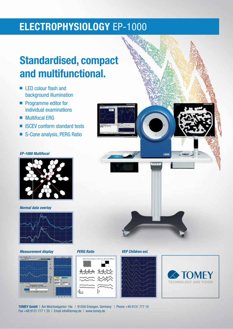
ELECTROPHYSIOLOGY EP-1000
LED colour fl ash and background illumination Programme editor for individual examinations Multifocal ERG ISCEV conform standard tests S-Cone analysis, PERG Ratio
Standardised, compact and multifunctional.
Normal data overlay
Measurement display
EP-1000 Multifocal
VEP Children ext.PERG Ratio
TOMEY GmbH | Am Weichselgarten 19a | 91058 Erlangen, Germany | Phone +49 9131 777 10Fax +49 9131 777 1 20 | Email [email protected] | www.tomey.de
TOM_Anzeige_A4_EP1000.indd 1 21.05.15 16:38
xxxiii
ELECTROPHYSIOLOGY EP-1000
LED colour fl ash and background illumination Programme editor for individual examinations Multifocal ERG ISCEV conform standard tests S-Cone analysis, PERG Ratio
Standardised, compact and multifunctional.
Normal data overlay
Measurement display
EP-1000 Multifocal
VEP Children ext.PERG Ratio
TOMEY GmbH | Am Weichselgarten 19a | 91058 Erlangen, Germany | Phone +49 9131 777 10Fax +49 9131 777 1 20 | Email [email protected] | www.tomey.de
TOM_Anzeige_A4_EP1000.indd 1 21.05.15 16:38
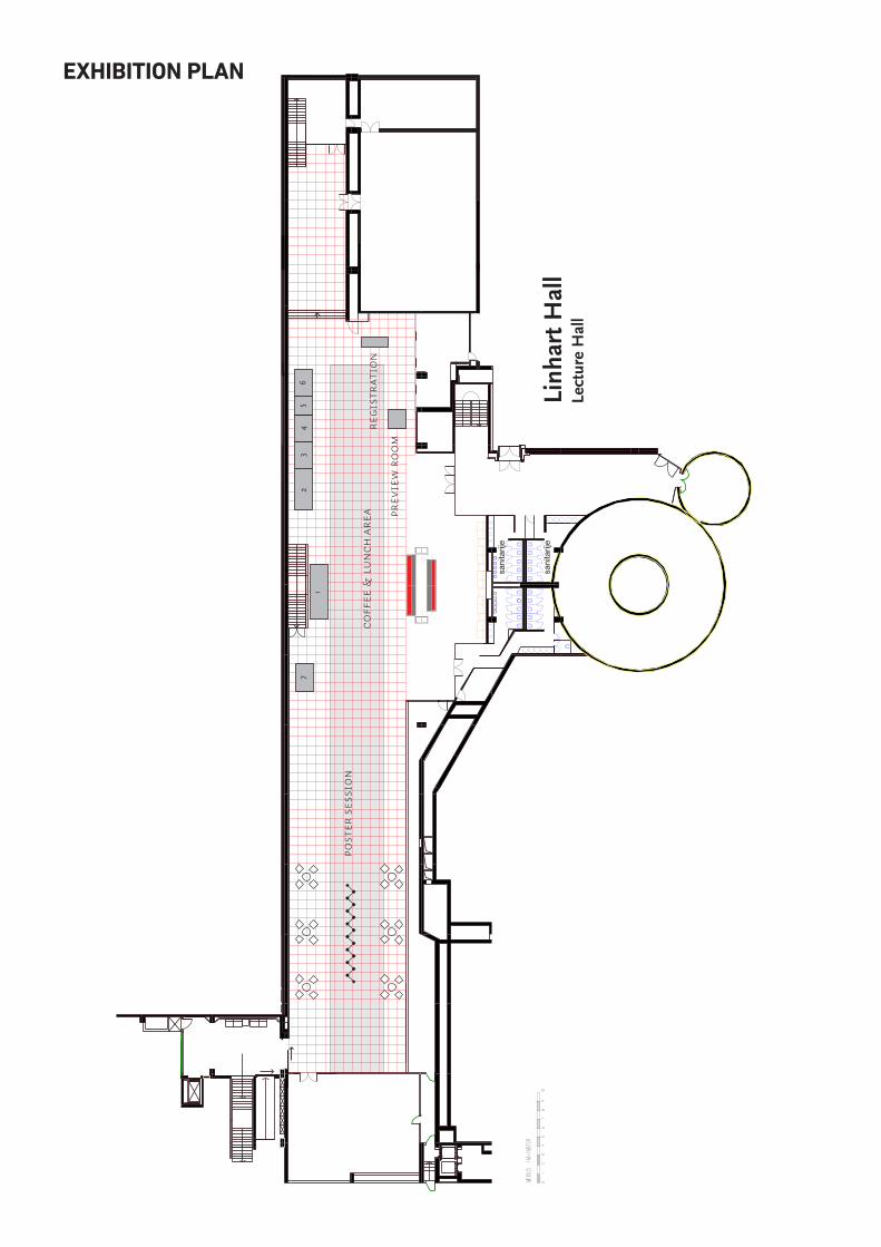
EXHIBITION PLAN

NOTES

NOTES