Embed Size (px)
Citation preview
© 1999 Macmillan Magazines Ltd
letters to nature
800 NATURE | VOL 401 | 21 OCTOBER 1999 | www.nature.com
3. Mohler, H. & Okada, T. Benzodiazepine receptor: demonstration in the central nervous system.
Science 198, 849–851 (1977).
4. Braestrup, C., Albrechtsen, R. & Squires, R. F. High densities of benzodiazepine receptors in human
cortical areas. Nature 269, 702–704 (1977).
5. Wieland, H. A., Luddens, H. & Seeburg, P. H. A single histidine in GABAA receptors is essential for
benzodiazepine agonist binding. J. Biol. Chem. 267, 1426–1429 (1992).
6. Kleingoor, C., Wieland, H. A., Korpi, E. R., Seeburg, P. H. & Kettenmann, H. Current potentiation by
diazepam but not GABA sensitivity is determined by a single histidine residue. Neuroreport 4, 187–
190 (1993).
7. Benson, J. A., Low, K., Keist, R., Mohler, H. & Rudolph, U. Pharmacology of recombinant g-
aminobutyric acidA receptors rendered diazepam-insensitive by point-mutated a-subunits. FEBS
Lett. 431, 400–404 (1998).
8. Fritschy, J.-M. et al. Five subtypes of type A g-aminobutyric acid receptors identified in neurons by
double and triple immunofluorescence staining with subunit-specific antibodies. Proc. Natl Acad. Sci.
USA 89, 6726–6730 (1992).
9. Lakso, M. et al. Efficient in vivo manipulation of mouse genomic sequences at the zygote stage. Proc.
Natl Acad. Sci. USA 93, 5860–5865 (1996).
10. Knoflach, F. et al. Pharmacological modulation of the diazepam-insensitive recombinant g-amino-
butyric acidA receptors a4b2g2 and a6b2g2. Mol. Pharmacol. 50, 1253–1261 (1996).
11. Bernard, F. et al. in Zolpidem: an Update of its Pharmacological Properties and the Therapeutic Place in
the Management of Insomnia (eds Freeman, H., Puech, A. J. & Roth, T.) 21–31 (Elsevier, Paris, 1996).
12. Melchior, C. L. & Allen, P. M. Interaction of pregnanolone and pregnanolone sulfate with ethanol and
pentobarbital. Pharmacol. Biochem. Behav. 42, 605–611 (1992).
13. Beuzen, A. & Belzung, C. Link between emotional memory and anxiety states: a study by principal
component analysis. Physiol. Behav. 58, 111–118 (1995).
14. Vogel, J. R., Beer, B. & Clody, D. E. A simple and reliable conflict procedure for testing anti-anxiety
agents. Psychopharmacology 21, 1–7 (1971).
15. Bonetti, E. P. et al. Benzodiazepine antagonist Ro 15-1788: neurological and behavioral effects.
Psychopharmacology 78, 8–18 (1982).
16. Hunkeler, W. et al. Selective antagonists of benzodiazepines. Nature 290, 514–516 (1981).
17. Bonetti, E. P. et al. Ro 15-4513: Partial inverse agonism at the BZR and interaction with ethanol.
Pharmacol. Biochem. Behav. 31, 733–749 (1988).
18. Misslin, R., Belzung, C. & Vogel, E. Behavioural validation of a light/dark choice procedure for testing
anti-anxiety agents. Behav. Proc. 18, 119–132 (1989).
19. Lister, R. G. The use of a plus-maze to measure anxiety in the mouse. Psychopharmacology 92, 180–185
(1987).
Supplementary information is available on Nature’s World-Wide Web site (http://www.nature.com) or as paper copy from the London editorial office of Nature.
AcknowledgementsWe thank Y. Lang for blastocyst injections, D. Blaser, S. Ganz, H. Pochetti and G. Schmidfor animal care, R. Keist and C. Michel for technical assistance and H. Westphal forproviding Ella-cre mice. This work was supported by a grant from the Swiss NationalScience Foundation.
Correspondence and requests for materials should be addressed to U.R.(e-mail: [email protected]).
.................................................................P/Q-type calcium channels mediatethe activity-dependent feedbackof syntaxin-1AKathy G. Sutton*†, John E. McRory*†, Heather Guthrie*,Timothy H. Murphy‡ & Terrance P. Snutch*
* Biotechnology Laboratory and ‡ Kinsmen Laboratory of Neurological Research,Dept Psychiatry University of British Columbia, Vancouver, V6T 1Z3, Canada† These authors contributed equally to this work.
.................................. ......................... ......................... ......................... ......................... ........
Spatial and temporal changes in intracellular calcium concentra-tions are critical for controlling gene expression in neurons1–5. Inmany neurons, activity-dependent calcium influx through L-typechannels stimulates transcription that depends on the transcrip-tion factor CREB by activating a calmodulin-dependent path-way6–11. Here we show that selective influx of calcium through P/Q-type channels12–14 is responsible for activating expression ofsyntaxin-1A, a presynaptic protein that mediates vesicle docking,fusion and neurotransmitter release. The initial P/Q-type calciumsignal is amplified by release of calcium from intracellular stores
and acts through phosphorylation that is dependent on thecalmodulin-dependent kinase CaM K II/IV, protein kinase Aand mitogen-activated protein kinase kinase. Initiation of syn-taxin-1A expression is rapid and short-lived, with syntaxin-1Aultimately interacting with the P/Q-type calcium channel todecrease channel availability. Our results define an activity-dependent feedback pathway that may regulate synaptic efficacyand function in the nervous system.
Syntaxin-1A interacts with the synaptic core (SNARE) complexand is tightly regulated by a number of proteins including munc-13,munc-18 and tomosyn15–18. Syntaxin-1A also binds to the domainII–III linker of N- and P/Q-type voltage-gated Ca2+ channels andshifts current inactivation properties to more negative poten-tials19–23. Transient expression of human a1A (P/Q-type) Ca2+
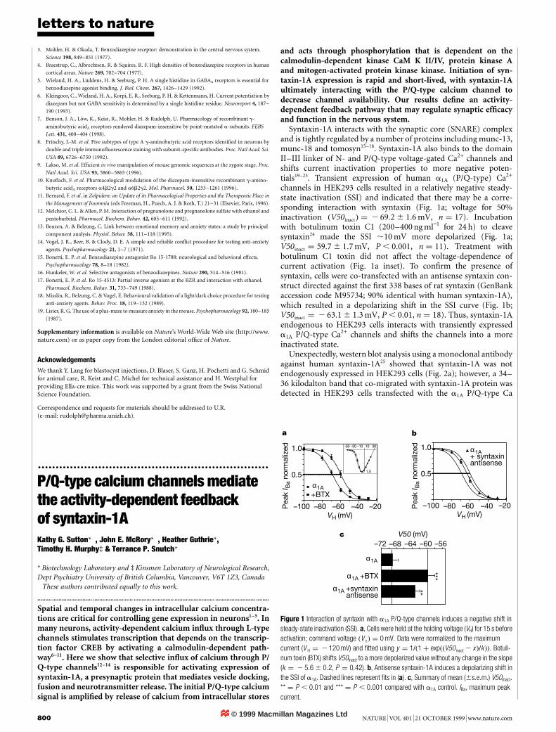
channels in HEK293 cells resulted in a relatively negative steady-state inactivation (SSI) and indicated that there may be a corre-sponding interaction with syntaxin (Fig. 1a; voltage for 50%inactivation ðV50inactÞ ¼ 2 69:2 6 1:6 mV, n ¼ 17). Incubationwith botulinum toxin C1 (200–400 ng ml−1 for 24 h) to cleavesyntaxin24 made the SSI ,10 mV more depolarized (Fig. 1a;V50inact ¼ 59:7 6 1:7 mV, P , 0:001, n ¼ 11). Treatment withbotulinum C1 toxin did not affect the voltage-dependence ofcurrent activation (Fig. 1a inset). To confirm the presence ofsyntaxin, cells were co-transfected with an antisense syntaxin con-struct directed against the first 338 bases of rat syntaxin (GenBankaccession code M95734; 90% identical with human syntaxin-1A),which resulted in a depolarizing shift in the SSI curve (Fig. 1b;V50inact ¼ 2 63:1 6 1:3 mV, P , 0:01, n ¼ 18). Thus, syntaxin-1Aendogenous to HEK293 cells interacts with transiently expresseda1A P/Q-type Ca2+ channels and shifts the channels into a moreinactivated state.
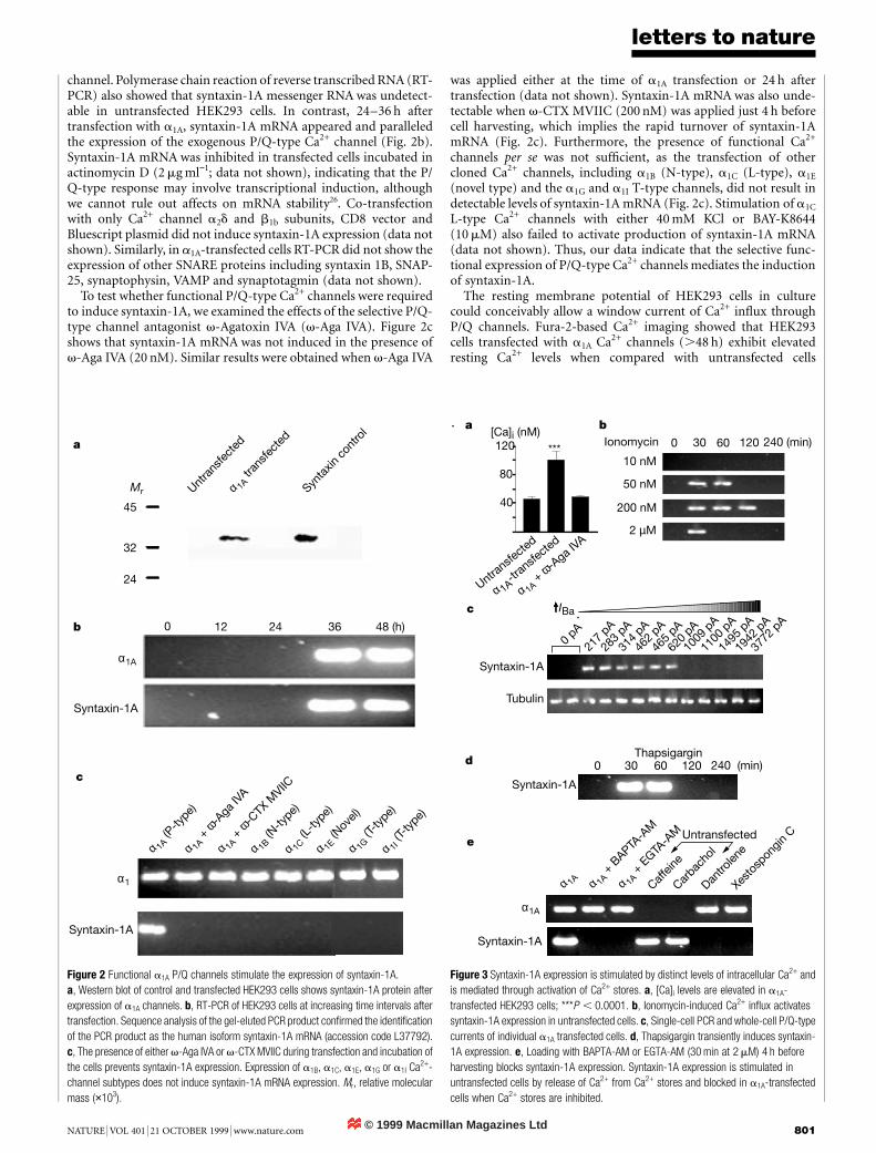
Unexpectedly, western blot analysis using a monoclonal antibodyagainst human syntaxin-1A25 showed that syntaxin-1A was notendogenously expressed in HEK293 cells (Fig. 2a); however, a 34–36 kilodalton band that co-migrated with syntaxin-1A protein wasdetected in HEK293 cells transfected with the a1A P/Q-type Ca
1.0
0.5
–100 –80 –60 –40 –20VH (mV)
Pea
k I B
a no
rmal
ized
a b
c
α1A
–50 –30 –10 3010
1.0
+BTX
1.0
0.5
–100
–72 –68 –64 –60 –56
–80 –60 –40 –20VH (mV)
Pea
k I B
a no
rmal
ized α1A
+ syntaxinantisense
α1A
α1A +BTX
α1A +syntaxinantisense
V50 (mV)
***
**
Figure 1 Interaction of syntaxin with a1A P/Q-type channels induces a negative shift insteady-state inactivation (SSI). a, Cells were held at the holding voltage (VH) for 15 s beforeactivation; command voltage ðV cÞ ¼ 0 mV. Data were normalized to the maximumcurrent (V H ¼ 2 120 mV) and fitted using y ¼ 1=ð1 þ expððV50inact 2 x Þ=k ÞÞ. Botuli-num toxin (BTX) shifts V50inact to a more depolarized value without any change in the slope(k ¼ 2 5:6 6 0:2, P ¼ 0:42). b, Antisense syntaxin-1A induces a depolarizing shift inthe SSI of a1A. Dashed lines represent fits in (a). c, Summary of mean (6s.e.m.) V50inact.** ¼ P , 0:01 and *** ¼ P , 0:001 compared with a1A control. IBa, maximum peakcurrent.
© 1999 Macmillan Magazines Ltd
letters to nature
NATURE | VOL 401 | 21 OCTOBER 1999 | www.nature.com 801
channel. Polymerase chain reaction of reverse transcribed RNA (RT-PCR) also showed that syntaxin-1A messenger RNA was undetect-able in untransfected HEK293 cells. In contrast, 24–36 h aftertransfection with a1A, syntaxin-1A mRNA appeared and paralleledthe expression of the exogenous P/Q-type Ca2+ channel (Fig. 2b).Syntaxin-1A mRNA was inhibited in transfected cells incubated inactinomycin D (2 mg ml−1; data not shown), indicating that the P/Q-type response may involve transcriptional induction, althoughwe cannot rule out affects on mRNA stability26. Co-transfectionwith only Ca2+ channel a2d and b1b subunits, CD8 vector andBluescript plasmid did not induce syntaxin-1A expression (data notshown). Similarly, in a1A-transfected cells RT-PCR did not show theexpression of other SNARE proteins including syntaxin 1B, SNAP-25, synaptophysin, VAMP and synaptotagmin (data not shown).
To test whether functional P/Q-type Ca2+ channels were requiredto induce syntaxin-1A, we examined the effects of the selective P/Q-type channel antagonist v-Agatoxin IVA (v-Aga IVA). Figure 2cshows that syntaxin-1A mRNA was not induced in the presence ofv-Aga IVA (20 nM). Similar results were obtained when v-Aga IVA
was applied either at the time of a1A transfection or 24 h aftertransfection (data not shown). Syntaxin-1A mRNA was also unde-tectable when v-CTX MVIIC (200 nM) was applied just 4 h beforecell harvesting, which implies the rapid turnover of syntaxin-1AmRNA (Fig. 2c). Furthermore, the presence of functional Ca2+
channels per se was not sufficient, as the transfection of othercloned Ca2+ channels, including a1B (N-type), a1C (L-type), a1E
(novel type) and the a1G and a1I T-type channels, did not result indetectable levels of syntaxin-1A mRNA (Fig. 2c). Stimulation of a1C
L-type Ca2+ channels with either 40 mM KCl or BAY-K8644(10 mM) also failed to activate production of syntaxin-1A mRNA(data not shown). Thus, our data indicate that the selective func-tional expression of P/Q-type Ca2+ channels mediates the inductionof syntaxin-1A.
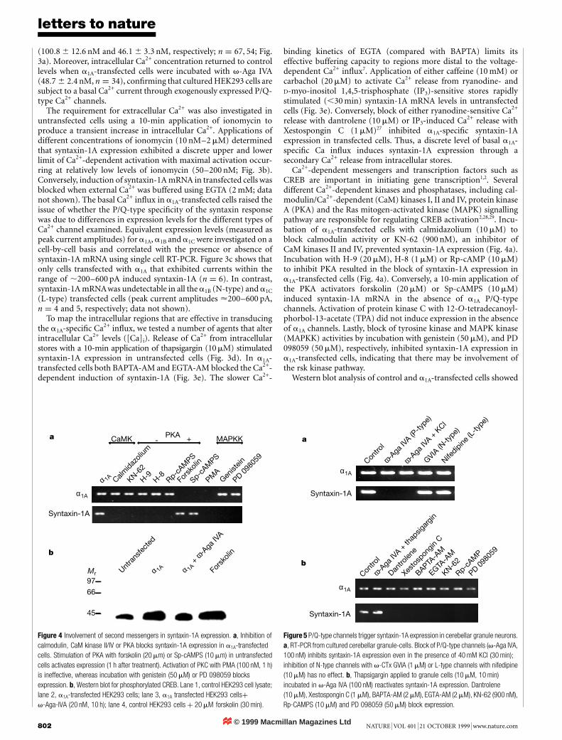
The resting membrane potential of HEK293 cells in culturecould conceivably allow a window current of Ca2+ influx throughP/Q channels. Fura-2-based Ca2+ imaging showed that HEK293cells transfected with a1A Ca2+ channels (.48 h) exhibit elevatedresting Ca2+ levels when compared with untransfected cells
0
α1A
Syntaxin-1A
Syntaxin-1A
α 1A +
ω-A
ga IV
A
α 1A +
ω-C
TX M
VIIC
α 1B (N
-type)
α 1C (L
-type)
α 1E (N
ovel)
a
b
c
α1
α 1A (P
-type
)
Mr
32
45
24
Untra
nsfe
cted
Synta
xin co
ntro
l
α 1A tr
ansfe
cted
α 1G (T
-type)
α 1I (T
-type)
48 (h)362412
Figure 2 Functional a1A P/Q channels stimulate the expression of syntaxin-1A.a, Western blot of control and transfected HEK293 cells shows syntaxin-1A protein afterexpression of a1A channels. b, RT-PCR of HEK293 cells at increasing time intervals aftertransfection. Sequence analysis of the gel-eluted PCR product confirmed the identificationof the PCR product as the human isoform syntaxin-1A mRNA (accession code L37792).c, The presence of either v-Aga IVA or v-CTX MVIIC during transfection and incubation ofthe cells prevents syntaxin-1A expression. Expression of a1B, a1C, a1E, a1G or a1I Ca2+-channel subtypes does not induce syntaxin-1A mRNA expression. Mr, relative molecularmass (×103).
c
d
e
0 30 60 120 240 (min)
Syntaxin-1A
Thapsigargin
0 pA
217
pA
283
pA
314
pA
462
pA
465
pA
620
pA
1009
pA
1100
pA
1495
pA
1942
pA
3772
pA
Syntaxin-1A
IBa
Tubulin
ba
0 30 60 120 240 (min)Ionomycin
2 µM
200 nM
50 nM
10 nM
Untransfe
cted
α 1A-tr
ansfe
cted
α 1A + ω-A
ga IVA
40
80
120[Ca]i (nM)
***
α 1A α 1A +
BAPTA
-AM
α 1A +
EGTA
-AM
Caffein
e
Carbac
hol
Syntaxin-1A
α1A
Dantro
lene
Xesto
spon
gin CUntransfected
Figure 3 Syntaxin-1A expression is stimulated by distinct levels of intracellular Ca2+ andis mediated through activation of Ca2+ stores. a, [Ca]i levels are elevated in a1A-transfected HEK293 cells; ***P , 0:0001. b, Ionomycin-induced Ca2+ influx activatessyntaxin-1A expression in untransfected cells. c, Single-cell PCR and whole-cell P/Q-typecurrents of individual a1A transfected cells. d, Thapsigargin transiently induces syntaxin-1A expression. e, Loading with BAPTA-AM or EGTA-AM (30 min at 2 mM) 4 h beforeharvesting blocks syntaxin-1A expression. Syntaxin-1A expression is stimulated inuntransfected cells by release of Ca2+ from Ca2+ stores and blocked in a1A-transfectedcells when Ca2+ stores are inhibited.
© 1999 Macmillan Magazines Ltd
letters to nature
802 NATURE | VOL 401 | 21 OCTOBER 1999 | www.nature.com
(100:8 6 12:6 nM and 46:1 6 3:3 nM, respectively; n ¼ 67; 54; Fig.3a). Moreover, intracellular Ca2+ concentration returned to controllevels when a1A-transfected cells were incubated with v-Aga IVA(48:7 6 2:4 nM, n ¼ 34), confirming that cultured HEK293 cells aresubject to a basal Ca2+ current through exogenously expressed P/Q-type Ca2+ channels.
The requirement for extracellular Ca2+ was also investigated inuntransfected cells using a 10-min application of ionomycin toproduce a transient increase in intracellular Ca2+. Applications ofdifferent concentrations of ionomycin (10 nM–2 mM) determinedthat syntaxin-1A expression exhibited a discrete upper and lowerlimit of Ca2+-dependent activation with maximal activation occur-ring at relatively low levels of ionomycin (50–200 nM; Fig. 3b).Conversely, induction of syntaxin-1A mRNA in transfected cells wasblocked when external Ca2+ was buffered using EGTA (2 mM; datanot shown). The basal Ca2+ influx in a1A-transfected cells raised theissue of whether the P/Q-type specificity of the syntaxin responsewas due to differences in expression levels for the different types ofCa2+ channel examined. Equivalent expression levels (measured aspeak current amplitudes) for a1A, a1B and a1C were investigated on acell-by-cell basis and correlated with the presence or absence ofsyntaxin-1A mRNA using single cell RT-PCR. Figure 3c shows thatonly cells transfected with a1A that exhibited currents within therange of ,200–600 pA induced syntaxin-1A (n ¼ 6). In contrast,syntaxin-1A mRNAwas undetectable in all the a1B (N-type) and a1C
(L-type) transfected cells (peak current amplitudes <200–600 pA,n ¼ 4 and 5, respectively; data not shown).
To map the intracellular regions that are effective in transducingthe a1A-specific Ca2+ influx, we tested a number of agents that alterintracellular Ca2+ levels ([Ca]i). Release of Ca2+ from intracellularstores with a 10-min application of thapsigargin (10 mM) stimulatedsyntaxin-1A expression in untransfected cells (Fig. 3d). In a1A-transfected cells both BAPTA-AM and EGTA-AM blocked the Ca2+-dependent induction of syntaxin-1A (Fig. 3e). The slower Ca2+-
binding kinetics of EGTA (compared with BAPTA) limits itseffective buffering capacity to regions more distal to the voltage-dependent Ca2+ influx7. Application of either caffeine (10 mM) orcarbachol (20 mM) to activate Ca2+ release from ryanodine- andD-myo-inositol 1,4,5-trisphosphate (IP3)-sensitive stores rapidlystimulated (,30 min) syntaxin-1A mRNA levels in untransfectedcells (Fig. 3e). Conversely, block of either ryanodine-sensitive Ca2+
release with dantrolene (10 mM) or IP3-induced Ca2+ release withXestospongin C (1 mM)27 inhibited a1A-specific syntaxin-1Aexpression in transfected cells. Thus, a discrete level of basal a1A-specific Ca influx induces syntaxin-1A expression through asecondary Ca2+ release from intracellular stores.
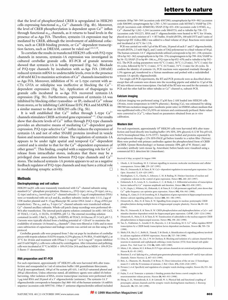
Ca2+-dependent messengers and transcription factors such asCREB are important in initiating gene transcription1,2. Severaldifferent Ca2+-dependent kinases and phosphatases, including cal-modulin/Ca2+-dependent (CaM) kinases I, II and IV, protein kinaseA (PKA) and the Ras mitogen-activated kinase (MAPK) signallingpathway are responsible for regulating CREB activation2,28,29. Incu-bation of a1A-transfected cells with calmidazolium (10 mM) toblock calmodulin activity or KN-62 (900 nM), an inhibitor ofCaM kinases II and IV, prevented syntaxin-1A expression (Fig. 4a).Incubation with H-9 (20 mM), H-8 (1 mM) or Rp-cAMP (10 mM)to inhibit PKA resulted in the block of syntaxin-1A expression ina1A-transfected cells (Fig. 4a). Conversely, a 10-min application ofthe PKA activators forskolin (20 mM) or Sp-cAMPS (10 mM)induced syntaxin-1A mRNA in the absence of a1A P/Q-typechannels. Activation of protein kinase C with 12-O-tetradecanoyl-phorbol-13-acetate (TPA) did not induce expression in the absenceof a1A channels. Lastly, block of tyrosine kinase and MAPK kinase(MAPKK) activities by incubation with genistein (50 mM), and PD098059 (50 mM), respectively, inhibited syntaxin-1A expression ina1A-transfected cells, indicating that there may be involvement ofthe rsk kinase pathway.
Western blot analysis of control and a1A-transfected cells showed
α 1A Calmidaz
olium
KN-62
H-9 H-8 Rp-cAM
PS
Fors
kolin
Sp-cAM
PS
PMA
Genist
ein
PD 098
059
Fors
kolin
α 1A +
ω-A
ga IV
A
α 1AUntra
nsfe
cted
a
b
α1A
Syntaxin-1A
Mr
66
97
45
CaMK PKA MAPKK- +
Figure 4 Involvement of second messengers in syntaxin-1A expression. a, Inhibition ofcalmodulin, CaM kinase II/IV or PKA blocks syntaxin-1A expression in a1A-transfectedcells. Stimulation of PKA with forskolin (20 mm) or Sp-cAMPS (10 mm) in untransfectedcells activates expression (1 h after treatment). Activation of PKC with PMA (100 nM, 1 h)is ineffective, whereas incubation with genistein (50 mM) or PD 098059 blocksexpression. b, Western blot for phosphorylated CREB. Lane 1, control HEK293 cell lysate;lane 2, a1A-transfected HEK293 cells; lane 3, a1A transfected HEK293 cellsþv-Aga-IVA (20 nM, 10 h); lane 4, control HEK293 cells þ 20 mM forskolin (30 min).
Contro
l
ω-Aga
IVA +
thap
sigar
gin
BAPTA-A
M
EGTA-A
M
KN-62
Rp-cAM
P
PD 098
059
α1A
Syntaxin-1A
Contro
l
ω-Aga
IVA (P
-type)
ω-Aga
IVA +
KCl
GVIA (N
-type)
Nifedipine
(L-ty
pe)
α1A
Syntaxin-1A
Dantro
lene
Xesto
spon
gin C
a
b
Figure 5 P/Q-type channels trigger syntaxin-1A expression in cerebellar granule neurons.a, RT-PCR from cultured cerebellar granule-cells. Block of P/Q-type channels (v-Aga IVA,100 nM) inhibits syntaxin-1A expression even in the presence of 40 mM KCl (30 min);inhibition of N-type channels with v-CTx GVIA (1 mM) or L-type channels with nifedipine(10 mM) has no effect. b, Thapsigargin applied to granule cells (10 mM, 10 min)incubated in v-Aga IVA (100 nM) reactivates syntaxin-1A expression. Dantrolene(10 mM), Xestospongin C (1 mM), BAPTA-AM (2 mM), EGTA-AM (2 mM), KN-62 (900 nM),Rp-CAMPS (10 mM) and PD 098059 (50 mM) block expression.
© 1999 Macmillan Magazines Ltd
letters to nature
NATURE | VOL 401 | 21 OCTOBER 1999 | www.nature.com 803
that the level of phosphorylated CREB is upregulated in HEK293cells expressing functional a1A Ca2+ channels (Fig. 4b). Moreover,the level of CREB phosphorylation is dependent upon Ca2+ influxthrough functional a1A channels, as it returns to basal levels in thepresence of v-Aga IVA. Therefore, syntaxin-1A expression may bemediated by CREB, although the involvement of additional cofac-tors, such as CREB-binding protein, or Ca2+-dependent transcrip-tion factors, such as DREAM, cannot be ruled out2,10,11,30.
To correlate the results in transfected HEK293 cells with P/Q-typechannels in neurons, syntaxin-1A expression was investigated incultured cerebellar granule cells. RT-PCR of granule neuronsshowed that syntaxin-1A is basally expressed (Fig. 5a). Blockadeof P/Q-type channels by incubation with v-Aga IVA (100 nM)reduced syntaxin mRNA to undetectable levels, even in the presenceof 40 mM KCl to maximize activation of Ca2+ channels insensitive tov-Aga IVA. Moreover, inhibition of N- or L-type current with v-CTx GVIA or nifedipine was ineffective at blocking the Ca2+-dependent expression (Fig. 5a). Application of thapsigargin togranule cells incubated in v-Aga IVA recovered syntaxin-1Aexpression (Fig. 5b). Furthermore, expression of syntaxin-1A wasinhibited by blocking either ryanodine- or IP3-induced Ca2+ releasefrom stores, or by inhibiting CaM Kinase II/IV, PKA and MAPKK ina similar manner to that in HEK293 cells (Fig. 5b).
It is well established that Ca2+ influx through L-type Ca2+
channels stimulates CREB-activated gene expression6–11. Our resultsshow that discrete levels of Ca2+ influx through P/Q-type channelsprovides an alternative means of mediating Ca2+-dependent geneexpression. P/Q-type-selective Ca2+ influx induces the expression ofsyntaxin-1A and not of other SNARE proteins involved in vesiclefusion and neurotransmitter release. The regulation of syntaxin-1Aappears to be under tight spatial and temporal Ca2+-dependentcontrol and is similar to that for the Ca2+-dependent expression ofother genes4,5. This finding, coupled with a supporting role for Ca2+
release from intracellular stores, indicates that there may be aprivileged close association between P/Q-type channels and Ca2+
stores. The induced syntaxin-1A protein appears to act as a negativefeedback regulator of P/Q-type channels and may have a critical rolein modulating synaptic activity. M
MethodsElectrophysiology and cell cultureHEK293 tsa201 cells were transiently transfected with Ca2+-channel subunits usingstandard Ca2+-phosphate precipitation. Human a1A (P/Q-type), rat a1B (N-type), rat a1C
(L-type) and rat a1E (novel type) Ca2+-channel subunits (1–3 mg of complementary DNA)were co-transfected with rat a2d and b1b ancillary subunits (molar ratio 1:1:1), 2 mg ofCD8 marker plasmid and 9–15 mg Bluescript SK carrier DNA (total ¼ 20 mg cDNA pertransfection). The a1G and a1I T-type Ca2+-channel subunits were transfected withoutCa2+-channel ancillary subunits. Whole-cell patch clamp recordings were performed 48–96 h after transfection. The internal patch-pipette solution contained (in mM): 105 CsCl,25 TEACl, 1 CaCl2, 11 EGTA, 10 HEPES, pH 7.2. The external recording solutioncontained (in mM): 5 BaCl2, 1 MgCl2, 10 HEPES, 40 TEACl, 10 Glucose, 87.5 CsCl, pH 7.2.Currents were typically elicited from a holding potential of −120 mV to maximize theavailability of syntaxin-1A-bound a1A channels20. Data were filtered at 1 kHz and in mostcases subtraction of capacitance and leakage currents was carried out on-line using a P/4protocol.
Cerebellar granule cells were prepared from 7-day rat pups by incubation of cerebellarpieces with trypsin solution (0.025% trypsin, 0.25% glucose, 0.3% BSA, 35 mM MgSO4) at37 8C for 20 min and treatment with DNase I (1 mg ml−1 in 0.1 mg ml−1 trypsin inhibitorand 35 mM MgSO4); cells were collected by centrifugation. After trituration and pelleting,cells were incubated at 37 8C in MEM þ 10% FCS for 24 h and then in MEM þ 10% FCSþ
5 fluoro 59-deoxyuridine.
RNA preparation and RT-PCRFor each experiment, approximately 106 HEK293 cells were harvested 48 h after trans-fection and resuspended in 1 ml of extraction buffer (4M guanidinium thiocyanate,20 ml b mercaptoethanol, 100 ml of Na-acetate pH 4.0), 1 ml H2O-saturated phenol and200 ml chloroform. Unless otherwise stated, all inhibitory agents were added 4 h beforeharvesting. After isolation of RNA, reverse transcriptase reactions were performed with1 mg total RNA and 20 pmol of a 39 oligonucleotide (antisense). The syntaxin-1A 39oligonucleotide corresponds to basepairs (bp) 860–841 of the human syntaxin-1A mRNAsequence (accession code M95734). Other 39 antisense oligonucleotides utilized included
syntaxin 1B bp 749–769 (accession code 4507290); synaptophysin bp 919–902 (accessioncode X06389); synaptotagmin bp 1,783–1,765 (accession code M55047); VAMP bp 279–262 pb (accession code AF060538); SNAP-25 bp 453–436 (accession code L19760), a1A
Ca2+ channel bp 1,262–1,244 (accession code M64373); and a-tubulin bp 738–721(accession code V01227). RNA and 39 oligonucleotides were heated to 90 8C for 10 min,placed on ice and a mixture of 1 3 RT buffer, 10 mM dNTPs, 100 mM DTT and 5 units ofSuperscript RT (Gibco BRL) was added to a final volume of 20 ml. Reactions were mixedand incubated at 42 8C for 1.5 h.
PCR was carried out with 5 ml of the RTmix, 20 pmol of each 59 and 39 oligonucleotide,10 mM dNTPs, 2.5 mM MgCl2 and 2 units of TAQ polymerase to a final volume of 50 ml.The human syntaxin-1A 59 oligonucleotide utilized corresponds to bp 241–258; syntaxin-1B to bp 342–362; synaptophysin to bp 556–574; synaptotagmin to bp 799–816; VAMP tobp 34–52; SNAP-25 to bp 89–106; a1A P/Q-type to bp 652–670; and a-tubulin to bp 592–612. The PCR cycling parameters were 92 8C (1 min), 50 8C (1.25 min), 74 8C (1 min) for10 cycles, followed by 92 8C (1 min), 57 8C (0.75 min), 74 8C (1 min) for 20 cycles. Toconfirm the identity of syntaxin-1A, PCR reactions were separated routinely through a1.3% agarose gel, transferred to a Nytran membrane and probed with a radiolabelledsyntaxin-1A-specific oligonucleotide.
For single-cell PCR experiments, the RT and PCR protocols were as described aboveexcept that cell contents were drawn into the recording pipette and added to 12 ml of theRTmix without reverse transcriptase. One half of the RTmix was used for the syntaxin-1APCR and the other half for either tubulin or Ca2+-channel a1-subunit PCR.
Calcium imagingCalcium imaging was performed using 17 mM fura-2 AM loaded into HEK293 cells(30 min, room temperature in 0.067% pluronic). Resting [Ca]i was estimated by taking380/360 nm excitation image pairs (isosbestic point ratio) in DMEM culture medium thatwas equilibrated with a 5% CO2/95% air mixture and continuously perfused. Ratio valueswere converted to [Ca2+]i values based on parameters obtained from an in vitrocalibration.
Western blotFor each experiment, approximately 106 HEK293 cells were harvested 48 h after trans-fection and lysed directly into loading buffer (4% SDS, 20% glycerol, 0.12 M Tris pH 6.8,0.01% bromophenol blue, 0.1% DTT). Samples were boiled and proteins separated byelectrophoresis through a 12% SDS-PAGE gel before transfer to a nylon filter (Nytran).Blots were probed with primary antibodies against the phosphorylated form of CREB(pCREB, Upstate Biotechnology) or human syntaxin (SP8, gift of W. Honer), andsecondary antibody (anti-mouse Ig, Amersham) before bands were visualized using acommercial ECL detection kit (Amersham).
Received 4 May; accepted 26 August 1999.
1. Ghosh, A. & Greenburg, M. E. Calcium signalling in neurons: molecular mechanisms and cellular
consequences. Science 268, 239–247 (1995).
2. Bito, H., Deisseroth, K. & Tsien, R. W. Ca2+-dependent regulation in neuronal gene expression. Curr.
Opin. Neurobiol. 7, 419–429 (1997).
3. Hardingham, G. E., Chawla, S., Johnson, C. M. & Bading, H. Distinct functions of nuclear and
cytoplasmic calcium in the control of gene expression. Nature 385, 260–265 (1997).
4. Dolmetsch, R. E., Lewis, R. S., Goodnow, C. C. & Healy, J. I. Differential activation of transcription
factors induced by Ca2+ response amplitude and duration. Nature 386, 855–858 (1997).
5. Li, W., Llopis, J., Whitney, M., Zlokarnik, G. & Tsien, R. Y. Cell-permeant caged InsP3 ester shows that
Ca2+ spike frequency can optimize gene expression. Nature 392, 936–941 (1998).
6. Bading, H., Ginty, D. D. & Greenberg, M. E. Regulation of gene expression in hippocampal neurons by
distinct calcium signaling pathways. Science 260, 181–186 (1993).
7. Deisseroth, K., Bito, H. & Tsien, R. W. Signalling from synapse to nucleus: postsynaptic CREB
phosphorylation during multiple forms of hippocampal synaptic plasticity. Neuron 16, 89–101
(1996).
8. Bito, H., Deisseroth, K. & Tsien, R. W. CREB phosphorylation and dephosphorylation: a Ca2+- and
stimulus duration-dependent switch for hippocampal gene expression. Cell 87, 1203–1214 (1996).
9. Diesseroth, K., Heist, E. K. & Tsien, R. W. Translocation of calmodulin to the nucleus supports CREB
phosphorylation in hippocampal neurons. Nature 392, 198–202 (1998).
10. Tao, X., Finkbeiner, S., Arnold, D. B., Shaywitz, A. J. & Greenberg, M. E. Ca2+ influx regulates BDNF
transcription by a CREB family transcription factor-dependent mechanism. Neuron 20, 709–726
(1998).
11. Shieh, P. B., Hu, S.-C., Bobb, K., Timusk, T. & Ghosh, A. Identification of a signaling pathway involved
in calcium regulation of BDNF expression. Neuron 20, 727–740 (1998).
12. Llina, R., Sugimori, M., Lin, J. W. & Cherksey, B. Blocking and isolation of a calcium channel from
neurons in mammals and cephalopods utilizing a toxin fraction (FTX) from funnel-web spider
poison. Proc. Natl Acad. Sci. USA 86, 1689–1693 (1989).
13. Mintz, I. M., Adams, M. E. & Bean, B. P. P-type calcium channels in rat central and peripheral neurons.
Neuron 9, 85–95 (1992).
14. Bourinet, E. et al. Splicing of a1A subunit gene generates phenotypic variants of P- and Q-type calcium
channels. Nature Neurosci. 2, 407–415 (1999).
15. Betz, A., Okamoto, M., Benseler, F. & Brose, N. Direct interaction of the rat unc-13 homologue
munc13-1 with the N-terminus of syntaxin. J. Biol. Chem. 272, 2520–2526 (1997).
16. Pevsner, J. et al. Specificity and regulation of a synaptic vesicle docking complex. Neuron 13, 353–361
(1994).
17. Fujita, Y. et al. Tomosyn: a syntaxin-1-binding protein that forms a novel complex in the
neurotransmitter release process. Neuron 20, 905–915 (1998).
18. Sheng, Z.-H., Westenbroek, R. E. & Catterall, W. A. Physical link and functional coupling of
presynaptic calcium channels and the synaptic vesicle docking/fusion machinery. J. Bioenerg.
Biomembr. 30, 335–345 (1998).
© 1999 Macmillan Magazines Ltd
letters to nature
804 NATURE | VOL 401 | 21 OCTOBER 1999 | www.nature.com
19. Sheng, Z.-H., Rettig, J., Takahashi, M. & Catterall, W. A. Identification of a syntaxin-binding site on
N-type calcium channels. Neuron 13, 1303–1313 (1994).
20. Bezprozvanny, I., Scheller, R. H. & Tsien, R. W. Functional impact of syntaxin on gating of N-type and
Q-type calcium channels. Nature 378, 623–626 (1995).
21. Martin-Moutot, N. et al. Interaction of SNARE complexes with P/Q-type calcium channels in rat
cerebellar synaptosomes. J. Biol. Chem. 271, 6567–6570 (1996).
22. Rettig, J. et al. Isoform-specific interaction of the a1A subunits of brain Ca2+ channels with the
presynaptic proteins syntaxin and SNAP-25. Proc. Natl Acad. Sci. USA 93, 7363–7368 (1996).
23. Kim, D. K. & Catterall, W. A. Ca2+-dependent and -independent interactions of the isoforms of the a1A
subunit of brain Ca2+ channels with presynaptic SNARE proteins. Proc. Natl Acad. Sci. USA 94,
14782–14786 (1997).
24. Williamson, L. C., Halpern, J. L., Montecucco, C., Brown, J. E. & Neale, E. A. Clostridial neurotoxins
and substrate proteolysis in intact neurons: botulinum neurotoxin C acts on synaptosomal-associated
protein of 25 kDa. J. Biol. Chem. 271, 7694–7699 (1996).
25. Honer, W. G., Hu, L, & Davies, P. Human synaptic proteins with a heterogenous distribution in
cerebellum and visual cortex. Brain Res. 609, 9–20 (1993).
26. Daly, C. & Ziff, E. B. Post-transcriptional regulation of synaptic vesicle protein expression and the
developmental control of synaptic vesicle formation. J. Neurosci. 17, 2365–2375 (1997).
27. Gafni, J. et al. Xestospongins: potent membrane permeable blockers of the inositol 1,4,5-trisphosphate
receptor. Neuron 19, 723–733 (1997).
28. Gonzalez, G. A. & Montminy, M. R. Cyclic AMP stimulates somatostatin gene transcription by
phosphorylation of CREB at serine 133. Cell 59, 675–680 (1989).
29. Ginty, D. D., Bonni, A. & Greenberg, M. E. Nerve growth factor activates a Ras-dependent protein
kinase that stimulates c-fos transcription via phosphorylation of CREB. Cell 77, 713–725 (1994).
30. Carrion, A. M., Link, W. A., Ledo, F., Mellstrom, B. & Naranjo, J. R. DREAM is a Ca2+-regulated
transcriptional repressor. Nature 398, 80–84 (1999).
AcknowledgementsWe thank M. Gilbert, C. Santi, A. Stea and G. Zamponi for comments and discussions. Wealso thank W. Honer for providing the SP8 human syntaxin-1A monoclonal antibody andD. Brink and V. Leuranguer for tissue culture support. The research was supported bygrants from the Medical Research Council (MRC) of Canada (T.P.S. and T.H.M.),postdoctoral support from the Amyotrophic Lateral Sclerosis Society of Canada (K.S.),and studentship and postdoctoral fellowship support from the MRC (H.G. and J.M.,respectively). T.P.S. and T.H.M. are recipients of MRC of Canada Scientist awards.
Correspondence and requests for materials should be addressed to T.P.S.(e-mail: [email protected]).
.................................................................Extraintestinal disseminationof Salmonella byCD18-expressing phagocytesAndres Vazquez-Torres*, Jessica Jones-Carson†, Andreas J. Baumler‡,Stanley Falkow§, Raphael Valdivia§k, William Brown*¶, Mysan Le*¶,Ruth Berggren*k, W. Tony Parks# & Ferric C. Fang*
* Departments of Medicine, Pathology, and Microbiology, University of ColoradoHealth Sciences Center, Denver, Colorado 80262, USA† Department of Immunology, National Jewish Center for Immunology andRespiratory Medicine, Denver, Colorado 80262, USA‡ Department of Medical Microbiology and Immunology, Texas A&M University,College Station, Texas 77843-1114, USA§ Department of Microbiology and Immunology, Stanford University School ofMedicine, Stanford, California 94305, USA¶ Department of Medicine, Veterans Affairs Medical Center, Denver,Colorado 80262, USA# Laboratory of Cell Regulation and Carcinogenesis, National Cancer Institute,National Institutes of Health, Bethesda, Maryland 20892, USA
.................................. ......................... ......................... ......................... ......................... ........
Specialized epithelia known as M cells overlying the lymphoidfollicles of Peyer’s patches are important in the mucosal immunesystem, but also provide a portal of entry for pathogens such asSalmonella typhimurium, Mycobacterium bovis, Shigella flexneri,Yersinia enterocolitica and reoviruses1–4. Penetration of intestinal
kPresent addresses: Department of Biochemistry, University of California at Berkeley, Berkeley, California94720, USA (R.V.); Department of Medicine, University of Texas Southwestern Medical Center, Dallas,Texas 75325, USA (R.B.).
M cells and epithelial cells by Salmonella typhimurium requiresthe invasion genes of Salmonella Pathogenicity Island 1 (SPI1)3,5–9.SPI1-deficient S. typhimurium strains gain access to the spleenfollowing oral administration and cause lethal infection in mice5
without invading M cells3,9 or localizing in Peyer’s patches10,which indicates that Salmonella uses an alternative strategy todisseminate from the gastrointestinal tract. Here we report thatSalmonella is transported from the gastrointestinal tract to thebloodstream by CD18-expressing phagocytes, and that CD18-deficient mice are resistant to dissemination of Salmonella tothe liver and spleen after oral administration. This CD18-depen-dent pathway of extraintestinal dissemination may be importantfor the development of systemic immunity to gastrointestinalpathogens, because oral challenge with SPI1-deficient S. typhi-murium elicits a specific systemic IgG humoral immune response,despite an inability to stimulate production of specific mucosalIgA.
As phagocytic cells transmigrate across normal tissue barriers11
and Salmonella can survive within mononuclear phagocytic cells12,we proposed that tissue macrophages transport Salmonella from theintestine to the systemic circulation; CD18-deficient mice13 pro-vided a model in which to test this hypothesis. Among its manyfunctions, the b2-integrin CD18 mediates leukocyte transmigrationin response to various stimuli, including S. typhimurium14,15.
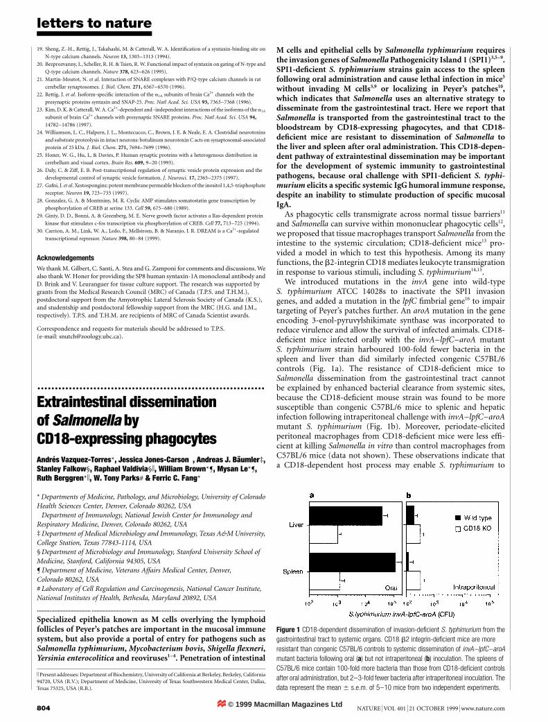
We introduced mutations in the invA gene into wild-typeS. typhimurium ATCC 14028s to inactivate the SPI1 invasiongenes, and added a mutation in the lpfC fimbrial gene16 to impairtargeting of Peyer’s patches further. An aroA mutation in the geneencoding 3-enol-pyruvylshikimate synthase was incorporated toreduce virulence and allow the survival of infected animals. CD18-deficient mice infected orally with the invA–lpfC–aroA mutantS. typhimurium strain harboured 100-fold fewer bacteria in thespleen and liver than did similarly infected congenic C57BL/6controls (Fig. 1a). The resistance of CD18-deficient mice toSalmonella dissemination from the gastrointestinal tract cannotbe explained by enhanced bacterial clearance from systemic sites,because the CD18-deficient mouse strain was found to be moresusceptible than congenic C57BL/6 mice to splenic and hepaticinfection following intraperitoneal challenge with invA–lpfC–aroAmutant S. typhimurium (Fig. 1b). Moreover, periodate-elicitedperitoneal macrophages from CD18-deficient mice were less effi-cient at killing Salmonella in vitro than control macrophages fromC57BL/6 mice (data not shown). These observations indicate thata CD18-dependent host process may enable S. typhimurium to
Figure 1 CD18-dependent dissemination of invasion-deficient S. typhimurium from thegastrointestinal tract to systemic organs. CD18 b2 integrin-deficient mice are moreresistant than congenic C57BL/6 controls to systemic dissemination of invA–lpfC–aroAmutant bacteria following oral (a) but not intraperitoneal (b) inoculation. The spleens ofC57BL/6 mice contain 100-fold more bacteria than those from CD18-deficient controlsafter oral administration, but 2–3-fold fewer bacteria after intraperitoneal inoculation. Thedata represent the mean 6 s.e.m. of 5–10 mice from two independent experiments.


![Integrating the Healthcare Enterprise€¦ · Document Source Document ConsumerOn Entry [ITI Document Registry Document Repository Provide&Register Document Set – b [ITI-41] →](https://img.pdfslide.net/doc/110x75/5f08a1eb7e708231d422f7c5/integrating-the-healthcare-enterprise-document-source-document-consumeron-entry.jpg)


