Embed Size (px)
Citation preview
Rolf D. Issels and Hermann WagnerSanghamitra Ghose, Carsten J. Kirschning, Ramunas M. Vabulas, Parviz Ahmad-Nejad,
PathwayToll/Interleukin-1 Receptor Signal HSP70 as Endogenous Stimulus of theTRANSDUCTION:MECHANISMS OF SIGNAL
2002, 277:15107-15112.J. Biol. Chem.
http://www.jbc.org/content/277/17/15107Access the most updated version of this article at
.JBC Affinity SitesFind articles, minireviews, Reflections and Classics on similar topics on the
Alerts:
When a correction for this article is posted•
When this article is cited•
to choose from all of JBC's e-mail alertsClick here
http://www.jbc.org/content/277/17/15107.full.html#ref-list-1
This article cites 30 references, 10 of which can be accessed free at
at VIV
A, V
A C
omm
onwealth U
niv on June 6, 2014http://w
ww
.jbc.org/D
ownloaded from
at V
IVA
, VA
Com
monw
ealth Univ on June 6, 2014
http://ww
w.jbc.org/
Dow
nloaded from
HSP70 as Endogenous Stimulus of the Toll/Interleukin-1 ReceptorSignal Pathway*
Received for publication, November 23, 2001, and in revised form, January 16, 2002Published, JBC Papers in Press, February 12, 2002, DOI 10.1074/jbc.M111204200
Ramunas M. Vabulas‡§, Parviz Ahmad-Nejad‡¶, Sanghamitra Ghose¶�, Carsten J. Kirschning‡,Rolf D. Issels�**, and Hermann Wagner‡ ‡‡
From the ‡Institute of Medical Microbiology, Immunology and Hygiene, Technical University of Munich, Trogerstrasse 9,Munich 81675, Germany, the §Institute of Immunology, Moletu� pl. 29, Vilnius 2021, Lithuania, the �KlinikumGrosshadern, Medical Clinic III, Ludwig-Maximilians-University, Munich 81377, Germany, and the **National ResearchCenter for Environment and Health, KKG Hyperthermie, Institute of Molecular Immunology, Munich 81377, Germany
Human heat-shock protein (HSP)70 activates innateimmune cells and hence requires no additional adju-vants to render bound peptides immunogenic. Here wetested the assumption that endogenous HSP70 activatesthe Toll/IL-1 receptor signal pathway similar to HSP60and pathogen-derived molecular patterns. We show thatHSP70 induces interleukin-12 (IL-12) and endothelialcell-leukocyte adhesion molecule-1 (ELAM-1) promotersin macrophages and that this is controlled by MyD88and TRAF6. Furthermore, HSP70 causes MyD88 relocal-ization and MyD88-deficient dendritic cells do not re-spond to HSP70 with proinflammatory cytokine produc-tion. Using the system of genetic complementation withToll-like receptors (TLR) we found that TLR2 and TLR4confer responsiveness to HSP70 in 293T fibroblasts. Theexpanding list of endogenous ligands able to activatethe ancient Toll/IL-1 receptor signal pathway is in linewith the “danger hypothesis” proposing that the innateimmune system senses danger signals even if they orig-inate from self.
Stimuli that activate innate immune cells such as dendriticcells (DCs)1 are at present subject of intensive studies, becauseof the central role of these cells in initiating and controllingadaptive immune responses. There are at least two lines ofthought in regard to the nature of such stimuli. The firstimplies that “exogenous” pathogen-associated (hence, “for-eign”) molecular patterns (PAMPs) are selectively recognizedby germ line encoded Toll-like receptors (TLRs), which subse-quently drive innate immune cell activation (1). In favor of thisview, PAMPs such as bacterial lipopeptides, lipopolysaccha-rides (LPS) from Gram-negative bacteria, flagellin, and bacte-rial CpG-DNA are recognized by TLR2, TLR4, TLR5, andTLR9, respectively (2). Ligand-driven TLR activation causes
consecutive recruitment of the adaptor molecule MyD88, theIL-1 receptor-associated kinases and the adaptor moleculeTRAF6 (3). This ancient Toll/IL-1 receptor (TIR) signal path-way ultimately leads to activation of transcription factors thatswitch on production of proinflammatory mediators such asIL-12 and tumor necrosis factor (TNF)�, costimulatory mole-cules from B7 family, and adhesion molecules, for example,endothelial cell-leukocyte adhesion molecule (ELAM-1; E-se-lectin). Given the importance of IL-12 in cell-mediated immu-nity (4), the indispensability of costimulatory molecules for fullactivation of lymphocytes (5), and the role of adhesion mole-cules in recruiting leukocytes (6) and forming the proper im-munological synapses between interacting cells (7), it becomesclear why TLRs have the central position in both innate andadaptive anti-infectious responses.
Alternatively, it has been suggested that DCs act as senti-nels of “endogenous” ligands released from cells undergoingunprogrammed necrotic death as opposed to “silent” pro-grammed cell death, i.e. apoptosis (8, 9). According to this viewnot the “foreign-ness” is sensed but the danger, thus the name“danger hypothesis” (10). Identification of endogenous dangersignals will help to better understand how the immune systemfunctions. First candidates in line are family members of in-tracellular HSPs, which, upon necrotic cell death might bereleased and thus could be able to activate DCs (11, 12). Forexample HSP60 (13), HSP70 (14, 15), as well as gp96 (16)deliver activation signals to innate immune cells. Because in-duction of specific CD8 T cell responses by HSP�peptide com-plexes appears not to require adjuvants to confer immunoge-nicity to bound peptides (17) the ability of HSPs to activate DCsmay not only explain the immunogenicity of HSP�peptide com-plexes but also it represents a strong argument in favor of thedanger hypothesis.
Members of the HSP70 cytosolic group are either constitu-tively expressed (HSC70) or can be induced by a broad range ofstress factors (HSP70). The inducible HSP70 has been recentlycharacterized as a potent maturation stimulus for DCs (18).Driven by the quest to identify/compare the pathways throughwhich exogenous and endogenous ligands activate innate im-mune cells, and encouraged by recent data on HSP60 signaling(19), we searched for further cases of dual specificity (endoge-nous and exogenous) of the Toll/IL-1 receptor signal pathwayby examining HSP70-triggered intracellular events. Our datadefine HSP70 as an endogenous stimulus for the Toll/IL-1receptor signal pathway that engages TLR2 and TLR4.
EXPERIMENTAL PROCEDURES
Plasmids and Reagents—A FLAG epitope-tagged C terminus of mu-rine MyD88 (dnMyD88) was described previously (19). The expressionvectors for the C terminus of human TRAF6 (dnTRAF6), human flag-
* This work was supported by the Deutsche ForschungsgemeinschaftSFB456, partly by SFB455/B9, and by the Fonds der ChemischenIndustrie. The costs of publication of this article were defrayed in partby the payment of page charges. This article must therefore be herebymarked “advertisement” in accordance with 18 U.S.C. Section 1734solely to indicate this fact.
¶ Both authors contributed equally to this work.‡‡ To whom correspondence should be addressed: Tel.: 49-89-4140-
4120; Fax: 49-89-4140-4868; E-mail: [email protected] The abbreviations used are: DCs, dendritic cells; PAMPs, pathogen-
associated molecular patterns; TLRs, Toll-like receptors; LPS, lipopo-lysaccharide; TIR, Toll/IL-1 receptor; TNF, tumor necrosis factor;ELAM-1, endothelial cell-leukocyte adhesion molecule; IL, interleukin;HSP, heat-shock protein; BMDC, bone marrow-derived dendritic cells;EGFP, enhanced green fluorescence protein; PMA, phorbol 12-myris-tate 13-acetate; FCS, fetal calf serum; ELISA, enzyme-linked immu-nosorbent assay; dn, dominant negative.
THE JOURNAL OF BIOLOGICAL CHEMISTRY Vol. 277, No. 17, Issue of April 26, pp. 15107–15112, 2002© 2002 by The American Society for Biochemistry and Molecular Biology, Inc. Printed in U.S.A.
This paper is available on line at http://www.jbc.org 15107
at VIV
A, V
A C
omm
onwealth U
niv on June 6, 2014http://w
ww
.jbc.org/D
ownloaded from
tagged TLR2, TLR4, and E-selectin (ELAM-1) promoter luciferase con-struct were gifts from Tularik, Inc. (South San Francisco, CA); murineTLR9 in pcDNA3 was a gift from S. Bauer (Technical University ofMunich, Munich, Germany); the human MD-2 expression vector waskindly provided by K. Miyake (Saga Medical School, Nabeshima,Japan). The luciferase reporter driven by a synthetic enhancer harbor-ing 6 NF-�B binding consensus sites was a gift from P. Baeuerle(Munich, Germany). The IL-12p40 promoter (�703 to �53) luciferaseconstruct was a gift from K. M. Murphy (Washington University, St.Louis, MO).
Recombinant human HSP70 was purchased from StressGen Biotech-nologies (Victoria, Canada). To control contamination with LPS, HSP70preparations were boiled for 30–60 min where indicated. Phosphothio-ate-stabilized CpG oligonucleotide 1668 (TCC ATG ACG TTC CTG ATGCT) was purchased from TIB Molbiol (Berlin, Germany). LPS fromEscherichia coli 055:B5 and PMA were from Sigma-Aldrich (Munich,Germany).
Cell Culture, Transfection, and Luciferase Assays—The murinemacrophage cell line RAW264.7 was grown in VLE-RPMI medium(Biochrom KG, Berlin, Germany) supplemented with 10% fetal calfserum (FCS), 100 IU/ml penicillin G, and 100 IU/ml streptomycinsulfate (all from Biochrom KG). 5–10 � 106 RAW264.7 cells weretransfected by electroporation in a 400-�l final volume (RPMI/25%FCS) at 300 V and 960 microfarads in a Gene Pulser (Bio-Rad Labora-tories, Munich, Germany). 5 �g of reporter plasmids was used fortransfection together with different amounts of specific expression vec-tors as indicated in the figure legends. The overall amount of plasmidDNA was held constant at 20 �g per electroporation by addition of theappropriate empty expression vector. After electroporation, cells werewashed and split into 12-well plates for subsequent stimulation andlysis.
The human embryonic kidney fibroblasts 293T were cultured inDulbecco’s modified Eagle’s medium (Biochrom KG) with the samesupplements as for RAW264.7 macrophage cell cultures. For luciferasereporter assays the 293T cells were transfected by electroporation sim-ilar as RAW264.7 except that 200 V were used. 10 ng of 6� NF-�Bluciferase reporter together with 0.1 �g of respective expression vectoror empty control vector (as indicated in figure legend) per transfectionwere used. The overall DNA amount was held constant at 20 �g byaddition of empty vector.
For luciferase assays, transfected cells were stimulated as de-scribed in the Fig. 5 legend, lysed, and luciferase activity in extractswas measured with the Luciferase Assay System kit from Promega(Mannheim, Germany) according to manufacturer’s instruction.
Mice, Generation of BMDC, and Determination of Cytokines—MyD88-deficient mice were a kind gift from S. Akira (Osaka University,Osaka, Japan) (20). Gene-targeted mice deficient for TLR2 (TLR2�/�)were granted by Tularik Inc. (21). C3H/HeJ mice (TLR4d/d) and C3H/HeN (TLR4�/�) were purchased from Harlan (Germany) for 5-fold back-crossing of TLR2�/� mice toward the C3H genetic background. Micewere genotyped for the point mutation of the TLR4 gene responsible forthe amino acid exchange P712H of the intracellular domain of TLR4and characteristic for the C3H/HeJ strain (22). F1 generation mice ofparent mice 5-fold backcrossed toward the C3H background carryingboth the TLR2 and the TLR4 mutations heterozygously were used forfurther crossing. Offspring mice carrying one of each, the mutated orthe wild type allele homozygously (TLR2�/�/TLR4�/�, TLR2�/�/TLR4�/�, TLR2�/�/TLR4d/d, TLR2�/�/TLR4d/d) were selected pairwisefor further breeding. Age-matched groups of wild-type and mutant micewere used for the experiments.
Bone marrow-derived dendritic cells (BMDC) were prepared as de-scribed previously (23). For stimulation, nonadherent BMDC at days6–7 were washed, plated at 3.8–7.5 � 105/ml, and after a 2-h rest cellswere stimulated in triplicates for 20 h. IL-12p40 and TNF� levels inculture supernatants were determined by commercially availableELISA kits (R&D Systems, Wiesbaden-Nordenstadt, Germany) accord-ing to the instructions of the manufacturer.
Confocal Laser Scanning Microscopy—7 � 105 RAW264.7 macro-phages stably expressing MyD88-EGFP2 were plated 1 day before im-age collection on round glass slides in 12-well dishes in 10% FCS,VLE-RPMI medium containing 500 units/ml interferon �. 3 h prior tostimulation, medium was exchanged with fresh 0% FCS-containingmedium. Cells were stimulated as indicated in the legend of Fig. 3,washed, and formalin-fixed. Samples were viewed with a Zeiss LSM 510confocal microscope (Carl Zeiss, Jena, Germany) using a Argon 488-nm
laser, a Plan-Neofluar 40 1.3 oil lens, and LSM 510 version 2.02 soft-ware. Slices 1-�m thick were collected at a resolution of 1024 � 1024pixels.
RESULTS
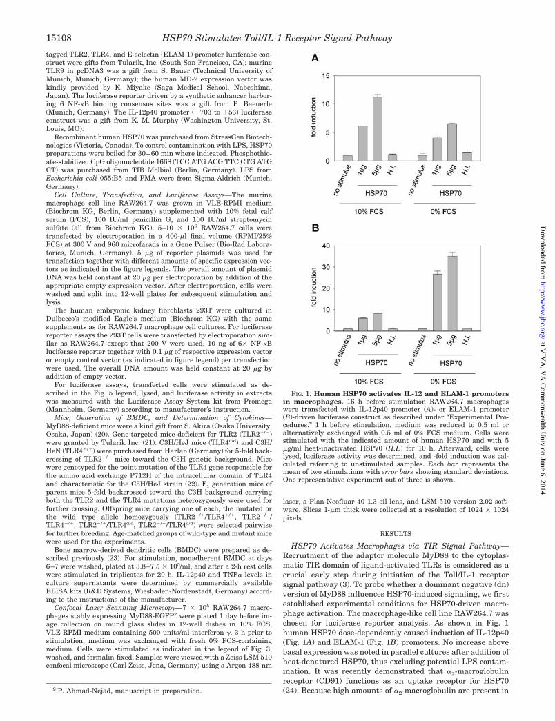
HSP70 Activates Macrophages via TIR Signal Pathway—Recruitment of the adaptor molecule MyD88 to the cytoplas-matic TIR domain of ligand-activated TLRs is considered as acrucial early step during initiation of the Toll/IL-1 receptorsignal pathway (3). To probe whether a dominant negative (dn)version of MyD88 influences HSP70-induced signaling, we firstestablished experimental conditions for HSP70-driven macro-phage activation. The macrophage-like cell line RAW264.7 waschosen for luciferase reporter analysis. As shown in Fig. 1human HSP70 dose-dependently caused induction of IL-12p40(Fig. 1A) and ELAM-1 (Fig. 1B) promoters. No increase abovebasal expression was noted in parallel cultures after addition ofheat-denatured HSP70, thus excluding potential LPS contam-ination. It was recently demonstrated that �2-macroglobulinreceptor (CD91) functions as an uptake receptor for HSP70(24). Because high amounts of �2-macroglobulin are present in2 P. Ahmad-Nejad, manuscript in preparation.
FIG. 1. Human HSP70 activates IL-12 and ELAM-1 promotersin macrophages. 16 h before stimulation RAW264.7 macrophageswere transfected with IL-12p40 promoter (A)- or ELAM-1 promoter(B)-driven luciferase construct as described under “Experimental Pro-cedures.” 1 h before stimulation, medium was reduced to 0.5 ml oralternatively exchanged with 0.5 ml of 0% FCS medium. Cells werestimulated with the indicated amount of human HSP70 and with 5�g/ml heat-inactivated HSP70 (H.I.) for 10 h. Afterward, cells werelysed, luciferase activity was determined, and -fold induction was cal-culated referring to unstimulated samples. Each bar represents themean of two stimulations with error bars showing standard deviations.One representative experiment out of three is shown.
HSP70 Stimulates Toll/IL-1 Receptor Signal Pathway15108
at VIV
A, V
A C
omm
onwealth U
niv on June 6, 2014http://w
ww
.jbc.org/D
ownloaded from
FCS one could presume an inhibitory effect of serum on HSP70interaction with macrophages. To probe for that possibilityserum-free stimulation with HSP70 was performed. As shownin Fig. 1 this impaired IL-12p40 reporter activity but greatlyenhanced ELAM-1 promoter induction by HSP70.
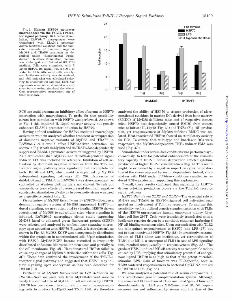
Having defined conditions for HSP70-mediated macrophageactivation we next analyzed whether transient overexpressionof dominant negative variants of MyD88 and TRAF6 inRAW264.7 cells would affect HSP70-driven activation. Asshown in Fig. 2 both dnMyD88 and dnTRAF6 dose-dependentlysuppressed ELAM-1 promoter activation triggered by HSP70.As a well-established MyD88- and TRAF6-dependent signalinducer, LPS was included for reference. Inhibition of cell ac-tivation by dominant negative molecules from the Toll/IL-1receptor signal pathway was significant but incomplete forboth HSP70 and LPS, which could be explained by MyD88-independent signaling pathways (25, 26). Expression ofdnMyD88 and dnTRAF6 in RAW264.7 was dose-dependent, ascontrolled by Western blotting (data not shown). To rule outunspecific or toxic effects of overexpressed dominant negativeconstructs, stimulation of cells by hypo-osmotic stress was usedas a specificity control (Fig. 2).
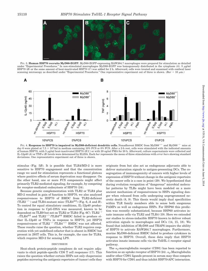
Visualization of MyD88 Recruitment by HSP70—Because adominant negative version of MyD88 suppressed HSP70-in-duced signaling, we next attempted to visualize HSP70-drivenrecruitment of MyD88 to subcellular sites where signaling isinitiated. RAW264.7 macrophage clones stably expressingMyD88 fused to enhanced green fluorescent protein (EGFP)were selected and analyzed by confocal laser scanning micros-copy upon activation with HSP70 (5 �g/ml, 2-h stimulation). Asshown in Fig. 3A MyD88-EGFP was homogenously distributedwithin the cytoplasm in nonstimulated cells. Upon stimulationwith HSP70, MyD88-EGFP became recruited to irregularlydistributed endosome-like vesicular structures and partially tothe cell membrane (Fig. 3B). Heat-denatured HSP70 failed tocause any changes of MyD88 distribution within the cells (Fig.3C). These data confirmed the involvement of the Toll/IL-1receptor signal pathway and suggested that HSP70 may ini-tiate signaling upon endocytosis as has been described forHSP60 (19).
Verification of MyD88 Involvement in Cell Activation byHSP70—Next we used cells from MyD88-deficient mice toverify the central role of MyD88 in cell activation by HSP70.HSP70 has been shown to stimulate murine antigen-present-ing cells to produce IL-12p40 and TNF� (14). We therefore
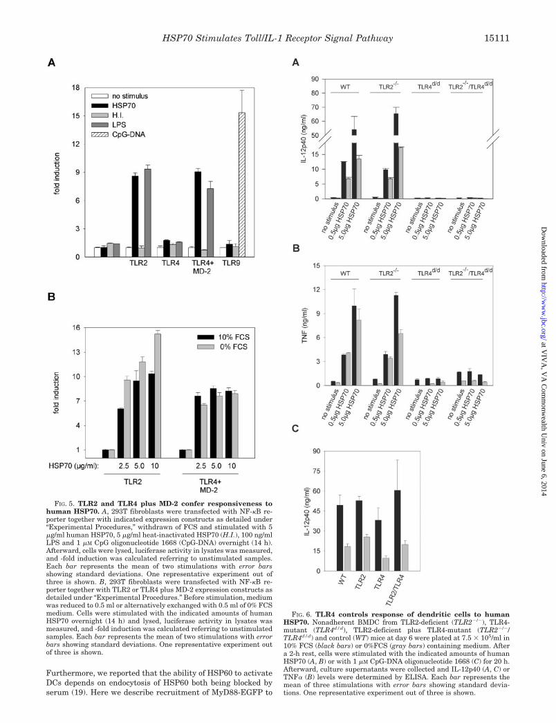
analyzed the ability of HSP70 to trigger production of afore-mentioned cytokines in murine DCs derived from bone marrow(BMDC) of MyD88-defficient mice and of respective controlmice. HSP70 dose-dependently caused BMDC from controlmice to initiate IL-12p40 (Fig. 4A) and TNF� (Fig. 4B) produc-tion, yet responsiveness of MyD88-deficient BMDC was ab-lated. Heat-inactivated HSP70 showed no stimulatory activityfor DCs. To control that wild-type and knock-out DCs wereresponsive, the MyD88-independent TNF� inducer PMA wasused (Fig. 4B).
Stimulation under serum-free conditions was performed syn-chronously, to test for potential enhancement of the stimula-tory capacity of HSP70. Serum deprivation affected cytokineproduction at higher HSP70 concentrations (Fig. 4). This resultmight be explained by a negative impact on cytokine produc-tion of the stress imposed by serum deprivation. Indeed, stim-ulation with PMA under FCS-free conditions resulted in re-duced TNF� production, supporting this explanation.
Overall, these results confirmed that signaling for HSP70-driven cytokine production occurs via the Toll/IL-1 receptorsignal pathway.
HSP70 Signals via TLR2 and TLR4—The essential role ofMyD88 and TRAF6 in HSP70-triggered cell activation sug-gested an involvement of Toll-like receptors. To analyze thispossibility we first utilized genetic complementation with TLRsof the HSP70-unresponsive human embryonic kidney fibro-blast cell line 293T. Cells were transiently transfected with aluciferase reporter driven by a synthetic enhancer harboring 6NF-�B binding consensus sites. Upon cotransfection with TLR2the cells gained responsiveness to HSP70 (and LPS (27)) butnot to heat-inactivated HSP70 (Fig. 5A). Interestingly, cotrans-fection of TLR4 alone was ineffective, yet cotransfection ofTLR4 plus MD-2, a coreceptor of TLR4 in case of LPS signaling(28), resulted unequivocally in responsiveness (Fig. 5A). Thegrade of HSP70-induced NF-�B activity was comparable to thatcaused by LPS, implying that stimulatory potential of endoge-nous ligand HSP70 is as high as that of the potent microbialstimulus LPS. Gain of function was TLR-specific, becauseTLR9 conferred responsiveness to bacterial CpG-DNA but notto HSP70 or LPS (Fig. 5A).
We also analyzed a potential role of serum components inthis reductionist genetic complementation system. Althoughthe absence of FCS-enhanced TLR2 mediated “gain of function”dose-dependently, TLR4 plus MD-2-mediated HSP70 respon-siveness was not influenced by serum and the dose of the
FIG. 2. Human HSP70 activatesmacrophages via the Toll/IL-1 recep-tor signal pathway. 16 h before stimu-lation, RAW264.7 macrophages weretransfected with ELAM-1 promoter-driven luciferase construct and the indi-cated amounts of dominant negativeMyD88 and TRAF6 constructs as de-scribed under “Experimental Proce-dures.” 1 h before stimulation, mediumwas exchanged with 0.5 ml of 0% FCSmedium. Cells were stimulated with 5�g/ml HSP70, 100 ng/ml LPS, or 500 �l ofwater for 10 h. Afterward, cells were ly-sed, luciferase activity was determined,and -fold induction was calculated refer-ring to unstimulated samples. Each barrepresents mean of two stimulations witherror bars showing standard deviations.One representative experiment out ofthree is shown.
HSP70 Stimulates Toll/IL-1 Receptor Signal Pathway 15109
at VIV
A, V
A C
omm
onwealth U
niv on June 6, 2014http://w
ww
.jbc.org/D
ownloaded from
stimulus (Fig. 5B). It is possible that TLR4/MD-2 is moresensitive to HSP70 engagement and that the concentrationrange we used for stimulation represents a functional plateauwhere positive effects of serum deprivation may disappear. Onthe other hand, one or more FCS components might affectprimarily TLR2-mediated signaling, for example, by competingfor receptor-mediated endocytosis of HSP70 (24).
Because genetic complementation with TLR2 or TLR4 plusMD-2 resulted in gain of function to HSP70, we also analyzedresponsiveness to HSP70 of BMDC from TLR2-deficient(TLR2�/�) and TLR4-mutant mice (TLR4d/d) (Fig. 6, A and B).To control for equal stimulatory conditions, IL-12p40 produc-tion in response to CpG-DNA was measured, known to bedependent on TLR9 but not on TLR2 or TLR4 (Fig. 6C). TLR2�/
�/TLR4d/d and TLR2�/�/TLR4d/d BMDC failed to produce ei-ther IL-12p40 or TNF� in response to HSP70, yet HSP70responsiveness of TLR2�/�/TLR4�/� BMDC was not affected.These results raise the question, whether TLR2 requires coop-eration with yet undefined cofactor that is absent in BMDC butpresent in 293T cells. This is, for example, the case for TLR4,which requires MD-2 for sensing LPS.
DISCUSSION
Heat-shock protein�peptide complexes do not require adju-vants to elicit peptide-specific CD8 T cell responses (17). Thisraises the question whether certain HSPs not only chaperonizepeptides mirroring the antigenic repertoire of (tumor) cells they
originate from but also act as endogenous adjuvants able todeliver maturation signals to antigen-presenting DCs. The co-segregation of immunogenicity of cancers with higher levels ofexpression of HSP70 without change in the antigenic repertoireof the cancer cells is a case in point (29). We hypothesized thatduring evolution recognition of “dangerous” microbial molecu-lar patterns by TLRs might have been modeled on a moreancient mechanism of responsiveness to HSPs signaling dan-ger when released from cells undergoing unprogrammed ne-crotic death (8, 9). This thesis would imply dual specificitieswithin TLR family members able to sense both exogenousPAMPs as well as endogenous HSPs. For HSP60 this predic-tion was recently substantiated, because HSP60 activates in-nate immune cells via TLR2 and TLR4 (19). Here we extendedour studies to stress-inducible HSP70 known to deliver robustactivation signals to macrophages and DCs (14, 15, 18). Wefound that inhibition of MyD88 and TRAF6 impairs the abilityof HSP70 to activate RAW264.7 macrophages. Furthermore,murine MyD88-deficient BMDC failed to produce cytokines inresponse to HSP70. Overall, these data imply that HSP70activates innate immune cells via the Toll/IL-1 receptor signalpathway.
The �2-macroglobulin receptor (CD91) has been reported tofunction as an uptake receptor of HSP70 (24). �2-Macroglobulinand/or other CD91 ligands present in serum may thus competewith HSP70 for CD91 and thus inhibit HSP70�APC interaction.
FIG. 3. Human HSP70 recruits MyD88-EGFP. MyD88-EGFP-expressing RAW264.7 macrophages were prepared for stimulation as detailedunder “Experimental Procedures.” In non-stimulated macrophages MyD88-EGFP was homogenously distributed in the cytoplasm (A). 5 �g/mlHSP70 (B) or the same amount of heat-inactivated HSP70 (C) was added for 2 h. Afterward, cells were treated and examined with confocal laserscanning microscopy as described under “Experimental Procedures.” One representative experiment out of three is shown. (Bar � 10 �m.)
FIG. 4. Response to HSP70 is impaired in MyD88-deficient dendritic cells. Nonadherent BMDC from MyD88�/� and MyD88�/� mice atday 6 were plated at 7.5 � 105/ml in medium containing 10% FCS or 0% FCS. After a 2-h rest, cells were stimulated with the indicated amountsof human HSP70, with 5 �g/ml heat-inactivated HSP70 (H.I.) or with 20 ng/ml PMA for 20 h. Afterward, culture supernatants were collected andIL-12p40 (A) or TNF� (B) levels were determined by ELISA. Each bar represents the mean of three stimulations with error bars showing standarddeviations. One representative experiment out of three is shown.
HSP70 Stimulates Toll/IL-1 Receptor Signal Pathway15110
at VIV
A, V
A C
omm
onwealth U
niv on June 6, 2014http://w
ww
.jbc.org/D
ownloaded from
Furthermore, we reported that the ability of HSP60 to activateDCs depends on endocytosis of HSP60 both being blocked byserum (19). Here we describe recruitment of MyD88-EGFP to
FIG. 6. TLR4 controls response of dendritic cells to humanHSP70. Nonadherent BMDC from TLR2-deficient (TLR2�/�), TLR4-mutant (TLR4d/d), TLR2-deficient plus TLR4-mutant (TLR2�/�/TLR4d/d) and control (WT) mice at day 6 were plated at 7.5 � 105/ml in10% FCS (black bars) or 0%FCS (gray bars) containing medium. Aftera 2-h rest, cells were stimulated with the indicated amounts of humanHSP70 (A, B) or with 1 �M CpG-DNA oligonucleotide 1668 (C) for 20 h.Afterward, culture supernatants were collected and IL-12p40 (A, C) orTNF� (B) levels were determined by ELISA. Each bar represents themean of three stimulations with error bars showing standard devia-tions. One representative experiment out of three is shown.
FIG. 5. TLR2 and TLR4 plus MD-2 confer responsiveness tohuman HSP70. A, 293T fibroblasts were transfected with NF-�B re-porter together with indicated expression constructs as detailed under“Experimental Procedures,” withdrawn of FCS and stimulated with 5�g/ml human HSP70, 5 �g/ml heat-inactivated HSP70 (H.I.), 100 ng/mlLPS and 1 �M CpG oligonucleotide 1668 (CpG-DNA) overnight (14 h).Afterward, cells were lysed, luciferase activity in lysates was measured,and -fold induction was calculated referring to unstimulated samples.Each bar represents the mean of two stimulations with error barsshowing standard deviations. One representative experiment out ofthree is shown. B, 293T fibroblasts were transfected with NF-�B re-porter together with TLR2 or TLR4 plus MD-2 expression constructs asdetailed under “Experimental Procedures.” Before stimulation, mediumwas reduced to 0.5 ml or alternatively exchanged with 0.5 ml of 0% FCSmedium. Cells were stimulated with the indicated amounts of humanHSP70 overnight (14 h) and lysed, luciferase activity in lysates wasmeasured, and -fold induction was calculated referring to unstimulatedsamples. Each bar represents the mean of two stimulations with errorbars showing standard deviations. One representative experiment outof three is shown.
HSP70 Stimulates Toll/IL-1 Receptor Signal Pathway 15111
at VIV
A, V
A C
omm
onwealth U
niv on June 6, 2014http://w
ww
.jbc.org/D
ownloaded from
endosome-like vesicular structures after stimulation of macro-phages with HSP70 implying that endocytosis of HSP70 mayprecede signaling (Fig. 3A). This conclusion bears a caveat,because TLR2-transfected fibroblasts showed a clear and dose-dependent inhibitory effect of serum on the activity of HSP70whereas TLR4 plus MD-2-transfected cells did not (Fig. 5B). Itcould be that HSP70 activates TLR2 only upon endocytosis,whereas TLR4 is activated independently of internalization, ashas been shown for CpG-DNA and LPS, respectively.2 Thisquestion needs to be addressed by analyzing whether inhibitionof endocytosis differentially affects HSP70-driven activation ofTLR2-transfected versus TLR4 plus MD-2-transfected cells.
Experimentally, TLR2 conferred responsiveness of 293Tcells to HSP70, yet TLR2-deficient BMDC responded to HSP70.Because TLR4-defective BMDC were completely unresponsivetoward HSP70, we conclude that TLR2 does not autonomouslyfunction as a HSP70 receptor. Whether a coreceptor is missingor a threshold concentration for HSP70-TLR2 signaling ismuch higher in BMDC needs to be analyzed.
As reported for HSP60 (19, 30) we show here that HSP70also acts as an endogenous TLR ligand. It follows that duringevolution TLR family members have been selected as receptorsfor different HSPs released from necrotic cells. Because se-quences and structures are homologous only between membersof a given HSP subfamily, the question arises why differentHSPs engage selectively TLR2 and TLR4. This intriguing ques-tion may only be solved by analyzing TLR�HSP interactions atthe molecular level. Because of its robust inflammatory re-sponse the innate immune system can now be characterized asbeing highly reactive to HSPs released from dying cells. Fur-thermore, the ancient Toll/IL-1 receptor signal pathway origi-nally thought to alert toward invading pathogens turns out toadditionally be a sensor of endogenous ligands. In essence, ourdata support the danger hypothesis (10) because the immunesystem does not appear to care whether danger signals origi-nate from the self or the non-self.
In addition we anticipate practical implications. Differentexpression of heat-shock proteins has been described in malig-nant versus normal human tissue (12). Furthermore, genetransfer of HSP70 into B16 and CMT93 melanoma cells en-hanced tumor immunogenicity (29). Our finding that HSP70activates the Toll/IL-1 receptor signal pathway classifiesHSP70 as an endogenous natural adjuvant, similar to LPS. Itfollows that the immunogenicity of tumors, at least in part,may be linked to their ability to release the endogenous adju-vant HSP70. The notion that typing of individual tumors fortheir endogenous HSP70 content represents a basis for thera-peutic protocols aimed at increasing HSP70 release and thus
tumor immunogenicity needs to be analyzed.
Acknowledgments—We thank Dr. I. Forster for critical discussion ofthe manuscript, M. Hammel for excellent technical assistance, and Dr.C. da Costa and S. Durr for advice with DC culture and ELISA. We arealso grateful to Dr. S. Akira for MyD88-deficient mice and Drs. P.Baeuerle, S. Bauer, K. Miyake, and K. M. Murphy for vectors.
REFERENCES
1. Medzhitov, R., and Janeway, C. A., Jr. (1997) Cell 91, 295–2982. Akira, S., Takeda, K., and Kaisho, T. (2001) Nat. Immunol. 2, 675–6803. O’neill, L. (2000) Biochem. Soc. Trans. 28, 557–5634. Gately, M. K., Renzetti, L. M., Magram, J., Stern, A. S., Adorini, L., Gubler, U.,
and Presky, D. H. (1998) Annu. Rev. Immunol. 16, 495–5215. Hunter, C. A., and Reiner, S. L. (2000) Curr. Opin. Immunol. 12, 413–4186. Ebnet, K., and Vestweber, D. (1999) Histochem. Cell Biol. 112, 1–237. Krummel, M. F., and Davis, M. M. (2002) Curr. Opin. Immunol. 14, 66–748. Gallucci, S., Lolkema, M., and Matzinger, P. (1999) Nat. Med. 5, 1249–12559. Sauter, B., Albert, M. L., Francisco, L., Larsson, M., Somersan, S., and
Bhardwaj, N. (2000) J. Exp. Med. 191, 423–43410. Matzinger, P. (1998) Semin. Immunol. 10, 399–41511. Basu, S., Binder, R. J., Suto, R., Anderson, K. M., and Srivastava, P. K. (2000)
Int. Immunol. 12, 1539–154612. Somersan, S., Larsson, M., Fonteneau, J. F., Basu, S., Srivastava, P., and
Bhardwaj, N. (2001) J. Immunol. 167, 4844–485213. Chen, W., Syldath, U., Bellmann, K., Burkart, V., and Kolb, H. (1999) J. Im-
munol. 162, 3212–321914. Moroi, Y., Mayhew, M., Trcka, J., Hoe, M. H., Takechi, Y., Hartl, F. U.,
Rothman, J. E., and Houghton, A. N. (2000) Proc. Natl. Acad. Sci. U. S. A.97, 3485–3490
15. Medzhitov, R., Preston-Hurlburt, P., and Janeway, C. A., Jr. (1997) Nature388, 394–397
16. Singh-Jasuja, H., Scherer, H. U., Hilf, N., Arnold-Schild, D., Rammensee,H. G., Toes, R. E., and Schild, H. (2000) Eur. J. Immunol. 30, 2211–2215
17. Srivastava, P. K., Menoret, A., Basu, S., Binder, R. J., and McQuade, K. L.(1998) Immunity 8, 657–665
18. Kuppner, M. C., Gastpar, R., Gelwer, S., Nossner, E., Ochmann, O., Scharner,A., and Issels, R. D. (2001) Eur. J. Immunol. 31, 1602–1609
19. Vabulas, R. M., Ahmad-Nejad, P., da Costa, C., Miethke, T., Kirschning, C. J.,Hacker, H., and Wagner, H. (2001) J. Biol. Chem. 276, 31332–31339
20. Adachi, O., Kawai, T., Takeda, K., Matsumoto, M., Tsutsui, H., Sakagami, M.,Nakanishi, K., and Akira, S. (1998) Immunity. 9, 143–150
21. Werts, C., Tapping, R. I., Mathison, J. C., Chuang, T. H., Kravchenko, V.,Saint, G. I., Haake, D. A., Godowski, P. J., Hayashi, F., Ozinsky, A.,Underhill, D. M., Kirschning, C. J., Wagner, H., Aderem, A., Tobias, P. S.,and Ulevitch, R. J. (2001) Nat. Immunol. 2, 346–352
22. Poltorak, A., He, X., Smirnova, I., Liu, M. Y., Huffel, C. V., Du, X., Birdwell, D.,Alejos, E., Silva, M., Galanos, C., Freudenberg, M., Ricciardi-Castagnoli, P.,Layton, B., and Beutler, B. (1998) Science 282, 2085–2088
23. Lutz, M. B., Kukutsch, N., Ogilvie, A. L., Rossner, S., Koch, F., Romani, N., andSchuler, G. (1999) J. Immunol. Methods 223, 77–92
24. Basu, S., Binder, R. J., Ramalingam, T., and Srivastava, P. K. (2001) Immunity14, 303–313
25. Horng, T., Barton, G. M., and Medzhitov, R. (2001) Nat. Immunol. 2, 835–84126. Fitzgerald, K. A., Palsson-McDermott, E. M., Bowie, A. G., Jefferies, C. A.,
Mansell, A. S., Brady, G., Brint, E., Dunne, A., Gray, P., Harte, M. T.,McMurray, D., Smith, D. E., Sims, J. E., Bird, T. A., and O’Neill, L. A. (2001)Nature 413, 78–83
27. Hirschfeld, M., Ma, Y., Weis, J. H., Vogel, S. N., and Weis, J. J. (2000)J. Immunol. 165, 618–622
28. Shimazu, R., Akashi, S., Ogata, H., Nagai, Y., Fukudome, K., Miyake, K., andKimoto, M. (1999) J. Exp. Med. 189, 1777–1782
29. Melcher, A., Todryk, S., Hardwick, N., Ford, M., Jacobson, M., and Vile, R. G.(1998) Nat. Med. 4, 581–587
30. Ohashi, K., Burkart, V., Flohe, S., and Kolb, H. (2000) J. Immunol. 164,558–561
HSP70 Stimulates Toll/IL-1 Receptor Signal Pathway15112
at VIV
A, V
A C
omm
onwealth U
niv on June 6, 2014http://w
ww
.jbc.org/D
ownloaded from


![Integrating the Healthcare Enterprise€¦ · Document Source Document ConsumerOn Entry [ITI Document Registry Document Repository Provide&Register Document Set – b [ITI-41] →](https://img.pdfslide.net/doc/110x75/5f08a1eb7e708231d422f7c5/integrating-the-healthcare-enterprise-document-source-document-consumeron-entry.jpg)