Embed Size (px)
Citation preview

Essential idea: The blood system continuously transports substances to cells and simultaneously collects waste products.
6.2 The blood system
By Chris Paine
https://bioknowledgy.weebly.com/
The image shows a capillary in adipose tissue (body fat). You can clearly see the red blood cells in the capillary lumen. Pores in the capillary wall allows plasma to leak into surrounding tissues facilitating the exchange of substances with body tissues.
http://medcell.med.yale.edu/histology/blood_vessels_lab/images/capillary.jpg
UnderstandingsStatement Guidance
6.2.U1 Arteries convey blood at high pressure from the ventricles to the
tissues of the body.
6.2.U2 Arteries have muscle cells and elastic fibres in their walls.
6.2.U3 The muscle and elastic fibres assist in maintaining blood pressure
between pump cycles.
6.2.U4 Blood flows through tissues in capillaries. Capillaries have
permeable walls that allow exchange of materials between cells in
the tissue and the blood in the capillary.
6.2.U5 Veins collect blood at low pressure from the tissues of the body and
return it to the atria of the heart.
6.2.U6 Valves in veins and the heart ensure circulation of blood by
preventing backflow.
6.2.U7 There is a separate circulation for the lungs.
6.2.U8 The heart beat is initiated by a group of specialized muscle cells in
the right atrium called the sinoatrial node.
6.2.U9 The sinoatrial node acts as a pacemaker.
6.2.U10 The sinoatrial node sends out an electrical signal that stimulates
contraction as it is propagated through the walls of the atria and then
the walls of the ventricles.
6.2.U11 The heart rate can be increased or decreased by impulses brought
to the heart through two nerves from the medulla of the brain.
6.2.U12 Epinephrine increases the heart rate to prepare for vigorous physical
activity.

Applications and SkillsStatement Guidance
6.2.A1 William Harvey’s discovery of the circulation of the
blood with the heart acting as the pump.
6.2.A2 Pressure changes in the left atrium, left ventricle and
aorta during the cardiac cycle.
6.2.A3 Causes and consequences of occlusion of the
coronary arteries.
6.2.S1 Identification of blood vessels as arteries, capillaries
or veins from the structure of their walls.
6.2.S2 Recognition of the chambers and valves of the heart
and the blood vessels connected to it in dissected
hearts or in diagrams of heart structure.

There are three main types of blood vessel:
Arteries carry high pressure blood away from the heart to tissues that need it
Capillaries are very small (< 10 μm diameter) and therefore can penetrate virtually every tissue in the body. Blood moves slowly through them under low pressure providing opportunities for the exchange of substances.
Veins carry the low pressure blood back to the heart using valves to ensure blood flows in the correct direction.
n.b. arteries and veins tend to be large structures, smaller arteries are known as arterioles and correspondingly smaller veins are venules.
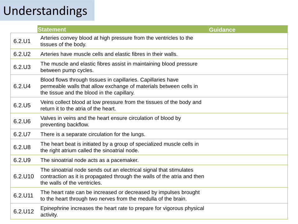
6.2.U1 Arteries convey blood at high pressure from the ventricles to the tissues of the body. AND 6.2.U2 Arteries have muscle cells
and elastic fibres in their walls. AND 6.2.U3 The muscle and elastic fibres assist in maintaining blood pressure between pump cycles.
Muscle contracts to decrease the size of the lumen. This causes an increase blood pressure and therefore maintains high blood pressure between the pulses of high pressure blood travelling from the heart.
Elastic fibres stretch to increase the lumen with each pulse of blood. After the pulse of blood passes the fibres recoil decreasing the lumen size and therefore helping to maintain a high blood pressure.
Relatively (to the wall) small lumen maintains high blood pressure.
The structure of arteriesThick muscular wall and fibrous outer layer help the artery to withstand high pressure
https://commons.wikimedia.org/wiki/File:Blausen_0055_ArteryWallStructure.png

6.2.U4 Blood flows through tissues in capillaries. Capillaries have permeable walls that allow exchange of
materials between cells in the tissue and the blood in the capillary.
https://commons.wikimedia.org/wiki/File:Capillary_system_CERT.jpg
The structure of capillaries
Capillaries are the smallest blood vessels and are adapted for the exchange of substances to and from the blood.
This enables tissues to gain nutrients and molecules such as oxygen and to rid themselves of waste material.
Capillaries also allow substances to enter and leave the organism, e.g. gas exchange of oxygen and carbon dioxide in the lungs.
Wall is one cell thick allows easy diffusion of substances in and out of the capillary due to the short diffusion distance.
Blood travels slowly under low pressure allowing more opportunity for exchange.
Basement membrane is permeable to many substances
The walls and membrane can contain pores to further aid the diffusion of substances
Due the the massive number of capillaries present and the small lumen the surface area available for the exchange of substances is very large.
Image adapted from: https://commons.wikimedia.org/wiki/File:Capillary.svg
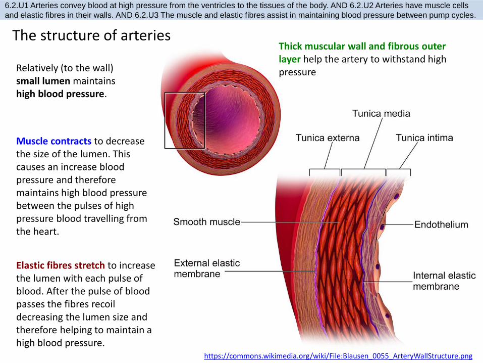
6.2.U5 Veins collect blood at low pressure from the tissues of the body and return it to the atria of the heart. AND
6.2.U6 Valves in veins and the heart ensure circulation of blood by preventing backflow.
The structure of veins
https://commons.wikimedia.org/wiki/File:Venous_valve.svg
http://40.media.tumblr.com/tumblr_m0dwjt3WKQ1qzcf71o1_500.jpg
Veins return blood to the heart for re-circulation.
Because of the low pressure valves are required to prevent back-flow of the blood and therefore ensure that the blood moves towards to heart.
Because there is less pressure to resist the walls of the veins are thinner and less elastic than arteries. They also contain less muscle than the arteries.
The large lumen (compared to arteries and the thickness of the wall) means that the blood is under low pressure.
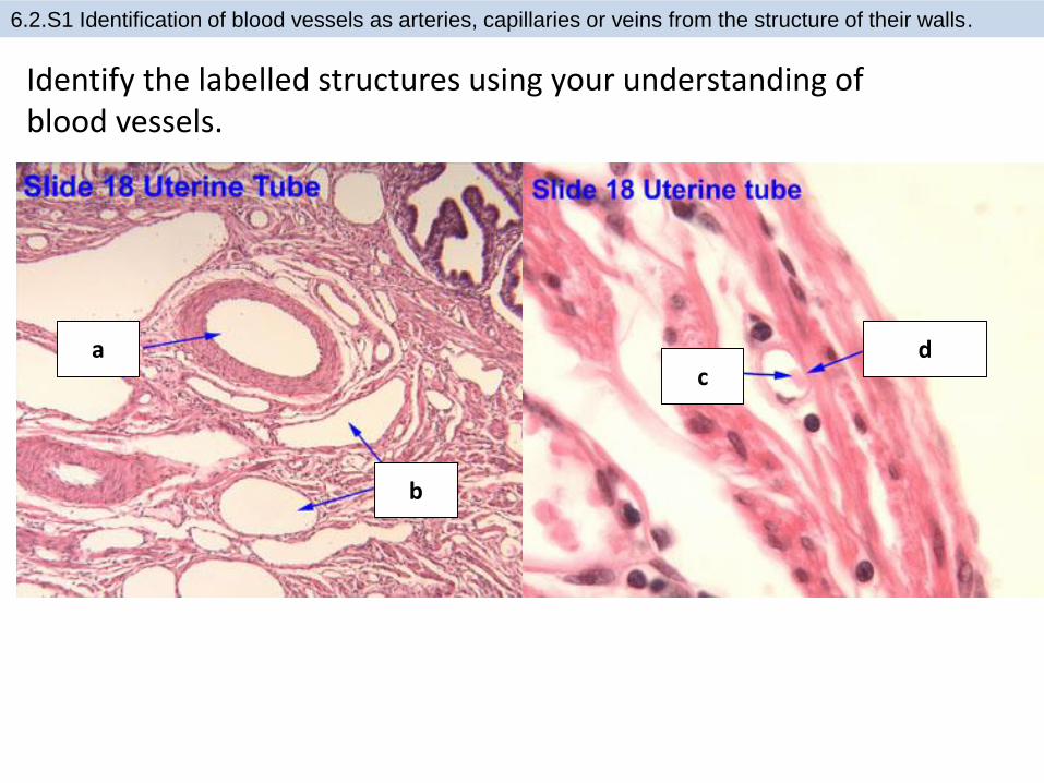
6.2.S1 Identification of blood vessels as arteries, capillaries or veins from the structure of their walls.
Identify the labelled structures using your understanding of blood vessels.
a
b
cd

6.2.S1 Identification of blood vessels as arteries, capillaries or veins from the structure of their walls.
Identify the labelled structures using your understanding of blood vessels.
a
b
cd
https://www.ouhsc.edu/histology/Text%20Sections/Cardiovascular.html

6.2.S1 Identification of blood vessels as arteries, capillaries or veins from the structure of their walls.
Identify the labelled structures using your understanding of blood vessels.
https://www.ouhsc.edu/histology/Text%20Sections/Cardiovascular.html

6.2.A1 William Harvey’s discovery of the circulation of the blood with the heart acting as the pump.
William Harvey was an English physician who made a key
contribution to our knowledge of anatomy and physiology. He was the first to describe completely the systemic circulation.
http://md.rcm.upr.edu/romanfranco/wp-content/uploads/sites/41/2015/02/harveypic.jpg
https://commons.wikimedia.org/wiki/File:William_Harvey_%281578-1657%29_Venenbild.jpg
Learn more about him:• http://www.indiana.edu/~liblilly/anatomia/bloodcirc.html• http://www.bbc.co.uk/history/historic_figures/harvey_william.shtml• http://www.sciencemuseum.org.uk/broughttolife/people/williamharve
y.aspx
Repeat Harvey’s experiment with guidance from The Naked Scientists:• http://www.thenakedscientists.com/HTML/experiments/exp/veins/

Nature of Science: Theories are regarded as uncertain - William Harvey overturned theories developed by
the ancient Greek philosopher Galen on movement of blood in the body. (1.9)
Galen’s ideas were first challenged by by an Arab physician, Ibn-al-Nafiz in the thirteenth century, but despite this Galen’s theory remained accepted by society until William Harvey (1578 - 1657) published “De Motu Cordis” in 1628. Even then it took many years for his theory of systemic circulation (similar to modern accepted theory) to succeed Galen’s.
http://md.rcm.upr.edu/romanfranco/wp-content/uploads/sites/41/2015/02/harveypic.jpghttps://commons.wikimedia.org/wiki/File:William_Harvey_%281578-1657%29_Venenbild.jpg
Theories are by definition the best accepted explanations and predictions
of natural phenomena. Although they are usually thoroughly tested based on evidence and reason theories theories are the only the current best accepted explanation. Theories if successfully questioned can be modified or even rejected/falsified if a better explanation arises.
http://famousbiologists.org/wp-content/uploads/2013/06/galen.jpg
Galen (129 - c216) thought that “blood is created in the liver from ingested food and flows to the right side of the heart. Some of it flows to the lungs where it gives off ‘sooty vapors’ and some flows through invisible pores into the left side of the heart, where it gains ‘vital spirits’ when mixed with pneuma brought in by the trachea.”
http://membercentral.aaas.org/blogs/scientia/circulatory-system-galen-harvey

6.2.U7 There is a separate circulation for the lungs.
http://www.kscience.co.uk/animations/blood_system.swf

6.2.S2 Recognition of the chambers and valves of the heart and the blood vessels connected to it in
dissected hearts or in diagrams of heart structure.
Label the heart diagram

6.2.S2 Recognition of the chambers and valves of the heart and the blood vessels connected to it in
dissected hearts or in diagrams of heart structure.
Label the heart diagram
Now try to label this heart: http://sciencelearn.org.nz/Contexts/See-through-Body/Sci-Media/Animation/Label-the-heart
bicuspid valve

6.2.U8 The heart beat is initiated by a group of specialized muscle cells in the right atrium called the sinoatrial node. AND
6.2.U9 The sinoatrial node acts as a pacemaker. AND 6.2.U10 The sinoatrial node sends out an electrical signal that
stimulates contraction as it is propagated through the walls of the atria and then the walls of the ventricles.
http://www2.estrellamountain.edu/faculty/farabee/Biobk/heartbeat.gif

6.2.U11 The heart rate can be increased or decreased by impulses brought to the heart through two nerves from the
medulla of the brain. AND 6.2.U12 Epinephrine increases the heart rate to prepare for vigorous physical activity.
http://goo.gl/YeoeJ
n.b. adrenalin is also known as epinephrine

6.2.A2 Pressure changes in the left atrium, left ventricle and aorta during the cardiac cycle.

6.2.A2 Pressure changes in the left atrium, left ventricle and aorta during the cardiac cycle.
Top-quality animations available from www.medmovie.com :
http://www.fed.cuhk.edu.hk/~johnson/teaching/transport/animations/HyperHeart.swf

6.2.A2 Pressure changes in the left atrium, left ventricle and aorta during the cardiac cycle.

6.2.A2 Pressure changes in the left atrium, left ventricle and aorta during the cardiac cycle.

6.2.A3 Causes and consequences of occlusion of the coronary arteries.
http://www.hhmi.org/biointeractive/obesity/heart_attack.html

6.2.A3 Causes and consequences of occlusion of the coronary arteries.
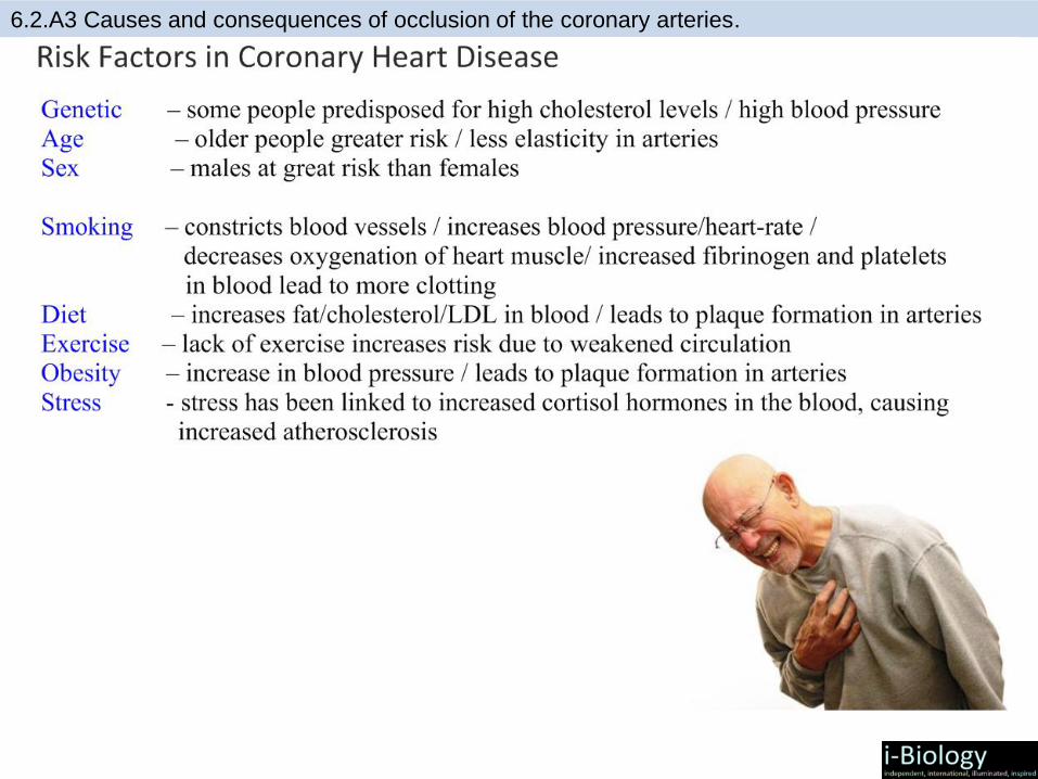
6.2.A3 Causes and consequences of occlusion of the coronary arteries.
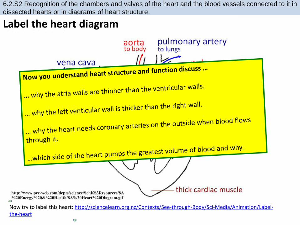
6.2.S2 Recognition of the chambers and valves of the heart and the blood vessels connected to it in
dissected hearts or in diagrams of heart structure.
Label the heart diagram
Now try to label this heart: http://sciencelearn.org.nz/Contexts/See-through-Body/Sci-Media/Animation/Label-the-heart
bicuspid valve

Bibliography / Acknowledgments
Bob Smullen