Embed Size (px)
Citation preview

64-year-old male with new onset of
severe headache & double vision
Kaitlin Lipner, MD
Brad Kincaid, MD
Leo Wolansky, MD
CT Head Non-
Contrast
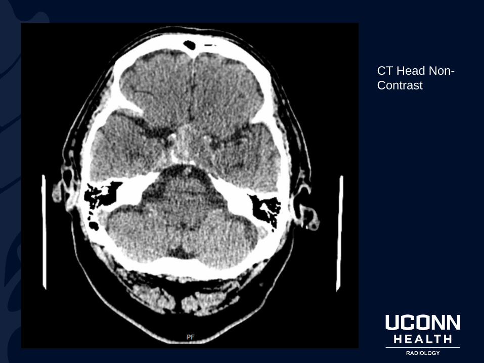
Axial MRI T1
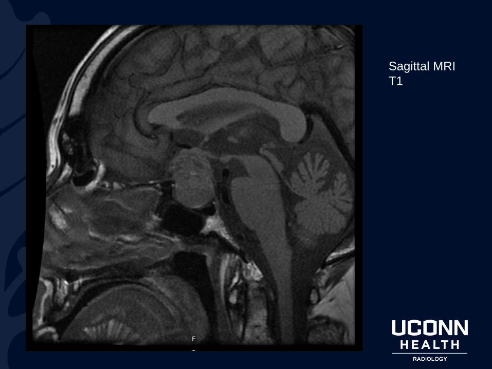
Sagittal MRI
T1
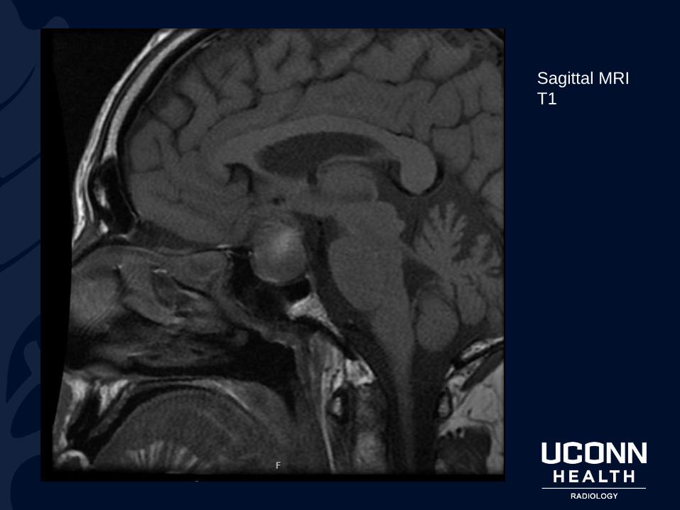
Sagittal MRI
T1
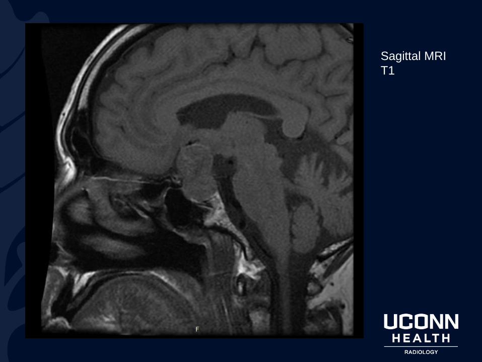
Sagittal MRI
T1

?

Pituitary Apoplexy:
Pituitary Macroadenoma
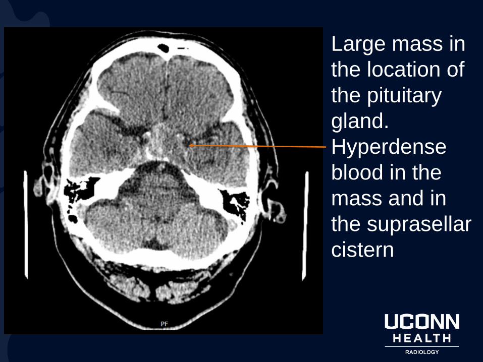
Large mass in
the location of
the pituitary
gland.
Hyperdense
blood in the
mass and in
the suprasellar
cistern
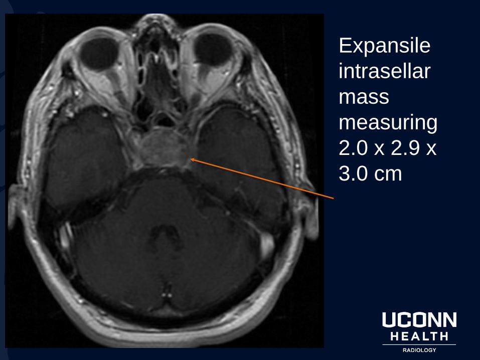
Expansile
intrasellar
mass
measuring
2.0 x 2.9 x
3.0 cm
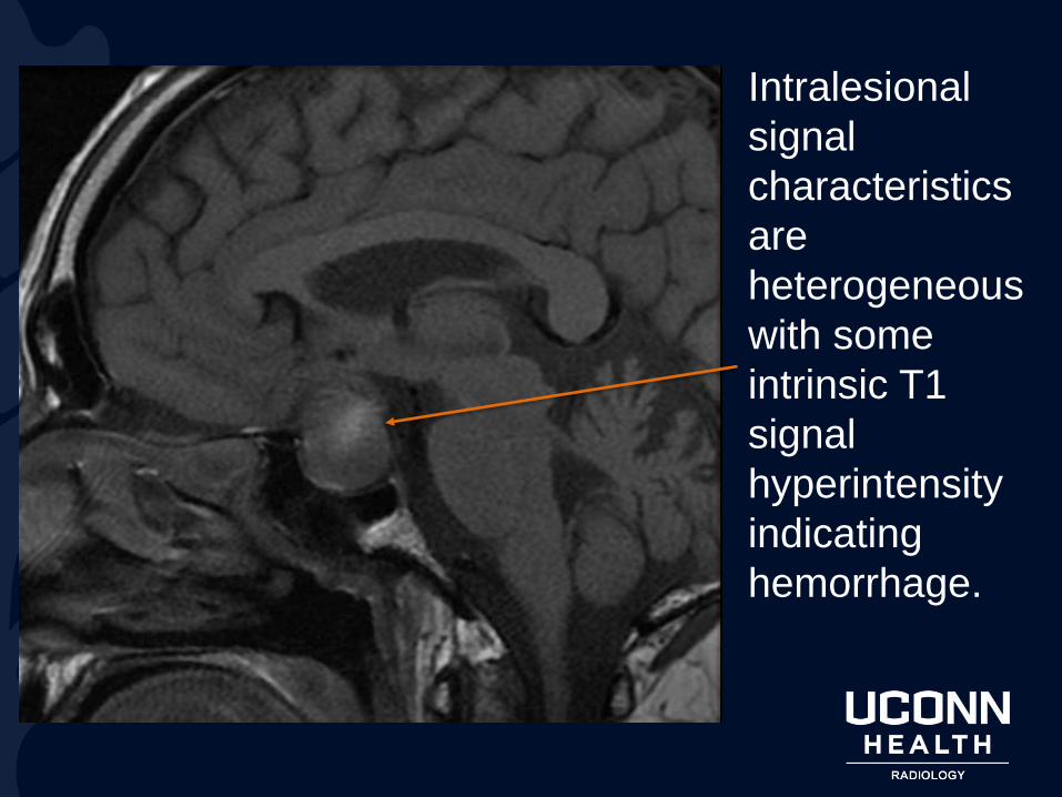
Intralesional
signal
characteristics
are
heterogeneous
with some
intrinsic T1
signal
hyperintensity
indicating
hemorrhage.
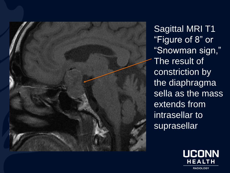
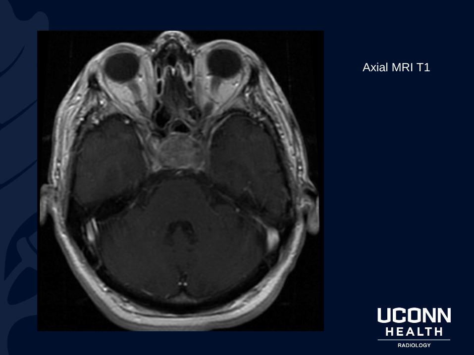
Sagittal MRI T1
“Figure of 8” or
“Snowman sign,”
The result of
constriction by
the diaphragma
sella as the mass
extends from
intrasellar to
suprasellar
Pituitary Macroadenoma
Definitions:
Macroadenoma: Pituitary Adenoma larger than 10 mm
Pituitary Apolplexy: Pituitary Stroke (sudden onset)
usually associated with hemorrhagic adenoma.
Epidemiology
• Adenoma is most common cause of sellar mass
• accounts for up to 10 percent of all intracranial
neoplasms
Pituitary Macroadenoma
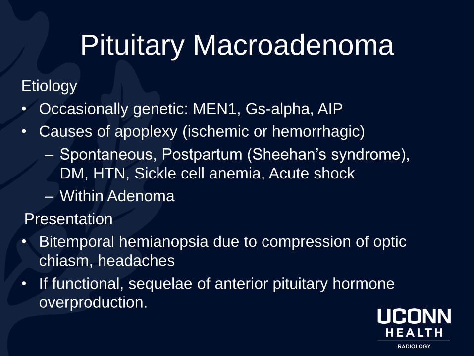
Etiology
• Occasionally genetic: MEN1, Gs-alpha, AIP
• Causes of apoplexy (ischemic or hemorrhagic)
– Spontaneous, Postpartum (Sheehan’s syndrome),
DM, HTN, Sickle cell anemia, Acute shock
– Within Adenoma
Presentation
• Bitemporal hemianopsia due to compression of optic
chiasm, headaches
• If functional, sequelae of anterior pituitary hormone
overproduction.

Diagnosis
• Labs: prolactin, insulin like growth factor 1, 24-hour urinary free cortisol, testosterone LH, FSH, TSH with T4
• Mild to moderate elevation of Prolactin is nonspecific due to loss of inhibition by dopamine (“Stalk-effect”)
• Differential for adenoma: hyperplasia, craniopharyngioma, meningioma, pituicytoma, Rathke’s cleft cyst, abscess, hypophysitis
Pituitary Macroadenoma
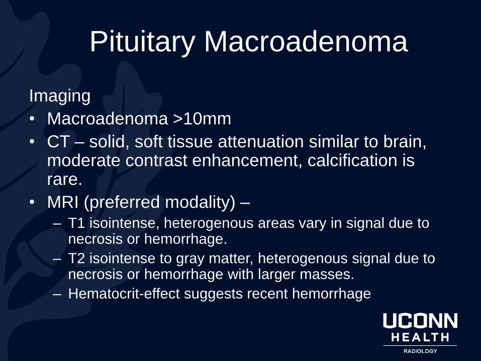
Imaging
• Macroadenoma >10mm
• CT – solid, soft tissue attenuation similar to brain, moderate contrast enhancement, calcification is rare.
• MRI (preferred modality) –– T1 isointense, heterogenous areas vary in signal due to
necrosis or hemorrhage.
– T2 isointense to gray matter, heterogenous signal due to necrosis or hemorrhage with larger masses.
– Hematocrit-effect suggests recent hemorrhage
Pituitary Macroadenoma

References1. Harrison's Principles of Internal Medicine. 20th ed., McGraw-Hill Education, 2018. Access
Medicine, accessmedicine-mhmedical-com.online.uchc.edu/book.aspx?bookid=2129. Accessed 2
Mar. 2019.
2. "Pituitary Tumors." DynaMed,
www.dynamed.com.online.uchc.edu/topics/dmp~AN~T900666/Pituitary-tumors. Accessed 2 Mar.
2019.
3. Schwedt, Todd J., and David W. Dodick. "Approach to the patient with thunderclap headache."
UptoDate, www-uptodate-com.online.uchc.edu/contents/approach-to-the-patient-with-thunderclap-
headache?search=pituitary%20apoplexy&source=search_result&selectedTitle=2~41&usage_type
=default&display_rank=2. Accessed 2 Mar. 2019.
4. Snyder, Peter J. "Causes, presentation, and evaluation of sellar masses." UptoDate, www-
uptodate-com.online.uchc.edu/contents/causes-presentation-and-evaluation-of-sellar-
masses?search=pituitary%20adenoma&source=search_result&selectedTitle=1~150&usage_type=
default&display_rank=1#H4. Accessed 2 Mar. 2019.
5. Lipner, K., Kincaid, B. Pituitary Macroadenoma: Pituitary Apoplexy. Radiology Online. 2020.