Embed Size (px)
Citation preview

Minimally Invasive Esophagectomy6A006
www.amegroups.com
Min
imally In
vasiv
e Esoph
agec
tom
y
Editors: Zhigang LiToni LerutSuzanne S. Gisbertz
Associate Editors: Luigi BonavinaHarushi OsugiSook Whan SungBin Li
Editors: Zhigang Li
Toni LerutS
uzanne S. G
isbertz
AME Surgery Series 6A006

Minimally Invasive Esophagectomy
Editors: Zhigang LiToni LerutSuzanne S. Gisbertz
Associate Editors: Luigi BonavinaHarushi OsugiSook Whan SungBin Li
AME Surgery Series 6A006

AME Publishing Company
Room C 16F, Kings Wing Plaza 1, NO. 3 on Kwan Street, Shatin, NT, Hong Kong
Information on this title: www.amegroups.com For more information, contact [email protected]
Copyright © AME Publishing Company. All rights reserved.
This publication is in copyright. Subject to statutory exception and to the provisions of relevant collective licensing agreements, no reproduction of any part may take place without the written permission of AME Publishing Company.
First published in 2018 Printed in China by AME Publishing Company
Editors: Zhigang Li, Toni Lerut, Suzanne S. GisbertzCover Image Illustrator: Anthony P. Yim, HongKong, China
Minimally Invasive Esophagectomy(Hard Cover)
ISBN 978-988-79496-4-0AME Publishing Company, Hong Kong
AME Publishing Company has no responsibility for the persistence or accuracy of URLs for external or third-party internet websites referred to in this publication, and does not guarantee that any content on such websites is, or will remain, accurate or appropriate.
The advice and opinions expressed in this book are solely those of the authors and do not necessarily represent the views or practices of the publisher. No representation is made by the publisher about the suitability of the information contained in this book, and there is no consent, endorsement or recommendation provided by the publisher, express or implied, with regard to its contents.

© AME Publishing Company. All rights reserved. www.amegroups.com
I
EDITORS
Zhigang LiDepartment of Thoracic Surgery, Section of Esophageal Surgery, Shanghai Chest Hospital, Shanghai Jiaotong University, Shanghai 200030, China
Toni LerutDepartment of Thoracic Surgery, University Hospitals Leuven, Leuven, Belgium
Suzanne S. GisbertzDepartment of Surgery, Academic Medical Center, Amsterdam, The Netherlands
ASSOCIATE EDITORS
Luigi BonavinaDepartment of Biomedical Sciences for Health, Division of General Surgery, University of Milan School of Medicine, Milano, Italy
Harushi OsugiDepartment of Surgery, Institute of Gastroenterology, Tokyo Women’s Medical University, Kawada-Cho, Shinjuku-ku, Japan
Sook Whan SungDepartment of Thoracic and Cardiovascular Surgery, Seoul St. Mary’s Hospital, College of Medicine, The Catholic University of Korea, Seoul, Republic of Korea
Bin LiDepartment of Thoracic Surgery, Section of Esophageal Surgery, Shanghai Chest Hospital, Shanghai Jiaotong University, Shanghai 200030, China
AUTHORS
Mohamed M. AbdelfatahGastroenterology and Hepatology Section, Baylor College of Medicine, Houston, TX, USA
Alberto AiolfiDivision of General Surgery, Department of Biomedical Sciences for Health, University of Milan, IRCCS Policlinico San Donato, Milan, Italy
Emanuele AstiDivision of General Surgery, Department of Biomedical Sciences for Health, University of Milan, IRCCS Policlinico San Donato, Milan, Italy
Adolfo BadaloniEsophageal Institute, Hospital Universitario Fundacion Favaloro, Buenos Aires, Argentina
Hasan F. BatirelThoracic Surgery Department, Marmara University Hospital, Istanbul, Turkey
Gijs H. BerkelmansDepartment of surgery, Catharina hospital, Eindhoven, the Netherlands
Daniele BernardiDivision of General Surgery, Department of Biomedical Sciences for Health, University of Milan, IRCCS Policlinico San Donato, Milan, Italy
Ronaldus L. A. W. BleysDepartment of Anatomy, University Medical Center Utrecht, Utrecht, The Netherlands
Chantal BoelensDepartment of Nursing, Operating Theatres, University Hospitals Leuven, Leuven, Belgium
Luigi BonavinaDivision of General Surgery, Department of Biomedical Sciences for Health, University of Milan, IRCCS Policlinico San Donato, Milan, Italy
Michael BouvetDepartment of Surgery, University of California San Diego, San Diego, CA, USA
Minimally Invasive Esophagectomy(FIRST EDITION)

© AME Publishing Company. All rights reserved. www.amegroups.com
II
Raphael BuenoBrigham and Women’s Hospital, Division of Thoracic Surgery, Boston, MA, USA
Philip Wai-Yan ChiuDivision of Upper Gastrointestinal and Metabolic Surgery, Department of Surgery, Prince of Wales Hospital, the Chinese University of Hong Kong, Hong Kong, China
Franco CiotolaEsophageal Institute, Hospital Universitario Fundacion Favaloro, Buenos Aires, Argentina
Willy CoosemansDepartment of Thoracic Surgery, University Hospitals Leuven, Leuven, Belgium
Steve CoppensDepartment of Anaesthesiology, University Hospitals Leuven, Leuven, Belgium
Miguel A. CuestaDepartment of Surgery, VU Medisch Centrum, Amsterdam, The Netherlands
Jonathan C. DeLongDepartment of Surgery, University of California San Diego, San Diego, CA, USA
Lieven DepypereDepartment of Thoracic Surgery, University Hospitals Leuven, Leuven, Belgium
Chun DingDepartment of Thoracic Surgery, Section of Esophageal Surgery, Shanghai Chest Hospital, Shanghai Jiaotong University, Shanghai 200030, China
Bart F. GeertsDepartment of Anaesthesiology, Academic Medical Center, Amsterdam, The Netherlands
Suzanne S. GisbertzDepartment of Surgery, Academic Medical Center, Amsterdam, The Netherlands
Haiyong GuDepartment of Thoracic Surgery, Section of Esophageal Surgery, Shanghai Chest Hospital, Shanghai Jiaotong University, Shanghai 200030, China
Xufeng GuoDepartment of Thoracic Surgery, Section of Esophageal Surgery, Shanghai Chest Hospital, Shanghai Jiaotong University, Shanghai 200030, China
Noboru HanaokaDepartment of Gastrointestinal Oncology, Osaka Medical Center for Cancer and Cardiovascular Diseases, 3-3 Nakamichi 1-chome, Higashinari-ku, Osaka 537-8511, Japan
Nicholas R. HessDepartment of Cardiothoracic Surgery, University of Pittsburgh School of Medicine and the University of Pittsburgh Medical Center, Pittsburgh, PA, USA
Koji HigashinoDepartment of Gastrointestinal Oncology, Osaka Medical Center for Cancer and Cardiovascular Diseases, 3-3 Nakamichi 1-chome, Higashinari-ku, Osaka 537-8511, Japan
Santiago HorganDepartment of Surgery, University of California San Diego, San Diego, CA, USA
Rong HuaDepartment of Thoracic Surgery, Section of Esophageal Surgery, Shanghai Chest Hospital, Shanghai Jiaotong University, Shanghai 200030, China
Hiroyasu IishiDepartment of Gastrointestinal Oncology, Osaka Medical Center for Cancer and Cardiovascular Diseases, 3-3 Nakamichi 1-chome, Higashinari-ku, Osaka 537-8511, Japan
Ryu IshiharaDepartment of Gastrointestinal Oncology, Osaka Medical Center for Cancer and Cardiovascular Diseases, 3-3 Nakamichi 1-chome, Higashinari-ku, Osaka 537-8511, Japan

© AME Publishing Company. All rights reserved. www.amegroups.com
III
Hajime IsomotoDivision of Medicine and Clinical Science, Tottori University, Yonago, Japan
Garth R. JacobsenDepartment of Surgery, University of California San Diego, San Diego, CA, USA
Hyun Woo JeonDepartment of Thoracic and Cardiovascular Surgery, Bucheon St. Mary’s Hospital, College of Medicine, The Catholic University of Korea, Seoul, Republic of Korea
Egle JezerskyteDepartment of Surgery, Academic Medical Center, Amsterdam, The Netherlands
Haoyao JiangDepartment of Thoracic Surgery, Section of Esophageal Surgery, Shanghai Chest Hospital, Shanghai Jiao Tong University, Shanghai 200030, China
Koichiro KawaguchiDivision of Medicine and Clinical Science, Tottori University, Yonago, Japan
Kaitlyn J. KellyDepartment of Surgery, University of California San Diego, San Diego, CA, USA
Bastiaan R. KlarenbeekDepartment of surgery, Radboudumc, Nijmegen, the Netherlands
Kazuo KoyanagiDepartment of Esophageal Surgery, National Cancer Center Hospital, Tokyo, Japan
Kenji KudouDepartment of Surgery, Institute of Gastroenterology, Tokyo Women’s Medical University, Kawada-Cho, Shinjuku-ku, Japan
Hiroki KurumiDivision of Medicine and Clinical Science, Tottori University, Yonago, Japan
Kristel LaesDepartment of Nursing, Operating Theatres, University Hospitals Leuven, Leuven, Belgium
Toni LerutDepartment of Thoracic Surgery, University Hospitals Leuven, Leuven, Belgium
Ryan M. LevyDepartment of Cardiothoracic Surgery, University of Pittsburgh School of Medicine and the University of Pittsburgh Medical Center, Pittsburgh, PA, USA
Zhigang LiDepartment of Thoracic Surgery, Section of Esophageal Surgery, Shanghai Chest Hospital, Shanghai Jiaotong University, Shanghai 200030, China
Bin LiDepartment of Thoracic Surgery, Section of Esophageal Surgery, Shanghai Chest Hospital, Shanghai Jiaotong University, Shanghai 200030, China
James D. LuketichDepartment of Cardiothoracic Surgery, University of Pittsburgh School of Medicine and the University of Pittsburgh Medical Center, Pittsburgh, PA, USA
Chenguang LuoDepartment of Thoracic Surgery, The first people’s Hospital of Yangquan, Shanxi 045000, China
Michael D. P. LuyerDepartment of Surgery, Catharina Hospital, Eindhoven, The Netherlands
Misha D. P. LuyerDepartment of surgery, Catharina hospital, Eindhoven, the Netherlands
Teng MaoDepartment of Thoracic Surgery, Section of Esophageal Surgery, Shanghai Chest Hospital, Shanghai Jiaotong University, Shanghai 200030, China
Fabio NachmanEsophageal Institute, Hospital Universitario Fundacion Favaloro, Buenos Aires, Argentina

© AME Publishing Company. All rights reserved. www.amegroups.com
IV
Philippe NafteuxDepartment of Thoracic Surgery, University Hospitals Leuven, Leuven, Belgium
Kousuke NarumiyaDepartment of Surgery, Institute of Gastroenterology, Tokyo Women’s Medical University, Kawada-Cho, Shinjuku-ku, Japan
Katie S. NasonDepartment of Cardiothoracic Surgery, University of Pittsburgh School of Medicine and the University of Pittsburgh Medical Center, Pittsburgh, PA, USA
Arne NeyrinckDepartment of Anaesthesiology, University Hospitals Leuven, Leuven, Belgium
Alejandro NieponiceEsophageal Institute, Hospital Universitario Fundacion Favaloro, Buenos Aires, Argentina
Grard A. P. NieuwenhuijzenDepartment of surgery, Catharina hospital, Eindhoven, the Netherlands
Bo NingThe Second Affiliated Hospital of Chongqing Medical University, Chongqing 400010, China
Olugbenga T. OkusanyaDepartment of Cardiothoracic Surgery, University of Pittsburgh School of Medicine and the University of Pittsburgh Medical Center, Pittsburgh, PA, USA
Harushi OsugiDepartment of Surgery, Institute of Gastroenterology, Tokyo Women’s Medical University, Kawada-Cho, Shinjuku-ku, Japan
Mohamed O. OthmanDivision of Gastroenterology, Department of Internal Medicine, East Carolina University, Greenville, NC, USA
Arjun PennathurDepartment of Cardiothoracic Surgery, University of Pittsburgh School of Medicine and the University of Pittsburgh Medical Center, Pittsburgh, PA, USA
Mauricio RamirezEsophageal Institute, Hospital Universitario Fundacion Favaloro, Buenos Aires, Argentina
Camiel RosmanDepartment of surgery, Radboudumc, Nijmegen, the Netherlands
Jelle P. RuurdaDepartment of Surgery, University Medical Center Utrecht, Utrecht, The Netherlands
Manuel Villa SanchezDepartment of Cardiothoracic Surgery, University of Pittsburgh School of Medicine and the University of Pittsburgh Medical Center, Pittsburgh, PA, USA
Bryan J. SandlerDepartment of Surgery, University of California San Diego, San Diego, CA, USA
Inderpal S. SarkariaDepartment of Cardiothoracic Surgery, University of Pittsburgh School of Medicine and the University of Pittsburgh Medical Center, Pittsburgh, PA, USA
Andrea SironiDivision of General Surgery, Department of Biomedical Sciences for Health, University of Milan, IRCCS Policlinico San Donato, Milan, Italy
Yuchen SuDepartment of Thoracic Surgery, Section of Esophageal Surgery, Shanghai Chest Hospital, Shanghai Jiao Tong University, Shanghai 200030, China
Yifeng SunDepartment of Thoracic Surgery, Section of Esophageal Surgery, Shanghai Chest Hospital, Shanghai Jiaotong University, Shanghai 200030, China
Sook Whan SungDepartment of Thoracic and Cardiovascular Surgery, Seoul St. Mary’s Hospital, College of Medicine, The Catholic University of Korea, Seoul, Republic of Korea
Scott J. SwansonBrigham and Women’s Hospital, Division of Thoracic Surgery, Boston, MA, USA

© AME Publishing Company. All rights reserved. www.amegroups.com
V
Cover Image Illustrator Executive Typesetting Editor
Anthony P. Yim, HongKong, China Li Wang, AME Publishing Company
Yuji TachimoriCancer Care Center, Kawasaki Saiwai Hospital, Kawasaki, Japan
Yohei TakedaDivision of Medicine and Clinical Science, Tottori University, Yonago, Japan
Yoji TakeuchiDepartment of Gastrointestinal Oncology, Osaka Medical Center for Cancer and Cardiovascular Diseases, 3-3 Nakamichi 1-chome, Higashinari-ku, Osaka 537-8511, Japan
Noriya UedoDepartment of Gastrointestinal Oncology, Osaka Medical Center for Cancer and Cardiovascular Diseases, 3-3 Nakamichi 1-chome, Higashinari-ku, Osaka 537-8511, Japan
Mark I. van Berge HenegouwenDepartment of Surgery, Academic Medical Center, Amsterdam, The Netherlands
Richard van HillegersbergDepartment of Surgery, University Medical Center Utrecht, Utrecht, The Netherlands
Hans Van VeerDepartment of Thoracic Surgery, University Hospitals Leuven, Leuven, Belgium
Frans van WorkumDepartment of surgery, Radboudumc, Nijmegen, the Netherlands
Denise P. VeeloDepartment of Anaesthesiology, Academic Medical Center, Amsterdam, The Netherlands
Jon O. WeeBrigham and Women’s Hospital, Division of Thoracic Surgery, Boston, MA, USA
Teun J. WeijsDepartment of Surgery, University Medical Center Utrecht, Utrecht, The Netherlands
Sachiko YamamotoDepartment of Gastrointestinal Oncology, Osaka Medical Center for Cancer and Cardiovascular Diseases, 3-3 Nakamichi 1-chome, Higashinari-ku, Osaka 537-8511, Japan
Yu YangDepartment of Thoracic Surgery, Section of Esophageal Surgery, Shanghai Chest Hospital, Shanghai Jiaotong University, Shanghai 200030, China
Yang YangDepartment of Thoracic Surgery, Section of Esophageal Surgery, Shanghai Chest Hospital, Shanghai Jiao Tong University, Shanghai 200030, China
Kazuo YashimaDivision of Medicine and Clinical Science, Tottori University, Yonago, Japan
Bo YeDepartment of Thoracic Surgery, Section of Esophageal Surgery, Shanghai Chest Hospital, Shanghai Jiao Tong University, Shanghai 200030, China
Hon-Chi YipDivision of Upper Gastrointestinal and Metabolic Surgery, Department of Surgery, Prince of Wales Hospital, the Chinese University of Hong Kong, Hong Kong, China
Xiaobin ZhangDepartment of Thoracic Surgery, Section of Esophageal Surgery, Shanghai Chest Hospital, Shanghai Jiao Tong University, Shanghai 200030, China
Cecilia ZubietaEsophageal Institute, Hospital Universitario Fundacion Favaloro, Buenos Aires, Argentina

© AME Publishing Company. All rights reserved. www.amegroups.com
The Annals of Cardiothoracic Surgery, one of AME's peer-reviewed journals, is lucky to have an author from Rochester, USA. He is left-handed. When he began his training in surgery, he encountered a particularly intractable obstacle: when using scissors or doing knotting during a surgery, his actions were the mirror opposite of what was described in textbooks. Therefore, he often “took a beating” from his mentors when performing a surgery.
Later, he summarized his experience and published it in a journal in an attempt to find other surgeons that “suffer from the same fate”. Surprisingly, after his article was published, many surgeons e-mailed him, asking him how left-handed doctors should undergo surgical training, and so on. Then he met Professor Tristan D. Yan, the editor-in-chief of Annals of Cardiothoracic Surgery, who also happens to be a left-handed doctor. Tristan encouraged him to become a heart surgeon because there are steps in cardiac surgery that require the use of the left hand to complete the suture threading technique. Tristan’s view was that it was better if surgeons were trained to use both their left and right hands.
A few days ago, on my daughter’s first day of kindergarten, I chatted with her teacher for a while; finally, she asked me if there was anything about my daughter that she should take note of . “Please do not correct my daughter's left-handedness,” I said, “Just let it be.” “Why?” the teacher asked in wonder.
On December 7th, 2013, we held the second AME Academic Salon in the Hospital Affiliated to Nantong University. After dinner, Dr. Shen Yaxing from the Department of Thoracic Surgery of Shanghai Zhongshan Hospital invited several attendees to have tea in his room. The elevator was in the middle of the hotel. After we walked out of the elevator, he led us to the left, then to the left, then to the left, then to the left, and finally to the door of his room. Although we were confused and disoriented, some of us figured out out that the door was just diagonally across the elevator. We all burst into laughter. Yaxing divulged that he took this route the first time he entered his room, and so he decided to bring us on the same route the second time. Yaxing then said that this was the behavior of a “typical” surgeon!
During the training to be a surgeon, each step and each action are done under the strict direction and supervision of a senior surgeon. Thus, many surgeons like to affectionately address their mentors as their "masters".
How, then, can you become a master of surgery? In addition to your own intelligence and diligence, the expertise and mentorship offered by a “master” is also very important. Just like in the world of martial arts, there are many different schools that are independent from each other and have their own strengths and weaknesses, and the surgical world is very much the same.
Therefore, it is important for a young surgeon to gain knowledge and skills from different masters by taking in only the essence and discarding the dregs. With this in mind, we have here determined to publish the AME Surgery Series, in an attempt to share with our readers the surgical skills of some of the prominent surgical teams in China and from abroad, as well as their philosophical thinking and some interesting stories. We sincerely hope that our colleagues in the surgical departments find these books insightful and helpful.
Stephen D. WangFounder and CEO,
AME Publishing Company
Foreword VI

© AME Publishing Company. All rights reserved. www.amegroups.com
VII
Esophageal cancer (EC) is one of the leading causes of cancer death worldwide with an estimated 456,000 new cases and 400,000 deaths in 2012 (1). The incidence rate of it varies in different regions and more than 80% of cases occur in developing countries (2). The highest incidence rates are reported in Eastern Asia and Southern Africa, with a relative high rate in America and Europe, and the lowest prevalence in Western Asia (3). In the past few decades, in order to help reduce EC, various researches have been conducted to study the epidemiology, pathogenesis, treatment and prognosis of this malignant cancer. In this new book Minimally Invasive Esophagectomy, we focus on the new surgical technology—minimally invasive esophagectomy (MIE).
The book is a comprehensive collection of articles written by international leading experts in the field of MIE. It is organized of seven chapters, including general introduction, anatomy background, preoperative preparation, endoscopic resection, thoracoscopic esophagectomy, robot for MIE and postoperative care. Every section provides an insightful review of MIE and aims to help clinicians and investigators receive more up-to-date scientific information.
Chapter one is the general introduction in which the history and benefits of MIE are introduced. In western countries, adenocarcinomas (AC) is more prevalent while squamous cell carcinomas (SCC) is the predominant type in eastern countries, which led to a different history of MIE in both worlds (4,5). It has been proved that patients in whom MIE was performed may benefit from less complications, shorter hospital stay and better short-term quality of life (6,7) Thus, MIE is crucial in the era of enhanced recovery protocols.
The second chapter is about new insights into the surgical anatomy of the esophagus. Better understanding of the anatomy of the vagus nerve and also immune response may contribute to brand-new insights into the surgical approaches and techniques and thus to improve the outcome after surgery.
Chapter three begins with an article discussing patient selection for MIE. Despite the rapidly development of treatment and management approaches in recent years, surgery-related morbidity is still a common problem. With this regard, optimal preoperative evaluation and patient selection are required. Enhanced recovery after surgery (ERAS) is a relative new concept and it is believed that the combination of ERAS and MIE will reduce the occurrence of surgical trauma and expect a speedy recovery of patients after surgery (8). The last article in this chapter presents an overview of open and laparoscopic surgery from the anaesthetist’s perspective.
The following three chapters give a comprehensive review on endoscopic resection, thoracoscopic esophagectomy (TSE) and the use of robotic technique for MIE. Traditionally, surgery is the preferred option for early-stage esophageal cancer. However, the high mortality and poor postoperative quality of life after open surgery necessitated better treatment procedures. Bearing this in mind, experts in the field kept exploring new avenues and with the advent of novel minimally invasive techniques, TSE and MIE have been developed. The past two decades has witnessed a wide acceptance of TSE and MIE due to their less invasive characters.
The poor prognosis of EC, in part, is attributed to the poorly controlled postoperative care. Therefore, attention should be paid to the management of the main surgical complications, such as anastomotic leakage, chylothorax, recurrent laryngeal nerve injury, tracheoesophageal fistula, gastrointestinal reflux and pulmonary complications. For the surgeon, it is of paramount importance to reduce the incidence of complications. Indeed reducing complications opens the door to ERAS (Enhanced Recovery After Surgery) in particular allowing for early feeding after esophagectomy. In the last chapter, the cause, clinical manifestations and diagnosis, treatment and prevention of all these complications are well described.
We hope that the book will be a valuable resource for medical staffs in this field and calling for further international collaborations aiming to improve the treatment and management of patients suffering from esophageal cancer.
References
1. International Agency for Research on Cancer. GLOBOCAN 2012, Estimated Cancer Incidence, Mortality and Prevalence Worldwide in 2012. Geneva: World Health Organization, 2012.
2. Liang H, Fan JH, Qiao YL. Epidemiology, etiology, and prevention of esophageal squamous cell carcinoma in China. Cancer Biology & Medicine 2017;14(1):33-41.
3. Torre LA, Bray F, Siegel RL, et al. Global cancer statistics, 2012. CA Cancer J Clin 2015;65(2):87-108.
Preface

© AME Publishing Company. All rights reserved. www.amegroups.com
VIII
4. Napier KJ, Scheerer M, Misra S. Esophageal cancer: A Review of epidemiology, pathogenesis, staging workup and treatment modalities. World J Gastrointest Oncol 2014;6(5):112-20.
5. Dirix P, Weytjens R, Vanderkam S, et al. Chapter 36 Oesophageal cancer. In: Kerr DJ, Haller DG, van de Velde CJ, et al. editor(s). Oxford Textbook of Oncology (3 ed.). Oxford University Press 2016:365-388.
6. Li Z, Sun Y, Yang Y, et al. The era of minimally invasive esophagectomy. Shanghai Chest 2018;2:51.7. Biere SS, van Berge Henegouwen MI, Maas KW, et al. Minimally invasive versus open oesophagectomy for patients with
oesophageal cancer: a multicentre, open-label, randomised controlled trial. Lancet 2012 ;379(9829):1887-92.8. Guo X, Ding C, Luo C, et al. ERAS prior to minimally invasive esophagectomy. Shanghai Chest 2018;2:50.
Zhigang Li, MD, PhDDepartment of Thoracic Surgery, Section of Esophageal Surgery,
Shanghai Chest Hospital, Shanghai Jiaotong University, 241 Huaihai West Rd, Shanghai 200030, China
Toni Lerut, MD, PhD Emeritus Professor of Surgery and Chairman Department of Thoracic Surgery,
University Hospitals Leuven, Herestraat 49, 3000Leuven, Belgium
Zhigang Li Toni Lerut

© AME Publishing Company. All rights reserved. www.amegroups.com
IX
The practice of minimally invasive esophagectomy for carcinoma started in 1992 after the pioneer report of Cuschieri, and is still evolving. At that time, only a few surgeons adopted the thoracoscopic approach. Instead, most surgeons decided to take full advantage of their improving expertise in basic and more or less advanced laparoscopic surgery to mobilize the stomach, perform a celiac lymphadenectomy, and prepare the gastric conduit for esophageal replacement; therefore, the laparoscopic approach set the foundation for the hybrid procedures which incorporated the trans-hiatal, the Ivor Lewis, and the McKeown techniques. A fully minimally invasive esophagectomy was performed in 1999. Later on, the first proof of concept that the minimally invasive approach was the way to go came in 2012 with a multicenter randomized clinical trial published on Lancet (TIME trial), which showed a significant reduction of respiratory complications compared to open esophagectomy. Today, laparoscopic and thoracoscopic techniques represent the preferred approach in many institutions worldwide and a major component of the enhanced recovery programs after esophagectomy. Yet, the learning curve remains substantial and the reported differences in outcomes may reflect patient selection, selective use of neo-adjuvant therapy, and lack of centralization of this complex operation.
The Journal of Thoracic Disease, a relatively young medical publication, already has a well-established reputation among surgeons worldwide and has gained a remarkable impact factor over the past few years. The publisher and the editors of Journal of Thoracic Disease have collected a series of recent articles on minimally invasive esophagectomy written by experts from respected international institutions. The final result of this endeavor is a comprehensive, highly educational, state-of-art volume that provides an easy-to consult and updated source of valued information for the surgeon. This book represents a broad overview of the research and clinical work related to minimally invasive esophagectomy, and depicts the evolution and outcomes of the resectional and reconstructive techniques over the past quarter of century. The topics are organized in six main sections spanning from surgical anatomy through preoperative assessment and preparation, endoscopic surgery, thoracoscopic surgery, and robotics, to postoperative care. The innovative contents and the overall quality of data and figures make the book really instructive and worth-reading for both the trainee and the expert surgeon.
I am sure that the reader will especially appreciate the fact that the contents of this publication reflect not only surgical and technological advances, but also the progress in anesthesiology, perioperative care, and medical oncology that have accompanied the extraordinary development of esophageal surgery. As such, this book will represent an important and useful reference for the general and thoracic surgeons and for all components of the multidisciplinary team dedicated to the care and cure of esophageal cancer patients.
Preface
Luigi Bonavina
Luigi Bonavina, MD, FACSProfessor of Surgery, University of Milan Medical School, Milano, Italy;
Director, Division of General Surgery, IRCCS Policlinico San Donato, Italy

© AME Publishing Company. All rights reserved. www.amegroups.com
X
Esophageal cancer is the 6th most common cancer in the world and the incidence of gastro-esophageal cancer will increase by 24% by 2027. Esophageal cancer is a rather common disease but is challenging not only for patients but also for physicians, and it requires particular knowledge and technique of the physicians who treat it. Because of its high potential of malignancy and anatomical features, the treatment insults the patients substantially. Therefore, minimally invasive approaches confer great benefit to patients. Recently, multimodality therapy has been developing rapidly, but resection of the esophageal lesion remains a mainstay for curative-intent treatment of this disease. This book covers all aspects of minimally invasive treatment of esophageal cancer and presents the state of the art in this field. It consists of 24 chapters, assorted into 4 categories, ranging from a general overview of this disease, to endoscopic treatment, thoracoscopic esophagectomy including robot-assisted surgery, and even pre- and post-treatment care. The contributing authors, who are all authorities and experts in their fields, come from all over the world (USA: 19; China: 18; Japan: 17; Netherlands: 17; Belgium: 9; Argentina: 6; Italy: 5; Republic of Korea: 2; and Turkey: 1).
As I am a surgeon, I would like to give special mention to the surgical issue which comprises the major portion of this book. Esophageal cancer has two main subtypes. One is adenocarcinoma, which is dominant in the West, and the other is squamous cell carcinoma, which accounts for the majority of cases in the East. The surgical strategy, therefore, differs between the West and the East. This book completely covers the surgical techniques on the basis of both concepts. Each chapter is well documented with comprehensive figures and tables. On the other hand, minimally invasive esophagectomy (MIE) may not be fully accepted at all surgical institutes. For example, a little over 30% of esophagectomies are performed thoracoscopically in Japan. Randomized trials and meta-analyses have concluded that MIE is associated with less blood loss, fewer postoperative complications, and shorter hospital stay, despite the longer duration of the procedure compared with open esophagectomy. However, a National Clinical Database survey conducted in the UK and Japan revealed that MIE was associated with a significantly higher incidence of complications. Probably, the disadvantage of MIE in the survey study was due to poor outcomes obtained through low-volume centers. It is well known that operative mortality after esophagectomy is inversely related to hospital volume. Esophagectomy itself requires surgeons to have substantial skill, but when done thoracoscopically, it requires additional skill. Proficiency is, therefore, essential to performing MIE efficiently. This book provides up-to-date detailed information on procedures and techniques.
This book is sure to help surgeons and endoscopists who intend to perform high-quality minimally invasive treatment for esophageal cancer to overcome the steep learning curve required.
Preface
Harushi Osugi, MD Department of Surgery, Institute of Gastroenterology,
Tokyo Women’s Medical University, Kawada-Cho, Shinjuku-ku, Japan
Harushi Osugi

© AME Publishing Company. All rights reserved. www.amegroups.com
XI
Table of Contents
General Introduction
1 The era of minimally invasive esophagectomyZhigang Li, Yifeng Sun, Yu Yang, Rong Hua, Xufeng Guo, Bin Li, Bo Ye, Haiyong Gu, Teng Mao
Anatomy Background
5 New insights into the surgical anatomy of the esophagusTeun J. Weijs, Jelle P. Ruurda, Michael D. P. Luyer, Miguel A. Cuesta, Richard van Hillegersberg, Ronaldus L. A. W. Bleys
Preoperative Preparation
11 Evaluation and patient selection for minimally invasive esophagectomyYifeng Sun, Yang Yang, Haiyong Gu, Yu Yang, Xufeng Guo, Bin Li, Rong Hua, Bo Ye, Teng Mao, Zhigang Li
16 ERAS prior to minimally invasive esophagectomyXufeng Guo, Chun Ding, Chenguang Luo, Yu Yang, Bin Li, Rong Hua, Bo Ye, Haiyong Gu, Yifeng Sun, Teng Mao, Zhigang Li
20 Anaesthesia during esophagectomyDenise P. Veelo, Bart F. Geerts
Endoscopic Resection
28 Endoscopic diagnosis and management of early squamous cell carcinoma of esophagusHon-Chi Yip, Philip Wai-Yan Chiu
36 Management of strictures after endoscopic submucosal dissection for superficial esophageal cancerKoichiro Kawaguchi, Hiroki Kurumi, Yohei Takeda, Kazuo Yashima, Hajime Isomoto
43 The impact of flexible endoscopy in esophageal surgeryAlejandro Nieponice, Fabio Nachman, Adolfo Badaloni, Franco Ciotola, Cecilia Zubieta, Mauricio Ramirez
51 Endoscopic submucosal dissection and endoscopic mucosal resection for early stage esophageal cancerBo Ning, Mohamed M. Abdelfatah, Mohamed O. Othman
62 Endoscopic submucosal dissection for superficial Barrett’s esophageal cancer in the Japanese state and perspectiveRyu Ishihara, Sachiko Yamamoto, Noboru Hanaoka, Yoji Takeuchi, Koji Higashino, Noriya Uedo, Hiroyasu Iishi

© AME Publishing Company. All rights reserved. www.amegroups.com
XII
Thoracoscopic Esophagectomy
69 Video-assisted thoracoscopic surgery and open chest surgery in esophageal cancer treatment: present and futureLieven Depypere, Willy Coosemans, Philippe Nafteux, Hans Van Veer, Arne Neyrinck, Steve Coppens, Chantal Boelens, Kristel Laes, Toni Lerut
82 Minimally invasive esophagectomy for esophageal squamous cell carcinoma—Shanghai Chest Hospital experienceBin Li, Yu Yang, Yifeng Sun, Rong Hua, Xiaobin Zhang, Xufeng Guo, Haiyong Gu, Bo Ye, Zhigang Li, Teng Mao
90 Supracarinal dissection of the esophagus and lymphadenectomy by MIEHarushi Osugi, Kousuke Narumiya, Kenji Kudou
100 Minimally invasive Ivor Lewis esophagectomy for esophageal cancerHyun Woo Jeon, Sook Whan Sung
105 Gastro-esophageal junction cancers: what is the best minimally invasive approach?Egle Jezerskyte, Mark I. van Berge Henegouwen, Miguel A. Cuesta, Suzanne S. Gisbertz
115 McKeown or Ivor Lewis totally minimally invasive esophagectomy for cancer of the esophagus and gastroesophageal junction: systematic review and meta-analysisFrans van Workum, Gijs H. Berkelmans, Bastiaan R. Klarenbeek, Grard A. P. Nieuwenhuijzen, Misha D. P. Luyer, Camiel Rosman
123 Minimally invasive esophagectomy: the Brigham and Women’s Hospital experienceJon O. Wee, Raphael Bueno, Scott J. Swanson
127 Hybrid and total minimally invasive esophagectomy: how I do itLuigi Bonavina, Emanuele Asti, Andrea Sironi, Daniele Bernardi, Alberto Aiolfi
139 Uniportal video-assisted thoracic surgery for esophageal cancerHasan F. Batirel
Robot for Minimally Invasive Esophagectomy
146 Robot assisted esophagectomy for esophageal squamous cell carcinomaXiaobin Zhang, Yuchen Su, Yu Yang, Yifeng Sun, Bo Ye, Xufeng Guo, Teng Mao, Rong Hua, Zhigang Li
155 The benefits and limitations of robotic assisted transhiatal esophagectomy for esophageal cancerJonathan C. DeLong, Kaitlyn J. Kelly, Garth R. Jacobsen, Bryan J. Sandler, Santiago Horgan, Michael Bouvet
161 Robotic assisted minimally invasive esophagectomy (RAMIE): the University of Pittsburgh Medical Center initial experienceOlugbenga T. Okusanya, Inderpal S. Sarkaria, Nicholas R. Hess, Katie S. Nason, Manuel Villa Sanchez, Ryan M. Levy, Arjun Pennathur, James D. Luketich

© AME Publishing Company. All rights reserved. www.amegroups.com
XIII
Postoperative Care
168 Early oral nutrition plays an active role in enhanced recovery after minimally invasive esophagectomyKazuo Koyanagi, Yuji Tachimori
173 Postoperative complications and management of minimally invasive esophagectomyRong Hua, Haoyao Jiang, Yifeng Sun, Xufeng Guo, Yu Yang, Bin Li, Bo Ye, Haiyong Gu, Teng Mao, Zhigang Li


© AME Publishing Company. All rights reserved. www.amegroups.com
Since the minimally invasive surgery (MIS) was clinically introduced into biliary tract and urologic surgery in the 1990s, it has evolved with its application in gynecological, obstetric and general surgery and finally become a prevalent procedure in thoracic surgery. Interestingly, the MIS technique is now advanced in thoracic surgery rather than other disciplines. In the Asian region, where the use of minimally invasive techniques is more prevalent, the rate of minimally invasive resection for lung cancer has exceeded 80%, and the rate of minimally invasive esophagectomy (MIE) in large centers has exceeded 60%.
Below is an outlined description of MIE: (I) The high complication rate of conventional
esophageal cancer surgery has stimulated interest in exploring minimally invasive techniques, which is expected to reduce the associated mortality;
(II) With the development of high-definition surgical imaging systems and fine surgical instruments, MIE gained attention in more accurate tumor control, especially after the introduction of robot-assisted surgical techniques;
(III) Given the low esophageal surgeries volume at most medical centers and long learning curve, the early benefits of MIE for surgical treatment of esophageal cancer have not been widely confirmed. This feature directly showed that the patient's long-term survival was not improved. A randomized controlled trial is needed;
(IV) The improvement of surgical instruments and modification of surgical approaches are the keys to gaining greater clinical advantage in the future of MIE, especially the popularity of robotic surgery. However, a full understanding of esophageal cancer is the key to ultimately improve long-term survival.
History of MIE
The history of MIE in eastern and western countries are different because of the different pathological types of esophageal cancer between these two worlds, which lead to a different selection in surgical approach. In western countries, adenocarcinoma is predominant histologic type
General Introduction
The era of minimally invasive esophagectomy
Zhigang Li, Yifeng Sun, Yu Yang, Rong Hua, Xufeng Guo, Bin Li, Bo Ye, Haiyong Gu, Teng Mao
Department of Thoracic Surgery, Section of Esophageal Surgery, Shanghai Chest Hospital, Shanghai Jiaotong University, Shanghai 200030, China
Contributions: (I) Conception and design: Z Li; (II) Administrative support: T Mao; (III) Provision of study materials or patients: Y Sun, Y Yang; (IV)
Collection and assembly of data: R Hua, X Guo; (V) Data analysis and interpretation: B Li, B Ye, H Gu; (VI) Manuscript writing: All authors; (VII)
Final approval of manuscript: All authors.
Correspondence to: Zhigang Li, MD, PhD. Department of Thoracic Surgery, Section of Esophageal Surgery, Shanghai Chest Hospital, Shanghai
Jiaotong University, 241 Huaihai West Rd, Shanghai 200030, China. Email: [email protected].
Abstract: Minimally Invasive Esophagectomy (MIE) has become a standard surgical approach for esophageal cancer in the world. In this report, we introduce the history of MIE which is different between eastern and western countries. With the development of surgical techniques, including single-lumen endotracheal intubation, CO2 artificial pneumothorax, and surgical positioning, the MIE has made great progress. It was approved that due to significantly reduced surgical trauma, MIE lowers postoperative morbidity and mortality while achieving a tumor resection efficacy comparable with the conventional open esophagectomy. The improvement of surgical instruments and modification of surgical approaches are the keys to gaining greater clinical advantage in the future of MIE, especially the popularity of robotic surgery.
Keywords: Minimally invasive esophagectomy (MIE); history; clinical outcome
Received: 10 April 2018; Accepted: 07 June 2018; Published: 28 June 2018.
doi: 10.21037/shc.2018.06.09
View this article at: http://dx.doi.org/10.21037/shc.2018.06.09

© AME Publishing Company. All rights reserved. www.amegroups.com
2 Li et al. The era of minimally invasive esophagectomy
and the gastroesophageal junction is the most common lesion site. Because the abdominal and low mediastinal lymph nodes (below the carina of the trachea) are the common metastasis sites, the Ivor Lewis esophagectomy is considered as the main option for surgical treatment of esophageal adenocarcinoma. In the early period of MIE in the West, experience in performing anti-reflux surgery played an important role to shorten the learning curve of the minimally invasive Ivor Lewis procedure. Dr. James D. Luketich is a pioneer and advocator in this field (1). In contrast, squamous cell carcinoma is the most common pathological type of esophageal cancer in Asian. Lymph node dissection is essential along the recurrent laryngeal nerve (RLN) in the upper mediastinum. Considering thorough lymph node dissection and effective tumor margin, McKeown technology is more often used in the surgical treatment of esophageal squamous cell carcinoma. Because of the stringent requirements of lymph node dissection in surgical treatment of esophageal squamous cell carcinoma surgery, early thoracoscopic image quality is not sufficient to perfectly support the above techniques. Therefore, the MIE in Asia is slightly later than that of western countries. However, when HD thoracoscopic camera appeared, their advantages in lymph node dissection were even greater than those of open surgery, which led to the rapid prosperity of MIE in Asia.
The following key techniques have played significant roles in promoting the development of MIE: (I) a single-lumen endotracheal intubation and CO2 artificial pneumothorax greatly improves visualization of the operative details. CO2 inflation can spontaneously separate the space around the esophagus. This allows the esophagus resection and lymph node dissection to be performed easily. The use of a single-lumen tracheal tube can favor the operation to expose the lymph nodes in the upper mediastinum very much, especially those surrounding the left RLN. The bronchial blocker is a good technical supplement for single-lumen intubation. When conversion is needed, we can easily switch to single-lung ventilation mode; (II) surgical positioning was modified for post-mediastinal approach, the patient can be placed in a prone or hemi-prone position. These positions facilitate the operation in the mediastinum, surgeons do not have to perform the operation with an uncomfortable elevation of their hands. In the case of using the prone position, a surgeon may even sit on a stool while performing the operation. Moreover, bleeding in the mediastinum does not affect visualization of the operative field since blood drains
to the lower part of the chest cavity in the prone position; (III) a high-definition monitor, 3D camera and, especially, robot-assisted techniques should be a booster for future MIE. With the assistance of a 3D camera and robotic system, surgeons can perform delicate esophagectomy and lymph node dissection in the mediastinum. It has been proven that robotic surgery can achieve more satisfactory outcomes of local lymph node dissection in the superior mediastinum.
Short-term benefits of MIE
The original intent of MIE was to reduce the high complication rate of esophagectomy. Does MIE really improve perioperative results? The most influential clinical study was the randomized controlled trial (2) published in 2012, which showed that the incidence of pneumonia was significantly lower in the MIE group than open surgery group (9% vs. 29%, P<0.005); the rate of RLN injury was also significantly lower in the MIE group. Lesser postoperative pain and better protection of RLN may be the main reasons for the decrease in the incidence of pneumonia. However, other perioperative parameters, including surgical mortality, showed no differences between MIE and OPEN group. We have not yet seen the long-term results of MIE, especially regarding the difference in local tumor recurrence. Although the use of MIE can achieve the same outcomes for lymph node dissection as open surgery and better protect the RLN, the outcome of local tumor control still needs to be studied with long-term follow-up. If the technical superiority of MIE in the protection of lung function was attributed to the omission of para-RLN lymph node dissection, we should be cautious to advocate the use of MIE. In addition, this study only included less than 60 patients in each group. Randomized studies with large sample sizes are needed to verify the perioperative advantage of MIE. Japanese researchers are working on this (3). A national data review in Japan (MIE 1,751 vs. OPEN 3,601) reported higher perioperative complications in patients undergoing MIE than in patients undergoing open surgery (44.3% vs. 40.8%, P=0.016) (4). MIE showed longer operative time, higher anastomotic leak rate and re-operation rate in this retrospective study. Morality within 30 days after surgery showed no differences between the two groups. A multicenter, retrospective study in North America also showed only moderate improvements in perioperative recovery in patients undergoing MIE (5).
According to the current clinical data, MIE seems to

Minimally Invasive Esophagectomy
© AME Publishing Company. All rights reserved. www.amegroups.com
3
benefit early recovery after operation. However, this result is uncertain due to the following factors: First, the early studies did not eliminate the impact of the learning curve and many factors can compromise the outcomes in the early stages of performing MIE. Second, the initial thoracoscopic equipment cannot provide a high-definition, stable surgical view. This certainly has an impact on the quality of surgery. Finally, the pioneer doctors who perform MIE were mainly young surgeons with less experience. This will certainly have a great impact on the anastomotic leak rate. Therefore, we are confident that we will see better and more stable outcomes of modified MIE in the future.
Does MIE improve long-term survival?
A new surgical technique, especially for the treatment of cancer, should be assessed based on the tumor control rate and improvement in long-term survival. To date, accurate data on the long-term survival of patients with esophageal cancer undergoing MIE are not available, and most retrospective studies have shown an equivalent result. An European, multicenter clinical trial has shown there is no difference in the 3-year survival, regardless of overall survival or disease-free survival, between open surgery and MIE groups (overall survival, 40.4% vs. 50.5%, respectively, P=0.207) (6). In the past, multiple meta-analyses and a few clinical trials have failed to verify the advantage of MIE in improving long-term survival. Several retrospective studies had bias in grouping patients. More patients with early-stage tumors were assigned to the MIE group. Moreover, delicate lymph node dissection, which was performed under a high-definition surgical view, resulted in a shift in the patient’s tumor staging. Therefore, the data of superior survival rate in patients undergoing MIE is not reliable.
The reasons why MIE cannot improve the long-term survival of patients with esophageal cancer are as follows. First, the surgical treatment of esophageal cancer has been evolving for decades. The principles of tumor resection and lymph node dissection were established 20 years ago and served as bible for the surgeries. MIE only changed the surgical approach but did not change the treatment strategy. Thus, the long-term survival of patients with esophageal cancer cannot be changed. Second, MIE was developed within the past 10 years and has become popular in large centers within the past five years. Therefore, the learning curve can seriously interfere with the analysis of long-term survival (7). Third, after gaining proficiency in performing
MIE, most of the excellent surgeons refuse to perform open surgery. Therefore, it is becoming more and more difficult to carry out a randomized, controlled trial.
The future
In the future, MIE can provide the following benefits to patients. First, the minimally invasive effects of MIE should be further improved to realistically reduce perioperative complications. Fully programmed surgical techniques can play a role in reducing complications, especially anastomotic leaks, and even improving overall survival with minimally invasive interventions. Assuming that the overall complication rate of esophageal cancer could be reduced to less than 20% after MIE, the patient’s long-term survival shall be improved. Second, robot-assisted surgery may improve the short- and long-term efficacy of esophageal cancer treatment. The robotics can provide a high-definition view of the surgical site and perform a delicate operation and the same quality of surgery. It has been confirmed that robot-assisted surgery can significantly improve the effects of bilateral lymph node dissection in patients with esophageal squamous cell carcinoma (8). The sites near the RLN are the most common locations of lymph node metastasis of esophageal cancer. Good local tumor control will further improve long-term survival. A randomized controlled trial in Europe has confirmed the improved survival in patients with esophageal cancer undergoing robot-assisted esophagectomy (9). Third, comprehensive treatment is still the key to ultimate improvements in the treatment of esophageal cancer.
Summary
The current clinical data have shown the advantages of MIE in reducing postoperative complications and improving quality of life. However, these data need to be verified by large-sized studies, especially data from the patients operated by surgeons who have had proficiency in performing MIE after his learning curve. High-definition endoscopic instruments and a more skilled hand can improve the long-term prognosis of patients with esophageal cancer undergoing MIE. In addition, decreased postoperative complications, improved long-term quality of life and an increased success rate of retreatment will help improve overall survival. The main problem for MIE in the future is how to improve surgical outcomes after neoadjuvant and radical chemoradiotherapy.

© AME Publishing Company. All rights reserved. www.amegroups.com
4 Li et al. The era of minimally invasive esophagectomy
Acknowledgements
None.
Footnote
Conflicts of Interest: The authors have no conflicts of interest to declare.
References
1. Luketich JD, Pennathur A, Awais O, et al. Outcomes after minimally invasive esophagectomy: review of over 1000 patients. Ann Surg 2012;256:95-103.
2. Biere SS, van Berge Henegouwen MI, Maas KW, et al. Minimally invasive versus open oesophagectomy for patients with oesophageal cancer: a multicentre, open-label, randomised controlled trial. Lancet 2012;379:1887-92.
3. Kataoka K, Takeuchi H, Mizusawa J, et al. A randomized Phase III trial of thoracoscopic versus open esophagectomy for thoracic esophageal cancer: Japan Clinical Oncology Group Study JCOG1409. Jpn J Clin Oncol 2016;46:174-7.
4. Takeuchi H, Miyata H, Gotoh M, et al. A risk model for esophagectomy using data of 5354 patients included
in a Japanese nationwide web-based database. Ann Surg 2014;260:259-66.
5. Yerokun BA, Sun Z, Yang CJ, et al. Minimally Invasive Versus Open Esophagectomy for Esophageal Cancer: A Population-Based Analysis. Ann Thorac Surg 2016;102:416-23.
6. Straatman J, van der Wielen N, Cuesta MA, et al. Minimally Invasive Versus Open Esophageal Resection: Three-year Follow-up of the Previously Reported Randomized Controlled Trial: the TIME Trial. Ann Surg 2017;266:232-6.
7. van Workum F, Stenstra MHBC, Berkelmans GHK, et al. Learning Curve and Associated Morbidity of Minimally Invasive Esophagectomy: A Retrospective Multicenter Study. Ann Surg 2017. [Epub ahead of print].
8. Kim DJ, Park SY, Lee S, et al. Feasibility of a robot-assisted thoracoscopic lymphadenectomy along the recurrent laryngeal nerves in radical esophagectomy for esophageal squamous carcinoma. Surg Endosc 2014;28:1866-73.
9. van der Sluis PC, Ruurda JP, Verhage RJ, et al. Oncologic Long-Term Results of Robot-Assisted Minimally Invasive Thoraco-Laparoscopic Esophagectomy with Two-Field Lymphadenectomy for Esophageal Cancer. Ann Surg Oncol 2015;22 Suppl 3:S1350-6.
doi: 10.21037/shc.2018.06.09Cite this article as: Li Z, Sun Y, Yang Y, Hua R, Guo X, Li B, Ye B, Gu H, Mao T. The era of minimally invasive esophagectomy. Shanghai Chest 2018;2:51.

© AME Publishing Company. All rights reserved. www.amegroups.com
Introduction
In 1913 the first successful resection of the thoracic esophagus for cancer was performed, the patient surviving for 12 years (1). The esophagus was replaced by an extracorporeal rubber tube that connected the remnant of the cervical esophagus to the stomach. Despite this initial success only 2 of the subsequent 25 patients survived the postoperative period (2). Fortunately, in the following century the desperately needed technical innovation resulted in acceptable postoperative survival and quality of life (3-9). The most recent development was the minimally invasive transthoracic esophagectomy (8). Morbidity was significantly reduced compared to the former open approaches, with similar oncologic outcome (9).
Following introduction of minimally invasive surgery
several anatomical issues arose that required further study. First, robot assisted and conventional minimally invasive surgery optimize the view of the surgical field by using up to 10× magnification. This enabled visualization of previously undescribed tissue planes in the mediastinum. These naturally existing tissue planes may be used as dissection plane during minimally invasive surgery. Secondly, a function of the vagus nerve has been identified that may important for patients undergoing an esophagectomy. The inflammatory response is regulated via the vagus nerve and vagus nerve stimulation has been shown to be effective in many inflammatory models (10,11). Since pulmonary (inflammatory) complications occur relatively frequently following esophagectomy, compared to other thoracic procedures, we hypothesized that pulmonary vagotomy, an integral part of esophagectomy, may be an important factor
Anatomy Background
New insights into the surgical anatomy of the esophagus
Teun J. Weijs1, Jelle P. Ruurda1, Michael D. P. Luyer2, Miguel A. Cuesta3, Richard van Hillegersberg1, Ronaldus L. A. W. Bleys4
1Department of Surgery, University Medical Center Utrecht, Utrecht, The Netherlands; 2Department of Surgery, Catharina Hospital, Eindhoven,
The Netherlands; 3Department of Surgery, VU Medisch Centrum, Amsterdam, The Netherlands; 4Department of Anatomy, University Medical
Center Utrecht, Utrecht, The Netherlands
Contributions: (I) Conception and design: All authors; (II) Administrative support: None; (III) Provision of study materials or patients: MA Cuesta,
RLAW Bleys; (IV) Collection and assembly of data: TJ Weijs, MA Cuesta, R van Hillegersberg, JP Ruurda; (V) Data analysis and interpretation: All
authors; (VI) Manuscript writing: All authors; (VII) Final approval of manuscript: All authors.
Correspondence to: Teun J. Weijs, MD. Department of Surgery, University Medical Center Utrecht, Heidelberglaan 100, 3508 GA Utrecht, The
Netherlands. Email: [email protected].
Abstract: Implementation of (robot assisted) minimally invasive esophagectomy and increased knowledge of the relation between the autonomic nervous system and the immune response have led to new insights regarding the surgical anatomy of the esophagus. First, two layers of connective tissue were identified; the aorto-esophageal and aorto-pleural ligaments that separate the peri-esophageal compartment, containing vagus nerves, carinal lymph nodes and trachea, from the para-aortic compartment; containing thoracic duct and azygos vein. Second the surgical anatomy of the pulmonary vagus nerve branches has been described in detail. Based on the hypothesis that sparing the vagal nerve branches may be important a method to spare the pulmonary branches of the vagus nerve during thoracoscopic esophagectomy was validated in a cadaver study. Further studies will now investigate the impact of these new insights in the surgical anatomy of the esophagus in clinical practice.
Keywords: Esophagus; anatomy; connective tissue layers; vagus nerve
Submitted Dec 30, 2016. Accepted for publication Mar 13, 2017.
doi: 10.21037/jtd.2017.03.172
View this article at: http://dx.doi.org/10.21037/jtd.2017.03.172
10

Weijs et al. Surgical anatomy of the esophagus
© AME Publishing Company. All rights reserved. www.amegroups.com
6
in this regard.In this review the general anatomy of the esophagus
will be summarized briefly followed by new insights in the surgical anatomy of the esophagus including the course of the pulmonary vagus nerve branches.
General anatomy
The esophagus is a slender tube traversing part of the neck, the thorax and abdomen in its course from the pharynx to the stomach. From inside outwards it is constituted of mucosa, submucosa, a circular muscle layer, a longitudinal muscle layer and adventitia (12). Important structures that are intimately related to the esophagus are the trachea and pericardium ventrally; the azygos vein and right pleura on the right laterally, the spine and thoracic duct dorsally, and the aorta and left pleura left laterally.
The esophagus requires sphincters to prevent air and liquid uncontrollably being sucked into the esophagus due to the negative intrathoracic pressure. In the neck the upper esophageal sphincter is found, which is the caudal part of the inferior pharyngeal constrictor, located at the pharynx-esophagus transition. The lower esophageal sphincter is the part of the esophageal musculature at the level of the diaphragm up to the stomach which is able to generate a higher pressure. It serves as a functional sphincter and cannot be distinguished morphologically. Its sphincteric action is reinforced by the right crus of the diaphragm which envelops the esophagus and contracts during inspiration, thereby serving as an external sphincter.
The esophageal mucosa and submucosa contain a dense uninterrupted network of arterioles (13). In the neck these are supplied by multiple small branches from the inferior thyroid artery. In the thorax 4–5 esophageal arteries arise directly from the aorta. Also 1–2 esophageal branches arise from bronchial arteries and occasionally (20%) from an intercostal artery. In the abdomen there are generally multiple branches, from the left inferior phrenic artery, left gastric artery and short gastric arteries (13,14). Due to the uninterrupted network of intramural arterioles it is possible to leave a completely mobilized thoracic esophagus in situ when incurable cancer is discovered preoperatively, without esophageal ischemia or perforation in 72% percent of patients (15). The veins draining the esophagus generally course next to the supplying arteries.
The esophagus is characterized by a dense network of submucosal lymph channels that are mainly longitudinally oriented. Lymph node stations collecting the lymph are
located in the neck, mediastinum, and along the left gastric artery up to the coeliac lymph nodes. The lymph node map of the International Association for the Study of Lung Cancer is commonly used to classify the mediastinal lymph nodes (16). Importantly, the variation in the number of mediastinal lymph nodes is very large, ranging from 11 to 54 lymph nodes (17). Regarding the abdominal lymph nodes the lymph node map of the Japanese Society for gastric cancer is used (18). The thoracic duct arises from the cisterna chyli and courses dorsal to the esophagus, receiving esophageal lymph channels on its course to the left venous angle.
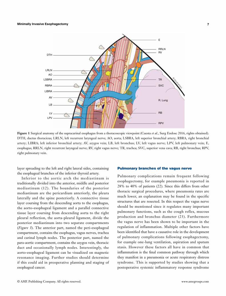
Recently a study of 25 thoracoscopic esophagectomies was performed, aiming to describe the anatomy of the supracarinal esophagus from a thoracoscopic viewpoint (19). The resulting anatomic description is shown in Figure 1, emphasizing the complex anatomy of this confined region.
Connective tissue layers and compartments
Recently the TIME trial has shown that thoracoscopic esophagectomy reduces pulmonary complications, which was followed by increased application of this approach (9). During thoracoscopic esophageal resections in vivo previously undescribed connective tissue layers were encountered. The knowledge and application of naturally existing tissue planes and compartments has been shown to be crucial in colorectal surgery. Therefore, the connective tissue layers and compartments surrounding the esophagus were recently studied in vivo during thoracoscopic esophagectomies (n=55) and using MRI, and in cadavers (n=2) with the aid of magnetic resonance imaging (MRI) and histology of large tissue sections (20,21). In these studies two regions were distinguished: a region superior to the aortic arch (superior mediastinum and neck) and one inferior to the aortic arch.
The connective tissue planes and compartments superior to the aortic arch are summarized in Figure 2. Above the aortic arch the esophagus and trachea traverse the visceral compartment. The posterior and posterolateral border of this compartment is formed by the alar fascia, which connects the right and left carotid sheets, passing dorsally to the esophagus. The anterior and anterolateral border is formed by the strap muscles. This visceral compartment also contains recurrent laryngeal nerves, the thyroid gland and lymph nodes. During thoracoscopic esophagectomy and dissection in the superior mediastinum the alar fascia can be seen posterior to the esophagus as a connective tissue
Minimally Invasive Esophagectomy
© AME Publishing Company. All rights reserved. www.amegroups.com
7
layer spreading to the left and right lateral sides, containing the esophageal branches of the inferior thyroid artery.
Inferior to the aort ic arch the mediast inum is traditionally divided into the anterior, middle and posterior mediastinum (12). The boundaries of the posterior mediastinum are the pericardium anteriorly, the pleura laterally and the spine posteriorly. A connective tissue layer coursing from the descending aorta to the esophagus, the aorto-esophageal ligament and a parallel connective tissue layer coursing from descending aorta to the right pleural reflection, the aorta-pleural ligament, divide the posterior mediastinum into two separate compartments (Figure 3). The anterior part, named the peri-esophageal compartment, contains the esophagus, vagus nerves, trachea and carinal lymph nodes. The posterior part, named the para-aortic compartment, contains the azygos vein, thoracic duct and occasionally lymph nodes. Interestingly, the aorto-esophageal ligament can be visualized on magnetic resonance imaging. Further studies should determine if this could aid in preoperative planning and staging of esophageal cancer.
Pulmonary branches of the vagus nerve
Pulmonary complications remain frequent following esophagectomy, for example pneumonia is reported in 28% to 40% of patients (22). Since this differs from other thoracic surgical procedures, where pneumonia rates are much lower, an explanation may be found in the specific structures that are resected. In this respect the vagus nerve should be mentioned since it regulates many important pulmonary functions, such as the cough reflex, mucous production and bronchus diameter (23). Furthermore the vagus nerve has been shown to be important in the regulation of inflammation. Multiple other factors have been identified that have a causative role in the development of pulmonary complications following esophagectomy, for example one-lung ventilation, aspiration and sputum stasis. However these factors all have in common that inflammation is the final common pathway through which they manifest in a pneumonia or acute respiratory distress syndrome. This is supported by studies showing that a postoperative systemic inflammatory response syndrome
Figure 1 Surgical anatomy of the supracarinal esophagus from a thoracoscopic viewpoint (Cuesta et al., Surg Endosc 2016, rights obtained). DTH, ductus thoracicus; LRLN, left recurrent laryngeal nerve; AO, aorta; LSBRA, left superior bronchial artery; RBRA, right bronchial artery; LIBRA; left inferior bronchial artery; AV, azygos vein; LB, left bronchus; LV, left vagus nerve; LPV, left pulmonary vein; E, esophagus; RRLN, right recurrent laryngeal nerve; RV, right vagus nerve; TR, trachea; SVC, superior vena cava; RB, right bronchus; RPV, right pulmonary vein.
DTH
LRLN
AO
LSBRA
RBRA
LIBRA
AV
LB
LV
LPV
E
RRLNRV
TR
SVC
RB
RPV
R. Lung

Weijs et al. Surgical anatomy of the esophagus
© AME Publishing Company. All rights reserved. www.amegroups.com
8
Figure 2 Peri-esophageal connective tissue layers above the aortic arch (Weijs et al. 2016, rights obtained). The blue line represents the alar fascia and the carotid sheaths; the green line represents the visceral fascia and the red line represents the perivertebral fascia. Car, carotid artery; Eso, esophagus; Jug, internal jugular vein; LCM, longus colli muscle; Ln, lymph node; Rln, recurrent laryngeal nerve; SCA, subclavian artery; SCM, sternocleidomastoid muscle; V, vagus nerve; VA, vertebral artery.
Figure 3 Peri-esophageal connective tissue layers below the level of the aortic arch (Weijs et al. 2016, rights obtained). The green line represents pleura, the purple line represents a connective tissue layer coursing from left to right main bronchus, the black line represents the aorto-esophageal ligament and the gray line represents the aorto-pleural ligament. Av, azygos vein; LMB, left main bronchus; Ln, lymph node; RMB, right main bronchus; TD, thoracic duct.
following esophagectomy is predictive of subsequent pulmonary complications (24). Therefore pulmonary vagotomy during esophagectomy may be a pivotal factor in development of postoperative pulmonary complications and sparing the pulmonary branches of the vagus nerve may be
beneficial. As the precise anatomy of the pulmonary vagus nerve
branches was unclear, an anatomical study was performed in six human cadavers (25). This study provided a map for the development of a method to spare the pulmonary branches

Minimally Invasive Esophagectomy
© AME Publishing Company. All rights reserved. www.amegroups.com
9
of the vagus nerve in 10 human cadavers (26).Both lungs are supplied by a small anterior pulmonary
plexus and a large posterior pulmonary plexus (containing 74–77% of the total lung supply). The right anterior plexus is located just above the right pulmonary artery and the left anterior plexus just anterior to the left pulmonary artery. The right posterior pulmonary plexus consists of a median of 13 nerve branches that sequentially arise from the vagus nerve from where it crosses the superior edge of the right main bronchus (Figure 4). The left posterior pulmonary plexus consists of a median of 13 nerve branches that sequentially arise from the vagus nerve between where it crosses the superior edge of the pulmonary artery and the lower edge of the pulmonary vein. The posterior pulmonary plexus is segmentally organized: the cranialmost branches innervate the superior lung lobe, the caudalmost innervate the middle and inferior lung lobes. In order to spare the innervation of both superior and middle/inferior lung lobes, all branches up to the caudalmost largest branch of the vagus nerve should be spared. The right caudalmost large pulmonary vagus nerve branch is found a median of 11 mm caudal to inferior edge of the right main bronchus and the left caudalmost large pulmonary vagus nerve branch is located a median of 13 mm caudal to the inferior edge of the left main bronchus.
It was feasible to spare the pulmonary branches of the vagus nerve during thoracoscopic esophagectomy in human cadavers, using these descriptions. On the right side, a median of nine pulmonary vagus nerve branches could be spared, of which four coursed to the middle and inferior lung lobes. On the left side a median of 10 pulmonary vagus nerve branches could be spared, of which four coursed to
the inferior lung lobe. The subcarinal (station 7) lymph nodes were always removed completely. Peri-bronchial lymph nodes (station 10L and R) were left behind in eight cases, however, it is questionable if these would have been resected during conventional esophagectomy.
Conclusions
Introduction of minimally invasive surgery and new insights in the function of the vagus nerve required a better understanding of the corresponding anatomy. For the esophagus this resulted in refined descriptions of the surgical anatomy from a thoracoscopic viewpoint, the peri-esophageal fascias and mapping of pulmonary vagus nerve branches.
Acknowledgements
None.
Footnote
Conflicts of Interest: The authors have no conflicts of interest to declare.
References
1. Torek F. The first successful case of resection of the thoracic portion of the oesophagus for carcinoma. Surg Gynecol Obstet 1913;16:614-7.
2. Torek F. Carcinoma of the thoracic portion of the esophagus. Arch Surg 1925;10:353-60.
Figure 4 Anatomy of the pulmonary branches of the vagus nerve from the viewpoint of right transthoracic esophagectomy (Weijs et al. 2015, rights obtained). A, azygos vein; Ao, aorta; Oeso, esophagus; RLN, left recurrent laryngeal nerve; S, sympathetic trunk; T, trachea; V, vagus nerve.
Right pulmonary hilum
Right pulmonary hilum
Posterior Posterior
Infe
rior
Infe
rior
Superior
Superior
BA

Weijs et al. Surgical anatomy of the esophagus
© AME Publishing Company. All rights reserved. www.amegroups.com
10
3. Ohsawa T. The surgery of the oesophagus. Arch Jpn Chir 1933;10:605-95.
4. Lewis I. The surgical treatment of carcinoma of the oesophagus; with special reference to a new operation for growths of the middle third. Br J Surg 1946;34:18-31.
5. McKeown KC. Total three-stage oesophagectomy for cancer of the oesophagus. Br J Surg 1976;63:259-62.
6. Orringer MB, Sloan H. Esophagectomy without thoracotomy. J Thorac Cardiovasc Surg 1978;76:643-54.
7. Omloo JM, Lagarde SM, Hulscher JB, et al. Extended transthoracic resection compared with limited transhiatal resection for adenocarcinoma of the mid/distal esophagus: five-year survival of a randomized clinical trial. Ann Surg 2007;246:992-1000; discussion 1000-1.
8. Cuschieri A. Endoscopic subtotal oesophagectomy for cancer using the right thoracoscopic approach. Surg Oncol 1993;2 Suppl 1:3-11.
9. Biere SS, van Berge Henegouwen MI, Maas KW, et al. Minimally invasive versus open oesophagectomy for patients with oesophageal cancer: a multicentre, open-label, randomised controlled trial. Lancet 2012;379:1887-92.
10. Tracey KJ. Reflex control of immunity. Nat Rev Immunol 2009;9:418-28.
11. Luyer MD, Greve JW, Hadfoune M, et al. Nutritional stimulation of cholecystokinin receptors inhibits inflammation via the vagus nerve. J Exp Med 2005;202:1023-9.
12. Gray H. Chapter XI: Splanchnology. In: Lewis WH (editor). Anatomy of the human body, 20th edition. New York: Philadelphia, Lea & Febiger; 1918.
13. Liebermann-Meffert DM, Luescher U, Neff U, et al. Esophagectomy without thoracotomy: is there a risk of intramediastinal bleeding? A study on blood supply of the esophagus. Ann Surg 1987;206:184-92.
14. Swigart LL, Siekert RG, et al. The esophageal arteries; an anatomic study of 150 specimens. Surg Gynecol Obstet 1950;90:234-43, illust.
15. Weijs TJ, Toxopeus EL, Ruurda JP, et al. Leaving a mobilized thoracic esophagus in situ when incurable cancer is discovered intraoperatively. Ann Thorac Surg 2015;99:490-4.
16. Rusch VW, Asamura H, Watanabe H, et al. The IASLC lung cancer staging project: a proposal for a new international lymph node map in the forthcoming seventh edition of the TNM classification for lung cancer. J Thorac Oncol 2009;4:568-77.
17. Ziyade S, Pinarbasili NB, Ziyade N, et al. Determination of standard number, size and weight of mediastinal lymph nodes in postmortem examinations: reflection on lung cancer surgery. J Cardiothorac Surg 2013;8:94.
18. Kajitani T. The general rules for the gastric cancer study in surgery and pathology. Part I. Clinical classification. Jpn J Surg 1981;11:127-39.
19. Cuesta MA, van der Wielen N, Weijs TJ, et al. Surgical anatomy of the supracarinal esophagus based on a minimally invasive approach: vascular and nervous anatomy and technical steps to resection and lymphadenectomy. Surg Endosc 2017;31:1863-70.
20. Cuesta MA, Weijs TJ, Bleys RL, et al. A new concept of the anatomy of the thoracic oesophagus: the meso-oesophagus. Observational study during thoracoscopic esophagectomy. Surg Endosc 2015;29:2576-82.
21. Weijs TJ, Goense L, van Rossum PS, et al. The peri-esophageal connective tissue layers and related compartments: visualization by histology and magnetic resonance imaging. J Anat 2017;230:262-71.
22. Weijs TJ, Berkelmans GH, Nieuwenhuijzen GA, et al. Immediate Postoperative Oral Nutrition Following Esophagectomy: A Multicenter Clinical Trial. Ann Thorac Surg 2016;102:1141-8.
23. Mazzone SB, Canning BJ. Autonomic neural control of the airways. Handb Clin Neurol 2013;117:215-28.
24. D’Journo XB, Michelet P, Marin V, et al. An early inflammatory response to oesophagectomy predicts the occurrence of pulmonary complications. Eur J Cardiothorac Surg 2010;37:1144-51.
25. Weijs TJ, Ruurda JP, Luyer MD, et al. Topography and extent of pulmonary vagus nerve supply with respect to transthoracic oesophagectomy. J Anat 2015;227:431-9.
26. Weijs TJ, Ruurda JP, Luyer MD, et al. Preserving the pulmonary vagus nerve branches during thoracoscopic esophagectomy. Surg Endosc 2016;30:3816-22.
Cite this article as: Weijs TJ, Ruurda JP, Luyer MD, Cuesta MA, van Hillegersberg R, Bleys RL. New insights into the surgical anatomy of the esophagus. J Thorac Dis 2017;9(Suppl 8):S675-S680. doi: 10.21037/jtd.2017.03.172

© AME Publishing Company. All rights reserved. www.amegroups.com
Introduction
Minimally invasive esophagectomy (MIE) techniques involve either complete endoscopic resection, via a thoracoscopic or laparoscopic approach, or a hybrid approach in which one part of the procedure is performed endoscopically. The principal purpose of MIE is to reduce surgical trauma and its effect on postoperative quality of life, rather than to expand the indications for surgery (1). Compared with open esophagectomy, MIE has advantages with respect to blood loss, operative trauma, postoperative recovery time and hospital stay (2,3). However, given the relatively high risk of surgery-related morbidity, adequate preoperative evaluation and patient selection are essential for MIE (4).
Preoperative evaluation
Patients in our institution are selected for MIE after the following standard preoperative work-up.
History and physical examination
There is no substitute for a careful history and physical examination performed by an experienced clinician. In our institution, a complete history and physical examination is performed, with particular attention to the severity of dysphagia. The clinicians evaluating a patient for MIE have several purposes during the evaluation process. First, the most important is to provide all parties with an assessment of both the short- and long-term risks of morbidity and
Preoperative Preparation
Evaluation and patient selection for minimally invasive esophagectomy
Yifeng Sun, Yang Yang, Haiyong Gu, Yu Yang, Xufeng Guo, Bin Li, Rong Hua, Bo Ye, Teng Mao, Zhigang Li
Department of Thoracic Surgery, Section of Esophageal Surgery, Shanghai Chest Hospital, Shanghai Jiao Tong University, Shanghai 200030, China
Correspondence to: Zhigang Li, MD, PhD. Department of Thoracic Surgery, Section of Esophageal Surgery, Shanghai Chest Hospital, Shanghai Jiao
Tong University, 241 Huaihai West Rd, Shanghai 200030, China. Email: [email protected].
Abstract: Minimally invasive esophagectomy (MIE) is an evolving surgical alternative to traditional open esophagectomy. Despite considerable technical challenges, it was considered that MIE could be performed effectively by surgeons experienced in open esophageal resection and advanced laparoscopic surgery. This chapter illustrates the preoperative evaluation and operative indications of MIE for esophageal cancer. Firstly, a complete history and physical exam is required for counseling on preoperative optimization. Then, the operation can be conducted after standard preoperative work-up includes several parts, such as positron-emission tomography (PET), endoscopic ultrasound (EUS), esophagography and computed tomography (CT). To our knowledge, the operative indications for MIE is now extended due to the rapid development of surgical technique and detailed preoperative evaluation. Limited node invasion and neoadjuvant chemoradiation are not rigorous contraindications for MIE any more. Optimal results require elaborate evaluation, appropriate patient selection and a multidisciplinary team experienced in the management of esophageal cancer.
Keywords: Preoperative evaluation; minimally invasive esophagectomy (MIE); operative indication
Received: 10 April 2018; Accepted: 07 June 2018; Published: 28 June 2018.
doi: 10.21037/shc.2018.06.08
View this article at: http://dx.doi.org/10.21037/shc.2018.06.08

© AME Publishing Company. All rights reserved. www.amegroups.com
12 Sun et al. Evaluation and patient selection for minimally invasive esophagectomy
mortality from the MIE procedure and to simultaneously identify factors that can be addressed to reduce the possibility of adverse events. In addition, the comprehensive evaluation of a patient as part of the preoperative assessment allows the identification of risk factors and health issues, such as diabetes, high blood pressure, and heart or lung problems, that should be under control.
Upper endoscopy
During the preoperative assessment, the most important aspect is to perform complete upper endoscopy and biopsy of the lesion to confirm the presence of a resectable esophageal lesion and to obtain pathological diagnosis (5). Deep biopsy with an endoscopic technique is a safe, high-yield, diagnostic method in patients with esophageal tumors. Pathologic confirmation may improve clinical decision making in the management of the patient (6). Kawamura and colleagues reported that endoscopists who do not allot adequate examination time may overlook neoplastic lesions in the upper gastrointestinal tract (7).
CT and PET/CT scanning
Since the overall accuracy rates of computed tomography (CT) scanning for the assessment of the depth of esophageal cancers are relatively poor, CT scanning is used to identify distant metastases and suspicious regional nodes rather than tumor depth (8). In our institution, CT scanning is used to evaluate the extent of lymph node involvement and distant metastasis. Before the advent of CT scanning for the staging of esophageal cancer, other noninvasive tests including linear tomography and nuclear scintigraphy accurately staged esophagus cancer in less than 30% of cases (9). Moreover, CT scanning may be useful to predict the efficacy of preoperative chemotherapy and the subsequent prognosis for patients with advanced esophageal cancer (10). New interest is developing in staging based on tumor measurements made on radiographs, with assessment of treatment response based on tumor length (9).
Routine CT scanning has improved the detection of distant metastases, but it generally has been replaced by the more sensitive PET/CT (positron emission tomography/computer tomography). The minimum lesion size that can be detected by PET scan alone is 5 mm; however, with PET/CT, there may be improvements in the resolution because lesion size and intensity influence detectability (11). PET is very sensitive in more than 95% of cases for
detecting primary tumors if the primary tumor is hypermetabolic (12,13). One of the major advantages of PET over CT is the three-dimensional imaging that can be accomplished with PET. This modality is also more likely than CT to identify secondary tumors (14). PET is not typically used to diagnose esophageal cancer; however, it is used to evaluate regional nodal disease and distant metastases. Just as endoscopic ultrasound (EUS) contributes to the preoperative evaluation and management of the new esophageal cancer patients, PET/CT adds additional biological information about the primary tumor as well as important staging information (15). FDG PET/CT scanning is valuable for assessing treatment response after neoadjuvant chemotherapy and predicting survival outcomes after surgery (16).
EUS
EUS staging of esophageal cancer was firstly reported by Lightdale in 1992 (17). In the next decade, the modality became a standard part of staging and follow-up of esophageal cancer in newly diagnosed patients (18,19). Although clinical signs and symptoms can determine T stage with a fair degree of accuracy, with substernal chest pain, dysphagia, and weight loss all being highly suggestive of T3 or T4 disease, symptoms alone are probably not enough to determine surgical resectability (20). EUS remains the most accurate modality for determination of T stage, with accuracy rates ranging from 64% to 80% with a low-frequency probe and up to 85% to 92% with a high-frequency probe (21). In studies with pathologic confirmation, the accuracy rates of EUS for determining N stage range from 70% to 86% (22). With the increased utility of preoperative chemoradiation, EUS can help to determine the locoregional stage of the cancer so that neoadjuvant treatment can potentially be offered to those with locally advanced disease (23). In addition, EUS is also helpful for confirming metastases to the celiac lymph nodes, which determines stage IV cancer (24).
Recently, with the widespread use of endoscopic therapy for early esophageal cancers, accurate staging of T1 adenocarcinoma with EUS is important to determine which patients may be offered endoscopic therapy for a potential cure and which patients should undergo esophagectomy. In experienced EUS groups, the accuracy of staging of intramucosal (T1a) cancer ranged from 82% to 94% (21,25). A T1b tumor has an approximately 20% likelihood of lymph node metastases compared with intramucosal

Minimally Invasive Esophagectomy
© AME Publishing Company. All rights reserved. www.amegroups.com
13
lesions, which have a less than 5% likelihood; thus, the EUS assessment of T stage may be helpful in deciding between endoscopic treatment and surgical resection (26). Performing EUS before endoscopic resection of T1 tumors remains controversial, and resection may be diagnostic (27).
Esophagography
Esophagography, which is also known as a barium swallow, is the radiographic or fluoroscopic examination of the pharynx and the esophagus after ingestion of thick and thin mixtures of barium sulfate, respectively. This test, which is commonly performed as part of an upper GI series, is indicated for patients with a history of dysphagia and regurgitation. The purpose is to diagnose hiatal hernias, diverticula, strictures, ulcers, tumors, and motility disorders. However, with regard to esophageal cancer, further testing is usually required for a definitive diagnosis (28).
In summary, preoperative evaluation using imaging techniques, such as EUS, CT and PET/CT scanning, remains the mainstay for the diagnosis of esophageal cancer (8). In addition, esophagography can be used to confirm the location of the lesion and evaluate the continuity of esophagus. Complete and appropriate evaluation of esophageal cancer can improve the selection of patients for MIE and may improve patient outcomes (29).
Patient selection
Although MIE has been widely used for nearly 20 years, there are not yet specific criteria regarding the indications for MIE. In general, MIE is indicated in the same patients as open techniques with a few exceptions (30). It should not be undertaken in patients who are unable to survive the physiological insult of surgery. Evaluating individual risk is important for patient selection and proper preoperative management. A critical component of the preoperative evaluation is the assessment of a patient’s functional status. Functional status is an important component of the decision algorithm for both the pulmonary and cardiac elements of the preoperative evaluation (31). It is not easy to predict which patients will develop with complications after surgery. Even so, we must know which indicators can increase postoperative morbidity and mortality. Many risk factors have been identified including poor cardiopulmonary function, advanced age, tumor stage, diabetes mellitus, impaired general health, and hepatic or renal dysfunction (32).
Combined with the previous reports and our experiences
at home and abroad, the following items are considered as common indications for MIE: (I) early or middle stage esophageal cancer that does not invade the full thickness of the esophagus; (II) no severe pleural adhesions; (III) lesion diameter <5 cm; (IV) lesion diameter >5 cm with the lesion mainly within the esophageal lumen; (V) no adjacent lymphadenopathy or distant metastasis.
Tumor staging is essential for planning surgical treatment, and surgery should not be performed in patients without a chance of cure such as in those with distant metastases or in those unable to survive the physiological insult of surgery. The main objective of surgery must be to achieve a R0 resection. For thoracic surgeons, particularly those who are still learning MIE, it is very important to choose patients according to tumor staging. Generally, patients with a clinical stage of T1-3N0-1M0 (AJCC TNM staging) are suitable for direct surgery. Patients with locally advanced esophageal cancer can receive neoadjuvant chemoradiation followed by MIE. Currently, neoadjuvant chemoradiation plays a key role because it downstages the tumor and allows for adequate tumor resection. From a surgical perspective, the drawback is that neoadjuvant chemoradiation may further complicate the procedure by causing significant fibrosis, which necessitates an important discussion about the effectiveness of MIE in these situations. However, in a multicenter, open-label, randomized controlled trial published in 2012 by Biere et al. that compared open surgery with MIE for advanced tumors in a similar number of patients, there were not significant differences between groups in terms of the margins obtained after resection (33). Moreover, the recent literature does not show that neoadjuvant treatment is a contraindication for minimally invasive techniques (34). Thus, a minimally invasive approach would not be a contraindication for patients with advanced stages of cancer.
Overall, the field of MIE is stirring, and innovations continue to occur in rapid succession. At this moment, the most prominent question is related to long-term outcomes. Studies on the indications and contraindications for MIE are required to explicit the specific selection criteria for MIE.
Acknowledgements
None.
Footnote
Conflicts of Interest: The authors have no conflicts of interest

© AME Publishing Company. All rights reserved. www.amegroups.com
14 Sun et al. Evaluation and patient selection for minimally invasive esophagectomy
to declare.
References
1. Luketich JD, Schauer P, Christie N. Minimally invasive esophagectomy. Ann Thorac Surg 2000;70:906-11; discussion 911-2.
2. Smithers BM, Gotley DC, Martin I, et al. Comparison of the outcomes between open and minimally invasive esophagectomy. Ann Surg 2007;245:232-40.
3. Kauppila JH, Xie S, Johar A, et al. Meta-analysis of health-related quality of life after minimally invasive versus open oesophagectomy for oesophageal cancer. Br J Surg 2017;104:1131-40.
4. Mehta K, Bianco V, Awais O, et al. Minimally invasive staging of esophageal cancer. Ann Cardiothorac Surg 2017;6:110-8.
5. Winiker M, Mantziari S, Figueiredo SG, et al. Accuracy of preoperative staging for a priori resectable esophageal cancer. Dis Esophagus 2018;31:1-6.
6. Tae HJ, Lee HL, Lee KN, et al. Deep biopsy via endoscopic submucosal dissection in upper gastrointestinal subepithelial tumors: a prospective study. Endoscopy 2014;46:845-50.
7. Kawamura T, Wada H, Sakiyama N, et al. Examination time as a quality indicator of screening upper gastrointestinal endoscopy for asymptomatic examinees. Dig Endosc 2017;29:569-75.
8. Pennathur A, Gibson MK, Jobe BA, et al. Oesophageal carcinoma. Lancet 2013;381:400-12.
9. Roedl JB, Harisinghani MG, Colen RR, et al. Assessment of treatment response and recurrence in esophageal carcinoma based on tumor length and standardized uptake value on positron emission tomography–computed tomography. Ann Thorac Surg 2008;86:1131-8.
10. Wakatsuki K, Matsumoto S, Migita K, et al. Usefulness of computed tomography density of a tumor in predicting the response of advanced esophageal cancer to preoperative chemotherapy. Surgery 2017;162:823-35.
11. Ott K, Weber W, Siewert JR. The importance of PET in the diagnosis and response evaluation of esophageal cancer. Dis Esophagus 2006;19:433-42.
12. Flamen P, Lerut A, Van Cutsem E, et al. Utility of positron emission tomography for the staging of patients with potentially operable esophageal carcinoma. J Clin Oncol 2000;18:3202-10.
13. Himeno S, Yasuda S, Shimada H, et al. Evaluation of esophageal cancer by positron emission tomography. Jpn J
Clin Oncol 2002;32:340-6. 14. Block MI, Patterson GA, Sundaresan RS, et al.
Improvement in staging of esophageal cancer with the addition of positron emission tomography. Ann Thorac Surg 1997;64:770-6; discussion 776-7.
15. Meyers BF, Downey RJ, Decker PA, et al. The utility of positron emission tomography in staging of potentially operable carcinoma of the thoracic esophagus: Results of the American College of Surgeons Oncology Group Z0060 trial. J Thorac Cardiovasc Surg 2007;133:738-45.
16. Goel R, Subramaniam RM, Wachsmann JW. PET/Computed Tomography Scanning and Precision Medicine: Esophageal Cancer. PET Clin 2017;12:373-91.
17. Lightdale CJ. Endoscopic ultrasonography in the diagnosis, staging and follow-up of esophageal and gastric cancer. Endoscopy 1992;24:297-303.
18. Lightdale CJ, Kulkarni KG. Role of endoscopic ultrasonography in the staging and follow-up of esophageal cancer. J Clin Oncol 2005;23:4483-9.
19. Zhang X, Watson DI, Lally C, et al. Endoscopic ultrasound for preoperative staging of esophageal carcinoma. Surg Endosc 2005;19:1618-21.
20. Heidemann J, Schilling MK, Schmassmann A, et al. Accuracy of endoscopic ultrasonography in preoperative staging of esophageal carcinoma. Dig Surg 2000;17:219-24.
21. Cen P, Hofstetter WL, Lee JH, et al. Value of endoscopic ultrasound staging in conjunction with the evaluation of lymphovascular invasion in identifying low-risk esophageal carcinoma. Cancer 2008;112:503-10.
22. Barbour AP, Rizk NP, Gerdes H, et al. Endoscopic ultrasound predicts outcomes for patients with adenocarcinoma of the gastroesophageal junction. J Am Coll Surg 2007;205:593-601.
23. Luu C, Amaral M, Klapman J, et al. Endoscopic ultrasound staging for early esophageal cancer: Are we denying patients neoadjuvant chemo-radiation? World J Gastroenterol 2017;23:8193-9.
24. Eloubeidi MA, Wallace MB, Hoffman BJ, et al. Predictors of survival for esophageal cancer patients with and without celiac axis lymphadenopathy: Impact of staging endosonography. Ann Thorac Surg 2001;72:212-9; discussion 219-20.
25. Murata Y, Suzuki S, Ohta M, et al. Small ultrasonic probes for determination of the depth of superficial esophageal cancer. Gastrointest Endosc 1996;44:23-8.
26. He LJ, Shan HB, Luo GY, et al. Endoscopic ultrasonography for staging of T1a and T1b esophageal

Minimally Invasive Esophagectomy
© AME Publishing Company. All rights reserved. www.amegroups.com
15
squamous cell carcinoma. World J Gastroenterol 2014;20:1340-7.
27. Li JJ, Shan HB, Gu MF, et al. Endoscopic ultrasound combined with submucosal saline injection for differentiation of T1a and T1b esophageal squamous cell carcinoma: a novel technique. Endoscopy 2013;45:667-70.
28. Levine MS, Rubesin SE. History and Evolution of the Barium Swallow for Evaluation of the Pharynx and Esophagus. Dysphagia 2017;32:55-72.
29. Kaushik N, Khalid A, Brody D, et al. Endoscopic ultrasound compared with laparoscopy for staging esophageal cancer. Ann Thorac Surg 2007;83:2000-2.
30. Decker G, Coosemans W, De Leyn P, et al. Minimally invasive esophagectomy for cancer. Eur J Cardiothorac Surg 2009;35:13-20.
31. Zingg U, Smithers BM, Gotley DC, et al. Factors
associated with postoperative pulmonary morbidity after esophagectomy for cancer. Ann Surg Oncol 2011;18:1460-8.
32. Uchihara T, Yoshida N, Baba Y, et al. Risk factors for pulmonary morbidities after minimally invasive esophagectomy for esophageal cancer. Surg Endosc 2018;32:2852-8.
33. Biere SS, van Berge Henegouwen MI, Maas KW, et al. Minimally invasive versus open oesophagectomy for patients with oesophageal cancer: a multicentre, open-label, randomised controlled trial. Lancet 2012;379:1887-92.
34. Merritt RE, Whyte RI, D’Arcy NT, et al. Morbidity and mortality after esophagectomy following neoadjuvant chemoradiation. Ann Thorac Surg 2011;92:2034-40.
doi: 10.21037/shc.2018.06.08Cite this article as: Sun Y, Yang Y, Gu H, Yang Y, Guo X, Li B, Hua R, Ye B, Mao T, Li Z. Evaluation and patient selection for minimally invasive esophagectomy. Shanghai Chest 2018;2:49.

© AME Publishing Company. All rights reserved. www.amegroups.com
Enhanced recovery after surgery (ERAS) was first proposed and practiced by the Danish surgeon Kehlet in 1997 (1,2). As a new concept and therapeutic rehabilitation model, ERAS refers to the application of a series of optimization measures with evidence-based medical indications during the perioperative period. These measures are intended to reduce the physiological and psychological traumatic stress in patients who undergo surgery and to accelerate rehabilitation. The benefits of the ERAS concept are reflected in the following: (I) improvement in the physical and psychological efficacy of the treatment; (II) reduction in postoperative complications; (III) acceleration of rehabilitation; (IV) shortening of the hospital stay; (V) reduction in medical expenses; (VI) reduction in social and family burdens. To date, ERAS has been successfully applied in surgeries for colorectal surgery (3-5). Moreover, several large esophageal cancer centers in China and other countries have also gradually applied the ERAS to esophagectomy (6-10). Currently, minimally invasive esophagectomy (MIE), which is represented by thoracoscopic laparoscopic esophagectomy (TLE), has been widely used
in clinical practice. In addition, robotic assisted minimally invasive esophagectomy (RAMIE) has also been gradually introduced and performed to further optimize the minimally invasive surgical approach of MIE. It is believed that in the near future, the combination of ERAS and MIE will greatly reduce the occurrence of surgical trauma and accelerate the rehabilitation of patients after esophagectomy.
Section I: preoperative considerations of ERAS for esophagectomy
For esophagectomy, perioperative ERAS mainly focuses on the preoperative, intraoperative and postoperative periods. This study uses MIE as an example to describe the details of the preoperative preparation and the features that are assessed in perioperative MIE-ERAS, as described below.
Preoperative education
Enhanced preoperative evaluation for patients, which
Preoperative Preparation
ERAS prior to minimally invasive esophagectomy
Xufeng Guo1, Chun Ding1, Chenguang Luo2, Yu Yang1, Bin Li1, Rong Hua1, Bo Ye1, Haiyong Gu1, Yifeng Sun1, Teng Mao1, Zhigang Li1
1Department of Thoracic Surgery, Section of Esophageal Surgery, Shanghai Chest Hospital, Shanghai Jiaotong University, Shanghai 200030, China; 2Department of Thoracic Surgery, The first people’s Hospital of Yangquan, Shanxi 045000, China
Correspondence to: Zhigang Li, MD, PhD. Department of Thoracic Surgery, Section of Esophageal Surgery, Shanghai Chest Hospital, Shanghai
Jiaotong University, 241 Huaihai West Rd, Shanghai 200030, China. Email: [email protected].
Abstract: The concept and pattern of enhanced recovery after surgery (ERAS) have been used for nearly 20 years. However, the clinical application and popularization of ERAS in the esophagectomy have been greatly restricted due to the complexity of operation and the high incidence of postoperative complications. In recent years, with the growing maturity of the minimally invasive technique of thoracic laparoscopy, the attention of the concept of tissue and organ protection, the improvement of making gastric tube and the presentation and application of new concepts, ERAS has made a breakthrough in the field of esophagectomy. In this paper, the ERAS clinical pathway prior to minimally invasive esophagectomy (MIE) of Shanghai chest hospital was summarized, including preoperative preparation and evaluation, preoperative education and so on. The aim is to continuously improve and popularize the clinical pathway of ERAS in MIE, applying in more and more centers, so as to benefit more and more patients.
Keywords: Esophageal cancer; minimally invasive esophagectomy (MIE); enhanced recovery after surgery (ERAS)
Received: 10 April 2018; Accepted: 07 June 2018; Published: 28 June 2018.
doi: 10.21037/shc.2018.06.07
View this article at: http://dx.doi.org/10.21037/shc.2018.06.07

Minimally Invasive Esophagectomy
© AME Publishing Company. All rights reserved. www.amegroups.com
17
includes an assessment of the surgical risk and tolerability, is conducive to postoperative rehabilitation. Preoperative education is a non-independent predictor of the successful implementation of accelerated rehabilitation (11,12). Preoperative education should focus on the introduction of the treatment process and surgical plan to facilitate cooperation among patients with respect to postoperative rehabilitation and the development of an early discharge plan. The patients should be informed of their important role in this plan, including in postoperative posture, early nasogastric enteral nutrition, early ambulation, postoperative feeding methods, and for patients with neck anastomosis, the precautions of applying pressure on the neck incisions during cough and expectoration.
Preoperative nutrition and other risk assessment
Patients with esophageal cancer often experience concomitant preoperative malnutrition due to the limited food intake and tumor depletion, which may directly affect postoperative recovery. Studies have shown that preoperative malnutrition increases the incidence of postoperative complications (13,14). Therefore, patients with severe malnutrition should receive nutritional support therapy, and the operation should be rescheduled. Patients who receive preoperative radical radiochemotherapy, neoadjuvant radiochemotherapy, and those with severe diabetes have an increased risk of postoperative anastomotic leakage. These patients should therefore undergo cautious, accelerated rehabilitation.
Preoperative fasting
As a traditional concept, food intake should be prohibited the night before surgery to reduce the risk of mis-aspiration during anesthesia. However, no evidence supports the idea that long-term fasting before esophagectomy can prevent mis-aspiration. Eating can reduce catabolism, psychological stress and potential insulin resistance. The consumption of liquid foods 6 hours before surgery is safe and can reduce preoperative thirst, hunger and irritability, as well as postoperative muscle loss; this can also reduce the symptoms of nausea and vomiting and can significantly reduce the incidence of postoperative insulin resistance (15). Patients with severe esophageal obstruction should avoid eating 6 hours before surgery to avoid anesthesia-related aspiration.
Preoperative bowel preparation
Preoperative enema may cause stress in the patient and may lead to dehydration and electrolyte imbalances, especially in frail elderly patients. Therefore, preoperative bowel preparation for esophagectomy is only recommended for patients with severe constipation; in these cases, laxatives such as lactulose oral solution and phenolphthalein tablets are recommended.
Preoperative anesthetic medication
Sedative medication given the night before the surgery may help relieve patient stress.
Use of prophylactic antibiotics
Antibiotic prophylaxis is beneficial for the reduction of infection in patients undergoing esophageal cancer surgeries. Antibiotics should be given half an hour before the start of surgery. If the operation time is more than 3 hours, antibiotics can be given again during surgery.
Section II: preoperative clinical approach of MIE-ERAS in the Esophageal Center of Shanghai Chest Hospital
Preoperative education
Psychological guidanceThe patients are encouraged to face their own illness with open-mindedness and positivity.
Dietary guidanceThe preoperative diet should consist of high-protein and high-calorie foods, which should also be rich in vitamins and low in fiber. Coarse, hard, pickled, and spicy food should be avoided. Alcohol, including beer, rice wine, and liquor is prohibited. Food in liquid form along with post-meal liquid (1% streptomycin) rinsing and administration of metronidazole (one tablet of po bid after a meal) should be provided to the patients with obvious obstructions.
Sanitation guidancePatients should brush their teeth at least twice a day and rinse with warm boiled water after meals. Patients with oral diseases should be treated promptly.

© AME Publishing Company. All rights reserved. www.amegroups.com
18 Guo et al. ERAS prior to minimally invasive esophagectomy
Preoperative preparation
Respiratory tract preparation(I) The patients with a history of smoking should have
completely quit smoking for at least 2 weeks and should not smoke during their hospitalization;
(II) Terbutaline Sulfate Injection 2 & Pulmicort 2 (inhalation bid): Mucosolvan 60 mg (po tid); Theophylline 0.1 g (po Qn)
Cardiopulmonary exercise(I) Climbing stairs: the patient is asked to continuously
walk upstairs without any break based on the individual’s strength, for 5 floors 3–4 times/day;
(II) Deep breathing exercises: abdominal breathing, deep breathing, and coughing are performed under the guidance of a doctor 400 times.
Special preoperative preparation (I) For patients with hypertension, blood pressure
should be monitored daily, and preoperative oral antihypertensive drugs should be administered to maintain a stable blood pressure;
(II) For patients with diabetes, blood sugar should be tested before each meal and at bedtime to ensure a stable level of preoperative blood glucose;
(III) For patients whose medical status is complicated by other systemic diseases, experts in the relevant
departments should be consulted if necessary;(IV) All self-administered drugs and supplements (such as
aspirin) should be discontinued under the guidance of a physician;
(V) For patients with severe eating obstructions, severe malnutrition, and those who cannot eat, preoperative nutritional support should be provided (oral ENSURE or parenteral nutrition support);
(VI) For pat ients with suspected supraclav icular lymphadenopathy, lymph node biopsy should be performed.
Preoperative examination
Examination of cardiopulmonary function (I) Blood test: routine blood/urine/fecal tests, liver/
kidney funct ion/electrolyte , b lood glucose/glycosylated hemoglobin, coagulation function, blood type, hepatitis/syphilis/AIDS;
(II) Assessment of cardiac function: electrocardiogram, dynamic electrocardiogram (for patients with a history of arrhythmia), treadmill exercise test (for those >60 years of age), heart echocardiography (for patients >60 years of age), coronary angiography (for patients with a history of coronary heart disease);
(III) Pulmonary function assessment: lung function, blood gas analysis, breath-holding test, and stair climbing test.
Tumor examination(I) Blood tumor markers;(II) Esophagoscopy/EUS: biopsy;(III) Gastrointestinal (GI) tumors: general classification
and location of tumors;(IV) Neck ultrasound, chest-enhanced CT, abdomen-
enhanced CT, PET-CT (selective);
Staging assessment and therapeutic strategies
According to the preoperative examination, a cTNM staging assessment was performed, and a treatment plan was individually developed according to the guidelines for the diagnosis and treatment of esophageal cancer (Figure 1).
Acknowledgements
None.
Local-reginoal tumor I-IIIA
Early stage (IB-IIA) LNM (-)
(IB-IIB) LNM (+)
Early early stage (IA)
ESD/MIE/THE MIE LN dissection
Induction therapy
Adjuvant treatment
Figure 1 Surgical treatment approach for resectable esophageal cancer at the Esophageal Center of Shanghai Chest Hospital.

Minimally Invasive Esophagectomy
© AME Publishing Company. All rights reserved. www.amegroups.com
19
Footnote
Conflicts of Interest: The authors have no conflicts of interest to declare.
References
1. Kehlet H. Multimodal approach to control postoperative pathophysiology and rehabilitation. Br J Anaesth 1997;78:606-17.
2. Kehlet H, Wilmore DW. Multimodal strategies to improve surgical outcome. Am J Surg 2002;183:630-41.
3. Basse L, Hjort Jakobsen D, Billesbølle P, et al. A clinical pathway to accelerate recovery after colonic resection. Ann Surg 2000;232:51-7.
4. Basse L, Jacobsen DH, Billesbølle P, et al. Colostomy closure after Hartmann's procedure with fast-track rehabilitation. Dis Colon Rectum 2002;45:1661-4.
5. Basse L, Jakobsen DH, Bardram L, et al. Functional recovery after open versus laparoscopic colonic resection: a randomized, blinded study. Ann Surg 2005;241:416-23.
6. Sun HB, Liu XB, Zhang RX, et al. Early oral feeding following thoracolaparoscopic oesophagectomy for oesophageal cancer. Eur J Cardiothorac Surg 2015;47:227-33.
7. Shewale JB, Correa AM, Baker CM, et al. Impact of a Fast-track Esophagectomy Protocol on Esophageal Cancer Patient Outcomes and Hospital Charges. Ann Surg 2015;261:1114-23.
8. Zhao G, Cao S, Cui J. Fast-track surgery improves postoperative clinical recovery and reduces postoperative
insulin resistance after esophagectomy for esophageal cancer. Support Care Cancer 2014;22:351-8.
9. Cao S, Zhao G, Cui J, et al. Fast-track rehabilitation program and conventional care after esophagectomy: a retrospective controlled cohort study. Support Care Cancer 2013;21:707-14.
10. Pan H, Hu X, Yu Z, et al. Use of a fast-track surgery protocol on patients undergoing minimally invasive oesophagectomy: preliminary results. Interact Cardiovasc Thorac Surg 2014;19:441-7.
11. Aarts MA, Okrainec A, Glicksman A, et al. Adoption of enhanced recovery after surgery (ERAS) strategies for colorectal surgery at academic teaching hospitals and impact on total length of hospital stay. Surg Endosc 2012;26:442-50.
12. Younis J, Salerno G, Fanto D, et al. Focused preoperative patient stoma education, prior to ileostomy formation after anterior resection, contributes to a reduction in delayed discharge within the enhanced recovery programme. Int J Colorectal Dis 2012;27:43-7.
13. Kunisaki C, Shimada H, Nomura M, et al. Immunonutrition risk factors of respiratory complications after esophagectomy. Nutrition 2004;20:364-7.
14. Nozoe T, Kimura Y, Ishida M, et al. Correlation of pre-operative nutritional condition with post-operative complications in surgical treatment for oesophageal carcinoma. Eur J Surg Oncol 2002;28:396-400.
15. Brady M, Kinn S, Stuart P. Preoperative fasting for adults to prevent perioperative complications. Cochrane Database Syst Rev 2003;(4):CD004423.
doi: 10.21037/shc.2018.06.07Cite this article as: Guo X, Ding C, Luo C, Yang Y, Li B, Hua R, Ye B, Gu H, Sun Y, Mao T, Li Z. ERAS prior to minimally invasive esophagectomy. Shanghai Chest 2018;2:50.

© AME Publishing Company. All rights reserved. www.amegroups.com
Introduction
Esophageal surgery is high-risk. Despite innovations in surgical techniques (open vs. laparo-thoracoscopic) and the addition of neo-adjuvant chemo-radiotherapy, major morbidity still can be up to 65% and 30-day mortality rate as high as 4% (1,2). Pulmonary infections and anastomotic dehiscence make up for the majority of reported complications. To reduce complications minimally invasive techniques were introduced and some studies report a more favourable outcome (1). This especially translates into a reduction in pulmonary complications, which are reported to decrease by 60% (2). However, outcome seems especially related to patient and tumour characteristics, surgical experience and hospital volume (3,4). Over the years, it has become clear that a multimodal, multispecialty and dedicated-team approach is essential for these patients, including strict patient selection, work-up, and enhanced recovery after surgery (ERAS) protocols (5). The anaesthetist is an essential member of the multidisciplinary team. Pain and stress, fluid, hemodynamic, and ventilation management can influence outcome significantly. Best practices may differ
depending on the surgical techniques used. In this review, we will provide an overview of the
current state of the art perioperative practices for open and laparoscopic surgery from the anaesthetist’s perspective.
Preoperative screening and optimisation
Patients with esophageal cancer often have comorbidities, suffer from significant weight loss, poor nutritional state and are more fragile (6,7). An overweight patient will have a higher chance of wound infections while an underweight patient has odds of death go up 5-fold (8). Timely screening of the preoperative patient will allow the possibility to improve the health status of the patient and to reduce chances of adverse outcome (9). The anaesthetist should be involved early in a multidisciplinary evaluation that also includes a debate whether or not to proceed to surgery based on expected perioperative morbidity.
To assess the perioperative risk of morbidity and mortality some general and some esophagectomy specific risk scoring systems exist. Warnell and co-workers reviewed ten of these models that were externally validated (3). The accuracy of
Preoperative Preparation
Anaesthesia during esophagectomy
Denise P. Veelo, Bart F. Geerts
Department of Anaesthesiology, Academic Medical Center, Amsterdam, The Netherlands
Contributions: (I) Conception and design: DP Veelo; (II) Administrative support: None; (III) Provision of study materials or patients: All authors;
(IV) Collection and assembly of data: DP Veelo; (V) Data analysis and interpretation: All authors; (VI) Manuscript writing: All authors; (VII) Final
approval of manuscript: All authors.
Correspondence to: Denise P. Veelo, MD, PhD. Department of Anaesthesiology, Academic Medical Center, Amsterdam, Meibergdreef 9, 1105 AZ, The
Netherlands. Email: [email protected].
Abstract: In this review, we will provide an overview of the current state of the art of perioperative practices for open and laparoscopic esophagus surgery from the anaesthetist’s perspective. Morbidity and mortality after esophagectomy is still high despite multidisciplinary and enhanced recovery pathways showing promising results. The anaesthetist has an important role in the complex care of the esophageal cancer patient. Minimizing unnecessary fluid administration, adequate pain management, hypotension, and protective lung ventilation are examples of proven strategies that can improve outcome after this high-risk surgery.
Keywords: Esophagectomy; anaesthesia; review
Submitted Nov 29, 2016. Accepted for publication Mar 10, 2017.
doi: 10.21037/jtd.2017.03.153
View this article at: http://dx.doi.org/10.21037/jtd.2017.03.153
27
Minimally Invasive Esophagectomy
© AME Publishing Company. All rights reserved. www.amegroups.com
21
these scoring varies widely with overestimation of mortality occurring frequently (5–200% of cases) and a reported area under the curve between 0.58 and 0.78. Common risk factors are age, comorbidities (cardiopulmonary, diabetes, renal insufficiency, liver dysfunction), preoperative treatment (neoadjuvant chemotherapy), tumor staging and hospital characteristics (hospital volume of esophagectomies) (3).
More elaborate general preoperative risk scores have also recently been developed from large databases of over one million patients. The POSPOM scoring system is such a scoring system (10). Unfortunately, it fails to consider the patient comorbidities completely neither does it reflect the full scope of the esophagectomy (both abdominal and thoracic surgery). However, this score does provide the patient and all specialists involved in the care with a more reliable (albeit likely underestimation) of what would happen if esophageal surgery were performed.
A structured preoperative screening in an anaesthesia outpatient clinic has become the standard for anaesthesia care in most countries. During the pre-assessment cardiac function should be evaluated by assessing functional disabilities and the MET score. An EKG can provide information about dysrhythmias, conduction delays, previous myocardial infarction, and hypertrophic development of atrium and/or ventricle. When wall motion or valvular issues are suspected, an echocardiography can provide new insights. Most patients will receive chemoradiation, which may impact cardiac function. Lund et al. found that baseline cardiac output can be decreased by as much as 15% due to chemo-radiotherapy during rest (11). Although the impact has been described as mild, anaesthetic drugs, surgery, one lung ventilation (shunting), and laparo-thoracoscopy may further influence heart function.
Additional work-up should include blood testing for renal and liver function, haematology, irregular antibodies and clotting upon indication. When esophagectomy is to be performed, functional assessment of the lungs needs to be performed. The patients need to be able to undergo one-lung ventilation (OLV). As most patients in this population have been or are smokers, the incidence of significant emphysema is higher. Exact cut-offs to perform one-lung ventilation have not been clearly determined. Acute lung injury after esophagectomy has been reported in as many as 25% of all cases after surgery (12). Risk factors are low pre-operative body mass index, smoking, the experience of the surgeon, the duration of surgery and OLV, post-operative anastomotic leak, peri-operative hypoxaemia, hemodynamic instability requiring additional fluids or vasoactive support
(12).Weight loss is often pathognomonic for poor outcome
after surgery and albumin levels can be a marker of very poor nutritional state (13). A dietician should be consulted to optimize weight, fat and protein status. Sometimes it is warranted to delay surgery to supplement proteins as this might improve wound healing and prevent infections or anastomotic breakdown. Early involvement of physiotherapist to improve physical or cardiopulmonary fitness and a dietician for a good nutritional state seems rational but studies into the effect on outcome are contradictory (14-17).
Best anaesthetic practices
Type of anaesthesia
The discussion of the advantages over the use of one anaesthetic over the other has led to a number of studies to be performed. However, this discussion is complicated by the small number of studies available, small number of patients included and the differences in endpoints and methodology, which makes them difficult to compare.
Some studies have described immune-modulatory benefits and reduced ischemia-reperfusion injury markers of volatile anaesthetics during one lung ventilation. However, after thoracic surgery there seems to be no clear relation between inflammatory markers and pulmonary morbidity as the results of clinical studies are conflicting (18-22).
During one lung ventilation hypoxic pulmonary vasoconstriction (HPV) influences intrapulmonary shunting and oxygenation. Volatile anaesthetics have been shown to impair HPV in a dose dependent matter in contrast to propofol in animal models (23). However, when titrate to effect the influence of volatile anaesthetics on intrapulmonary shunting may be equal to that of propofol (24).
Thoracic epidural analgesia
There seems to be no benefit of using either volatile anaesthetics or propofol on the occurrence and severity of post-operative pain (25). But the evidence on the use of multimodal treatment regimes during esophagectomy and especially the thoracic epidural analgesia (TEA) seems clear. This benefit has been shown for both open as minimally invasive esophagectomy. TEA provides superior analgesia, reduces respiratory complications, need for postoperative mechanical ventilation, rehabilitations and hospital length of stay (26-30). Most studies show mainly an effect on

© AME Publishing Company. All rights reserved. www.amegroups.com
22 Veelo and Geerts. Anaesthesia during esophagectomy
pulmonary morbidity although a reduction in the incidence of anastomotic leakage has also been suggested (31). TEA has no clear anti-inflammatory effects (32).
Ventilatory management
Ventilatory management during transthoracic esophagectomy is usually managed with OLV by means of a double-lumen tube (DLT). This technique enables easy separation of both lungs but has also been associated with complications such as hoarseness and damage to the vocal cords, and tracheo-bronchial lacerations. Conformation of position requires fiber-optic bronchoscopy. Recently the video DLT has been introduced. This DLT has an integrated high-resolution camera, which would remove the need for fiber-optic confirmation (33). Although the first reports with this technique are promising, conclusive studies are needed to confirm added safety, utility and cost-effectiveness of this device. An alternative technique for separated lung ventilation is the use of a bronchus blocker. This device is thought to be similar in terms of performance for patients with normal airways. In patients with airway abnormalities and difficult intubation bronchus blockers may be preferred (34). During minimally invasive transthoracic and trans-hiatal surgery the use of one lung ventilation may not be obligatory. One Chinese group reported the use of single lumen intubation for thoracoscopy as feasible and safe (35). Indeed, the need for OLV might also depend on the position of the anastomosis and the need for optimal surgical views. Challenges for the anaesthesiologist during OLV are deoxygenation and hypercapnia due to shunting and atelectasis. The latter may especially be difficult to manage during thoracoscopy, which may take place in the left lateral or prone position (36,37). Laparoscopic surgery in the prone position is described to be associated with better oxygenation due to lower shunt fractions and better ventilation/perfusion matching (38,39). In addition, it may decrease blood loss and improve surgical ergonomics.
The incidence of acute respiratory distress syndrome (ARDS) or acute lung injury (ALI) after esophagectomy is high with a reported incidence of 16% up to 33% (40). Important etiologic factors are fluid overload, vascular leakage, damage of lung lymphatics and pulmonary endothelium. These are induced by peripheral and alveolar inflammatory mediator production and cellular infiltration. Patient and procedure related risk factors for ALI have been discussed earlier. The severity of the inflammatory response may be a predictive factor in postoperative pulmonary
morbidity (41). The use of OLV may aggravate this process. The use of lung protective ventilation strategies during one lung ventilation such as the use of smaller tidal volumes (5 mL/kg), plateau pressures below 35 cmH2O and the application of PEEP has been shown to decrease the inflammatory response and improve oxygenation and resulted in shorter times until extubation and pulmonary complications (41,42). Although no large outcome studies have been done for patients after esophagectomy, the benefits of the use of lung protective ventilation in the prevention and treatment of ARDS/ALI in critically ill patients and the general surgical population are well established (43).
Fluid management
Intravenous fluids and outcome
Both hypervolemia and hypovolemia may be associated with increased morbidity (44). Fluid management in this patient group has until recently focused on restricting fluid administration to prevent pulmonary and cardiac complications (40). The majority of studies focus on patients after lung surgery and only a few small retrospective studies are available on esophageal surgery showing a reduction in pulmonary complications with fluid restriction (45,46). However, it remains unclear whether a reduction in anastomotic leakage can be achieved by fluid restriction as surgical and anatomical factors may play a more important etiologic role. This can also be concluded from the data of Wei et al. (45). A relationship between fluid balance and anastomotic leakage was not found. Indeed, a too restrictive approach may also increase the possibility of post-operative complications, such as cardiac ischemia, and kidney failure (44).
A review of Ishikawa et al. on the development of acute lung injury after lung surgery highlights this fact (47). They state that although the incidence of renal injury in thoracic surgical patients has been estimated to be 1.4%, outcome was mainly based on incidence of patients requiring renal replacement therapy. If other criteria would be used the incidence of kidney injury may be much higher varying between 6% and 33%.
Goal directed therapy
Perioperative goal-directed fluid therapy (PGDT) aims to optimize fluid administration by using objective parameters

Minimally Invasive Esophagectomy
© AME Publishing Company. All rights reserved. www.amegroups.com
23
predicting fluid responsiveness such as pulse pressure and stroke volume variation, stroke volume or cardiac output. Its application has been shown to improve outcome in high risk surgery patients and may either reduce or increase the amount of infused fluids depending on the population studied, pre-PGDT fluid habits, the hemodynamic algorithm and type of fluid used (48). However, most studies have focused on abdominal and vascular surgery patients and outcome data is lacking on those for thoracic surgery, especially those receiving open and laparo-thoracoscopic esophagectomy. Minimally invasive technologies currently available to guide goal-directed fluid therapy include esophageal Doppler, arterial waveform analysis, photoplethysmography, and bioimpedance. Some experiences in thoracic surgery have been made using arterial waveform analysis targeting dynamic markers of preload responsiveness such as stroke volume variation (SVV), pulse pressure variation (PPV) and stroke volume index (SVI) (49-51). The accuracy of SVV and PVV are influenced by the tidal volume given and chest compliance, which is affected during open chest surgery. The use of this marker in these patients remains controversial (40). EVLW has been used as a predictor for the development of acute lung injury in patients after thoracotomy. Recently, Haas et al. showed that a GDFT algorithm using SVV did not increase extravascular lung water (EVLW) in patients undergoing thoracotomy for lung resection and esophagectomy suggesting the safety of use of such protocols (49). Unfortunately no large prospective outcome studies have been done as yet and especially the utility of these markers with surgery by means of thoracoscopy is unknown.
Presently restrictive fluid regimes are most advocated based on the evidence available. However, one can make the argument for goal directed approaches generated from experience in the general surgical population, especially for patients with pre-existent kidney disorders.
Haemodynamics vs. integrity of the anastomosis
During esophagectomy multiple arteries are ligated. The newly formed gastric tube depends only on the right gastro-epiploic artery leaving the fundus (and future anastomosis) dependent on passive diffusion of blood. Poor local perfusion is thought to be the main etiologic factor in development of anastomotic leakage (52). Optimally, local perfusion pressure and flow would be monitored during the operation and during the first postoperative days. However, until now this
has only been done in experimental settings (53-58).
Monitoring techniques
Standard intraoperative haemodynamic monitoring includes EKG, (continuous) arterial blood pressure, and central venous pressure. Some experimental perfusion or microcirculation monitor techniques have been described in esophagus surgery (53-59). Examples are Laser Doppler Flowmetry, Near Infrared Spectroscopy (NIRS), Laser Speckle (Contrast) Imaging (LSI), Fluorescence Imaging (FI), Sidestream Darkfield Microscopy (SDF) and Optical Coherence Tomography (OCT). Although these techniques are very promising most are not yet validated and may be difficult to use and interpret at the bedside. Intraoperatively a real-time widefield overview of the flow of the gastric tube may be preferable, such as LSI (59). The surgeon may then be able to adjust location of the anastomosis based on flow parameters and determining borders between vital and less vital (ischemic) tissue regions. Furthermore anaesthesiologists may adjust hemodynamic and fluid management and titrate on effect. Postoperatively other techniques, measuring oxygenation or flow may be more useful. Previous studies researched by Miyazaki, Ikeda and Pierie et al. reported that anastomotic leakage was more common in patients with lower local flow values (52,56,57). However, large prospective clinical studies are needed to show the usefulness of these techniques in influencing outcome.
Pressure and/or flow?
For the anaesthesiologist it is important to consider whether to optimize perfusion pressure, flow or both in order to improve outcome, especially anastomotic dehiscence. The evidence on this topic is scarce. One recent observational study studied the effect of hypotensive episodes (systolic pressure decline of >30% of baseline value for more than 5 minutes) during esophagectomy and the occurrence of anastomotic leak in 84 patients (60). They found that more anastomotic leakages were seen in patients with hypotensive episodes and high vasopressor use. Interestingly, hypotensive episodes seemed more frequent in patients in prone positioning and with the use of epidural catheters. Although this was a small study the results are in line with recent large studies in the general surgical population showing the correlation between low blood pressures and adverse outcome (61). As discussed above little evidence is available

© AME Publishing Company. All rights reserved. www.amegroups.com
24 Veelo and Geerts. Anaesthesia during esophagectomy
on the influence of flow parameters and outcome. The usefulness of monitoring SVI in relation to outcome has also been suggested in a small study of Sugasawa et al. (51). They showed that those patients that had a SVI <35 mL/m2 at the end of esophagectomy had a higher chance of developing acute kidney injury.
Some efforts have been made to investigate whether the anaesthesiologist can influence perfusion of the gastric tube directly. Most studies confirm that the presence of systemic hypotension negatively affects flow over the gastric tube (52,54,56,58,62). However, increasing MAP above normal levels likely has no additional benefits. Venous congestion may be an additional factor in decreasing flow over the gastric tube. The local application of nitroglycerin is recommended by some investigators under those circumstances (53,55).
Enhanced recovery
Enhanced recovery programs have gained traction in all areas of surgery. The goal is to achieve independence from medical treatment, decrease complication rates and achieve early discharge. Length of stay has been reduced with the help of ERAS protocols in esophagectomy patients (63). Although most topics mentioned above are part of the ERAS protocol, other items that should be named are early extubation, preoperative carbohydrate loading up to two hours prior to surgery, and early and adequate postoperative feeding (5).
It is unclear if esophagectomy patients should be transferred to a post-anaesthesia care unit, intensive care unit or normal recovery after surgery. Patient allocation differs between hospitals and is often based on historical choices. It seems rational to have patients stay in a high-care environment to spot early neo-oesophagus breakdown, sepsis, inadequate pain management, and persistent hemodynamic instability. Experience with the protocols and specificities of post-operative care of esophagectomy is essential.
Aside from achieving early and adequate feeding, diligent fluid titration in the post-operative setting and ward seems a rational approach. Studies on this topic are lacking. Finally, we would like to point out that for the longest periods of their hospital stay esophagectomy patients are not monitored for their vital signs. Miniaturisation and wireless techniques now allow heart rate, temperature and respiratory rate monitoring with the application of a small patch (64,65). Data is not yet available on the value in spotting the morbid patient by means of these devices but
this may be an important possibility to improve care for these patients. With ICU outreach teams and MEWS on one end and wireless monitoring tools on the other, the gap for failure to rescue seems to be closing.
Conclusions
Morbidity and mortality after esophagectomy is still high despite multidisciplinary and enhanced recovery pathways showing promising results. The anaesthetist has an important role in the care of the complex care of the esophageal cancer patient. Minimising unnecessary fluid administration, adequate pain management, hypotension, and protective lung ventilation are examples of proven strategies that can improve outcome after this high-risk surgery. Future possibilities for improvement may especially lie in the early rescue of deteriorating patients in the postoperative surgical wards.
Acknowledgements
None.
Footnote
Conflicts of Interest: DP Veelo and BF Geerts have received travel and research grants from Edwards Lifesciences LLC.
References
1. Biere SS, van Berge Henegouwen MI, Maas KW, et al. Minimally invasive versus open oesophagectomy for patients with oesophageal cancer: a multicentre, open-label, randomised controlled trial. Lancet 2012;379:1887-92.
2. Briez N, Piessen G, Torres F, et al. Effects of hybrid minimally invasive oesophagectomy on major postoperative pulmonary complications. Br J Surg 2012;99:1547-53.
3. Warnell I, Chincholkar M, Eccles M. Predicting perioperative mortality after oesophagectomy: a systematic review of performance and methods of multivariate models. Br J Anaesth 2015;114:32-43.
4. Dikken JL, Dassen AE, Lemmens VE, et al. Effect of hospital volume on postoperative mortality and survival after oesophageal and gastric cancer surgery in the Netherlands between 1989 and 2009. Eur J Cancer 2012;48:1004-13.
5. Ford SJ, Adams D, Dudnikov S, et al. The implementation and effectiveness of an enhanced recovery programme

Minimally Invasive Esophagectomy
© AME Publishing Company. All rights reserved. www.amegroups.com
25
after oesophago-gastrectomy: a prospective cohort study. Int J Surg 2014;12:320-4.
6. Robinson TN, Wu DS, Pointer L, et al. Simple frailty score predicts postoperative complications across surgical specialties. Am J Surg 2013;206:544-50.
7. Yoshida N, Baba Y, Shigaki H, et al. Preoperative Nutritional Assessment by Controlling Nutritional Status (CONUT) is Useful to estimate Postoperative Morbidity After Esophagectomy for Esophageal Cancer. World J Surg 2016;40:1910-7.
8. Mullen JT, Davenport DL, Hutter MM, et al. Impact of body mass index on perioperative outcomes in patients undergoing major intra-abdominal cancer surgery. Ann Surg Oncol 2008;15:2164-72.
9. Guinan EM, Dowds J, Donohoe C, et al. The physiotherapist and the esophageal cancer patient: from prehabilitation to rehabilitation. Dis Esophagus 2017;30:1-12.
10. Le Manach Y, Collins G, Rodseth R, et al. Preoperative Score to Predict Postoperative Mortality (POSPOM): Derivation and Validation. Anesthesiology 2016;124:570-9.
11. Lund M, Tsai JA, Nilsson M, et al. Effects of neoadjuvant chemo or chemoradiotherapy for oesophageal cancer on perioperative haemodynamics: A prospective cohort study within a randomised clinical trial. Eur J Anaesthesiol 2016;33:653-61.
12. Tandon S, Batchelor A, Bullock R, et al. Peri-operative risk factors for acute lung injury after elective oesophagectomy. Br J Anaesth 2001;86:633-8.
13. Goh SL, De Silva RP, Dhital K, et al. Is low serum albumin associated with postoperative complications in patients undergoing oesophagectomy for oesophageal malignancies? Interact Cardiovasc Thorac Surg 2015;20:107-13.
14. Inoue T, Ito S, Ando M, et al. Changes in exercise capacity, muscle strength, and health-related quality of life in esophageal cancer patients undergoing esophagectomy. BMC Sports Sci Med Rehabil 2016;8:34.
15. Inoue J, Ono R, Makiura D, et al. Prevention of postoperative pulmonary complications through intensive preoperative respiratory rehabilitation in patients with esophageal cancer. Dis Esophagus 2013;26:68-74.
16. van Egmond MA, van der Schaaf M, Klinkenbijl JH, et al. Preoperative functional status is not associated with postoperative surgical complications in low risk patients undergoing esophagectomy. Dis Esophagus 2017;30:1-7.
17. Sakurai Y, Masui T, Yoshida I, et al. Randomized clinical trial of the effects of perioperative use of
immune-enhancing enteral formula on metabolic and immunological status in patients undergoing esophagectomy. World J Surg 2007;31:2150-7; discussion 2158-9.
18. Erturk E, Topaloglu S, Dohman D, et al. The comparison of the effects of sevoflurane inhalation anesthesia and intravenous propofol anesthesia on oxidative stress in one lung ventilation. Biomed Res Int 2014;2014:360936.
19. Sugasawa Y, Yamaguchi K, Kumakura S, et al. Effects of sevoflurane and propofol on pulmonary inflammatory responses during lung resection. J Anesth 2012;26:62-9.
20. Schilling T, Kozian A, Senturk M, et al. Effects of volatile and intravenous anesthesia on the alveolar and systemic inflammatory response in thoracic surgical patients. Anesthesiology 2011;115:65-74.
21. Lee JJ, Kim GH, Kim JA, et al. Comparison of pulmonary morbidity using sevoflurane or propofol-remifentanil anesthesia in an Ivor Lewis operation. J Cardiothorac Vasc Anesth 2012;26:857-62.
22. De Conno E, Steurer MP, Wittlinger M, et al. Anesthetic-induced improvement of the inflammatory response to one-lung ventilation. Anesthesiology 2009;110:1316-26.
23. Loer SA, Scheeren TW, Tarnow J. Desflurane inhibits hypoxic pulmonary vasoconstriction in isolated rabbit lungs. Anesthesiology 1995;83:552-6.
24. Schwarzkopf K, Schreiber T, Preussler NP, et al. Lung perfusion, shunt fraction, and oxygenation during one-lung ventilation in pigs: the effects of desflurane, isoflurane, and propofol. J Cardiothorac Vasc Anesth 2003;17:73-5.
25. Peng K, Liu HY, Wu SR, et al. Does Propofol Anesthesia Lead to Less Postoperative Pain Compared With Inhalational Anesthesia?: A Systematic Review and Meta-analysis. Anesth Analg. 2016;123:846-58.
26. Saeki H, Ishimura H, Higashi H, et al. Postoperative management using intensive patient-controlled epidural analgesia and early rehabilitation after an esophagectomy. Surg Today 2009;39:476-80.
27. Buise M, Van Bommel J, Mehra M, et al. Pulmonary morbidity following esophagectomy is decreased after introduction of a multimodal anesthetic regimen. Acta Anaesthesiol Belg 2008;59:257-61.
28. Cense HA, Lagarde SM, de Jong K, et al. Association of no epidural analgesia with postoperative morbidity and mortality after transthoracic esophageal cancer resection. J Am Coll Surg 2006;202:395-400.
29. Neal JM, Wilcox RT, Allen HW, et al. Near-total esophagectomy: the influence of standardized multimodal management and intraoperative fluid restriction. Reg
© AME Publishing Company. All rights reserved. www.amegroups.com
26 Veelo and Geerts. Anaesthesia during esophagectomy
Anesth Pain Med 2003;28:328-34.30. Flisberg P, Törnebrandt K, Walther B, et al. Pain relief
after esophagectomy: Thoracic epidural analgesia is better than parenteral opioids. J Cardiothorac Vasc Anesth 2001;15:282-7.
31. Michelet P, D'Journo XB, Roch A, et al. Perioperative risk factors for anastomotic leakage after esophagectomy: influence of thoracic epidural analgesia. Chest 2005;128:3461-6.
32. Yokoyama M, Itano Y, Katayama H, et al. The effects of continuous epidural anesthesia and analgesia on stress response and immune function in patients undergoing radical esophagectomy. Anesth Analg 2005;101:1521-7.
33. Koopman EM, Barak M, Weber E, et al. Evaluation of a new double-lumen endobronchial tube with an integrated camera (VivaSight-DL(™) ): a prospective multicentre observational study. Anaesthesia 2015;70:962-8.
34. Campos JH. Which device should be considered the best for lung isolation: double-lumen endotracheal tube versus bronchial blockers. Curr Opin Anaesthesiol 2007;20:27-31.
35. Zhang R, Liu S, Sun H, et al. The application of single-lumen endotracheal tube anaesthesia with artificial pneumothorax in thoracolaparoscopic oesophagectomy. Interact Cardiovasc Thorac Surg 2014;19:308-10.
36. Fabian T, Martin J, Katigbak M, et al. Thoracoscopic esophageal mobilization during minimally invasive esophagectomy: a head-to-head comparison of prone versus decubitus positions. Surg Endosc 2008;22:2485-91.
37. Mao QX, Guo W, Huang BQ, et al. Impact of artificial capnothorax on coagulation in patients during video-assisted thoracoscopic esophagectomy for squamous cell carcinoma. Surg Endosc 2016;30:2766-72.
38. Tanaka E, Okabe H, Kinjo Y, et al. Advantages of the prone position for minimally invasive esophagectomy in comparison to the left decubitus position: better oxygenation after minimally invasive esophagectomy. Surg Today 2015;45:819-25.
39. Bonavina L, Laface L, Abate E, et al. Comparison of ventilation and cardiovascular parameters between prone thoracoscopic and Ivor Lewis esophagectomy. Updates Surg 2012;64:81-5.
40. Chau EH, Slinger P. Perioperative fluid management for pulmonary resection surgery and esophagectomy. Semin Cardiothorac Vasc Anesth 2014;18:36-44.
41. Michelet P, D'Journo XB, Roch A, et al. Protective ventilation influences systemic inflammation after esophagectomy: a randomized controlled study. Anesthesiology 2006;105:911-9.
42. Shen Y, Zhong M, Wu W, et al. The impact of tidal volume on pulmonary complications following minimally invasive esophagectomy: a randomized and controlled study. J Thorac Cardiovasc Surg 2013;146:1267-73.
43. Guay J, Ochroch EA. Intraoperative use of low volume ventilation to decrease postoperative mortality, mechanical ventilation, lengths of stay and lung injury in patients without acute lung injury. Cochrane Database Syst Rev 2015;(12):CD011151.
44. Bellamy MC. Wet, dry or something else? Br J Anaesth 2006;97:755-7.
45. Wei S, Tian J, Song X, et al. Association of perioperative fluid balance and adverse surgical outcomes in esophageal cancer and esophagogastric junction cancer. Ann Thorac Surg 2008;86:266-72.
46. Kita T, Mammoto T, Kishi Y. Fluid management and postoperative respiratory disturbances in patients with transthoracic esophagectomy for carcinoma. J Clin Anesth 2002;14:252-6.
47. Ishikawa S, Griesdale DE, Lohser J. Acute kidney injury after lung resection surgery: incidence and perioperative risk factors. Anesth Analg 2012;114:1256-62.
48. Grocott MP, Dushianthan A, Hamilton MA, et al. Perioperative increase in global blood flow to explicit defined goals and outcomes following surgery. Cochrane Database Syst Rev 2012;11:CD004082.
49. Haas S, Eichhorn V, Hasbach T, et al. Goal-directed fluid therapy using stroke volume variation does not result in pulmonary fluid overload in thoracic surgery requiring one-lung ventilation. Crit Care Res Pract 2012;2012:687018.
50. Kobayashi M, Koh M, Irinoda T, et al. Stroke volume variation as a predictor of intravascular volume depression and possible hypotension during the early postoperative period after esophagectomy. Ann Surg Oncol 2009;16:1371-7.
51. Sugasawa Y, Hayashida M, Yamaguchi K, et al. Usefulness of stroke volume index obtained with the FloTrac/ Vigileo system for the prediction of acute kidney injury after radical esophagectomy. Ann Surg Oncol 2013;20:3992-8.
52. Miyazaki T, Kuwano H, Kato H, et al. Predictive value of blood flow in the gastric tube in anastomotic insufficiency after thoracic esophagectomy. World J Surg 2002;26:1319-23.
53. Van Bommel J, De Jonge J, Buise MP, et al. The effects of intravenous nitroglycerine and norepinephrine on gastric microvascular perfusion in an experimental model of gastric tube reconstruction. Surgery 2010;148:71-7.

Minimally Invasive Esophagectomy
© AME Publishing Company. All rights reserved. www.amegroups.com
27
54. Pathak D, Pennefather SH, Russell GN, et al. Phenylephrine infusion improves blood flow to the stomach during oesophagectomy in the presence of a thoracic epidural analgesia. Eur J Cardiothorac Surg 2013;44:130-3.
55. Buise MP, Ince C, Tilanus HW, et al. The effect of nitroglycerin on microvascular perfusion and oxygenation during gastric tube reconstruction. Anesth Analg 2005;100:1107-11.
56. Ikeda Y, Niimi M, Kan S, et al. Clinical significance of tissue blood flow during esophagectomy by laser Doppler flowmetry. J Thorac Cardiovasc Surg 2001;122:1101-6.
57. Pierie JP, De Graaf PW, Poen H, et al. Impaired healing of cervical oesophagogastrostomies can be predicted by estimation of gastric serosal blood perfusion by laser Doppler flowmetry. Eur J Surg 1994;160:599-603.
58. Klijn E, Niehof S, de Jonge J, et al. The effect of perfusion pressure on gastric tissue blood flow in an experimental gastric tube model. Anesth Analg 2010;110:541-6.
59. Milstein DM, Ince C, Gisbertz SS, et al. Laser speckle contrast imaging identifies ischemic areas on gastric tube reconstructions following esophagectomy. Medicine (Baltimore) 2016;95:e3875.
60. Fumagalli U Melis A, Balazova J, et al. Intra-operative hypotensive episodes may be associated with post-operative esophageal anastomotic leak. Updates Surg 2016;68:185-90.
61. Salmasi V, Maheshwari K, Yang D, et al. Relationship between Intraoperative Hypotension, Defined by Either Reduction from Baseline or Absolute Thresholds, and Acute Kidney and Myocardial Injury after Noncardiac Surgery: A Retrospective Cohort Analysis. Anesthesiology 2017;126:47-65.
62. Al-Rawi OY, Pennefather SH, Page RD, et al. The effect of thoracic epidural bupivacaine and an intravenous adrenaline infusion on gastric tube blood flow during esophagectomy. Anesth Analg 2008;106:884-7.
63. Gemmill EH, Humes DJ, Catton JA. Systematic review of enhanced recovery after gastro-oesophageal cancer surgery. Ann R Coll Surg Engl 2015;97:173-9.
64. Stoelting RK. Continuous Postoperative Electronic Monitoring and the Will to Require It. Anesth Analg 2015;121:579-81.
65. Hernandez-Silveira M, Ahmed K, Ang SS, et al. Assessment of the feasibility of an ultra-low power, wireless digital patch for the continuous ambulatory monitoring of vital signs. BMJ Open 2015;5:e006606.
Cite this article as: Veelo DP, Geerts BF. Anaesthesia during esophagectomy. J Thorac Dis 2017;9(Suppl 8):S705-S712. doi: 10.21037/jtd.2017.03.153

© AME Publishing Company. All rights reserved. www.amegroups.com
Endoscopic Resection
Introduction
Squamous cell carcinoma (SCC) remains the most common histological subtype of esophageal cancers in Asia, in particular China and Japan. The disease is associated with poor prognosis and most patients were diagnosed at a late stage when curative treatment is no longer possible. For patients with localized disease, surgery provides a chance of cure but is also associated with significant surgical morbidity and mortality. Much progress has been made in the past decade to improve endoscopic detection of early esophageal cancers. Potential curative endoscopic therapy has also been developed to reduce the morbidity associated with the treatment for esophageal cancers. This article aims to provide an updated review on the latest development of endoscopic diagnosis and treatment of early esophageal SCC.
Endoscopic detection and diagnosis of esophageal SCC
Conventional white light imaging (WLI) endoscopy with endoluminal biopsy has been the gold standard for detection and diagnosis of esophageal cancers. For patients presenting with symptoms such as dysphagia, the tumors are likely of significant size and conventional WLE would be adequate for diagnosis. However, when the endoscopy was performed as a screening or surveillance, the sensitivity of WLE in detecting early lesions would be much lower.
Chromoendoscopy with Lugol’s iodine has been utilized as the preferred method of screening in high-risk patients since early 2000s. The agent stains to glycogen in normal squamous epithelium, giving off its brown color under white light endoscopy. In glycogen depleted epithelium such as dysplasia, the mucosa would
Endoscopic diagnosis and management of early squamous cell carcinoma of esophagus
Hon-Chi Yip, Philip Wai-Yan Chiu
Division of Upper Gastrointestinal and Metabolic Surgery, Department of Surgery, Prince of Wales Hospital, the Chinese University of Hong Kong,
Hong Kong, China
Contributions: (I) Conception and design: All authors; (II) Administrative support: All authors; (III) Provision of study materials or patients:
All authors; (IV) Collection and assembly of data: HC Yip; (V) Data analysis and interpretation: All authors; (VI) Manuscript writing: All authors;
(VII) Final approval of manuscript: All authors.
Correspondence to: Professor Philip Wai-Yan Chiu. Division of Upper Gastrointestinal and Metabolic Surgery, Department of Surgery, Prince of
Wales Hospital, The Chinese University of Hong Kong, 4/F Lui Che Woo Clinical Sciences Building, Prince of Wales Hospital, Shatin, NT, Hong
Kong, China. Email: [email protected].
Abstract: In recent years, diagnosis of early squamous cell carcinoma (SCC) of the esophagus has been increasingly emphasized. Utilization of image enhanced technology such as narrow band imaging (NBI) and magnification endoscopy allowed detailed examination of the esophageal mucosa. Different patterns of intrapapillary capillary loops (IPCL) have been proven to accurately diagnose and predict the depth of invasion of the tumors. In addition, the application of endoscopic submucosal dissection (ESD) has enabled safe en bloc resection of esophageal lesions. Promising results of ESD have been published and ESD is now the standard of therapy in early SCC of esophagus.
Keywords: Esophageal neoplasms; narrow band imaging (NBI); endoscopic mucosal resection
Submitted May 18, 2017. Accepted for publication Jun 09, 2017.
doi: 10.21037/jtd.2017.06.57
View this article at: http://dx.doi.org/10.21037/jtd.2017.06.57
35

29Minimally Invasive Esophagectomy
© AME Publishing Company. All rights reserved. www.amegroups.com
appear “unstained”. In one early prospective study of 225 adults from Linxian, China who suffered from esophageal dysplasia or carcinoma, unstained mucosal areas after iodine application had sensitivities of 63%, 93%, 96%, and 100% for identifying mild, moderate, severe dysplasia and early invasive carcinoma, respectively (1). Its use among patients with head and neck cancers had been validated in multiple prospective studies (2-4). However, the use of Lugol’s iodine is associated with a number of problems. First, the solution irritates the esophageal mucosa and can cause chest pain or discomfort. It could also cause hypersensitivity reaction, leading to mucosal damage of the esophagus and stomach (5-8). Second, Lugol chromoendoscopy has low specificity for esophageal neoplasia, leading to a high false positive rate and the need for unnecessary biopsies (1-4). The need for application of the dye also would also potentially increase the procedural time.
Narrow band imaging (NBI) technology was introduced in the early 2000 to facilitate endoscopic diagnosis of gastrointestinal lesions. By using filter of two specific peak wavelengths (415 and 540 nm), the mucosal surface and vascular pattern of the gastrointestinal tract could be enhanced, allowing endoscopists to detect and characterize lesions (9). The system is incorporated now with ordinary endoscopes and could be easily activated by pressing a button. Two different approaches of utilizing NBI technology have been described for screening of esophageal lesions: the non-magnifying endoscopy for detection of lesion and the combination of magnifying endoscopy for characterization of these lesions.
Using non-magnifying NBI endoscopy, normal esophageal mucosa would appear green in color, while in the presence of lesions there would be brownish discoloration. This is an invaluable tool for screening of abnormal lesions in the esophagus as well as the hypopharyngeal area. The NBI mode could be switched on when the endoscope is inserted into the oral cavity. Upon passage of the upper esophageal sphincter examination of the esophagus could be completed without changing of the mode. Conventional white light endoscopic examination of the stomach is currently still the gold standard due to the limitation of the brightness with the NBI technology. After complete examination of the stomach, the esophagus could be examined again using WLI. However, at the level of the cervical esophagus, the NBI mode should be switched on again to avoid missing lesions at this region during scope insertion.
Multiple prospective studies have shown that non-
magnified NBI examination is superior to WLI in detection of early esophageal lesions for screening of high-risk patients (10-13). The performance of non-magnified NBI and Lugol chromoendoscopy were similar in these studies. With the addition of magnified endoscopy, characterization of surface vascular pattern by observing the intrapapillary capillary loops (IPCL) would help to increase the accuracy of NBI endoscopy. In a multicenter randomized study by Muto et al, NBI with magnification was compared with WLI as screening modality for patients with head and neck SCC (14). Among 320 enrolled patients, 212 esophageal superficial cancers were detected. NBI with magnifying endoscopy achieved a significantly higher sensitivity (97.2% vs. 55.2%), accuracy (88.9% vs. 56.5%), and NPV (72.8% vs. 20.3%) than WLI endoscopy. A recent meta-analysis including 11 cross sectional studies and 1 randomized study with a total of 1,911 patients, found no difference in sensitivity between NBI and Lugol chromoendoscopy for diagnosing early esophageal cancer (15). In addition, NBI endoscopy also had a higher specificity comparing to Lugol chromoendoscopy (per lesion analysis 82% vs. 37%). Although Lugol chromoendoscopy is still considered as the gold standard, NBI endoscopy should be regarded as a reliable alternative option for screening of early esophageal cancers, with potential additional benefit of less patient discomfort and shorter procedural time.
Evaluation of IPCL
Inoue et al. first reported his observation of esophageal mucosal microvascular pattern utilizing magnifying WLI endoscopy (16,17). A progressive change in the IPCL was also noted with increasing destruction of the mucosa by neoplastic transformation of the esophagus. Characterization of IPCL using WLI is particularly challenging due to poor contrast of the vessels comparing with background pinkish mucosa. The use of NBI greatly facilitates observation of changes in the microvascular pattern of the esophagus by selectively enhancing the brown colored IPCL. According to the original classification, a total of 5 subtypes of IPCL were identified (18,19).
IPCL I & II—normal esophagus or esophagitis
Using NBI endoscopy with magnification, IPCL can be visualized readily as brown colored loops. Occasionally flow of individual red blood cells within the IPCL could be

30
© AME Publishing Company. All rights reserved. www.amegroups.com
Yip and Chiu. Endoscopic management of SCC esophagus
observed as well. In normal esophageal mucosa, there would not be any color change of the mucosa on NBI, i.e., absence of brownish discolored area. The IPCL would appear as small open coiled loops with a diameter of ~7–10 nm (IPCL-I) (Figure 1). With inflammatory change of the esophagus, there would typically be dilatation and elongation of IPCL over the margin of the lesion (IPCL-II).
IPCL III & IV—tissue atypia or early neoplastic change
Lesions with brownish discoloration on NBI should be further evaluated with magnifying endoscopy. Those with minimal microvascular proliferation can be categorized as IPCL type III (Figure 2). These lesions are most likely regional atrophic mucosa or low-grade intraepithelial neoplasia, and regular endoscopic surveillance should be performed. IPCL type IV is characterized by dilatation and elongation of the vessels, representing high-grade intraepithelial neoplasia (Figure 3).
IPCL V1–3 and VN—from carcinoma in-situ to submucosal invasive carcinoma
In carcinoma in situ, four characteristic changes of IPCL in the esophageal brown discolored areas have been observed (IPCL V1): dilatation, meandering, caliber change and non-uniformity in the appearance (Figure 4). Progressive destruction of the IPCL would occur in deeper extension of the esophageal carcinoma. In IPCL V2 corresponding to M2 invasive carcinoma, the morphology of IPCL demonstrated additional elongation of the vessels in the vertical plane (Figure 5). IPCL V3 is characterized by loss of the loop configuration of the vessels (Figure 6). On histology, these usually represent M3 to SM1 invasive carcinoma. When large new abnormal vessels are observed (usually >3 times of V3 IPCL), they likely correspond to deep submucosal invasive carcinoma and are classified as IPCL type VN.
Using the above classification, Sato et al. analyzed 446 lesions from 358 patients with esophageal neoplasia (20). The sensitivity and specificity for IPCL type V1–2 for M1–2 disease was 89.5% and 79.6% respectively. This is an important finding as M1–2 carcinomas are lesions amenable for endoscopic resection, which would be discussed further in this review. A substantial interobserver and intraobserver agreement for the IPCL classification was reported as well, but only three reviewers were involved in the calculation of the kappa value in their study.
Figure 1 Normal intrapapillary capillary loops (IPCL); Inoue type 1, JES type A.
Figure 3 Presence of brownish discoloration with associated dilatation of IPCL. Inoue type IV, JES type B1.
Figure 2 Some brownish discoloration but minimum change in microvascular pattern. IPCL Inoue type III, JES type A.

31Minimally Invasive Esophagectomy
© AME Publishing Company. All rights reserved. www.amegroups.com
Figure 5 Further destruction of IPCL with elongation of microvessels in vertical plan. Inoue type V2, JES type B1.
Figure 4 Demonstration of dilatation, meandering, caliber change and non-uniformity of the IPCL. Inoue type V1, JES type B1.
Figure 6 Loss of loop like appearance in the advanced IPCL. Inoue type V3, JES type B2.
On the other hand, Arima et al. proposed another classification based on magnifying endoscopy (21). The vascular patterns were divided into four subtypes. In addition, the concept of avascular areas (AVA) was also introduced, with the larger size AVA representing deeper invasion of the esophageal carcinoma.
In an attempt to avoid multiplicity of classification systems and complicated criteria, the Japanese Esophageal Society (JES) proposed a new classification in 2012 (22). In this new system, morphology of IPCL is classified into type A and B based on the presence of abnormality including weaving, dilatation, irregular caliber, and difference in shape (Figures 1,2). Type B vessels are further subclassified into B1–B3 based on the size of the abnormal IPCL and whether a loop-like appearance is preserved. AVA were also classified into small (<0.5 mm), medium 0.5–3 mm), large size (>3 mm), and further incorporated with the IPCL morphological classification in predicting the depth of invasion (Figures 3-6). A prospective multicenter study was reported using this classification (23). The overall accuracy of the system was 90.5%. The sensitivity and positive predictive value of B1 vessels for M1–M2 tumors were 97.5% and 92.4% respectively, reflecting optimal diagnostic accuracy in deciding for endoscopic resection.
Endoscopic treatment of esophageal SCC
Two prerequisites are required for successful endoscopic treatment of esophageal SCC: complete removal of the primary tumor in the absence of regional lymph node metastasis. In order to achieve that, reliable method of endoscopic resection is mandatory, ideally with en bloc removal of the tumor, as well as an accurate prediction of the risk of lymph node metastasis. In Japan, endoscopists have been performing endoscopic mucosal resection (EMR) for early esophageal cancers for disease confined to the mucosa since the 1990s. In a large nationwide study of 2,418 patients with early esophageal cancers, the risks of lymph node metastasis were 0% and 3.3% for M1 (disease confined to epithelium) and M2 (disease confined to lamina propria mucosa) respectively (24). Tumors invading to muscularis mucosae (M3) or superficial third of submucosa (SM1) had a much higher risk of lymph node metastasis at 10.2% and 26.5%. In another study of 240 surgically resected early carcinomas, tumors that invade beyond lamina propria (M3 & SM1) had no lymph node metastasis if there was absence of lymphovascular permeation, vertical tumor invasion <200 μm and tumor grading of 1 or 2 (25).

32
© AME Publishing Company. All rights reserved. www.amegroups.com
Yip and Chiu. Endoscopic management of SCC esophagus
As a result, endoscopic resection has been recommended only for SCC confined to M1 or M2 level (absolute indication). M3 or SM1 tumors <200 μm are considered relative indications if there is no clinical evidence of lymph node metastasis (26).
EMR involves the use of endoscopic snare for resection of a lesion usually after artificially raising the lesion with submucosal injection of a mixed solution. Various techniques have been used to facilitate the EMR procedure, such as the band assisted or cap assisted techniques. The major limitation of EMR lies in the difficulty in achieving en bloc resection for larger size lesions. In the aforementioned nationwide study, piecemeal resection was required in 94% of the cases if the tumor diameter is larger than 2 cm (24). Pathological assessment of the resected tumor becomes inaccurate if tumors are resected in piecemeal manner, in particular determination of margin clearance and the depth of invasion. Moreover, residual tumor could be left at the edge of each snare application during piecemeal EMR and led to an increased risk of local recurrence (27).
Endoscopic submucosal dissection (ESD)
ESD is an endoscopic technique initially developed for resection of gastric neoplasms (28-30). Compared with EMR where lesion size is the main factor in determining the need for piecemeal resection, ESD could achieve en bloc resection regardless of the lesion size and is also less affected by fibrosis in the submucosal layer. The technique of ESD has now been extended to the rest of the gastrointestinal tract including early esophageal neoplasia. Compared to gastric ESD, esophageal ESD is more difficult to perform due to narrow space in the lumen as well as a higher risk of perforation owing to a thin muscular layer. Favorable outcomes have been reported and will be elaborated further below.
Esophageal ESD could be performed under conscious sedation or general anaesthesia. Generally, we prefer procedure under general anaesthesia especially for cases with expected long duration and lesions locating in the proximal esophagus as the risk of perforation significantly increase if the patient could not cooperate well during conscious sedation. Special endoscopic electrosurgical knives are required during the ESD procedures. These are specially designed devices for precise tissue cutting and hemostasis. Two types of knives have been developed: the non-insulated and the insulated tip knives. In our ESD procedures we usually use the Dual Knife J (KD655Q, Olympus Medical
Systems, Tokyo, Japan), a type of non-insulated knife with a knob-shaped tip and injection port. A high definition endoscope with water-jet function and a transparent hood mounted at the tip is preferred. Esophageal ESD involves four steps: Marking, lifting, incision and dissection. Precise marking of the margin of the lesion is imperative as once the lesion is lifted the margins would become indistinct. Next, lifting of the lesion is performed by submucosal injection of a mixed solution. Normal saline, hyaluronic acid or glycerin solution have all been used for injection, with the addition of adrenaline and indigo carmine as a dye to highlight the submucosal plane. Circumferential mucosal incision would then be performed, usually from the anal side of the lesion. Particular attention has to be made with regard to the effect of gravity, as pooling of fluid in the dependent area could significantly obscure the endoscopic view. After mucosal incision, complete submucosal dissection could be performed by clearly visualizing the submucosal plane between the mucosa and the muscularis propria. Various retraction methods have been reported to facilitate dissection. The “clip traction” method is one of the easiest techniques reported (31,32). It involves the use of a long thread of suture tied to an endoscopic clip, which is applied on the oral side of the lesion after mucosal incision and the suture retrieved in the mouth. Upon pulling of the suture externally, countertraction could be achieved for better exposure of the submucosal plane. A shorter duration of procedure using the “clip traction” method was required compared to conventional ESD (33). Careful hemostasis is needed to avoid reactionary and delayed hemorrhage. Large submucosal vessels encountered during dissection could be coagulated with the electrosurgical knives or hemostatic forceps (Coagrasper, FD-410LR, Olympus Medical Systems, Tokyo, Japan). Resected specimen should be pinned on a block fixed in formalin for dedicated pathological assessment.
Outcomes of endoscopic resection of early esophageal cancers
Early reports on clinical outcomes of esophageal ESD have been promising with a high en bloc resection rate of 95–100% and a low complication rate (Bleeding 0%, perforation 3–6%) (34-36). In a recent meta-analysis of 8 comparative studies between esophageal ESD and EMR, ESD achieved a significant higher rate of en bloc resection (odds ratio =52.8, 95% CI: 25.6–108.8) but at a higher risk of perforation (odds ratio =2.19, 95% CI: 1.08–4.47) (37).

33Minimally Invasive Esophagectomy
© AME Publishing Company. All rights reserved. www.amegroups.com
A longer procedural time was required with ESD. Risk of local recurrence was significantly lower with ESD when compared to EMR (0.3% versus 11.5%; odds ratio =0.08, 95% CI: 0.03–0.23). Ono et al. reported the long-term outcomes of esophageal ESD of 84 patients with early squamous cell cancers (36). The 5-year cause-specific survival was 100% for M1–M2 carcinomas and 85% for M3/SM1 invasive carcinomas. A comparable cause specific survival at 5 years was also reported in an earlier study between conventional EMR and surgery for M3/SM1 carcinomas (95% and 93.5%) (38).
In recent years, post-procedural strictures have become one of the major concerns for esophageal ESD. Studies with multivariate analysis have identified dissection of >3/4 circumference of the lumen as the most important risk factor for occurrence of such complication (39-41). Risk of stricture after near circumferential ESD could be as high as 100%. Numerous preventive strategies have been proposed, including the use of topical or systemic anti-inflammatory agents, prophylactic endoscopic balloon dilation and tissue engineering approaches (42-46). Unfortunately, the efficacy of these strategies is not well established, and there is currently a lack of standardized approach in prevention of this potentially debilitating complication.
Conclusions
In the recent decade, numerous advances have been made in accurate endoscopic diagnosis of early esophageal SCC, as well as the advent of novel endoscopic approach in curative resection of such lesions. With increased in detection and endoscopic resection of early esophageal carcinoma, patients suffering from this traditionally lethal disease could hopefully enjoy an extended survival with improved quality of life.
Acknowledgements
None.
Footnote
Conflicts of Interest: The authors have no conflicts of interest to declare.
References
1. Dawsey SM, Fleischer DE, Wang GQ, et al. Mucosal
iodine staining improves endoscopic visualization of squamous dysplasia and squamous cell carcinoma of the esophagus in Linxian, China. Cancer 1998;83:220-31.
2. Shiozaki H, Tahara H, Kobayashi K, et al. Endoscopic screening of early esophageal cancer with the Lugol dye method in patients with head and neck cancers. Cancer 1990;66:2068-71.
3. Dubuc J, Legoux J, Winnock M, et al. Endoscopic screening for esophageal squamous-cell carcinoma in high-risk patients: a prospective study conducted in 62 French endoscopy centers. Endoscopy 2006;38:690-5.
4. Hashimoto CL, Iriya K, Baba ER, et al. Lugol's dye spray chromoendoscopy establishes early diagnosis of esophageal cancer in patients with primary head and neck cancer. Am J Gastroenterol 2005;100:275-82.
5. Sreedharan A, Rembacken BJ, Rotimi O. Acute toxic gastric mucosal damage induced by Lugol's iodine spray during chromoendoscopy. Gut 2005;54:886-7.
6. Park JM, Seok Lee I, Young Kang J, et al. Acute esophageal and gastric injury: complication of Lugol's solution. Scand J Gastroenterol 2007;42:135-7.
7. Thuler FP, de Paulo GA, Ferrari AP. Chemical esophagitis after chromoendoscopy with Lugol's solution for esophageal cancer: case report. Gastrointest Endosc 2004;59:925-6.
8. Kondo H, Fukuda H, Ono H, et al. Sodium thiosulfate solution spray for relief of irritation caused by Lugol's stain in chromoendoscopy. Gastrointest Endosc 2001;53:199-202.
9. Gono K, Yamazaki K, Doguchi N, et al. Endoscopic Observation of Tissue by Narrowband Illumination. Optical Review 2003;10:211-5.
10. Lecleire S, Antonietti M, Iwanicki-Caron I, et al. Lugol chromo-endoscopy versus narrow band imaging for endoscopic screening of esophageal squamous-cell carcinoma in patients with a history of cured esophageal cancer: a feasibility study. Dis Esophagus 2011;24:418-22.
11. Ide E, Maluf-Filho F, Chaves DM, et al. Narrow-band imaging without magnification for detecting early esophageal squamous cell carcinoma. World J Gastroenterol 2011;17:4408-13.
12. Yokoyama A, Ichimasa K, Ishiguro T, et al. Is it proper to use non-magnified narrow-band imaging for esophageal neoplasia screening? Japanese single-center, prospective study. Dig Endosc 2012;24:412-8.
13. Kawai T, Takagi Y, Yamamoto K, et al. Narrow-band imaging on screening of esophageal lesions using an ultrathin transnasal endoscopy. J Gastroenterol Hepatol 2012;27 Suppl 3:34-9.

34
© AME Publishing Company. All rights reserved. www.amegroups.com
Yip and Chiu. Endoscopic management of SCC esophagus
14. Muto M, Minashi K, Yano T, et al. Early detection of superficial squamous cell carcinoma in the head and neck region and esophagus by narrow band imaging: a multicenter randomized controlled trial. J Clin Oncol 2010;28:1566-72.
15. Morita FH, Bernardo WM, Ide E, et al. Narrow band imaging versus lugol chromoendoscopy to diagnose squamous cell carcinoma of the esophagus: a systematic review and meta-analysis. BMC Cancer 2017;17:54.
16. Inoue H, Honda T, Yoshida T, et al. Ultra-high Magnification Endoscopy of the Normal Esophageal Mucosa. Dig Endosc 1996;8:134-8.
17. Inoue H, Honda T, Nagai K, et al. Ultra-high Magnification Endoscopic Observation of Carcinoma in situ of the Esophagus. Dig Endosc 1997;9:16-8.
18. Inoue H. Magnification endoscopy in the esophagus and stomach. Digestive Endoscopy 2001;13:S40-1.
19. Inoue H, Kaga M, Ikeda H, et al. Magnification endoscopy in esophageal squamous cell carcinoma: a review of the intrapapillary capillary loop classification. Ann Gastroenterol 2015;28:41-8.
20. Sato H, Inoue H, Ikeda H, et al. Utility of intrapapillary capillary loops seen on magnifying narrow-band imaging in estimating invasive depth of esophageal squamous cell carcinoma. Endoscopy 2015;47:122-8.
21. Arima M, Tada M, Arima H. Evaluation of microvascular patterns of superficial esophageal cancers by magnifying endoscopy. Esophagus 2005;2:191-7.
22. Oyama T, Momma K, Makuuchi H. Japan esophageal society classification of superficial esophageal squamous cell carcinoma. Endosc Dig 2012;24:466–8.
23. Oyama T, Inoue H, Arima M, et al. Prediction of the invasion depth of superficial squamous cell carcinoma based on microvessel morphology: magnifying endoscopic classification of the Japan Esophageal Society. Esophagus 2017;14:105-12.
24. Kodama M, Kakegawa T. Treatment of superficial cancer of the esophagus: a summary of responses to a questionnaire on superficial cancer of the esophagus in Japan. Surgery 1998;123:432-9.
25. Tajima Y, Nakanishi Y, Ochiai A, et al. Histopathologic findings predicting lymph node metastasis and prognosis of patients with superficial esophageal carcinoma: analysis of 240 surgically resected tumors. Cancer 2000;88:1285-93.
26. Kuwano H, Nishimura Y, Oyama T, et al. Guidelines for Diagnosis and Treatment of Carcinoma of the Esophagus April 2012 edited by the Japan Esophageal Society. Esophagus 2015;12:1-30.
27. Katada C, Muto M, Manabe T, et al. Local recurrence of squamous-cell carcinoma of the esophagus after EMR. Gastrointest Endosc 2005;61:219-25.
28. Yamamoto H, Kawata H, Sunada K, et al. Success rate of curative endoscopic mucosal resection with circumferential mucosal incision assisted by submucosal injection of sodium hyaluronate. Gastrointest Endosc 2002;56:507-12.
29. Yahagi N, Fujishiro M, Kakushima N, et al. Endoscopic submucosal dissection for early gastric cancer using the tip of an electrosurgical snare (thin type). Digestive Endoscopy 2004;16:34-8.
30. Oda I, Gotoda T, Hamanaka H, et al. Endoscopic submucosal dissection for early gastric cancer: technical feasibility, operation time and complications from a large consecutive series. Dig Endosc 2005;17:54-8.
31. Ota M, Nakamura T, Hayashi K, et al. Usefulness of clip traction in the early phase of esophageal endoscopic submucosal dissection. Digestive Endoscopy 2012;24:315-8.
32. Oyama T. Counter traction makes endoscopic submucosal dissection easier. Clin Endosc 2012;45:375-8.
33. Koike Y, Hirasawa D, Fujita N, et al. Usefulness of the thread-traction method in esophageal endoscopic submucosal dissection: Randomized controlled trial. Digestive Endoscopy 2015;27:303-9.
34. Oyama T, Tomori A, Hotta K, et al. Endoscopic submucosal dissection of early esophageal cancer. Clin Gastroenterol Hepatol 2005;3:S67-70.
35. Ishihara R, Iishi H, Uedo N, et al. Comparison of EMR and endoscopic submucosal dissection for en bloc resection of early esophageal cancers in Japan. Gastrointest Endosc 2008;68:1066-72.
36. Ono S, Fujishiro M, Niimi K, et al. Long-term outcomes of endoscopic submucosal dissection for superficial esophageal squamous cell neoplasms. Gastrointest Endosc 2009;70:860-6.
37. Guo HM, Zhang XQ, Chen M, et al. Endoscopic submucosal dissection vs endoscopic mucosal resection for superficial esophageal cancer. World J Gastroenterol 2014;20:5540-7.
38. Shimizu Y, Tsukagoshi H, Fujita M, et al. Long-term outcome after endoscopic mucosal resection in patients with esophageal squamous cell carcinoma invading the muscularis mucosae or deeper. Gastrointest Endosc 2002;56:387-90.
39. Ono S, Fujishiro M, Niimi K, et al. Predictors of postoperative stricture after esophageal endoscopic submucosal dissection for superficial squamous cell neoplasms. Endoscopy 2009;41:661-5.

35Minimally Invasive Esophagectomy
© AME Publishing Company. All rights reserved. www.amegroups.com
40. Takahashi H, Arimura Y, Okahara S, et al. Risk of perforation during dilation for esophageal strictures after endoscopic resection in patients with early squamous cell carcinoma. Endoscopy 2011;43:184-9.
41. Shi Q, Ju H, Yao LQ, et al. Risk factors for postoperative stricture after endoscopic submucosal dissection for superficial esophageal carcinoma. Endoscopy 2014;46:640-4.
42. Ezoe Y, Muto M, Horimatsu T, et al. Efficacy of preventive endoscopic balloon dilation for esophageal stricture after endoscopic resection. J Clin Gastroenterol 2011;45:222-7.
43. Hashimoto S, Kobayashi M, Takeuchi M, et al. The efficacy of endoscopic triamcinolone injection for the prevention of esophageal stricture after endoscopic submucosal dissection.
Gastrointest Endosc 2011;74:1389-93.44. Yamaguchi N, Isomoto H, Nakayama T, et al. Usefulness
of oral prednisolone in the treatment of esophageal stricture after endoscopic submucosal dissection for superficial esophageal squamous cell carcinoma. Gastrointest Endosc 2011;73:1115-21.
45. Ohki T, Yamato M, Ota M, et al. Prevention of esophageal stricture after endoscopic submucosal dissection using tissue-engineered cell sheets. Gastroenterology 2012;143:582-8.e1-2.
46. Sakaguchi Y, Tsuji Y, Ono S, et al. Polyglycolic acid sheets with fibrin glue can prevent esophageal stricture after endoscopic submucosal dissection. Endoscopy 2015;47:336-40.
Cite this article as: Yip HC, Chiu PW. Endoscopic diagnosis and management of early squamous cell carcinoma of esophagus. J Thorac Dis 2017;9(Suppl 8):S689-S696. doi: 10.21037/jtd.2017.06.57

© AME Publishing Company. All rights reserved. www.amegroups.com
Endoscopic Resection
Management of strictures after endoscopic submucosal dissection for superficial esophageal cancer
Koichiro Kawaguchi, Hiroki Kurumi, Yohei Takeda, Kazuo Yashima, Hajime Isomoto
Division of Medicine and Clinical Science, Tottori University, Yonago, Japan
Contributions: (I) Conception and design: K Kawaguchi, H Isomoto; (II) Administrative support: K Yashima; (III) Provision of study materials or
patients: K Kawaguchi, K Yashima, Y Takeda, H Kurumii; (IV) Collection and assembly of data: K Kawaguchi, Y Takeda, H Kurumi; (V) Data
analysis and interpretation: K Kawaguchi, K Yashima, H Isomoto; (VI) Manuscript writing: All authors; (VII) Final approval of manuscript: All
authors.
Correspondence to: Koichiro Kawaguchi. 36-1 Nishi-cho, Yonago City, Tottori, 683-8504, Japan. Email: [email protected].
Abstract: Endoscopic resection of early esophageal cancer has a high therapeutic effect while being minimally invasive. Especially, the establishment of the endoscopic submucosal dissection (ESD) procedure has made it possible to resect large lesions in an en-bloc manner. As a result, accurate pathology evaluation became possible, and the risk of local recurrence was extremely low. On the other hand, esophageal strictures after endoscopic treatment of an extensive circumferential lesion are a potential problem. Previously, for the prevention and treatment of esophageal strictures, patients had to undergo painful endoscopic balloon dilation (EBD) many times. It is, however, associated with complications (perforation, bleeding, etc.). For this, oral intake and/or a local injection of steroids were given and EBD sessions were less frequently or even unnecessarily performed. Furthermore, oral mucosa epithelial cell sheet transplantation and biodegradable stents are applied for controlling post-ESD stricture. Nevertheless, EBD was still the treatment option for refractory cases of stenosis. Recently, endoscopic radial incision and cutting methods have been applied to esophageal post-procedural strictures and even for anastomotic strictures following surgery. Thus, it is now becoming possible to treat circumferential lesions with ESD and to control the resultant stenosis.
Keywords: Esophageal cancer; endoscopic submucosal dissection (ESD); esophageal stricture; steroid; sequential
steroid therapy; endoscopic radial incision and cutting method (ERIC); epithelial sheet engraftment
Submitted Mar 17, 2017. Accepted for publication Apr 17, 2017.
doi: 10.21037/atm.2017.04.22
View this article at: http://dx.doi.org//10.21037/atm.2017.04.22
Introduction
The burden between endoscopic treatment [endoscopic mucosal resection (EMR), endoscopic submucosal dissection (ESD)] and surgical therapy for esophageal cancer are greatly different (1), therefore it is extremely important to find an early lesion that is an indication for endoscopic resection. Advances in image enhancement endoscopy and magnified endoscopy have increased the number of cases of early esophageal cancer detection (2), and the correct diagnosis rate for pretreatment diagnosis has also increased (3). The ESD procedure, an application of conventional EMR, has been developed and established in
Japan (4). ESD has relatively high complication rates; the notification of perforation risk is essential especially in the esophagus (1,5). Bleeding during ESD can be managed by endoscopic hemostasis with soft coagulation by forceps. Even with these ESD-related incident risks taken into consideration, the merit that large lesions (i.e., superficial spreading carcinoma) can be resected en-bloc is more beneficial (6). And the number of lesions for endoscopic therapy including diagnostic treatment is increasing because of the invasiveness of surgery and chemo-radio therapy (CRT) (1). Furthermore, as the result of JCOG0508 (phase III) coming out in 2016, the relative adaptation of endoscopic therapy is expected to expand (described later).
184

37Minimally Invasive Esophagectomy
© AME Publishing Company. All rights reserved. www.amegroups.com
Indication for EMR/ESD in superficial esophageal cancer
For early esophageal cancer involving the epithelium (EP) or the lamina propria (LPM), endoscopic treatment (EMR/ESD) is indicated, because of the almost nominal possibility of lymph node metastasis at that depth reaching these layers (7). For esophageal cancer invading the muscularis mucosa (MM), the lymph node metastasis rate is reported as 9%, and for cancer with shallow (<200 μm) submucosal (s-SM) invasion it increases to 19% (8). However, in these settings, there are reports that the frequency of metastasis is no less than 5% except for lesions of 50 mm or more, macroscopic type 0-I/0-III, or positive cases of vascular invasion (8), and it is regarded as a relative indication of there being lesions from EMR/ESD there (9). Table 1 shows the risk factor of lymph node metastasis in MM/s-SM esophageal cancer. In submucosal cancer that invades deeper than SM 200 μm (d-SM), metastasis is seen in 30–50% (7,8), so there is originally no indication for endoscopic therapy. But the diagnostic accuracy of MM/s-SM cancer is not satisfactory enough (3), especially for so-called superficial spreading carcinoma, it is necessary to consider a diagnostic treatment for esophageal cancer with a high surgical-related mortality rate (2–3%) (1). Furthermore, as JCOG0508 (phase III) shows, non-surgical treatments combining endoscopic resection and CRT for esophageal cancer with a suspected SM invasion were examined and proved to be effective and safe (the results shown online in Japanese). In this situation, as above, the larger lesions were also endoscopically resected by the ESD technique. Accordingly, esophageal luminal stenosis after endoscopic treatment appeared as a problem and remains unresolved.
Risk and prevention of post-procedural stenosis
The risk of stenosis after esophageal ESD is primarily affected by the circumference of the resected area, and if the mucosal defect after resection exceeds 3/4 of the circumference, there is a possibility of a stricture that is clinically problematic (10). If it is resected in a complete circular (or semi-circumferential) manner, a post-procedural structure must occur. For this reason, in the 2007 edition of esophageal cancer diagnosis and treatment guidelines in Japan, an absolute indication for endoscopic therapy was described as having a circumference of less than 2/3. If larger than 2/3 the lesion is resected, and it may be necessary to dilate frequently for prevention
of post-procedural stenosis. For prevention of esophageal strictures after ESD, endoscopic balloon dilation (EBD), starting from an early postoperative day, was effective, but the burden on the patients compromises their quality of life.
However, in recent years, it has been reported that the occurrence of stenosis after ESD and the frequency of required EBD sessions can be substantially decreased by local injection or oral administration of steroids (11-13), and such prophylactic ways have been widely spread in clinical practice. As a result, the limitation on the circumference of a lesion was deleted from the guidelines of esophageal cancer in the current version (9), and added on as the following comment: “When the mucosal defect is over 3/4 of circumference, the occurrence of scar stricture after mucosal resection is predicted. Therefore, sufficient preoperative explanation and prevention of stenosis are necessary.” And, it is now becoming possible to treat the whole circumference lesion via ESD and to control the stenosis (14).
Table 2 shows prevention methods of post-procedural strictures by steroids. Such treatment with steroids can be more effective than preventive EBD leading to less of a burden on patients. Currently, it is under examination by JCOG 1217 as a phase III study as to whether the steroid administration method is superior to local injections or oral administration (15). In turn, we propose that local steroid injections of triamcinolone on the day of ESD followed by oral intake of prednisolone a few days later would be “sequential steroid therapy” for post-procedural strictures against extensive excision of large esophageal cancer lesions, and is an emerging concept in single institutional preliminary trials which requires confirmation with larger prospective studies.
According to the method reported by Yamaguchi et al. (11), prednisolone 0.5 mg/kg/day (30 mg/day)
Table 1 Risk of lymph node metastasis of esophageal cancer with infiltration in MM, s-SM
Category Risk factor
Macroscopic type 0-I, 0-III
Longitudinal diameter 50 mm
Infiltrative growth pattern Inf b, c
Vascular invasion Ly (+), v (+)
Pre-procedural diagnosis SM deep
MM, muscularis mucosa; SM, submucosal.

38
© AME Publishing Company. All rights reserved. www.amegroups.com
Kawaguchi et al. Management of esophageal strictures post ESD
starts to gradually taper. Oral steroid intake is easier than the local injection method and it was reported that the prevention effect of stenosis was rather high. On the other hand, there are reports that it is difficult to use in cases of diabetes, osteoporosis and certain psychiatric disorders etc. and it can develop infectious diseases that can become lethal as a result of systemic administration (16). Characteristics of steroid refractory cases are shown in Table 3 (17). Figure 1 shows our case.
Hashimoto et al. for the first time reported a method of using a local injection of triamcinolone on days 3, 7, and 10 after ESD (12). However, it was technically difficult to locally inject steroids into the ulcer base after ESD, and there were risks such as perforation. Therefore, Hanaoka et al. reported about their results of locally pouring triamcinolone into the ulcer base for only one session immediately after ESD procedure (13), and this method is now mainstream. Figure 2 shows our case.
Recently, oral mucosal epithelial cell sheet transplantation applying regenerative medicine technology (18,19), and the usefulness of biodegradable stents (20) have been reported and new clinical applications are expected. In cell sheet transplantation, expensive medical expenses are required to prepare cell sheets, and there are problems to be solved such as it taking no less than 2 weeks at Tokyo Women’s Medical University Hospital. However, a clinical study of cell sheet transplantation accompanied by the transfer of a patient at Nagasaki University Hospital was conducted, and it was found to be clinically applicable to patients in remote areas and proved effective for the prevention of stenosis. The summarized results were briefly as follows. Ten patients who underwent complete circular or semicircular ESD for ESCC were transplanted with autologous oral epithelial cell sheets. The safety in every process throughout the cell sheet preparation, transport, and transplantation was confirmed. Using cell sheet transplantation, the luminal stenosis rate was 40%, while the median EBD session was 0. Median
post-ESD ulcer healing period was rather short at 36 days (21). In fact, Ohki et al. have already applied endoscopic transplantation of autologous oral mucosal epithelial cell sheets in 9 patients with superficial esophageal squamous carcinoma to prevent post-ESD stricture in Advanced Biomedical Engineering and Science, Tokyo Women’s Medical University, Tokyo. Eight of the 9 patients had no experience of esophageal strictures and the procedure was safely performed without complications (19). Nevertheless, oral mucosal epithelial cell sheets transplantation has potential disadvantages. The fabrication of cell sheets is still technically and financially difficult in clinical practice even in most tertiary university hospital settings. To resolve this issue we would have to create ready-made oral mucosal epithelial cell sheets that can be transported from production facilities equipped with a cell culture facility (CCF) to a remote hospital that does not have CCFs to fabricate cell sheets, where they will be transplanted (schematic Figure 3). Then, this treatment can be performed in almost all hospitals everywhere in Japan without the necessity for the hospitals themselves to fabricate the cell sheets in their own CCF, promising regenerative medicine technology that offers a safe treatment option to prevent esophageal strictures after extensive ESD through faster healing until epithelialization. It has not yet been conclusively determined whether cell sheet transplantation is more effective for stenosis than steroid
Table 2 Prevention methods of stenosis by steroids
Methods Case No. Success rate (%)
Oral administration of Prednisolone for 8 weeks (tapered gradually) vs. Preventive EBD* 19, 22 95, 68
Local injection of Triamcinolone for 3 times after ESD procedure vs. Preventive or Therapeutic EBD 21, 20 81, 25
Local injection of Triamcinolone at once immediately post-procedure vs. Preventive EBD 30, 29 90, 34
*, endoscopic balloon dilatation. ESD, endoscopic submucosal dissection; EBD, endoscopic balloon dilatation.
Table 3 Characteristics of steroid refractory case
Resected circumference: 9/10 or more
Resected longitudinal diameter: 50 mm
Location of esophagus: cervical esophagus
History of chemo-radio therapy
A case with two or more factors is at high risk of refractory to oral administration of steroid.

39Minimally Invasive Esophagectomy
© AME Publishing Company. All rights reserved. www.amegroups.com
Figure 1 Case of 0-IIc type superficial esophageal cancer with submucosal (SM) infiltration in a small part resected en bloc via ESD. Judged by narrow band imaging (NBI). (A) The lesion size was 4/5 of circumference; (B) resected specimen size was 80×45 mm; (C) maximum esophageal mucosal defect was 5/6 of circumference and 12 cm of longitudinal diameter; (D) oral prednisolone was prescribed at an initial dose of 30 mg on the second day of ESD, then tapered gradually, and discontinued after 8 weeks. The ulcer healed without stenosis, 1 week, (E) 5 weeks, and (F) 3 months after ESD. ESD, endoscopic submucosal dissection.
Figure 2 Case of 0-IIb type early esophageal cancer (EP) resected en-bloc via ESD. (A) The lesion size was about 2/3 of circumference; (B) this lesion included previous treatment scar; (C) resected specimen size was 55×45 mm; (D) maximum esophageal mucosal defect was 3/4 of circumference; triamcinolone was injected onto the artificial ulcer bed immediately post procedure. The ulcer healed without stenosis, (E) 2 week and (F) 5 weeks after ESD. ESD, endoscopic submucosal dissection.
A B C
D E F
E
B
A B
D FE
C

40
© AME Publishing Company. All rights reserved. www.amegroups.com
Kawaguchi et al. Management of esophageal strictures post ESD
administration, and further prospective studies are required.
Treatment of post-procedural stenosis
Even for cases in which stenosis had occurred, the main treatment was frequent EBD. However, there were cases where adequate effects were not obtained, and complications such as bleeding and perforation were reported, which above all else might cause a tremendous burden on patients.
In recent years, Muto et al. reported on the usefulness of endoscopic radial incision and cutting method (ERIC) (22). This method was carried out as follows: (I) the stricture area was incised radially by using an IT knife endoscopically; (II) the virtual line that connects the esophageal lumen on the oral side and the lumen on the anal side was assumed, and an incision was performed along this line; (III) the incision area was sliced off with an IT knife; and (IV) after RIC, preventive EBD was performed repeatedly at the
frequency of once per week, to maintain patency until the cutting surface became a scar. It can be expected there will be refractory stenosis after ESD, except for extremely hard scarring stenosis and long stenosis (23). Figure 4 shows our case.
Conclusions
ESD against superficial esophageal cancer has been technically capable of resecting a large lesion including wholly circumference lesions, but it remains a major task for postoperative stenosis. Although problems remain in the risk of infection and the certainty of the effect, the steroid administration method is established and the problem is being overcome by spreading it. Facilities that can administer oral mucosal epithelial cell sheet transplantation at present are limited, but clinical trials among multi-centers for using transferring cell sheets are also planned.
Figure 3 First, the patients’ oral mucosa is taken in Nagasaki University Hospital. Then, along with autoserra it is transferred to Tokyo Women’s Medical University Hospital where the oral mucosal cell sheets are manipulated. The cell sheets are to be transported again to Nagasaki University Hospital, and transplanted onto the post-ESD ulcer. ESD, endoscopic submucosal dissection.
Autosera oral mucosa sampling Oral mucosal cell sheet fabrication
Nagasaki
Tokyo

41Minimally Invasive Esophagectomy
© AME Publishing Company. All rights reserved. www.amegroups.com
The usefulness of the ERIC method has also been reported for refractory stenosis cases. In the future, solving the problem of stenosis after esophagus ESD will lead to the expansion of esophageal ESD indication.
Acknowledgements
None.
Footnote
Conflicts of Interest: The authors have no conflicts of interest
to declare.
References
1. Tachimori Y, Ozawa S, Numasaki H, et al. Comprehensive
Registry of Esophageal Cancer in Japan, 2009. Esophagus
2016;13:110-37.
2. Muto M, Minashi K, Yano T, et al. Early detection of
superficial squamous cell carcinoma in the head and
neck region and esophagus by narrow band imaging: a
multicenter randomized controlled trial. J Clin Oncol
Figure 4 Case of 0-IIb type early esophageal cancer (MM) resected en-bloc via ESD. (A) This lesion was located beside the previous treatment scar on the anal side and occupied 4/5 of circumference or more; (B) maximum esophageal mucosal defect was 9/10 of circumference; (C) resected specimen size was 55×35 mm. Though 100 mg of triamcinolone was injected onto the ulcer bed immediately post-procedure, the patient experienced a post-procedural stricture, requiring 10 sessions of EBD. The ulcer healed with stenosis at 1 month (D) and 3 months (E) after ESD. It resulted in a very deep laceration that reached the muscularis propria (F) in the 11th session of EBD 3 months after ESD. Thus, the patients underwent ERIC for the severe stenosis at 5 months after ESD (G). After the procedure, the stricture improved immediately, and the patient could eat solid foods. The patient has maintained patency for 3 months after ERIC (I). ESD, endoscopic submucosal dissection; EBD, endoscopic balloon dilatation; ERIC, endoscopic radial incision and cutting method.
A
D
G H I
E F
B C

42
© AME Publishing Company. All rights reserved. www.amegroups.com
Kawaguchi et al. Management of esophageal strictures post ESD
Cite this article as: Kawaguchi K, Kurumi H, Takeda Y, Yashima K, Isomoto H. Management of strictures after endoscopic submucosal dissection for superficial esophageal cancer. Ann Transl Med 2017;5(8):184. doi: 10.21037/atm.2017.04.22
2010;28:1566-72.3. Oyama T, Inoue H, Arima M, et al. Prediction of the
invasion depth of superficial squamous cell carcinoma based on microvessel morphology: magnifying endoscopic classification of the Japan Esophageal Society. Esophagus 2017;14:105-12.
4. Gotoda T, Kondo H, Ono H, et al. A new endoscopic mucosal resection procedure using an insulation-tipped electrosurgical knife for rectal flat lesions: report of two cases. Gastrointest Endosc 1999;50:560-3.
5. Ono S, Fujishiro M, Niimi K, et al. Long-term outcomes of endoscopic submucosal dissection for superficial esophageal squamous cell neoplasms. Gastrointest Endosc 2009;70:860-6.
6. Takahashi H, Arimura Y, Masao H, et al. Endoscopic submucosal dissection is superior to conventional endoscopic resection as a curative treatment for early squamous cell carcinoma of the esophagus (with video). Gastrointest Endosc 2010;72:255-64, 264.e1-2.
7. Kodama M, Kakegawa T. Treatment of superficial cancer of the esophagus: a summary of responses to a questionnaire on superficial cancer of the esophagus in Japan. Surgery 1998;123:432-9.
8. Oyama T, Miyata Y, Shimaya S. Lymph nodal metastasis of m3, sm1 esophageal cancer (in Japanese). Stom Intest 2002;37:71-4.
9. Kuwano H, Nishimura Y, Oyama T, et al. Guidelines for Diagnosis and Treatment of Carcinoma of the Esophagus April 2012 edited by the Japan Esophageal Society. Esophagus 2015;12:1-30.
10. Katada C, Muto M, Manabe T, et al. Esophageal stenosis after endoscopic mucosal resection of superficial esophageal lesions. Gastrointest Endosc 2003;57:165-9.
11. Yamaguchi N, Isomoto H, Nakayama T, et al. Usefulness of oral prednisolone in the treatment of esophageal stricture after endoscopic submucosal dissection for superficial esophageal squamous cell carcinoma. Gastrointest Endosc 2011;73:1115-21.
12. Hashimoto S, Kobayashi M, Takeuchi M, et al. The efficacy of endoscopic triamcinolone injection for the prevention of esophageal stricture after endoscopic submucosal dissection. Gastrointest Endosc 2011;74:1389-93.
13. Hanaoka N, Ishihara R, Takeuchi Y, et al. Intralesional steroid injection to prevent stricture after endoscopic submucosal dissection for esophageal cancer: a controlled prospective study. Endoscopy 2012;44:1007-11.
14. Isomoto H, Yamaguchi N, Nakayama T, et al. Management
of esophageal stricture after complete circular endoscopic submucosal dissection for superficial esophageal squamous cell carcinoma. BMC Gastroenterol 2011;11:46.
15. Mizutani T, Tanaka M, Eba J, et al. A Phase III study of oral steroid administration versus local steroid injection therapy for the prevention of esophageal stricture after endoscopic submucosal dissection (JCOG1217, Steroid EESD P3). Jpn J Clin Oncol 2015;45:1087-90.
16. Ishida T, Morita Y, Hoshi N, et al. Disseminated nocardiosis during systemic steroid therapy for the prevention of esophageal stricture after endoscopic submucosal dissection. Dig Endosc 2015;27:388-91.
17. Yamaguchi N, Isomoto H, Ohnita K, et al. The prevention of stricuture arter esophageal endoscopic submucosal dissection by oral or injected steroids (in Japanese). Endosc Dig 2012;24:91-100.
18. Ohki T, Yamato M, Murakami D, et al. Treatment of oesophageal ulcerations using endoscopic transplantation of tissue-engineered autologous oral mucosal epithelial cell sheets in a canine model. Gut 2006;55:1704-10.
19. Ohki T, Yamato M, Ota M, et al. Prevention of esophageal stricture after endoscopic submucosal dissection using tissue-engineered cell sheets. Gastroenterology 2012;143:582-8.e1-2.
20. Saito Y, Tanaka T, Andoh A, et al. Novel biodegradable stents for benign esophageal strictures following endoscopic submucosal dissection. Dig Dis Sci 2008;53:330-3.
21. Yamaguchi N, Isomoto H, Fukuda H, et al. Endoscopic Strategy for Superficial Esophageal Cancer. Jpn Cancer Clin 2015;61:165-83.
22. Muto M, Ezoe Y, Yano T, et al. Usefulness of endoscopic radial incision and cutting method for refractory esophagogastric anastomotic stricture (with video). Gastrointest Endosc 2012;75:965-72.
23. Minamino H, Machida H, Tominaga K, et al. Endoscopic radial incision and cutting method for refractory esophageal stricture after endoscopic submucosal dissection of superficial esophageal carcinoma. Dig Endosc 2013;25:200-3.

© AME Publishing Company. All rights reserved. www.amegroups.com
Introduction
Flexible endoscopy has recently become a unique tool for esophageal surgeons in the aim of reducing morbidity and advancing the minimally invasive era. The early legacy of natural orifice transluminal endoscopic surgery has yielded a number of tools that allow complete endoscopic treatment of cases that a decade ago could only be treated with surgery. The two main areas within esophageal surgery where endoscopic approaches have gained attention are treatment of achalasia with endoscopic myotomy and esophageal preservation in esophageal cancer. This manuscript reviews the main achievements in both fields and summarizes the main technical features. The impact on the field is enormous and compulsive training in flexible endoscopy should be mandatory for esophageal surgeons.
Peroral endoscopic myotomy (POEM)
Achalasia is a rare esophageal motility disorder. It is characterized by the loss of peristalsis of the esophageal body and the absence or lack of adequate relaxation of the lower esophageal sphincter (LES), due to a selective alteration of the inhibitory neurons at that level. Therapeutic alternatives include surgical myotomy and endoscopic methods such as balloon dilatation and botulinum toxin injection aimed at weakening or relaxing the LES (1).
In recent years peroral endoscopic myotomy appeared as a new alternative and has been widely adopted due to the low morbidity and encouraging results in the different series (2).
The initial reports of the different published series
The impact of flexible endoscopy in esophageal surgery
Alejandro Nieponice, Fabio Nachman, Adolfo Badaloni, Franco Ciotola, Cecilia Zubieta, Mauricio Ramirez
Esophageal Institute, Hospital Universitario Fundacion Favaloro, Buenos Aires, Argentina
Contributions: (I) Conception and design: N Alejandro, Z Cecilia; (II) Administrative support: N Fabio, B Adolfo, N Alejandro; (III) Provision of
study materials or patients: N Fabio, B Adolfo, N Alejandro; (IV) Collection and assembly of data: R Mauricio, C Franco, Z Cecilia, N Alejandro; (V)
Data analysis and interpretation: B Adolfo, N Alejandro; (VI) Manuscript writing: All authors; (VII) Final approval of manuscript: All authors.
Correspondence to: Alejandro Nieponice, MD, PhD. Av Belgrano 1746, CABA, Argentina. Email: [email protected].
Abstract: Achalasia and Treatment of esophageal Adenocarcinoma are commonly associated to surgical resection. Newer technologies in interventional endoscopy gave way to a substantial paradigm shift in the management of these conditions. In the case of achalasia, endoscopic myotomy is rapidly displacing Heller’s myotomy as the gold standard in many centers. Early stage neoplasia in Barrett’s esophagus (BE) comprising high-grade dysplasia (HGD), intramucosal and, in some cases, submucosal carcinoma is now being treated without the need of esophagectomy. This review presents a summary of the most relevant endoscopic techniques for both achalasia and esophageal cancer. Endoscopic advances in diagnostic and therapeutic arenas allow for minimally invasive therapies and organ preservation in most settings of achalasia and early stage neoplasia of the esophagus provided that the clinical setting and physician’s expertise are prepared for this approach.
Keywords: Achalasia; esophageal cancer; peroral endoscopic myotomy (POEM); Barrett; endoscopic submucosal
dissection (ESD); mucosal resection
Submitted Jan 27, 2017. Accepted for publication Apr 09, 2017.
doi: 10.21037/jtd.2017.05.61
View this article at: http://dx.doi.org/10.21037/jtd.2017.05.61
50
Endoscopic Resection

44
© AME Publishing Company. All rights reserved. www.amegroups.com
Nieponice et al. Endoscopy in esophageal surgery
present the POEM as an effective and low morbidity alternative for the treatment of classical achalasia (3).
There are also reports of good POEM results in cases of other motor disorders such as diffuse esophageal spasm, hypertensive LES, nutcracker or jackhammer esophagus where the outcome of laparoscopic myotomy is not as effective. POEM is recommended in these diffuse motor disorders and not the classical Heller myotomy, since it allows a more extended myotomy from the proximal esophagus (4).
Patients with prior therapies make the procedure more difficult, but it can safely be done in experienced groups. In patients who have undergone a Heller myotomy, a new myotomy on the anterior face is subject to a high risk of mucosal opening with the possible complications that this entails. POEM performed in hour 7 (posterior face), eliminates this risk and adds the possibility of a better myotomy (5). This is probably the clearest and most relevant indication for this method.
POEM technique
In order to perform an endoscopic myotomy, it is generally
necessary to have a working overtube, a high resolution endoscope, and a CO2 insufflator. Tools include a semi-rigid cap, an injector, a coagulation grasper and an endoscopic knife with a monopolar power source with spray mode.
The site of the mucosal incision is located at least 15 cm distant from the UEG and ideally at hour 2 or 5. Saline solution stained with indigo carmine is instilled in the submucosal layer to lift the mucosa and allow safe entering into the submucosal space. Under direct vision, a submucosal tunnel is created along the esophagus, through the EGJ up to 2–3 cm distal on the gastric side. Myotomy starts 5 cm below the mucosal incision and extends to the rest of the tunnel. Although the precise indication of the myotomy is only of the circular layer, there are studies reporting the safety of full-thickness myotomy (6). Closure of the mucosal incision is safely performed with endoclips, usually in number of 4 (Figure 1).
Results
A comprehensive review and tabulation of efficacy results with papers published up to the beginning of 2014 reports excellent efficacy rates (90–100% at 3–12 months), except
Figure 1 Outline of procedure. (A) mucosal incision; (B) tunnel creation; (C) myotomy; (D) mucosal closure with endoclips.
A
C
B
D

45Minimally Invasive Esophagectomy
© AME Publishing Company. All rights reserved. www.amegroups.com
in the European multicentric series, where it was 82% in patients who completed 1-year follow-up. Efficacy was measured using the parameter of decreased Eckardt Score (ES) to 3 or less (7).
Studies have shown that centers need to perform at least 20–40 procedures to complete the learning curve (8,9). In the last 2 years, new publications provided data with longer-term follow-up. Four Western series of pioneer centers in Portland (USA), Chicago (USA), Mineola (USA), and Rome (Italy) with 100, 41, 93, and 100 patients respectively, reported clinical success rates of 92%, 93%, 96%, 94% at the mean follow-up of 21.5, 12, 22, and 11 months respectively (Table 1). In the largest series of cases to date, Inoue et al. reported results in 500 patients, with 105 patients with more than three years of follow-up (10). The procedure was successfully performed in all patients. Moderate adverse events occurred in 3.2% including pneumothorax, bleeding, mucosal lesions, postoperative hematomas, pleural effusion, and inflammation of minor omentum. Most were managed conservatively. There were no serious adverse events. Clinical success was achieved in 91.7%. At endoscopic follow-up, 65% had signs of reflux esophagitis, but only 17% of patients complained of GERD symptoms. At three years, overall success remained high in 88.5%, with GERD symptomatic in 21% and signs of reflux esophagitis in 56%. All reflux symptoms were effectively controlled with proton pump inhibitors (10).
Reflux disease (GERD) and POEM
The problem of GERD after POEM is of great interest because it is rapidly displacing Heller’s myotomy as the first line therapy for achalasia in most patients. To date, only four series have presented substantial data on the evaluation of GERD in their patients using all three methods (systematic symptom assessment, endoscopic evaluation and outpatient pH study) (11,12). These studies found that
27–59% of patients had endoscopic reflux symptoms (mainly mild esophagitis class A or B of Los Angeles), 29–38% had abnormally high acid exposure in the pH studies, and 15–23% had frequent reflux symptoms. These patients have been treated effectively with PPI. It should be noted that the fundoplication of Dor or Toupet performed in conjunction with a laparoscopic Heller myotomy in patients with achalasia has modest efficacy. High-quality studies of laparoscopy centers have shown that 18–42% of patients present abnormal exposure to the acid in the postoperative period, similar to that observed in the post-POEM study (13,14). It is not clear why the rate of GERD after POEM is not substantially greater than after a Heller myotomy combined with fundoplication. It may be due to no hiatal dissection during POEM compared to extensive dissection of the hiatus during a standard myotomy. This extensive dissection disrupts important ligaments of the esophagus, which are thought to contribute to the maintenance of the angle of His, which is the main barrier remaining after myotomy. This mechanism is not altered during POEM.
Our experience
Fifty cases of POEM were analyzed prospectively between December 2013 and August 2016. The mean follow-up was 10 months (6/32). The extension was limited until obtaining a Hill type II valve and never exceeded 2 cm. Endpoints included the clinical outcome measured by the Eckardt score (ES), presence of symptomatic reflux of the related Quality of Life Questionnaire (GERD HQRL), need for PPI, and esophagitis discarded by endoscope. The poem was completed in 100% of the patients. Follow-up was 100%. Efficacy (ES ≤3) was 47/50 (94.2%) at a short-term follow-up and 44/50 (88.6%) at long-term follow-up, with a mean ES decline from 9 to 1.2 (P=0.0001). There were intraoperative complications n=2 (mucosal bleeding and perforation) and immediate post-operative n=1 (massive
Table 1 Longer-term follow-up publications
Study Year N Median follow-up (months) Eckardt Score (pre/post) Pressure EEI (mmHg) Effectiveness (%)
Von Renteln 2013 70 12 6.9/1 27.6/8.9 82.4
Teitelbaum 2014 41 12 7/1 22/9 93
Sharata 2015 100 21.5 6/1 44.3/19.6 92
Patel 2015 93 22 7.8/6.44 43/18 96
Familiari 2016 100 11 8.1/1.1 41.4/19 94.5

46
© AME Publishing Company. All rights reserved. www.amegroups.com
Nieponice et al. Endoscopy in esophageal surgery
capnothorax) managed in a conservative procedure that did not require conversion or reintervention. The average number of days of hospitalization was 1.3 days. The cases of symptomatic reflux were 10/50 (20%) with signs of endoscopic esophagitis in 4/50 (8%). Patients currently requiring PPIs are 4/50 (8%). Additional treatment (endoscopic dilatation) was performed in 10/50 cases (20%).
Conclusions
The POEM is a safe and effective method that allows thinking about a paradigm shift regarding laparoscopic myotomy. Encouraging results and low morbidity yield to a faster recovery of the patient that stimulates adoption of the procedure. The need to have a multidisciplinary team with extensive experience in therapeutic endoscopy makes it advisable to limit this procedure to centers of reference and high volume in this disease.
Endoscopic therapies for early esophageal adenocarcinoma
Esophageal adenocarcinoma is increasing at a rate greater than any cancer in the Western hemisphere. Treatment most often requires esophageal resection, a procedure that is associated with considerable morbidity and mortality (15,16). In the last two decades, efforts have been made to diagnose esophageal cancer at an earlier stage so as to facilitate preservation of the esophagus and improve long-term survival and quality of life (17-19). With minimally invasive surgical and endoscopic techniques evolving rapidly, there has been a substantial paradigm shift in the management of early stage neoplasia in Barrett’s esophagus (BE) comprising high-grade dysplasia (HGD), intramucosal and, in some cases, submucosal carcinoma (20,21). The availability of more therapeutic options interjects an increasing degree of complexity regarding the optimal therapeutic algorithm to be employed.
Previously, the majority of patients with early-stage esophageal neoplasia would undergo surgical resection of the esophagus in order to eliminate the risk of occult disease progression and ensure long-term survival. However, in these cases, esophageal resection was performed at the expense of the related to having a gastric interposition (i.e., regurgitation, early satiety, stricture, aspiration) (22). Organ preservation, defined as any endoluminal procedure used in an attempt to completely eradicate disease while preserving the anatomic structure and physiology of the esophagus
has now become an option for patients with early stage neoplasia (23).
R e c e n t l y, t h e A m e r i c a n G a s t r o e n t e r o l o g i c a l Association (13) published a position statement on the management of BE with an analysis of available evidence to support decision-making related to diagnosis, screening and treatment of BE (24). In parallel, a consensus statement was created by a group of experts on the management of BE and early cancer including diagnosis, staging and therapeutic approaches (21).
Radiofrequency ablation (RFA)
RFA using the HALO system (Covidien Inc., Mansfield, MA) is the most commonly performed ablation therapy. This system includes either an ablation balloon catheter (HALO360) for circumferential ablation or an endoscope-mounted device (HALO90,60,Ultra) for focal ablation to deliver a high-power short burst of ablative energy to the abnormal esophageal epithelium.
The energy delivered provides uniform treatment to a depth of 500 μm. The depth of treatment is therefore limited to the mucosal layer and the risk of stricture formation is significantly reduced compared to other ablative techniques (21,25-30). Success rates in eradicating dysplasia are reported to be over 90% with near complete eradication of intestinal metaplasia in controlled trials. The overall complication rate ranges from 3–7% with the most common being stricture (31). Durability of the method has been shown to be over 85% at 3 years and disease progression has been reported to be 1.37% per patient year in 127 patients with a 3-year follow-up (29). Limitations of this method include the lack of sample retrieval for histology analysis and the possibility of leaving undetected buried glands (32,33). In this manuscript we review the main endoscopic tools or procedures that enable organ preservation and discuss its performance.
Endoscopic mucosal resection (EMR)
Currently, EMR is used as both a diagnostic and a therapeutic tool. The endoscopic cap resection technique and the ligate-and-cut technique are the most commonly used methods for EMR. A randomized trial to compare these 2 techniques has shown similar efficacy. EMR is usually indicated for tumors/nodules <2 cm in diameter (34-38). Long-term success rates are 96.6% in specialized centers with an 84% 5-year survival. Metachronous lesions

47Minimally Invasive Esophagectomy
© AME Publishing Company. All rights reserved. www.amegroups.com
developed during follow-up in up to 20% of patients (39). Limitations include the piecemeal resection that can hinder the histology analysis particularly when multifocality is present, stricture after extended resections (>50% circumference) and risk of perforation (40).
Endoscopic submucosal dissection (ESD)
ESD is an advanced endoscopic resection technique for en-bloc resection of lesions larger than 2 cm in diameter, thus providing more accurate histologic assessment for the lateral and deep margins of discrete nodules. In certain instances, this technique reduces the occurrence of metachronous lesions compared to EMR (41,42). In more advanced settings, the ESD technique may be extended to a circumferential sleeve mucosal resection to remove the entire abnormal epithelium, followed by a biological scaffold deployment for stricture prevention. This approach has been reported in a single center experience (23,43,44).
There is consensus that esophageal preservation could be attempted in patients with any length of HGD and in elderly patients with early cancer without submucosal invasion (T1a). In young and middle-aged patients with T1a adenocarcinoma, the esophageal preservation strategies remain controversial and should only be considered based on the expertise of each center, and on the availability of the appropriate technology. It is likely that with further follow-up on safety and durability of endoscopic therapies, esophageal preservation in these patients will become more broadly accepted. However, in patients with submucosal invasion (T1b), the consensus is that esophageal preservation has a limited role and surgical resection remains the preferred option due to the high probability of lymph node metastases in this patient group (Table 2) (45).
In order to attempt esophageal preservation it is advised that centers should be prepared with state-of-the art equipment and technology for interventional endoscopic procedures, as well as trained physicians with a high volume practice in the esophageal field. Gastroenterology, pathology, and foregut surgery units that work in a multidisciplinary manner are beneficial when dealing with these complex management algorithms. Recently, a multidisciplinary consensus from a group of experts was published for the management of BE and early stage esophageal adenocarcinoma ranging from diagnostic to therapeutic implications (7). They have provided over 80 consensus statements for all topics. Specifically for treatment of early stage neoplasia, many of their conclusions reached >80% agreement in support of the consensus in this manuscript. Briefly, it is stated that “For patients with HGD in an endoscopically visible abnormality, endoscopic resection is essential for proper diagnosis and staging. Endoscopic treatment should be preferred over surgical treatment for the management of most patients with BE with HGD and endoscopic treatment of HGD/T1m should only be performed in tertiary referral care centers after proper training of the endoscopists and pathologists involved” (7). The patient’s condition is also important when making decisions regarding the preservation of the esophagus and it should be assessed from several areas such as performance status and nutritional state.
We acknowledge that the pathology report is the main tool to predict risk of lymph node involvement in early stage neoplasia. It has been shown that lack of lymphovascular invasion, depth of invasion up to 500 μm (intramucosal), and well to moderately well differentiated adenocarcinoma are associated with very low risk of node metastasis and are ideal candidates for esophageal preservation (42,43).
Table 2 Summary of consensus recommendations
Clinical scenario When to perform esophagectomy
Short segment BE with HGD
Only if adequate technology for or expertise for EP is not available or patient is unable to follow-up with repeated treatment and surveillance intervals
Long segment BE HGD Only if adequate technology for or expertise for EP is not available or patient is unable to follow-up with repeated treatment and surveillance intervals
BE with intramucosal adenocarcinoma (T1a)
When there is inadequate technology or expertise, lack of adequate staging, multifocality and nodular lesions in long-segment BE, or patient is unable to follow-up with repeated treatment and surveillance intervals
BE with submucosal adenocarcinoma (T1b)
Always unless not a surgical candidate
BE, Barrett’s esophagus; HGD, high-grade dysplasia; EP, esophageal preservation.

48
© AME Publishing Company. All rights reserved. www.amegroups.com
Nieponice et al. Endoscopy in esophageal surgery
Yes Yes
No No No Yes
Yes
Yes No
No
Yes
No
HGD T1a Adenocarcinoma T1b Adenocarcinoma
Risk Factor?Multifocality?Long
segment BE?
Nodular lesions? EMRHigh expertise and
resources available?
Surgical resectionESD/Inner layer esophagectomy
Follow-up with Seattle protocol every 3 months (x2), every 6 monthS (x2),
yearly.
BE remaining?RFA
Figure 2 Therapeutic algorithm outlining consensus statements.
Although no clinical studies were published comparing esophagectomy versus endoscopic therapies for early stage cancer, a recent systematic review has found that only 2% of the patients with T1a were reported to have lymph node metastasis in the esophagectomy specimen which compares to the mortality rate of the esophagectomy in the best centers. Given the high morbidity of the procedure and the fact that cure cannot be warranted even after esophagectomy in patients that already have lymphatic spread, the authors conclude that risk of lymph node metastasis does not warrant the choice of esophagectomy over endoscopic therapies (44).
Functional status and co-morbidity is another relevant issue when a decision on a preservation strategy is ambiguous. Patients with very low surgical risk that have lesions where complete endoscopic eradication is unlikely due to technical or anatomical reasons should be referred to surgery instead of attempting preservation. Finally, the
patient’s socioeconomic environment and his/her ability to follow-up treatment guidelines are very relevant at the time of making a decision. Any endoscopic therapy requires strict acid suppression therapy and intensive surveillance. Also, repeated interventions will be required. This needs to be discussed with patients prior to initiation of the treatment. Preservation should only be attempted in those patients who have full access to a complete health system and are willing and able to maintain a follow-up treatment over the course of several years. This is, at least, until further evidence on the need of follow-up is published which better defines increasing surveillance intervals (Figure 2) (7).
Conclusions
In conclusion, endoscopic advances in diagnostic and therapeutic arenas allow for organ preservation in most settings of early stage neoplasia of the esophagus provided

49Minimally Invasive Esophagectomy
© AME Publishing Company. All rights reserved. www.amegroups.com
that the clinical setting and physician’s expertise are prepared for this approach and the patient understands the implications of this decision. Thorough discussions with the patient on therapeutic options should precede any procedure and active involvement of the patient in the final decision is strongly encouraged. Surgical treatment remains the standard of care for invasive carcinoma, a paradigm that will shift as we learn which approaches prove to be safe and effective over long-term follow-up.
Acknowledgements
None.
Footnote
Conflicts of Interest: The authors have no conflicts of interest to declare.
References
1. Mayberry JF. Epidemiology and demographics of achalasia. Gastrointest Endosc Clin N Am 2001;11:235-48, v.
2. Inoue H, Minami H, Kobayashi Y, et al. Peroral endoscopic myotomy (POEM) for esophageal achalasia. Endoscopy 2010;42:265-71.
3. NOSCAR POEM White Paper Committee., Stavropoulos SN, Desilets DJ, et al. Per-oral endoscopic myotomy white paper summary. Gastrointest Endosc 2014;80:1-15.
4. Swanström LL, Rieder E, Dunst CM. A stepwise approach and early clinical experience in peroral endoscopic myotomy for the treatment of achalasia and esophageal motility disorders. J Am Coll Surg 2011;213:751-6.
5. Stefanidis D, Richardson W, Farrell TM, et al. SAGES guidelines for the surgical treatment of esophageal achalasia. Surg Endosc 2012;26:296-311.
6. Li QL, Chen WF, Zhou PH, et al. Peroral endoscopic myotomy for the treatment of achalasia: a clinical comparative study of endoscopic full-thickness and circular muscle myotomy. J Am Coll Surg 2013;217:442-51.
7. Von Renteln D, Fuchs KH, Fockens P, et al. Peroral endoscopic myotomy for the treatment of achalasia: an international prospective multicenter study. Gastroenterology 2013;145:309-11.e1-3.
8. Kurian AA, Dunst CM, Sharata A, et al. Peroral endoscopic esophageal myotomy: defining the learning curve. Gastrointest Endosc 2013;77:719-25.
9. Patel KS, Calixte R, Modayil RJ, et al. The light at the
end of the tunnel: a single-operator learning curve analysis for per oral endoscopic myotomy. Gastrointest Endosc 2015;81:1181-7.
10. Inoue H, Sato H, Ikeda H, et al. Per-Oral Endoscopic Myotomy: A Series of 500 Patients. J Am Coll Surg 2015;221:256-64.
11. Stavropoulos SN, Modayil RJ, Brathwaite CE, et al. 181 Outcomes of a 5-Year, Large Prospective Series of Per Oral Endoscopic Myotomy (POEM). Emphasis on Objective Assessment for GERD and Luminal Patency. Gastrointest Endosc 2015;81:AB118-9.
12. Familiari P, Greco S, Gigante G, et al. Gastroesophageal reflux disease after peroral endoscopic myotomy: Analysis of clinical, procedural and functional factors, associated with gastroesophageal reflux disease and esophagitis. Dig Endosc 2016;28:33-41.
13. Kumagai K, Kjellin A, Tsai JA, et al. Toupet versus Dor as a procedure to prevent reflux after cardiomyotomy for achalasia: results of a randomised clinical trial. Int J Surg 2014;12:673-80.
14. Rawlings A, Soper N, Oelschlager B, et al. Laparoscopic Dor versus Toupet fundoplication following Heller myotomy for achalasia: results of a multicenter, prospective, randomized-controlled trial. Surg Endosc 2012;26:18-26.
15. Luketich JD, Alvelo-Rivera M, Buenaventura PO, et al. Minimally invasive esophagectomy: outcomes in 222 patients. Ann Surg 2003;238:486-94; discussion 94-5.
16. Orringer MB, Marshall B, Chang AC, et al. Two thousand transhiatal esophagectomies: changing trends, lessons learned. Ann Surg 2007;246:363-72; discussion 72-4.
17. DeMeester SR. Evaluation and treatment of superficial esophageal cancer. J Gastrointest Surg 2010;14 Suppl 1:S94-100.
18. DeMeester SR. Endoscopic mucosal resection and vagal-sparing esophagectomy for high-grade dysplasia and adenocarcinoma of the esophagus. Semin Thorac Cardiovasc Surg 2005;17:320-5.
19. Amano Y, Kinoshita Y. Barrett esophagus: perspectives on its diagnosis and management in asian populations. Gastroenterol Hepatol (N Y) 2008;4:45-53.
20. Shaheen NJ, Inadomi JM, Overholt BF, et al. What is the best management strategy for high grade dysplasia in Barrett's oesophagus? A cost effectiveness analysis. Gut 2004;53:1736-44.
21. Bennett C, Vakil N, Bergman J, et al. Consensus statements for management of Barrett's dysplasia and early-stage esophageal adenocarcinoma, based on a Delphi

50
© AME Publishing Company. All rights reserved. www.amegroups.com
Nieponice et al. Endoscopy in esophageal surgery
process. Gastroenterology 2012;143:336-46.22. D'Journo XB, Martin J, Ferraro P, et al. The esophageal
remnant after gastric interposition. Dis Esophagus 2008;21:377-88.
23. Witteman BP, Foxwell TJ, Monsheimer S, et al. Transoral endoscopic inner layer esophagectomy: management of high-grade dysplasia and superficial cancer with organ preservation. J Gastrointest Surg 2009;13:2104-12.
24. Spechler SJ, Sharma P, Souza RF, et al. American Gastroenterological Association medical position statement on the management of Barrett's esophagus. Gastroenterology 2011;140:1084-91.
25. Semlitsch T, Jeitler K, Schoefl R, et al. A systematic review of the evidence for radiofrequency ablation for Barrett's esophagus. Surg Endosc 2010;24:2935-43.
26. Pouw RE, Gondrie JJ, Curvers WL, et al. Successful balloon-based radiofrequency ablation of a widespread early squamous cell carcinoma and high-grade dysplasia of the esophagus: a case report. Gastrointest Endosc 2008;68:537-41.
27. Pouw RE, Sharma VK, Bergman JJ, et al. Radiofrequency ablation for total Barrett's eradication: a description of the endoscopic technique, its clinical results and future prospects. Endoscopy 2008;40:1033-40.
28. Krishnan K, Komanduri S, Cluley J, et al. Radiofrequency ablation for dysplasia in Barrett's esophagus restores beta-catenin activation within esophageal progenitor cells. Dig Dis Sci 2012;57:294-302.
29. Shaheen NJ, Overholt BF, Sampliner RE, et al. Durability of radiofrequency ablation in Barrett's esophagus with dysplasia. Gastroenterology 2011;141:460-8.
30. Ganz RA, Overholt BF, Sharma VK, et al. Circumferential ablation of Barrett's esophagus that contains high-grade dysplasia: a U.S. Multicenter Registry. Gastrointest Endosc 2008;68:35-40.
31. Shaheen NJ, Sharma P, Overholt BF, et al. Radiofrequency ablation in Barrett's esophagus with dysplasia. N Engl J Med 2009;360:2277-88.
32. Titi M, Overhiser A, Ulusarac O, et al. Development of subsquamous high-grade dysplasia and adenocarcinoma after successful radiofrequency ablation of Barrett's esophagus. Gastroenterology 2012;143:564-6.e1.
33. Gray NA, Odze RD, Spechler SJ. Buried metaplasia after endoscopic ablation of Barrett's esophagus: a systematic review. Am J Gastroenterol 2011;106:1899-908; quiz 909.
34. Thomas T, Ayaru L, Lee EY, et al. Length of Barrett's
segment predicts success of extensive endomucosal resection for eradication of Barrett's esophagus with early neoplasia. Surg Endosc 2011;25:3627-35.
35. Thomas T, Singh R, Ragunath K. Trimodal imaging-assisted endoscopic mucosal resection of early Barrett's neoplasia. Surg Endosc 2009;23:1609-13.
36. Inoue H. Treatment of esophageal and gastric tumors. Endoscopy 2001;33:119-25.
37. Nijhawan PK, Wang KK. Endoscopic mucosal resection for lesions with endoscopic features suggestive of malignancy and high-grade dysplasia within Barrett's esophagus. Gastrointest Endosc 2000;52:328-32.
38. Pech O, Ell C. Early esophageal cancer: pro-endoscopic resection. Chirurg 2011;82:490-4.
39. Groome M, Lindsay J, Ross PE, et al. Use of oesophageal stress response proteins as potential biomarkers in the screening for Barrett's oesophagus. Eur J Gastroenterol Hepatol 2008;20:961-5.
40. Lewis JJ, Rubenstein JH, Singal AG, et al. Factors associated with esophageal stricture formation after endoscopic mucosal resection for neoplastic Barrett's esophagus. Gastrointest Endosc 2011;74:753-60.
41. Van Den Eynde M, Jouret-Mourin A, Sempoux C, et al. Endoscopic mucosal or submucosal resection of early neoplasia in Barrett's esophagus after antireflux surgery. Gastrointest Endosc 2010;72:855-61.
42. Inoue H. Endoscopic mucosal resection for the entire gastrointestinal mucosal lesions. Gastrointest Endosc Clin N Am 2001;11:459-78.
43. Nieponice A, McGrath K, Qureshi I, et al. An extracellular matrix scaffold for esophageal stricture prevention after circumferential EMR. Gastrointest Endosc 2009;69:289-96.
44. Badylak SF, Hoppo T, Nieponice A, et al. Esophageal preservation in five male patients after endoscopic inner-layer circumferential resection in the setting of superficial cancer: a regenerative medicine approach with a biologic scaffold. Tissue Eng Part A 2011;17:1643-50.
45. Nieponice A, Badaloni AE, Jobe BA, et al. Management of early-stage esophageal neoplasia (MESEN) consensus. World J Surg 2014;38:96-105.
Cite this article as: Nieponice A, Nachman F, Badaloni A, Ciotola F, Zubieta C, Ramirez M. The impact of flexible endoscopy in esophageal surgery. J Thorac Dis 2017;9(Suppl 8):S681-S688. doi: 10.21037/jtd.2017.05.61

© AME Publishing Company. All rights reserved. www.amegroups.com
Endoscopic submucosal dissection and endoscopic mucosal resection for early stage esophageal cancer
Bo Ning1, Mohamed M. Abdelfatah2, Mohamed O. Othman3
1The Second Affiliated Hospital of Chongqing Medical University, Chongqing 400010, China; 2Gastroenterology and Hepatology Section, Baylor
College of Medicine, Houston, TX, USA; 3Division of Gastroenterology, Department of Internal Medicine, East Carolina University, Greenville,
NC, USA
Correspondence to: Mohamed O. Othman, MD, Director of Advanced Endoscopy, Assistant Professor of Medicine. Gastroenterology Section, Baylor
College of Medicine, 7200 Cambridge Street, Suite 10C, Houston, Texas 77030, USA. Email: [email protected].
Abstract: Mortality from esophageal cancer remains high despite advances in medical therapy. Although the incidence of squamous cell carcinoma of the esophagus remains unchanged, the incidence of the esophageal adenocarcinoma has increased over time. Gastroesophageal reflux disease (GERD and obesity are contributing factors to the development of Barrett’s esophagus and subsequent development of adenocarcinoma. Early recognition of the disease can lead to resection of esophageal cancer prior to the development of lymphovascular invasion. Various modalities have been implemented to aid identification of precancerous lesions and early esophageal cancer. Chromoendoscopy, narrowband imaging and endoscopic ultrasound examination are typically used for evaluating early esophageal lesions. Recently, confocal laser endomicroscopy (CLE) and volumetric laser scanning were implemented with promising results. Endoscopic management of early esophageal cancer may be done using endoscopic mucosal resection (EMR) or endoscopic submucosal dissection (ESD). Both techniques allow resection of the mucosa (and possibly a portion of the submucosa) containing the early tumor without interruption of deeper layers. A submucosal injection creating a cushion coupled with snare resection or cap assisted mucosal suction followed by ligation and snare resection are the most common techniques of EMR. EMR can remove lesions less than 2 cm in size en bloc. Larger lesions may require resection in piecemeal fashion. This may limit assessment of the margins of the lesion and orienting the lesion’s border. ESD offers en bloc dissection of the lesion regardless of its size. ESD is performed with specialized needle knives, which allow incision followed by careful dissection of the lesion within the submucosal layer. Tumor recurrence after ESD is rare but the technique is labor intensive and has an increased risk of perforation. Esophageal stenosis remains a concern after extensive EMR or ESD. Dilation with balloon or stent placement is usually sufficient to treat post-resection stenosis.
Keywords: Endoscopic submucosal dissection (ESD); endoscopic mucosal resection (EMR); early esophageal
cancer
Submitted Nov 14, 2016. Accepted for publication Feb 16, 2017.
doi: 10.21037/acs.2017.03.15
View this article at: http://dx.doi.org/10.21037/acs.2017.03.15
Introduction
Historically, radical esophagectomy was the standard of care for early esophageal cancer. In the last two decades, endoscopic mucosal resection (EMR) and endoscopic submucosal dissection (ESD) have been evolving with promising results. Several studies comparing endoscopic
therapy versus surgical resection in patients with TisN0M0 and T1N0M0 esophageal cancer have been recently published. Patients treated with endoscopic therapy had similar median cancer-free survival rates compared with those treated with surgery. Moreover, patients who underwent endoscopic therapy had a significantly lower morbidity rate compared with patients who underwent
Endoscopic Resection
52
© AME Publishing Company. All rights reserved. www.amegroups.com
Ning et al. Endoscopic tissue resection in early esophageal cancer
surgery (1).EMR and ESD offer non-invasive, less expensive
treatments for esophageal cancer limited to the mucosa and without lymph nodes metastasis (2). In this article, we will discuss endoscopic management options for early esophageal cancer.
Endoscopic assessment of early esophageal cancer
Patient selection after extensive and accurate diagnosis and staging is crucial before commitment to endoscopic therapy. An upper endoscopy with multiple mucosal biopsies can diagnose EsC with sensitivity up to 96% (3). Adding brush cytology in a structured esophageal segment can increase the diagnostic accuracy for esophageal cancer to 98.8% (4). Meanwhile, other non-tissue based measures such as chromoendoscopy, narrow band imaging (NBI), confocal endoscopy, spectroscopy, magnification endoscopy, endoscopic ultrasonography (EUS) and other advanced endoscopic imaging techniques are needed to detect the extent and depth of the esophageal cancer. They provide vital information in diagnosing early esophageal cancer as well as guiding the appropriate therapy.
The precise staging of esophageal cancer is crucial for endoscopic therapy qualification.
Depth of tumor invasion, recognition of tumor margins and evaluation of lymph node involvement are essential to determine the feasibility and choice of endoscopic management. Below are some techniques which can aid in selection of lesions amenable to endoscopic treatment.
Chromoendoscopy
Macroscopic features of esophageal cancer can be identified by traditional white-light endoscopies, such as nodules, ulcers or strictures. However, some early esophageal cancers, particularly in high-grade dysplasia, appear macroscopically normal.
Some dyes are applied under white-light endoscopy. Stains used have three major mechanisms, absorptive stains, contrast stains, and reactive stains. Absorptive stains have an affinity for some mucosal elements, including Lugol’s iodine, methylene blue, toluidine blue, and crystal violet (gentian violet). Lugol’s iodine is a solution of elemental iodine and potassium iodide in water. When sprayed onto the surface of the esophageal mucosa, the iodine acts upon the starch of normal squamous epithelium and stain
them into black, dark brown, or green-brown after a few minutes. The carcinoma and precancerous lesions (CAPs) which lack starch remain unstained or lightly stained (5). Lugol’s iodine has been the chromoendoscopy agent of choice for evaluation of early esophageal cell carcinoma (6). Methylene blue (MB) is another chromoendoscopy agent that is absorbed by enteric epithelium but not by squamous or gastric epithelium. This selectivity toward enteric epithelium makes it an ideal agent for staining Barrett’s esophagus and highlighting dysplasia in a background of esophageal squamous mucosa. Crystal violet has similar properties to methylene blue (5). Toluidine blue is a basic dye that stains cellular nuclei. It stains malignant tissues, which have an increased DNA synthesis and a high nuclear-to-cytoplasmic ratio, into blue. Toluidine blue is beneficial for both squamous esophageal cancers and esophageal columnar epithelium of Barrett’s esophagus. Unlike vital stains, contrast stains such as indigo carmine are nonabsorbable by the tissue. However, it adds value in highlighting mucosal irregularities (5).
Endoscopy staining with different dyes or combinations can make the presence and extent of esophageal lesions clearer. Chromoendoscopy has a critical role in identifying the borders of the early lesions prior to EMR and ESD.
NBI/FICE/i-scan and magnifying endoscopy
Various new technologies have been applied to better delineate esophageal mucosa. Unlike traditional white-light endoscopy with wave-length ranging from approximately 400 to 800 nm for illumination, narrow-banding imaging (NBI) is a technique where narrow bandwidths of blue and green light are used. The depth of light penetration into the tissue depends on its wavelength. The narrow band light used in NBI preferentially enhances blue light, which penetrates superficially and highlights the superficial capillary network and mucosal pit patterns. The combination of NBI with magnifying or high-resolution endoscopy technology allows visualization of minute structures of the mucosa and fine vascular network (7) (Figure 1). This leads to the recognition of the intrapapillary capillary loops (IPCLs) pattern within the squamous mucosa which results in higher detection of early esophageal carcinoma. There are reports indicating that NBI plus magnifying endoscopy will improve the detection of specialized columnar epithelium and dysplastic epithelium in Barrett’s esophagus. It has high sensitivity and high negative predictive value for detecting superficial

53Minimally Invasive Esophagectomy
© AME Publishing Company. All rights reserved. www.amegroups.com
esophageal SCC and produces results comparable to those obtained with Lugol’s chromoendoscopy. The other two similar techniques to NBI which have been used in endoscopes are Fuji Intelligent Chromoendoscopy (FICE) (manufactured by Fujinon) and i-SCAN (manufactured by Pentax). Both techniques utilize optical filters or electronic methods to highlight the details of surface patterns and vascular structures. They have all been referred to as virtual chromoendoscopy and have similar diagnostic value for early esophageal cancer. Virtual chromoendoscopy is easier to use, negates the time used for spraying dye and in addition, it can be applied on and off by the click of a button in the endoscope.
Endoscopic ultrasonography (EUS)
Since EUS was first introduced in the early 1980s, it has been evolving into a valuable diagnostic and therapeutic tool. EUS utilizes echo waves to visualize the histological layers of the esophagus and surrounding tissues. EUS is more sensitive than other imaging modalities in evaluating the depth of invasion of the local tumor and the regional lymph nodes (8). Accuracy for local tumor staging reaches 90% in superficial and partially obstructing esophageal cancers. In a meta-analysis, EUS had a sensitivity of 81.6% and specificity of 99.4% in diagnosing T1. EUS had a pooled sensitivity of 92.4% and specificity of 97.4% in diagnosing T4 lesions. Fine needle aspiration (FNA), increased the sensitivity of EUS to diagnose N stage from 84.7% to 96.7% (9).
EUS using high-frequency ultrasound probes is more accurate than conventional EUS for evaluating the depth
of invasion of early esophageal cancer. High-frequency ultrasound probes can accurately detect the depth of invasion in 70–88% of intramucosal cancer and in 83%-94% of submucosal cancer. However, the sensitivity of high-frequency ultrasound probes for the diagnosis of submucosal invasive cancer was relatively low (10,11).
Some reports suggest that the low sensitivity of EUS staging of early-stage esophageal cancers results in under or over treatment of a significant number of patients (12).
The major limitation of EUS is that it is operator dependent and require certain expertise and training to reach proper skills for staging. In addition, EUS is less sensitive in diagnosing GEJ tumors (12). Finally, esophageal cancer with associated stricture could limit the accuracy of EUS due to inability to advance the EUS scope and possible increased risk of perforation.
Local nodes larger than 1 cm with a hypoechogenic round shape are typical for malignant nodes. The sensitivity of EUS in detecting the malignant features of local lymph nodes is 80% (13). EUS guided FNA can prove to be a minimally invasive, safe method to obtain cytology specimens for staging. Adding FNA can improve accuracy up to 92–98% (8,14). Overall, EUS staging for precancerous lesions within Barrett’s esophagus may not yield sufficient information to differentiate mucosal from submucosal invasion but it is helpful in ruling out lymph node metastasis.
Other novel endoscopic techniques available for early esophageal diagnosis
Confocal laser endomicroscopy (CLE)CLE is an imaging technique which illuminates tissue with a low-power laser allowing a microscopic view of the surface epithelium. The technology requires a contrast injection such as fluorescein. Contrast material diffuses through the capillary to the extracellular matrix with subsequent detection of reflected fluorescent light from the tissue by the laser beam. CLE is capable of obtaining very high magnification and resolution images of the mucosal layer of the GI tract (14). A CLE system could be a through-the-scope based probe (Probe-base CLE, pCLE) or dedicated endoscopy with integrated CLE systems (Endoscope-base CLE, eCLE). Some reports demonstrated that pCLE could visualize tissues at the cellular and subcellular levels with a magnification of 1,000 times, enabling a real-time “optical biopsy” diagnosis of suspicious lesions. CLE is a valid method to differentiate neoplasms from non-neoplasms in BE accurately (15-17). In a meta-analysis by Xiong et al., the sensitivity and specificity
Figure 1 NBI imaging of the esophagus in retroflexion view showing small islands of Barrett’s mucosa.

54
© AME Publishing Company. All rights reserved. www.amegroups.com
Ning et al. Endoscopic tissue resection in early esophageal cancer
of CLE in detecting neoplasia within Barrett’s esophagus was 89% and 83% respectively (18).
Optical coherence tomography (OCT)OCT can be thought of as a technique analogous to ultrasonography. But unlike EUS, which uses sound waves scattering to produce images, OCT uses infrared light from a laser and optical scattering to create a 2-dimensional image, based on differences in tissue composition. By using a method called interferometry, OCT is capable of measuring interference patterns of a wide-field tissue. The depth-dependent tissue microstructure information can be derived by transforming interference patterns into images in real time with signal-processing algorithms. OCT can differentiate between normal squamous, Barrett’s and gastric mucosa. Volumetric laser endomicroscopy (VLE) is novel balloon-based OCT imaging technique. With this system, the optical components of the catheter are positioned within the esophageal lumen via a balloon-centered probe placed over a guide wire under endoscopic control. The entire portion of the esophageal mucosa in contact with the balloon is scanned in a circumferential and helical manner when the balloon is inflated. VLE can provide a 6-cm long circumferential volumetric scan of the subsurface esophageal wall layers up to 3 mm deep with near microscopic resolution in 96 seconds (19,20) (Figure 2).
Other novel imaging modalities which are beyond the scope of this discussion are fluorescence endoscope, autofluorescence imaging and trimodal imaging (21,22).
Section B: EMR
BackgroundEMR is a minimally invasive endoscopic technique for directed removal of superficial gastrointestinal benign or early malignant lesions. EMR and ESD have an advantage over ablation techniques in providing enough tissue for adequate histological staging (16).
The basic technique of EMR is cutting and removal of lesions by a through-the-scope snare with or without cautery. Since the majority of early esophageal cancer lesions are flat, it is challenging to trap the lesion into the snare properly. Some auxiliary techniques are developed to handle flat lesions. These techniques include the double-channel endoscope, submucosal injection, cap and ligation assisted EMR.
EMR has a 98.8% complete eradication rate in patients with Barrett’s esophagus without high-risk characteristics (submucosal invasion, poorly differentiated tumors, or
evidence of lymphatic or vascular invasion). In patients who had high-risk characteristics, the reported rate was 80.6%. Recurrence rates for both cancer and high-grade dysplasia were 1.4% (17). A 90% sustained remission rate of Barrett’s esophagus-associated neoplasia and intestinal metaplasia was reported in Europe, which was achieved by the combination of initial EMR and subsequent circumferential radiofrequency ablation at least six weeks later (23). As for squamous cell cancers in Asia, in a meta-analysis, the reported en bloc resection rate was 49.3%, and the recurrence rate was 11.5% (17).
EMR successfully eradicates 91% to 98% of T1a cancer (24,25). EMR is considered a relatively safe technique, with complications including bleeding (10%) (24,26), perforation (3%) (24,26) and stricture formation. The risk of stricture formation is proportionate to the size and circumference of the lesion, with up to a 37% risk. Endoscopic dilation successfully manages the majority of strictures (27).
EMR is the preferred technique for nodular lesions in Barrett’s esophagus (25). Combination EMR with radiofrequency ablation has been described in dysplastic Barrett’s esophagus with good results. In a recent meta-analysis, Desai et al. compared patients with Barrett’s esophagus-related high-grade dysplasia and/or intra-mucosal cancer who underwent standard EMR to the patient underwent EMR followed by RFA. The result showed that both techniques had equal eradication rate. However, standard EMR had a higher incidence of bleeding, perforation and stricture formation (28).
Technique for performing EMR:(I) Injection-assisted EMRIn this technique, injecting solution into the submucosal
Figure 2 Volumetric Laser Scanning of the esophagus showing one large branched Barrett’s gland within the mucosa.

55Minimally Invasive Esophagectomy
© AME Publishing Company. All rights reserved. www.amegroups.com
space beneath the lesion can create a safety cushion. The lesion is then lifted for a snare to cut. The water cushion under the lesion facilitates the capture by snare and minimize mechanical and cautery damage to deeper layers. The submucosal injection is accomplished by injecting saline solution via a needle through the endoscopic channel. Normal saline solution is often used for submucosal injection. However, a cushion made with normal saline solution often dissipates within minutes. Various agents including hyaluronic acid (HA), hydroxypropyl methylcellulose (HPMC), succinylated gelatin, glycerol, and a fibrinogen solution are added for increased cushioning time (29-31). There are currently no specially approved submucosal injection solutions for EMR by the U.S. Food and Drug Administration. However, an approved 0.4% solution of HA in Japan (MucoUp; Johnson & John- son, Tokyo, Japan) demonstrates sustained effect of mucosal lifting and reduced injecting volume (30). Dilute epinephrine (1:100,000–1:200,000) is another agent added into the submucosal injection solution. It had potential benefits of reduced bleeding and sustained submucosal cushion, by decreasing blood flow and subsequently delaying absorption of the fluid. Intraprocedural muscularis propria injury and perforation are easily noticed by staining. The volume of submucosal injection depends on the size of lesion and type of solution. Repeat injections may be required for complete removal.(II) Ligation-assisted EMR (EMR-L)Ligation-assisted EMR uses a band ligation device to resect the targeted lesions. Before the procedure, the band ligation device is attached to the tip of the endoscope with a releasing wire through the channel. The endoscope with the ligation device is then wheeled to approach the targeted lesion. When the banding cap of the ligation device is positioned over the targeted lesion, suction is applied to retract the lesion into the cap. The band is then released
to ligate the lesion, creating a pseudopolyp. Submucosal injection can be used before suctioning to facilitate creating the pseudopolyp. Once ligated, the target lesion can be removed by electrocautery snare above or below the band. For large lesions, the procedure can be repeatedly applied until complete resection. Multiband mucosectomy (MBM) is a device that uses a modified variceal band ligator without submucosal lifting. The single-use Duette Multi-Band Mucosectomy Kit (Cook Medical, Winston Salem, NC, USA) is one of the ligation devices used for MBM. It consists of six rubber bands on a transparent cap (inner diameter 9 mm), releasing wires attached to a specially designed releasing handle, and a 7-Fr hexagonal braided polypectomy snare, which can be reused for multiple resections (Figure 3). Although both are highly effective and safe, MBM is faster and cheaper than the cap-assisted EMR (32). In recent data, the complete resection rate of MBM was 92.3% with a low acute bleeding complication rate of 7.6%. Delayed bleeding and stenosis complication rates of MBM in this research were both 1.9% (33). A 2.4% local recurrence rate of MBM was reported in a long-term follow-up research of early esophageal squamous cell neoplasia treatment (34).(III) Cap-assisted EMR (EMR-C)In cap-assisted EMR, a transparent cap is first affixed to the tip of the endoscope. A specially designed crescent-shaped electrocautery snare is then opened and positioned on the internal circumferential ridge at the tip of the cap. After being located over the target lesion, suctioning the lesion into the cap is attempted. Once the lesion is retracted completely into the cap, electrocautery snare close, capture and resect the lesion. A submucosal injection is often needed to facilitate suction and provide a cushion. Caps are soft or hard clear plastic, cylindrical and available with a flat circular (straight)—or oval (oblique)—shaped tip. Like ligation-assisted EMR, due to diameters of the cap ranging from 12.9 to 18 mm, larger lesions can only be removed by piece-meal resection, which may increase the risk of residual neoplasia and potential metastasis. Conio et al. reported a 91% complete eradication of neoplasia and metaplasia rate in BE by circumferential cap-assisted EMR. The median follow-up period was 18.4 months. This method had a high stenosis rate of 40% which was treated with dilations and covered stent endoscopically (35).
Section C: enodscopic submucosal dissection (ESD)
BackgroundESD was firstly introduced in 1988 by Japanese endoscopists
Figure 3 Multiband mucosectomy (MBM) device.
56
© AME Publishing Company. All rights reserved. www.amegroups.com
Ning et al. Endoscopic tissue resection in early esophageal cancer
for early superficial gastric cancer treatment and biopsy (36). Over the ensuing decades, many needle knives were developed and ESD evolved into an advanced endoscopic procedure which can provide en bloc resection of large GI mucosal and submucosal lesions. In a recent systemic review and meta-analysis of ESD in gastroesophageal junction lesions, en bloc resection and complete resection was achieved in 98.6% and 87.0% of lesions respectively. When curative resections are achieved, no local recurrence and distant metastasis occurred (37).
Generally, ESD indications for esophageal cancer are stricter than for gastric cancer. ESD can only be considered in patients without lymphovascular invasion. The lymphovascular invasion is mainly based on the tumor’s depth, which could be evaluated by a pre-procedure assessment on the macroscopic type, magnifying narrow-band imaging endoscopy for squamous cell carcinoma and high-frequency probe-based EUS. Although the actual depth of invasion is unknown until pathologic analysis, ESD is beneficial in providing en bloc specimen, such that noncurative resections can be more easily detected and referred for further oncologic surgery (37).
The Japanese Esophageal Society issued absolute indications for esophageal cancer ESD which are intramucosal cancers involving the epithelium and lamina propria occupying <2/3 of the lumen of the esophagus along with relative indications, which are esophageal cancer involving the muscularis mucosa or <200 μm invasion of the submucosa (38). In western society, a majority of early esophageal cancer is adenocarcinomas originating from BE. The reasonable indication for ESD in this category is high grade dysplasia or intramucosal adenocarcinoma. The current guidelines do not set any limitations for performing ESD based on tumor length (38).
This recommendation was based on incidence of lymph metastasis in T1a EAC of 0–2.6% (39). In comparison, the incidence of lymph node metastasis is 0–33% in T1b SM1 tumors (tumors extending to the upper third of the submucosa) and up to 60% for T1b SM2-3 tumors (tumors extending to the middle and lower third of the submucosa) (40). Giving the high risk of lymph node metastasis in T1b SM2-3 tumors, these lesions should be managed with surgery. ESD is favored over EMR for lesions larger than 15 mm, lesion with poor lifting and to better assess depth of invasion if submucosal invasion was to be suspected (41).
Technique of esophageal ESD procedureESD is performed with a standard, single accessory-
channel endoscope. Carbon dioxide is used for insufflation. Special equipment necessary for ESD are a transparent cap, submucosal injection needle and solutions, ESD knives, coagulation devices, and endoclips. Typical ESD is accomplished in a stepwise manner including marking the lesion, incision and submucosal dissection with simultaneous hemostasis.
Marking the lesionAbsolute delineation and definition of the border of esophageal neoplasms is crucial. Chromoendoscopy using several dyes, or NBI with magnification are often used for pre-procedural assessment. Once the margins of the lesion are fully visualized, an argon plasma coagulation (APC) or ESD knife using soft coagulation current can be applied to mark the resection borders with dots around the lesion at least 5 mm away from the margin. The marked resection border is easily recognized during circumferential incising, especially when the submucosal injection distorts the appearance of the lesion.
Creating a submucosal fluid cushionAfter the resection borders are marked, fluids can be injected beneath the mucosa by a submucosal injection needle through the endoscopic channel to create a cushion. Normal saline solution is safe and economical solution for injection but does not provide long-lasting cushion. Hypertonic saline solution and dextrose may cause local tissue damage (42). Sodium hyaluronate 0.4% (MucoUp; Johnson and Johnson, Tokyo, Japan) is widely used in Asian centers of expertise. In non-Asian countries, 0.4% hydroxypropyl methylcellulose is widely accepted and it is relatively inexpensive. Several dyes, typically indigo carmine, can be added into the solution to help differentiate tissue planes. The addition of epinephrine is somewhat controversial. It can help reduce procedural bleeding but was reported to increase the risk of gastric ischemia and myocardial infarction (43,44). Recently, new submucosal injection solutions with audodissection properties are under evaluation (45,46).
Circumferential incisingCircumferential incision is made along the dots marked around the lesion. The incisions between marking dots connect to form a circle which separates the lesion from normal mucosa (Figure 4). For complete and en bloc resection, it is recommended that the circumferential incision should be started outside the dots rather than

57Minimally Invasive Esophagectomy
© AME Publishing Company. All rights reserved. www.amegroups.com
inside. There are several specially designed commercial ESD knives for cutting such as dual knife, IT knife, IT nano knife, hook knife and flex knife (47). Most ESD knives can be used in multiple steps of ESD. The utility of these knives depend on the operator’s personal experiences and preference.
Dissecting the submucosal layer beneath the lesionSubmucosal dissection is a challenging and time-consuming step. The entire lesion is stripped or peeled from the muscularis propria by ESD knives in the submucosal space. During this step, the submucosal injection needle and ESD knives are used interchangeably to lift the lesion and dissect the submucosal tissue. HybridKnife is an ESD knife specially designed for both purposes. It has a fine capillary in the core of the 5-mm cutting knife, which can serve as a 120-mm water jet when connected to a foot pedal and computerized jet lavage unit (ERBEJET 2 system; ERBE USA). The HybridKnife system allows the operator to perform the submucosal injection and dissection without changing the device. Water is injected with proper pressure
by ultrafine water jets that can penetrate the mucosa and submucosal space to lift and provide a cushion without needle punctuation.
Minor oozing from small blood vessels can be treated with current coagulation flow directly delivered by ESD knives from Electrosurgical units (ESU). For more significant bleeding, hemostatic forceps or a coagulation grasper can be used with a relative electrosurgical current to stop the bleeding. Several newer ESUs provide multiple pre-settings and functionality that facilitate safe and effective ESD. One of the commonly used units is ERBE VIO300D unit (ERBE USA), it has a SOFT COAG mode which provides a continuous current of less than 190Vp. SOFT COAG mode is very useful for vessel coagulation with hemostatic forceps (i.e., Coagrasper). Its other modes like DRY CUT and ENDOCUT also provide different cutting and coagulation effects by using different duty cycle and electrosurgical waveforms. It is recommended to reduce intraprocedural bleeding by prophylactic coagulation with hemostatic forceps to handle larger non-bleeding submucosal vessels during the dissection (Figure 5).
Treatment of artificial ulcer after ESDAfter accomplishing the dissection, the lesion can be removed by forceps, transparent cap or basket and processed for histological evaluation. An artificial ulcer is then created with muscularis propria. It is important to inspect the ulcer bed for micro-perforation or exposed blood vessels. Hemoclips are commonly used for closure of perforations and possible bleeding vessels during the inspection. Liquid antacids such as sucralfate are applied by spraying the surface of the ulcer through the endoscope via a catheter or injection needle for facilitating healing. Intravenous administration of proton pump inhibitors (PPI) in the first several days after the procedure followed by oral administration for several weeks is recommend standard of care treatment for the post ESD ulcer.
Specimen processing and histological evaluationProper specimen handling is crucial to provide a consistent and accurate diagnosis. Several factors such as maintenance of proper orientation, meticulous macroscopic examination, accurate mapping of the les ion, and appropriate morphologic diagnosis are the main concerns. The specimen needs to be pinned against a plate peripherally by stainless-steel pins and then immersed in formaldehyde immediately to preserve the tissue size, shape, and orientation. Lugol’s solution staining can be used again
Figure 4 Circumferential incision of esophageal mucosal lesion.
Figure 5 Post ESD resection bed.

58
© AME Publishing Company. All rights reserved. www.amegroups.com
Ning et al. Endoscopic tissue resection in early esophageal cancer
for macroscopic delineation of the lesion. After immersion in formalin overnight, the specimen is measured in 2 dimensions according to the location and closest margin of the lesion. Then the specimen is sectioned at 2–3 mm (optimally 2.5 mm but no less than 2 mm) parallel to the oral/anal plane or accommodate to the interested margin. A picture with all of these annotations and ruler in place is recommended before sectioning. The maintenance of orientation is crucial in the following; slicing, histological analysis and reporting. Specimens are always entirely submitted in sequential order for histopathologic evaluation. Factors relevant to prognosis and further treatment decisions including histologic type, the size of the lesion, depth of invasion, association conditions (ulcer/scar), lymphovascular/venous invasion, and cut margin status (horizontal and vertical) should always be carefully evaluated and reported.
Outcomes of ESDIsomoto et al. reported en bloc resection rates of 90–100% for esophageal SCC and 97–100% for esophageal AC using ESD. Curative resection rates were 88–99.1% for SCC vs. 79–97% for AC (48). Probst et al. studied the outcome of 24 patients with esophageal SCC and 87 patients with esophageal AC who underwent ESD. The en bloc resection rates were 100% for SCC vs. 95.4% for AC. R0 resection rates were 91.7% for SCC vs. 83.9% for AC. R0 resection was higher in Barrett’s lesion ≤ M3 (90%) compared to lesions > M3 (70.4%). The curative resection rates were 45.8 % for SCC vs. 72.4 % for AC. Only AC was observed with local recurrence of 2.4% (49).
In a recent meta-analysis from Asian populations comparing ESD to EMR, ESD had significantly higher curative resection rates and lower local recurrence rate than EMR, particularly in lesions less than 2cm. However, operative time and perforation rate were significantly higher in the ESD group compared to EMR group. Risk of bleeding or stricture were equal between the two groups (17).
Complications of esophageal endoscopic resection include pain, intra-procedural and delayed bleeding, stricture, perforation with subsequent potential pneumothorax, hemopneumothorax and pneumomediastinum. The most frequent complication of ESD is intra-procedural bleeding. A recent review estimated complication rates after ESD for esophageal cancer to be around 2.6–10% perforation rate and 0.7–5.2% bleeding rate (48).
Most perforations can be identified during the procedure and managed by clip closure. Delayed perforation due to artificial ulcer after esophageal ESD is rare but may result in
severe or even life-threatening conditions like mediastinal emphysema or mediastinitis (50). Early recognition and subsequent surgical management are essential. Minimal subcutaneous emphysema may result due to escaped air from esophageal muscles fibers, and can be treated with conservative management. CO2 is highly recommended for insufflation during esophageal ESD. Over-the-scope clip (OTSC) system (Ovesco, Germany), which is delivered over the scope can provide better tissue capture compared to conventional clips (51).
Strictures are another frequently mentioned complication post-ESD. Due to the tube-like structure, the esophagus has the highest rates of stricture complication compared to other areas in the GI tract. Post ESD esophageal stricture is defined as narrowing due to esophageal ESD procedure through which a standard endoscope can’t pass. Circumference and length of resection area are the main risk factors. Esophageal stricture occurs in patients who undergo more than a 75% circumference ESD resection of the esophagus. Multiple management aimed at preventing and treating post-ESD esophageal stricture can be applied, which include multiple sessions of endoscopic balloon dilatation (EBD), local injection of steroids (triamcinolone, betamethasone), implantation of a temporal esophageal stent, systemic steroid (prednisolone) administration, and systemic N-acetylcysteine administration. Some new methods are under investigation in animal models at the moment, such as endoscopic injection of autologous oral mucosal epithelial or adipose tissue-derived stromal cells and endoscopic transplantation of tissue-engineered cell sheet of autologous oral mucosal epithelial cells.
We typically recommend follow up endoscopy in 3 months for surveillance after performing ESD or EMR. Although EMR and ESD can achieve complete resection of early esophageal adenocarcinoma, it is difficult to completely eradicate the surrounding Barrett’s esophagus with ESD or EMR alone. Radiofrequency ablation of the residual Barrett’s tissue is recommended after ESD or EMR to decrease the risk of recurrent tumor (52).
Conclusions
Endoscopic resection of early esophageal cancer is a feasible and safe treatment modality for esophageal cancer. EMR and ESD are acceptable treatment modalities for early esophageal cancer. ESD requires technical expertise but is associated with higher rates of en bloc, R0, and curative resections in addition to lower recurrence rates compared to

59Minimally Invasive Esophagectomy
© AME Publishing Company. All rights reserved. www.amegroups.com
EMR. Sufficient training is crucial to ensure safe and high-quality resections.
Acknowledgements
None.
Footnote
Conflicts of Interest: The authors have no conflicts of interest to declare.
References
1. Das A, Singh V, Fleischer DE, et al. A comparison of endoscopic treatment and surgery in early esophageal cancer: an analysis of surveillance epidemiology and end results data. Am J Gastroenterol 2008;103:1340-5.
2. Kruszewski WJ. Endoscopic methods in the treatment of early-stage esophageal cancer. Wideochir Inne Tech Maloinwazyjne 2014;9:125-30.
3. ASGE Standards of Practice Committee, Evans JA, Early DS, et al. The role of endoscopy in the assessment and treatment of esophageal cancer. Gastrointest Endosc 2013;77:328-34.
4. Zargar SA, Khuroo MS, Jan GM, et al. Prospective comparison of the value of brushings before and after biopsy in the endoscopic diagnosis of gastroesophageal malignancy. Acta Cytol 1991;35:549-52.
5. Canto MI. Staining in gastrointestinal endoscopy: the basics. Endoscopy 1999;31:479-86.
6. Ragunath K, Krasner N, Raman VS, et al. A randomized, prospective cross-over trial comparing methylene blue-directed biopsy and conventional random biopsy for detecting intestinal metaplasia and dysplasia in Barrett's esophagus. Endoscopy 2003;35:998-1003.
7. Mochizuki Y, Saito Y, Kobori A, et al. Magnified endoscopy combined with narrow band imaging of minimal superficial esophageal neoplasia-indicators to differentiate intraepithelial neoplasias. J Gastrointest Cancer 2012;43:599-606.
8. Wiersema MJ, Vilmann P, Giovannini M, et al. Endosonography-guided fine-needle aspiration biopsy: diagnostic accuracy and complication assessment. Gastroenterology 1997;112:1087-95.
9. Puli SR, Reddy JB, Bechtold ML, et al. Staging accuracy of esophageal cancer by endoscopic ultrasound: a meta-analysis and systematic review. World J Gastroenterol
2008;14:1479-90.10. Yoshinaga S, Oda I, Nonaka S, et al. Endoscopic
ultrasound using ultrasound probes for the diagnosis of early esophageal and gastric cancers. World J Gastrointest Endosc 2012;4:218-26.
11. Thomas T, Gilbert D, Kaye PV, et al. High-resolution endoscopy and endoscopic ultrasound for evaluation of early neoplasia in Barrett's esophagus. Surg Endosc 2010;24:1110-6.
12. Bergeron EJ, Lin J, Chang AC, et al. Endoscopic ultrasound is inadequate to determine which T1/T2 esophageal tumors are candidates for endoluminal therapies. J Thorac Cardiovasc Surg 2014;147:765-71: Discussion 771-3.
13. Bhutani MS, Hawes RH, Hoffman BJ. A comparison of the accuracy of echo features during endoscopic ultrasound (EUS) and EUS-guided fine-needle aspiration for diagnosis of malignant lymph node invasion. Gastrointest Endosc 1997;45:474-9.
14. Eloubeidi MA, Wallace MB, Reed CE, et al. The utility of EUS and EUS-guided fine needle aspiration in detecting celiac lymph node metastasis in patients with esophageal cancer: a single-center experience. Gastrointest Endosc 2001;54:714-9.
15. Cerfolio RJ, Bryant AS, Ohja B, et al. The accuracy of endoscopic ultrasonography with fine-needle aspiration, integrated positron emission tomography with computed tomography, and computed tomography in restaging patients with esophageal cancer after neoadjuvant chemoradiotherapy. J Thorac Cardiovasc Surg 2005;129:1232-41.
16. Moss A, Bourke MJ, Hourigan LF, et al. Endoscopic resection for Barrett's high-grade dysplasia and early esophageal adenocarcinoma: an essential staging procedure with long-term therapeutic benefit. Am J Gastroenterol 2010;105:1276-83.
17. Guo HM, Zhang XQ, Chen M, et al. Endoscopic submucosal dissection vs endoscopic mucosal resection for superficial esophageal cancer. World J Gastroenterol 2014;20:5540-7.
18. Xiong YQ, Ma SJ, Zhou JH, et al. A meta-analysis of confocal laser endomicroscopy for the detection of neoplasia in patients with Barrett's esophagus. J Gastroenterol Hepatol 2016;31:1102-10.
19. Swager A, Boerwinkel DF, de Bruin DM, et al. Volumetric laser endomicroscopy in Barrett's esophagus: a feasibility study on histological correlation. Dis Esophagus 2016;29:505-12.

60
© AME Publishing Company. All rights reserved. www.amegroups.com
Ning et al. Endoscopic tissue resection in early esophageal cancer
20. Lightdale CJ. Optical coherence tomography in Barrett's esophagus. Gastrointest Endosc Clin N Am 2013;23:549-63.
21. Boerwinkel DF, Di Pietro M, Liu X, et al. Endoscopic TriModal imaging and biomarkers for neoplasia conjoined: a feasibility study in Barrett's esophagus. Dis Esophagus 2014;27:435-43.
22. Sturm MB, Wang TD. Emerging optical methods for surveillance of Barrett's oesophagus. Gut 2015;64:1816-23.
23. Phoa KN, Pouw RE, van Vilsteren FG, et al. Remission of Barrett's esophagus with early neoplasia 5 years after radiofrequency ablation with endoscopic resection: a Netherlands cohort study. Gastroenterology 2013;145:96-104.
24. Pech O, Behrens A, May A, et al. Long-term results and risk factor analysis for recurrence after curative endoscopic therapy in 349 patients with high-grade intraepithelial neoplasia and mucosal adenocarcinoma in Barrett's oesophagus. Gut 2008;57:1200-6.
25. Ell C, May A, Pech O, et al. Curative endoscopic resection of early esophageal adenocarcinomas (Barrett's cancer). Gastrointest Endosc 2007;65:3-10.
26. Pouw RE, van Vilsteren FG, Peters FP, et al. Randomized trial on endoscopic resection-cap versus multiband mucosectomy for piecemeal endoscopic resection of early Barrett's neoplasia. Gastrointest Endosc 2011;74:35-43.
27. Chennat J, Konda VJ, Ross AS, et al. Complete Barrett's eradication endoscopic mucosal resection: an effective treatment modality for high-grade dysplasia and intramucosal carcinoma--an American single-center experience. Am J Gastroenterol 2009;104:2684-92.
28. Desai M, Saligram S, Gupta N, et al. Efficacy and safety outcomes of multimodal endoscopic eradication therapy in Barrett's esophagus-related neoplasia: a systematic review and pooled analysis. Gastrointest Endosc 2017;85:482-95.e4.
29. Fujishiro M, Yahagi N, Kashimura K, et al. Comparison of various submucosal injection solutions for maintaining mucosal elevation during endoscopic mucosal resection. Endoscopy 2004;36:579-83.
30. Yamamoto H, Yahagi N, Oyama T, et al. Usefulness and safety of 0.4% sodium hyaluronate solution as a submucosal fluid "cushion" in endoscopic resection for gastric neoplasms: a prospective multicenter trial. Gastrointest Endosc 2008;67:830-9.
31. Arantes V, Albuquerque W, Benfica E, et al. Submucosal injection of 0.4% hydroxypropyl methylcellulose facilitates endoscopic mucosal resection of early gastrointestinal
tumors. J Clin Gastroenterol 2010;44:615-9.32. Zhang YM, Boerwinkel DF, Qin X, et al. A randomized
trial comparing multiband mucosectomy and cap-assisted endoscopic resection for endoscopic piecemeal resection of early squamous neoplasia of the esophagus. Endoscopy 2016;48:330-8.
33. Jin XF, Sun QY, Chai TH, et al. Clinical value of multiband mucosectomy for the treatment of squamous intraepithelial neoplasia of the esophagus. J Gastroenterol Hepatol 2013;28:650-5.
34. Wang Z, Lu H, Wu L, et al. Long-term outcomes of endoscopic multiband mucosectomy for early esophageal squamous cell neoplasia: a retrospective, single-center study. Gastrointest Endosc 2016;84:893-9.
35. Conio M, Fisher DA, Blanchi S, et al. One-step circumferential endoscopic mucosal cap resection of Barrett's esophagus with early neoplasia. Clin Res Hepatol Gastroenterol 2014;38:81-91.
36. Hirao M, Masuda K, Asanuma T, et al. Endoscopic resection of early gastric cancer and other tumors with local injection of hypertonic saline-epinephrine. Gastrointest Endosc 1988;34:264-9.
37. Park CH, Kim EH, Kim HY, et al. Clinical outcomes of endoscopic submucosal dissection for early stage esophagogastric junction cancer: a systematic review and meta-analysis. Dig Liver Dis 2015;47:37-44.
38. Ono S, Fujishiro M, Koike K. Endoscopic submucosal dissection for superficial esophageal neoplasms. World J Gastrointest Endosc 2012;4:162-6.
39. Bhatt A, Abe S, Kumaravel A, et al. Indications and Techniques for Endoscopic Submucosal Dissection. Am J Gastroenterol 2015;110:784-91.
40. Ancona E, Rampado S, Cassaro M, et al. Prediction of lymph node status in superficial esophageal carcinoma. Ann Surg Oncol 2008;15:3278-88.
41. Pimentel-Nunes P, Dinis-Ribeiro M, Ponchon T, et al. Endoscopic submucosal dissection: European Society of Gastrointestinal Endoscopy (ESGE) Guideline. Endoscopy 2015;47:829-54.
42. Fujishiro M, Yahagi N, Kashimura K, et al. Tissue damage of different submucosal injection solutions for EMR. Gastrointest Endosc 2005;62:933-42.
43. Probst A, Maerkl B, Bittinger M, et al. Gastric ischemia following endoscopic submucosal dissection of early gastric cancer. Gastric Cancer 2010;13:58-61.
44. Kim HH, Park MI, Park SJ, et al. Myocardial infarction thought to be provoked by local epinephrine injection during endoscopic submucosal dissection. J Clin Med Res

61Minimally Invasive Esophagectomy
© AME Publishing Company. All rights reserved. www.amegroups.com
2011;3:143-6.45. Sumiyama K, Toyoizumi H, Ohya TR, et al. A double-
blind, block-randomized, placebo-controlled trial to identify the chemical assistance effect of mesna submucosal injection for gastric endoscopic submucosal dissection. Gastrointest Endosc 2014;79:756-64.
46. Khashab MA, Saxena P, Sharaiha RZ, et al. A novel submucosal gel permits simple and efficient gastric endoscopic submucosal dissection. Gastroenterology 2013;144:505-7.
47. Inoue H, Minami H, Kobayashi Y, et al. Peroral endoscopic myotomy (POEM) for esophageal achalasia. Endoscopy 2010;42:265-71.
48. Isomoto H, Yamaguchi N, Minami H, et al. Management of complications associated with endoscopic submucosal dissection/ endoscopic mucosal resection for esophageal
cancer. Dig Endosc 2013;25 Suppl 1:29-38.49. Probst A, Aust D, Markl B, et al. Early esophageal
cancer in Europe: endoscopic treatment by endoscopic submucosal dissection. Endoscopy 2015;47:113-21.
50. Hanaoka N, Uedo N, Ishihara R, et al. Clinical features and outcomes of delayed perforation after endoscopic submucosal dissection for early gastric cancer. Endoscopy 2010;42:1112-5.
51. Nishiyama N, Mori H, Kobara H, et al. Efficacy and safety of over-the-scope clip: including complications after endoscopic submucosal dissection. World J Gastroenterol 2013;19:2752-60.
52. Neuhaus H, Terheggen G, Rutz EM, et al. Endoscopic submucosal dissection plus radiofrequency ablation of neoplastic Barrett's esophagus. Endoscopy 2012;44:1105-13.
Cite this article as: Ning B, Abdelfatah MM, Othman MO. Endoscopic submucosal dissection and endoscopic mucosal resection for early stage esophageal cancer. Ann Cardiothorac Surg 2017;6(2):88-98. doi: 10.21037/acs.2017.03.15

© AME Publishing Company. All rights reserved. www.amegroups.com
Endoscopic submucosal dissection for superficial Barrett’s esophageal cancer in the Japanese state and perspective
Ryu Ishihara, Sachiko Yamamoto, Noboru Hanaoka, Yoji Takeuchi, Koji Higashino, Noriya Uedo, Hiroyasu Iishi
Department of Gastrointestinal Oncology, Osaka Medical Center for Cancer and Cardiovascular Diseases, 3-3 Nakamichi 1-chome, Higashinari-ku,
Osaka 537-8511, Japan
Correspondence to: Ryu Ishihara, MD. Department of Gastrointestinal Oncology, Osaka Medical Center for Cancer and Cardiovascular Diseases, 3-3
Nakamichi 1-chome, Higashinari-ku, Osaka 537-8511, Japan. Email: [email protected].
Abstract: The incidence of Barrett’s esophageal cancer is one of the most rapidly increasing among all cancers
in the West, and it is also expected to increase in Japan. The optimal treatment for early Barrett’s esophageal
cancer remains controversial. En bloc esophagectomy with regional lymph node dissection has been considered the
standard therapy. Endoscopic therapies are currently being evaluated as alternatives to esophagectomy because they
can provide the least postoperative morbidity and the best quality of life. Endoscopic mucosal resection (EMR) and
endoscopic submucosal dissection (ESD) allow for removal of visible lesions and histopathologic review of resected
tissue, which help in diagnostic staging of the disease. EMR is limited with respect to resection size, and large
lesions must be resected in several fragments. Piecemeal resection of lesions is associated with high local recurrence
rates, probably because of minor remnants of neoplastic tissue being left in situ. ESD provides larger specimens than
does EMR in patients with early Barrett’s neoplasia. This in turn allows for more precise histological analysis and
higher en bloc and curative resection rates, potentially reducing the incidence of recurrence. Detailed endoscopic
examination to determine the invasion depth and spread of Barrett’s esophageal cancer is essential before ESD.
The initial inspection is usually conducted with white-light imaging followed by narrow-band imaging. The ESD
procedure is similar to that for lesions in other parts of the gastrointestinal tract. However, the narrow space of
the esophagogastric junction and contraction of the lower esophageal sphincter sometimes disturb the visual
field and endoscopic control. Skilled endoscope handling, sometimes including retroflexion, is required during
ESD for Barrett’s esophageal cancer. Previous reports have shown that ESD achieves en bloc resection in >80% of
lesions. Although promising short-term results are reported, a long-term, large-scale study is required for better
understanding of ESD for Barrett’s esophageal cancer.
Keywords: Barrett’s esophageal cancer; Barrett’s esophagus (BE); endoscopic resection; endoscopic submucosal
dissection (ESD); endoscopic treatment
Submitted Jan 19, 2014. Accepted for publication Feb 13, 2014.
doi: 10.3978/j.issn.2305-5839.2014.02.03
View this article at: http://www.atmjournal.org/article/view/3534/4377
Barrett’s esophageal (BE) cancer
BE was first described in 1950 (1). This condition is thought to be a complication of chronic gastroesophageal reflux disease and may be found in both symptomatic and asymptomatic individuals (2). The annual incidence of adenocarcinoma arising from BE is 0.12% to 0.50% (3-7). There is geographic variation in the prevalence of BE, which
is much more common in the West than in the East (8). The increase in the incidence of BE has led to a four-fold increase in the incidence of BE cancer in the West (9). Similar data are not available from the East. However, it is suggested that the rate of BE and BE cancer will increase in Asia in the future (10,11) because of the decreasing prevalence of Helicobacter pylori infection and Westernization of the diet.
Endoscopic Resection

63Minimally Invasive Esophagectomy
© AME Publishing Company. All rights reserved. www.amegroups.com
Barrett’s esophageal cancer in Japan
BE is defined as replacement of the stratified squamous epithelium that normally lines the distal esophagus with columnar epithelium (12). Histological confirmation of intestinal metaplasia is not required for the diagnosis of BE in Japan. In Japan, there are few reports on the prevalence of BE and incidence of BE cancer. BE is usually classified into two categories according to the extent of columnar epithelium above the gastroesophageal junction: (I) long-segment BE, in which the extent of the columnar epithelium is ≥3 cm; and (II) short-segment BE, in which the extent of the columnar epithelium is <3 cm (13). In Japanese patients, because the prevalence of long-segment BE (≥3 cm) is extremely low (11), most esophageal adenocarcinoma in Japanese patients arises from short-segment BE. The risk of cancer in BE appears to vary with the extent of BE; therefore, patients with long-segment disease may have a higher incidence of adenocarcinoma than those with short-segment BE (14). In a Spanish cohort, for example, the annual risk of BE cancer was 0.57% for patients with long-segment BE and only 0.26% for patients with short-segment disease (15).
Treatment for Barrett’s esophageal cancer
BE cancer survival rates correlate with the disease stage. Locally advanced diseases show a 5-year survival rate of approximately 20% (16,17). Because of the poor 5-year survival rates for advanced BE cancer, surveillance and early detection of BE cancer has become a critical issue (18,19). Rigorous surveillance of BE and a systematic biopsy protocol improves detection of dysplasia and early cancer (20).
The optimal treatment for early BE cancer remains controversial. En bloc esophagectomy with regional lymph node dissection has been considered to be the standard therapy. Esophagectomy definitively eliminates all portions of the esophagus lined by BE and allows for the removal of associated lymph nodes that could harbor metastases. Nevertheless, en bloc esophagectomy is associated with high mortality (4-19%) (21), high postoperative morbidity (20-47%) (22), and low postoperative quality of life (23). The morbidity and mortality associated with surgical esophagectomy and the low rates of metastases associated with early esophageal cancer have led to an interest in newer, less invasive therapies as alternatives to esophagectomy.
New modalities such as endoscopic therapies or less
aggressive surgical operations are currently being evaluated in an effort to achieve the least postoperative morbidity and the best quality of life. Although limited data are available on the risk of metastasis related to subdivisions of T1 lesions, studies of esophagectomy specimens indicate that a low risk is present, ranging from 0.0% to 1.3% for T1a carcinomas and 18.0% to 22.0% for T1b tumors (24-26). This low rate of metastasis has provided a rationale for the endoscopic treatment of mucosal (T1a) BE cancer or high-grade dysplasia for curative intent. Endoscopic therapies can be further subdivided into tissue-acquiring and non-tissue-acquiring modalities. Tissue acquisition can be achieved through endoscopic submucosal dissection (ESD) and endoscopic mucosal resection (EMR), while thermal, photochemical, or radiofrequency energy is used to destroy the BE without providing a tissue specimen (27-29). Favorable outcomes have been reported after endoscopic ablative techniques such as photodynamic therapy, radiofrequency ablation, and cryotherapy. Modalities such as argon plasma coagulation, multipolar electrocoagulation, and laser therapy are not current mainstay therapies because of high BE relapse rates and their infrequent usage. In endoscopic eradication treatment, it is recommended that any visible abnormalities be removed by endoscopic resection followed by ablation of all remaining BE according to United States guidelines (30). However, this strategy is not commonly utilized in Japan because of the unknown risk of metachronous lesion development in the residual BE after endoscopic resection in the Japanese population.
ESD and EMR for Barrett’s esophageal cancer
Endoscopic resection in the form of EMR and ESD allows for removal of visible lesions and histopathologic review of resected tissue, facilitating more accurate diagnostic staging of the disease. If submucosal invasion is found, patients can then be referred for surgical resection because these lesions have a substantial risk of metastasis. If the lesion is confined to the mucosa and the resection margins are clear, endoscopic resection can be curative because of the very low risk of lymph node metastases. Notably, most adverse events associated with endoscopic resection are amenable to endoscopic treatment (31-33).
The various modalities of EMR include the use of a transparent cap, two-channel endoscope, and ligation. These modalities are limited with respect to resection size, and large lesions must be resected in several fragments. In addition, the targeted area cannot be precisely controlled by

64
© AME Publishing Company. All rights reserved. www.amegroups.com
Ishihara et al. ESD for Barrett’s esophageal cancer
the endoscopist, which might result in unnecessary resection of non-neoplastic mucosa. When lesions are resected in small fragments, histological assessment of cancer invasion depth can be inaccurate. Histological evaluation of several specimens cannot usually identify the outer margins of the neoplastic area, and thus complete resection cannot be confirmed. In addition, piecemeal resection of early neoplasia in BE is associated with a high local recurrence rate, probably because of minor remnants of neoplastic tissue left in situ (34-37). In one trial, the rate of complete resection (R0) was only 30% with a lesion diameter of <20 mm (36). Repeated sessions of EMR are sometimes needed to achieve complete local remission, and recurrent lesions develop in 10% to 30% of cases after EMR without eradication of the residual non-neoplastic BE (34-36,38,39).
In patients with early BE neoplasia, ESD provides larger specimens than does EMR, for more precise histological analysis and higher en bloc and curative resection rates, potentially reducing the incidence of recurrence. Variations of this method have been used increasingly more frequently for early gastrointestinal neoplasia, mainly in Asian countries. Although no large randomized prospective studies of ESD and EMR for neoplastic lesions have been performed, the results of several retrospective studies have been reported (40-42). A recent meta-analysis of nonrandomized studies showed that ESD for early gastrointestinal tumors is superior to EMR in terms of en bloc and curative resection rates, but that it is more time-consuming and is associated with higher rates of bleeding and perforation (43). Because limited data are available on ESD for BE cancer, we herein introduce our view of the Japanese standard practice of ESD for BE cancer.
Endoscopic examination before ESD
Detailed endoscopic examination to determine invasion depth and lesion spread is usually performed before ESD. Initial inspection is conducted with white-light imaging (Figure 1). Cancer invasion depth is diagnosed based on the lesion color, elevation, depression, and hardness. Spread of the lesion is determined by the presence of redness, an irregular surface, slight elevation, or slight depression. Non-magnifying white-light imaging observation is usually followed by magnifying narrow-band imaging observation. Lesion spread is determined by the presence of an irregular surface pattern or irregular vessel pattern with narrow-band imaging (Figure 2). Endoscopic diagnosis of the lateral extension of BE cancer is sometimes difficult because the margin can be unclear and the cancer can spread under the squamous epithelium. When these two modalities fail to delineate the lesion, biopsies are taken for further assessment. Screening for synchronous lesions is also performed with white-light imaging and narrow-band imaging. Autofluorescence imaging is commercially available, but the combination of this modality and random biopsy is not commonly used in clinical practice of BE cancer treatment in Japan.
Indication for ESD
ESD is indicated when a lesion is diagnosed as high-grade dysplasia or mucosal cancer during the pretreatment evaluation. The depth of cancer invasion is further assessed by histological examination of the resected specimen. When the lesion is identified as high-grade dysplasia or cancer limited to the lamina propria, ESD is regarded as
Figure 1 Endoscopic image of Barrett’s esophageal cancer with small elevations and slightly reddish areas.
Figure 2 Narrow-band image of a slightly reddish area shows an irregular surface pattern.

65Minimally Invasive Esophagectomy
© AME Publishing Company. All rights reserved. www.amegroups.com
curative. When the lesion invades the muscularis mucosa, a substantial risk of metastasis exists and additional surgical resection is considered based on the patient’s condition. When submucosal invasion is confirmed histologically, additional surgical resection is usually performed. A lesion with a circumferential spread of two-thirds or less is a generally accepted indication for ESD. Lesions with a circumferential spread of more than two-thirds can be treated by ESD; however, surgical resection is sometimes indicated because of the risk of severe stricture after ESD.
Process of ESD
Marking dots are usually made 2 to 3 mm outside the margins of the lesion. However, the margin of BE cancer is sometimes unclear and difficult to delineate. Marking dots are made
5 to 10 mm outside lesions with unclear margins. When the oral side of the lesion is adjacent to the squamous epithelium, marking dots are made at least 10 mm outside the oral margins because the cancer can spread invisibly under the squamous epithelium (Figure 3). A solution such as glycerin solution or hyaluronic acid is injected into the submucosa, and the mucosa is incised outside the marking dots. In the lower part of the esophagus, most of the submucosal vessels run longitudinally. Mucosal incision in the transverse direction readily results in bleeding when longitudinally running vessels are cut. The submucosal layer beneath the lesion is then meticulously dissected until total removal of the lesion has been achieved (Figures 4,5). This part of the procedure is the most challenging and requires expert control and skill. Most BE cancers in Japan arise from short-segment BE, which is usually located near the esophagogastric junction. The narrow space of the esophagogastric junction and contraction of the lower esophageal sphincter sometimes disturb the visual field and control of the endoscope. Detailed handling of the endoscope, sometimes retroflexed handling, is required in the narrow space during ESD for BE cancer.
Management of adverse events associated with ESD
The adverse event profile of endoscopic resection includes stricture formation, bleeding, and perforation. Perforation is usually treated by endoscopic clipping, and bleeding is treated by ablation with hemostatic forceps. The risk of stricture rises with the extent of the resection area. When more than three-fourths of the circumference is resected by ESD, the risk of stricture increases (44). Repeated balloon
Figure 3 Endoscopic image of Barrett’s esophageal cancer after marking.
Figure 5 Endoscopic image of resected specimen.
Figure 4 Esophageal ulcer after endoscopic submucosal dissection.

66
© AME Publishing Company. All rights reserved. www.amegroups.com
Ishihara et al. ESD for Barrett’s esophageal cancer
dilatation was previously required to treat stricture after ESD. However, triamcinolone injection (45,46) or oral prednisolone (47) can reportedly reduce the stricture after wide spread endoscopic resection.
Outcome after endoscopic resection for Barrett’s esophageal cancer
Only two English-language case series of ESD for BE cancer (48,49), and four peer-reviewed English articles on ESD for esophagogastric junctional cancer have been published (50-53). BE cancer is probably included within the group of esophagogastric junctional cancers; however, the actual number of cases of BE cancers is not described in these articles. Some non-peer-reviewed Japanese articles involving five to six patients with BE cancer have also been published (54,55). Short-term outcomes were evaluated in these Japanese articles. En bloc resection was achieved in 80% to 83% of lesions, and en bloc resection with cancer-free margins was achieved in 80% to 83% of patients.
Comparison of long-term survival after surgical resection and endoscopic resection would provide helpful information with regard to the most optimal standard treatment. Although the ideal design would be a randomized controlled trial to compare outcomes between these two treatment modalities, this would be difficult to achieve given the small number of cases of mucosal BE cancer and the difficulty in randomizing patients to these two radically different treatment approaches. The available literature suggests that the long-term outcomes of endoscopic therapy for early esophageal cancer, including the median cancer-free survival period, are similar to those of surgical therapy but with fewer adverse events (37,56-58). ESD allows for detailed histologic examination and a reduced risk of recurrence. Improved outcomes are expected with the use of an ESD-based treatment strategy for BE cancer. Although previous reports show promising short-term results (48-55), a long-term, large-scale study is required for better understanding of ESD for BE cancer.
Acknowledgements
None.
Footnote
Conflicts of Interest: The authors have no conflicts of interest to declare.
References
1. Barrett NR. Chronic peptic ulcer of the oesophagus and ‘oesophagitis’. Br J Surg 1950;38:175-82.
2. Lieberman DA, Oehlke M, Helfand M. Risk factors for Barrett’s esophagus in community-based practice. GORGE consortium. Gastroenterology Outcomes Research Group in Endoscopy. Am J Gastroenterol 1997;92:1293-7.
3. Hvid-Jensen F, Pedersen L, Drewes AM, et al. Incidence of adenocarcinoma among patients with Barrett’s esophagus. N Engl J Med 2011;365:1375-83.
4. Wani S, Falk G, Hall M, et al. Patients with nondysplastic Barrett’s esophagus have low risks for developing dysplasia or esophageal adenocarcinoma. Clin Gastroenterol Hepatol 2011;9:220-7; quiz e26.
5. Drewitz DJ, Sampliner RE, Garewal HS. The incidence of adenocarcinoma in Barrett’s esophagus: a prospective study of 170 patients followed 4.8 years. Am J Gastroenterol 1997;92:212-5.
6. Shaheen NJ, Crosby MA, Bozymski EM, et al. Is there publication bias in the reporting of cancer risk in Barrett’s esophagus? Gastroenterology 2000;119:333-8.
7. Hameeteman W, Tytgat GN, Houthoff HJ, et al. Barrett’s esophagus: development of dysplasia and adenocarcinoma. Gastroenterology 1989;96:1249-56.
8. Shaheen NJ, Richter JE. Barrett’s oesophagus. Lancet 2009;373:850-61.
9. Brown LM, Devesa SS, Chow WH. Incidence of adenocarcinoma of the esophagus among white Americans by sex, stage, and age. J Natl Cancer Inst 2008;100:1184-7.
10. Wu JC. Gastroesophageal reflux disease: an Asian perspective. J Gastroenterol Hepatol 2008;23:1785-93.
11. Hongo M, Nagasaki Y, Shoji T. Epidemiology of esophageal cancer: Orient to Occident. Effects of chronology, geography and ethnicity. J Gastroenterol Hepatol 2009;24:729-35.
12. American Gastroenterological Association, Spechler SJ, Sharma P, et al. American gastroenterological association medical position statement on the management of Barrett’s esophagus. Gastroenterology 2011;140:1084-91.
13. Sharma P, Morales TG, Sampliner RE. Short segment Barrett’s esophagus--the need for standardization of the definition and of endoscopic criteria. Am J Gastroenterol 1998;93:1033-6.
14. Thomas T, Abrams KR, De Caestecker JS, et al. Meta analysis: cancer risk in Barrett’s oesophagus. Aliment Pharmacol Ther 2007;26:1465-77.
15. Alcedo J, Ferrández A, Arenas J, et al. Trends in Barrett’s

67Minimally Invasive Esophagectomy
© AME Publishing Company. All rights reserved. www.amegroups.com
esophagus diagnosis in southern Europe: implications for surveillance. Dis Esophagus 2009;22:239-48.
16. Gillison EW, Powell J, Mcconkey CC, et al. Surgical workload and outcome after resection for carcinoma of the oesophagus and cardia. Br J Surg 2002;89:344-8.
17. Liu JF, Wang QZ, Hou J. Surgical treatment for cancer of the oesophagus and gastric cardia in Hebei, China. Br J Surg 2004;91:90-8.
18. Inadomi JM, Sampliner R, Lagergren J, et al. Screening and surveillance for Barrett esophagus in high-risk groups: a cost-utility analysis. Ann Intern Med 2003;138:176-86.
19. Sharma P, Sidorenko EI. Are screening and surveillance for Barrett’s oesophagus really worthwhile? Gut 2005;54 Suppl 1:i27-32.
20. Abela JE, Going JJ, Mackenzie JF, et al. Systematic four-quadrant biopsy detects Barrett’s dysplasia in more patients than nonsystematic biopsy. Am J Gastroenterol 2008;103:850-5.
21. Swisher SG, Deford L, Merriman KW, et al. Effect of operative volume on morbidity, mortality, and hospital use after esophagectomy for cancer. J Thorac Cardiovasc Surg 2000;119:1126-32.
22. Begg CB, Cramer LD, Hoskins WJ, et al. Impact of hospital volume on operative mortality for major cancer surgery. JAMA 1998;280:1747-51.
23. Barr H. High-grade dysplasia in Barrett’s oesophagus. The case against oesophageal resection. Ann R Coll Surg Engl 2007;89:586-8.
24. Stein HJ, Feith M, Bruecher BL, et al. Early esophageal cancer: pattern of lymphatic spread and prognostic factors for long-term survival after surgical resection. Ann Surg 2005;242:566-73; discussion 573-5.
25. Leers JM, Demeester SR, Oezcelik A, et al. The prevalence of lymph node metastases in patients with T1 esophageal adenocarcinoma a retrospective review of esophagectomy specimens. Ann Surg 2011;253:271-8.
26. Barbour AP, Jones M, Brown I, et al. Risk stratification for early esophageal adenocarcinoma: analysis of lymphatic spread and prognostic factors. Ann Surg Oncol 2010;17:2494-502.
27. Sharma VK, Jae Kim H, Das A, et al. Circumferential and focal ablation of Barrett’s esophagus containing dysplasia. Am J Gastroenterol 2009;104:310-7.
28. Shaheen NJ, Sharma P, Overholt BF, et al. Radiofrequency ablation in Barrett’s esophagus with dysplasia. N Engl J Med 2009;360:2277-88.
29. Pech O, Gossner L, May A, et al. Long-term results of photodynamic therapy with 5-aminolevulinic acid for
superficial Barrett’s cancer and high-grade intraepithelial neoplasia. Gastrointest Endosc 2005;62:24-30.
30. Spechler SJ, Sharma P, Souza RF, et al. American Gastroenterological Association technical review on the management of Barrett’s esophagus. Gastroenterology 2011;140:e18-52; quiz e13.
31. Seewald S, Akaraviputh T, Seitz U, et al. Circumferential EMR and complete removal of Barrett’s epithelium: a new approach to management of Barrett’s esophagus containing high-grade intraepithelial neoplasia and intramucosal carcinoma. Gastrointest Endosc 2003;57:854-9.
32. Peters FP, Kara MA, Rosmolen WD, et al. Stepwise radical endoscopic resection is effective for complete removal of Barrett’s esophagus with early neoplasia: a prospective study. Am J Gastroenterol 2006;101:1449-57.
33. Chennat J, Konda VJ, Ross AS, et al. Complete Barrett’s eradication endoscopic mucosal resection: an effective treatment modality for high-grade dysplasia and intramucosal carcinoma--an American single-center experience. Am J Gastroenterol 2009;104:2684-92.
34. Ell C, May A, Gossner L, et al. Endoscopic mucosal resection of early cancer and high-grade dysplasia in Barrett’s esophagus. Gastroenterology 2000;118:670-7.
35. Peters FP, Kara MA, Rosmolen WD, et al. Endoscopic treatment of high-grade dysplasia and early stage cancer in Barrett’s esophagus. Gastrointest Endosc 2005;61:506-14.
36. Ell C, May A, Pech O, et al. Curative endoscopic resection of early esophageal adenocarcinomas (Barrett’s cancer). Gastrointest Endosc 2007;65:3-10.
37. Pech O, Behrens A, May A, et al. Long-term results and risk factor analysis for recurrence after curative endoscopic therapy in 349 patients with high-grade intraepithelial neoplasia and mucosal adenocarcinoma in Barrett’s oesophagus. Gut 2008;57:1200-6.
38. May A, Gossner L, Pech O, et al. Local endoscopic therapy for intraepithelial high-grade neoplasia and early adenocarcinoma in Barrett’s oesophagus: acute-phase and intermediate results of a new treatment approach. Eur J Gastroenterol Hepatol 2002;14:1085-91.
39. Behrens A, May A, Gossner L, et al. Curative treatment for high-grade intraepithelial neoplasia in Barrett’s esophagus. Endoscopy 2005;37:999-1005.
40. Ishihara R, Iishi H, Uedo N, et al. Comparison of EMR and endoscopic submucosal dissection for en bloc resection of early esophageal cancers in Japan. Gastrointest Endosc 2008;68:1066-72.
41. Oka S, Tanaka S, Kaneko I, et al. Advantage of endoscopic submucosal dissection compared with EMR for early

68
© AME Publishing Company. All rights reserved. www.amegroups.com
Ishihara et al. ESD for Barrett’s esophageal cancer
gastric cancer. Gastrointest Endosc 2006;64:877-83.42. Takahashi H, Arimura Y, Masao H, et al. Endoscopic
submucosal dissection is superior to conventional endoscopic resection as a curative treatment for early squamous cell carcinoma of the esophagus (with video) Gastrointest Endosc 2010;72:255-64, 264.e1.
43. Cao Y, Liao C, Tan A, et al. Meta-analysis of endoscopic submucosal dissection versus endoscopic mucosal resection for tumors of the gastrointestinal tract. Endoscopy 2009;41:751-7.
44. Ono S, Fujishiro M, Niimi K, et al. Predictors of postoperative stricture after esophageal endoscopic submucosal dissection for superficial squamous cell neoplasms. Endoscopy 2009;41:661-5.
45. Hashimoto S, Kobayashi M, Takeuchi M, et al. The efficacy of endoscopic triamcinolone injection for the prevention of esophageal stricture after endoscopic submucosal dissection. Gastrointest Endosc 2011;74:1389-93.
46. Hanaoka N, Ishihara R, Takeuchi Y, et al. Intralesional steroid injection to prevent stricture after endoscopic submucosal dissection for esophageal cancer: a controlled prospective study. Endoscopy 2012;44:1007-11.
47. Yamaguchi N, Isomoto H, Nakayama T, et al. Usefulness of oral prednisolone in the treatment of esophageal stricture after endoscopic submucosal dissection for superficial esophageal squamous cell carcinoma. Gastrointest Endosc 2011;73:1115-21.
48. Hoteya S, Matsui A, Iizuka T, et al. Comparison of the clinicopathological characteristics and results of endoscopic submucosal dissection for esophagogastric junction and non-junctional cancers. Digestion 2013;87:29-33.
49. Ikeda K, Isomoto H, Oda H, et al. Endoscopic submucosal dissection of a minute intramucosal adenocarcinoma in Barrett’s esophagus. Dig Endosc 2009;21:34-6.
50. Yoshinaga S, Gotoda T, Kusano C, et al. Clinical impact of endoscopic submucosal dissection for superficial adenocarcinoma located at the esophagogastric junction. Gastrointest Endosc 2008;67:202-9.
51. Hirasawa K, Kokawa A, Oka H, et al. Superficial adenocarcinoma of the esophagogastric junction: long-term results of endoscopic submucosal dissection. Gastrointest Endosc 2010;72:960-6.
52. Kakushima N, Yahagi N, Fujishiro M, et al. Efficacy and safety of endoscopic submucosal dissection for tumors of the esophagogastric junction. Endoscopy 2006;38:170-4.
53. Omae M, Fujisaki J, Horiuchi Y, et al. Safety, efficacy, and long-term outcomes for endoscopic submucosal dissection of early esophagogastric junction cancer. Gastric Cancer 2013;16:147-54.
54. Hayashi S, Aoki H, Yano M, et al. Case series of Barrett’s esophageal cancer treated by endoscopic submucosal dissection. Journal of Tokushima Prefectural Central Hospital 2013;34:53-8.
55. Fumizono U, Fujisaki J, Imai M, et al. Five cases of Barrett’s esophageal cancer treated by endoscopic submucosal dissection. Progress of Digestive Endoscopy 2010;77:31-4.
56. Prasad GA, Wu TT, Wigle DA, et al. Endoscopic and surgical treatment of mucosal (T1a) esophageal adenocarcinoma in Barrett’s esophagus. Gastroenterology 2009;137:815-23.
57. Pech O, Bollschweiler E, Manner H, et al. Comparison between endoscopic and surgical resection of mucosal esophageal adenocarcinoma in Barrett’s esophagus at two high-volume centers. Ann Surg 2011;254:67-72.
58. Das A, Singh V, Fleischer DE, et al. A comparison of endoscopic treatment and surgery in early esophageal cancer: an analysis of surveillance epidemiology and end results data. Am J Gastroenterol 2008;103:1340-5.
Cite this article as: Ishihara R, Yamamoto S, Hanaoka N, Takeuchi Y, Higashino K, Uedo N, Iishi H. Endoscopic submucosal dissection for superficial Barrett’s esophageal cancer in the Japanese state and perspective. Ann Transl Med 2014;2(3):24. doi: 10.3978/j.issn.2305-5839.2014.02.03
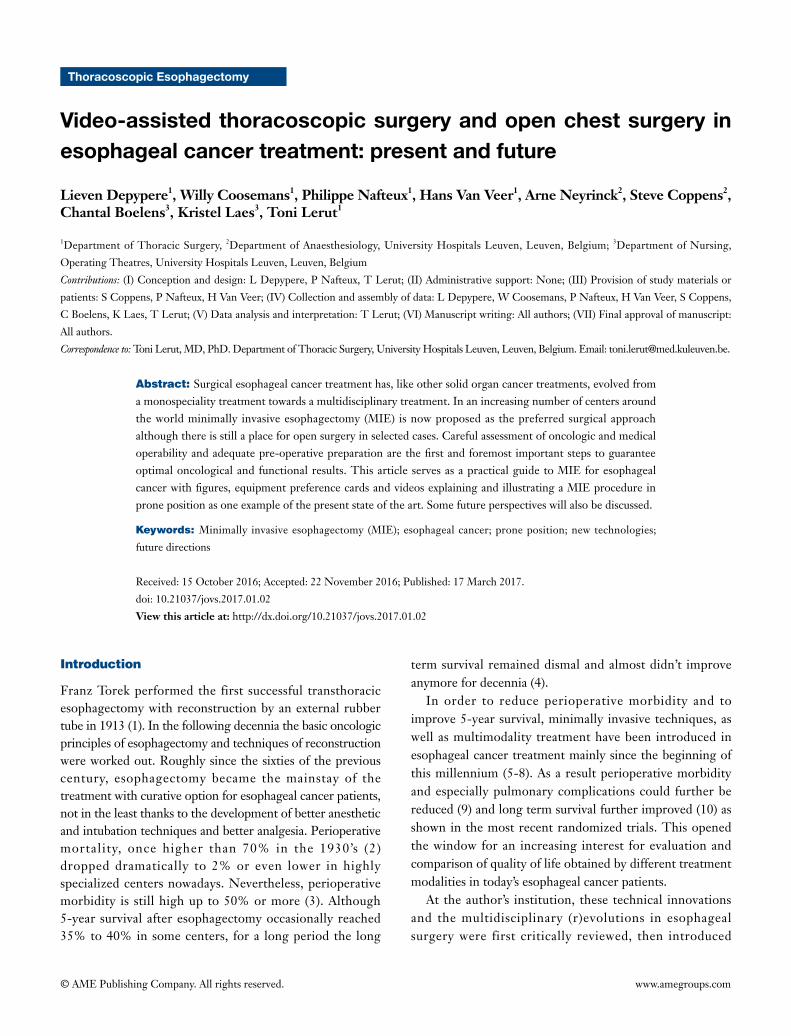
© AME Publishing Company. All rights reserved. www.amegroups.com
Introduction
Franz Torek performed the first successful transthoracic esophagectomy with reconstruction by an external rubber tube in 1913 (1). In the following decennia the basic oncologic principles of esophagectomy and techniques of reconstruction were worked out. Roughly since the sixties of the previous century, esophagectomy became the mainstay of the treatment with curative option for esophageal cancer patients, not in the least thanks to the development of better anesthetic and intubation techniques and better analgesia. Perioperative mortality, once higher than 70% in the 1930’s (2) dropped dramatically to 2% or even lower in highly specialized centers nowadays. Nevertheless, perioperative morbidity is still high up to 50% or more (3). Although 5-year survival after esophagectomy occasionally reached 35% to 40% in some centers, for a long period the long
term survival remained dismal and almost didn’t improve anymore for decennia (4).
In order to reduce perioperative morbidity and to improve 5-year survival, minimally invasive techniques, as well as multimodality treatment have been introduced in esophageal cancer treatment mainly since the beginning of this millennium (5-8). As a result perioperative morbidity and especially pulmonary complications could further be reduced (9) and long term survival further improved (10) as shown in the most recent randomized trials. This opened the window for an increasing interest for evaluation and comparison of quality of life obtained by different treatment modalities in today’s esophageal cancer patients.
At the author’s institution, these technical innovations and the multidisciplinary (r)evolutions in esophageal surgery were first critically reviewed, then introduced
Video-assisted thoracoscopic surgery and open chest surgery in esophageal cancer treatment: present and future
Lieven Depypere1, Willy Coosemans1, Philippe Nafteux1, Hans Van Veer1, Arne Neyrinck2, Steve Coppens2, Chantal Boelens3, Kristel Laes3, Toni Lerut1
1Department of Thoracic Surgery, 2Department of Anaesthesiology, University Hospitals Leuven, Leuven, Belgium; 3Department of Nursing,
Operating Theatres, University Hospitals Leuven, Leuven, Belgium
Contributions: (I) Conception and design: L Depypere, P Nafteux, T Lerut; (II) Administrative support: None; (III) Provision of study materials or
patients: S Coppens, P Nafteux, H Van Veer; (IV) Collection and assembly of data: L Depypere, W Coosemans, P Nafteux, H Van Veer, S Coppens,
C Boelens, K Laes, T Lerut; (V) Data analysis and interpretation: T Lerut; (VI) Manuscript writing: All authors; (VII) Final approval of manuscript:
All authors.
Correspondence to: Toni Lerut, MD, PhD. Department of Thoracic Surgery, University Hospitals Leuven, Leuven, Belgium. Email: [email protected].
Abstract: Surgical esophageal cancer treatment has, like other solid organ cancer treatments, evolved from a monospeciality treatment towards a multidisciplinary treatment. In an increasing number of centers around the world minimally invasive esophagectomy (MIE) is now proposed as the preferred surgical approach although there is still a place for open surgery in selected cases. Careful assessment of oncologic and medical operability and adequate pre-operative preparation are the first and foremost important steps to guarantee optimal oncological and functional results. This article serves as a practical guide to MIE for esophageal cancer with figures, equipment preference cards and videos explaining and illustrating a MIE procedure in prone position as one example of the present state of the art. Some future perspectives will also be discussed.
Keywords: Minimally invasive esophagectomy (MIE); esophageal cancer; prone position; new technologies;
future directions
Received: 15 October 2016; Accepted: 22 November 2016; Published: 17 March 2017.
doi: 10.21037/jovs.2017.01.02
View this article at: http://dx.doi.org/10.21037/jovs.2017.01.02
Thoracoscopic Esophagectomy

© AME Publishing Company. All rights reserved. www.amegroups.com
70 Depypere et al. VATS and open esophagectomy
and further evaluated thereafter always keeping in mind the greatest respect for the basic surgical and oncological principles as well as the well-being of the patient. Tri-modality treatment for esophageal cancer in separate teams evolved into a multidisciplinary team approach within an individual therapeutic plan tailored for every single patient presenting with a cancer of the esophagus or gastro esophageal junction (GEJ).
The first totally minimally invasive esophagectomy (MIE) in Leuven was done in 2003 in a patient with an early (T1aN0) stage cancer that today would have been treated by endoscopic mucosal resection (EMR). The procedure was a three-stage procedure performed through a right sided VATS in left lateral decubitus followed by a totally laparoscopic procedure and left cervicotomy in supine position. After a single surgeon learning curve in over 100 patients (11) with special attention for not compromising the oncologic principle as applied in open surgery (in particular the lymph node dissection), more advanced esophageal cancers became accepted for this type of surgery, also including resections after neoadjuvant chemoradiation therapy. Together with this learning curve, mean number of resected lymph nodes became even higher than in open surgery (12). Other surgeons of the team subsequently became familiarized with MIE as well. Based on several literature reports and personal on-site visits of expert centers, the next step was to perform the right VATS procedure in prone position, which facilitated visualization, lymph node dissection and teaching possibilities (13-15), but challenged the anesthesiological team to keep airway control in any situation.
Therefore a standard protocol was written including practical guidelines allowing surgeons, anesthesiologists and operating theater nurses to familiarize themselves with this technique. In this article minimally invasive esophageal cancer treatment will be discussed as part of a multidisciplinary treatment in our department.
Principles of surgical treatment
It is generally accepted that surgical resection should only be performed with curative intent. Resection is ill-advised when macroscopically incomplete resection (due to invasion of adjacent structures and/or non-resectable metastases) is to be expected. Absolute contra-indications for esophagectomy include local tumor invasion of non-resectable neighbouring structures (T4), carcinomatosis peritonei or thoracalis, hematogenous parenchymatous
metastases involving e.g., the liver and non-resectable metastatic lymph nodes.
The pattern of lymphatic dissemination is difficult to predict, but carcinomas of the proximal and middle thirds of the esophagus preferably metastasise to the cervical region, whereas more distally situated tumors and tumors of the gastro-esophageal junction more commonly metastasise to the lymph nodes around the celiac artery.
Resectable metastatic lymph nodes in the region of the primary tumor, including the celiac trunk and its trifurcation for distal third tumors and cervical nodes for middle and proximal tumors, are not necessarily a contra-indication for surgery. The presence of lymph node metastases, however, has a negative influence on survival, even following extensive lymphadenectomy.
The early-stage lymphatic dissemination as well as completeness of tumoral resection (R0) pose challenges for radical surgical treatment and are still a matter of debate. The concept of extensive en bloc resections was already reported in 1963 (16), but its associated mortality of more than 20% in the original report, discouraged general acceptance. Skinner (17) and Akiyama et al. (18) reintroduced the concept of en bloc resection combined with extensive lymphadenectomy. Ultimately, they were able to reduce operative mortality to 5%, with 5-year survival rates of 18% and 42% respectively. These numbers are opposed to these after transhiatal esophagectomy introduced in the western world by Orringer et al. in 1978 (19). The rationale here is that the surgical intervention will not be able to influence the natural course of the disease. Therefore the intervention is merely restricted to the esophagectomy without making efforts to perform more extensive lymphadenectomies.
The radical en bloc resection, as opposed to the standard resection, aims at performing an as wide as possible peritumoral with an en bloc lymph-node resection of the middle and distal thirds of the posterior mediastinum.
The two-field lymph node dissection incorporates, besides a wide local excision of the primary tumor, a lymphadenectomy of the entire posterior mediastinum, also including the subcarinal nodes and up to the nodes along the left recurrent nerve and the brachiocephalic trunk. In the abdomen it includes the lymph nodes along the celiac trunk, common hepatic and splenic arteries, as well as the lymph nodes along the lesser gastric curvature and in the lesser omentum.
There is an increasing consensus that the optimum of resected lymph nodes should at least be 23 nodes (20).

Minimally Invasive Esophagectomy
© AME Publishing Company. All rights reserved. www.amegroups.com
71
The three-field lymph node dissection. The pattern of lymphatic dissemination is not restricted to the thorax and abdomen. About 20% of the patients with a distal tumor present with metastasis in the cervical region (21). In this operation, besides the already mentioned removal of thoracic and abdominal nodes, the cervical field is defined as the third field and includes the paraesophageal nodes and the nodes lateral to the carotid vessels as well as the supraclavicular nodes. Three field lymphadenectomy is mainly practiced in the far East and is recommended in squamous cell carcinoma of the middle and upper third of the esophagus.
These considerations on radicality of resection and extent of lymphadenectomy are the rationale to justify a transthoracic approach as opposed to the transhiatal approach for which the rationale is merely based on an effort to decrease perioperative morbidity and possibly postoperative mortality.
Recent observations indicate that the results of radical esophagectomy are much better than commonly quoted and today with a proper selection of patients overall 5-year survival figures reaching 40% are frequently reported.
Patient selection and workup
All patients (minimally invasive or open surgery) undergo an oncological workup including flexible endoscopy with biopsy of the tumor, endoscopic ultrasonography of the tumor and reachable lymph nodes (in case there is no stenosis) and full body PET-CT with intravenous contrast. For early tumours, an endomucosal resection (EMR) can be performed as a staging procedure (distinguishing T1a fromT1b) becoming subsequently a definitive treatment in case of T1a lesion, unless lymphatic invasion and /or poor differentiation. A bronchoscopy can be performed in selected cases to rule out T4 stage or second primary cancer in the lung especially in case of squamous cell carcinoma.
Over the recent years neoadjuvant therapy has become the standard in locally advanced cancer of the esophagus and GEJ accounting for about two thirds of all patients to be treated with curative intent.
Medical operability assessment also includes a functional assessment including electrocardiogram, pulmonary function tests and cardiac stress testing. Echo-Doppler of the carotids will be performed in vascular high risk patients. Any other co-morbidity will be thoroughly evaluated and treated lege artis when needed. The medical operability will be tested not only at the start of neoadjuvant therapy but
both oncologic and medical operability will be reassessed again afterwards before the surgery in order to evaluate the impact of the neoadjuvant therapy.
Open versus MIE: indications
Our center is a high volume tertiary referral center performing approximately 120 esophagectomies for cancer per year.
Patients with resectable distal esophageal or GEJ tumors and small mid-thoracic tumors are usually operated on by totally minimally invasive surgery, being VATS and laparoscopy, in general a McKeown procedure. Patients with bigger mid-esophageal tumors, especially patients with squamous cell carcinomas after neoadjuvant therapy, are usually operated on in a hybrid fashion by classic right thoracotomy, followed by laparoscopy and left cervicotomy. Patients over 80 years or more fragile patients suffering from multiple co-morbidities ( approximately 15% of cases) with distal or GEJ tumors are preferably operated on by open left thoraco-abdominal approach, as it is faster (shorter narcosis time) and in most of these patients an intrathoracic anastomosis can be performed (lower risk of anastomotic leakage). Other indications for open surgery are patients presenting with subcardia tumors extending downwards on the fundus and who will require total gastrectomy (approximately 15% of cases) or patients who have had major upper GI surgery e.g., multiple redo antireflux surgery, bariatric surgery…..
Finally the choice of open versus MIE is left to the discretion of the surgeons. As not all surgeons have stepped in into the prone MIE resections notwithstanding a still growing trend to adopt the MIE in prone position.
All together this resulted in a 47% prone MIE in 2016.
Pre-operative preparation
Patients are prepared for surgery with an epidural anaesthetic catheter, placed at the level of the 7th thoracic intervertebral space under local anaesthetic. Afterwards, the patient is positioned in supine position to prepare for general anaesthesia. An arterial line is brought in a radial artery. After induction with propofol and starting the general anaesthesia with sevoflurane, a nasogastric tube is inserted and the patient is intubated using a single lumen reinforced endotracheal tube. An endo-bronchial blocker in then positioned under bronchoscopic evaluation. The use of a blocker with two balloons (each of them positioned in

© AME Publishing Company. All rights reserved. www.amegroups.com
72 Depypere et al. VATS and open esophagectomy
Figure 1 Single lumen reinforced endotracheal tube with endo-bronchial double balloon blocker.
Figure 2 Positioning of the patient. (A) Table preparation for prone position without patient with bean bag, side supports, pillows for chest and pelvis and head support with integrated mirror; (B) patient installation in the same position with arm rests and pneumatic compression devices.
one bronchus) (Figure 1) helps stabilizing the blocker in a correct position.
Next, a central line is inserted preferably in the right subclavian vein. This allows the surgical team to drape the neck for the left cervicotomy but even so for a possible bilateral cervicotomy if a three-field lymphadenectomy is indicated. It also facilitates the postoperative care for the central line compared to a jugular central line in preventing central line infections. Finally a urinary catheter is inserted to monitor urinary output during and after the intervention and sequential pneumatic compression devices are put around the legs during the whole procedure as prophylactic treatment for thrombo-embolic phenomenons.
Positioning and equipment preference card
First stage: the patient is positioned in prone position for
thoracic esophageal dissection and lymphadenectomy. Installation in prone decubitus position requires a standard device in order to support on the head, chest and pelvis. Pillows are placed to support shoulders and pelvis leaving the abdomen free for breathing excursions. The head is placed in a special support with integrated mirror allowing the anesthesiologist, even in prone position, to evaluate the face of the patient and the position of the tube (Figure 2A). Positioning of the right arm is very important in order to get abduction of the scapula. The arms are positioned on a support device in flexion of the shoulder and elbow (Figure 2B). In this way the area between the spine and the inner edge of the scapula is widened.
The patient is further stabilized on the table using a bean-bag and two side supports on the left side of the chest (Figure 2A). This allows, if necessary, rotation of the table up to 45° causing the patient to slide alongside those supports in a more lateral decubitus position, in that way making an urgent thoracotomy easier.
Equipment for patient installation and surgical equipment are described in card 1 in supplementary.
Second and third stage: the patient is positioned in supine position with neck in hyperextension and the face turned to the right for laparoscopic mobilization of the stomach, gastric tubulation and lymphadenectomy combined with the anastomosis via a left cervicotomy.
The legs are placed in the leg support without flexion in the hips in order to have maximal range of motion with the laparoscopic instruments. The neck is already placed in hyperextension and somewhat to the right to be ready for stage 3. The abdomen and neck are prepped and draped together.
A B

Minimally Invasive Esophagectomy
© AME Publishing Company. All rights reserved. www.amegroups.com
73
Figure 3 Abdominal part second time: supine position. (A) Patient installation second time; (B) trocar positions second time.
Figure 5 Trocar positions thoracic time.
Figure 4 Operating theatre setup for the first stage (A = anesthesiologist, AS = assisting surgeon, E = electrocautery, H = ultrasonic device, S = first surgeon, SN = scrub nurse, V = video tower).
A B
Equipment for patient installation and surgical equipment are described in cards 2 and 3 in supplementary.
Procedure
First stage: the surgeon stands on the right side of the patient with the first assistant on his or her right side, both looking to the monitor in front of each of them (Figure 4). The right hemithorax is prepped and draped, making sure that enough lateral skin is free to perform an urgent thoracotomy when needed.
Four trocars are placed along the inner edge of the right scapula: the first at the level of the tip of the scapula for the thoracoscope (usually a 10 mm 30°), the second at the level of the 4th intercostal space (5 mm), the third at the level of the 8th intercostal space (12 mm) and the last one (5 mm) at the level of the 2nd intercostal space, as working trocar for the assistant (suction, retraction) (Figure 5). After introduction of the first trocar, insufflation with carbon dioxide with positive pressure of 5 to 8 mmHg is initiated in order to compress the right lung allowing for an adequate visualization of the posterior mediastinum.
Typically, the dissection is started with the transection of the inferior pulmonary ligament. The dissection is extended by incising the mediastinal pleura at the level of the pericardium and alongside the inferior pulmonary vein up to the right main stem bronchus. The lower edge of the bronchus is visualized by dissecting the right lower part of the subcarinal lymph nodes adjacent to the right main stem bronchus. This package of nodes is subsequently dissected away from the pericardium, staying en bloc and connected with the esophagus (Figure 6). The pleura overlying the right main stem bronchus is incised up to the crossing azygos vein. This arch of the azygos vein is completely dissected and transected using vascular endostaplers

© AME Publishing Company. All rights reserved. www.amegroups.com
74 Depypere et al. VATS and open esophagectomy
membranous part of the trachea is carefully divided pushing the esophagus away from the trachea. This dissection is continued to the left border of the trachea until the left paratracheal gutter is entered (Figure 8). Now, very importantly, careful dissection will expose the left recurrent nerve that has to be further dissected out without using any electro cautery (Figure 9). Having created a window behind the esophagus, the esophagus is pulled downwards. From above the esophagus a section plane between this structure and the beginning of the aortic arch is made starting from the distal border of that earlier created window. This will bring into view the posterior wall of the left main stem bronchus which is carefully liberated exposing in that way the carina and the right trachea-bronchial corner and the left border of the subcarinal lymph node package. This lymphatic tissue is dissected off the main stem bronchus and thus completing the en bloc removal of the subcarinal lymph nodes in one piece (Figure 10). Now the dissection
Figure 9 Identification L recurrent nerve (25). Available online: http://www.asvide.com/articles/1392
Figure 7 Azygos (23). Available online: http://www.asvide.com/articles/1390
Figure 10 Aorta + subcarinal (26). Available online: http://www.asvide.com/articles/1393
Figure 6 Start dissection (22). Available online: http://www.asvide.com/articles/1389
Figure 8 Pars membranacea (24). Available online: http://www.asvide.com/articles/1391
(2 mm stapler height).The underlying intercosto-bronchial artery is clipped and transected as well (Figure 7). The esophagus with the concomitant right vagus nerve is pushed upwards and the connective tissue between esophagus and
Video 1. Start dissection
Lieven Depypere, Willy Coosemans, Toni Lerut*, et al.
Department of Thoracic Surgery, University Hospitals Leuven, Leuven, Belgium
▲
Video 4. Identification L recurrent nerve
Lieven Depypere, Willy Coosemans, Toni Lerut*, et al.
Department of Thoracic Surgery, University Hospitals Leuven, Leuven, Belgium
▲
Video 5. Aorta + subcarinal
Lieven Depypere, Willy Coosemans, Toni Lerut*, et al.
Department of Thoracic Surgery, University Hospitals Leuven, Leuven, Belgium
▲
Video 2. Azygos
Lieven Depypere, Willy Coosemans, Toni Lerut*, et al.
Department of Thoracic Surgery, University Hospitals Leuven, Leuven, Belgium
▲
Video 3. Pars membranacea
Lieven Depypere, Willy Coosemans, Toni Lerut*, et al.
Department of Thoracic Surgery, University Hospitals Leuven, Leuven, Belgium
▲

Minimally Invasive Esophagectomy
© AME Publishing Company. All rights reserved. www.amegroups.com
75
Figure 11 Thoracic duct supradiaphragmatic (27). Available online: http://www.asvide.com/articles/1394
is continued flush along the descending aorta taking the thoracic duct en bloc with the esophagus. The thoracic duct is clipped and transected several centimeters below the tumor, above the diaphragm. Also at the opposite side,
Figure 13 Right recurrent (29). Available online: http://www.asvide.com/articles/1396
Figure 12 Paratracheal (28). Available online: http://www.asvide.com/articles/1395
Figure 14 Left recurrent (30). Available online: http://www.asvide.com/articles/1397
Figure 15 Overview final (31). Available online: http://www.asvide.com/articles/1398
the dissection is performed away from the esophagus flush along the pericardium taking all lymph nodes and this until the diaphragmatic pillars are reached (Figure 11) Attention is now moved towards the superior mediastinum. After incising the mediastinal pleura left from the superior caval vein, the fatty tissue and lymph nodes between the vein and the left anterolateral part of the trachea are dissected and removed (Figure 12). This is followed by the lymph node dissection along the right recurrent nerve. Pulling on the remaining proximal part of the right vagal nerve facilitates the visualization of and dissection along the right recurrent nerve that is turning around the right subclavian artery. All small vascular branches are clipped and transected as no electrocoagulation is allowed here (Figure 13). Next comes the dissection along the already in part dissected left recurrent nerve removing all adjacent lymph node up well in to the base of the neck. Again electro cautery in the neighborhood of the recurrent nerve is avoided (Figure 14).
Video 6. Thoracic duct supradiaphragmatic
Lieven Depypere, Willy Coosemans, Toni Lerut*, et al.
Department of Thoracic Surgery, University Hospitals Leuven, Leuven, Belgium
▲
Video 9. Left recurrent
Lieven Depypere, Willy Coosemans, Toni Lerut*, et al.
Department of Thoracic Surgery, University Hospitals Leuven, Leuven, Belgium
▲
Video 10. Overview final
Lieven Depypere, Willy Coosemans, Toni Lerut*, et al.
Department of Thoracic Surgery, University Hospitals Leuven, Leuven, Belgium
▲
Video 7. Paratracheal
Lieven Depypere, Willy Coosemans, Toni Lerut*, et al.
Department of Thoracic Surgery, University Hospitals Leuven, Leuven, Belgium
▲
Video 8. Right recurrent
Lieven Depypere, Willy Coosemans, Toni Lerut*, et al.
Department of Thoracic Surgery, University Hospitals Leuven, Leuven, Belgium
▲

© AME Publishing Company. All rights reserved. www.amegroups.com
76 Depypere et al. VATS and open esophagectomy
Figure 18 Left gastric artery (33). Available online: http://www.asvide.com/articles/1400
Figure 17 Lesser curvature (32). Available online: http://www.asvide.com/articles/1399
Figure 16 Operating theatre setup for the second and third stage (A = anesthesiologist, AS = assisting surgeon, E = electrocautery, H = ultrasonic device, S = first surgeon, SN = scrub nurse, V = video tower).
Figure 15 shows nicely the end result i.e., the thorough lymphadenectomy and en bloc dissection of the entire posterior mediastinum.
When this has been achieved, careful hemostasis is performed and classically two chest drains are placed using therefore the two most basal ports. One classic 28 F chest drain apical and one flexible 19 F silicon drain on the diaphragm. The right lung is reinflated and the ports are closed.
Second stage: the surgeon takes place in between the legs, the first assistant on the right side of the patient, the second assistant on the left side of the patient (Figure 16). Instead of a second assistant, a table-mounted laparoscopic liver retractor can be used for retraction. Typically five abdominal ports are placed in a similar fashion to the approach used for a laparoscopic Nissen fundoplication (Figure 3B). The liver is retracted medially and the dissection starts with transection of the gastro-hepatic ligament and transection of the right gastric artery using the harmonic scalpel. After opening the lesser sac the right pillar of the hiatus esophagei is identified and the gastro-esophageal junction is dissected out of the hiatus creating a window towards the greater sac behind the esophagus (Figure 17). From the lesser curvature side, the left gastric artery and vein are prepared and clipped at their base and transected taking all lymph nodes down to the offspring of the artery from the coeliac axis as to ensure complete lymph node clearance in along both vessels (Figure 18). The dissection continues along the greater curvature of the stomach taking great care not to damage the right gastro-epiploïc artery: the greater sac is incised and opened above the transverse colon and divided in the direction of the spleen. The short gastric vessels are divided and the dissection continues until the fundus is completely freed and the hiatus and left pillar are coming in to view. This is followed by the mobilization of the distal part of the greater curvature always away from the gastro-epiploic artery close to the transverse colon. In skinny patients care is taken not to open and erroneously divide the mesenterium of the transverse colon. All adhesions between the posterior gastric wall and pancreas are freed until the pylorus is reached. During the entire dissection process tissues are grasped only on the lesser curvature which later on will be resected so to avoid traumatization of the tissues of what will become the gastric tube (Figure 19). The gastric tubulation is performed using endostaplers introduced from the right upper quadrant, after having the liver retractor placed in the subxiphoidal position during tubulation. The first stapler is placed at the lesser curvature about 5 cm
Video 11. Lesser curvature
Lieven Depypere, Willy Coosemans, Toni Lerut*, et al.
Department of Thoracic Surgery, University Hospitals Leuven, Leuven, Belgium
▲
Video 12. Left gastric artery
Lieven Depypere, Willy Coosemans, Toni Lerut*, et al.
Department of Thoracic Surgery, University Hospitals Leuven, Leuven, Belgium
▲
AS AS
SSN

Minimally Invasive Esophagectomy
© AME Publishing Company. All rights reserved. www.amegroups.com
77
Figure 20 Gastric tube (35). Available online: http://www.asvide.com/articles/1402
Figure 22 Anastomosis (37). Available online: http://www.asvide.com/articles/1404
Figure 21 Lymph node dissection (36). Available online: http://www.asvide.com/articles/1403
Figure 19 Greater curvature (34). Available online: http://www.asvide.com/articles/1401
proximally from the pylorus at the level of the crow’s foot to achieve a gastric tube of 4 to 5 cm width. Of course the nasogastric tube has been pulled back beforehand. The gastric tube is most of the times at least 30 cm long. No
pyloric drainage procedure is performed (Figure 20). Now there is ample space to perform the lymph node dissection along the common hepatic artery, the splenic artery and the base of the coeliac axis to achieve a DII lymphadenectomy (Figure 21).
The esophagus is than dissected in the hiatus with en bloc resection of the peri-esophageal tissue till the chest cavity is entered, freeing completely the esophagus. At this time, it can be necessary to lower the intra-abdominal pressure or to put the chest drains on active suction in order to prevent intrathoracic overpressure.
To avoid loss of pressure due to the open communication between abdomen and chest through the hiatus, the proximal gastrectomy piece is used to plug the hiatus, allowing the intra-abdominal pressure to remain high enough as to ensure good visualization during the lymph node dissection. The gastric tube is than fixed to the proximal gastrectomy specimen using two temporary stitches.
Third stage: (Figure 22): an 8 cm long left cervical incision anterior to the sternocleidomastoid muscle and finishing in the suprasternal notch is performed. Omohyoid and strapped muscles are transected. Dissection is performed medially from the great vessels and laterally from the thyroid gland clipping and transecting eventually its feeding arteries. During this procedure, again great care is taken not to damage the left recurrent nerve. The cervical esophagus is then mobilized until the thoracic part of the dissection is reached. The esophagus with the proximal gastrectomy specimen can be exteriorized through the neck incision thus pulling up the gastric conduit during temporary ventilation stop and under direct laparoscopic vision in order to prevent torsion. At this point a classic
Video 16. Anastomosis
Lieven Depypere, Willy Coosemans, Toni Lerut*, et al.
Department of Thoracic Surgery, University Hospitals Leuven, Leuven, Belgium
▲
Video 14. Gastric tube
Lieven Depypere, Willy Coosemans, Toni Lerut*, et al.
Department of Thoracic Surgery, University Hospitals Leuven, Leuven, Belgium
▲
Video 15. Lymph node dissection
Lieven Depypere, Willy Coosemans, Toni Lerut*, et al.
Department of Thoracic Surgery, University Hospitals Leuven, Leuven, Belgium
▲
Video 13. Greater curvature
Lieven Depypere, Willy Coosemans, Toni Lerut*, et al.
Department of Thoracic Surgery, University Hospitals Leuven, Leuven, Belgium
▲

© AME Publishing Company. All rights reserved. www.amegroups.com
78 Depypere et al. VATS and open esophagectomy
end-to-side anastomosis is performed: we prefer a semi-mechanical anastomosis, using an endostapler for the posterior wall of the esophagogastric anastomosis and finishing the anterior part of the anastomosis using a double layer continuous stitch after transection and resection of the esophagus. The nasogastric tube is subsequently repositioned into the gastric conduit under direct visual control before finishing the anterior part of the anastomosis. The anastomosis is then reduced from the neck into the thoracic inlet behind the trachea. The neck incision is closed after placement of a closed suction drain system.
Finally the gastric tube is then retracted towards the abdomen and fixed to the hiatus using separate stay sutures as to avoid intrathoracic migration of abdominal organs.
If deemed necessary, a feeding jejunostomy can be placed.Tr o c a r s a r e r e m o v e d u n d e r v i s u a l c o n t r o l ,
pneumoperitoneum is deflated and incisions are closed. A pyloric drainage procedure is not routinely performed. No drain is left in the abdominal cavity.
Role of team members
Surgeon, first assistant, scrub nurse and anesthesiologist are all of pivotal importance during this complex and sometimes challenging procedure. The individual tasks of each team member are already described in the text. If a new team member is not familiar with the procedure, an experienced colleague will help and assist during the procedure (and during the learning curve) and this works for all team members.
Post-operative management
To optimize the results of surgery, this type of procedure should be performed in an appropriately equipped center that is familiar with the meticulous management of the postoperative course. If necessary, bronchoscopy or eventually a mini-tracheostomy placed either at the time of surgery or sometimes thereafter can be used to ensure adequate bronchial toilet. In the postoperative management, the role of the physiotherapist is crucial in order to prevent pulmonary complications.
Fluid balance and oxygen saturation should be closely monitored and oxygen supplementation is mandatory. Fluid restriction is essential to avoid cardiac and respiratory complications. It is also vital to maintain adequate and balanced nutrition during the whole postoperative period and therefore total parenteral nutrition or enteral feeding by jejunostomy, when placed at the time of surgery, is given.
Thrombosis prophylaxis is continued by sequential pneumatic compress ion devices for the f irst two postoperative days and subcutaneous injection of low molecular weight heparins. Prophylactic antibiotics are given for 24 hours.
Physiotherapy with gradual breathing exercises and general condition exercises is performed from the day of surgery to the day of discharge at least twice a day.
Since it is essential to avoid stasis in the gastric tube and subsequent respiratory complications secondary to aspiration, the nasogastric tube is kept in place and gastroprokinetic drugs (domperidon and erythromycin) are administered.
In case these preventive measures are of no avail and gastric stasis and subsequent gastric dilation occur, endoscopic balloon dilation of the pylorus will be performed.
A contrast study to check the integrity of the anastomosis in case of cervical location is not routinely performed. At day 5, if there is no clinical signs of anastomotic leak, oral fluids are started. In case of intrathoracic anastomosis a gastrografin swallow will be performed at day 5 and oral fluid and soft diet are started if no leak is visualized. On the same day the epidural catheter is removed and the patient is encouraged to mobilize fully. Oral analgetics are administered lege artis. The chest drain will be removed when the effluent amounts to less than 300 mL of fluids. Patients are discharged when they are able to tolerate the soft diet and the pain is sufficiently controlled to permit normal mobilization. The patient is than seen in the outpatient clinic one month after discharge.
Tips, tricks and pitfalls
v Patient selection is crucial;v This procedure should be performed in high volume
centers by an experienced team that can also offer alternative treatment possibilities;
v Correct pat ient insta l lat ion together with the anesthesiological team is pivotal;
v Gentle tissue handling, especially using a no-touch technique for the gastric tube;
v If a bleeding occurs during the procedure, compression allows to evaluate the situation and can also be the sole treatment;
v In case of doubts, conversion is the safest option.
Where are we going from here?
Today esophagectomy whether open or with MIE can

Minimally Invasive Esophagectomy
© AME Publishing Company. All rights reserved. www.amegroups.com
79
be performed with a minimum of blood loss and blood transfusion is only needed in a minority of patients. MIE is thought to have a positive impact on pain resulting in less need for analgesia, less pulmonary complications and shorter ICU stay.
All incremental steps of progress have resulted in reduced postoperative mortality and morbidity and thus better oncological outcome.
Despite all efforts, nowadays one in two patients will still die from recurrence and postoperative morbidity still occurs in one out of three patients.
New technologies, techniques and treatment modalities are emerging in an ongoing search to improve results. Experience with robotic esophagectomy and uniportal VATS is growing reportedly reducing even more postoperative complications and related morbidity (7).
Sentinel lymph node mapping and navigational techniques are tested hoping to avoid in the future the surgical trauma created by extensive lymphadenectomy in selected patients.
Treatment modalit ies aiming at preserving the esophagus such as EMR for early T1 cancers and definitive chemoradiation are already in place and in some centers definitive chemoradiation is the standard of care for advanced squamous cell carcinoma. These patients are closely monitored and in case of local recurrence salvage esophagectomy is offered as the last option in selected patients.
A wide spectrum of biologicals in the search for a more targeted therapy based on individual genetic profiles of the cancer is promising a tailored individualized treatment.
Interventional endoscopic techniques are booming stimulated by the ever increasing miniaturization of instrumentation. The recent introduction of POEM challenging the classic laparoscopic Heller myotomy for achalasia is one perfect example of what can be expected in the future. Here the sky seems to be the limit, not to speak about the perspectives offered by the nanotechnology both in the area of diagnostics as well as in the therapeutic arena.
From all this it must become clear that there is a definite need for superspecialisation in the field of esophageal pathology both benign and malignant.
The surgeon of tomorrow dealing with esophageal cancer will work in close collaboration with his/her peers in oncology, (interventional) radiology, interventional endoscopy….
This means that the different specialties as they are
existing today will gradually grow towards each other eventually intertwining or merging. The result will be a complete re-engineering from surgery as a technical act into a unique form of therapy for each individual patient and for the better of the patient.
But this will require a strong engagement of from the surgeon to master the knowledge of esophageal cancer and to master the specifics of diagnosis and therapy (including complications and failure) of esophageal cancer.
Now is the time to act.
Acknowledgements
None.
Footnote
Conflicts of Interest: The authors have no conflicts of interest to declare.
References
1. Torek F. The first successful case of resection of the thoracic portion of the esophagus for carcinoma. Surg Gynecol Obstet 1913;16:614.
2. De Bakey M,Ochsner A. Carcinoma of the esophagus. Postgrad Med 1948;3:192-8.
3. Raymond DP, Seder CW, Wright CD, et al. Predictors of Major Morbidity or Mortality After Resection for Esophageal Cancer: A Society of Thoracic Surgeons General Thoracic Surgery Database Risk Adjustment Model. Ann Thorac Surg 2016;102:207-14.
4. Surveillance, Epidemiology, and End Results Program. SEER Stat Fact Sheets: esophageal cancer. Available online: http://seer.cancer.gov/statfacts/html/esoph.html, accessed September 17, 2014.
5. Cuschieri A, Shimi S, Banting S. Endoscopic oesophagectomy through a right thoracoscopic approach. J R Coll Surg Edinb 1992;37:7-11.
6. Luketich JD, Alvelo-Rivera M, Buenaventura PO, et al. Minimally invasive esophagectomy: outcomes in 222 patients. Ann Surg 2003;238:486-94; discussion 494-5.
7. Qureshi YA, Dawas KI, Mughal M, et al. Minimally invasive and robotic esophagectomy: Evolution and evidence. J Surg Oncol 2016;114:731-735.
8. Jang R, Darling G, Wong RK. Multimodality approaches for the curative treatment of esophageal cancer. J Natl

© AME Publishing Company. All rights reserved. www.amegroups.com
80 Depypere et al. VATS and open esophagectomy
Compr Canc Netw 2015;13:229-38. 9. Biere SS, van Berge Henegouwen MI, Maas KW, et al.
Minimally invasive versus open oesophagectomy for patients with oesophageal cancer: a multicentre, open-label, randomised controlled trial. Lancet 2012;379:1887-92.
10. Shapiro J, van Lanschot JJ, Hulshof MC, et al. Neoadjuvant chemoradiotherapy plus surgery versus surgery alone for oesophageal or junctional cancer (CROSS): long-term results of a randomised controlled trial. Lancet Oncol 2015;16:1090-8.
11. Nafteux P, Moons J, Coosemans W, et al. Minimally invasive oesophagectomy: a valuable alternative to open oesophagectomy for the treatment of early oesophageal and gastro-oesophageal junction carcinoma. Eur J Cardiothorac Surg 2011;40:1455-63; discussion 1463-4.
12. Chapter 1: changing trends and lessons learned from 2500 esophagectomies. In: Nafteux P. The continuous quest for quality improvement after esophagectomy for cancer. Leuven: Thesis Academic press, 2016:16-42.
13. Markar SR, Wiggins T, Antonowicz S, et al. Minimally invasive esophagectomy: Lateral decubitus vs. prone positioning; systematic review and pooled analysis. Surg Oncol 2015;24:212-9.
14. Tanaka E, Okabe H, Kinjo Y, et al. Advantages of the prone position for minimally invasive esophagectomy in comparison to the left decubitus position: better oxygenation after minimally invasive esophagectomy. Surg Today 2015;45:819-25.
15. Shen Y, Feng M, Tan L, et al. Thoracoscopic esophagectomy in prone versus decubitus position: ergonomic evaluation from a randomized and controlled study. Ann Thorac Surg 2014;98:1072-8.
16. Logan A. The surgical treatment of carcinoma of the esophagus and cardia. J Thorac Cardiovasc Surg 1963;46:150-61.
17. Skinner DB. En bloc resection for neoplasms of the esophagus and cardia. J Thorac Cardiovasc Surg 1983;85:59-71.
18. Akiyama H, Tsurumaru M, Kawamura T, et al. Principles of surgical treatment for carcinoma of the esophagus: analysis of lymph node involvement. Ann Surg 1981;194:438-46.
19. Orringer MB, Sloan H. Esophagectomy without thoracotomy. J Thorac Cardiovasc Surg 1978;76:643-54.
20. Peyre CG, Hagen JA, DeMeester SR, et al. The number of lymph nodes removed predicts survival in esophageal cancer: an international study on the impact of extent of
surgical resection. Ann Surg 2008;248:549-56.21. Lerut T, Nafteux P, Moons J, et al. Three-field
lymphadenectomy for carcinoma of the esophagus and gastroesophageal junction in 174 R0 resections: impact on staging, disease-free survival, and outcome: a plea for adaptation of TNM classification in upper-half esophageal carcinoma. Ann Surg 2004;240:962-72; discussion 972-4.
22. Depypere L, Coosemans W, Lerut T, et al. Start dissection. Asvide 2017;4:081. Available online: http://www.asvide.com/articles/1389
23. Depypere L, Coosemans W, Lerut T, et al. Azygos. Asvide 2017;4:082. Available online: http://www.asvide.com/articles/1390
24. Depypere L, Coosemans W, Lerut T, et al. Pars membranacea. Asvide 2017;4:083. Available online: http://www.asvide.com/articles/1391
25. Depypere L, Coosemans W, Lerut T, et al. Identification L recurrent nerve. Asvide 2017;4:084. Available online: http://www.asvide.com/articles/1392
26. Depypere L, Coosemans W, Lerut T, et al. Aorta + subcarinal. Asvide 2017;4:085. Available online: http://www.asvide.com/articles/1393
27. Depypere L, Coosemans W, Lerut T, et al. Thoracic duct supradiaphragmatic. Asvide 2017;4:086. Available online: http://www.asvide.com/articles/1394
28. Depypere L, Coosemans W, Lerut T, et al. Paratracheal. Asvide 2017;4:087. Available online: http://www.asvide.com/articles/1395
29. Depypere L, Coosemans W, Lerut T, et al. Right recurrent. Asvide 2017;4:088. Available online: http://www.asvide.com/articles/1396
30. Depypere L, Coosemans W, Lerut T, et al. Left recurrent. Asvide 2017;4:089. Available online: http://www.asvide.com/articles/1397
31. Depypere L, Coosemans W, Lerut T, et al. Overview final. Asvide 2017;4:090. Available online: http://www.asvide.com/articles/1398
32. Depypere L, Coosemans W, Lerut T, et al. Lesser curvature. Asvide 2017;4:091. Available online: http://www.asvide.com/articles/1399
33. Depypere L, Coosemans W, Lerut T, et al. Left gastric artery. Asvide 2017;4:092. Available online: http://www.asvide.com/articles/1400
34. Depypere L, Coosemans W, Lerut T, et al. Greater curvature. Asvide 2017;4:093. Available online: http://www.asvide.com/articles/1401
35. Depypere L, Coosemans W, Lerut T, et al. Gastric tube.

Minimally Invasive Esophagectomy
© AME Publishing Company. All rights reserved. www.amegroups.com
81
Asvide 2017;4:094. Available online: http://www.asvide.com/articles/1402
36. Depypere L, Coosemans W, Lerut T, et al. Lymph node dissection. Asvide 2017;4:095. Available online: http://
www.asvide.com/articles/140337. Depypere L, Coosemans W, Lerut T, et al. Anastomosis.
Asvide 2017;4:096. Available online: http://www.asvide.com/articles/1404
doi: 10.21037/jovs.2017.01.02Cite this article as: Depypere L, Coosemans W, Nafteux P, Van Veer H, Neyrinck A, Coppens S, Boelens C, Laes K, Lerut T. Video-assisted thoracoscopic surgery and open chest surgery in esophageal cancer treatment: present and future. J Vis Surg 2017;3:30.

© AME Publishing Company. All rights reserved. www.amegroups.com
Thoracoscopic Esophagectomy
Esophageal cancer is a common gastrointestinal cancer and ranks sixth in tumor-related death. Esophageal adenocarcinoma is prevalence in European countries and the United States, while esophageal squamous cell carcinoma is frequently observed in Asian countries (1). Radical surgical resection combined with systematic lymph node dissection has always been used as a significant approach for treating esophageal cancer. In recent years, preoperative neoadjuvant therapy has significantly improved the long-term outcomes of the surgical treatment of esophageal cancer (2,3). Conventional open surgical procedures for esophageal cancer are very traumatic and lead to a high incidence of postoperative complications, especially pulmonary complications, as well as high postoperative mortality, despite continuous advances in perioperative management and surgical techniques. Since Cuschieri et al. (4) first reported thoracoscopic surgery in minimally invasive esophagectomy (MIE) in 1992, MIE has become a standard surgical approach for esophageal cancer in the world (5-7). It was approved that due to significantly reduced surgical trauma, MIE lowers postoperative morbidity and mortality while achieving a tumor resection efficacy comparable with the conventional open esophagectomy (6,8-10).
In recent years, the Department of Esophageal Surgery of Shanghai Chest Hospital has been committed to the promotion and practice of minimally invasive techniques, and from the conventional thoraco-laparoscopic assisted surgery to the Da Vinci robot assisted esophageal resection, MIE has gradually become a routine surgical treatment of resectable esophageal cancer. In this report, we summarize
the past MIE experiences performed at the Shanghai Chest Hospital and analyze their progress in early postoperative recovery and effectiveness in achieving satisfactory oncological outcomes.
Preoperative assessment of patients
All patients underwent preoperative staging and evaluation, including enhanced computed tomography (CT) scan of the chest and abdomen, neck ultrasound, upper gastrointestinal endoscopy under ordinary white light, and endoscopy coupled with endoscopic ultrasound (EUS), as well as bronchoscopy for patients with lesions above the carina. Fluoroxyglucose-18 positron emission computed tomography (PET) was only used on those with regional or distant metastases that were difficult to determine. Patients with multiple lymph node metastases and over stage T3 development were subjected to preoperative induction therapy. All patients received complete two-field lymph node dissection, including those along the bilateral recurrent laryngeal nerve. As long as the imaging assessment indicated it was surgically resectable, it was regarded as an indication for minimally invasive esophageal surgery.
Choice of surgical approach
Whether conventional esophagectomy or MIE, the approach for esophagus resection has been rather controversial. Open esophagectomy typically use the
Minimally invasive esophagectomy for esophageal squamous cell carcinoma—Shanghai Chest Hospital experience
Bin Li#, Yu Yang#, Yifeng Sun, Rong Hua, Xiaobin Zhang, Xufeng Guo, Haiyong Gu, Bo Ye, Zhigang Li, Teng Mao
Department of Thoracic Surgery, Section of Esophageal Surgery, Shanghai Chest Hospital, Shanghai Jiao Tong University, Shanghai 200030, China#These authors contributed equally to this work.
Correspondence to: Teng Mao, MD, PhD. Department of Thoracic Surgery, Section of Esophageal Surgery, Shanghai Chest Hospital, Shanghai Jiao
Tong University, 241 Huaihai West Rd, Shanghai 200030, China. Email: [email protected].
Submitted Apr 10, 2018. Accepted for publication Jun 08, 2018.
doi: 10.21037/jtd.2018.06.75
View this article at: http://dx.doi.org/10.21037/jtd.2018.06.75
89

Minimally Invasive Esophagectomy
© AME Publishing Company. All rights reserved. www.amegroups.com
83
transhiatal approach and the transthoracic approach, the latter including the Ivor Lewis, McKeown, and Sweet procedures. Each surgical option has certain advantages. Currently, randomized control led studies on the advantages and disadvantages of various surgical choices are lacking. Previously, Hulscher and his colleagues reported a large, randomized comparative study with 220 patients conducted in the Netherlands in which some had undergone transthoracic esophagectomy (TTE) and some had undergone transhiatal esophagectomy (THE) (11) and found that the patients who received THE had a shorter operation time, less blood loss, and lower incidence of postoperative complications, but showed no difference in perioperative mortality. However, the patients who received TTE had a higher number of lymph nodes dissected. The 5-year disease-free survival and the overall survival of the TTE group demonstrated improvement but were not statistically significant. Although the open TTE exhibited better survival tendencies and better lymph node dissection, its incidence of postoperative complications was higher than that of THE. In theory, transthoracic MIE can achieve the same oncological surgical outcome while lowering the incidence of postoperative complications. Relative to the high incidence of adenocarcinoma in the lower esophageal segment and the esophagogastric junction in European countries and the United States, esophageal cancer in China are dominated by squamous cell carcinoma, with lesions mostly in the middle and the lower esophageal segments. In our center, esophageal cancer in the middle and the lower esophageal segments accounted for 88.1% of the total cases. Different pathological types and different tumor location require different surgical strategies for the treatment of esophageal cancer. In China, MIE mostly adopts the Ivor Lewis or the McKeown procedure (12-14). A review study revealed that compared with the McKeown procedure, the Ivor Lewis procedure shows a lower incidence of intraoperative damage of the recurrent laryngeal nerve, shorter postoperative hospital stay, and less intraoperative blood loss, but a similar incidence of anastomotic leakage (15). However, this study lacked pathological and long-term follow-up results, and a high proportion of the cases were esophageal adenocarcinoma, it only has limited implications to the treatment of esophageal cancer in China.
In the Shanghai Chest Hospital, the McKeown procedure was mainly adopted; among 207 MIE procedures performed, 193 were completed via the McKeown procedure, accounting for 93.2% of the total cases.
Anesthesia and surgical posture
General anesthesia and anesthesia ventilation via single-lumen endotracheal tube was routinely performed. A “single-lumen endotracheal tube + CO2 artificial pneumothorax” technique was adopted for intraoperative lung collapse. The advantage of this technique is that after the establishment of artificial pneumothorax, the lung collapse is more rapid and complete, the gap between the adipose tissue in the mediastinum is widened, and so is the gap between tissues peripheral to lymph nodes, which makes it easier to separate them while reducing blood loss. The pressure of artificial pneumothorax is generally 6–8 mmHg, exerting little effect on hemodynamics. In addition, the application of a single-lumen endotracheal tube is also conducive to the surgical resection of paratracheal tissue.
Two postures, the left lateral decubitus position and the prone position, were usually adopted for the chest operation. Due to anatomical similarities, the left lateral decubitus position was adopted in the initial applications of MIE. Luketich et al. (16) reported that the lateral decubitus position was chosen for more than 1,000 minimally invasive surgical operations for esophageal cancer. At the same time, the prone position exposes longitudinal organs and structures more clearly and has been gradually introduced in MIE and widely practiced (8,17,18). In a review study, Markar et al. (19) showed that compared with that in a lateral position, MIE performed with the patient in a prone position could reduce the incidence of postoperative pulmonary complications and intraoperative blood loss while increasing the number of mediastinal lymph nodes dissected. Two other studies (18,20) also showed that using a prone position for MIE could significantly improve postoperative oxygen delivery, reduce the incidence of pulmonary complications, and facilitate early postoperative recovery. However, one of the major drawbacks of the prone position is the need to change position when switching to thoracotomy during an emergency. In the Shanghai Chest Hospital, patients were in the left lateral recumbent position and leaned forward 30° with artificial pneumothorax, which not only provides a clear surgical area but also avoids posture change in the case of emergency. At present, this position has also been accepted by many hospitals in the world (Figure 1). In the abdominal laparoscopy-assisted operation, the surgical position is to tilt the patient with the head raised and the feet lowered, with the left side raised 30 degrees, which is conducive to the

© AME Publishing Company. All rights reserved. www.amegroups.com
84 Li et al. Minimally invasive esophagectomy for ESCC
downward and rightward movements of the omentum and colon and a better exposure of the splenic portal, facilitating the separation of short gastric vessels (Figure 2).
Surgical procedure
Thoracic procedure
The patient assumed the left lateral position, leaning forward 30 degrees to establish CO2 artificial pneumothorax (pressured at approximately 6–8 mmHg), and the surgeon stood on the ventral side of the patient. The seventh intercostal of the right anterior axillary line was set as the laparoendoscopic observation hole, the third intercostal was set as the primary operation hole, the sixth intercostal of the right midaxillary line was set as the secondary operation hole, and the ninth intercostal of the right midaxillary line was set as the operation hole for the assistant surgeon. In addition, a purse string suture was punctured through the third intercostal, located between the posterior edge of the scapula and the spine, to make it possible to pull the esophagus during the operation (Figure 1).
First, the mediastinal pleura anterior and posterior to the esophagus was longitudinally incised above the azygos vein with an electrocautery hook, and the incision was advanced along the right side of the vagus nerve until reaching the upper edge of the right supraclavicular artery. After carefully
exposing the right recurrent laryngeal nerve, the soft tissues and lymph nodes from the rear of the nerve, in front of the esophagus, and up to the bottom of the thyroid were completely dissected (Figure 3). Then, the azygous vein was detached, the mediastinal pleura were incised at the anterior edge of the azygos vein, and the soft tissue in front of the aorta was pushed toward the esophagus and dissected. In the case of stage T3 or above, the azygos vein and the aortic thoracic duct were also dissected. The middle and lower esophagus and the surrounding soft tissues, together with the subcarinal lymphatic nodes, were completely dissected. After the medial esophagus was properly separated from the mediastinum in an upward direction, the esophagus was pulled to the right and back with a pulling wire. The left recurrent laryngeal nerve lymph nodes were dissected from the left mediastinal pleura and the recurrent laryngeal nerve (Figure 4). The thoracic parabronchial lymph nodes and the lymph nodes inferior to the aortic arch were dissected along the external wall of the left main bronchus. At the end of the chest surgery, one 28-Fr chest tube and two mediastinal drainage tubes were installed.
Abdominal procedure
The patient assumed a supine position, with the head raised and the feet lowered, at approximately a 20-degree
Figure 1 Thoracic surgical posture: left lateral recumbent position and leaned forward 30°; the thoracic port design. Four trocars are placed in: the3rd/7th intercostal spaces of the right anterior axillary line, the 6th/9th intercostal spaces of the right midaxillary line.

Minimally Invasive Esophagectomy
© AME Publishing Company. All rights reserved. www.amegroups.com
85
angle. CO2 artificial pneumothorax was established (at a pressure of approximately 14 mmHg). Five abdominal Traco units were used, and a Trocar (camera, 10 mm) was placed 1 cm below the umbilicus. One operation hole was set at the xiphoid to make it easier to pull the liver and stomach, a second operation hole for the ultrasonic surgical knife was set between the mid clavicular line and the umbilicus, a third operation hole was set between the anterior axillary line and the midaxillary line, and the operation hole for the assistant surgeon was set at
the umbilicus level of the upper left abdomen (Figure 2). After being dissociated via skeletalization, the left gastric artery was disarticulated by clipping with a HemoLock clip (Figure 5). The lymph nodes in the area from the para-cardia to the left gastric artery were dissected. After retaining the right gastroepiploic artery that was disarticulated from the stomach, a longitudinal midline abdominal incision of approximately 5–8 cm in length was made from under the xiphoid, the stomach was removed of the body, and tubular gastroplasty was performed and raised to the neck through the posterior sternum or esophageal bed. The pyloric sphincter was disassociated through conventional pressing to facilitate postoperative emptying. A nasogastric tube and duodenal feeding tube were arranged.
Neck operation
An oblique incision approximately 6 cm in length was made along the left anterior edge of the sternocleidomastoid muscle to expose and mobilize the cervical esophagus, and the paraesophageal lymph nodes were dissected. In the case of upper segment esophageal cancer, routine bilateral neck lymph node dissection was performed. Esophagogastric anastomosis or manual esophagogastric anastomosis on the neck was performed with a circular stapler device (Figure 6).
Figure 2 Abdominal surgical posture: a supine position, with the head raised at approximately a 20-degree angle. Five abdominal Traco units were used: a Trocar (camera, 10 mm) was placed 1 cm below the umbilicus. One operation hole was set at the xiphoid, a second operation hole was set between the mid clavicular line and the umbilicus, a third operation hole was set between the anterior axillary line and the midaxillary line, and the operation hole for the assistant surgeon was set at the umbilicus level of the upper left abdomen.
Figure 3 The right recurrent laryngeal nerve was exposed and lymph nodes dissection. RRLN, right recurrent laryngeal nerve; RRLNLN, right recurrent laryngeal nerve lymph node; RSA, right subclavian artery.
5 mm trocar
5 mm trocar
5 mm trocar10 mm trocar10 mm trocar
20°

© AME Publishing Company. All rights reserved. www.amegroups.com
86 Li et al. Minimally invasive esophagectomy for ESCC
After placing the drainage flap, the neck was closed.
Postoperative treatment and follow-up
Postoperative patients were routinely fasted and given intravenous nutrition, and enteral nutrition was initiated on the second day after the operation. The anteroposterior and lateral chest radiographs were examined on the third day after the operation, when the chest drainage tube and gastric tube were removed as appropriate. The mediastinal drainage tubes were removed 7–10 days after the operation, and the patient was discharged and continued on enteral nutrition support. Two weeks after discharge from the hospital, the patient was scheduled for a follow-up examination in the
hospital to perform upper gastrointestinal radiography using iodine solution to confirm anastomotic healing. If no abnormalities were found, the duodenal feeding tube was removed, and the patient began a fluid diet and gradually increased to a semifluid diet.
The patient was scheduled with regular postoperative outpatient patient follow-ups once every 3 months in the first year after surgery, then once every 6 months until the fifth year after surgery. Follow-up items include chest CT, neck ultrasound, upper gastrointestinal radiography, tumor marker blood tests (every 6 months), and esophagoscopy (annually).
Perioperative outcomes
The application of minimally invasive surgery and the improvement in surgical details have resulted in improved perioperative outcomes. Several studies have shown that the application of MIE reduces intraoperative bleeding and the incidence of perioperative complications, especially pulmonary complications, and shortens postoperative hospital stay and intensive care unit (ICU) stay lengths. Luketich et al. (5) reported clinical data that retrospectively analyzed the surgical MIE treatment of 222 cases and found that the postoperative ICU stay and postoperative hospital stay lengths were 1 and 7 days, respectively, and the perioperative mortality rate, the incidence of postoperative anastomotic leakage, and the incidence of postoperative pneumonia were 1.4%,
Figure 4 The left recurrent laryngeal nerve was exposed and lymph nodes dissection. AV, azygos vein; TD, thoracic duct; LPA, left pulmonary artery; AA, aortic arch; LRLN, left recurrent laryngeal nerve; LMB, left main bronchus; T, trachea.
Figure 6 Cervical incision and esophagogastric anastomosis was performed with a circular stapler device.
Figure 5 The left gastric artery was exposed and disarticulated by clipping with a HemoLock clip. L, liver; S, stomach; LGA, left gastric artery; SA, splenic artery; CHA, common hepatic artery.

Minimally Invasive Esophagectomy
© AME Publishing Company. All rights reserved. www.amegroups.com
87
11.7%, and 7.7%, respectively, which demonstrated advantages compared with those of esophagectomy on esophageal cancer of the same stage. Subsequently, randomized controlled studies have been conducted to compare the efficacies of MIE and open esophageal surgery. In 2012, Biere et al. (6) conducted a multicenter randomized controlled study that included 56 cases of open esophagectomy and 59 cases of MIE to compare the incidence of postoperative complications. The results showed that the incidence of postoperative pulmonary complications was significantly lower in the MIE group (12%) than in the open esophagectomy group (34%).
In the Shanghai Chest Hospital, the incidence of postoperative pneumonia was 13.5%, which is close to that reported in previous studies. Another meta-analysis (21) came to a similar conclusion that MIE reduces the incidence of postoperative pulmonary complications and intraoperative bleeding. However, in terms of other postoperative complications such as anastomotic leakage or stenosis, recurrent laryngeal nerve palsy, and perioperative mortality, the two groups exhibited no differences.
In addi t ion to the inc idence of per ioperat ive complications, the surgery quality evaluation includes surgical oncology indicators, e.g., lymph node dissection efficiency and radical treatment outcome of the tumor site. In a multicenter randomized controlled study conducted in the Netherlands that compared the efficacies of MIE and open esophagectomy, it was found that the R0 resection rates of the MIE and open esophagectomy groups were 92% and 84%, respectively (P=0.08), the average numbers of lymph nodes dissected were 20 and 21, respectively, and the difference was statistically insignificant (6). Luketich was involved in another multicenter Stage II clinical trial that included 110 patients and purported to investigate the feasibility of MIE for esophageal cancer; it was found that the R0 resection rate was 96.1%, and the average number of lymph nodes dissected was 19 (10). Thirunavukarasu et al. (22) analyzed the 2010–2012 patient data from the US National Cancer Database and showed that among 4,047 patients with esophagectomy, of which 997 were performed with MIE, the incidence of a positive surgical margin in the MIE and open esophagectomy groups was 8.1% and 7.4%, respectively, differing insignificantly; however, the lymph node dissection outcome of the MIE group was superior to that of the open esophagectomy group. Therefore, MIE achieves a tumor radical treatment outcome similar to that of open esophagectomy, but a better outcome in terms of lymph node dissection.
In our study, the R0 tumor resection rate was 95.9%, which is similar to that of previous studies. Thanks to the clear field of view of the laparoscope, after the initial learning curve, MIE is able to achieve stable lymph node dissection efficiency, especially in the resection of the bilateral lymph nodes of the recurrent laryngeal nerve chain, which plays a key role in the lymphatic metastasis of advanced esophageal squamous cell carcinoma. In our study, the average number of lymph nodes dissected was 12, and the lymph node sampling rates of the left and right recurrent laryngeal nerve were 55.4% and 74.1%, respectively.
Long-term survival results
MIE has been widely accepted by the majority of thoracic surgeons due to its better perioperative recovery but is not yet accepted by some who are doubtful of its long-term effects. Currently, randomized controlled trials assessing the long-term effects of MIE are rare. A recently published multi-center randomized controlled trial by the TIME team (23) compared the long-term follow-up results of 56 open esophagectomy cases and 59 MIE cases and showed no statistically significant differences in 3-year overall survival and disease-free survival between the two groups. Although it was a multi-center study, it only included a small number of cases that were pathologically dominated by adenocarcinoma, so more studies on the long-term efficacy of MIE in squamous cell carcinoma are needed. In another meta-analysis on 1,549 patients (21), the results showed that MIE achieved a 5-year survival comparable to that of open esophagectomy while showing certain superiority in terms of 2-year survival. Yerokun et al. (24) used data from the US National Cancer Database and the propensity scores to analyze the long-term outcomes of MIE and open esophagectomy. The results showed that regardless of squamous or adenocarcinoma, the 3-year survival rates of the two groups exhibited no statistically significant differences, and the 3-year overall survival rates of the MIE and open esophagectomy groups of squamous cell carcinoma were 54.7% and 56.3%, respectively.
In the Shanghai Chest Hospital, the 3- and 5-year overall survival was 73.6% and 60.7%, respectively, which shows significant improvement in the long-term survival of esophageal cancer treatment compared with previous studies. Our previous study (25) indicated that for locally advanced stage T3 esophageal cancer, MIE was able to achieve a mid- and long-term outcome comparable to that

© AME Publishing Company. All rights reserved. www.amegroups.com
88 Li et al. Minimally invasive esophagectomy for ESCC
of open esophagectomy (Figure 7).
Conclusions
MIE is a safe and feasible method for treating esophageal cancer, and after overcoming the learning curve, surgeons who are skilled in open esophagectomy are also able to master this minimally invasive technique. By reducing the incidence of postoperative complications, especially pulmonary complicat ions , MIE can improve the postoperative quality of life of patients with esophageal cancer and achieve long-term survival outcomes comparable to those of traditional open esophagectomy.
Acknowledgements
None.
Footnote
Conflicts of Interest: The authors have no conflicts of interest to declare.
References
1 Global Burden of Disease Cancer Collaboration, Fitzmaurice C, Allen C, et al. Global, Regional, and
National Cancer Incidence, Mortality, Years of Life Lost, Years Lived With Disability, and Disability-Adjusted Life-years for 32 Cancer Groups, 1990 to 2015: A Systematic Analysis for the Global Burden of Disease Study. JAMA Oncol 2017;3:524-48.
2 van Hagen P, Hulshof MC, van Lanschot JJ, et al. Preoperative chemoradiotherapy for esophageal or junctional cancer. N Engl J Med 2012;366:2074-84.
3 Ando N, Kato H, Igaki H, et al. A randomized trial comparing postoperative adjuvant chemotherapy with cisplatin and 5-fluorouracil versus preoperative chemotherapy for localized advanced squamous cell carcinoma of the thoracic esophagus (JCOG9907). Ann Surg Oncol 2012;19: 68-74.
4 Cuschieri A, Shimi S, Banting S. Endoscopic oesophagectomy through a right thoracoscopic approach. J R Coll Surg Edinb 1992;37:7-11.
5 Luketich JD, Alvelo-Rivera M, Buenaventura PO, et al. Minimally invasive esophagectomy: outcomes in 222 patients. Ann Surg 2003;238:486-94; discussion 494-85.
6 Biere SS, van Berge Henegouwen MI, Maas KW, et al. Minimally invasive versus open oesophagectomy for patients with oesophageal cancer: a multicentre, open-label, randomised controlled trial. Lancet 2012;379:1887-92.
7 Kawahara K, Maekawa T, Okabayashi K, et al. Video-assisted thoracoscopic esophagectomy for esophageal cancer. Surg Endosc 1999;13:218-23.
Figure 7 Kaplan-Meier curves of the survival among 118 patients with pathologic T3 esophageal cancer after matching. (A) The overall survival curve of the two groups after propensity score matching; (B) the disease-free survival curve of the two groups after propensity score matching. MIE, minimally invasive esophagectomy.
1.0
0.8
0.6
0.4
0.2
0.0
1.0
0.8
0.6
0.4
0.2
0.0
Ove
rall
surv
ival
Dis
ease
free
sur
viva
l
00.0 10.0 20.0 30.0 40.0 00.0 10.0 20.0 30.0 40.0
Months
P=0.800
Number at risk
MIE: 59 57 27 2 0
OPEN: 59 53 36 19 0
Number at risk
MIE: 59 51 19 2 0
OPEN: 59 45 28 14 0
GroupMIEOPENMIE-cencoredOPEN-cencored
MIEOPENMIE-cencoredOPEN-cencored
Group
Months
P=0.216
A B

Minimally Invasive Esophagectomy
© AME Publishing Company. All rights reserved. www.amegroups.com
89
Cite this article as: Li B, Yang Y, Sun Y, Hua R, Zhang X, Guo X, Gu H, Ye B, Li Z, Mao T. Minimally invasive esophagectomy for esophageal squamous cell carcinoma—Shanghai Chest Hospital experience. J Thorac Dis 2018;10(6):3800-3807. doi: 10.21037/jtd.2018.06.75
8 Wang H, Shen Y, Feng M, et al. Outcomes, quality of life, and survival after esophagectomy for squamous cell carcinoma: A propensity score-matched comparison of operative approaches. J Thorac Cardiovasc Surg 2015;149:1006-14; discussion 1014-5.e4.
9 Takeuchi H, Miyata H, Ozawa S, et al. Comparison of Short-Term Outcomes Between Open and Minimally Invasive Esophagectomy for Esophageal Cancer Using a Nationwide Database in Japan. Ann Surg Oncol 2017;24:1821-7.
10 Luketich JD, Pennathur A, Franchetti Y, et al. Minimally invasive esophagectomy: results of a prospective phase II multicenter trial-the eastern cooperative oncology group (E2202) study. Ann Surg 2015;261:702-7.
11 Hulscher JB, van Sandick JW, de Boer AG, et al. Extended transthoracic resection compared with limited transhiatal resection for adenocarcinoma of the esophagus. N Engl J Med 2002;347:1662-9.
12 Li KK, Wang YJ, Liu XH, et al. The effect of postoperative complications on survival of patients after minimally invasive esophagectomy for esophageal cancer. Surg Endosc 2017,31:3475-82.
13 Lin M, Shen Y, Feng M, et al. Minimally invasive esophagectomy: Chinese experiences. J Vis Surg 2016;2:134.
14 Ye B, Zhong CX, Yang Y, et al. Lymph node dissection in esophageal carcinoma: Minimally invasive esophagectomy vs open surgery. World J Gastroenterol 2016;22:4750-6.
15 van Workum F, Berkelmans GH, Klarenbeek BR, et al. McKeown or Ivor Lewis totally minimally invasive esophagectomy for cancer of the esophagus and gastroesophageal junction: systematic review and meta-analysis. J Thorac Dis 2017;9:S826-S833.
16 Luketich JD, Pennathur A, Awais O, et al. Outcomes after minimally invasive esophagectomy: review of over 1000 patients. Ann Surg 2012;256:95-103.
17 Palanivelu C, Prakash A, Senthilkumar R, et al. Minimally
invasive esophagectomy: thoracoscopic mobilization of the esophagus and mediastinal lymphadenectomy in prone position--experience of 130 patients. J Am Coll Surg 2006;203:7-16.
18 Tanaka E, Okabe H, Kinjo Y, et al. Advantages of the prone position for minimally invasive esophagectomy in comparison to the left decubitus position: better oxygenation after minimally invasive esophagectomy. Surg Today 2015;45:819-25.
19 Markar SR, Wiggins T, Antonowicz S, et al. Minimally invasive esophagectomy: Lateral decubitus vs. prone positioning; systematic review and pooled analysis. Surg Oncol 2015;24:212-9.
20 Otsubo D, Nakamura T, Yamamoto M, et al. Prone position in thoracoscopic esophagectomy improves postoperative oxygenation and reduces pulmonary complications. Surg Endosc 2017;31:1136-41.
21 Guo W, Ma X, Yang S, et al. Combined thoracoscopic-laparoscopic esophagectomy versus open esophagectomy: a meta-analysis of outcomes. Surg Endosc 2016;30:3873-81.
22 Thirunavukarasu P, Gabriel E, Attwood K, et al. Nationwide analysis of short-term surgical outcomes of minimally invasive esophagectomy for malignancy. Int J Surg 2016;25:69-75.
23 Straatman J, van der Wielen N, Cuesta MA, et al. Minimally Invasive Versus Open Esophageal Resection: Three-year Follow-up of the Previously Reported Randomized Controlled Trial: the TIME Trial. Ann Surg 2017;266:232-6.
24 Yerokun BA, Sun Z, Yang CJ, et al. Minimally Invasive Versus Open Esophagectomy for Esophageal Cancer: A Population-Based Analysis. Ann Thorac Surg 2016;102:416-23.
25 Zhang Xb, Yang Y, Ye B, et al. Minimally invasive esophagectomy is a safe surgical treatment for locally advanced pathologic T3 esophageal squamous cell carcinoma. J Thorac Dis 2017;9:2982-91.

© AME Publishing Company. All rights reserved. www.amegroups.com
Since the first report by Dallemagne with the patient in the left lateral position in 1991 and by Cuschieri in the prone position in 1992, minimally invasive esophagectomy for cancer has gradually become popular and has been performed widely. After learning the technique,
the previously undescribed fine anatomy, namely the microanatomy, became obvious under magnified view obtained by positioning the camera at close vicinity to the dissection and the thoracoscopic surgeon’s knowledge of the layer structure in the mediastinum became profounder.
Supracarinal dissection of the esophagus and lymphadenectomy by MIE
Harushi Osugi, Kousuke Narumiya, Kenji Kudou
Department of Surgery, Institute of Gastroenterology, Tokyo Women’s Medical University, Kawada-Cho, Shinjuku-ku, Japan
Contributions: (I) Conception and design: H Osugi; (II) Administrative support: All authors; (III) Provision of study materials or patients: H Osugi; (IV)
Collection and assembly of data: H Osugi; (V) Data analysis and interpretation: H Osugi; (VI) Manuscript writing: All authors; (VII) Final approval
of manuscript: All authors.
Correspondence to: Harushi Osugi. Department of Surgery, Institute of Gastroenterology, Tokyo Women’s Medical University, 162-8666, 8-1 Kawada-
Cho, Shinjuku-ku, Japan. Email: [email protected].
Abstract: Since 1995, video-assisted thoracoscopic esophagectomy (VATS), according the same surgical principles as the Japanese open surgery, has been completed in 700 patients with esophageal cancer. Our indication for VATS is (I) no extensive pleural adhesion; (II) no contiguous tumor spread; (III) pulmonary function capable of sustaining single-lung ventilation, and (IV) non radiated patients. We use 4 ports around a 5 cm mini-thoracotomy on 5th intercostal space. We laid emphasis on utilizing magnifying effect of video (5 to 20 magnifications), obtained by positioning the camera at close vicinity to the dissection. Magnified view facilitates recognizing the fine layer structure of the mediastinum. The dissection should be performed following this layer structure just like open the page of a book. Tearing the layer makes the dissection irrational and cause unnecessary bleeding and invasiveness. The microanatomies we recognize during upper mediastinal dissection are (I) the most outer layer below the mediastinal pleura are branches from the vagus nerve and thoracic sympathetic trunk; (II) there is no vessel flow in the nerves or out, in the field of dissection; (III) the ideal layer of dissection along the nerve is exposing the epineurium; (IV) the strongest fixing structures in the mediastinum are the vagal nerves and nerves form thoracic sympathetic trunk; (V) the stump of thoracic duct shows particular appearance because of the intramural smooth muscle; (VI) the lymphonodes in the mediastinum are fixed strongly with nerves and gently with vessels; (VII) the aorta is covered with fine fibrous membrane consisting of branches form thoracic sympathetic trunk, etc. Magnified view shows the microstructure of the lymph node such as the afferent lymphatics penetrating the capsule and the hilum structure consisting the efferent lymphatics, artery, vein and nerve. The direction of the hilum of nodes is defined in each region. Therefore, understanding the hilum direction facilitates rational dissection. The hospital mortality was four patients (0.6%). The rate of regional control was 95%. The 5-year survival rates of the patients with pStage 0, 1, 2, 3, 4 were 92%, 88%, 69%, 52% and 24%, respectively, which were favorably compared with open surgery.
Keywords: Thoracoscopy; supracarinal dissection; microanatomy; esophagectomy
Submitted Jan 27, 2017. Accepted for publication Apr 10, 2017.
doi: 10.21037/jtd.2017.05.25
View this article at: http://dx.doi.org/10.21037/jtd.2017.05.25
99
Thoracoscopic Esophagectomy

91Minimally Invasive Esophagectomy
© AME Publishing Company. All rights reserved. www.amegroups.com
Reducing surgical trauma in the mediastinum by rational dissection along the anatomical layers is an important factor in minimally invasive surgery together with reducing the thoracic wound.
Three-field lymphadenectomy has been performed routinely since the mid-1980s in Japan (1), but the extent of lymph node dissection is still in discussion. According to the Efficacy Index [the incidence of metastasis to a region (%), multiplied by the 5-year survival rate (%) of patients with metastasis to that region and divided by 100] (2), the upper mediastinal nodes, such as the bilateral recurrent nodes and tracheobronchial nodes, should be dissected precisely, although dissection of these nodes requires substantial effort by surgeons and is associated with a risk of postoperative complications. The sensitivity for diagnosing the presence of metastasis in each lymph node station is low (3); therefore when retrieving nodes likely being metastasized, there is no excuse for omitting dissection of all nodes. In this chapter, the upper mediastinal microanatomy, which is essential for precise dissection through thoracoscopy, will be demonstrated.
As the left lateral position has been the preferred approach since introduction of thoracoscopy at our institute (4), all figures shown here were obtained from patients in the left lateral position (the upper and left sides correspond with the ventral and cranial aspects, respectively). In order to correspond with monitor images obtained from patients in the prone position, the figures need to be rotated 180 degrees to the right.
Indication for video-assisted thoracoscopic surgery (VATS)
Table 1 shows our indication for VATS. Principally, we do not indicate VATS for patients who received radiation, because mediastinal fibrosis caused by radiation makes microanatomy obscure.
Principle of thoracoscopic dissection and layer structures in the upper mediastinum
Figure 1 demonstrates the layer structure of the upper mediastinum. The outermost structure under the mediastinal pleura consists of the neural branches. The sympathetic branches from the right trunk dominate over the left and surround the esophagus and thoracic duct. Black heavy arrows indicate our layer of dissection for total mobilization in the upper mediastinum. Almost all structures divided in the mediastinal mobilization run transversally, except the esophagus, vagal nerves, and thoracic duct. Therefore, mobilization should be done transversally or orthogonally to the aorta and
Table 1 Our indication for thoracoscopic esophagectomy
No extensive pleural adhesion preventing camera insertion
No contiguous tumor spread
Pulmonary function capable of sustaining single-lung ventilation
Radiation-naive patients
Patient’s consent
Figure 1 Illustration of the anatomy cranial to the aortic arch. Black heavy arrows indicate layers of dissection for total mobilization in the upper mediastinum. The sympathetic branches from the right trunk dominate over the left and encase the thoracic duct. RMP; right mediastinal pleura; LMP, left mediastinal pleura; RST, right trunk of the sympathetic nerve (b1 is the branch between the esophagus and thoracic duct and the landmark of dissection when the thoracic duct is preserved; b2 is the Branche encasing the thoracic duct and is divided for total mediastinal mobilization); LST, left trunk of the sympathetic nerve; AZ, azygos vein, td; thoracic duct; RSA, right subclavian artery; CS, cardiac branches of the sympathetic nerve from the cervical ganglion; LRN, left recurrent nerve; LN, nodes along the left recurrent nerve; VN, vessels of the left recurrent nodes (commonly present in front of the node).
TRACHEA
VERTEBRA
ESOPHAGUS
Thoracic duct
RST
Trachea
Vertebra
Oesophagus
AZ
td
LRNCS
LMP
LN
RSA
LST
td
RMP
b1
b2
VN

92
© AME Publishing Company. All rights reserved. www.amegroups.com
Osugi. Thoracoscopic supracranial dissection
tracheobronchus. Identifying the structures under magnif ied v iew, the neural branches are div ided without sealing to avoid the use of energy devices and unnecessary tissue damage. Under magnified view, the epineurium of the recurrent and vagal nerves can be identified easily as shiny fine membrane with fine vessels running longitudinally (Figure 2). As no vessel penetrates the epineurium in the dissection field, exposing the epineurium is the ideal layer of dissection. Under magnified view, the tiny lymph node structure is clearly visible. Histologically, only the afferent lymphatic vessels are
located on the convex capsule of the lymph node (Figure 3), while the artery and vasoactive unmyelinated nerve, and the vein and efferent lymphatic vessel are located at the hilum. These hilar structures serve to fix the node in place. In other words, each node has its own direction of hilar fixation. An understanding of the direction of the fixation facilitates nodal dissection (Figure 4).
Dissection of the right recurrent nodes
First, the mediastinal pleura are incised along the right vagal nerve, the right subclavian artery, and the ventral margin of the vertebra. Dividing the esophagotracheal artery, arising from the right subclavian artery and running on the right side of the esophagus to the anterior aspect of the trachea, at the anterior edge of the vertebra, the fatty tissue consisting of the recurrent nodes is mobilized. Then the epineurium of the vagal nerve is exposed and the right recurrent nerve is identified at its recurring point (just caudal to the right subclavian artery) (Figure 5). The dissection along the recurrent nerve is carried out by exposing the epineurium and dividing the esophageal branches (commonly 4 or 5 esophageal branches are divided) to the caudal border of the right lobe of the thyroid gland. The nodes present dorsal to the recurrent nerve. The recurrent nerve should be carefully differentiated from the sympathetic nerve from the cervical ganglion (Figure 6). The sympathetic nerve runs along the
Figure 3 Magnified view of lymph nodes with anthracosis, its vessels and fine layer structure. V, fine vein of the lymph node; AL, afferent lymphatic vessel of the lymph node; OTM, esophagotracheal muscle; LN, small lymph node with anthracosis; N, fine branch of nerve. Under magnified view, even the thickness of the wall of the vein and lymphatics can be compared, also two lymph nodes were separated with a layer consisting fine branches of nerve (n).
Figure 2 Epineurium of the left recurrent nerve. Under magnified view, the glossy appearance with the fine vessels running longitudinally is recognized. No vessel penetrates the epineurium in the dissection field. LRN, left recurrent nerve; ST, stump of the branch.
Figure 4 Illustration of the direction of the hilum of the lymph nodes in the upper mediastinum. Arrows indicate the direction of the hilum. Black, green and red arrows indicate the right and left recurrent nodes, and infracarinal nodes, respectively.
Azygosvein
trachea
subclavian
artery
LRN
VentralCran
ial Aoritc arch
ST
ST
V
AL
V
ALOTM
LN
N
NN

93Minimally Invasive Esophagectomy
© AME Publishing Company. All rights reserved. www.amegroups.com
right subclavian artery, through the arch of the recurrent nerve, and to the frontal aspect of the trachea, and forms a V shape together with the vagal nerve, while in contrast the recurrent nerve forms a U shape. In some patients, the tracheoesophageal artery branches off proximal to the subclavian artery, near the recurrent nerve (Figure 7). In these patients, care should be taken not to injure the artery, so to avoid incurring palsy of the nerve. Depend on each patient’s anatomy, the right inferior thyroidal artery can be recognized (Figure 8).
Mobilization of the dorsal aspect of the esophagus
Cranial to the aortic arch, the dorsal aspect of the esophagus is rather avascular and anatomically simple. However, there can be three planes of dissection according to the right sympathetic branches (Figure 9). When the thoracic duct is preserved, dissection should be conducted along b1 in Figure 1. For total mediastinal mobilization, b2 in Figure 1 is excised and the branches of the left sympathetic trunk are cut (Figure 10), then the left mediastinal pleura are exposed (Figure 11). The azygos arch is mobilized and divided following double ligation. The ligated ends are retracted through the chest wall ventrally and dorsally to enhance mediastinal exposure.
Figure 5 Dissection along the right recurrent nerve. Under magnified view, the epineurium of the recurrent nerve is clearly recognized. RVN, right vagal nerve; SA, subclavian artery; RRN, right recurrent nerve; OTA, esophagotracheal artery; OB, esophageal branch of the recurrent nerve;.
Figure 6 Differentiation of the right recurrent nerve from the sympathetic nerve. T, trachea; RVN, right vagal nerve; SA, subclavian artery; SN, sympathetic nerve from the cervical ganglion (The nerve runs on the subclavian artery, through the arch of the recurrent nerve, and to the frontal aspect of the trachea); RRN, right recurrent nerve; OB, esophageal branch of the recurrent nerve; OTA, esophagotracheal artery (this picture shows the common site of the artery).
Figure 7 Anomalous branching of the esophagotracheal artery. SA, subclavian artery; RRN, right recurrent nerve; OB, esophageal branch of the recurrent nerve; OTA, esophagotracheal artery (in this case, the artery branches off proximally very close to the recurrent nerve).
Figure 8 Dissection along the right recurrent nerve up to the thyroid gland. SA, subclavian artery; RRN, right recurrent nerve; T, trachea; TA, inferior thyroidal artery; TG, thyroid gland; PT, parathyroid gland (in this case, the parathyroid gland is removed together with lymph nodes).
RVNSA VentralCran
ial
RRNOB
OTA
RVN
RRN
OB
SN
SA
T
OTA
Ventral
Cran
ial
SA OB
OB
OTA
RRNVentral
Cran
ial
OB
SAVentral
Cran
ial
RRN
RRN
TG
T
TA
PT

94
© AME Publishing Company. All rights reserved. www.amegroups.com
Osugi. Thoracoscopic supracranial dissection
Figure 10 Mobilization of the dorsal aspect of the esophagus (deeper layer). After dividing the leftmost branches of the right trunk, the left mediastinal pleura can be exposed properly. RP, cut edge of the right mediastinal pleura; ALL, anterior longitudinal ligament of the vertebra; b2, branches of the right trunk of the sympathetic nerve, encasing the thoracic duct (indicating b2 in Figure 1); bl, branches of the left trunk of the sympathetic nerve; LP, left mediastinal pleura.
Figure 9 Mobilization of the dorsal aspect of the esophagus (shallower layer). RP, cut edge of the right mediastinal pleura; SN, branch of sympathetic nerve (indicating b1 in Figure 1); C, cut edge of the layer consisting b1 in Figure 1; SF, Sibson’s fascia (the thickened portion of endothoracic fascia between the first rib and the transverse process of the seventh cervical vertebra. The vessel runs parallel commonly).
Figure 11 Exposure of the left mediastinal pleura and aortic arch. After dissection removing LST in Fig. 1, the left mediastinal pleura is properly exposed. O, esophagus; A, aortic arch; LSA, left subclavian artery; LP, reflection of left mediastinal pleura on the aortic arch.
Figure 12 Intercostobronchial artery (third intercostal artery) and thoracic duct. TD, thoracic duct (the thoracic duct runs most dorsally at the root of the intercostobronchial artery and is encased with the sympathetic nerve); O, esophagus; ICBA, intercostobronchial artery; ICA, third intercostal artery; BA, right bronchial artery (the fine sympathetic nerve is seen running along the artery); SN, band of sympathetic nerve from the right thoracic trunk (in this case, the band is very thick).
The pleura is then incised along the anterior edge of the vertebral column dorsally and the right bronchial artery is doubly clipped and divided at its root as it bifurcates from the intercostobrachial artery (third intercostal artery) (Figure 12). Dissection is continued exposing the ventral aspect of the intercostobrachial artery as far as the right wall of the aortic arch. Then the right wall of the aortic arch is exposed. Cranial to the aortic arch, dissection
is carried out ventrally, exposing the left mediastinal pleura, until pulsation of the left subclavian artery is recognized. Because the thoracic duct is covered with this fibrous membrane, the fibrous membrane should be divided for combined resection of the duct (Figure 13). The stump of thoracic duct shows particular appearance because of its intramural smooth muscle (Figure 14). At the level of the pulmonary hilum, the lymphatic collecting ducts from the chest wall and the mediastinum, draining into
vertebra
b2
b2 blLP
Ventral
Cran
ial
RP RP RP
ALL
Lt.mediastinalpleura
Ventral
Cran
ial Lung
O
A
LSA
LP
Aorta
TD
O
TD
ICBA
Ventral
Cran
ial
BA
BA
SN
ICA
Vertebra
CSN
SF
VentralCran
ial
Oesophagus
RP RP
SN
C
Esophagus

95Minimally Invasive Esophagectomy
© AME Publishing Company. All rights reserved. www.amegroups.com
Figure 14 Stump of the thoracic duct. The upper right picture shows proximal stump of the thoracic duct at the level of crossing-over the left subclavian artery. The lower left picture shows distal stump of the thoracic duct at the level of the pulmonary hilum. Both pictures demonstrate the particular appearance of the stump of the thoracic duct because of its intramural smooth muscle.
Figure 13 Thoracic duct. RP, cut edge of the right mediastinal pleura; FM, fibrous membrane (encases the thoracic duct together with the aorta); L, ligation on the thoracic duct; OV, fine vessel on the esophagus.
the thoracic duct, are observed (Figure 15).
Mobilization of the ventral aspect of the esophagus
The right vagal nerve is divided at the level of the tracheal bifurcation, just caudal to the pulmonary branches. The esophagus is mobilized from the trachea by dividing the
neural and vascular communication between the bilateral edges of the tracheal cartilage and the esophagus (Figure 16). There is no vascular communication between the membranous part of the trachea and the esophagus. At the level of the tracheal bifurcation, the esophagus makes contact with the membranous part of the left main bronchus and is kept in place by the left vagal nerve and branches of the bronchial artery coming from the left side of the
Figure 15 Lymphatic collecting duct. The upper right picture shows the collecting duct from the thoracic wall and the lower left picture shows the collecting duct from the mediastinum. The collecting duct appears thicker than the afferent lymphatic vessel (Figure 3) because of its intramural smooth muscle. TD, thoracic duct; CD, collecting duct; ICBA, intercostobronchial artery; SN, branches of the right trunk of the sympathetic nerve; LN, lymph node.
Figure 16 Dividing the esophagotracheal fibrous band (right side). SA, right subclavian artery; MT, membranous part of the trachea; OTF, esophagotracheal fibrous band; SN, fine branch of sympathetic nerve in the esophagotracheal fibrous band; V, vessels in the esophagotracheal fibrous band.
OTF
SA
trachealcartilage
SNV
Ventral
Cran
ial
MT
SN
RPFM
lOV
VentralCran
ial
Thoracicduct
OesophagusEsophagus
Ventral
Cran
ial
Ventral
Cran
ial
OesophagusTD
ICBA
TD
TD
CD
CDCD
SN
LN
Esophagus

96
© AME Publishing Company. All rights reserved. www.amegroups.com
Osugi. Thoracoscopic supracranial dissection
esophagus. Under magnified view, the esophagus is found to be fixed in place by the muscular structure. A bundle of the longitudinal muscle of the esophagus separates from the wall, runs cranially and enters on the left edge of the cartilage part of the tracheobronchus (the esophagotracheal muscle) (Figure 17).
Dissection of the left recurrent nodes
Following mobilization of the dorsal and left aspects of the esophagus, the tracheobronchus is retracted ventrally to separate it from the now dorsally retracted esophagus. The right esophagotracheal fibrous band is excised and the trachea is gradually retracted ventrally and rotated to the left applying a retractor on the right edge of the tracheal cartilage in order to expose the left side. Then the left esophagotracheal fibrous band is excised (Figure 18), and with the aid of an angulated camera and progressive dorsal retraction of the esophagus, the dissection is continued on the left side of the cartilage part of the trachea where the fine pretracheal branches of the left recurrent laryngeal nerve are cut (Figure 19). As a result, the sympathetic cardiac branches from the cervical ganglion can be recognized under the fine membrane (Figures 1,20). Because there are no vessels penetrating this fine membrane, mobilization of the tissue from this membrane can be performed bluntly without any bleeding. Following this mobilization, the left recurrent laryngeal nerve together with its surrounding lymph nodes can be retracted dorsally by retracting the esophagus and applying traction on the esophageal branches of the nerve (Figures 21,22). This improves the exposure which facilitates further cranial dissection. Superiorly in the neck, several fine branches arising from the left recurrent laryngeal nerve give this area a characteristic appearance like a rake signifying the upper limit of the thoracic dissection. Finally, the left recurrent nerve is separated from the tissue including the lymph nodes and the esophagus by dividing 5 to 10 of its esophageal branches. For safe and complete isolation of the nerve, its epineurium (Figures 2,23), which appears glossy with fine vessels running longitudinally, should be exposed. After total isolation of the left recurrent laryngeal nerve, the left side of the lymphatic tissue is separated by exposing the left subclavian artery and dividing the thoracic duct as it approaches the left subclavian artery. Cranial border of dissection along the left recurrent nerve
Figure 17 Muscular fixation of the esophagus. LMB, left main bronchus; TBLN, tracheobronchial lymph node; OBM, esophagobronchial muscle; VN, fine vessels and nerves between the esophagus and bronchus. The muscle was confirmed to consist of the smooth muscle histologically.
Figure 18 Dividing the esophagotracheal fibrous band (left side). ETC, left edge of the tracheal cartilage; V, fine vessels in the esophagotracheal fibrous band.
Figure 19 Mobilization of the esophagus from the left side of the trachea. ETC, left edge of the tracheal cartilage; EBC, left edge of the bronchial cartilage; N, fine nerves in the esophagotracheal fibrous band; V, fine vessels in the esophagotracheal fibrous band.
Ventral
Cran
ial
VN
ETC
N
EBC
Ventral
Cran
ial
Oesophagus
tracheaETC ETC ETC
VVV
V
Esophagus
OBM
LMB
VN
VentralCran
ial
TBLN
Esophagus

97Minimally Invasive Esophagectomy
© AME Publishing Company. All rights reserved. www.amegroups.com
Figure 22 Dissection along the left recurrent nerve. RN, left recurrent verve; FM, fine fibrous membrane covering the sympathetic cardiac nerve (there is no vessel penetrating this membrane); V, vessel of a lymph node; OTFB, cut edge of the esophagotracheal fibrous band; OB, esophageal branch of the left recurrent nerve (bending of the recurrent nerve to the esophagus indicates there is a esophageal branch); TB, tracheal branch of the left recurrent nerve (bending of the recurrent nerve to the trachea indicates there is a tracheal branch).
Figure 21 Dissection along the left recurrent nerve. RN, left recurrent verve; FM, fine fibrous membrane covering the sympathetic cardiac nerve (there is no vessel penetrating this membrane); V, vessel of a lymph node (commonly seen in front of the nodes. cf. Figure 1, vn); LN, lymph node; OTFB, cut edge of the esophagotracheal fibrous band; OB, esophageal branch of the left recurrent nerve.
Figure 23 Isolation of the left recurrent nerve. SC, sympathetic cardiac nerve from the cervical ganglion; FM, fine fibrous membrane covering the sympathetic cardiac nerve; RN, left recurrent nerve; OB, esophageal branch of the recurrent nerve; FB, fine esophageal and tracheal branches of the recurrent nerve (a rake appearance signifies the upper limit of the thoracic dissection).
Figure 20 Exposure of cardiac branch of sympathetic nerve from the cervical ganglion. ETC, edge of the tracheal cartilage; EBC, edge of the bronchial cartilage; AA, right wall of the aortic arch; RN, left recurrent verve; SC, sympathetic cardiac nerve from the cervical ganglion (this nerve presents in front of the recurrent nerve and is covered with the fine fibrous membrane. There are no vessels penetrating this membrane).
VentralCran
ial
Trachea
Oesophagus
AAETC EBC
RN
SC
Esophagus OB
Ventral
Cran
ial
Trachea
RN
RNRN
RN
TB
FM
VOTFB
Esophagus
Ventral
RN
RN
LN
OB
OB
V
V
OTFB
FMCran
ial
LN
OTFB
Esophagus
Ventral
Cran
ial
Ventral
Cran
ial
Oesophagus
Oesophagus
FM
SC
RN
RN
RN
FB
OB
Esophagus
Esophagus

98
© AME Publishing Company. All rights reserved. www.amegroups.com
Osugi. Thoracoscopic supracranial dissection
Figure 24 Cranial border of dissection along the left recurrent nerve. RSC, right subclavian artery; RRN, right recurrent nerve; ITA, left inferior thyroidal artery; TC, cystic lesion in the left lobe of the thyroid (in this case cystic lesion in the left lobe of the thyroid was detected before surgery); LRN, left recurrent nerve; LN, lymph nodes along the left recurrent nerve; SC, sympathetic cardiac nerve from the cervical ganglion.
Figure 26 Survival after thoracoscopic esophagectomy by p-Stage in patients followed for 3 years or more.
Figure 25 After supracarinal dissection. SVC, superior vena cava; RSC, right subclavian artery; LSC, left subclavian artery; LRN, left recurrent nerve; AA, aortic arch; LP, left mediastinal pleura; 3IC, third intercostal artery; 4IC, 4th intercostal artery.
from the chest is at the caudal pole of the thyroid gland, similar as on the right side (Figure 24). Overall, anatomical boundaries for the dissection of the left recurrent laryngeal lymph nodes include the left side of the cartilage part of the trachea, cardiac branches of the sympathetic nerve, the left subclavian artery and the left mediastinal pleura where en-bloc resection without direct traction on the recurrent laryngeal nerve is the main surgical principle (Figure 25).
Efficacy of MIE
Recent population-based analyses revealed that slightly more than 30% of cases of esophagectomy for cancer are performed thoracoscopically and the percentage is increasing (5-7). However, there is insufficient high-level evidence justifying the thoracoscopic approach. Only one prospective randomized controlled trial (8) and five meta-analyses (9-13) evaluating the benefits of thoracoscopy over open surgery have been reported. A British study demonstrated that there was no significant difference in outcomes between the minimally invasive group and the open surgery group, except for the former having a significantly higher risk of reintervention with increasing odds ratio with every progressive study year (5). Another study from Japan revealed that, compared with open surgery, minimally invasive esophagectomy was associated with a higher incidence of overall morbidities (40.8% vs. 44.3%), anastomotic leakage (12.5% vs. 14.9%), and reintervention (5.6% vs. 8.0%) (6). Thoracoscopic esophagectomies done by surgeons in the learning phase which are inevitably included in population-based analyses might be the cause of variance with the other studies. The operative and in-hospital mortality after esophagectomy is conversely related with hospital volume (14) and the same result is seen after thoracoscopic esophagectomy. The outcome of esophagectomy strongly depends on the surgeon’s experience. When it is performed thoracoscopically, additional experience and skill are required of surgeons (15).
No oncologic adverse effects were detected in meta-analyses (9-13). The thoracoscopic approach did not negatively affect the quality of mediastinal dissection, retrieval of mediastinal nodes, or survival. In our experience, survival after thoracoscopic esophagectomy was 92%, 88%, 69%, 52% and 27% at 5 years for pStage 0, 1, 2, 3, and 4, respectively (Figure 26). The indication was the same as that for open surgery and perioperative treatment consisted of neoadjuvant and/or adjuvant chemotherapy. Although the data are retrospective, survival was favorable compared with
0
20
40
60
80
100
Ove
rall
surv
ival
0 2 4 6 8 10 12Years after operation
Stage IVa ; 27%
Stage III ; 52%
Stage II ; 69%
Stage I ; 88%
Stage 0 ; 92%
354 patients followed 3 years or more
Survival after VATS by p-Stage
SC
TCITA
LN
VentralCran
ial
RSC
RRN
LRNEsophagus
Ventral
Cran
ial
LRN
SVC RSC LSC
3IC4IC
LP
AA
ESO
PH
AG
US

99Minimally Invasive Esophagectomy
© AME Publishing Company. All rights reserved. www.amegroups.com
that after open esophagectomy and similar to that for gastric cancer. However, the evidence of oncologic superiority of thoracoscopy over open surgery is still being evaluated with pragmatic randomized controlled trials.
The quality of dissection has improved with increased understanding of the mediastinal anatomy in vivo under magnified view. The novel anatomical knowledge enhanced through thoracoscopy can serve as feedback for open surgery to improve the quality of mediastinal dissection. In this chapter, the microanatomy usually recognized in the treatment of non radiated patients is presented. It is supposed that in patients undergoing neoadjuvant treatment, especially radiation, the fine anatomy may become obscure because of mediastinal fibrosis. Even so, understanding of the innate microanatomy is essential to ultimately perform the ideal esophagectomy, even in patients after neoadjuvant therapy.
Acknowledgements
None.
Footnote
Conflicts of Interest: The authors have no conflicts of interest to declare.
References
1. Fujita H. History of lymphadenectomy for esophageal cancer and the future prospects for esophageal cancer surgery. Surg Today 2015;45:140-9.
2. Udagawa H, Ueno M, Shinohara H, et al. The importance of grouping of lymph node stations and rationale of three-field lymphoadenectomy for thoracic esophageal cancer. J Surg Oncol 2012;106:742-7.
3. Funai T, Osugi H, Higashino M, et al. Estimation of lymph node metastasis by size in patients with intrathoracic oesophageal cancer. Br J Surg 2000;87:1234-9.
4. Osugi H, Takemura M, Higashino M, et al. Video-assisted thoracoscopic esophagectomy and radical lymph node dissection for esophageal cancer. A series of 75 cases. Surg
Endosc 2002;16:1588-93.5. Mamidanna R, Bottle A, Aylin P, et al. Short-term
outcomes following open versus minimally invasive esophagectomy for cancer in England: a population-based national study. Ann Surg 2012;255:197-203.
6. Takeuchi H, Miyata H, Gotoh M, et al. A risk model for esophagectomy using data of 5354 patients included in a Japanese nationwide web-based database. Ann Surg 2014;260:259-66.
7. Yerokun BA, Sun Z, Yang CJ, et al. Minimally Invasive Versus Open Esophagectomy for Esophageal Cancer: A Population-Based Analysis. Ann Thorac Surg 2016;102:416-23.
8. Biere SS, van Berge Henegouwen MI, Maas KW, et al. Minimally invasive versus open oesophagectomy for patients with oesophageal cancer: a multicentre, open-label, randomised controlled trial. Lancet 2012;379:1887-92.
9. Biere SS, Cuesta MA, van der Peet DL. Minimally invasive versus open esophagectomy for cancer: a systematic review and meta-analysis. Minerva Chir 2009;64:121-33.
10. Nagpal K, Ahmed K, Vats A, et al. Is minimally invasive surgery beneficial in the management of esophageal cancer? A meta-analysis. Surg Endosc 2010;24:1621-9.
11. Sgourakis G, Gockel I, Radtke A, et al. Minimally invasive versus open esophagectomy: meta-analysis of outcomes. Dig Dis Sci 2010;55:3031-40.
12. Dantoc M, Cox MR, Eslick GD. Evidence to support the use of minimally invasive esophagectomy for esophageal cancer: a meta-analysis. Arch Surg 2012;147:768-76.
13. Guo W, Ma X, Yang S, et al. Combined thoracoscopic-laparoscopic esophagectomy versus open esophagectomy: a meta-analysis of outcomes. Surg Endosc 2016;30:3873-81.
14. Fujita H, Ozawa S, Kuwano H, et al. Esophagectomy for cancer: clinical concerns support centralizing operations within the larger hospitals. Dis Esophagus 2010;23:145-52.
15. Osugi H, Takemura M, Higashino M, et al. Learning curve of video-assisted thoracoscopic esophagectomy and extensive lymphadenectomy for squamous cell cancer of the thoracic esophagus and results. Surg Endosc 2003;17:515-9.
Cite this article as: Osugi H, Narumiya K, Kudou K. Supracarinal dissection of the esophagus and lymphadenectomy by MIE. J Thorac Dis 2017;9(Suppl 8):S741-S750. doi: 10.21037/jtd.2017.05.25

© AME Publishing Company. All rights reserved. www.amegroups.com
Thoracoscopic Esophagectomy
Introduction
Treatment of esophageal cancer contains various surgical approaches and open surgical techniques still considered as the standard method (1). Ivor Lewis procedure involves laparotomy, thoracotomy followed by intrathoracic anastomosis. Transhiatal esophagectomy includes laparotomy and cervical anastomosis without thoracotomy. McKeown procedure involves a three-stage esophagectomy with cervical anastomosis. Each technique has potential advantage and disadvantage. Open Ivor Lewis and McKeown esophagectomy has a high morbidity and mortality. Pulmonary complications are the primary concern because of synchronous laparotomy and thoracotomy. Therefore, the transhiatal procedure is preferred in patients with reduced lung function. However, in the transhiatal procedure, mediastinal lymph node dissection is omitted, and the final stage could not be determined, resulted in a potential risk of recurrence.
Anastomotic leakage is another major problem in esophageal cancer surgery because of poor blood supply of gastric conduit. Anastomotic leakage in the cervical anastomosis is more frequent but less severe than intrathoracic anastomosis.
It is unclear that which procedure is optimal. The level of the anastomosis depends on the tumor location, underlying disease, and surgeon’s preference.
Minimal ly invas ive esophagectomy (MIE) has been introduced since 1990’s and reduced pulmonary complications while technical challenging (2). MIE with cervical anastomosis is preferred due to technical feasibility in the early period. However, the cervical anastomosis is related to more leakage, stricture, and nerve injury compared with intrathoracic anastomosis (3).
With growing experience of minimally invasive surgery, intrathoracic anastomosis has been performed gradually for middle and lower esophageal cancer.
Minimally invasive Ivor Lewis esophagectomy for esophageal cancer
Hyun Woo Jeon1, Sook Whan Sung2
1Department of Thoracic and Cardiovascular Surgery, Bucheon St. Mary’s Hospital, College of Medicine, The Catholic University of Korea, Seoul,
Republic of Korea; 2Department of Thoracic and Cardiovascular Surgery, Seoul St. Mary’s Hospital, College of Medicine, The Catholic University
of Korea, Seoul, Republic of Korea
Contributions: (I) Conception and design: SW Sung; (II) Administrative support: SW Sung; (III) Provision of study materials or patients: SW Sung; (IV)
Collection and assembly of data: HW Jeon; (V) Data analysis and interpretation: HW Jeon; (VI) Manuscript writing: All authors; (VII) Final approval
of manuscript: All authors.
Correspondence to: Sook-Whan Sung. Department of Thoracic and Cardiovascular Surgery, Seoul St. Mary’s Hospital, The Catholic University of
Korea, 222 Banpo-daero, Seocho-Gu, Seoul 06591, Republic of Korea. Email: [email protected].
Abstract: Esophageal cancer is the malignant tumor arising from the esophagus and has a poor prognosis. Squamous cell carcinoma and adenocarcinoma are the main subtypes of esophageal cancer with different risk factors. In the early stage, surgical resection is the most curative treatment modality. However, the procedure is considered an advanced and technically demanding surgery because esophageal cancer surgery includes esophagectomy, lymph node dissection, and a creation of esophageal conduit. Stomach is the commonest organ for the esophageal substitute. In open procedures, pulmonary complications and anastomotic failure are the most severe problems. Minimally invasive esophagectomy (MIE) has been introduced to decrease the postoperative pulmonary complications, but anastomotic failure remains a serious issue because of the extra-anatomical anastomosis between the esophagus and the conduit in the thorax or the neck.
Keywords: Esophageal cancer; Ivor Lewis esophagectomy; minimally invasive surgery
Received: 30 September 2016; Accepted: 09 October 2016; Published: 07 November 2016.
doi: 10.21037/jovs.2016.10.03
View this article at: http://dx.doi.org/10.21037/jovs.2016.10.03

Minimally Invasive Esophagectomy
© AME Publishing Company. All rights reserved. www.amegroups.com
101
We demonstrated the technique and feasibility of high intrathoracic anastomosis under thoracoscopy.
Patient selection and workup
Between October 2010 and June 2015, Total 87 patients underwent subtotal esophagectomy for esophageal cancer by a single surgeon in Seoul St. Mary’s Hospital. MIE was performed in the 81 patients. The tumor location primarily indicated the level of the anastomosis. MIE McKweon procedure was performed in 10 patients for upper thoracic esophageal cancer. The indications of MIE Ivor Lewis operation (71 patients) were mid to lower esophageal cancer, non-T4 lesion and no history of laparotomy or thoracotomy.
The patients had to receive chest computed tomography (CT), positron emission tomography (PET) and endoscopic ultrasonography (EUS) to determine the clinical stage.
Echocardiography and pulmonary function test were performed routinely before surgery. If the advanced clinical stage without distant metastasis was diagnosed, neoadjuvant treatment was performed followed by surgery.
Pre-operative preparation
No special preparation is needed.
Equipment preference card
v Primary thoracoscopy set;v Thoracoscopic instruments; v Harmonic scalpel (Ethicon Endo-Surgery, Inc.,
Cincinnati, Ohio, USA);
v Endostapler (TriStapler; Covidien, Norwalk, CT, USA);
v EEA (DST EEA 28; Tyco, Healthcare, Norwalk, CT, USA).
Procedure
Abdominal procedure (Figure 1)
Patients placed in the lithotomy position after a double-lumen endotracheal tube placement. Five-ports utilized for gastric mobilization. A 10.5-mm port placed in the umbilicus for a 30° angle scope. Two 5-mm ports were placed bilaterally in the subcostal region at the mid-clavicular line, and 12-mm ports were placed between these two 5-mm ports on both sides. The assistant for the scope positioned between the patient's legs and the operator performed the procedure on the right aspect of the patient. The first assistant placed on the left side of the patient. Dissection of the omentum was carried out along the greater curvature of the stomach from the insertion of the right gastroepiploic artery using the scalpel (Ethicon Endo-Surgery, Inc.). The gastrosplenic ligament and short gastric vessels were divided, and the lesser omentum incised, then the dissection was performed to avoid the injury of the capsule of the pancreas. The left gastric artery and vein were identified and divided using a laparoscopic clip. The gastrohepatic ligament was then divided. The esophageal hiatus was identified and the lower esophageal part mobilized from the hiatus. To avoid stomach compression at the hiatus after the operation, we elect to widen the hiatus by a slight release and incision in the right crura. Pyloromyotomy was performed and surgical glue was applied. Partial gastric tubing procedure was performed using a linear stapler at distal two-third of the stomach alongside lesser curvature with a creation of new stomach pouch of 5 to 6 cm width. During the procedure, regional lymph nodes were dissected, and jejunostomy was not needed routinely (5).
Thoracic procedure (Figure 2)
After completion of the gastric procedure part, the patient’s position was changed to the left lateral decubitus, and 6-cm utility incisions with four ports were made on the right side of the chest. Utility incision was placed in the 6th intercostal space at the anterior axillary line with 6cm length. First port (10 mm sized) was positioned in the 8th
Video 1. Laparoscopic gastric tubing
Hyun Woo Jeon, Sook Whan Sung*
Department of Thoracic and Cardiovascular Surgery, Seoul St. Mary’s Hospital, College of Medicine, The
Catholic University of Korea, Seoul, Republic of Korea
▲
Figure 1 Laparoscopic gastric tubing (4). Available online: http://www.asvide.com/articles/1208

© AME Publishing Company. All rights reserved. www.amegroups.com
102 Jeon and Sung. Minimally invasive Ivor Lewis esophagectomy for esophageal cancer
intercostal space at the mid-axillary line for the thoracoscopy. The second port (10 mm sized) was placed in the 3rd intercostal space at the anterior axillary line. The third port (10 mm sized) was applied below the inferior angle of the scapula. The last port was implemented in the 6th intercostal space at the posterior axillary line (Figure 3). After selective single-lung ventilation, the azygos vein was divided using the Endostapler device (TriStapler; Covidien, Norwalk, CT). The mediastinal pleura were
opened over the thoracic esophagus. Starting from middle esophageal dissection, the enbloc esophageal dissection was performed with adjacent loose tissue and lymph nodes from the heart and left mediastinal pleura. The esophagus was encircled using umbilical tape for esophageal lifting. The upper thoracic esophagus was dissected to the level of thoracic inlet. During the dissection at the tracheal level, extensive dissection was avoided to prevent tracheal injury or fistula creation between the trachea and gastric conduit. We identify the left recurrent laryngeal nerve while we retract the trachea anteriorly, and we dissect the lymph node meticulously using endo-scissors. The lower thoracic esophagus dissected, and the thoracic duct was ligated routinely, followed by mediastinal lymph nodes clearance.
After mobilization of the intrathoracic esophagus, a manual purse-string suture of the muscular layer was placed at the highest level of the thoracic esophagus using 2–0 prolene® (Figure 4A).
Vertical esophagotomy was performed (3 to 4 centimeters) below the purse-string suture. The anvil of a 28-mm circular stapler was placed carefully in the proximal esophagus through the esophageal opening, and the purse-string suture was tied around the central rod (Figure 4B). Additionally, the esophagus was divided just below the tied purse string suture. The stomach pulled into the thorax through the hiatal opening, and the dissected esophagus and stomach were pulled out through the utility incision. The final gastric tubing procedure (>5 cm in width of gastric conduit) was carried out using a linear stapler, leaving a 4-cm opening at the top of gastric tubing for subsequent EEA body insertion (DST EEA 28; Tyco, Healthcare, Norwalk, CT; Figure 4C). The stomach graft was then returned to the thoracic cavity. The EEA body inserted into the stomach graft (Figure 4D), and the spike was penetrated the stomach wall. The anvil was approximated and attached to the EEA body, and the device was fired to create the anastomosis. After ensuring the internal mucosal integrity through the stomach opening, the opening was closed using a linear stapler, and after then we placed nasogastric tube. Frozen sectioning was checked proximal margins. The gastric tube was placed in the posterior mediastinum, and the whole length incised pleura closed with interrupted sutures for the prevention of gastric elongation or bulging into the pleural cavity (7). Finally, a single chest tube placed.
Role of team members
During the thoracoscopic phase, the operator has performed
Video 2. VATS Ivor Lewis operation
Hyun Woo Jeon, Sook Whan Sung*
Department of Thoracic and Cardiovascular Surgery, Seoul St. Mary’s Hospital, College of Medicine, The
Catholic University of Korea, Seoul, Republic of Korea
▲
Figure 3 Utility incisions with four ports on the right sided chest.
Figure 2 VATS Ivor Lewis operation (6). Available online: http://www.asvide.com/articles/1209

Minimally Invasive Esophagectomy
© AME Publishing Company. All rights reserved. www.amegroups.com
103
MIE Ivor Lewis operation on the patient’s left side. The first assistant was on the patient’s right side. The second assistant was next to the operator for the thoracoscopy.
The operator used utility incision for the grasper and 10 mm port in the 3rd intercostal space for the ultrasonic device.
The f irst assistant used 10 mm port below the subscapular area for endo-instrument and utility incision for the long curved suction device.
Postoperative management
The patients were referred to the general ward, and early ambulation started immediately. Patient’s education for deep breathing and active coughing exercise was delivered by specialized educators. Nasogastric tube aspiration was performed hourly for 4 hours and removed on a postoperative day 1 or 2 depend of amount of the drainage. Low molecular heparin was given to avoid deep vein thrombus.
Cephalosporin antibiotic was given for 2 days only in the
absence of evident infection. The chest tube was removed once the drainage is less
than 200 mL/day. Diet was started on a postoperative day 5 to 8 depending
on the patient’s condition. The esophagography was not requested routinely.
Tips, tricks, and pitfalls
After middle thoracic esophageal dissection, the esophagus was circled with an umbilical tape or long silastic drain tube. The lifting of the esophagus allowed upper thoracic esophageal mobilization and node dissection safer and efficiently. We made a muscular purse-string suture using 2–0 prolene. The needle holder was placed through the 10 mm port in the 3rd intercostal space. Vertical esophagotomy performed below the purse string suture. In the MIE Ivor Lewis operation, the placement of the anvil in the proximal esophagus is troublesome. For that, 90 degrees between anvil and esophagus allowed easier placement of anvil in the proximal esophagus therefore we used the port in the
Figure 4 Thoracoscopic Ivor Lewis procedures.
A B
C D

© AME Publishing Company. All rights reserved. www.amegroups.com
104 Jeon and Sung. Minimally invasive Ivor Lewis esophagectomy for esophageal cancer
3rd intercostal space. During the approximation of anvil and EEA body, a gentle and tensionless approximation of stomach and the esophagus without adjacent soft tissue is needed. Pushing the body toward the apex of the chest generally makes tensionless anastomosis.
Acknowledgments
None.
Footnote
Conflicts of Interest: The authors have no conflicts of interest to declare.
References
1. Muller JM, Erasmi H, Stelzner M, et al. Surgical therapy of oesophageal carcinoma. Br J Surg 1900;77:845-57.
2. Luketich JD, Pennathur A, Awais O, et al. Outcomes after
minimally invasive esophagectomy: review of over 1000 patients. Ann Surg 2012;256:95-103.
3. Rindani R, Martin CJ, Cox MR. Transhiatal versus Ivor-Lewis oesophagectomy: is there a difference? Aust N Z J Surg 1999;69:187-94.
4. Jeon HW, Sung SW. Laparoscopic gastric tubing. Asvide 2016;3:436. Available online: http://www.asvide.com/articles/1208
5. Lee JW, Sung SW, Park JK, et al. Laparoscopic gastric tube formation with pyloromyotomy for reconstruction in patients with esophageal cancer. Ann Surg Treat Res 2015;89:117-23.
6. Jeon HW, Sung SW. VATS Ivor Lewis operation. Asvide 2016;3:437. Available online: http://www.asvide.com/articles/1209
7. Jeon HW, Park JK, Song KY, et al. High Intrathoracic Anastomosis with Thoracoscopy Is Safe and Feasible for Treatment of Esophageal Squamous Cell Carcinoma. PLoS One 2016;11:e0152151.
doi: 10.21037/jovs.2016.10.03Cite this article as: Jeon HW, Sung SW. Minimally invasive Ivor Lewis esophagectomy for esophageal cancer. J Vis Surg 2016;2:165.

© AME Publishing Company. All rights reserved. www.amegroups.com
Introduction
Esophageal cancer is the sixth most-common cause of death from cancer worldwide with over 450,000 new cases annually (1). In Northern and Western Europe, and the USA the predominant histologic subtype of esophageal cancer is an adenocarcinoma and the prevalence of this subtype is increasing rapidly in these countries, particularly for males (2). Risk factors for developing an adenocarcinoma are symptomatic gastro-esophageal reflux disease (GERD), Helicobacter pylori infection, Barrett’s esophagus, obesity and a combination of alcohol and smoking (3). Mainly, adenocarcinomas develop in the distal third of the esophagus, in the esophagogastric junction
(EGJ) and gastric cardia. The main symptoms of patients with esophageal cancer are dysphagia, weight loss, pain in the stomach or symptoms of anemia (4). When patients present with symptoms, a variety of diagnostic instruments are available to assess the location and spread of the tumor and to check for local or distant metastases. To assess the precise location of the tumor and to confirm the diagnosis, an endoscopy with biopsy has to be performed. For the treatment of EGJ tumors in particular it is important to assess the spread of the tumor into the esophagus and the gastric cardia. Lymphatic dissemination and the possibility of distant metastases are further investigated by endoscopic ultrasound (EUS), computed tomography scan (CT-scan) of the neck, thorax and abdomen, ultrasound of the
Gastro-esophageal junction cancers: what is the best minimally invasive approach?
Egle Jezerskyte1, Mark I. van Berge Henegouwen1, Miguel A. Cuesta2, Suzanne S. Gisbertz1
1Department of Surgery, Academic Medical Center, Amsterdam, The Netherlands; 2Department of Surgery, VU University Medical Center,
Amsterdam, The Netherlands
Contributions: (I) Conception and design: MA Cuesta, SS Gisbertz, E Jezerskyte; (II) Administrative support: E Jezerskyte, MI van Berge
Henegouwen, MA Cuesta, SS Gisbertz; (III) Provision of study materials or patients: MA Cuesta, SS Gisbertz, MI van Berge Henegouwen; (IV)
Collection and assembly of data: SS Gisbertz, E Jezerskyte, MA Cuesta; (V) Data analysis and interpretation: E Jezerskyte, SS Gisbertz, MI van
Berge Henegouwen, MA Cuesta; (VI) Manuscript writing: All authors; (VII) Final approval of manuscript: All authors.
Correspondence to: Suzanne S. Gisbertz. Department of Surgery, Academic Medical Center, PO Box 22660, 1100 DD, Amsterdam, The Netherlands.
Email: [email protected].
Abstract: The short-term advantages of minimally invasive esophagectomy (MIE) in terms of less morbidity and better quality of life (QoL) in comparison with open esophagectomy (OE) became visible in the last few years. There are two main MIE approaches: a transthoracic esophagectomy (TTE) (either accompanied by an intrathoracic or cervical anastomosis) or a transhiatal esophagectomy (THE) (accompanied by a cervical anastomosis). Additionally, minimally invasive gastrectomy is increasingly gaining popularity over open gastrectomy. Controversy still exists about what approach is the best for esophagogastric junction tumors (EGJ) and the choice of the approach is currently based on the surgeons’ discretion. In this study, we describe the definition, staging and classification, indications for each minimally invasive approach for EGJ tumors, the surgical technique, current developments and problems regarding surgical treatment for patients with cancer of the EGJ.
Keywords: Minimally invasive surgery; esophagogastric tumors; junction tumors; Ivor Lewis; esophagogasric
anastomosis; esophagectomy
Submitted May 18, 2017. Accepted for publication Jun 09, 2017.
doi: 10.21037/jtd.2017.06.56
View this article at: http://dx.doi.org/10.21037/jtd.2017.06.56
114
Thoracoscopic Esophagectomy

106
© AME Publishing Company. All rights reserved. www.amegroups.com
Jezerskyte et al. Minimally invasive surgery for EGJ cancer
neck, and a PET-CT-scan. If local tumor ingrowth and/or distant metastases are suspected, biopsies can confirm this. Esophageal carcinomas are staged according to the 7th edition of the American Joint Committee on Cancer staging system (5). This classification predicts the overall survival (OS) rates per stage groupings for adenocarcinomas and squamous cell carcinomas separately. It shows a risk-adjusted 5-year OS ranging from 15% to 85% for adenocarcinomas and ranging from 15% to 75% for squamous cell carcinomas, depending on the stage group. Moreover, if tumors are staged with T1a or lower it should be treated by endomucosal resection and above stage T1a patients will undergo an esophageal or gastric resection. For the surgical classification of EGJ adenocarcinomas, the Siewert classification is used despite its limitations. This classification divides tumors in type I–III based on anatomical criteria (6): v Type I: adenocarcinoma of the distal esophagus
with the center located within 1 to 5 cm above the anatomic EGJ.
v Type II: true cardia carcinoma infiltrating from 1 centimeter on the side of the esophagus up to 2 cm below the GEJ in the stomach.
v Type III: subcardial gastric carcinoma with the tumor center between 2–5 cm below the GEJ.
Important is the definition of the EGJ or Z line and the gastric cardia. The histological and endoscopic definition of the EGJ may differ. This EGJ may be defined as the histological transition from the squamous epithelium of the esophagus to the gastric columnar epithelium or as the upper margin of the longitudinal folds of the stomach. It is clear that this transition does not occur exactly in the anatomical transition between the esophagus and stomach. Additionally, it is important to describe the level of the diaphragm in relation to the Z line, in patients with a type 2 hernia, these levels differ, and this may also influence the surgical approach. The word cardia, derived from the ancient Thracian city in the Gallipoli peninsula (narrowing between the Aegean and the Marmara sea) means for surgeons the area around the opening of the esophagus into the stomach. It is clear that these landmarks are not reliable with diseases such as cancer or hiatal hernias involving the EGJ. The UICC TNM 2016 has changed the definition of the EGJ cancers in relation with the Classification of 2009. Nowadays, a tumor the epicenter of which is within 2 cm of the EGJ and also extends into the esophagus is classified and staged using the esophageal scheme. Cancers involving the EGJ whose epicenter is within the proximal 2 cm of the
cardia (Siewert I/II) are to be staged as esophageal. Cancers with the epicenter more than 2 cm distal from the EGJ will be staged using the gastric cancer TNM staging system even if the EGJ is involved (7).
An esophageal resection has always been the main curative treatment of esophageal cancer. Since the promising results of a randomized controlled trial that introduced neoadjuvant chemoradiotherapy (nCRTx) as an important additional treatment to surgery for esophageal cancer, survival rates have been improved importantly, with the increase in the 5-year OS rate from 33% to 47% (8,9). However, esophagectomies are still associated with high morbidity and mortality rates. To reduce the morbidity and to increase the quality of life (QoL), a minimally invasive esophagectomy approach (MIE) was introduced in the early nineties. Looking for evidence, the outcomes of the TIME trial showed advantages of MIE when compared to open esophagectomy (OE), such as a decreasing incidence of postoperative pulmonary infections, a shorter length of hospital stay and better QoL scores, indicating an improved patient recovery (10). Concerning oncological safety, no differences were found in OS and disease-free survival after 1- and 3-year follow-up, with a better QoL of physical components at 1- and 3-year follow-up (11,12). Therefore, MIE is currently considered to be a safe surgical procedure and the majority of patients with a resectable esophageal or EGJ-tumor should be operated by a minimally invasive approach. In other countries patients are perioperatively treated with chemotherapy according to the results of the MAGIC trial (13). Recently it has been investigated that both treatments are equally effective in terms of oncologic outcome [radical resection rates, lymphadenectomy, patterns of recurrent disease, and (disease-free) survival]. However, neoadjuvant chemoradiotherapy is associated with a considerably lower level of toxicity, and nCRT could therefore be the preferred neoadjuvant regimen (14). However, these are data from a retrospective study, no results from RCT’s are available yet.
Staging of adenocarcinomas of the EGJ
Siewert classification may be considered practical, but at the same time many EGJ tumors may be difficult to classify. Endoscopy, endosonography and CT scan are the diagnostic tools that we dispose to stage them. Parry et al. has studied the subject using a prospective database of 266 adenocarcinomas located in the EGJ. The overall accuracy for tumor localization was better for the

107Minimally Invasive Esophagectomy
© AME Publishing Company. All rights reserved. www.amegroups.com
combination of endoscopy and endosonography that for the CT scan. Especially difficult was to determine the exact location for type II. The nodal status, specifically the upper mediastinal was diagnosed better with the CT scan. Interesting is, that in only 3% of the patients the type of surgery that was planned preoperatively changed due to other intraoperative findings (15). Also in the data from the HIVEX trial we see this discrepancy in preoperative (endoscopy/EUS) and postoperative (histopathology) location of the tumor. Based on the preoperative endoscopic examination, 40 of 220 patients (18%) were classified with a type II tumor. Based on the postoperative pathologic examination of the resection specimen (gold standard), 115 of 205 patients (56%) were classified with a type II tumor (16). In only 3 patients, the operative plan changed from an esophageal resection to a total gastrectomy because of tumor location. An additional remark has to be made: in the time of the HIVEX trial a CT scan was not part of the initial staging yet, and only a conventional X-ray of the thorax and an ultrasound of the abdomen was performed. Additionally, no neoadjuvant therapy was administered or minimally invasive therapy was performed during this trial.
Treatment possibilities for EGJ tumors
The discussion about neoadjuvant or perioperative treatment for EGJ cancer has already been described in the introduction. In daily practice, most multidisciplinary teams will recommend neoadjuvant chemotherapy for EGJ tumors with Siewert type II and III, whereas they will choose for Chemoradiotherapy (nCRTx) for Siewert type I tumors, or, differently described, nCRT for patients with limited tumor ingrowth in the stomach (8,9,14). In the CROSS trial, only patients with 2 or less than 2 cm ingrowth in the stomach were included (8). After neoadjuvant therapy, there are three main surgical approaches for resection of EGJ tumors: the transthoracic esophagectomy (the 2-staged Ivor-Lewis esophagectomy or 3-staged McKeown esophagectomy), the transhiatal esophagectomy (THE) or the total gastrectomy. The decision for the surgical approach is based on the surgeon’s discretion, since there is no clear evidence about the best surgical approach in terms of morbidity and oncological outcomes yet. Generally, a laparoscopic gastrectomy is performed for Siewert type III. For type II, a MIE Ivor-Lewis procedure is the main choice (although some prefer a cervical anastomosis) and, as an alternative procedure a laparoscopic total gastrectomy with a high
esophagogastrostomy anastomosis using the Orvil Circular Stapler [®Medtronic Inc., Minneapolis, MN, USA (17)] can be performed. Some surgeons indicate a laparoscopic THE with an anastomosis in the cervical area and in the case of extensive growth of the tumor along the lesser curvature an open esophageal and gastric resection followed by a colon interposition is indicated. The same MIE is indicated for the Siewert type I tumor.
Preparation for operation
Along with the use of neoadjuvant treatment, patients have to be optimally prepared for operation. This includes improvements of the general condition by optimal nutrition, and physiotherapy. Moreover, concerning the operative planning, it is important to study the radiotherapy charts (radiation field) to see if the proximal esophagus and mediastinum or parts of the stomach have been exposed to radiotherapy. The study of Goense shows increased complications with increased radiation of the gastric fundus, whereas the study by Koëter shows that the incidence of severe complications was associated with a high superior mediastinal planning target volume border (18,19). If the intrathoracic anastomosis is the preferred location of reconstruction, then the anastomosis should be created in a non-radiated area in order to reduce the leakage rate.
Surgical techniques
Two-staged minimally invasive Ivor-Lewis procedure
The Ivor-Lewis esophagectomy with intrathoracic anastomosis is a perfect operation for many infracarinal esophageal cancers, but has a high difficulty grade due to the creation of the intrathoracic anastomosis. The patient is intubated by selective intubation, which is only used for the anastomotic phase during thoracoscopy. The operation commences with a laparoscopy with extensive celiac trunk type D2 lymphadenectomy, gastric dissection along greater curvature with preservation of gastroepiploic vessels, creation of a gastric conduit by staplers and hiatal dissection, followed by right thoracoscopy in prone position of the patient, including dissection and mobilization of the esophagus, a mediastinal lymphadenectomy and division of the esophagus in the area between the carina and the azygos vein.
The gastric tube and the esophageal specimen are pulled
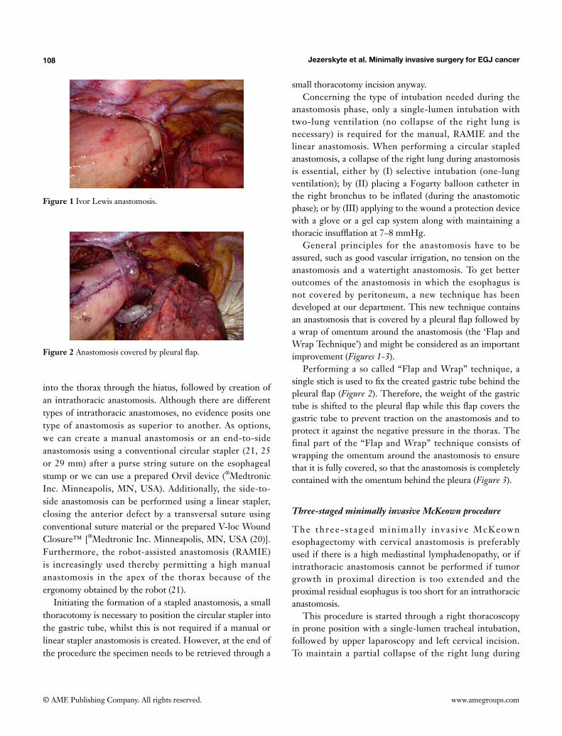
108
© AME Publishing Company. All rights reserved. www.amegroups.com
Jezerskyte et al. Minimally invasive surgery for EGJ cancer
into the thorax through the hiatus, followed by creation of an intrathoracic anastomosis. Although there are different types of intrathoracic anastomoses, no evidence posits one type of anastomosis as superior to another. As options, we can create a manual anastomosis or an end-to-side anastomosis using a conventional circular stapler (21, 25 or 29 mm) after a purse string suture on the esophageal stump or we can use a prepared Orvil device (®Medtronic Inc. Minneapolis, MN, USA). Additionally, the side-to-side anastomosis can be performed using a linear stapler, closing the anterior defect by a transversal suture using conventional suture material or the prepared V-loc Wound Closure™ [®Medtronic Inc. Minneapolis, MN, USA (20)]. Furthermore, the robot-assisted anastomosis (RAMIE) is increasingly used thereby permitting a high manual anastomosis in the apex of the thorax because of the ergonomy obtained by the robot (21).
Initiating the formation of a stapled anastomosis, a small thoracotomy is necessary to position the circular stapler into the gastric tube, whilst this is not required if a manual or linear stapler anastomosis is created. However, at the end of the procedure the specimen needs to be retrieved through a
small thoracotomy incision anyway. Concerning the type of intubation needed during the
anastomosis phase, only a single-lumen intubation with two-lung ventilation (no collapse of the right lung is necessary) is required for the manual, RAMIE and the linear anastomosis. When performing a circular stapled anastomosis, a collapse of the right lung during anastomosis is essential, either by (I) selective intubation (one-lung ventilation); by (II) placing a Fogarty balloon catheter in the right bronchus to be inflated (during the anastomotic phase); or by (III) applying to the wound a protection device with a glove or a gel cap system along with maintaining a thoracic insufflation at 7–8 mmHg.
General principles for the anastomosis have to be assured, such as good vascular irrigation, no tension on the anastomosis and a watertight anastomosis. To get better outcomes of the anastomosis in which the esophagus is not covered by peritoneum, a new technique has been developed at our department. This new technique contains an anastomosis that is covered by a pleural flap followed by a wrap of omentum around the anastomosis (the ‘Flap and Wrap Technique’) and might be considered as an important improvement (Figures 1-3).
Performing a so called “Flap and Wrap” technique, a single stich is used to fix the created gastric tube behind the pleural flap (Figure 2). Therefore, the weight of the gastric tube is shifted to the pleural flap while this flap covers the gastric tube to prevent traction on the anastomosis and to protect it against the negative pressure in the thorax. The final part of the “Flap and Wrap” technique consists of wrapping the omentum around the anastomosis to ensure that it is fully covered, so that the anastomosis is completely contained with the omentum behind the pleura (Figure 3).
Three-staged minimally invasive McKeown procedure
The three-s taged min imal ly invas ive McKeown esophagectomy with cervical anastomosis is preferably used if there is a high mediastinal lymphadenopathy, or if intrathoracic anastomosis cannot be performed if tumor growth in proximal direction is too extended and the proximal residual esophagus is too short for an intrathoracic anastomosis.
This procedure is started through a right thoracoscopy in prone position with a single-lumen tracheal intubation, followed by upper laparoscopy and left cervical incision. To maintain a partial collapse of the right lung during
Figure 1 Ivor Lewis anastomosis.
Figure 2 Anastomosis covered by pleural flap.

109Minimally Invasive Esophagectomy
© AME Publishing Company. All rights reserved. www.amegroups.com
thoracoscopy, the thoracic cavity must be insufflated with carbon dioxide at 6–8 mmHg. During thoracoscopy the esophagus is dissected and a mediastinal lymphadenectomy is performed as comparable to the previous described in the Ivor Lewis esophagectomy section.
After thoracoscopy, the patient is repositioned in French position to perform a laparoscopy with lymphadenectomy and formation of the gastric conduit as comparable with the Ivor-Lewis procedure. No Kocher maneuver or intervention to the pylorus is performed. Subsequently, a cervical anastomosis can be created starting with left cervical incision to decrease the risk of bilateral recurrent laryngeal nerve injury. The esophagus is divided and the gastric tube is pulled into the cervical region via the prevertebral route. A cervical end-to-end anastomosis is created manually. The advantages of a cervical anastomosis compared to an intrathoracic anastomosis (Ivor-Lewis procedure) are presumed better clinical management of leakages (e.g., by bedside opening of the cervical wound) and a larger proximal resection margin.
THE
This procedure is performed by laparoscopy and left cervical incision. Starting with transhiatal dissection of the esophagus (and tumor) from the pericardial sac and aortic planes up to the carina, it is followed by an extended D2 abdominal lymphadenectomy and gastric dissection. After dissection of the cervical esophagus, a small-assisting-protected laparotomy is performed to retrieve the whole specimen by stripping. Extracorporeal creation of gastric tube and resection of the specimen is then followed by pulling the gastric tube into the cervical wound where the anastomosis is made. Due to the transhiatal approach, the mediastinal lymphadenectomy is limited (16). Details of the
surgical techniques have been published elsewhere (22).
Total gastrectomy
A laparoscopic total gastrectomy is performed through a laparoscopy in the upper abdomen, usually using 4 trocars and a separate incision for the Nathanson liver retractor or 5 trocars. A formal D2 lymphadenectomy is performed and the duodenum is divided by a stapler. The distal esophagus is mobilized and both pleura’s may or may not be opened, depending on the tumor. Intrathoracic, only the distal paraesophageal lymph nodes can be resected. After transection of the vagal nerves and the esophagus above the level of the tumor, an end-side esophagojejunostomy is created, usually using the Orvil (®Medtronic Inc. Minneapolis, MN, USA), since a purse string suture in the confined space of the narrow hiatus so high intrathoracic is usually not possible. It is advocated to send the resection specimen for frozen section before the anastomosis is created.
Evidence for surgical techniques
A prospective database of 266 consecutive patients with surgical resectable EGJ adenocarcinomas is analyzed by Parry et al. (22). Twenty-five % of patients had a Siewert I, 66% had type II tumor and 5% a type III tumor. In total, 86% were treated with esophagectomy and 14% with gastrectomy. Overall 5 years survival was 38% (41% of patients did not receive any neoadjuvant therapy). In type II patients the type of operation did not significantly influence OS on multivariate analysis. A positive circumferential resection margin (CRM) at the site of the esophagus was more common with gastrectomy than with esophagectomy (29% vs. 11%; P=0.025). No significant differences in morbidity, mortality or disease recurrence were found. In patients with type II, upper mediastinal nodal involvement (subcarinal, paratracheal, and aortopulmonary window) was found in 11% of the patients. In 34% of patients treated with esophagectomy, paraesophageal lymph nodes metastases were harvested compared with 5% of patients treated with gastrectomy. They conclude that in patients with a type II EGJ adenocarcinoma, a positive CRM was more common with gastrectomy. Esophagectomy provides for a more complete paraesophageal lymphadenectomy. Furthermore, the high prevalence of mediastinal nodal involvement indicates that a full lymphadenectomy of these stations should be considered.
Figure 3 Anastomosis covered by omental wrap.

110
© AME Publishing Company. All rights reserved. www.amegroups.com
Jezerskyte et al. Minimally invasive surgery for EGJ cancer
In the era before the systematic use of neoadjuvant therapy, the Dutch HIVEX-trial compared the transhiatal approach versus the transthoracic approach for esophageal carcinoma (16). The trial revealed no differences in survival rates between the two approaches for EGJ tumors type II, whereas for type I the survival rates after transthoracic esophagectomy (TTE) were higher. In order to update this comparison in the current era of nCRTx and MIE, a comparable trial should be performed. The so-called IVORY-trial is currently in preparation and will compare minimally invasive transhiatal versus the transthoracic approach after nCRTx for distal and EGJ tumors type I and II according to the Siewert classification. The advantages of the transthoracic approach are an extensive esophageal dissection, a more complete mediastinal lymphadenectomy and possibly a better anastomosis. Because the thoracic anastomosis is more distally created than the cervical anastomosis, it is possible to perform a more extended gastric resection that in those type II tumors will help to achieve free resections margins. Moreover, the gastric tube may be shorter, but is better vascularized and consequently this may result in less morbidity, especially with less anastomotic leakages. It seems that Siewert type I tumors will metastasize to the paratracheal lymph nodes in 10% of the patients after neoadjuvant therapy, whilst this is less than 2% for type II tumors after neoadjuvant therapy (16). The role and extension of mediastinal lymphadenectomy is still controversial after the use of the nCRTx according to the CROSS-study (23).
Currently, after esophagectomy for cancer both cervical and intrathoracic esophagogastric anastomosis are used. Although a cervical anastomosis seems to be the best option for proximal and mid-esophageal tumors, yet a cervical anastomosis is followed by a higher frequency of anastomotic leakages—probably due to the worse perfusion at the top of the gastric tube (24). Additionally, after MIE with cervical anastomoses for distal or EGJ tumors, patients do have more complaints of dysphagia, dumping and regurgitation (25). This might be attributable to the higher incidence of strictures in these patients (26,27). A recent study showed that there seems to be a trend to create more Ivor-Lewis esophageal resections for EGJ-tumors than McKeown esophagectomies, involving in the period 2007 to 2014 an increase from 15% to 46% of intrathoracic anastomosis (28). In contrast, in the same period the incidence of three-stage McKeown esophagectomy decreased from 85% into 54% in the same study. To reach consensus about this ongoing topic the multicenter ICAN-
trial has been launched to compare the short-term outcomes of transthoracic resections between patients with a cervical anastomosis (McKeown procedure) versus patients with an intrathoracic anastomosis (Ivor-Lewis procedure). The first patients have been recently included (29).
Concerning the surgical techniques, another important improvement with the aim to decrease the anastomotic leakage rate is the covering of the intrathoracic anastomosis by wrapping the omentum around it. A systematic review not only showed a significant decrease in the anastomotic leakages rate, but also in the length of hospital stay (30).
Another point of discussion is the position of patients, lateral or in prone position during Ivor-Lewis or McKeown esophagectomy. Initially, the lateral decubitus position with selective intubation and ventilation of one lung was preferred. However, the introduction of the prone position by Cuschieri in 1994 described many advantages of this position over the lateral decubitus position, such as no necessity for a complete lung block and a better visualization of the esophageal area (31). A recently published systematic review from Markar et al., suggests that the prone position is associated with less pulmonary complications, less blood loss, and a higher number of resected lymph nodes (32). It must be noted that there was some evidence of heterogeneity for the analysis of pulmonary complications and blood loss in this review.
Thus, not only the proper approach or the extension of lymphadenectomy, but also the ideal thoracoscopic position of the patient will be important items for future studies in patients who undergo MIE.
Authors’ own experience and type of anastomosis
In the Netherlands in the last years, most esophageal resections have been centralized in centers performing more than 20 esophageal resections yearly. Along with the increased use of minimally invasive surgery, the morbidity and mortality rates in patients have since decreased (10,33,34). Moreover, neoadjuvant therapies such as neoadjuvant chemoradiotherapy and chemotherapy are now extensively used. Data from the National Dutch Register (DUCA) show that the use of the thoracic esophagectomy increased from 47% in 2011 to 74% in 2015; that the implementation of total MIE increased from 32% to 72%; and that neoadjuvant therapy was administered in 91% of the registered 846 patients in 2015 (34).
As distal esophageal and EGJ adenocarcinomas are more

111Minimally Invasive Esophagectomy
© AME Publishing Company. All rights reserved. www.amegroups.com
than 80% of all esophageal cancers in the Netherlands, our department has increased the use of MIE Ivor-Lewis approach—a comparable development found at the other upper GI centers in The Netherlands. The first multicenter study about MIE Ivor-Lewis esophagectomy included more than 282 patients from six centers, performing different types of anastomoses, and showed a leakage in more than 15% of the patients with a 30-days mortality of 2.1% (35). This high-leakage rate obliged the surgeons to analyze these numbers, the learning curve and the cause of this rate. The result of this analysis in our department produced a change in the used anastomosis technique, resulting in the “Flap and Wrap” technique as described in the intrathoracic anastomosis paragraph of this chapter. This anastomosis technique is increasingly used in our Department, rising from 24.2% of the total esophageal resections in 2014 up to 72% of the resections in 2016. With this anastomosis anastomotic leakage rate has dropped to less than 5% for intrathoracic anastomosis with the “Flap and Wrap” technique, whereas leakages are still found in 20% of the patients with a cervical anastomosis without “Flap and Wrap” technique (unpublished data). Moreover, an important decrease of overall morbidity, reoperation rate, readmissions to the ICU and in ICU length of stay have been found (LOS). It seems that a lower incidence of the morbidity rates is associated to a higher frequency of transthoracic resections.
There is still no consensus about the ideal type of intrathoracic anastomosis. Different types of anastomoses are used, including manual, linear stapler, circular stapler and robot manual-assisted anastomoses. There is no evidence that one technique is better than the other, but general principles for anastomoses such as the need of a well-vascularized gastric tube, no tension or traction on the anastomosis and adequate patency are important factors to respect. In our experience, the flap and wrap anastomosis technique in which the anastomosis is covered behind a flap of pleura, so fixing it with stitches and having wrapped the entire anastomosis in omentum, hence accounts for a relative low leakage rate.
Outcome of published series
Data about morbidity and mortality rates following a MIE after neoadjuvant therapy for EGJ tumors specifically are scarce. The largest prospective study described the morbidity and mortality rates of a series of more than 1,000 patients in whom minimally invasive Ivor-
Lewis esophagectomy and McKeown esophagectomy is performed (36). They did not find any differences in the frequency of anastomotic leakages requiring surgery (4% versus 5%, respectively). However, there were significant differences in the occurrence of vocal cord paralyses, with a higher incidence among McKeown esophagectomies than Ivor-Lewis procedures (8% versus 1%, respectively). Moreover, no difference in 30-days mortality was reported (0.9% versus 2.5%, respectively). In general, there seems to be a trend towards lower morbidity rates among MIE Ivor-Lewis resections as compared to MIE McKeown resections and THE, but results of evidence based studies comparing these different approaches are lacking (25,36).
Outcomes for minimally invasive TTE esophagectomy
Two studies that reported short-term outcomes of MIE Ivor-Lewis resections for EGJ tumors specifically are recently reported. One study reported a multicentric series of patients with a distal or a EGJ tumor of the esophagus treated by MIE Ivor Lewis and the other study compared two cohorts, MIE Ivor Lewis and MIE McKeown procedure. Straatman et al. investigated the short-term outcomes of 282 patients among 6 different European centers who underwent minimally invasive Ivor-Lewis esophagectomy for only distal and EGJ tumors and showed a morbidity rate of 44% (35). The most frequent complications were anastomotic leakages (15.2%), pulmonary complications (13.1%), and cardiac complications (4.3%). Perioperative outcomes were: a median operative time of 333 minutes, 242mL blood loss (median) and 1.8% conversions to open Ivor-Lewis esophagectomy. Radical resections (R0) were performed in 93% of the patients. Further postoperative outcomes were a median length of stay of 12 days, and a median length of ICU stay of 2 days and the 30-day morbidity was 2.1%.
The second retrospective study compared 356 patients who underwent Ivor-Lewis MIE (intrathoracic anastomosis, n=210) with patients who underwent McKeown or Transhiatal MIE (cervical anastomosis, n=146) (25). The incidence of recurrent laryngeal nerve palsy was 14.4% after a cervical anastomosis and 0% after an intrathoracic anastomosis. Dysphagia, dumping, and regurgitation were reported less frequently after creation of an intrathoracic anastomosis. Dilatation of benign strictures occurred in 43.8% of the cervical anastomoses versus in 6.2% of the intrathoracic anastomoses. If a benign stricture was identified, it was dilated for a median of 4 times in the

112
© AME Publishing Company. All rights reserved. www.amegroups.com
Jezerskyte et al. Minimally invasive surgery for EGJ cancer
cervical group and only once in the intrathoracic group. Anastomotic leakage for which reoperation was required occurred in 8.2% after cervical anastomosis and in 11.4% after intrathoracic anastomosis (not significant). Median ICU stay, hospital stay, in-hospital mortality, 30-day mortality, and 90-day mortality were similar between the groups (not significant). They conclude that MIE with an intrathoracic anastomosis is associated with better functional results with less dysphagia, less benign anastomotic strictures requiring fewer dilatations, and a lower incidence of recurrent laryngeal nerve palsy as compared to MIE with cervical anastomoses. Other postoperative morbidity and mortality did not differ between the groups.
Conclusions
The incidence of cancer of the distal esophagus and EGJ is increasing, whereby adenocarcinomas are paramount in the Western World (up to 80% of all esophageal carcinomas). The advantages of MIE as a treatment of esophageal or a EGJ cancer in comparison with OE are important improvements in the short-term outcomes, such as less blood loss, less respiratory infections, a better postoperative QoL, with similar 1- and 3-year survival rates. Tumors of the EGJ are classified by the Siewert classification, despite all its limitations. For Siewert type III a total or proximal gastrectomy is the indicated resection whereas for type II both the extended gastrectomy or the Ivor Lewis approach are the two possibilities. Importantly, a R0 radical resection should be performed. For Siewert types I and II, the Ivor-Lewis esophagectomy is an ideal operation following neoadjuvant therapy. There is an important increase in the use of this approach per year with 41% of all esophageal resections in 2015 in the Netherlands. This approach includes an intrathoracic anastomosis after laparoscopic preparation of the gastric tube and thoracoscopic esophageal resection and intrathoracic anastomosis in prone position. Other surgical options for an esophageal resection include a THE or the (transthoracic) McKeown resection, in which a cervical anastomosis is performed. The transhiatal approach has its limitations due to the incapacity to perform a mediastinal lymphadenectomy and is reserved to frail patients with a distal or EGJ tumor. In a non-randomized study, patients with intrathoracic anastomoses showed a better functional outcome than the group with cervical anastomoses along with less dysphagia, less benign strictures and lower incidence than recurrent
nerve palsy. Our experience with intrathoracic anastomoses accompanied by the ‘Flap and Wrap’ technique limited the postoperative leakage rate to approximately 5% whereas the leakage rate in patients with cervical anastomoses remains up to 20%.
Additionally, a more extended lymphadenectomy is possible in transthoracic surgery compared to THE or gastrectomy, and lymph node status is still the most important predictor for survival.
Acknowledgements
None.
Footnote
Conflicts of Interest: The authors have no conflicts of interest to declare.
References
1. Ferlay J, Steliarova-Foucher E, Lortet-Tieulent J, et al. Cancer incidence and mortality patterns in Europe: estimates for 40 countries in 2012. Eur J Cancer 2013;49:1374-403.
2. Arnold M, Soerjomataram I, Ferlay J, et al. Global incidence of oesophageal cancer by histological subtype in 2012. Gut 2015;64:381-7.
3. Abbas G, Krasna M. Overview of esophageal cancer. Ann Cardiothorac Surg 2017;6:131-6.
4. Pennathur A, Gibson MK, Jobe BA, et al. Oesophageal carcinoma. Lancet 2013;381:400-12.
5. Rice TW, Blackstone EH, Rusch VW, et al. 7th Edition of the AJCC Cancer Staging Manual: esophagus and esophagogastric junction. Ann Surg Oncol 2010;17:1721-4.
6. Rüdiger Siewert J, Feith M, Werner M, et al. Adenocarcinoma of the esophagogastric junction: results of surgical therapy based on anatomical/topographic classification in 1,002 consecutive patients. Ann Surg 2000;232:353-61.
7. Deng JY, Liang H. Adenocarcinoma of esophagogastric junction. Chin J Cancer Res 2014;26:362-3.
8. Shapiro J, van Lanschot JJ, Hulshof MC, et al. Neoadjuvant chemoradiotherapy plus surgery versus surgery alone for oesophageal or junctional cancer (CROSS): Long-term results of a randomised controlled trial. Lancet Oncol 2015;16:1090-8.

113Minimally Invasive Esophagectomy
© AME Publishing Company. All rights reserved. www.amegroups.com
9. van Hagen P, Hulshof MC, van Lanschot JJ, et al. Preoperative chemoradiotherapy for esophageal or junctional cancer. N Engl J Med 2012;366:2074-84.
10. Biere SS, Van Berge Henegouwen MI, Maas KW, et al. Minimally invasive versus open oesophagectomy for patients with oesophageal cancer: A multicentre, open-label, randomised controlled trial. Lancet 2012;379:1887-92.
11. Maas KW, Cuesta MA, Van Berge Henegouwen MI, et al. Quality of Life and Late Complications After Minimally Invasive Compared to Open Esophagectomy: Results of a Randomized Trial. World J Surg 2015;39:1986-93.
12. Straatman J, van der Wielen N, Cuesta MA, et al. Minimally Invasive Versus Open Esophageal Resection: Three-year Follow-up of the Previously Reported Randomized Controlled Trial: the TIME Trial. Ann Surg 2017. [Epub ahead of print].
13. Cunningham D, Allum WH, Stenning SP, et al. Perioperative Chemotherapy versus Surgery Alone for Resectable Gastroesophageal Cancer. N Engl J Med 2006;355:11-20.
14. Anderegg MC, van der Sluis PC, Ruurda JP, et al. Preoperative Chemoradiotherapy Versus Perioperative Chemotherapy for Patients With Resectable Esophageal or Gastroesophageal Junction Adenocarcinoma. Ann Surg Oncol 2017. [Epub ahead of print].
15. Parry K, Haverkamp L, Bruijnen RC, et al. Staging of adenocarcinoma of the gastroesophageal junction. Eur J Surg Oncol 2016;42:400-6.
16. Omloo JM, Lagarde SM, Hulscher JB, et al. Extended transthoracic resection compared with limited transhiatal resection for adenocarcinoma of the mid/distal esophagus: five-year survival of a randomized clinical trial. Ann Surg 2007;246:992-1000.
17. Medtronic Inc. (2013) Orvil Circular Stapler. 18. Koëter M, Kathiravetpillai N, Gooszen JA, et al. Influence
of the Extent and Dose of Radiation on Complications After Neoadjuvant Chemoradiation and Subsequent Esophagectomy With Gastric Tube Reconstruction With a Cervical Anastomosis. Int J Radiat Oncol Biol Phys 2017;97:813-21.
19. Goense L, van Rossum PS, Ruurda JP, et al. Radiation to the Gastric Fundus increases the risk of Anastomotic Leakage after Esophagectomy. Ann Thorac Surg 2016;102:1798-804.
20. Medtronic Inc. (2015) V-loc Wound Closure. 21. van Hillegersberg R, Seesing MF, Brenkman HJ, et al.
Robot-assisted minimally invasive esophagectomy. Chirurg
2016;87:635-42.22. Parry K, Haverkamp L, Bruijnen RC, et al. Surgical
treatment of adenocarcinomas of the gastro-esophageal junction. Ann Surg Oncol 2015;22:597-603.
23. Koen Talsma A, Shapiro J, Looman CW, et al. Lymph Node Retrieval During Esophagectomy With and Without Neoadjuvant Chemoradiotherapy. Ann Surg 2014;260:786-92.
24. Scheepers JJ, Veenhof XA, van der Peet DL, et al. Laparoscopic transhiatal resection for malignancies of the distal esophagus: Outcome of the first 50 resected patients. Surgery 2008;143:278-85.
25. van Workum F, van der Maas J, van den Wildenberg F, et al. Improved Functional Results After Minimally Invasive Esophagectomy: Intrathoracic Versus Cervical Anastomosis. Ann Thorac Surg 2017;103:267-73.
26. Biere SS, Maas KW, Cuesta MA, et al. Cervical or thoracic anastomosis after esophagectomy for cancer: A systematic review and meta-analysis. Dig Surg 2011;28:29-35.
27. Markar SR, Arya S, Karthikesalingam A, et al. Technical Factors that Affect Anastomotic Integrity Following Esophagectomy: Systematic Review and Meta-analysis. Ann Surg Oncol 2013;20:4274-81.
28. Haverkamp L, Seesing MF, Ruurda JP, et al. Worldwide trends in surgical techniques in the treatment of esophageal and gastroesophageal junction cancer. Dis Esophagus 2017;30:1-7.
29. van Workum F, Bouwense SA, Luyer MD, et al. Intrathoracic versus Cervical ANastomosis after minimally invasive esophagectomy for esophageal cancer: study protocol of the ICAN randomized controlled trial. Trials 2016;17:505.
30. Yuan Y, Zeng X, Hu Y, et al. Omentoplasty for oesophagogastrostomy after oesophagectomy. Cochrane Database Syst Rev 2014;(10):CD008446.
31. Cuschieri A. Thoracoscopic subtotal oesophagectomy. Endosc Surg Allied Technol 1994;2:21-5.
32. Markar SR, Wiggins T, Antonowicz S, et al. Minimally invasive esophagectomy: Lateral decubitus vs. prone positioning; Systematic review and pooled analysis. Surg Oncol 2015;24:212-9.
33. Wouters MW, Karim-Kos HE, le Cessie S, et al. Centralization of esophageal cancer surgery: does it improve clinical outcome? Ann Surg Oncol 2009;16:1789-98.
34. Dutch Upper GI Cancer Audit 2015. 2015. Available online: https://www.dica.nl/jaarrapportage-2015/duca
35. Straatman J, van der Wielen N, Nieuwenhuijzen GA, et al.

114
© AME Publishing Company. All rights reserved. www.amegroups.com
Jezerskyte et al. Minimally invasive surgery for EGJ cancer
Techniques and short-term outcomes for total minimally invasive Ivor Lewis esophageal resection in distal esophageal and gastroesophageal junction cancers: pooled data from six European centers. Surg Endosc 2017;31:119-26.
36. Luketich JD, Pennathur A, Awais O, et al. Outcomes after Minimaly Invasive Esophagectomy: review over 1000 patients. Ann Surg 2012;256:95-103.
Cite this article as: Jezerskyte E, van Berge Henegouwen MI, Cuesta MA, Gisbertz SS. Gastro-esophageal junction cancers: what is the best minimally invasive approach? J Thorac Dis 2017;9(Suppl 8):S751-S760. doi: 10.21037/jtd.2017.06.56

© AME Publishing Company. All rights reserved. www.amegroups.com
McKeown or Ivor Lewis totally minimally invasive esophagectomy for cancer of the esophagus and gastroesophageal junction: systematic review and meta-analysis
Frans van Workum1, Gijs H. Berkelmans2, Bastiaan R. Klarenbeek1, Grard A. P. Nieuwenhuijzen2, Misha D. P. Luyer2, Camiel Rosman1
1Department of surgery, Radboudumc, Nijmegen, the Netherlands; 2Department of surgery, Catharina hospital, Eindhoven, the Netherlands
Contributions: (I) Conception and design: F van Workum, BR Klarenbeek, C Rosman; (II) Administrative support: None; (III) Provision of study
materials or patients: None; (IV) Collection and assembly of data: F van Workum, BR Klarenbeek; (V) Data analysis and interpretation: All authors;
(VI) Manuscript writing: All authors; (VII) Final approval of manuscript: All authors.
Correspondence to: Frans van Workum. Radboud University Medical Centre, P.O. Box 9101, 6500 HB, Nijmegen, the Netherlands.
Email: [email protected].
Background: Minimally invasive esophagectomy (MIE) has consistently been associated with improved perioperative outcome and similar oncological safety compared to open esophagectomy. However, it is currently unclear what type of MIE is preferred for patients with resectable esophageal cancer.Methods: Literature was searched in Medline, Embase and the Cochrane library combining relevant search terms. Articles that included patients undergoing totally minimally invasive esophagectomy (TMIE) or hybrid minimally invasive esophagectomy (HMIE) and compared McKeown with Ivor Lewis procedures were included. Studies were excluded if they included >10% of patients undergoing a procedure other than MIE McKeown or MIE Ivor Lewis (i.e., transhiatal resections). The primary outcome parameter was anastomotic leakage. Secondary outcome parameters were: other complications, reinterventions, reoperations, hospital length of stay, ICU length of stay, postoperative mortality, operative time, blood loss, R0 resection rate, lymph nodes examined, quality of life and costs. Results: Five studies with a total of 1,681 patients undergoing TMIE were included. There were no studies comparing HMIE McKeown versus HMIE Ivor Lewis. There were no randomized controlled trials and all included studies were cohort studies with a moderate risk of bias. No meta-analysis could be performed for R0 resection rate, survival, quality of life and costs because there was insufficient data available for these parameters. The incidence of anastomotic leakage did not differ between the groups [relative risk (RR) =1.39, 95% confidence interval (CI) =0.90–10.38, P=0.14]. TMIE Ivor Lewis was associated with a lower incidence of recurrent laryngeal nerve (RLN) trauma (RR =6.70, 95% CI =3.09–14.55, P<0.001), a shorter hospital length of stay [standardized mean difference (SMD) =0.17, 95% CI =0.06–0.28, P=0.002] and less blood loss (SMD =0.69, 95% CI =0.25–1.12, P=0.002). Conclusions: TMIE Ivor Lewis is associated with improved outcome regarding RLN trauma, hospital length of stay and blood loss as compared to TMIE-McKeown, but the incidence of anastomotic leakage is not different. The evidence is limited, of low quality and at risk for bias. A randomized controlled trial is currently being performed in order to demonstrate whether a McKeown or Ivor Lewis procedure should be preferred in patients undergoing MIE.
Keywords: Minimally invasive esophagectomy (MIE); McKeown esophagectomy; Ivor Lewis esophagectomy;
cervical anastomosis; intrathoracic anastomosis
Submitted Jan 08, 2017. Accepted for publication Mar 23, 2017.
doi: 10.21037/jtd.2017.03.173
View this article at: http://dx.doi.org/10.21037/jtd.2017.03.173
122
Thoracoscopic Esophagectomy

116
© AME Publishing Company. All rights reserved. www.amegroups.com
van Workum et al. McKeown vs. Ivor Lewis MIE
Introduction
The annual incidence of esophageal carcinoma is increasing (1). Esophagectomy remains the cornerstone for curative treatment, most often after neoadjuvant therapy (2). Minimally invasive esophagectomy (MIE), consisting of hybrid minimally invasive esophagectomy (HMIE) or totally minimally invasive esophagectomy (TMIE), has been shown to be superior compared to open esophagectomy regarding perioperative outcome (3,4) without compromising oncologic safety (5,6). This has led to a progressive adoption of MIE and currently, 45% of all patients worldwide with resectable esophageal cancer undergo MIE (7).
Similar to open esophagectomy, MIE can consist of transhiatal esophagectomy (8), McKeown esophagectomy (9) or Ivor Lewis esophagectomy (10), but Ivor Lewis or McKeown procedures are usually performed since they allow adequate thoracic lymph node dissection (11,12). In patients with esophageal tumors above the level of the carina, an Ivor Lewis procedure is unfeasible because it might compromise adequate resection margins. For patients with lower esophageal or gastroesophageal junction tumors, both McKeown and Ivor Lewis procedures are considered to be oncologically feasible. Supposed benefits of cervical anastomosis are that it is technically less challenging than totally minimally invasive intrathoracic anastomosis and that if an anastomotic leak occurs, it can be managed more easily than intrathoracic leakage. However, intrathoracic anastomosis after MIE is believed to be associated with a lower incidence of anastomotic leakage and better functional results.
It is currently unknown whether minimally invasive McKeown or minimally invasive Ivor Lewis esophagectomy should be preferred for these patients and both cervical and intrathoracic anastomoses are performed (7). The aim of this article is therefore to perform a systematic review and meta-analysis of studies comparing minimally invasive McKeown esophagectomy with minimally invasive Ivor Lewis esophagectomy.
Methods
Literature search
The electronic databases of Medline, Embase and the Cochrane Central Register of Controlled Trials databases were searched using the following search terms (and combinations of these terms): minimal invasive, minimally
invasive, laparo-thoracoscop*, laparothoracoscop*, thoracolaparoscop* OR, thoraco-laparoscop*, laparoscop*, hybrid, video assisted thoracic surgery (VATS), video-assisted, video assisted, thoracoscop* esophagectom*, oesophagectom*, resection* oesophagus, oesophageal, oesophagal, esophagus, esophageal, esophagal. No language restrictions were applied and all results up to December 2016 were included. Medical ethical approval was not sought because no new patient data was obtained for this study.
Criteria for selecting studies for this review
Comparative cohort studies or randomized controlled trials that included patients undergoing HMIE or TMIE and that compared McKeown versus Ivor Lewis procedures were included. Exclusion criteria were: less than 10 patients in a treatment arm, unclear description of operative technique rendering classification into McKeown or Ivor Lewis procedures impossible and studies that contained more that 10% other procedures in one of the arms (i.e., minimally invasive transhiatal esophagectomy). VATS procedures and hand-assisted laparoscopic surgery (HALS) procedures were regarded as minimally invasive and were also included.
All references of studies were screened on title and abstract by two authors independently (BK and FvW). All studies that were not excluded in the screening stage were assessed in full text for eligibility. If discrepancies occurred, this was discussed in a meeting and if no consensus could be reached, another discussion meeting with a third author (CR) was held until consensus was reached. This process will be described in a flow chart according to the PRISMA statement (13).
Quality assessment
All studies were independently assessed for methodological quality by BK and FvW using the Newcastle-Ottawa rating scale (14). Discrepancies were resolved in discussion. In case of persisting discrepancy, a meeting with a third author (CR) was held and discrepancies were discussed until consensus was reached.
Outcome parameters and data extraction
The primary outcome parameter was anastomotic leakage. Secondary outcome parameters were: all complications, severe complications (CD ≥3), pneumonia, pulmonary

117Minimally Invasive Esophagectomy
© AME Publishing Company. All rights reserved. www.amegroups.com
Figure 1 Summary of screening and selection process—PRISMA diagram.
Articles identified through database searching (n=3,604)
Additional articles identified through other sources (n=26)
Articles after duplicates removed (n=2,041)
Articles screened (n=2,041)
Articles excluded (n=2,017)
Full-text articles assessed for eligibility (n=24)
Articles included in qualitative synthesis (n=5)
Articles included in quantitative synthesis (meta-analysis) (n=5)
Full-text articles excluded, with reasons (n=19)
- No comparison of McKeown versus Ivor Lewis (n=11)- No data per operative approach (n=3)- <10 patients per treatment group (n=3)- Unclear whether patients in an outcome group underwent McKeown or Ivor Lewis (n=2)
complications, chyle leakage, wound infection, recurrent laryngeal nerve (RLN) palsy, benign anastomotic strictures, operating time, blood loss, reoperation rate, reintervention rate, hospital length of stay, ICU length of stay, postoperative mortality (30-, 90-day and in hospital mortality), R0 resection rate, number of lymph nodes found, quality of life and costs. Data was extracted and was entered into review manager (version 5.3). Continuous variables were expressed as median and interquartile ratio or range, the mean and SD were estimated from the available data by methods described elsewhere (15,16).
Analysis
Meta-analysis was performed if data on an outcome parameter was reported in at least two studies in a way that was compatible with meta-analysis. The Mantel-Haenszel method for dichotomous data was used, presented as relative risks (RR) with 95% confidence intervals (CIs). The inverse variance method was used for meta-analysis of continuous data; results are presented as standardized mean difference (SMD) with 95% CIs. The statistical heterogeneity was assessed with I2. In the absence of substantial statistical
heterogeneity [I2 ≤50% (15)] a fixed-effect model was used. In case of substantial heterogeneity (I2 >50%), a random-effects model was used. Statistical analyses were performed with Review Manager (version 5.3).
Results
Studies
A summary of the screening and selection process according to PRISMA (13) is shown in Figure 1. No studies comparing McKeown versus Ivor Lewis procedure in patients undergoing HMIE were identified. Five studies with 1,681 patients undergoing TMIE were ultimately included for analysis (17-21). The characteristics of the included studies are summarized in Table 1.
Quality assessment
The results of the quality assessment of the included studies are shown in Table 2. There were no randomized controlled trials. Studies scored 6 or 7 stars out of 9 according to the Newcastle-Ottawa rating scale, corresponding to a moderate risk of bias. Four studies were retrospective

118
© AME Publishing Company. All rights reserved. www.amegroups.com
van Workum et al. McKeown vs. Ivor Lewis MIE
cohort studies and one was a prospective cohort study. One study clearly stated that MIE Ivor Lewis procedures were predominantly performed in a more recent time period (19). This was not described for the other studies.
Heterogeneity
Considerable heterogeneity was found for the outcome parameters pulmonary complications (I2 =73%), intensive care length of stay (I2 =95%), examined lymph nodes (I2 =79%), operating time (I2 =99%) and blood loss (I2 =70%). For these parameters, a random effects model was used. No sensitivity analysis was performed because this was considered unfeasible with only 2 or 3 studies available for the outcome parameters with high heterogeneity.
Meta-analysis
The outcome parameters severe complications (CD ≥3), pneumonia, wound infection, reintervention, quality of life and costs were not reported in any of the included studies. The outcome parameters chyle leakage, reoperation, R0 resection rate were reported, but not enough data was available to perform meta-analysis.
The results of the meta-analysis are shown in Table 3. The incidence of anastomotic leakage was 5.2% after McKeown esophagectomy and 4.7% after Ivor Lewis esophagectomy (RR =1.39, 95% CI =0.90–2.15, P=0.14) (Figure 2) . Totally minimally invasive Ivor Lewis esophagectomy was associated with a lower incidence of RLN trauma (RR =6.70, 95% CI =3.09–14.55, P<0.001),
Table 1 Characteristics of included studies
Study Study design NMcKeown
(N)Ivor Lewis
(N)Type of TMIE Outcome parameters
Hao 2014 Retrospective cohort
136 81 55 Thoracolaparoscopic AL, LOS, blood loss, OT, thoracic LN
Luketich 2012 Retrospective cohort
1,033 481 530 Thoracolaparoscopic Conversion, AL, gastric tube necrosis, ARDS, empyema, MI, heart failure, LOS, ICU LOS, RLN trauma, R0, LN, mortality
Nguyen 2008 Prospective cohort
104 47 57 Thoracolaparoscopic Blood loss, OT, AL, blood transfusion, major complications, mortality, strictures
Rajan 2010 Retrospective cohort
336 319 17 Thoracolaparoscopic Blood loss, OT, conversion, AL, LOS, ICU LOS, overall complications, pulmonary complications, mortality, strictures
Zhai 2015 Retrospective cohort
72 40 32 Thoracolaparoscopic Blood loss, OT, AL, blood transfusion, pulmonary complications, chylothorax, cardiac arrhythmia, delayed gastric emptying, RLN trauma, reoperations, LOS, ICU LOS, LN, mortality, strictures
TMIE, totally minimally invasive esophagectomy; AL, anastomotic leakage; RLN, recurrent laryngeal nerve; R0, R0 resection rate; LN, lymph nodes examined; ICU, intensive care unit; LOS, length of stay; OT, operating time.
Table 2 Risk of bias assessment
Article Representative SelectionAscertainment
of exposureDemonstration Compatibility Outcome Follow-up
Adequacy follow-up
Total stars
Hao 2014 1 0 1 1 0 1 1 1 6
Luketich 2012 1 0 1 1 0 1 1 1 6
Nguyen 2008 1 1 1 1 0 1 1 1 7
Rajan 2010 1 0 1 1 0 1 1 1 6
Zhai 2015 1 1 1 1 0 1 1 1 7

119Minimally Invasive Esophagectomy
© AME Publishing Company. All rights reserved. www.amegroups.com
Table 3 Outcomes of meta-analysis*
Parameter No. of studies McKeown (%) Ivor Lewis (%) RR/SMD (95% CI) I2 (%) P value
Anastomotic leakage 5 5.2 4.7 1.39 (0.90–10.38) 40 0.14
Pulmonary complications 2 9.7 16.3 1.16 (0.25–5.26) 73 0.85
Blood transfusion 2 11.5 8.4 1.40 (0.56–3.51) 0 0.48
RLN trauma 2 8.8 1.2 6.70 (3.09–14.55) 0 <0.001
Anastomotic stricture 3 7.4 18 1.30 (0.76–2.22) 49 0.34
Postoperative mortality 5 2.1 1.0 2.18 (0.95–4.98) 0 0.07
Intensive care LOS 2 – – −0.22 (–1.15–0.70) 95 0.63
Hospital LOS 4 – – 0.17 (0.06–0.28) 0 0.002
Examined lymph nodes 2 – – −0.23 (–0.75–0.29) 79 0.39
Operating time 3 – – 2.47 (−0.20–5.14) 99 0.07
Blood loss 3 – – 0.69 (0.25–1.12) 70 0.002
*, If parameters appear in Table 1 as reported outcome parameters but not in this table, the parameter was either reported by only one study or the parameters were described in a way that could not be pooled in meta-analysis. RR, relative risk; SMD, standardized mean difference; CI, confidence interval; RLN, recurrent laryngeal nerve; ICU, intensive care unit; LOS, length of stay.
Figure 2 Forest plot anastomotic leakage.
a shorter hospital length of stay (SMD =0.17, 95% CI =0.06–0.28, P=0.002) and less blood loss (SMD =0.69, 95% CI =0.25–1.12, P=0.002) compared to totally minimally invasive McKeown esophagectomy (Figures 3-5). There were no significant differences between the groups regarding the other outcome parameters.
Conclusions
In this meta-analysis, no difference in anastomotic leakage was found between McKeown and Ivor Lewis esophagectomy in patients undergoing TMIE. The incidence of anastomotic leakage was lower than in recent randomized controlled trials (2,3) and this might be explained by the fact that no standardized definitions of complications were
used across studies. For example, the largest study that was included in this meta-analysis only reported anastomotic leakage if a reoperation was required (19). Recently, the esophagectomy complications consensus group (ECCG) proposed standardized definitions for complications after esophagectomy and hopefully this will lead to more uniform definitions of complications in future studies (22).
For open esophagectomy, the anastomotic leakage incidence has been shown to be higher after cervical anastomosis compared to intrathoracic anastomosis in a meta-analysis of randomized controlled trials (23). However, the included RCTs were of moderate methodological quality and included limited numbers of patients. None of the studies included patients undergoing MIE and this is important, since especially the minimally invasive
Study or subgroupHao 2014Luketich 2012Nguyen 2008Rajan 2010Zhai 2015
Total (95% CI)Total eventsHeterogeneity: Chi2 =6.66, df =4 (P=0.16); I2=40%Test for overall effect: Z =1.48 (P=0.14)
Events4
2635
12
50
Events0
23513
32
Total81
48147
31940
968
Total55
530511732
685
Weight1.8%
67.3%14.8%5.8%
10.3%
100.0%
M-H, fixed, 95% CI6.15 (0.34, 111.92)
1.25 (0.72, 2.15)0.65 (0.16, 2.58)0.27 (0.03, 2.18)
3.20 (0.99, 10.38)
1.39 (0.90, 2.15)
McKeown Ivor Lewis Risk ratio Risk ratioM-H, fixed, 95% CI
0.01 0.1 1 10 100Favours McKeown Favours Ivor Lewis

120
© AME Publishing Company. All rights reserved. www.amegroups.com
van Workum et al. McKeown vs. Ivor Lewis MIE
Figure 3 Forest plot RLN trauma. RLN, recurrent laryngeal nerve.
Figure 4 Forest plot hospital length of stay.
Figure 5 Forest plot blood loss.
creation of an intrathoracic anastomosis is considered to be technically challenging and results of open surgery might not be applicable to TMIE. HMIE might combine the best of both worlds for the Ivor Lewis procedure because the technically challenging thoracoscopic creation of an intrathoracic anastomosis is avoided by performing a thoracotomy and pulmonary complications are reduced by performing laparoscopic gastric mobilization (4).
Minimally invasive Ivor Lewis esophagectomy was associated with a lower incidence of RLN trauma and less blood loss. The difference in the incidence of RLN trauma is consistent with the literature regarding open procedures (23) and is explained by avoiding a cervical dissection close to the RLN. This is important, since it has been shown that RLN trauma is associated with increased incidence of pulmonary complications, postoperative ventilation time, intensive care length of stay and hospital length of stay (24-26). The lower blood loss volume that was found in patients undergoing minimally invasive Ivor Lewis
esophagectomy can be explained by omitting a third stage, the incision and the associated blood loss.
An interesting finding is that the hospital length of stay was shorter after minimally invasive Ivor Lewis esophagectomy than after minimally invasive McKeown esophagectomy, despite the fact that no differences in postoperative complications were observed between the groups. This may be explained by the fact that patients with intrathoracic anastomosis have a lower incidence of functional morbidity. In addition to a lower RLN trauma incidence, fewer swallowing problems and a lower incidence of benign anastomotic dilatations (27) might contribute to a shorter hospital length of stay. Further, prospective research and comparisons of other groups are needed in order to assess whether this significant difference in functional results after intrathoracic anastomosis can be confirmed. Another explanation is that the lower length of stay in the Ivor Lewis group might have been caused by performance of the minimally invasive Ivor Lewis procedure in a more recent era, with increased surgeon
Study or subgroupLuketich 2012Zhai 2015
Total (95% CI)Total eventsHeterogeneity: Chi2 =0.87, df =1 (P=0.35); I2=0%Test for overall effect: Z =4.81 (P<0.00001)
Events379
46
Events52
7
Total48140
521
Total53032
562
Weight68.2%31.8%
100.0%
M-H, fixed, 95% CI8.15 [3.23, 20.58]3.60 [0.84, 15.50]
6.70 [3.09, 14.55]
McKeown Ivor Lewis Risk ratio Risk ratioM-H, fixed, 95% CI
0.01 0.1 1 10 100Favours McKeown Favours Ivor Lewis
–1 –0.5 0 0.5 1Favours Mckeown Favours Ivor Lewis
Study or subgroupHao 2014Luketich 2012Nguyen 2008Zhai 2015
Total (95% CI)Heterogeneity: Chi2 =0.36, df =3 (P=0.95); I2=0%Test for overall effect: Z =3.08 (P=0.002)
McKeownMean12.7
812.123.5
Mean12.4
79.7
20.3
SD3.935.9
12.215.1
SD1.065.98.1
10.5
Total81
4814740
649
Total55
5305132
668
Weight10.0%77.1%7.5%5.4%
100.0%
Std. mean difference Std. mean differenceIV, fixed, 95% CIIV, fixed, 95% CI
0.10 [–0.25, 0.44]0.17 [0.05, 0.29]
0.23 [–0.17, 0.63]0.24 [–0.23, 0.71]
0.17 [0.06, 0.28]
Ivor Lewis
Study or subgroupHao 2014Nguyen 2008Zhai 2015
Total (95% CI)Heterogeneity: Tau2 =0.10; Chi2 =6.60, df =3 (P=0.95); I2=0%Test for overall effect: Z =3.11 (P=0.002)
McKeownMean
110263
272.9
Mean86
146246.3
SD29.76
179136.3
SD10.29
11782.4
Total814740
168
Total555132
138
Ivor LewisWeight35.8%33.4%30.8%
100.0%
Std. mean differenceIV, fixed, 95% CI1.00 (0.64, 1.36)0.77 (0.36, 1.19)
0.23 (–0.24, 0.69)
0.69 (0.25, 1.12)
Std. mean differenceIV, fixed, 95% CI
Favours McKeown Favours Ivor Lewis–2 –1 0 1 2

121Minimally Invasive Esophagectomy
© AME Publishing Company. All rights reserved. www.amegroups.com
experience, increased expertise in postoperative management, increased use of enhanced recovery after surgery protocols and improved intensive care. However, only one of the included studies described this phenomenon (19). Therefore, it remains unknown to what extent this selection bias has influenced the results of the included studies.
Taking into account the limitations of the current evidence, it remains uncertain whether a McKeown or an Ivor Lewis should be preferred for MIE for patients in which both procedures are oncologically feasible. To answer this question, the ICAN randomized controlled trial is currently being conducted in the Netherlands and this trial randomizes 200 patients between TMIE McKeown and TMIE Ivor Lewis. In addition to postoperative morbidity and the severity of complications, this trial is also powered for finding differences in quality of life, functional results and cost-effectiveness (28).
Strengths of this study are the comprehensive search strategy and the fact that this is the first review comparing TMIE McKeown versus TMIE Ivor Lewis. Limitations are the heterogeneity of the included studies regarding definit ions of outcome parameters, the moderate methodological quality and the retrospective character of the included studies. In addition, selection bias might have played a significant role, but it is unclear to what extent it is present in the included studies. More research is needed in order to determine whether McKeown or Ivor Lewis MIE should be preferred for patients in whom both procedures are feasible.
Totally minimally invasive Ivor Lewis esophagectomy is associated with improved outcome regarding RLN trauma, hospital length of stay and blood loss compared to totally minimally invasive McKeown esophagectomy. However, the evidence is limited, of moderate quality and at risk for bias. A randomized controlled trial (Intrathoracic versus Cervical ANastomosis after transthoracic esophagectomy: ICAN trial) is currently being performed in order to demonstrate whether minimally invasive McKeown or minimally invasive Ivor Lewis esophagectomy should be preferred for patients in which both procedures are oncologically feasible.
Acknowledgements
None.
Footnote
Conflicts of Interest: The authors have no conflicts of interest to declare.
References
1. Jemal A, Bray F, Center MM, et al. Global cancer statistics. CA Cancer J Clin 2011;61:69-90.
2. van Hagen P, Hulshof MC, van Lanschot JJ, et al. Preoperative chemoradiotherapy for esophageal or junctional cancer. N Engl J Med 2012;366:2074-84.
3. Biere SS, van Berge Henegouwen MI, Maas KW, et al. Minimally invasive versus open oesophagectomy for patients with oesophageal cancer: a multicentre, open-label, randomised controlled trial. Lancet 2012;379:1887-92.
4. Mariette M, Meunier B, Pezet D, et al. Hybrid minimally invasive versus open oesophagectomy for patients with oesophageal cancer: A multicenter, open-label, randomized phase III controlled trial, the MIRO trial. J Clin Oncol 2015;33:suppl 3;abstr 5.
5. Dantoc M, Cox MR, Eslick GD. Evidence to support the use of minimally invasive esophagectomy for esophageal cancer: a meta-analysis. Arch Surg 2012;147:768-76.
6. Sihag S, Wright CD, Wain JC, et al. Comparison of perioperative outcomes following open versus minimally invasive Ivor Lewis oesophagectomy at a single, high-volume centre. Eur J Cardiothorac Surg 2012;42:430-7.
7. Haverkamp L, Seesing MF, Ruurda JP, et al. Worldwide trends in surgical techniques in the treatment of esophageal and gastroesophageal junction cancer. Dis Esophagus 2017;30:1-7.
8. Orringer MB, Sloan H. Esophagectomy without thoracotomy. J Thorac Cardiovasc Surg 1978;76:643-54.
9. McKeown KC. Total three-stage oesophagectomy for cancer of the oesophagus. Br J Surg 1976;63:259-62.
10. LEWIS I. The surgical treatment of carcinoma of the oesophagus; with special reference to a new operation for growths of the middle third. Br J Surg 1946;34:18-31.
11. Hulscher JB, van Sandick JW, de Boer AG, et al. Extended transthoracic resection compared with limited transhiatal resection for adenocarcinoma of the esophagus. N Engl J Med 2002;347:1662-9.
12. Peyre CG, Hagen JA, DeMeester SR, et al. The number of lymph nodes removed predicts survival in esophageal cancer: an international study on the impact of extent of surgical resection. Ann Surg 2008;248:549-56.
13. Liberati A, Altman DG, Tetzlaff J, et al. The PRISMA statement for reporting systematic reviews and meta-analyses of studies that evaluate healthcare interventions: explanation and elaboration. BMJ 2009;339:b2700.

122
© AME Publishing Company. All rights reserved. www.amegroups.com
van Workum et al. McKeown vs. Ivor Lewis MIE
14. Wells G, Shea B, O’Connell D, et al. The Newcastle-Ottawa Scale (NOS) for assessing the quality of nonrandomized studies in meta-analyses. Available online: http://www.ohri.ca/programs/clinical_epidemiology/oxford.asp
15. Higgins JP, Green S. Cochrane handbook for systematic reviews of interventions version 5.0.0. The Cochrane Collaboration. Available online: http://handbook.cochrane.org
16. Hozo SP, Djulbegovic B, Hozo I. Estimating the mean and variance from the median, range, and the size of a sample. BMC Med Res Methodol 2005;5:13.
17. Nguyen NT, Hinojosa MW, Smith BR, et al. Minimally invasive esophagectomy: lessons learned from 104 operations. Ann Surg 2008;248:1081-91.
18. Rajan PS, Vaithiswaran V, Rajapandian S, et al. Minimally invasive oesophagectomy for carcinoma oesophagus--approaches and options in a high volume tertiary centre. J Indian Med Assoc 2010;108:642-4.
19. Luketich JD, Pennathur A, Awais O, et al. Outcomes after minimally invasive esophagectomy: review of over 1000 patients. Ann Surg 2012;256:95-103.
20. Hao Z, Zhenya S, Lei W. Esophageal-gastric anastomosis in radical resection of esophageal cancer under thoracoscopy combined with laparoscopy. J Coll Physicians Surg Pak 2014;24:754-6.
21. Zhai C, Liu Y, Li W, et al. A comparison of short-term outcomes between Ivor Lewis and McKeown minimally
invasive esophagectomy. J Thorac Dis 2015;7:2352-8.22. Low DE, Alderson D, Cecconello I, et al. International
Consensus on Standardization of Data Collection for Complications Associated With Esophagectomy: Esophagectomy Complications Consensus Group (ECCG). Ann Surg 2015;262:286-94.
23. Biere SS, Maas KW, Cuesta MA, et al. Cervical or thoracic anastomosis after esophagectomy for cancer: a systematic review and meta-analysis. Dig Surg 2011;28:29-35.
24. Hulscher JB, van Sandick JW, Devriese PP, et al. Vocal cord paralysis after subtotal oesophagectomy. Br J Surg 1999;86:1583-7.
25. Baba M, Natsugoe S, Shimada M, et al. Does hoarseness of voice from recurrent nerve paralysis after esophagectomy for carcinoma influence patient quality of life? J Am Coll Surg 1999;188:231-6.
26. Hulscher JB, Tijssen JG, Obertop H, et al. Transthoracic versus transhiatal resection for carcinoma of the esophagus: a meta-analysis. Ann Thorac Surg 2001;72:306-13.
27. van Workum F, van der Maas J, van den Wildenberg FJ, et al. Improved Functional Results After Minimally Invasive Esophagectomy: Intrathoracic Versus Cervical Anastomosis. Ann Thorac Surg 2017;103:267-73.
28. van Workum F, Bouwense SA, Luyer MD, et al. Intrathoracic versus Cervical ANastomosis after minimally invasive esophagectomy for esophageal cancer: study protocol of the ICAN randomized controlled trial. Trials 2016;17:505.
Cite this article as: van Workum F, Berkelmans GH, K l a r e n b e e k B R , N i e u w e n h u i j z e n G A , L u y e r M D , Rosman C. McKeown or Ivor Lewis totally minimally invasive esophagectomy for cancer of the esophagus and gastroesophageal junction: systematic review and meta-analysis. J Thorac Dis 2017;9(Suppl 8):S826-S833. doi: 10.21037/jtd.2017.03.173

© AME Publishing Company. All rights reserved. www.amegroups.com
Minimally invasive esophagectomy: the Brigham and Women’s Hospital experience
Jon O. Wee, Raphael Bueno, Scott J. Swanson
Brigham and Women’s Hospital, Division of Thoracic Surgery, Boston, MA, USA
Correspondence to: Jon O. Wee, MD. Brigham and Women’s Hospital, 75 Francis St, Boston, MA 02115, USA. Email: [email protected].
Submitted Nov 16, 2016. Accepted for publication Mar 13, 2017.
doi: 10.21037/acs.2017.03.13
View this article at: http://dx.doi.org/10.21037/acs.2017.03.13
Clinical vignette
A 68-year-old male presented with a long history of recurrent dysphagia secondary to Schatzki’s ring for which he had previously required endoscopy and dilations in the past. He again developed symptoms of dysphagia that had progressed over several months resulting in a 15 pound weight loss and chronic fatigue. He had a barium swallow study that demonstrated a large mass causing partial obstruction in the distal esophagus as well as small hiatal hernia. Endoscopy demonstrated a large mass in the distal esophagus occupying nearly the entire lumen and was friable and with necrosis. The stomach and duodenum appeared normal. Biopsies of the esophageal mass demonstrated poorly differentiated adenocarcinoma, HER-2/neu positive. A computed tomography (CT) scan of the chest, abdomen and pelvis demonstrated a distal esophageal mass measuring 6.5 cm in length by 4.2 cm × 4.4 cm. The proximal esophagus above the mass was distended, but there was no evidence of any mediastinal, hilar or axillary lymphadenopathy. He had an endoscopic ultrasound that demonstrated a hypoechoic lesion that went through the muscularis propria measuring 3.1 cm × 3.8 cm. It was obstructing the lumen of the esophagus and the scope could not be advanced distally. He was staged at least a T3 Nx lesion. A positron emission tomography (PET) CT on 06/23/2016 demonstrated markedly hypermetabolic mass in the distal esophagus with SUVmax of 13.5. He underwent chemoradiotherapy neoadjuvant setting with cisplatin and etoposide and 5,040 cGy of XRT. Repeat PET/CT demonstrated some persistent avidity in the lower esophagus, but no additional activity. We proceeded onto surgical resection.
Surgical technique
Preparation
Patients are given a clear liquid diet 24 hours pre-operatively. We do not give a formal bowel prep as this leads to dehydration post-operatively.
Exposition
Our approach is a minimally invasive Ivor Lewis e sophagec tomy s t a r t ing in the sup ine pos i t ion laparoscopically, and then transitioning to a left laterally decubitus position for the chest direction. A foot board is utilized to allow for a step reverse trendelenburg position for laparoscopy.
Operation (Video 1)
The abdominal dissection is approached with dissection along the greater curvature. As we dissect up towards the cardia if the stomach, a large omental skirt is kept with the specimen for later use as an omental patch. The left gastric artery is dissected to keep all the lymph nodes within the specimen followed by the hiatal dissection to get some distal esophageal mobility. Once mobilized, we used an endoGIA stapler to resect the lesser curvature from the incisura all the way up to the cardia of the stomach to form a 4–5 cm gastric conduit. A feeding jejunostomy tube is placed using a 14 French feeding tube. We then examine the conduit and attached the distal portion of the specimen to the proximal portion of the conduit with some omentum to allow for later passage into the thoracic cavity. A Penrose is also placed around the distal esophagus to assist with the
Thoracoscopic Esophagectomy
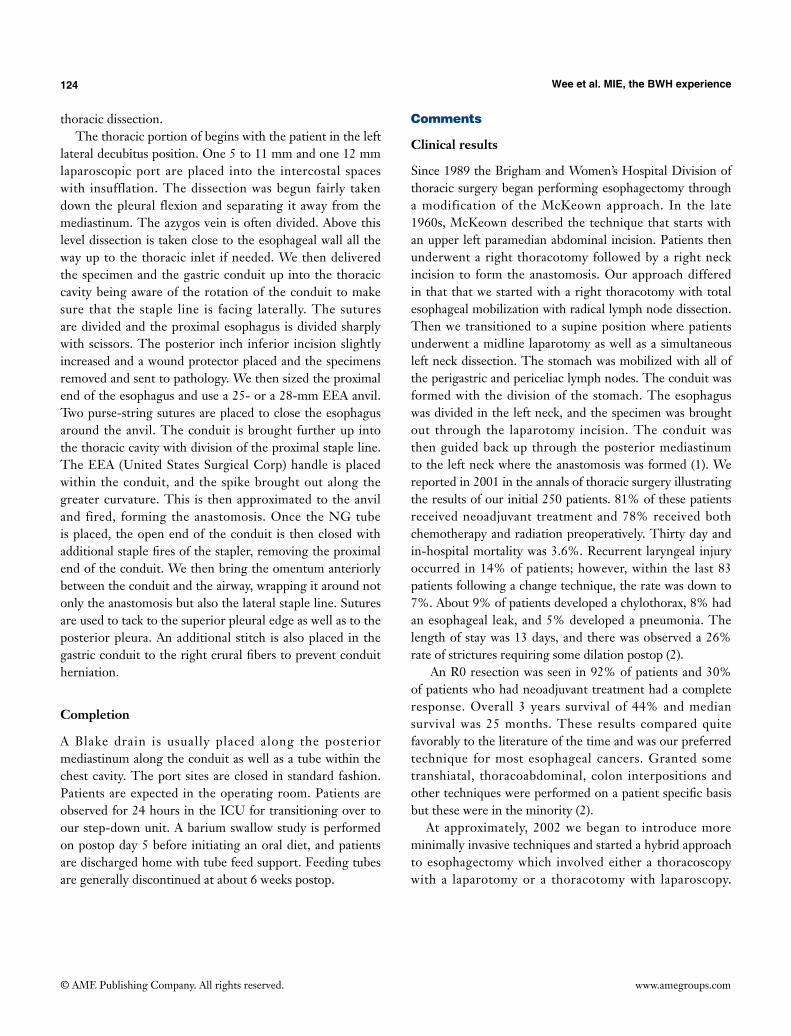
124
© AME Publishing Company. All rights reserved. www.amegroups.com
Wee et al. MIE, the BWH experience
thoracic dissection.The thoracic portion of begins with the patient in the left
lateral decubitus position. One 5 to 11 mm and one 12 mm laparoscopic port are placed into the intercostal spaces with insufflation. The dissection was begun fairly taken down the pleural flexion and separating it away from the mediastinum. The azygos vein is often divided. Above this level dissection is taken close to the esophageal wall all the way up to the thoracic inlet if needed. We then delivered the specimen and the gastric conduit up into the thoracic cavity being aware of the rotation of the conduit to make sure that the staple line is facing laterally. The sutures are divided and the proximal esophagus is divided sharply with scissors. The posterior inch inferior incision slightly increased and a wound protector placed and the specimens removed and sent to pathology. We then sized the proximal end of the esophagus and use a 25- or a 28-mm EEA anvil. Two purse-string sutures are placed to close the esophagus around the anvil. The conduit is brought further up into the thoracic cavity with division of the proximal staple line. The EEA (United States Surgical Corp) handle is placed within the conduit, and the spike brought out along the greater curvature. This is then approximated to the anvil and fired, forming the anastomosis. Once the NG tube is placed, the open end of the conduit is then closed with additional staple fires of the stapler, removing the proximal end of the conduit. We then bring the omentum anteriorly between the conduit and the airway, wrapping it around not only the anastomosis but also the lateral staple line. Sutures are used to tack to the superior pleural edge as well as to the posterior pleura. An additional stitch is also placed in the gastric conduit to the right crural fibers to prevent conduit herniation.
Completion
A Blake drain is usually placed along the posterior mediastinum along the conduit as well as a tube within the chest cavity. The port sites are closed in standard fashion. Patients are expected in the operating room. Patients are observed for 24 hours in the ICU for transitioning over to our step-down unit. A barium swallow study is performed on postop day 5 before initiating an oral diet, and patients are discharged home with tube feed support. Feeding tubes are generally discontinued at about 6 weeks postop.
Comments
Clinical results
Since 1989 the Brigham and Women’s Hospital Division of thoracic surgery began performing esophagectomy through a modification of the McKeown approach. In the late 1960s, McKeown described the technique that starts with an upper left paramedian abdominal incision. Patients then underwent a right thoracotomy followed by a right neck incision to form the anastomosis. Our approach differed in that that we started with a right thoracotomy with total esophageal mobilization with radical lymph node dissection. Then we transitioned to a supine position where patients underwent a midline laparotomy as well as a simultaneous left neck dissection. The stomach was mobilized with all of the perigastric and periceliac lymph nodes. The conduit was formed with the division of the stomach. The esophagus was divided in the left neck, and the specimen was brought out through the laparotomy incision. The conduit was then guided back up through the posterior mediastinum to the left neck where the anastomosis was formed (1). We reported in 2001 in the annals of thoracic surgery illustrating the results of our initial 250 patients. 81% of these patients received neoadjuvant treatment and 78% received both chemotherapy and radiation preoperatively. Thirty day and in-hospital mortality was 3.6%. Recurrent laryngeal injury occurred in 14% of patients; however, within the last 83 patients following a change technique, the rate was down to 7%. About 9% of patients developed a chylothorax, 8% had an esophageal leak, and 5% developed a pneumonia. The length of stay was 13 days, and there was observed a 26% rate of strictures requiring some dilation postop (2).
An R0 resection was seen in 92% of patients and 30% of patients who had neoadjuvant treatment had a complete response. Overall 3 years survival of 44% and median survival was 25 months. These results compared quite favorably to the literature of the time and was our preferred technique for most esophageal cancers. Granted some transhiatal, thoracoabdominal, colon interpositions and other techniques were performed on a patient specific basis but these were in the minority (2).
At approximately, 2002 we began to introduce more minimally invasive techniques and started a hybrid approach to esophagectomy which involved either a thoracoscopy with a laparotomy or a thoracotomy with laparoscopy.

125Minimally Invasive Esophagectomy
© AME Publishing Company. All rights reserved. www.amegroups.com
This allowed a transition to a fully minimally invasive esophagectomy which began in 2006. We began to perform a fully port based thoracoscopy with laparoscopy minimally invasive esophagectomy. By 2008 we had performed 34 open esophagectomy, 17 hybrid esophagectomies, and 17 completely minimally invasive esophagectomy for that calendar year. By 2012, the transition continued wi th decreas ing open approaches and increased minimally invasive approaches. We performed 28 open esophagectomies, 21 hybrid approaches and 53 completely minimally invasive approaches in 2012. In total, as of 2012, we had performed 475 open, 153 hybrid and 200 completely minimally invasive approaches.
We also had a mix in our minimally invasive approaches. Initially, most of our esophagectomies were minimally invasive 3-hole esophagectomy uti l iz ing either a thoracoscopy or laparoscopy in the hybrid approach, finishing with a neck anastomosis. Since 2006, we began to perform more minimally invasive Ivor Lewis Esophagectomy starting with a laparoscopy and finishing with a thoracoscopic chest anastomosis as tumor location near the GE junction allowed adequate margins in the chest as well as having a desire to avoid a neck dissection and risk recurrent nerve injury when reasonable.
Our rate of conversion from a hybrid or minimally invasive to an open procedure was approximately 15%, but this decreased as our experience increased. The most common reason for conversion was due to the learning curve and surgeon comfort and not due to an unforeseen complication. Dealing with adhesions and adhesiolysis was the most common reason. The operative length was the longest for these converted cases at 462 minutes whereas the planned open procedure remained the fastest at 334 minutes. However, there was less blood loss in the completely minimally invasive group compared to the open (300 vs. 450 cc) and a day shorter length of stay in both the ICU and the hospital for the MIE group. Overall morbidity was not significantly different between the groups though there was significantly less pulmonary embolisms (12.4% vs. 4.5%, P=0.001) for the MIE, but also a higher stricture rate requiring dilation (11% vs. 4.8 %, P=0.009). Overall mortality remained the same with 0.8% 30 day mortality and 4% 90 day mortality in the open group compared to 0.5% 30 day and 2.5 % 90 day mortality in the MIE group. Overall 73% of cases were performed completely minimally invasively by 2012.
From May 2000 and to June 2012, 123 patients underwent a 3-hole minimally invasive approach and 77 patients
underwent a minimally invasive Ivor Lewis approach. Within the 3-hole minimally invasive group, 63% of patients received neoadjuvant chemoradiotherapy followed by surgery. The 30 day mortality for the entire cohort was 0.8% and with a 90 day mortality of 3%. There was no difference in complications in those who had neoadjuvant chemoradiotherapy compared to those who had primary surgical resection. The rates of anastomotic leak, strictures requiring dilation, vocal cord injury, and chylothorax were all not specifically different (3).
In 2013 we began our robotic esophagectomy program utilizing the DaVinci robot for thoracic mobilization and creation of the anastomosis. Our initial experience of 20 patients demonstrated 0% 90 day mortality. 80% of patients had neoadjuvant chemoradiotherapy. Rate of conversion was 0. Morbidity was seen in 55% of patients, mostly from atrial fibrillation at 15%. Length of stay was 8 days, and 85% of patients were discharged to home. Overall operative time was long at 455 minutes, but this may be related to the learning curve (4).
Currently, the vast majority of esophagectomies performed at Brigham and Women’s hospital are performed in a minimally invasive fashion. There is a diversity in approaches from a 3-hole modified McKweon minimally invasive approach to a minimally invasive Ivor Lewis , and a robotic assisted minimally invasive esophagectomy. The transition from the open technique to these minimally invasive techniques has resulted in decreased length of stay, decreased morbidity, and improved patient outcomes and satisfaction. This transitioned occurred with practicing established thoracic surgeons who were comfortable with the open approach. The fact that these surgeons were able to adapt new minimally invasive techniques and establish expertise is a testament to the commitment to improve outcomes and advance the care of patients.
Acknowledgements
None.
Footnote
Conflicts of Interest: The authors have no conflicts of interest to declare.
References
1. Swanson SJ, Sugarbaker DJ. The three-hole

126
© AME Publishing Company. All rights reserved. www.amegroups.com
Wee et al. MIE, the BWH experience
esophagectomy. The Brigham and Women's Hospital approach (modified McKeown technique). Chest Surg Clin N Am 2000;10:531-52.
2. Swanson SJ, Batirel HF, Bueno R, et al. Transthoracic esophagectomy with radical mediastinal and abdominal lymph node dissection and cervical esophagogastrostomy for esophageal carcinoma. Ann Thorac Surg 2001;72:1918-24; discussion 1924-5.
3. Spector R, Zheng Y, Yeap BY, et al. The 3-Hole Minimally Invasive Esophagectomy: A Safe Procedure Following Neoadjuvant Chemotherapy and Radiation. Semin Thorac Cardiovasc Surg 2015;27:205-15.
4. Wee JO, Bravo-Iñiguez CE, Jaklitsch MT. Early Experience of Robot-Assisted Esophagectomy With Circular End-to-End Stapled Anastomosis. Ann Thorac Surg 2016;102:253-9.
Cite this article as: Wee JO, Bueno R, Swanson SJ. Minimally invasive esophagectomy: the Brigham and Women’s Hospital experience. Ann Cardiothorac Surg 2017;6(2):175-178. doi: 10.21037/acs.2017.03.13

© AME Publishing Company. All rights reserved. www.amegroups.com
Introduction
Esophageal cancer ranks eight among the most common malignancies and the incidence is rapidly increasing in the Western world at a rate greater than any other type of solid tumor. Esophagectomy represents the current standard of care for patients with localized esophageal carcinoma staged as T1sm/N+ or higher. A protocol of multidisciplinary treatment is generally recommended in patients with locally advanced tumors, especially in those with squamous-cell carcinoma (1). Esophagectomy is a complex surgical procedure that requires a two or three-field access depending on preoperative clinical staging, location and histology of the tumor, comorbidity, and patient’s anatomy and physiological status. Although the current postoperative mortality has decreased to less than 5% in high-volume centers, complications related to anastomotic and respiratory
failure are still significant and appear to be independent of the surgical approach and the anastomotic technique. The overall morbidity of the operation has not been significantly reduced over the past few decades, even with the trans-hiatal approach, indicating that the pathogenesis of complications associated with esophagectomy is multifactorial and not entirely dependent on the surgical access and length of the skin incisions. Minimally invasive surgery was introduced in the 90s’ with the aim to decrease the rate of respiratory complications associated with the thoracotomy approach. A number of hybrid and total minimally invasive surgical approaches have been developed and are currently applied in several centers worldwide. The most commonly performed hybrid procedure is a modification of the classic 2-stage Ivor Lewis operation, in which the laparotomy is replaced by laparoscopy for gastric conduit preparation and celiac lymphadenectomy. The total minimally invasive
Hybrid and total minimally invasive esophagectomy: how I do it
Luigi Bonavina, Emanuele Asti, Andrea Sironi, Daniele Bernardi, Alberto Aiolfi
Division of General Surgery, Department of Biomedical Sciences for Health, University of Milan, IRCCS Policlinico San Donato, Milan, Italy
Contributions: (I) Conception and design: L Bonavina; (II) Administrative support: D Bernardi, A Sironi, A Aiolfi; (III) Provision of study materials
or patients: L Bonavina, E Asti; (IV) Collection and assembly of data: D Bernardi, A Sironi, A Aiolfi, E Asti; (V) Data analysis and interpretation: L
Bonavina; (VI) Manuscript writing: All authors; (VII) Final approval of manuscript: All authors.
Correspondence to: Prof. Luigi Bonavina. Divisione Universitaria di Chirurgia, IRCCS Policlinico San Donato, Via Morandi 30, San Donato Milanese
20097, Milano, Italy. Email: [email protected].
Abstract: Esophagectomy is a major surgical procedure associated with a significant risk of morbidity and mortality. Minimally invasive esophagectomy is becoming the preferred approach because of the potential to limit surgical trauma, reduce respiratory complications, and promote earlier functional recovery. Various hybrid and total minimally invasive surgical techniques have been introduced in clinical practice over the past 20 years, and minimally invasive esophagectomy has been shown equivalent to open surgery concerning the short-term outcomes. Implementation of a minimally invasive esophagectomy program is technically demanding and requires a significant learning curve and the infrastructure of a dedicated multidisciplinary center where optimal staging, individualized therapy, and perioperative care can be provided to the patient. Both hybrid and total minimally invasive techniques of esophagectomy have proven safe and effective in expert centers. The choice of the surgical approach should be driven by preoperative staging, tumor site and histology, comorbidity, patient’s anatomy and physiological status, and surgeon’s experience.
Keywords: Esophageal cancer; laparoscopy; minimally invasive esophagectomy; thoracoscopic prone
esophagectomy; hybrid Ivor Lewis esophagectomy; trans-hiatal esophagectomy
Submitted Apr 18, 2017. Accepted for publication Jun 07, 2017.
doi: 10.21037/jtd.2017.06.55
View this article at: http://dx.doi.org/10.21037/jtd.2017.06.55
138
Thoracoscopic Esophagectomy

128
© AME Publishing Company. All rights reserved. www.amegroups.com
Bonavina et al. Minimally invasive esophagectomy
esophagectomy includes two techniques: the 3-stage thoraco-laparoscopic esophagectomy, a modification of the classic McKeown operation in which right thoracotomy and laparotomy are replaced by thoracoscopy and laparoscopy, and the minimally invasive trans-hiatal esophagectomy, a modification of the classic 2-stage trans-hiatal esophagectomy, in which the laparotomy is replaced by laparoscopy (Figure 1).
Patient selection and surgical strategy
Patients with esophageal carcinoma require an extensive preoperative staging, including CT scan and/or endoscopic ultrasonography, flexible bronchoscopy, and PET scan when appropriate, to exclude locally advanced or metastatic disease. Assessment of the functional, nutritional and comorbidity status is necessary before considering the patient for immediate surgery or for a multimodality treatment plan determined in a multidisciplinary oncological team meeting (2). Preparation for surgery should include abstinence from smoking, daily walking activity, use of an incentive spirometer, and a 1-week preoperative enteral nutritional support in patients with long-lasting dysphagia, significant weight loss, and a pre-frail or frail phenotype.
Preoperative staging and tumor characteristics influence the choice of the surgical strategy, i.e., a 2-stage or a 3-stage procedure. In some circumstances, starting with laparoscopy or thoracoscopy may be useful to provide the ultimate staging. Initial laparoscopic approach for gastric conduit preparation, as part of a hybrid or total minimally invasive Ivor Lewis operation, is feasible in the majority of
patients with esophageal adenocarcinoma (3). When tumor resectability is doubtful, as in patients with squamous-cell cancer of the middle-upper thoracic esophagus after neoadjuvant therapy, a primary thoracoscopy/thoracotomy as part of a hybrid or total minimally invasive McKeown approach may be more appropriate. However, to avoid surprises during the subsequent phase of the operation, the presence of peritoneal carcinosis, tumor involvement of the gastric fundus, liver metastases, and important comorbidities such as liver cirrhosis with portal hypertension, should be ruled out beforehand (4). Anatomical factors such as a short “bull” neck may suggest to avoid a neck anastomosis if not strictly necessary. In some circumstances, following an initial thoracic approach, a decision can be made to return into the chest for the anastomosis after the gastric conduit has been prepared by laparoscopy.
From a surgical and oncological standpoint, the ideal candidate for a primary thoracic approach is a patient with a clinically staged T1–3 squamous-cell carcinoma of the upper/middle thoracic esophagus. Initial thoracoscopy may be an option also in patients with type I esophageal adenocarcinoma, especially in those with extra-long Barrett’s esophagus. A narrow upper mediastinum and the presence of spine abnormalities such as dorsal kyphosis, scoliosis, and vertebral osteophytosis, may represent a relative contraindication to the thoracoscopic approach in the prone position because of the technical difficulties to access the esophagus hidden by the vertebral bodies, and the altered anatomical relationships with the aorta and the tracheobronchial tree (5). In these circumstances, the semi-prone position has some advantages over the prone position.
Surgical approach for minimally invasive esophagectomy
Hybrid Total
Ivor Lewis(laparoscopy + thoracotomy)
Chest anastomosis
Mc Keown(thoracoscopy +
laparotomy +cervicotomy)
Neck anastomosis
Ivor Lewis(laparoscopy + thoracoscopy)
Chest anastomosis
Mc Keown(thoracoscopy +laparoscopy +cervicotomy)
Neck anastomosis
Orringer(laparoscopy + cervicotomy)
Neck anastomosis
Figure 1 Approach and site of anastomosis according to the type of minimally invasive esophagectomy.

129Minimally Invasive Esophagectomy
© AME Publishing Company. All rights reserved. www.amegroups.com
Surgical procedures
Hybrid Ivor Lewis esophagectomy
The operation consists of a 2-stage approach (laparoscopy + right thoracotomy). The laparoscopic phase is performed with the patient placed in a reverse Trendelenburg position. After induction of pneumoperitoneum with the Veress needle, five 5–12 mm ports are placed (Figure 2). Dissection is performed using the hook cautery and ultrasonic scissors beginning with division of the gastrohepatic ligament. The stomach is mobilized by dividing the left gastric vessels (Figure 3) and short gastrics, and separating the
right gastroepiploic arcade from the gastrocolic omentum. A standard D2-lymphadenectomy is performed. A 4-cm wide gastric conduit is constructed by sequential firings of 45–60 mm Endo-GIA® (Medtronic) cartridges parallel to the greater curvature. The first 45 mm cartridge is applied across the lesser curve cranial to the third branch of the right gastric artery and is directed almost at right angle toward the greater curve; special care is required to maintain a consistent width of the stomach and to avoid spiralization of the gastric tube during application of the subsequent cartridges (Figure 4). Interrupted 4-0 PDS stitches are applied at the intersection of the staple lines. To prevent diaphragmatic hernia, a posterior suture of the crura is placed and left temporarily untied into the lower mediastinum. A 15 Fr Blake® drain (Ethicon) is placed in the mediastinum through the upper abdominal port and then through the hiatus. For the thoracotomy phase of the operation, the right lung is excluded using a left double-lumen tube or an endobronchial blocker under fiberoptic bronchoscopic guidance, and the patients is turned to the left lateral position with a roll at the level of the tip of the scapula. A right postero-lateral incision sparing the serratus muscle is performed in the fifth intercostal space and the lung is retracted medially. The arch of the azygos vein is divided with Hemolock clips and the thoracic duct is selectively ligated above the diaphragm. A standard infracarinal lymphadenectomy is performed in patients with adenocarcinoma, whereas paratracheal nodes are routinely removed only in squamous-cell carcinoma. The esophago-gastric anastomosis is performed at the apex of the right chest using a 25 mm EEA® stapler (Medtronic) (Figure 5). The pleural cavity is drained by the trans-abdominal Blake drain.
Figure 2 Laparoscopic set-up for gastric mobilization and celiac lymphadenectomy.
Figure 3 Laparoscopic division of the left gastric artery between Hemolock clips.
Figure 4 Laparoscopic gastric tubulisation.

130
© AME Publishing Company. All rights reserved. www.amegroups.com
Bonavina et al. Minimally invasive esophagectomy
Total minimally invasive Ivor Lewis esophagectomy
The laparoscopic stage of the procedure, including lymphadenectomy and gastric mobilization, is similar to that of the hybrid operation. Interrupted 4-0 PDS stitches applied at the intersection of the staple lines along the lesser curve and the apex of greater curve are of utmost importance to help retrieval and proper orientation of the gastric tube from the chest cavity during the thoracoscopic stage. The patient is placed in the semi-prone position with the right arm positioned on a support device and the forearm flexed to improve abduction of the scapula. The chest is stabilized on the operative table using bean bag and side supports to allow rotation in a more lateral decubitus position (Figure 6).
This is helpful to aid mediastinal exposure in patients with a protruding spine or to expedite the switch to thoracotomy if necessary. After exclusion of the right lung and induction of pneumothorax with a Veress needle in the posterior axillary line, three trocars (two 12 mm and one 5 mm) are placed in the fourth, sixth and eighth intercostal space. The arch of the azygos vein is divided using Hemolock clips or a vascular EndoGIA stapler. Incision of the mediastinal pleura is performed on both sides of the esophagus, and the dissection preferably starts between the vagal trunk and the right main bronchus. This allows en-bloc lymphadenectomy of the carina with nerve preservation in most circumstances. The esophagus is then mobilized up to the level of the diaphragm and the inferior pulmonary ligament is divided. The thoracic duct is identified and ligated with a single Hemolock® clip (Figure 7). The 25 mm anvil of a circular stapler (Orvil®, Medtronic) can be inserted trans-orally and retrieved through a small hole close to the stapled line of the esophageal stump
Figure 5 Trans-thoracic esophago-gastric anastomosis. (A) Circular stapler introduced into the gastric tube through a gastrotomy at the apex of the lesser curve; (B) the anastomosis as viewed through the gastrotomy site.
A B
Figure 6 Semi-prone patient positioning with a typical 45° angle.
Figure 7 The thoracic duct is secured with Hemolock clip at the level of the diaphragm.

131Minimally Invasive Esophagectomy
© AME Publishing Company. All rights reserved. www.amegroups.com
(Figure 8). Another option is to insert the anvil attached to a 2-0 polypropylene suture in the esophageal lumen through an esophagotomy; the needle is then retrieved by reverse puncture of the anterior esophageal wall and, once the anvil rod is out, the esophagus is divided with a linear stapler (Figure 9). At this point, the incision corresponding to the lowermost trocar is enlarged to 5 cm in length toward the
anterior axillary line and a wound retractor device (Alexis®, Applied Medical) is inserted. The distal end of the circular stapler, enveloped in a surgical glove through a small cut in the middle finger, is inserted through the mini-thoracotomy wound with minimal dilatation of the intercostal space. The surgical glove, by adhering to the borders of the wound retractor, allows to maintain the pneumothorax during the
Figure 8 Thoracoscopic anastomosis using the ORVIL device introduced trans-orally. (A) The tube connected with the anvil is retrieved through a small incision above the stapled esophageal stump; (B) the tube is disconnected from the anvil; (C) the base of the anvil rod is grasped; (D) the anvil is engaged with the stapler.
Figure 9 Alternative thoracoscopic anastomosis. (A) The anvil connected to a prolene suture is inserted in the lumen through an esophagotomy; (B) reverse needle puncture of the anterior esophageal wall (arrow); (C) the esophagus is stapled below the anvil; (D) esophago-gastric anastomosis in progress.
A
C
B
D
A
C
B
D

132
© AME Publishing Company. All rights reserved. www.amegroups.com
Bonavina et al. Minimally invasive esophagectomy
anastomosis. The stapler is then advanced through a small gastrotomy on the lesser curve side, perforates the tip of the greater curve and engages the anvil (Figure 10). After checking the donoughts, transection of the excess stomach with a linear stapler is performed. The specimen is placed in
an endobag and extracted from the chest cavity through the minithoracotomy (Figure 11).
Hybrid and total minimally invasive McKeown esophagectomy
The operation consists of a 3-stage esophagectomy with cervical anastomosis. Cuschieri first reported a thoracoscopic esophageal mobilization with the patient in the prone position in 1992 (6). The thoracoscopic approach was later adopted by other surgeons with the patient in the left-lateral decubitus (7). The prone position was revisited and popularized by Palanivelu in 2006 (8). A number of reports have subsequently evaluated this procedure and it appears that the prone position has some advantages over the left-lateral decubitus position (9). Thoracoscopic esophagectomy in the prone position is performed after induction of anesthesia with a single-lumen tracheal tube. The patient is then turned to the prone position, and chest and pelvic supports to leave the abdomen free for breathing excursions. A special headrest support with an integrated mirror (Disposa-View®) allows the anesthesiologist to check the position of the tube. Collapse of the right lung is obtained by CO2 insufflation through a Veress needle. Three ports are placed in the fourth, sixth and eighth intercostal space (Figure 12). Gas insufflation
Figure 10 Operative field during the thoracoscopic part of total minimally invasive Ivor Lewis esophagectomy. (A) Thoracoscopic set-up; (B) the ring of the Alexis device seen from inside the chest; (C) mini-thoracotomy ready for stapler insertion; (D) the stapler is enveloped in a surgical glove to maintain pneumothorax.
A
C
B
D
Figure 11 Wound scars after total minimally invasive Ivor Lewis esophagectomy.
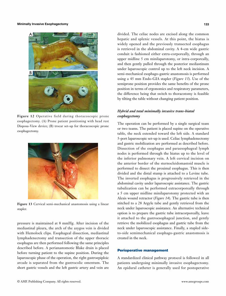
133Minimally Invasive Esophagectomy
© AME Publishing Company. All rights reserved. www.amegroups.com
pressure is maintained at 8 mmHg. After incision of the mediastinal pleura, the arch of the azygos vein is divided with Hemolock clips. Esophageal dissection, mediastinal lymphadenectomy and transection of the upper thoracic esophagus are then performed following the same principles described before. A perianastomotic Blake drain is placed before turning patient to the supine position. During the laparoscopic phase of the operation, the right gastroepiploic arcade is separated from the gastrocolic omentum. The short gastric vessels and the left gastric artery and vein are
divided. The celiac nodes are excised along the common hepatic and splenic vessels. At this point, the hiatus is widely opened and the previously transected esophagus is retrieved in the abdominal cavity. A 4-cm wide gastric conduit is fashioned either extra-corporeally, through an upper midline 5 cm minilaparotomy, or intra-corporeally, and then gently pulled through the posterior mediastinum under laparoscopic control up to the left neck incision. A semi-mechanical esophago-gastric anastomosis is performed using a 45 mm Endo-GIA stapler (Figure 13). Use of the semiprone position provides the same benefits of the prone position in terms of ergonomics and respiratory parameters, the difference being that switch to thoracotomy is feasible by tilting the table without changing patient position.
Hybrid and total minimally invasive trans-hiatal esophagectomy
The operation can be performed by a single surgical team or two teams. The patient is placed supine on the operative table, the neck extended toward the left side. A standard 5-port laparoscopic set-up is used. Celiac lymphadenectomy and gastric mobilization are performed as described before. Dissection of the esophagus and paraesophageal lymph nodes is performed through the hiatus up to the level of the inferior pulmonary vein. A left cervical incision on the anterior border of the sternocleidomastoid muscle is performed to dissect the proximal esophagus. This is then divided and the distal stump is attached to a Levine tube. The inverted esophagus is progressively retrieved in the abdominal cavity under laparoscopic assistance. The gastric tubulization can be performed extracorporeally through a 5 cm upper midline minilaparotomy protected with an Alexis wound retractor (Figure 14). The gastric tube is then stitched to a 28 Argyle tube and gently retrieved from the neck under laparoscopic assistance. An alternative technical option is to prepare the gastric tube intracorporeally, leave it attached to the gastroesophageal junction, and gently retrieve the mobilized esophagus and gastric tube from the neck under laparoscopic assistance. Finally, a stapled side-to-side semimechanical esophago-gastric anastomosis is created in the neck.
Perioperative management
A standardized clinical pathway protocol is followed in all patients undergoing minimally invasive esophagectomy. An epidural catheter is generally used for postoperative
Figure 12 Operative f ield during thoracoscopic prone esophagectomy. (A) Prone patient positioning with head rest Disposa-View device; (B) trocar set-up for thoracoscopic prone esophagectomy.
Figure 13 Cervical semi-mechanical anastomosis using a linear stapler.
A
B

134
© AME Publishing Company. All rights reserved. www.amegroups.com
Bonavina et al. Minimally invasive esophagectomy
analgesia. In selected patients with contraindications to epidural analgesia, a serratus anterior plane block is performed (10). Antibiotic prophylaxis with Cefazolin is given. An arterial line is routinely placed for blood pressure monitoring. Two large-bore intravenous lines are adequate, and a central line is rarely needed. Intraoperative normothermia is maintained using a warm air blanket and avoiding use of vasoconstrictor agents. The volume of fluid administration is restricted to a maximum of 2 L. Patients are usually extubated in the operating room and transferred to the intensive care unit. Pain management consists of levobupivacaine through the epidural catheter combined with intravenous paracetamol as needed. Nasogastric aspiration is maintained during the first 48 hours. Patients are allowed to ambulate and to begin pulmonary physiotherapy with an incentive spirometer on postoperative day one. Water sips and fruit jelly are allowed on day 3, and then the diet is gradually progressed. A gastrografin swallow study is routinely performed on day 5 or 6. Patients are discharged from the hospital when the following criteria are met: no laboratory or clinical evidence of infection, ability to fully ambulate without assistance, no major analgesic requirements, oral diet well tolerated without significant gastrointestinal discomfort.
Discussion
Incremental steps of innovation in esophageal surgery
have resulted in reduced postoperative mortality and improved oncological outcomes. With the advent of minimally invasive surgery, pros and cons of 2-stage and 3-stage procedures have been critically revisited. A recent survey has found a worldwide increase in the adoption of minimally invasive esophagectomy and a rise of high-volume centers. However, differences still exist regarding the extent of nodal dissection and site of anastomosis. The most favoured approach remains the minimally invasive McKeown operation followed by the minimally invasive Ivor Lewis operation. The preference for the transhiatal esophagectomy has decreased from 26% in 2007 to 15% in 2014 (11).
Today, the minimally invasive esophagectomy can be performed with minimal blood loss, controlled pain, and reduced intensive care unit stay and pulmonary complications compared to the open procedure. Short-term outcomes of minimally invasive esophagectomy have proven at least equivalent to the open approach in meta-analyses (12,13), a large administrative national database (14), and a randomized clinical trial (15). In addition, minimally invasive esophagectomy has been associated with a rapid restoration of health-related quality of life (16,17).
Both the hybrid and the total minimally invasive 2-stage and 3-stage esophagectomy are included in the definition of minimally invasive esophagectomy. The hybrid Ivor Lewis operation has been shown to have a reasonable learning curve and to be reproducible (18-20). Whether the laparoscopic component of the operation will decrease the major complication rate in esophageal cancer surgery has not been completely clarified yet. A French nationwide study has shown that laparoscopic gastric mobilization as a part of the hybrid esophagectomy significantly reduced postoperative mortality both at 30 and 90 days (21). Preliminary results of the Miro trial show a reduction in severe complications and major pulmonary complications without a negative impact on oncological outcomes and a trend toward better survival (22). On the other hand, Briez and colleagues (23) found that the hybrid Ivor Lewis approach was an independent factor protecting against major pulmonary complications when compared to open surgery. This may be related to the fact that laparoscopy can mitigate the mechanical and immunological stress associated with one lung ventilation and left lateral decubitus position. A total minimally invasive Ivor Lewis approach with thoracoscopic anastomosis is now preferred, but its use is not widespread due to the difficulties in performing the anastomosis. The increased prevalence of adenocarcinoma justifies the efforts to adopt the total minimally invasive
Figure 14 Extra-corporeal gastric tube formation after laparoscopic trans-hiatal esophagectomy.

135Minimally Invasive Esophagectomy
© AME Publishing Company. All rights reserved. www.amegroups.com
Ivor Lewis approach in the future despite the superior technical difficulties and the steep learning curve. It has been estimated that a reasonable learning curve for the total minimally invasive Ivor Lewis esophagectomy would require 35–40 patients to achieve improved results (24). The technique of intrathoracic anastomosis needs to be standardized and more data are needed to assess the efficacy of the various proposed methods. The results of the intrathoracic stapled anastomosis appear encouraging, but no single technique has proven superior to the others (25). Pooled data including 282 patients from 6 European centers showed a 15.2% incidence of anastomotic leakage. Only 13 patients (4.6%) had pleural empyema requiring thoracotomy for decortication, and the 30-day and in-hospital mortality rate was 2.1%. A R-0 resection was obtained in 92.5% of patients (26).
Concerning the 3-stage McKeown esophagectomy, the adoption of the prone and semiprone position has represented a major advance in the performance of the thoracoscopic phase of the operation. This approach has allowed 2-lung ventilation, further reduction of operative trauma, and improved surgical ergonomics compared to the left lateral decubitus (4,9,27). The TIME trial has provided evidence that thoracoscopic prone esophagectomy is associated with a lower incidence of in-hospital pulmonary infections and a shorter hospital stay compared to the open approach (15). More recently, there has been a shift in western countries from the 3-stage operation in favor of the total minimally invasive Ivor Lewis approach. This has been attributed to an increased referral of patients with esophageal adenocarcinoma and an effort to reduce the morbidity associated with recurrent nerve injuries (28). The 3-stage approach requires a cervical anastomosis and is also more time-consuming. Although the mean surgical time for the thoracoscopic esophagectomy is about one hour and is significantly reduced in the prone position compared to the left lateral decubitus (9), prone positioning and then repositioning requires extra-time in the operating room and at least 6 staff members including the anesthesiologist. Avoiding the thoracotomy incision and releasing the right chest and abdomen from compression during the prone position provides better oxygen delivery, decreases the pulmonary shunt, and improves the ventilation perfusion match (27). This could in turn decrease the incidence of respiratory (15) and anastomotic (29) complications. In addition, preservation of two-lung ventilation can significantly reduce the ischemia-reperfusion injury and the oxidative stress (30). In the future, the role of robot-assisted
esophagectomy may increase due to the benefits of a stable three-dimensional image and more precise dissection by avoiding the fulcrum effect of the instruments at the ribs and their parallel approach at the thoracic inlet and toward the diaphragm. In addition, robot-assisted surgery may enable to perform a safer manual anastomosis. A randomized trial comparing open and robot-assisted esophagectomy is ongoing (31). Common reservations about the safety of 3-stage esophagectomy (recurrent nerve injury, gastric conduit necrosis and twisting, “catastrophic” anastomotic leakage) have not been confirmed in our experience. The semimechanical anastomosis in the neck was as safe as a circular anastomosis at the apex of the chest (4), confirming the fact that neck and chest anastomoses are equally safe when performed in a standardized way (32).
Laparoscopic transhiatal esophagectomy, a revisitation of the procedure popularized in the 70s’ by Orringer and Sloan (33), was first described by DePaula et al. in 1995 (34). Later, a transhiatal esophagectomy through transcervical video-assisted mediastinoscopy combined with laparoscopy was reported (35). A recent systematic review has reported that the laparoscopic transhiatal esophagectomy is associated with a lower median blood loss and a shorter hospital stay compared to the open approach (36). However, this operation has become less popular over the past decade and has been largely replaced by the trans-thoracic approach which also provides an oncological advantage in patients with adenocarcinoma (37).
The efficacy of minimally invasive esophagectomy has been demonstrated even in patients treated with neoadjuvant chemoradiotherapy (15,38); however, in patients with advanced bulky tumors before treatment and in those who are candidates to salvage surgery the indication to minimally invasive surgery should be prudent. Radiation damage to the gastric fundus can increase the risk of anastomotic leakage in these patients (39). Microperfusion assessment with indocyanine green fluorescence angiography (Figure 15) may help to establish the best site for the anastomosis (40,41).
From the oncological standpoint, the question whether the minimally invasive techniques represent a viable alternative to the open procedures remains unanswered due to the heterogeneity of the procedures and the lack of long-term data. One meta-analysis found a that the statistically significant increase in lymph node yield associated with minimally invasive esophagectomy did not translate into a survival benefit as no difference was found in 1-, 2-, 3- and 5-year survival rates (42). In a prospective phase II multicenter trial including 104 patients in 17 centers, the

136
© AME Publishing Company. All rights reserved. www.amegroups.com
Bonavina et al. Minimally invasive esophagectomy
3-year overall survival and recurrence rate were 68% and 33.8%, respectively, following both 3-stage and 2-stage total minimally invasive esophagectomy (43). The most recent 3-year results of the TIME trial showed that the disease-free survival was 35.9% in the open versus 40.2% in the minimally invasive group (44).
Conclusions
Over the past three decades, a marked decrease in postoperative mortality due to better patient selection, improved perioperative care, and concentration of surgical procedures in high-volume centers has been observed. Transthoracic esophagectomy has emerged as the best surgical approach in fit patients. Minimally invasive esophagectomy has added value to this incremental progress by reducing postoperative pain and decreas ing the incidence of pulmonary complications.
Both the 3-stage and the 2-stage esophagectomy techniques, either hybrid or total minimally invasive, have proven safe and effective in expert centers and have led to a paradigm shift. Compared to the open surgical approach, the results of minimally invasive esophagectomy appear equivalent in terms of postoperative morbidity and mortality, node retrieval, completeness of resection, and early oncological results. Until further proof of effectiveness and generalizability is reached, the choice of the minimally invasive technique of esophagectomy depends on tumor location, histology, and surgeon’s experience and preference. The modern esophageal surgeon should continue to focus on performing a minimally invasive and maximally effective esophagectomy with low morbidity and mortality rates.
Acknowledgements
None.
Footnote
Conflicts of Interest: The authors have no conflicts of interest to declare.
References
1. Shapiro J, van Lanschot JJ, Hulshof MC, et al. Neoadjuvant chemoradiotherapy plus surgery versus surgery alone for oesophageal or junctional cancer (CROSS): long-term results of a randomized controlled trial. Lancet Oncol 2015;16:1090-98
2. Basta YL, Bolle S, Fockens P, et al. The value of multidisciplinary team meetings for patients with gastrointestinal malignancies: a systematic review. Ann Surg Oncol 2017. [Epub ahead of print].
3. Bonavina L, Incarbone R, Lattuada E, et al. Preoperative laparoscopy in management of patients with carcinoma of the esophagus and of the esophagogastric junction. J Surg Oncol 1997;65:171-4.
4. Bonavina L, Scolari F, Aiolfi A, et al. Early outcome of thoracoscopic and hybrid esophagectomy: propensity-matched comparative analysis. Surgery 2016;159:1073-81.
5. Okamura A, Watanabe M, Mine S, et al. Factors influencing difficulty of the thoracic procedure in minimally invasive esophagectomy. Surg Endosc 2016;30:4279-85.
6. Cuschieri A, Shimi S, Banting S. Endoscopic oesophagectomy through a right thoracoscopic approach. J R Coll Surg Edinb 1992;37:7-11.
7. Takeuchi H, Kawakubo H, Kitagawa Y. Current status of minimally invasive esophagectomy for patients with esophageal cancer. Gen Thorac Cardiovasc Surg 2013;61:513-21.
8. Palanivelu C, Prakash A, Senthilkumar R, et al. Minimally invasive esophagectomy: thoracoscopic mobilization of
Figure 15 Indocyanine green fluorescence angiography before the thoracoscopic anastomosis. The intramural vascular network is clearly visible both on the lesser curve side (A) and on the anastomotic site (B).
A B
Staple line
Anastomotic site
Gastroepiploic arcade

137Minimally Invasive Esophagectomy
© AME Publishing Company. All rights reserved. www.amegroups.com
the esophagus and mediastinal lymphadenectomy in prone position. Experience of 130 patients. J Am Coll Surg 2006;203:7-16.
9. Fabian T, Martin J, Katigbak M, et al. Thoracoscopic esophageal mobilization during minimally invasive esophagectomy: a head-to-head comparison of prone versus decubitus positions. Surg Endosc 2008;22:2485-91.
10. Barbera C, Milito P, Punturieri M, et al. Serratus anterior plane block for hybrid transthoracic esophagectomy: a pilot study. J Pain Res 2017;10:73-7.
11. Haverkamp L, Seesing MF, Ruurda JP, et al. Worldwide trends in surgical techniques in the treatment of esophageal and gastroesophageal junction cancer. Dis Esophagus 2017;30:1-7.
12. Nagpal K, Ahmed K, Vats A, et al. Is minimally invasive surgery beneficial in the management of esophageal cancer? A meta-analysis. Surg Endosc 2010;24:1621-9.
13. Sgourakis G, Gockel I, Radtke A, et al. Minimally invasive versus open esophagectomy: metaanalysis of outcomes. Dig Dis Sci 2010;55:3031-40.
14. Mamidanna R, Bottle A, Aylin P, et al. Short term outcomes following open versus minimally invasive esophagectomy for cancer in England: a population based national study. Ann Surg 2012;255:197-203.
15. Biere SS, van Berge Henegouwen MI, Maas KW, et al. Minimally invasive versus open oesophagectomy for patients with oesophageal cancer: a multicentre, open-label, randomised controlled trial. Lancet 2012;379:1887-92.
16. Parameswaran R, Blazeby JM, Hughes R, et al. Health-related quality of life after minimally invasive oesophagectomy. Br J Surg 2010;97:525-31.
17. Maas KW, Cuesta MA, van Berge Henegouwen MI, et al. Quality of Life and Late Complications After Minimally Invasive Compared to Open Esophagectomy: Results of a Randomized Trial. World J Surg 2015;39:1986-93.
18. Jagot P, Sauvanet A, Berthoux L, et al. Laparoscopic mobilization of the stomach for oesophageal replacement. Br J Surg 1996;83:540-2.
19. Bonavina L, Bona D, Binyom PR, et al. A laparoscopy-assisted surgical approach to esophageal carcinoma. J Surg Res 2004;117:52-7.
20. Bonavina L. Selected commentary to “Fifty-five minimally invasive esophagectomies: a single-center experience”. Eur Surg 2009;41:194-8.
21. Messager M, Pasquer A, Duhamel A, et al. Laparoscopic gastric mobilization reduces postoperative mortality after esophageal cancer surgery: a French nationwide study. Ann Surg 2015;262:817-22; discussion 822-3.
22. Briez N, Piessen G, Torres F, et al. Effects of hybrid minimally invasive oesophagectomy on major postoperative pulmonary complications. Br J Surg 2012;99:1547-53.
23. Briez N, Piessen G, Bonnetain F, et al. Open versus laparoscopically-assisted oesophagectomy for cancer: a multicentre randomised controlled phase III trial – the MIRO trial. BMC Cancer 2011;11:310.
24. Tapias LF, Morse CR. Minimally invasive Ivor Lewis esophagectomy: description of a learning curve. J Am Coll Surg 2014;218:1130-40.
25. Maas KW, Biere SS, Scheepers JJ, et al. Minimally invasive intrathoracic anastomosis after Ivor Lewis esophagectomy for cancer: a review of transoral or transthoracic use of staplers. Surg Endosc 2012;26:1795-802.
26. Straatman J, van der Wielen N, Nieuwenhuijzen G, et al. Techniques and short-term outcomes for total minimally invasive Ivor Lewis esophageal resection in distal esophageal and gastroesophageal cancers: pooled data from six European centers. Surg Endosc 2017;31:119-26.
27. Bonavina L, Laface L, Abate E, et al. Comparison of ventilation and cardiovascular parameters between prone thoracoscopic and Ivor Lewis esophagectomy. Updates Surg 2012;64:81-5.
28. Luketich JD, Pennathur A, Awais O, et al. Outcomes after minimally invasive esophagectomy. Review of over 1000 patients. Ann Surg 2012;256:95-103.
29. Kusano C, Baba M, Takao S, et al. Oxygen delivery as a factor in the development of fatal postoperative complications after oesophagectomy. Br J Surg 1997;84:252-7.
30. Cheng YJ, Chan KC, Chien CT, et al. Oxidative stress during 1-lung ventilation. J Thorac Cardiovasc Surg 2006;132:513-8.
31. van der Sluis PC, Ruurda JP, van der Horst S, et al. Robot-assisted minimally invasive thoraco-laparoscopic esophagectomy versus open transthoracic esophagectomy for resectable esophageal cancer, a randomized controlled trial (ROBOT trial). Trials 2012;13:230.
32. Walther B, Johansson J, Johnsson F, et al. Cervical or thoracic anastomosis after esophageal resection and gastric tube reconstruction. A prospective randomized trial comparing sutured neck anastomosis with stapled intrathoracic anastomosis. Ann Surg 2003;238:803-12.
33. Orringer MB, Sloan H. Esophagectomy without thoracotomy. J Thorac Cardiovasc Surg 1978;76:643-54.
34. DePaula AL, Hashiba K, Ferreira E, et al. Laparoscopic transhiatal esophagectomy with esophagogastroplasty. Surg Laparosc Endosc 1995;5:1-5.

138
© AME Publishing Company. All rights reserved. www.amegroups.com
Bonavina et al. Minimally invasive esophagectomy
35. Bonavina L, Incarbone R, Bona D, et al. Esophagectomy via laparoscopy and transmediastinal endodissection. J Laparoendosc Adv Surg Tech A 2004;14:13-6.
36. Parry K, Ruurda JP, van der Sluis PC, et al. Current status of laparoscopic transhiatal esophagectomy for esophageal cancer patients: a systematic review of the literature. Dis Esophagus 2017;30:1-7.
37. Omloo JM, Lagarde SM, Hulscher JB, et al. Extended transthoracic resection compared with limited transhiatal resection for adenocarcinoma of the mid/distal esophagus: five-year survival of a randomized clinical trial. Ann Surg 2007;246:992-1000.
38. Warner S, Chang YH, Paripati H, et al. Outcomes of minimally invasive esophagectomy in esophageal cancer after neoadjuvant chemoradiotherapy. Ann Thorac Surg 2014;97:439-45.
39. Goense L, van Rossum PS, Ruurda JP, et al. Radiation to the gastric fundus increases the risk of anastomotic leakage after esophagectomy. Ann Thorac Surg 2016;102;1798-804.
40. Zehetner J, DeMeester SR, Alicuben ET, et al.
Intraoperative assessment of perfusion of the gastric graft and correlation with anastomotic leaks after esophagectomy. Ann Surg 2015;262:74-8.
41. Yukaya T, Saeki H, Kasagi Y, et al. Indocyanine green fluorescence angiography for quantitative evaluation of gastric tube perfusion in patients undergoing esophagectomy. J Am Coll Surg 2015;221:e37-42.
42. Dantoc M, Cox MR, Eslick GD. Evidence to support the use of minimally invasive esophagectomy for esophageal cancer: a meta-analysis. Arch Surg 2012;147:768-76.
43. Luketich JD, Pennathur A, Franchetti Y, et al. Minimally invasive esophagectomy. Results of a prospective phase II multicenter trial-The Eastern cooperative oncology group (E2202) study. Ann Surg 2015;261:702-7.
44. Straatman J, van der Vielen N, Cuesta MA, et al. Minimally Invasive Versus Open Esophageal Resection: Three-year Follow-up of the Previously Reported Randomized Controlled Trial: the TIME Trial. Ann Surg 2017. [Epub ahead of print].
Cite this article as: Bonavina L, Asti E, Sironi A, Bernardi D, Aiolfi A. Hybrid and total minimally invasive esophagectomy: how I do it. J Thorac Dis 2017;9(Suppl 8):S761-S772. doi: 10.21037/jtd.2017.06.55

© AME Publishing Company. All rights reserved. www.amegroups.com
Minimally invasive (MI) esophageal mobilization for esophageal cancer is being performed for over two decades now. The technique was applied as a part of modified McKeown esophagectomy, which ended up with laparotomy and left cervical esophagogastrostomy (1). This technique is still used for tumors located at the level or higher than the carina. However in recent years, due to the disadvantages of a neck anastomosis (higher leak, stricture and recurrent laryngeal nerve injury rate), MI Ivor Lewis esophagectomy using a high thoracic anastomosis gained popularity (2). This was also evident in the practice of Dr. Luketich with a lower mortality (<2%) and leak (<5%) rate following a change of practice to MI Ivor Lewis esophagectomy (3).
Classical video-assisted thoracic surgery (VATS)
approach to esophageal cancer uses four incisions (4). The rationale for these incisions is to provide ease for anteroposterior movement of esophagus, angles for instruments and suturing especially during placement of a purse-string suture to esophagus for preparation of an intrathoracic anastomosis with circular stapler (4).
Uniportal VATS (U-VATS) presents a challenge for esophageal surgeons, as you have to do the same surgery from a single 3–5 cm incision. In this article we will describe the peculiarities of a uniportal approach for esophageal cancer.
U-VATS for esophageal mobilization
A left sided double lumen tube intubation is performed
Uniportal video-assisted thoracic surgery for esophageal cancer
Hasan F. Batirel
Thoracic Surgery Department, Marmara University Hospital, Istanbul, Turkey
Correspondence to: Hasan F. Batirel, MD, PhD. Thoracic Surgery Department, Marmara University Hospital, 7. Kat, Gogus Cerrahisi, Fevzi Cakmak
Mah., Mimar Sinan Cad., No: 41, 34899, Ust Kaynarca, Pendik, Istanbul, Turkey. Email: [email protected].
Abstract: Classical video-assisted thoracic surgery (VATS) approach to esophageal cancer uses four incisions. The rationale is to facilitate movement of the instruments and the esophagus and also suturing during placement of a purse-string suture for an intrathoracic anastomosis. Uniportal VATS (U-VATS) is challenge for surgeons, as you have to do an esophageal mobilization and anastomosis from a single 3–5 cm incision. The incision is placed either at the 5th or 6th intercostal space close to the posterior axillary line. Esophagus is mobilized en bloc with the subcarinal and periesophageal lymph nodes. The crucial parts are inclusion of subcarinal lymph node in the specimen, mobilization of the specimen from the left main bronchus and esophagogastric anastomosis. Esophagus is encircled with a thick penrose drain and retracted anterior and posteriorly during this dissection. Once the esophagus is completely mobilized, if an intrathoracic anastomosis is to be performed, gastric conduit is pulled inside the chest in correct orientation. A linear completely stapled side to side anastomosis is performed. A thick tissue endoscopic stapler is used for posterior and anterior wall. A single chest drain is placed and incision is closed. There are several intrathoracic anastomotic techniques. All of these techniques can be applied through a uniportal approach. Side to side completely stapled anastomosis is safe, fast and easy to perform. There is a single report on esophagectomy comparing uniportal and multiportal VATS approaches in esophageal cancer which showed comparable results in terms of duration of surgery, amount of bleeding, lymph node yield and leak rates. U-VATS for esophageal cancer is emerging as a new approach and the technique is feasible and certainly future studies will show if it is reproducible and provides a clinical advantage for the patient.
Keywords: Esophageal cancer; uniportal video-assisted thoracic surgery (U-VATS)
Received: 28 June 2017; Accepted: 25 September 2017; Published: 06 November 2017.
doi: 10.21037/jovs.2017.09.14
View this article at: http://dx.doi.org/10.21037/jovs.2017.09.14
Thoracoscopic Esophagectomy

© AME Publishing Company. All rights reserved. www.amegroups.com
140 Batirel. Uniportal video-assisted thoracic surgery for esophageal cancer
and then the patient is positioned in a left lateral decubitus position with 30 degrees tilting to the anterior side. The incision is placed either at the 5th or 6th intercostal space close to the posterior axillary line (Figure 1). The incision is placed on the 6th intercostal space if you have long endoscopic instruments or the patient has a smaller chest in the superoinferior axis. Otherwise 5th intercostal space provides access to the upper and lower chest cavity and mediastinum.
The chest is explored for adhesions, pleural implants or obvious lung metastasis. The lower lobe is grasped and retracted anterosuperiorly with a ring forceps. The retracting ring forceps is typically at the apex of the incision, while the camera in the middle and energy device
in the bottom part. The first move is to divide the inferior pulmonary ligament completely and lift station 9 lymph nodes to the esophagus. At this stage a forceps with a peanut just under the retracting forceps helps to lift the lung tissue and obviate the dissection plane. Then mediastinal pleura is incised close to the lung parenchyma over the inferior pulmonary vein and intermediary bronchus to the level of the azygos vein. The dissection is then advanced close to the azygos vein posteriorly and mediastinal pleura is incised until the hiatus anterior to the hemiazygos vein.
The dissection starts anteriorly and pericardium is identified at the level of the inferior pulmonary vein (Figure 2). Pericardium serves as a guide to lift the subcarinal lymph node laterally and posteriorly while you can gradually free the lymph node from intermediary
Figure 1 The uniportal incision is made on the 5th or 6th intercostal space, anterior axillary line. Postoperatively it is similar with a uniportal lung resection incision.
Figure 2 Posterior pericardium can be clearly seen (black star). Subcarinal lymph node (white circle) is freed from the intermediary bronchus en bloc with the esophagus.
Figure 3 Dissection of subcarinal lymph node en bloc with the esophagus (5). Available online: http://www.asvide.com/articles/1772
A B
Video 1. Dissection of subcarinal lymph node en bloc with the esophagus
Hasan F. Batirel*
Thoracic Surgery Department, Marmara University Hospital, Istanbul, Turkey
▲

Minimally Invasive Esophagectomy
© AME Publishing Company. All rights reserved. www.amegroups.com
141
bronchus and pericardium until carina (Figure 3). If
the tumor is below the carina, you can try to move
the esophagus using a forceps with a peanut to assess
resectability. If the tumor seems resectable, azygos vein
can be divided. The mediastinal pleura above azygos vein
is opened. A right angle clamp goes around the azygos vein and a 30–45 mm endoscopic vascular stapler divides the vein. The stapler is fully angled to the right side and it is placed from the anterior part of the incision (Figure 4).
Once the azygos ve in i s d iv ided, the anter ior dissection can be advanced posterior to the carina and to the membranous part of the trachea. Anterior to the pericardium, the plane is relatively avascular and can be advanced until you see the contralateral veins. During this dissection lung is retracted anteriorly with a ring forceps at the bottom of the incision. The other instruments are positioned as follows: forceps with peanut at the apex of the incision, camera in the middle and ultrasonic scalpel at the bottom part of the incision over the retracting forceps.
The fatty tissue between hemiazygos vein and esophagus is divided and descending aorta is visualized. Small aortic branches are divided with ultrasonic scalpel and dissection is carried out until we see the left vagus nerve and contralateral pleura/lung. A right angle clamp is passed from anterior to posterior, esophagus is lifted and encircled with a 2 cm thick penrose drain.
Then the dissection is carried out superiorly and inferiorly. When the penrose drain is retracted anteriorly at the bottom of the incision there is no further need for lung retraction. The posterior side of the esophagus is visualized very clearly and aortic branches are divided (Figure 5). An angled forceps with a peanut can help lift the esophagus to the lateral side opening the plane between left main bronchus and esophagus (Figure 6). If the subcarinal lymph node is very large, at this point the penrose drain is grasped very close to the esophagus with a ring forceps and the ring forceps is pushed inside the chest pulling the esophagus laterally and posteriorly. This move shows the plane between carina, left main bronchus and the subcarinal lymph node (Figure 7). The lymph node is then
Figure 5 Branches from the aorta (black star) are divided with an energy device. Esophagus (white circle) is retracted anteriorly to open up the posterior mediastinal space anterior to the aorta.
Figure 7 The esophagus is lifted anteriorly and laterally with either a peanut or a suction to expose the attachments between the left main bronchus (black star) and the esophagus.
Figure 6 Dissection on the left main bronchus and demonstration of the membranous side of left main bronchus (6). Available online: http://www.asvide.com/articles/1773
Figure 4 Azygos vein is divided with an endoscopic vascular stapler fully angled to the right side.
Video 2. Dissection on the left main bronchus and demonstration of the membranous side of left main
bronchus
Hasan F. Batirel*
Thoracic Surgery Department, Marmara University Hospital, Istanbul, Turkey
▲

© AME Publishing Company. All rights reserved. www.amegroups.com
142 Batirel. Uniportal video-assisted thoracic surgery for esophageal cancer
freed and esophagus is mobilized.The attachments of the esophagus to the divided azygos
vein stumps are divided with ultrasonic scalpel. We can visualize the whole carina, left paratracheal lymph node at this stage. Above the carina, the dissection is carried out close to the esophagus. Vagal nerves are divided. An angled forceps with a peanut is used to bluntly dissect the esophagus posteriorly and anteriorly to the cervical region. The right recurrent laryngeal nerve can be visualized and removed.
The dissection inferior to the inferior pulmonary vein is performed with an ultrasonic scalped. The pleura is incised along the diaphragmatic crura. Lymph nodes at the contralateral pulmonary ligament can be lifted to the specimen at this stage. The esophagus is lifted laterally and posteriorly to free the attachments to vena cava and pericardium (Figure 8).
If the anastomosis is to be performed in the cervical
region, the penrose drain is tied around the esophagus and pushed posteriorly to the neck for easy retrieval (Figure 9).
A single chest tube (28–32 Fr) is placed from the bottom of the incision, aligned inferiorly and advancing to the apex from the paravertebral sulcus adjacent to the stomach conduit.
Peculiarities of intrathoracic gastric interposition and esophagogastric anastomosis through a uniportal incision
The mobilization technique of the esophagus is the same in case of a MI Ivor Lewis esophagectomy. We avoid very high dissection of the esophagus at the apex of the chest. Once intrathoracic esophagus is mobilized, the specimen is pulled to the chest in correct orientation. The stapler line should be facing the lateral wall of the chest. The specimen is grasped and pulled until the apex of the chest. With this maneuver, we make sure that we have adequate length of stomach operation. Usually a 16–20 cm of conduit should be a sufficient length to reach to the apex of the chest.
Once this is checked, the stomach conduit is divided with a 60 mm thick tissue endoscopic stapler at its tip. At this point the tip is once more checked for its length. Then the esophagus is divided 1–2 cm above the azygos vein with a 60 mm thick tissue endoscopic stapler. Staplers are placed from the apex of the incision and angle is applied if necessary. It is important to make sure that nasogastric tube is completely removed at this stage.
At this stage, anastomosis can be performed (Figure 10). A no.1 silk suture is placed at the stapler line of the esophagus. The suture is retracted from the incision and an opening is
Figure 8 When esophagus is almost completely mobilized anterior part of the hiatus and retrocaval attachments are divided. The esophagus is lifted laterally and posteriorly at this stage.
Figure 9 If the anastomosis is to be performed in the neck, a penrose drain is pushed posterior to the esophagus to the neck for easy retrieval of the esophagus.
Figure 10 Technique of intrathoracic linear stapled side to side esophagogastrostomy (7). Available online: http://www.asvide.com/articles/1774
Video 3. Technique of intrathoracic linear stapled
side to side esophagogastrostomy
Hasan F. Batirel*
Thoracic Surgery Department, Marmara University Hospital, Istanbul, Turkey
▲

Minimally Invasive Esophagectomy
© AME Publishing Company. All rights reserved. www.amegroups.com
143
made at the medial side of the esophagus. Once the mucosa is opened nasogastric tube is advanced and pushed out of the esophagotomy for 2–3 cm (Figure 11). Then stomach conduit is pulled out of the single port incision in the same orientation. A 6–7 mm gastrotomy is made 3–4 cm distal to the tip of the stomach conduit (Figure 12). Gastrostomy can
be placed anterior or posterior depending on the preference of the surgeon. We typically make an anterior gastrotomy. Another no 1 silk suture is placed at the tip of the stomach for retraction. The thicker leg of a 60 mm thick tissue endoscopic stapler is placed inside the stomach. The stapler is advanced inside the chest while applying gentle traction to both silk sutures. Camera is at the apex and stapler is in the middle of the incision. No angle is applied to the stapler. The thin leg of the stapler is advanced inside the esophagus taking the nasogastric tube as a guide. Once both legs are inside the esophagus and stomach, nasogastric tube is completely removed. Edges of esophagus and stomach are aligned equally and stapler is fired to form the posterior side of the anastomosis (Figure 13). The stapler is removed and nasogastric tube is advanced under direct vision. In some cases nasogastric tube can be advanced after completion of the anastomosis. Both silk sutures are lifted inside the chest towards the lateral chest wall. One or two firings of 45–60 mm thick tissue endoscopic stapler complete the esophagogastrostomy (Figure 14). The stapler is frequently fully angled and placed from the apex of the incision. If present omentum is pulled over the stapler line and the stapler line is checked for its contact with membranous part of the trachea. Air is insufflated from the nasogastric tube and the stapler line is checked for leak and integrity.
Discussion
U-VATS for esophageal cancer is becoming possible in the current era of advanced MI thoracic surgery. There is a single report from China, showing that uniportal and multiportal VATS approaches in esophageal cancer are
Figure 11 A nasogastric catheter is pushed out of the small esophagotomy.
Figure 13 A 60 mm endoscopic thick tissue stapler is inserted in the gastric conduit and the esophagus to form the posterior wall of the anastomosis.
Figure 14 The lateral wall of the stomach and esophagus is divided with an endoscopic thick tissue stapler which completes the anastomosis.
Figure 12 The gastric conduit is pulled out of the uniportal incision in correct orientation and a small gastrotomy is performed in the anterior wall of the gastric conduit 3–4 cm away from its tip.

© AME Publishing Company. All rights reserved. www.amegroups.com
144 Batirel. Uniportal video-assisted thoracic surgery for esophageal cancer
comparable in terms of duration of surgery, amount of bleeding, lymph node yield and leak rates (8). The use of less number of incisions certainly further decreases trauma to the patient who is already undergoing a major gastrointestinal resection. We tried to simplify our technique by adding certain maneuvers which are detailed in the above sections. We used this technique in 18 patients (6 had neoadjuvant and 1 had curative chemoradiation) with no early postoperative leaks and only 1 patient experienced stapler line leak 22 days (was discharged on the 7th postoperative day) after surgery which was healed with drainage. The technique is feasible in patients who underwent chemoradiation, however if severe adhesions exist between aortic arch, left main bronchus and esophagus, a second port at the 8th intercostal space on the posterior axillary line provides better angle and control.
There are several intrathoracic anastomotic techniques. The most commonly applied technique is a circular stapler anastomosis which involves placement of an anvil and circular stapler inside a gastric conduit (4). Although there are several practical versions, the placement of a purse-string suture on the esophagus is technically demanding and sometimes muscular approximation is not perfect (4,9). Semi-stapled anastomosis involves a posterior stapler line, and separate sutures on the anterior part (10). Oral anvil placement is also popular, but a 28 anvil is sometimes difficult to pass through the upper esophageal sphincter (11). All of these techniques can be applied through a uniportal approach. Side to side completely stapled anastomosis is safe, fast and easy to perform. It makes a longitudinal stapler line which allows flow of fluids without any retention in a pouch like structure. The long stapler line appears to be a disadvantage, thus utmost attention should be given not to disrupt vascularity of both stomach and esophagus.
In conclusion, U-VATS for esophageal cancer is a demanding technique which needs flexibility in three dimensional thinking and manipulation. It should be performed by experienced esophageal surgeons and following a surgical evolution from multiportal to uniportal VATS (12). Surgeons confident with uniportal approach can perform the procedure, however familiarity with manipulations for posterior mediastinum and esophagus is essential. It is developing and emerging as a new approach and the technique is feasible and certainly future studies will show if it is reproducible and provides a clinical advantage for the patient.
Acknowledgements
None.
Footnote
Conflicts of Interest: Dr. Batirel is a consultant with Johnson and Johnson and receives fees and honoraria.
Informed Consent: Written informed consent was obtained from the patient for publication of this manuscript and any accompanying images.
References
1. Luketich JD, Alvelo-Rivera M, Buenaventura PO, et al. Minimally invasive esophagectomy: outcomes in 222 patients. Ann Surg 2003;238:486-94; discussion 494-5.
2. Zhang J, Wang R, Liu S, et al. Refinement of minimally invasive esophagectomy techniques after 15 years of experience. J Gastrointest Surg 2012;16:1768-74.
3. Luketich JD, Pennathur A, Awais O, et al. Outcomes after minimally invasive esophagectomy: review of over 1000 patients. Ann Surg 2012;256:95-103.
4. Pennathur A, Awais O, Luketich JD. Technique of minimally invasive Ivor Lewis esophagectomy. Ann Thorac Surg 2010;89:S2159-62.
5. Batirel HF. Dissection of subcarinal lymph node en bloc with the esophagus. Asvide 2017;4:455. Available online: http://www.asvide.com/articles/1772
6. Batirel HF. Dissection on the left main bronchus and demonstration of the membranous side of left main bronchus. Asvide 2017;4:456. Available online: http://www.asvide.com/articles/1773
7. Batirel HF. Technique of intrathoracic linear stapled side to side esophagogastrostomy. Asvide 2017;4:457. Available online: http://www.asvide.com/articles/1774
8. Guo W, Ma L, Zhang Y, et al. Totally minimally invasive Ivor-Lewis esophagectomy with single-utility incision video-assisted thoracoscopic surgery for treatment of mid-lower esophageal cancer. Dis Esophagus 2016;29:139-45.
9. Zhang R, Kang N, Xia W, et al. Thoracoscopic purse string technique for minimally invasive Ivor Lewis esophagectomy. J Thorac Dis 2014;6:148-51.
10. Gorenstein LA, Bessler M, Sonett JR. Intrathoracic linear stapled esophagogastric anastomosis: an alternative to the end to end anastomosis. Ann Thorac Surg 2011;91:314-6.
11. Nguyen NT, Nguyen XM, Masoomi H. Minimally

Minimally Invasive Esophagectomy
© AME Publishing Company. All rights reserved. www.amegroups.com
145
invasive intrathoracic esophagogastric anastomosis: circular stapler technique with transoral placement of the anvil. Semin Thorac Cardiovasc Surg 2010;22:253-5.
12. Migliore M, Choong CK, Lim E, et al. A surgeon's case
volume of oesophagectomy for cancer strongly influences the operative mortality rate. Eur J Cardiothorac Surg 2007;32:375-80.
doi: 10.21037/jovs.2017.09.14Cite this article as: Batirel HF. Uniportal video-assisted thoracic surgery for esophageal cancer. J Vis Surg 2017;3:156.

© AME Publishing Company. All rights reserved. www.amegroups.com
Robot for Minimally Invasive Esophagectomy
Introduction
Minimally invasive esophagectomy (MIE) has developed rapidly over the last two decades with the purpose of reducing postoperative complications and surgical-related mortality (1-3). Recent studies have demonstrated that
MIE is comparable to other procedures even prior to open esophagectomy with regard to short-term surgical outcomes and long-term oncological survival (4,5).
Robot assisted esophagectomy (RAE) has been introduced to overcome the limitations of MIE and uses steady robotic arms and a three-dimensional view. RAE has
Robot assisted esophagectomy for esophageal squamous cell carcinoma
Xiaobin Zhang#, Yuchen Su#, Yu Yang, Yifeng Sun, Bo Ye, Xufeng Guo, Teng Mao, Rong Hua, Zhigang Li
Department of Thoracic Surgery, Section of Esophageal Surgery, Shanghai Chest Hospital, Shanghai Jiao Tong University, Shanghai 200030, China
Contributions: (I) Conception and design: T Mao, Z Li; (II) Administrative support: None; (III) Provision of study materials or patients: None; (IV)
Collection and assembly of data: X Zhang, Y Yang, B Ye; (V) Data analysis and interpretation: Y Su, Y Sun, X Guo, R Hua; (VI) Manuscript writing:
All authors; (VII) Final approval of manuscript: All authors.#These authors contributed equally to this work.
Correspondence to: Zhigang Li, MD, PhD. Department of Thoracic Surgery, Section of Esophageal Surgery, Shanghai Chest Hospital, Shanghai Jiao
Tong University, 241 Huaihai West Rd, Shanghai 200030, China. Email: [email protected].
Background: This study aims to report our experience with robot assisted esophagectomy (RAE) for the treatment of resectable esophageal squamous cell carcinoma (ESCC).Methods: A series of 249 consecutive patients diagnosed with ESCC who underwent RAE from November 2015 to December 2017 at Shanghai Chest Hospital were evaluated, and their clinical data were reviewed retrospectively. One hundred patients were equally divided into four groups according to the surgery order, and the short-term outcomes in each group were analyzed.Results: Overall, 249 patients (201 males and 48 females) with a mean age of 63.4±7.3 years who underwent RAE were analyzed. The thoracic procedure was successfully performed with the assistance of a robot. The mean total duration was 250.6±58.4 mins, and the estimated blood loss was 215.5±87.6 mL. R0 resection was performed in 232 (93.2%) patients with a mean total number of dissected lymph nodes of 18.5±9.1 and mean yield of lymph nodes along the recurrent laryngeal nerve (RLN) of 4.4±3.2. The median postoperative hospital stay was 11 days, and no 90-day mortality was observed. Forty-five (18.1%) patients experienced pulmonary complications, and the recurrent laryngeal nerve injury were observed in 38 (15.3%) patients. A significant reduction in thoracic duration was observed after the initial 25 cases (P<0.001). After 50 cases, the dissection of total lymph nodes, mediastinum lymph nodes and lymph nodes along the RLN were significantly improved (P<0.001, P<0.001, P=0.001, respectively) with a shorter postoperative hospital stay (P=0.005).Conclusions: RAE is a safe and feasible alternative surgical approach for resectable esophageal carcinoma and is associated with a large yield of lymph nodes, especially along the RLN. The surgeon will reach a plateau of operative duration after 25 cases and a plateau of lymphadenectomy after 50 cases.
Keywords: Robot surgery; esophagectomy; learning curve; esophageal cancer
Submitted Apr 10, 2018. Accepted for publication Jun 08, 2018.
doi: 10.21037/jtd.2018.06.81
View this article at: http://dx.doi.org/10.21037/jtd.2018.06.81
154

Minimally Invasive Esophagectomy
© AME Publishing Company. All rights reserved. www.amegroups.com
147
advantages in terms of better three-dimensional images, hand-eye consistency and a flexible endowrist. Although recent studies of RAE have shown its safety and advantages in lymph node dissection, results from large scale samples are lacking (6-8).
The learning curve of MIE has been demonstrated to be 40–60 cases (9,10). However, reports on the learning curve of RAE are lacking, probably due to the slow promotion of this new surgery, which was introduced in 2004 (11). A previous small series on the learning curve of RAE had results of a learning curve of 6–20 cases, but the results remain controversial due to simplifying subgroup patients into two groups and defining the learning curve only according to surgical time.
This study aimed to present our short-term outcomes of RAE for ESCC to evaluate the safety and feasibility of RAE and to define the precise learning curve of RAE.
Methods
This is a retrospective study of 249 patients who underwent RAE at Shanghai Chest Hospital from November 2015 to December 2017. The inclusion criteria for RAE were as follows: (I) histologically diagnosed with ESCC; (II) tumor clinical stage T1–4a, N0–2, M0 according to the 8th edition of the American Joint Committee on Cancer tumor-node-metastasis (TNM) classification; (III) generally good physical shape to tolerate open esophagectomy; and (IV) allowed to have a McKeown esophagectomy. The exclusion criteria were: (I) a tumor clinical stage of T4b or M1; (II) American Society of Anesthesiologists grade greater than IV.
All patients underwent upper endoscopy and acquired a pathological diagnosis prior to surgery. Clinical staging was based on the findings of imaging examinations, including enhanced computed tomography of the chest and abdomen, endoscopic ultrasonography, and 18F-fluorodeoxyglucose positron emission tomography (18F-FDG PET). A bronchoscope was used in patients with upper esophageal cancer. Cranial magnetic resonance imaging was performed selectively.
The clinical data were collected in the Esophageal Surgery Section of Shanghai Chest Hospital database. To define the learning curve for RAE, the initial 100 patients were divided into four periods according to surgical consequence (25 in each group).
Approval for the study was obtained from the Ethics Committee of Shanghai Chest Hospital [ID of the ethic approval: KS(Y)1657]. Written informed consent was
obtained from each patient or his/her legal representative.
Operation
Thoracic stagePatients were induced with one-lung ventilation with a single lumen bronchial blocker and were then placed in a semi-prone position. Four ports were placed as follows: the camera port (12 mm trocar) was placed at the 6th intercostal place along the anterior of latissimus dorsi; the robotic left arm (10 mm trocar) was placed at the 8th intercostal place along the scapular line; the robotic right arm (10 mm trocar) was placed at the 4th intercostal place between the posterior axillary line and middle axillary line; and the accessory port (10 mm trocar) was placed at the 5th intercostal place along the middle axillary line. Moreover, to help retract the esophagus, a purse-string needle was passed into the thorax through the 4th intercostal place along the inner border of the scapula (Figure 1). The da Vinci robotic cart (Intuitive Surgical, Mountain View, CA, USA) was docked from the right rear of the patient.
The mediastinal pleura above the arch of the azygos vein was first divided, and the right recurrent laryngeal nerve (RLN) was identified after it was anatomically exposed along the right vagus nerve. Lymph nodes and fatty tissues along the right-RLN and the superior esophagus were dissected en bloc. The arch of azygos vein was divided, and the middle esophagus with surrounding tissue was en bloc dissected. Dissection continued from the right main bronchus up to the plane between the esophagus and the trachea membrane. The upper esophagus was retracted by the purse-string, and the left-RLN was pulled away from the tracheoesophageal groove. A lymphadenectomy was performed along the left-RLN from the thoracic outlet to the aortic-pulmonary window (Figure 2). The subcarinal lymph nodes were dissected after this step. Finally, the lower esophagus was dissected to the hiatus and the whole progress of thoracic esophagectomy was complete. The pulmonary branches of the vagus nerve were preserved, but the bronchial arteries were regularly transected to help with a rigid lymphadenectomy. The chest tube and a mediastinal drainage were inserted regularly.
Abdominal stageThe patient was placed in the reverse Trendelenburg position, and the robotic cart was docked from the head side of the patient. Five ports were used in the abdominal stage, including one camera port, two robotic arm port and two

© AME Publishing Company. All rights reserved. www.amegroups.com
148 Zhang et al. Robotic surgery for ESCC
assistant port (Figure 3). The greater curve was dissected first until the short gastric artery. Then, the celiac area was dissected and the left gastric artery was cut off (Figure 4). The operation converted to the hepatogastric ligament and the right crus was dissected. The last procedure with the robot assisted laparoscope was dissection of the left crus and fundus. A small incision was made at the sub-xiphoid. A narrow gastric tube (3–4 cm) was made from the abdomen. The anastomosis was completed at the neck. The patient was transported to the ICU with a nasogastric tube and nasoduodenal nutrition tube.
The durations of the two operative stages were recorded; the total operative duration was defined as the amount of time from the first incision at the thoracic stage to the closure of the abdominal incision. The thoracic operative duration was defined as the amount of time from the first
skin incision to the closure of the thoracic incision. Blood loss was estimated based on the suctioned volume and gauze pieces with blood during surgery.
Postoperative care
Patients were transferred to the intensive care unit (ICU) 2–3 h after the operation, and extubation was performed after an assessment of the patients’ respiratory function.
Statistical analysis was performed with SPSS version 22.0 (IBM Corp., Armonk, NY, USA). The mean, median, and standard deviation were calculated for continuous variables.
Figure 1 The robotic port placement for the thoracic procedure. C, camera port; L, left robotic arm port; R, right robotic arm port; A, assistant port; H, helping purse-string needle.
Figure 3 The “skeletonized” left recurrent laryngeal nerve and complete lymph node dissection. Lt RLN, left recurrent laryngeal nerve; LMB, left main bronchus; LPA, left pulmonary artery.
Figure 4 The robotic port placement for the abdominal procedure. C, camera port; L, left robotic arm port; R, right robotic arm port; A1, assistant port 1; A2, assistant port 2.
Figure 2 The “skeletonized” right recurrent laryngeal nerve and complete lymph node dissection. Rt RLN, right recurrent laryngeal nerve.

Minimally Invasive Esophagectomy
© AME Publishing Company. All rights reserved. www.amegroups.com
149
Student’s t-test, the chi-square test, and Fisher’s exact test were used to compare categorical variables between the two groups. One-way analysis of variance (ANOVA) was used to compare the quantitative variables between the four groups. A P value of <0.05 was considered statistically significant.
Results
The patient demographics and pathological data are listed in Table 1. From November 2015 to December 2017, a total of 249 patients diagnosed with ESCC received RAE. There were 201 (80.7%) males and 48 (19.3%) females with a mean age of 63.4±7.3 years. Most patients (62.7%) had a tumor located at the middle esophagus. The majority of patients (71.1%) were clinically classified as stage II or higher. Twenty patients (8.0%) underwent RAE subsequent
to neoadjuvant concurrent chemoradiotherapy (CCRT).The short-term outcomes are summarized in Table 2.
The thoracic procedures were successfully performed in all patients with the assistance of the robot, except for 2 conversions to open thoracotomy due to extensive pleural adhesion. In all, 74 (29.7%) patients received laparotomy, 174 (69.9%) patients underwent abdominal procedures performed with the assistance of the robot, and 8 patients developed conversions due to abdominal adhesions. One male patient failed to have a complete McKeown esophagectomy causing of intraoperative acute myocardial infarction. The cervical esophagus was excluded and the tumor was removed. The mean total operative duration was 250.6±58.4 min, and the mean operative duration of the thoracic procedure was 88.0±28.4 min. The estimated blood loss was 215.5±87.6 mL.
The mean total number of lymph nodes was 18.5±9.1, and the mean number of lymph nodes dissected along the RLN was 4.4±3.2. R0 resection was successfully performed in 232 (93.2%) patients. The pathological stages were stage I for 61 (24.5%) patients, stage II for 71 (28.5%) patients, stage III for 96 (38.6%) patients and stage IV for 21 (8.4%) patients. There was no 90-day mortality. Six patients required reoperation, one underwent conduit resection due to conduit necrosis and three required tracheotomy due to severe respiratory failure and the other two underwent jejunostomy due to anastomic leakage. The median ICU stay was 2 (range, 1–15) days, and the median postoperative hospital stay was 11 (range, 7–81) days. Postoperative complications were observed in 91 (36.5%) patients, and forty-five (18.1%) patients experienced pulmonary complications; recurrent laryngeal nerve injury were observed in 38 (15.3%) patients. Thirty-two (12.9%) patients had anastomotic leakage within 3–10 days after surgery 1 (0.4%). Table 3 shows the postoperative complications.
To identify the learning curve for RAE, we performed period to period comparisons of the short-term outcomes for the initial 100 patients (Table 4). The mean thoracic operative duration was 115.1±26.4 min in the first period, and the duration significantly decreased after the initial 25 cases and remained unchanged in the next 3 periods (115.1±26.4 vs. 88.4±16.0, 83.4±27.8, 87.0±18.3, P<0.001) (Figure 5A). Figure 5B,C,D shows that after the surgeon performed 50 RAE procedures, the number of lymph nodes (total, mediastinum, along RLN) was significantly increased (P<0.001, P<0.001, P=0.001, respectively) with a shorter postoperative hospital stay (P=0.005). However, no reduction was observed between the four periods with regard to the
Table 1 Patient demographics
Variables n (%)
Gender
Male 201 (80.7)
Female 48 (19.3)
Age (y) 63.4±7.3
BMI (kg/m2) 23.1±2.9
ASA
I 7 (2.8)
II 222 (89.2)
III 20 (8.0)
Tumor location
Upper 33 (13.3)
Middle 156 (62.7)
Lower 60 (24.0)
Clinical stage
I 72 (28.9)
II 112 (45.0)
III 43 (17.3)
IV 22 (8.8)
Neoadjuvant therapy
Yes 20 (8.0)
No 229 (92.0)
BMI, body mass index; ASA, American Society of Anesthesiologists.

© AME Publishing Company. All rights reserved. www.amegroups.com
150 Zhang et al. Robotic surgery for ESCC
surgical related complications of anastomotic leakage and recurrent laryngeal nerve injury.
Discussion
In this study, we confirmed that RAE was a safe and feasible surgical treatment for esophageal carcinoma. No 90-day mortality was observed. Additionally, complications were observed in 91 (36.5%) patients with a median postoperative hospital stay of 11 days. The short-term oncologic outcomes (including R0 resection and number of resected lymph nodes) were comparable with recent reports of traditional open, thoraco-laparoscopic and robotic esophagectomy (3,12,13). The results of the period to period comparisons showed that the learning curve of RAE was 25–50 cases.
Kernstine and colleagues first introduced transthoracic RAE in 2004, and the use of RAE has expanded over the last decade (11). At our institution, a McKeown esophagectomy
with two-field lymphadenectomy is the standard surgery for the treatment of intrathoracic esophageal carcinoma. We demonstrated the safety of RAE in our study, no 90-day mortality was observed and the rate of R0 resection was 93.2%. Numerous experts have described their experience with RAE, and most studies focus on safety, feasibility and short-term surgical outcomes. Chiu and colleagues (14) described their experience with 20 RAE procedures with a mean operative time of 499.5±70 min and blood loss of 355.7±329.6 mL. Boone et al. (15) conducted 47 RAE procedures with 3 incisions. The rate of R0 resection was 76.6% (36/47), and 48.6% of patients were in the pathological IVa stage. The rate of R0 resection in our study was 93.2%, which was comparable with conventional minimally invasive thoraco-laparoscopic esophagectomy (2-4). In a series of 114 consecutive RAE procedures, Park and colleagues (16) reported a mean number of total retrieved lymph nodes of 43.5±1.4, while in our study, the mean number of dissected lymph nodes was 18.5±9.1. This difference may be due to the different principles of pathologists who examined the lymph nodes.
Table 2 Short-term outcomes
Variables n (%)
Duration (minutes)
Total 250.6±58.4
Thoracic procedure 88.0±28.4
Estimated blood loss (mL) 215.5±87.6
Conversion 10 (4.0)
Number of yield lymph nodes
Total 18.5±9.1
Mediastinum 11.8±6.4
Along RLN chain 4.4±3.2
R0 resection 232 (93.2)
Pathological stage
I 61 (24.5)
II 71 (28.5)
III 96 (38.6)
IV 21 (8.4)
Reoperation 6 (2.4)
Postoperative hospital stay (days), median [range]
11 [7–81]
ICU stay (d), median [range] 2 [1–15]
90-day mortality 0
RLN, recurrent laryngeal nerve; ICU, intensive care unit.
Table 3 Complication data
Variables n (%)
Total complications 91 (36.5)
Pulmonary 45 (18.1)
Pneumonia 25 (10.0)
Pleural effusion requiring drainage procedure 18 (7.2)
Pneumothorax requiring treatment 4 (1.6)
Respiratory failure 5 (2.0)
Empyema 9 (3.6)
Cardiac
Atrial arrhythmias 9 (3.6)
Myocardial infarction 1 (0.4)
Gastrointestinal 32 (12.9)
Anastomotic leakage 32 (12.9)
Conduit necrosis 1 (0.4)
Tracheoesophageal fistula 1 (0.4)
Deep venous thrombosis 1 (0.4)
Recurrent laryngeal nerve injury 38 (15.3)
Wound infection 2 (0.8)
Chyle leak 3 (1.2)

Minimally Invasive Esophagectomy
© AME Publishing Company. All rights reserved. www.amegroups.com
151
Table 4 Period to period comparisons of the short-term outcomes
Variables Period 1 (n=25) Period 2 (n=25) Period 3 (n=25) Period 4 (n=25) P
Duration (thoracic procedure) 115.1±26.4 88.4±16.0 83.4±27.8 87.0±18.3 <0.001
Estimated blood loss 204.0±67.6 236.0±70.0 252.0±77.0 231.5±81.1 0.137
Number of yield lymph nodes
Total 13.5±7.1 13.0±8.6 23.0±14.0 21.0±7.0 <0.001
Mediastinum 7.8±5.2 7.6±3.0 12.7±3.8 15.5±9.6 <0.001
Along RLN 2.5±1.9 2.9±1.9 5.3±3.7 5.1±3.8 0.001
Recurrent laryngeal nerve injury 4 (16.0%) 5 (20.0%) 4 (16.0%) 3 (12.0%) 0.859
Anastomotic leakage 5 (20.0%) 4 (16.0%) 3 (12.0%) 3 (12.0%) 0.901
Postoperative hospital stay 21.0±13.0 20.2±15.7 13.6±8.1 11.6±3.3 0.005
RLN, recurrent laryngeal nerve.
Figure 5 The period-to-period results according to the sequence of operation. (A) The duration of thoracic procedure was significantly reduced after period 1 (25 cases); (B,C,D) the number of yield lymph nodes (along RLN, mediastinum, total) were significantly improved after period 2 (50 cases). RLN, recurrent laryngeal nerve.
200.0
150.0
100.0
50.0
0.0
50.0
40.0
30.0
20.0
10.0
0.0
60.0
40.0
20.0
0.0
20
15
10
5
0
Dur
atio
n (th
orac
ic p
roce
dure
) (m
inut
e)N
umbe
r of
yie
ld m
edia
stin
al ly
mph
nod
es
Num
ber
of y
ield
tota
l lym
ph n
odes
Num
ber
of y
ield
lym
ph n
odes
alo
ng R
LN
Period 1 Period 2 Period 3 Period 4
Period 1 Period 2 Period 3 Period 4
Period 1 Period 2 Period 3 Period 4
Period 1 Period 2 Period 3 Period 4
A
C
B
D

© AME Publishing Company. All rights reserved. www.amegroups.com
152 Zhang et al. Robotic surgery for ESCC
In our institution, a pathologic examination was performed for lymph nodes that were 5 mm and larger. A total of 256 patients underwent RAE within two years, and we assume that the statistical power was adequate due to the sufficient number and consistency of the patients enrolled.
Esophagectomy was associated with many complications (17). In a randomized controlled trial of MIE versus traditional open esophagectomy, Biere et al. (3) reported a higher rate of recurrent laryngeal nerve injury (14%) in open esophagectomy and 12% anastomotic leakage in MIE. In this study, the overall rate of complications was 36.5%, and recurrent laryngeal nerve injury was the most common complication (18.1%). Anastomotic leakage was observed in 32 (12.9%) patients, which was higher but comparable to the recent RAE studies. Park et al. (16) described 114 RAE procedures with extensive mediastinal lymphadenectomy (ML) for intrathoracic esophageal cancer, and 30 (26.3%) cases of recurrent laryngeal nerve injury and 17 (14.9%) cases of anastomotic leakage were observed. van der Sluis and colleagues (18) described 108 RAE procedures with a high rate of recurrent laryngeal nerve injury (9%) and anastomotic leakage (19%). We attribute the high rate of recurrent laryngeal nerve injury to the radical lymphadenectomy along the RLN and RLN “skeletonized” technique. Better visualization and a flexible but steady robot arm allowed the surgeon to precisely dissect the lymph nodes along the RLN where the narrow place cannot be easily mobilized under the long rigid thoracoscopic instrument. The surgeon preferred to skeletonize the RLN chain, and the tissue, including the lymph nodes, was thoroughly dissected, which may lead to transient recurrent laryngeal nerve injury. All 45 patients in our study recovered within 3 months after surgery. However, we failed to show a decline in the rate of recurrent laryngeal nerve injury or anastomotic leakage with period-to-period analyses. The high rates of recurrent laryngeal nerve injury and anastomotic leakage were assumed to remain in future RAE cases.
RAE was assumed to have a shorter learning curve than traditional minimally invasive thoraco-laparoscopic esophagectomy due to better 3D images and hand-eye consistency. In a study of 100 cases of esophageal cancer, Oshikiri et al. (19) described their experience with thoracoscopic esophagectomy. After 33 cases, the rate of lymphadenectomy in the chest reached a plateau, and less operative time and fewer nerve injuries were observed after period 2 (66 cases). The learning curve was assumed to be 30–60 cases. Hernandez et al. (6) reported on 52
patients with esophageal cancer who underwent RAE; the operative time was significantly decreased after 20 cases, and the complication rates remained low across the successive 10-patient cohorts. Surgeon-specific and team-related factors were assumed to be the main contributors to the decreased operative time. Kim et al. (7) reported in their series of twenty-one patients who received RAE that the mean robot console time of the thoracic phase was 108.8±46.3 min and reached a plateau after 6 cases. The two RAE reports were in accordance with the point that a surgeon can easily manipulate the robotic system, and the steep learning curve had been described as 8–20 cases. However, the learning curve was mainly defined according to the overall operative time and length of the postoperative hospital stay, and the studies lacked an adequate number of patients. To define the precise learning curve of the thoracic procedure, we recruited the initial 100 patients and divided the patients into 4 periods (25 each) according to the sequence of operation. All of the factors that were assumed to be associated with the surgical technique of the surgeon were recorded, including the duration of the thoracic operation, lymphadenectomy, length of hospital stay, duration of ICU stay and number of surgical-related complications, including anastomotic leakage and recurrent laryngeal nerve injury.
In this study, the thoracic procedures were performed with the assistance of the Da Vinci robotic system. There was a significant reduction in the mean operative duration of the thoracic procedure (P<0.001) after the initial 25 patients. We attribute this reduction to the surgeon’s proficiency with the robotic system and the surgical teamwork. Although we did not record the robotic docking time, the nurses and assistants had become more familiar with the robotic system and the operative procedure of the RAE after the initial learning curve. The number of dissected lymph nodes (total, mediastinum, along the RLN) was significantly increased (P<0.001, P<0.001, P=0.001, respectively). The surgeon was cautious with lymphadenectomy in the initial cases and was concerned about the safety of the anatomy under the robot system and the lack of haptic feedback from the robot console. With more experience, the surgeon became familiar with the RAE and the efficiency of lymphadenectomy was improved. In our experience, the surgeon reached a plateau of operative duration after 25 cases, which was fewer than traditional MIE. However, considering the surgical technique of lymphadenectomy and postoperative recovery, the precise learning curve for RAE was assumed to be 50 cases.

Minimally Invasive Esophagectomy
© AME Publishing Company. All rights reserved. www.amegroups.com
153
There are a few limitations of this study. First, this is a single center retrospective study, and the results of the learning curve may be affected by the surgeon’s experience with traditional thoracoscopic esophagectomy. Second, we did not record the robot docking time, which was an important factor in the total operative duration. Third, although the early oncological results showed that RAE was at least comparable to open and thoraco-laparoscopic esophagectomy, long-term follow-up is needed to clarify the long-term survival of patients treated with RAE.
Conclusions
In conclusion, RAE was a safe and feasible surgical approach for patients with esophageal cancer. The short-term outcomes were not inferior to open and thoracoscopic esophagectomy. After 25–50 cases, RAE reached a plateau with beneficial outcomes. Multi-center randomized controlled clinical trials of RAE will be needed to clarify the advantages and disadvantages of traditional thoraco-laparoscopic esophagectomy.
Acknowledgements
Funding: This study was supported by Three years of clinical innovation action plan (16CR1035B).
Footnote
Conflicts of Interest: The authors have no conflicts of interest to declare.
Ethical Statement: The study was approved by the Ethics Committee of Shanghai Chest Hospital [No. KS(Y)1657] and written informed consent was obtained from all patients.
References
1. Cuschieri A, Shimi S, Banting S. Endoscopic oesophagectomy through a right thoracoscopic approach. J R Coll Surg Edinb 1992;37:7-11.
2. Osugi H, Takemura M, Higashino M, et al. A comparison of video-assisted thoracoscopic oesophagectomy and radical lymph node dissection for squamous cell cancer of the oesophagus with open operation. Br J Surg 2003;90:108-13.
3. Biere SS, van Berge Henegouwen MI, Maas KW, et
al. Minimally invasive versus open oesophagectomy for patients with oesophageal cancer: a multicentre, open-label, randomised controlled trial. Lancet 2012;379:1887-92.
4. Ye B, Zhong CX, Yang Y, et al. Lymph node dissection in esophageal carcinoma: Minimally invasive esophagectomy versus open surgery. World J Gastroenterol 2016;22:4750-6.
5. Luketich JD, Pennathur A, Awais O, et al. Outcomes after minimally invasive esophagectomy: review of over 1000 patients. Ann Surg 2012;256:95-103.
6. Hernandez JM, Dimou F, Weber J, et al. Defining the learning curve for robotic-assisted esophago gastrectomy. J Gastrointest Surg 2013;17:1346-51.
7. Kim DJ, Hyung WJ, Lee CY, et al. Thoracoscopic esophagectomy for esophageal cancer: Feasibility and safety of robotic assistance in the prone position. J Thorac Cardiovasc Surg 2010;139:53-9.e1.
8. Suda K, Ishida Y, Kawamura Y, et al. Robot-assisted thoracoscopic lymphadenectomy along the left recurrent laryngeal nerve for esophageal squamous cell carcinoma in the prone position: Technical report and short-term outcomes. World J Surg 2012;36:1608-16.
9. Guo W, Zou YB, Ma Z, et al. One surgeon’s learning curve for video-assisted thoracoscopic esophagectomy for esophageal cancer with the patient in lateral position: how many cases are needed to reach competence? Surg Endosc 2013;27:1346-52.
10. Oshikiri T, Yasuda T, Hasegawa H, et al. Short-term outcomes and one surgeon’s learning curve for thoracoscopic esophagectomy performed with the patient in the prone position Surg Today 2017;47:313.
11. Kernstine KH, De Armond DT, Karimi M, et al. The robotic, 2-stage, 3-field esophago lymphadenectomy. J Thorac Cardiovasc Surg 2004;127:1847-9.
12. Akaishi T, Kaneda I, Higuchi N, et al. Thoracoscopic en bloc total esophagectomy with radical mediastinal lymphadenectomy. J Thorac Cardiovasc Surg 1996;112:1533-40; discussion 1540-1.
13. Weksler B, Sharma P, Moudgill N, et al. Robot-assisted minimally invasive esophagectomy is equivalent to thoracoscopic minimally invasive esophagectomy. Dis Esophagus 2012;25:403-9.
14. Chiu PW, Teoh AY, Wong VW, et al. Robotic-assisted minimally invasive esophagectomy for treatment of esophageal carcinoma. J Robot Surg 2017;11:193-9.
15. Boone J, Schipper ME, Moojen WA, et al. Robot-assisted thoracoscopic esophagectomy for cancer. Br J Surg 2009;96:878-86.

© AME Publishing Company. All rights reserved. www.amegroups.com
154 Zhang et al. Robotic surgery for ESCC
16. Park SY, Kim DJ, Yu WS, et al. Robot-assisted thoracoscopic esophagectomy with extensive mediastinal lymphadenectomy: experience with 114 consecutive patients with intrathoracic esophageal cancer. Dis Esophagus 2016;29:326-32.
17. Dolan JP, Kaur T, Diggs BS, et al. Impact of comorbidity on outcomes and overall survival after open and minimally invasive esophagectomy for locally advanced esophageal cancer. Surg Endosc 2013;27:4094-103.
18. van der Sluis PC, Ruurda JP, Verhage RJ, et al. Oncologic Long-Term Results of Robot-Assisted Minimally Invasive Thoraco-Laparoscopic Esophagectomy with Two-Field Lymphadenectomy for Esophageal Cancer. Ann Surg Oncol 2015;22 Suppl 3:S1350-6.
19. Oshikiri T, Yasuda T, Hasegawa H. Short-term outcomes and one surgeon's learning curve for thoracoscopic esophagectomy performed with the patient in the prone position. Surg Today 2017;47:313-9.
Cite this article as: Zhang X, Su Y, Yang Y, Sun Y, Ye B, Guo X, Mao T, Hua R, Li Z. Robot assisted esophagectomy for esophageal squamous cell carcinoma. J Thorac Dis 2018;10(6):3767-3775. doi: 10.21037/jtd.2018.06.81

© AME Publishing Company. All rights reserved. www.amegroups.com
Introduction
Robotic-assisted transhiatal esophagectomy (RATE) was first described by Horgan et al. in 2003 as a minimally invasive alternative to open total esophagectomy (1). In contrast to the classic open technique, minimally invasive total esophagectomy has less morbidity and mortality and results in a shorter length of stay including a drastic reduction in ICU-level care. Other minimally invasive approaches use a combination of laparoscopic and thoracoscopic techniques, which carry these same advantages, however, the learning curve is steep and requires entry into the chest with single lung ventilation for exposure (2,3). The robotic platform offers superior three-dimensional optics, innovative multi-articulated instruments, the ability to perform fine manipulations within the confines of the mediastinum, and intraoperative assessment of graft and anastomotic perfusion with fluorescence angiography, which is why RATE has been the
method of choice for total esophagectomy at our institution since 2006.
Operative technique
Our operative team consists of two surgeons, one an expert in minimally invasive and robotic surgery, the other an accomplished surgical oncologist who is well versed in the multiple techniques in total esophagectomy. Both surgeons evaluate the patients pre-operatively which includes endoscopic ultrasound for tumor depth as well as the presence of any lymph node metastasis. A PET-CT is also obtained to evaluate for metastatic disease. In overweight or obese patients, a 2 to 4 weeks bariatric liquid diet is prescribed in an attempt to reduce visceral and mediastinal fat and aid with visualization. Patients with locally advanced disease (T2 or greater or node positive) complete a course of chemoradiation prior surgery through our comprehensive cancer center. All patients receive an
The benefits and limitations of robotic assisted transhiatal esophagectomy for esophageal cancer
Jonathan C. DeLong, Kaitlyn J. Kelly, Garth R. Jacobsen, Bryan J. Sandler, Santiago Horgan, Michael Bouvet
Department of Surgery, University of California San Diego, San Diego, CA, USA
Contributions: (I) Conception and design: All authors; (II) Administrative support: M Bouvet, S Horgan; (III) Provision of study materials or patients:
All authors; (IV) Collection and assembly of data: J DeLong; (V) Data analysis and interpretation: All authors; (VI) Manuscript writing: All authors;
(VII) Final approval of manuscript: All authors.
Correspondence to: Michael Bouvet, MD. Department of Surgery, University of California San Diego, Moores Cancer Center, 3855 Health Sciences
Drive #0987, La Jolla, CA 92093-0987, USA. Email: [email protected].
Abstract: Robotic-assisted transhiatal esophagectomy (RATE) is a minimally invasive approach to total esophagectomy with less morbidity but equivalent efficacy when compared with the traditional open approach. The robotic platform offers numerous technical advantages that assist with the esophageal dissection, which allows the procedure to be completed without entry into the thoracic cavity. The major criticism of the transhiatal approach is that it forfeits the ability of the surgeon to perform a formal lymphadenectomy, but this does not appear to affect long-term survival.
Keywords: Esophageal cancer; esophagectomy; robotic-assisted transhiatal esophagectomy (RATE); indocyanine
green (ICG); fluorescence-guided surgery
Received: 05 August 2016; Accepted: 31 August 2016; Published: 08 September 2016.
doi: 10.21037/jovs.2016.09.01
View this article at: http://dx.doi.org/10.21037/jovs.2016.09.01
Robot for Minimally Invasive Esophagectomy

© AME Publishing Company. All rights reserved. www.amegroups.com
156 DeLong et al. Robotic-assisted transhiatal esophagectomy
upper GI endoscopy to once again directly visualize the lesion and assess for disease progression before proceeding with the case. Barring unanticipated progression of disease, the case is continued by the endoscopic injection of 200 units of BOTOX circumferentially into the pylorus to aid with postoperative gastric emptying (4). Pyloroplasty is not routinely performed.
The patient is positioned split leg on a beanbag device with heavy padded leg straps, which allows us to operate safely in a steep Trendelenburg position. Port sites are judiciously placed in a position that is optimal for both the laparoscopic and the robotic segments of the case. A 12-mm trocar is used for the robotic camera port and is positioned in the left upper quadrant just to the left of midline. Two 8-mm robotic trocars are placed in each the left and right upper quadrants which double as working ports for the laparoscopic portion of the case. A 10-mm assistant port is placed in the left lateral position, a second, 5 mm assistant port placed in the left mid-abdomen, and a Nathanson liver retractor is placed to aid with visualization and exposure (Figure 1).
We begin the procedure laparoscopically by mobilizing the greater curvature of the stomach and taking down the short gastric vessels with a laparoscopic ultrasonic scalpel. Care is taken to ensure the right gastroepiploic artery
and tributaries are avoided to prevent ischemia to the tubularized gastric graft. The dissection is continued until the left crus encountered at which point the esophagus is dissected circumferentially off of the crura of the diaphragm and encircled with a Penrose drain, which is used by the assistant to aid with retraction. The dissection is continued along the lesser curvature until the left gastric artery is identified and divided with an endoscopic vascular stapler. The robot platform is then docked, coming in at a 45-degree angle over the patients left shoulder. Starting the case laparoscopically allows the surgeon to begin the dissection while the surgical technician and circulating nurse set up and drape the robot, maximizing time and efficiency in the operating room.
The primary surgeon then continues the case at the robotic console. The assisting surgeon remains scrubbed in as the bedside assistant to provide critical traction of the esophagus. The circumferential dissection of the esophagus proceeds proximally with care to include all periesophageal tissue and lymph node-containing fat. In the obese patient, visualization and exposure are considerably improved when the patient has been adherent to the pre-operative bariatric liquid diet. Many patients receive neoadjuvant chemoradiation, which can cause inflammation and scarring, making the plane of dissection between the esophagus and pleura difficult to discern. In the event that the pleura are entered, it is immediately repaired with either a clip or a running simple suture. We do not routinely place chest tubes, even when the pleura are entered, as carbon dioxide pneumothoraces without parenchymal lung injury are self-limited and hemodynamically insignificant in nearly all cases. The dissection is carried as proximal as possible along the esophagus taking full advantage of the multi-articulating instruments, tremor reduction, and three-dimensional visualization that the robotic platform offers while operating within the confines of the mediastinum. At the completion of the esophageal dissection the azygos vein will be clearly visualized to the right with the aorta to the left (Figure 2).
Upon completion of the esophageal dissection the robotic portion of the case is completed when the esophagus is fully dissected. The robot is undocked and the patient cart is positioned away from the operating field. The surgical oncology team begins the left neck dissection to access the cervical esophagus while the minimally invasive team prepares for the laparoscopic creation of the neoesophagus. Care is taken to preserve the recurrent laryngeal nerve during the cervical dissection. While the
Figure 1 Trocar position for robotic-assisted transhiatal esophagectomy (RATE). The 8 mm ports are robotic trocars that double as the working ports for the laparoscopic portion of the case. The 12 mm port is a standard trocar that is used for the camera port, and the 5 and 10 mm ports are for the assisting surgeon to provide traction and suction.
8 mm 8 mm
5 mm
12 mm
10 mm
Nathanson

Minimally Invasive Esophagectomy
© AME Publishing Company. All rights reserved. www.amegroups.com
157
neck dissection is underway, the minimally invasive team re-insufflates the abdomen and laparoscopically creates the tubularized gastric conduit that will become the neoesophagus. The stomach is divided along the lesser curvature with a linear endoscopic stapler that is reinforced with a layer of polyglycolic acid:trimethylene carbonate (PGA:TMC), a synthetic bioabsorbable copolymer. When completed, the tube measures approximately 6 cm in width. At this point the perfusion of the newly created tubularized gastric graft can be assessed with indocyanine green (ICG)
fluorescence angiography. The technique involves the intravenous injection of 7.5 mg of ICG and assessment of the microvascular perfusion along the length of the graft with one of several commercially available laparoscopes that have fluorescence capability (Figure 3). Special attention is paid to the proximal tip of the tubularized gastric graft. Any poorly perfused areas can be visualized and avoided during the creation of the cervical esophagogastric anastomosis.
The next step is to position the neoesophagus in the mediastinum and create a tension free cervical esophagogastric anastomosis. The fundus of the graft is sutured to the distal end of the resected specimen to assist with optimal positioning of the gastric graft without twisting or kinking (Figure 4). The surgical oncologist pulls the fully mobilized esophageal specimen through the cervical neck incision as the minimally invasive surgeon visualizes the specimen and tubularized gastric graft pass through the hiatus from below. The proximal aspect of the esophagus is divided and the side-to-side esophagogastric anastomosis is created and then oversewn with interrupted silk sutures. A Jackson-Pratt drain is left in place in the cervical neck incision and maintained until that patient is tolerating an oral diet without an increase in drain output.
Most patients are extubated in the operating room and
Figure 2 Representative view of the mediastinum during the robotic portion of the case. At the completion of the mediastinal dissection the esophagus (asterisk) is retracted anterolaterally and the azygous vein (solid arrow) is clearly visualized to the right and the aorta (dashed arrow) is to the left.
Figure 3 Representative image of indocyanine green (ICG) fluorescence angiography, which assesses the microperfusion of the tubularized gastric graft. The green represents blood flow and can be seen all the way to the tip of the graft (solid arrow). The dashed arrow shows the reinforced staple line.
Figure 4 Schematic of the tubularized gastric graft that is sutured to the fully dissected esophagus and proximal stomach. Care is taken throughout the procedure to avoid damage to the right gastroepiploic artery, which will serve as the blood supply to the neoesophagus. The minimally invasive surgeon observes as the graft is pulled through the hiatus with care to ensure that the graft does not become twisted or kinked and is not under tension.

© AME Publishing Company. All rights reserved. www.amegroups.com
158 DeLong et al. Robotic-assisted transhiatal esophagectomy
transferred to either the ICU or the step-down unit at the surgeon’s discretion for post-operative care. We do not routinely place jejunostomy feeding tubes. All patients complete an esophagogram on postoperative day 3, and if no extravasation is noted the patient is advanced to a liquid diet. Routine follow up with scheduled imaging studies is arranged for each patient to monitor for local recurrence and metastatic disease (Figure 5).
Discussion
Minimally invasive esophagectomy has been shown to have perioperative outcomes that are superior to the open approach without compromising survival (6). The physiologic demands of an open procedure significantly outweigh those of a thoracoscopic and/or laparoscopic approach, which is why we see a consistently shorter length of stay in patients who have had the minimally invasive approach. Further, fewer resources are utilized in the postoperative management of these patients since they typically require far fewer days in the intensive care unit and sometimes are even transferred to the step-down unit on the same day of surgery.
As is true for open esophagectomy, there are a variety of minimally invasive techniques available for esophagectomy. Regardless of the minimally invasive technique selected the dissection is challenging to learn and the robotic platform offers a significant teaching advantage when two consoles are available. The experienced surgeon can take a junior surgeon through the mediastinal dissection using on-screen visual cues to guide them through the often times challenging surgical plane, and take over when appropriate.
Instruction during laparoscopic/thoracoscopic cases is reliant on verbal instruction, which can be considerably less effective. The learning curve for robotic over purely laparoscopic/thoracoscopic esophagectomy may therefore be lower.
The robotic platform has a number of features that are major assets during RATE. Because there are two closely spaced cameras, the operative field is displayed in three-dimension on the robotic surgeon’s console, which gives the surgeon the added benefit of depth perception for superior surgical navigation. Control of the robotic instruments is also superior to laparoscopic or open for a number of reasons. First, the instruments have multi-articulating arms so they have increased rotational freedom over standard laparoscopic tools. As for control of the instruments, there is tremor reduction and adjustable motion scaling which allows for greater precision. There is also an improvement in ergonomics for the surgeon since the controls can be adjusted at any time back to the optimal operating position, and when the surgeon releases the controls the robotic platform will hold the position of the instruments steadily in place. All of these features make the robotic platform an outstanding option for performing fine manipulations in small spaces like the mediastinum. Further, the robot has built-in ICG fluorescent angiography technology for localization and preservation of the right gastroepiploic artery, and to check perfusion of the gastric conduit.
The robotic-assisted transhiatal approach avoids the need for routine entry into the pleural cavity, which itself has numerous advantages. Thoracoscopic approaches require right lung collapse and single lung ventilation, which can be problematic in patients with underlying lung disease. Further, postoperative pain and discomfort may be reduced in patients where thoracic access is avoided and chest tubes are not routinely placed. And finally, by placing the anastomosis high in the cervical esophagus, any potential leak will typically drain to the skin incision rather than into the chest and mediastinum as is the case with a mid-esophageal anastomosis. A full summary of the advantages to RATE can be seen in Table 1.
The major criticism of this approach is that fewer lymph nodes are retrieved than with a thoracic exposure where one can perform a formal lymph node dissection. The National Comprehensive Cancer Network guidelines recommend retrieving at least 15 nodes to appropriately stage a patient without neoadjuvant chemoradiation (7). Institutions performing RATE report obtaining at least this many nodes in published case series (8,9). Some series report increased
Video 1. Supplemental video of key steps selected to demonstrate the advantages of robotic assisted transhiatal esophagectomy
Jonathan C. DeLong, Kaitlyn J. Kelly, Michael Bouvet*, et al.
Department of Surgery, University of California San Diego, San Diego, CA, USA
▲
Figure 5 Supplemental video of key steps selected to demonstrate the advantages of robotic assisted transhiatal esophagectomy (5). Available online: http://www.asvide.com/articles/1138

Minimally Invasive Esophagectomy
© AME Publishing Company. All rights reserved. www.amegroups.com
159
survival with en-bloc dissections where more lymph nodes are presumably obtained (10), but a recent meta-analysis with over 1,300 patients undergoing a minimally invasive approach showed no difference (6). The robotic approach is also not ideal for large or bulky tumors, and should not be used in there is concern for involvement of other mediastinal structures.
An additional criticism is the need to place the anastomosis proximally in the neck as opposed to in the mediastinum. When compared to mediastinal anastomoses, cervical anastomoses are theoretically at higher risk for ischemia and tension due to the greater distance that the conduit must reach. Additionally, pulling the conduit up through the thoracic inlet can cause some degree of venous congestion that may impact anastomotic healing. For these
reasons, some feel that the cervical anastomosis is at higher risk for leak or stricture than mediastinal anastomoses and prefer the latter. The flip side to this argument is that a cervical anastomotic leak much less morbid than a mediastinal anastomotic leak and can usually be managed conservatively with parenteral or distal enteral nutrition and continued drainage with the operatively placed drain.
A final drawback to this technique is the cost of using the robot and whether that cost is recouped by the benefits listed above. One obvious pitfall is in the case where the surgeon decides it is not safe to proceed robotically and the case is converted to open. In this scenario the case sustains all of the cost of a robotic procedure without any of the benefit. Additionally, as many of these cases are performed in busy tertiary care centers there may be high demand for block time for robotic cases. And finally, whenever there are two operating surgeons there can be the challenge of scheduling both surgeons for the same case. A summary of the drawbacks of RATE can be found in Table 2.
Patient selection for this technique is critical for it to be successful and to minimize the need for conversion to open. Patient body habitus is an important consideration. Whether open or robotic-assisted, the transhiatal approach is challenging in very tall patients or those with a long thorax. It can be very difficult to achieve communication between the proximal and distal dissection planes in these patients and access to the right chest may be required for a complete dissection. Similarly, patients with GEJ tumors extending down into the gastric cardia will require resection of a portion of the proximal stomach and the resulting gastric conduit may not reach the neck. In these cases the Ivor-Lewis technique with mediastinal anastomosis is required. A final consideration before embarking on RATE is surgeon experience and preparation of an appropriate operative team. It is critical to have an experienced minimally-invasive surgeon and surgical oncologist for this technique to be performed safely. Dissection in the mediastinum with the robot docked has the potential for significant bleeding with minimal exposure. A plan for rapid availability of blood products and rapid conversion to open must be in place and be well-understood by all team members.
Conclusions
In conclusion, RATE is a superior operation to the conventional open technique because it yields less perioperative morbidity while maintaining oncologic
Table 1 Advantages of robotic assisted transhiatal esophagectomy
Teaching advantage
Decreased length of stay
Technical advantages
Improved 3D optics
Multi-articulated arms/instruments
Motion scaling
Tremor reduction
Built-in fluorescence angiography
Improved ergonomics
Eliminates thoracic approach
Anastomosis in neck (improved drainage access)
Table 2 Disadvantages of robotic assisted transhiatal esophagectomy
Unable to perform lymphadenectomy
Fewer lymph nodes
Cost of robotic procedure
Risk of conversion to open
Learning curve
Scheduling multiple surgeons
Not ideal for large or bulky masses
Risk of hemorrhage with limited access
Anastomosis in neck (venous congestion, graft ischemia)

© AME Publishing Company. All rights reserved. www.amegroups.com
160 DeLong et al. Robotic-assisted transhiatal esophagectomy
efficacy. The robotic platform offers numerous technical advantages over other minimally invasive techniques and can achieve a complete resection without entering the thoracic cavity. The major disadvantage of this technique is the inability to perform a formal lymphadenectomy, but this does not appear to have a deleterious effect on long-term oncologic outcomes.
As with open approaches, the choice of minimally-invasive technique for esophagectomy is likely to be dependent on surgeon experience and preference. RATE is an excellent option for well selected patients such as those with mid-to-distal esophageal tumors not invading adjacent structures and not extending into the proximal stomach, and those with underlying lung disease.
Acknowledgements
None.
Footnote
Conflicts of Interest: The authors have no conflicts of interest to declare.
References
1. Horgan S, Berger RA, Elli EF, et al. Robotic-assisted minimally invasive transhiatal esophagectomy. Am Surg 2003;69:624-6.
2. Luketich JD, Alvelo-Rivera M, Buenaventura PO, et al. Minimally invasive esophagectomy: outcomes in 222 patients. Ann Surg 2003;238:486-94; discussion 494-5.
3. Montenovo MI, Chambers K, Pellegrini CA, et al. Outcomes of laparoscopic-assisted transhiatal esophagectomy for adenocarcinoma of the esophagus and esophago-gastric junction. Dis Esophagus 2011;24:430-6.
4. Fuchs HF, Broderick RC, Harnsberger CR, et al. Intraoperative Endoscopic Botox Injection During Total Esophagectomy Prevents the Need for Pyloromyotomy or Dilatation. J Laparoendosc Adv Surg Tech A 2016;26:433-8.
5. DeLong JC, Kelly KJ, Bouvet M, et al. Supplemental video of key steps selected to demonstrate the advantages of robotic assisted transhiatal esophagectomy. Asvide 2016;3:369. Available online: http://www.asvide.com/articles/1138
6. Yerokun BA, Sun Z, Jeffrey Yang CF, et al. Minimally Invasive Versus Open Esophagectomy for Esophageal Cancer: A Population-Based Analysis. Ann Thorac Surg 2016;102:416-23.
7. Ajani JA, D'Amico TA, Almhanna K, et al. Esophageal and esophagogastric junction cancers, version 1.2015. J Natl Compr Canc Netw 2015;13:194-227.
8. Coker AM, Barajas-Gamboa JS, Cheverie J, et al. Outcomes of robotic-assisted transhiatal esophagectomy for esophageal cancer after neoadjuvant chemoradiation. J Laparoendosc Adv Surg Tech A 2014;24:89-94.
9. Dunn DH, Johnson EM, Morphew JA, et al. Robot-assisted transhiatal esophagectomy: a 3-year single-center experience. Dis Esophagus 2013;26:159-66.
10. Altorki N, Skinner D. Should en bloc esophagectomy be the standard of care for esophageal carcinoma? Ann Surg 2001;234:581-7.
doi: 10.21037/jovs.2016.09.01Cite this article as: DeLong JC, Kelly KJ, Jacobsen GR, Sandler BJ, Horgan S, Bouvet M. The benefits and limitations of robotic assisted transhiatal esophagectomy for esophageal cancer. J Vis Surg 2016;2:156.
© AME Publishing Company. All rights reserved. www.amegroups.com
Robotic assisted minimally invasive esophagectomy (RAMIE): the University of Pittsburgh Medical Center initial experience
Olugbenga T. Okusanya*, Inderpal S. Sarkaria*, Nicholas R. Hess, Katie S. Nason, Manuel Villa Sanchez, Ryan M. Levy, Arjun Pennathur, James D. Luketich
Department of Cardiothoracic Surgery, University of Pittsburgh School of Medicine and the University of Pittsburgh Medical Center, Pittsburgh,
PA, USA
*These authors contributed equally to this work.
Correspondence to: Inderpal S. Sarkaria, MD, FACS. Department of Cardiothoracic Surgery, Division of Thoracic Surgery, University of Pittsburgh
Medical Center, Shadyside Medical Building, Suite 715.27, Pittsburgh, PA 15232, USA. Email: [email protected].
Submitted Feb 14, 2017. Accepted for publication Mar 09, 2017.
doi: 10.21037/acs.2017.03.12
View this article at: http://dx.doi.org/10.21037/acs.2017.03.12
Introduction
Esophagectomy is the mainstay of therapy in appropriately selected patients with resectable malignant esophageal disease (1). However, esophagectomy remains a technically challenging procedure that has the potential for significant postoperative morbidity and mortality (2,3).
Over the last 20 years, minimally invasive esophagectomy (MIE) has become increasingly adopted as a means to potentially decrease the perioperative morbidity of these operations. At the University of Pittsburgh Medical Center (UPMC), MIE has been shown to be a safe and effective procedure with broad applicability and equivalent oncologic outcomes (4-6).
More recently, robotic assisted approaches to these operations have been increasingly described with early series reporting varying techniques and outcomes (7-12). Larger single institution series, including from Memorial Sloan Kettering Cancer Center and others, have reported systematic approaches in the development of a robotic assisted minimally invasive esophagectomy (RAMIE) program yielding excellent outcomes with increasing proficiency over the course of the learning curve (13-15). The primary purported benefit of the robotic assisted approach largely centers around the markedly increased control over the conduct of the operation afforded to the operator over open or alternative minimally invasive operations. The primary purpose of this study is to report the initial experience with RAMIE at the UPMC, a high volume teaching program with extensive experience in
minimally invasive esophageal operations.
Methods
Patient selection
Between 2014 and 2016 patients seen for consideration of MIE were also considered for RAMIE. No specific selection criteria were specified, and patients considered appropriate for MIE were also considered appropriate for RAMIE. All patients underwent preoperative staging and evaluation including a full history and exam, esophagogastroduodenoscopy (EGD) with biopsy, fluorodeoxyglucose-18 positron emission tomography, computed tomography of the chest abdomen and pelvis and endoscopic ultrasound. Patients with suspected T3 or node positive tumors were referred for neoadjuvant chemotherapy or chemoradiation therapy and reevaluated for surgery following induction treatment.
The co-first author (I.S.S.), an experienced robotic thoracic surgeon including expertise in RAMIE, acted as primary or co-surgeon on the robotic console for all cases. The majority of cases were also performed with the senior author (J.D.L.), a highly experienced minimally invasive and esophageal senior surgeon, as co-surgeon. All cases were also assisted by surgical trainees who took part in various aspects of the case at the teaching console or bedside, as well as a single experienced physician assistant as the bedside operator. The same protocols used to manage the post-operative care of the MIE patients was used in the care
Robot for Minimally Invasive Esophagectomy

162
© AME Publishing Company. All rights reserved. www.amegroups.com
Okusanya et al. Robotic assisted minimally invasive esophagectomy
of the RAMIE patients.
Data collection
This study was granted a waiver from the institutional review board (IRB) for retrospective study and review. Patient characteristics and outcomes were collected and recorded in prospective fashion in accordance with an ongoing esophageal surgery database. Postoperative complications and long term follow up was collected prospectively and retrospectively by chart review. Complications were graded using the Clavien Dindo Grading Score (16).
Operative technique
Abdominal approachOur approach to RAMIE has been previously described by the co-first author, and was largely adapted from the MIE approach originally described and developed at UPMC (5,13). To summarize, EGD and bronchoscopy are performed at the beginning of every case. A midline 8 mm robotic port is placed at the level of the umbilicus. Three more 8 mm ports are placed in left and right mid clavicular line and at the left costal margins. A 5-mm non-robotic port is placed at the right costal margin through which a liver retractor is placed. A robotic bipolar forceps are used in the right midclavicular port, ultrasonic shears in the left midclavicular port and an atraumatic grasper in the leftmost costal port. An assistant 12 mm non-robotic port is placed in the right para umbilical position, as well as a second 5 mm assistant port further lateral in the same para umbilical line.
The dissection is generally begun with division of the lesser omentum, initial assessment and mobilization of the crura and esophageal hiatus, and exposure of the left gastric vascular pedicle. Complete celiac axis lymphadenectomy is performed, dissecting and sweeping all celiac, splenic and retrogastric lymphatic bearing tissues up along the vascular pedicle for later en bloc removal with the specimen. The left gastric and short gastric vessels are divided and the gastroepiploic arcade preserved in its entirety during gastric mobilization. Near infrared fluorescence imaging with indocyanine green may be utilized to clearly identify and preserve the gastroepiploic arcade to its termination point (15) . In the set t ing of prev ious induct ion chemoradiation therapy, an omental flap based off of 2–3 omental perforating arteries may be harvested for
later reinforcement of the gastroesophageal anastomosis. Complete gastric mobilization from the hiatus to the pylorus is performed. The gastric conduit is created with sequential applications of the endogastrointestinal stapler. The conduit is secured to the specimen for later traverse into the chest in proper orientation. The omental flap, if created, should be secured to the tip of the conduit to simplify transit into the chest as well.
A pyloroplasty is routinely performed in the majority of cases. The pylorus is open longitudinally with the ultrasonic shears and closed transversely with robotic suturing using interrupted sutures in a Heinicke-Mickulicz fashion. A feeding jejunostomy is placed and the abdominal portion concluded. The specimen is secured to the conduit and the abdominal portion is concluded.
Thoracic approachThe patient is placed in standard left lateral decubitus position. CO2 insufflation is initiated with an entry needle just below the tip of the scapula. Eight mm robotic ports are placed at the eighth intercostal space at the posterior axillary line, the third intercostal space in the mid to posterior axillary, fifth intercostal place into the mid axillary line, and at the ninth intercostal space approximately in line with the tip of the scapula. An assistant non-robotic port is placed at the site of the diaphragmatic insertion. Complete circumferential esophageal mobilization is performed from the level of the hiatus to the azygous vein with careful attention to harvest all periesophageal lymph node bearing tissues en bloc with the specimen. During dissection of the subcarinal lymph node packet, great care must be taken to avoid energy associated thermal injury to the membranous wall of the airways. Judicious use of both bipolar energy sources and non-energy dependent sharp and blunt dissection, and clear visualization and exposure of the dependent anatomy are critical to avoid these injuries which may result in esophageal/conduit airway fistulas, a known pitfall of MIE, robotic or otherwise (7,13-14). Additional mobilization of the esophagus towards the thoracic inlet is completed with careful attention to avoid traction or direct injury to the recurrent laryngeal nerve. The conduit is delivered into the chest and sutured to the diaphragm. The caudal to cranial deep dissection along the contralateral pleura and left mainstem bronchus is completed with lateral retraction of the specimen once divided from the conduit.
The esophagus is divided approximately 2–3 centimeters above the azygos vein, although more proximal division may be performed dependent on the margins necessary.

163Minimally Invasive Esophagectomy
© AME Publishing Company. All rights reserved. www.amegroups.com
A 4–5 cm access incision in made through the operator’s “left” hand robotic working port to deliver the specimen out of the chest. A robotically placed running “baseball” suture is placed around the opening of the divided proximal esophagus, and the anvil of the 28 mm end to end anastomotic (EEA) stapler is inserted and secured. An additional reinforcing superficial purse string suture is placed to ensure tissue apposition around the stem of the anvil during deployment of the stapler. The EEA stapler is introduced through a gastrotomy site created in the proximal conduit tip, and the spike brought out through the lateral wall of the conduit ideally just above the level of the vascular arcade insertion. The stapler is then docked to the anvil and fired, creating the anastomosis, and the redundant conduit resected. If an omental flap has been harvested, it is loosely secured around the newly created anastomosis at this time. A drain is left posterior to the conduit and a chest tube is left in the right pleural space.
Per our post-operative pathways for non-complicated cases, patients are generally admitted to intensive care unit (ICU) on the day of surgery and discharged to step-down on postoperative day 1 or 2. Tube feeding is initiated on day 2. A barium swallow is performed after removal of the nasogastric tube on postoperative day 4–5 and a liquid diet initiated. Patients are discharged with the perianastomotic drain which is removed at the first postoperative visit if no evidence of anastomotic leak is observed.
Results
Patient demographics
Patient demographics and tumor characteristics are summarized in Table 1. Twenty-five patients underwent RAMIE from June of 2014 until October of 2016. The mean age of these patients was 67 years old with a range from 39 to 84 years. Eighty percent of these patients were male. Fourteen (56%) of these patient received neoadjuvant chemoradiation while four (16%) received neoadjuvant chemotherapy. Seven (28%) patients underwent RAMIE without previous neoadjuvant therapy.
Preoperative tumor characteristics
Eighteen (72%) of the patients underwent RAMIE for esophageal adenocarcinoma (ACC), six (24%) for squamous cell carcinoma (SCC), and one (4%) for adenosquamous carcinoma. The majority of patients presented with stage
Table 1 Patient demographics and tumor characteristics in 25 patients undergoing RAMIE
Variable Value (range or %)
Median age 67 (range, 39–84)
Male gender 20 [80]
Induction therapy
None 7 [28]
Chemotherapy 4 [16]
Chemotherapy and radiation 14 [56]
ASA risk class
2 5 [20]
3 20 [80]
Approach
Ivor Lewis 23 [92]
McKeown 2 [8]
Histology
Adenocarcinoma 18 [72]
Squamous cell carcinoma 6 [24]
Adenosquamous carcinoma 1 [4]
Clinical stage
IA 1 [4]
IB 2 [8]
IIA 4 [16]
IIB 2 [8]
IIIA 8 [32]
IIIB 6 [24]
IIIC 2 [8]
Pathologic stage
0 (complete response) 4 [16]
IA 1 [4]
IB 2 [8]
IIA 2 [8]
IIB 6 [24]
IIIA 4 [16]
IIIB 4 [16]
IIIC 2 [8]
Table 1 (continued)

164
© AME Publishing Company. All rights reserved. www.amegroups.com
Okusanya et al. Robotic assisted minimally invasive esophagectomy
IIIA or IIIB disease (32% and 24% respectively).
Operative variables
Perioperative outcomes and complications are summarized in Table 2. The median operative time (skin incision to skin closure) was 661 minutes with a range of 503 to 902 minutes. Median estimated blood loss was 250 cc. A mean number of 26 lymph nodes were harvested with a range of 11 to 78. There were 4 total conversions with 2 (8%) unplanned conversions. One conversion was to open laparotomy due to extensive intra-abdominal adhesions and the other to non-robotic minimally invasive surgery for routine thoracoscopic creation of the anastomosis.
Postoperative outcomes
The median length of stay was 8 days with a median
Table 2 Perioperative outcomes and complications in 25 patients undergoing RAMIE
Variable Value (range or %)
Mean operative time (minutes) 661 (range, 503–902)
Median estimated blood loss (mL) 250 (range, 50–700)
Conversions (unplanned)
To non-robotic MIS 1 [4]
To open 1 [4]
Median ICU length of stay (days) 2 (range, 1–10)
Median hospital length of stay (days) 8 (range, 6–20)
Complications (Clavien Dindo)
Class I 3
Postoperative ileus 1
Urinary retention 1
Incisional cellulitis 1
Class II 16
Atrial fibrillation 6
Pneumonia 3
Pleural effusion requiring catheter drainage
2
SVT 1
Hyponatremia 1
Delirium 1
Decubitus ulcer 1
Chyle leak 1
Class IIIa 3
Respiratory failure requiring ICU readmission
1
Class IIIb 1
Respiratory failure requiring tracheostomy
1
Class IV 1
Endocarditis 1
Anastomotic leak ≥ grade 2 1 [4]
90 day mortality 0
Follow up time (months) 9.2 (range, 0.9–27.3)
RAMIE, robotic assisted minimally invasive esophagectomy.
Table 1 (continued)
Variable Value (range or %)
Pathologic T stage
0 (full response) 4 [16]
In situ 1 [4]
1 4 [16]
2 4 [16]
3 12 [48]
N stage
0 12 [48]
1 6 [24]
2 5 [20]
3 2 [8]
Completeness of resection
R0 24 [96]
R1/R2 1 [4]
Angiolymphatic invasion 10 [40]
Perineural invasion 9 [36]
Median lymph node harvest 26 (range, 11–78)
RAMIE, robotic assisted minimally invasive esophagectomy.

165Minimally Invasive Esophagectomy
© AME Publishing Company. All rights reserved. www.amegroups.com
ICU length of stay of 2 days. Eight (32%) patients had uncomplicated hospital stays. One patient (4%) suffered a grade 2 or greater anastomotic leak. All other complications are summarized in Table 2.
There were no deaths within the 30- or 90-day postoperative period. No patients were lost to follow up. The mean follow up time was 9 months with a range of one to 27 months. In 24 (96%) patients, a complete resection with microscopically negative margins (R0) was obtained. Four patients (16%) had complete pathologic responses after neoadjuvant therapy.
Discussion
This study represents our initial experience with RAMIE at the UPMC, and suggests that the safe introduction of these procedures can be accomplished with excellent outcomes in the setting of a high volume esophageal practice with surgeons already proficient in MIE and robotic surgery. Compared to a large series of over 1,000 patients undergoing MIE at the UPMC, RAMIE and MIE patients had similar 30 day mortality (0% vs. 2.8%), clinically significant anastomotic leak (4% vs. 5%), median lymph nodes harvested (27 vs. 21), conversion rates (8% vs. 5%), and R0 resection (96% vs. 98%) (Table 3) (4). RAMIE
operative times were greater and likely represent an early learning curve phenomenon, similar to that observed early and subsequent series from Memorial Sloan Kettering Cancer Center reported by the current co-first author (I.S.S.) (7,14). Interestingly, this learning curve phenomenon did not appear to be attenuated with the presence of an experienced RAMIE surgeon, suggesting an institutional learning curve, at least for time, independent of the operating surgeon’s alone. Other elements of the learning curve in the current series were decreased compared to the early MSKCC series, including rates of conversion (8% vs. 42%) and early rates of anastomotic leak (4% vs. 14%). Of note, there were no enteric-airway fistulas in this series, potentially representing the extensive accumulated previous experience of the senior surgeons in robotic.
There are several potential advantages to the robotic platform in these procedures. Tissue dissection in areas such as the hiatus and mediastinum, especially in patients with marked response to neoadjuvant therapy, may be facilitated by the superior optics and visualization, as well as instrumentation with multiple degrees of freedom, afforded by the robotic platform. The addition of a central camera, as well as an additional “assistant” arm, both under direct control of the surgeon, decrease the reliance on surgical assistants and greatly elevate the surgeon’s control over the
Table 3 Comparative outcomes of minimally invasive esophagectomy (MIE) and robotic assisted minimally invasive esophagectomy (RAMIE)
Variable Luketich 2012, MIE Sarkaria 2013, RAMIE Current study 2017, RAMIE
Patient number 1,011 21 25
Age, median 64 62 67
Histology, n [%]
Squamous cell carcinoma 105 [11] 2 [10] 6 [24]
Adenocarcinoma 727 [76] 18 [85] 18 [72]
Other 179 [13] 1 [5] 1 [4]
Median operative time, minutes NR 556 661
Median estimated blood loss, mL NR 307 250
Adequacy of cancer resection
Negative margins, n [%] 939 [98] 17 [81] 24 [96]
Median lymph nodes examined 19 20 26
Median hospital length of stay, days 8 10 8
Anastomotic leak, n [%] 26 [5] 2 (9.5) 1 [4]
30-day mortality, n [%] 17 [1.7] 0 [0] 0 [0]

166
© AME Publishing Company. All rights reserved. www.amegroups.com
Okusanya et al. Robotic assisted minimally invasive esophagectomy
conduct of the operation. Technically challenging portions of the MIE operation with long learning curves, such as pyloroplasty and creation of the stapled anastomosis, may be greatly facilitated with robotic suturing capabilities. While direct clinical benefit to the patient may be difficult to quantify, the benefits to the surgeon in terms of ease and simplification of self-orchestrated operative performance, and potential decrease in chronic work-related trauma and injuries, particularly involving long and complex operations, may be significant. As a caveat beyond the scope of this study, the financial and cost implications of these procedures are not currently well delineated within a large university practice with multiple surgical service lines utilizing robotic platforms. These potential costs in contrast to the potential benefits merit additional study to quantitatively characterize.
While our early RAMIE data is naturally limited by its relatively low volume of cases, the initial results are encouraging and do not suggest a compromise in surgical and early oncological outcomes with inception of the program within a high volume esophageal center of excellence with expertise in MIE. These institutional traits may represent a “best-case” scenario for development of a RAMIE program, but also represent a limitation of the study in that it is not clear what the applicability of these findings may be to other centers with less a priori experience. Regardless of practice specific background, much care and consideration must be taken to balance the needs of training surgeons in these complex robotic procedures without subjecting patients to unnecessary or undue risk. Preclinical observation of cases, simulation and stylized curriculum based training at established robotic RAMIE programs, case proctorship, and careful and graded accumulation of RAMIE experience with a priority on maintaining patient safety and outcomes may all help promote successful navigation of the learning curve without recapitulation of recognized and preventable procedural pitfalls, morbidity, and mortality.
Acknowledgements
None.
Footnote
Conflicts of Interest: The authors have no conflicts of interest to declare.
References
1. Abrams JA, Buono DL, Strauss J, et al. Esophagectomy compared with chemoradiation for early stage esophageal cancer in the elderly. Cancer 2009;115:4924-33.
2. Briel JW, Tamhankar AP, Hagen JA, et al. Prevalence and risk factors for ischemia, leak, and stricture of esophageal anastomosis: gastric pull-up versus colon interposition. J Am Coll Surg 2004;198:536-41; discussion 541-2.
3. Raymond D. Complications of esophagectomy. Surg Clin North Am 2012;92:1299-313.
4. Luketich JD, Pennathur A, Awais O, et al. Outcomes after minimally invasive esophagectomy: review of over 1000 patients. Ann Surg 2012;256:95-103.
5. Pennathur A, Awais O, Luketich JD. Technique of minimally invasive Ivor Lewis esophagectomy. Ann Thorac Surg 2010;89:S2159-62.
6. Luketich JD, Pennathur A, Franchetti Y, et al. Minimally invasive esophagectomy: results of a prospective phase II multicenter trial-the eastern cooperative oncology group (E2202) study. Ann Surg 2015;261:702-7.
7. Sarkaria IS, Rizk NP, Finley DJ, et al. Combined thoracoscopic and laparoscopic robotic-assisted minimally invasive esophagectomy using a four-arm platform: experience, technique and cautions during early procedure development. Eur J Cardiothorac Surg 2013;43:e107-15.
8. Ruurda JP, Draaisma WA, van Hillegersberg R, et al. Robot-assisted endoscopic surgery: a four-year single-center experience. Dig Surg 2005;22:313-20.
9. van Hillegersberg R, Boone J, Draaisma WA, et al. First experience with robot-assisted thoracoscopic esophagolymphadenectomy for esophageal cancer. Surg Endosc 2006;20:1435-9.
10. Anderson C, Hellan M, Kernstine K, et al. Robotic surgery for gastrointestinal malignancies. Int J Med Robot 2007;3:297-300.
11. Galvani CA, Gorodner MV, Moser F, et al. Robotically assisted laparoscopic transhiatal esophagectomy. Surg Endosc 2008;22:188-95.
12. Cerfolio RJ, Wei B, Hawn MT, et al. Robotic Esophagectomy for Cancer: Early Results and Lessons Learned. Semin Thorac Cardiovasc Surg 2016;28:160-9.
13. Sarkaria IS, Rizk NP. Robotic-assisted minimally invasive esophagectomy: the Ivor Lewis approach. Thorac Surg Clin 2014;24:211-22, vii.
14. Sarkaria IS, Rizk NP, Grosser R, et al. Attaining

167Minimally Invasive Esophagectomy
© AME Publishing Company. All rights reserved. www.amegroups.com
Cite this article as: Okusanya OT, Sarkaria IS, Hess NR, Nason KS, Sanchez MV, Levy RM, Pennathur A, Luketich JD. Robotic assisted minimally invasive esophagectomy (RAMIE): the University of Pittsburgh Medical Center initial experience. Ann Cardiothorac Surg 2017;6(2):179-185. doi: 10.21037/acs.2017.03.12
Proficiency in Robotic-Assisted Minimally Invasive Esophagectomy While Maximizing Safety During Procedure Development. Innovations (Phila) 2016;11:268-73.
15. van der Sluis PC, Ruurda JP, Verhage RJ, et al. Oncologic Long-Term Results of Robot-Assisted Minimally Invasive
Thoraco-Laparoscopic Esophagectomy with Two-Field Lymphadenectomy for Esophageal Cancer. Ann Surg Oncol 2015;22 Suppl 3:S1350-6.
16. Clavien PA, Barkun J, de Oliveira ML, et al. The Clavien-Dindo classification of surgical complications: five-year experience. Ann Surg 2009;250:187-96.

© AME Publishing Company. All rights reserved. www.amegroups.com
The incidence of esophageal cancer has been increasing over the past two decades (1). Despite improvement in treatment options, such as chemotherapy and radiotherapy, for patients with localized thoracic esophageal cancer, esophagectomy with regional lymph node dissection remains the mainstay of curative modality. Morbidity is a major concern during the follow-up period because of the invasive nature of esophagectomy and the complex operative procedures involved. However, recent studies have demonstrated a volume-outcome relationship for esophageal surgery; morbidity and mortality significantly decrease in high-volume hospitals (2). Improved outcomes in high-volume hospitals partly depend on thorough perioperative management by a multidisciplinary team using agreed written protocols throughout the patient’s hospital stay. In the late 1990s, Kehlet et al. advocated a fast-track multimodal program in colon cancer surgery and demonstrated both decreased postoperative complications and shortened length of hospital stay (3). This concept, originally developed to allow for a stress-free operation with minimal pain, has been shown to improve surgical outcomes based on understanding of the physiological and psychological role of various components of the surgical stress response that can be modified during perioperative period and has been applied to other cancers as enhanced recovery after surgery (ERAS). Cerfolio et al. first introduced this concept to esophageal surgical practice (4). Recent studies have demonstrated the feasibility and benefits of the ERAS protocol in esophageal cancer surgery (5),
and now the ERAS protocol is used not only to enhance patient recovery but also to reduce hospital costs.
In the study, Sun and coworkers conducted a single-center, open-labeled, randomized control trial to evaluate the impact of early oral feeding on postoperative course after minimally invasive esophagectomy (MIE) (6). Consistent with the above descriptions, the aim of this study was to determine the utility of MIE in the ERAS protocol. This study included 280 patients who underwent MIE, with patients divided into two groups: patients who were allowed to eat a regular diet on POD 1 (n=140; early oral feeding; EOF group) and patients who were restricted to eat until POD 6 and fed via a nasogastric or nasoenteral tube (n=140; late oral feeding; LOF group). Cardiac, respiratory, and gastrointestinal complications after MIE were assessed in both groups as the primary endpoint, with non-inferiority observed for the EOF group compared with the LOF group in terms of postoperative complications. The incidence of anastomotic leakage was similar between the two groups (EOF group, 3.6%; LOF group, 4.3%). Consequently, the EOF group had a significantly shorter length of hospital stay. The authors also revealed both early recovery of bowel movement and higher quality of life (QOL) status in the EOF group.
Early enteral nutrition after surgery is known to be a key component of the ERAS protocol, and previous studies have shown that early enteral feeding postoperatively preserves gut mucosal integrity and improves immunological functions (7). Considering the high risk of anastomotic
Early oral nutrition plays an active role in enhanced recovery after minimally invasive esophagectomy
Kazuo Koyanagi1, Yuji Tachimori2
1Department of Esophageal Surgery, National Cancer Center Hospital, Tokyo, Japan; 2Cancer Care Center, Kawasaki Saiwai Hospital, Kawasaki, Japan
Correspondence to: Kazuo Koyanagi, MD, PhD. Department of Esophageal Surgery, National Cancer Center Hospital, 5-1-1 Tsukiji, Chuo-ku, Tokyo
104-0045, Japan. Email: [email protected].
Provenance: This is an invited Editorial commissioned by Science Editor Dr. Maxing Feng.
Comment on: Sun HB, Li Y, Liu XB, et al. Early Oral Feeding Following McKeown Minimally Invasive Esophagectomy: An Open-label, Randomized,
Controlled, Noninferiority Trial. Ann Surg 2018;267:435-42.
Submitted Aug 31, 2017. Accepted for publication Sep 04, 2017.
doi: 10.21037/jtd.2017.09.26
View this article at: http://dx.doi.org/10.21037/jtd.2017.09.26
172
Postoperative Care

169Minimally Invasive Esophagectomy
© AME Publishing Company. All rights reserved. www.amegroups.com
leakage after esophagectomy, enteral nutrit ion is predominantly administered directly into the jejunum by a surgically placed jejunostomy or nasojejunal tube, not via oral intake. On the other hand, early oral intake has been shown to be feasible and safe in other gastrointestinal cancers (8). Furthermore, both artificial feeding routes, jejunostomy and nasojejunal tubes, are associated with additional costs and complications. Accordingly, the ERAS protocol recommends that the unnecessary placement of drains and feeding tubes should be avoided. Lassen et al. also conducted a randomized controlled trial comparing patients who were allowed to eat a normal diet at will with patients managed with a routine of nil by mouth and enteral tube nutrition after upper GI surgery, including esophagectomy. The results of this study demonstrated that allowing patients to eat a normal diet at will from the first day did not increase morbidities, including anastomotic leakage (9). However, only eight patients with esophagectomy were included in this study. Therefore, the study by Sun et al. is the first to describe the early oral intake after esophagectomy using large sample size, and suggests a clinical need to reassess the feasibility and safety of early oral intake after esophagectomy.
Although the authors selected the patients without comorbidities and organ dysfunction preoperatively and did not comment on their own multidisciplinary ERAS team in the study, patients who undergo esophageal surgery should be managed during pre-, intra-, and postoperative periods under the ERAS protocol. Comorbidities and organ dysfunction are occasionally associated with serious postoperative complications. Preoperative optimization of organ function can reclassify a patient from a high-risk group to a relatively low-risk group. Furthermore, patients and relatives are informed of the rehabilitation program after esophageal surgery and expected outcomes by a multidisciplinary team during the preoperative visit. In particular, education regarding a respiratory rehabilitation and swallowing training is important for patients who have undergone esophageal cancer surgery. We previously demonstrated that a preoperative care bundle could successfully prevent postoperative pneumonia after esophagectomy (10). Preservation of gastrointestinal function is also a key component of the ERAS protocol. Mechanical bowel preservation is selectively used to facilitate bowel handling, especially when reconstruction using the colon is planned after esophagectomy. Clear liquid intake should not be routinely prohibited until several hours preoperatively, and early enteral nutrition
should be enforced. As Sun and coworkers demonstrated, early postoperative feeding can improve the recovery of peristalsis, protects gut mucosal barrier function, and strengthens the immune response. However, because anastomosis is performed at the neck or upper mediastinum between the esophagus and gastric tube after esophagectomy, most surgeons prefer enteral tube feeding distal to the anastomosis for nutritional support. Even after esophagectomy, surgeons concerned with maintaining or increasing physiological and psychological patient activity, and unnecessary use of drains and nasogastric tubes should be avoided.
According to the ERAS protocol , pat ients are encouraged to ambulate immediately postoperatively. The ERAS protocol recommends that surgeons do not perform unnecessarily long skin incisions in order to reduce postoperative pain. Long thoracic and abdominal incisions occasionally cause intolerable postoperative pain that interferes with early mobilization. Accordingly, active pain control is critical for enhancing recovery after esophagectomy. Previous reports have demonstrated that thoracic epidural analgesia reduces postoperative pain and improves patient outcomes after esophagectomy (11). Epidural analgesia can support early postoperative mobilization, effective coughing, and vigorous physiotherapy. Furthermore, thoracic epidural anesthesia improves microcirculation of the gastric tube after esophagectomy and may decrease the incidence of anastomotic leakage (12). Although epidural analgesia is most effective for the control of the postoperative pain, skin incision length is known to be associated with postoperative pain, and epidural anesthesia is unable to completely control severe pain after esophagectomy.
As the authors performed, MIE has the potential to allow a quicker return to normal function and decrease morbidity among patients after esophagectomy (13) (Figure 1). Since Cuschieri et al. first reported the use of thoracoscopic esophagectomy for the treatment of esophageal cancer in 1992 (14), many surgeons have been interested in performing the procedure. In conjunction with the wide acceptance of the ERAS protocol, the number of MIE procedures that are being performed has been increasing, and large single-center studies have demonstrated that MIE may have some functional advantages, especially regarding respiratory function (15). Meta-analyses using individual institute reports comparing MIE with transthoracic esophagectomy have shown that MIE is associated with decreased operative blood loss, shorter length of intensive

170
© AME Publishing Company. All rights reserved. www.amegroups.com
Koyanagi and Tachimori. Contribution of MIE to ERAS protocol
care unit and hospital stays, and reduced incidence of postoperative respiratory complications (16,17). On the other hand, results from several nationwide database analyses have been disappointing, with results demonstrating no reduction in postoperative respiratory complications and higher reoperation or reintervention rates with MIE (18,19). However, these unexpected results may be attributable to the inclusion of a wide range of patients, surgeons, and hospitals in the nationwide database analyses. Therefore, we have recognized the necessity of a prospective study that will demonstrate lower invasiveness and improved QOL associated with MIE compared with transthoracic esophagectomy. However, multicenter randomized trials were not reported until quite recently because of the diversity of operative techniques used for MIE and the surgeons’ experience levels. After standardization of surgical techniques and perioperative management using the ERAS protocol, Biere et al. reported the results of a multicenter randomized control trial that compared MIE with the patient in the prone position and transthoracic esophagectomy (20). The results of this study demonstrated the apparent short-term benefits of MIE, such as fewer respiratory complications and shorter length of hospital stay, and also demonstrated that the reoperation rate was similar in both groups. Patients in the MIE group were satisfied with their QOL, with better physical status, better ability to speak, and lesser pain. Luketich et al. conducted a prospective phase II multicenter trial demonstrating the short-term feasibility and safety of MIE (21). Although the usefulness of laparoscopic gastric mobilization combined
with thoracoscopic surgery and differences between the left decubitus position and prone position are issues that require further assessment, MIE is now considered to be one of the key ERAS factors that can help reduce postoperative pain and enhance postoperative recovery after esophagectomy.
Sun et al. demonstrated the contribution of EOL and MIE to enhance postoperative management. To our knowledge, apparent benefits of MIE to the ERAS protocol have not yet been demonstrated. Recent studies have reported relatively shorter length of hospital stay in patients who underwent MIE compared with those who underwent transthoracic esophagectomy; however, the lengths of hospital stay for these patients were still longer than those for patients who underwent other gastrointestinal cancer surgeries. The length of hospital stay predominantly depends on the setting of the date to start first diet after gastrointestinal surgery. If early oral intake after esophagectomy is possible similar to other gastrointestinal surgeries, the length of hospital stay can be shortened without further interventions. Theoretically, MIE has the potential benefit of allowing the introduction of early oral intake because of significant reductions of surgical invasiveness. However, contrary to recent ERAS strategies being used in other gastrointestinal cancer surgeries, many esophageal surgeons remain reluctant to introduce early oral intake even after MIE. The reasons for continuing traditional nil by mouth after esophagectomy are concerns regarding anastomotic leakage and aspiration pneumonia associated with recurrent laryngeal nerve paralysis (22). In the study, Sun et al. demonstrated that anastomotic
v Relief from surgical pain
v Immunological function
v Mucosal integrity
Recovery after esophagectomy
Minimally invasive esophagectomy
Surgical invasiveness
Figure 1 Schema of the role of minimally invasive esophagectomy to the enhanced recovery after esophagectomy.

171Minimally Invasive Esophagectomy
© AME Publishing Company. All rights reserved. www.amegroups.com
adverse events and pneumonia were not increased in the EOF group. In addition, the authors reported a rat model in which early postoperative oral intake accelerates esophagogastric anastomotic healing (23). Therefore, the routine nil by mouth protocol after esophagectomy may not be justified, and early oral intake may have utility in improving postoperative recovery following MIE.
Although incidence of anastomotic leakage in the study was relatively lower, the anastomosis between the cervical esophagus and gastric conduit, commonly used for reconstruction after esophagectomy, is more likely to leak than other gastrointestinal anastomoses, and is consequently associated with higher postoperative mortality. Prevention of anastomotic leakage can improve the postoperative course of patients who undergo esophagectomy. Among several factors, such as preoperative nutritional status, reconstructed route, and site or technique of the anastomosis, there is a high probability that ischemia of the gastric conduit may contribute to anastomotic leakage. Perfusion and viability of the gastric conduit is commonly subjectively determined by clinical judgment according to color, movement, and pulsation of the vessels. If substantial intraoperative or postoperative measurement system of tissue blood flow is established, surgeons can obtain objective and reliable information to make decisions regarding the most appropriate management. We recently demonstrated near-infrared fluorescence using ICG as a promising intraoperative system for the assessment of gastric conduit wall blood flow, with an ability to predict anastomotic leakage after esophagectomy (24). Nishikawa et al. performed postoperative endoscopic examinations and demonstrated an association between ischemic change at the anastomosis and anastomotic complications (25). These objective parameters can be helpful for the safe management of early oral intake protocols after esophagectomy.
Sun and coworkers demonstrated higher QOL status in the EOF group. Early postoperative feeding may also be associated with higher QOL. In the study, all QOL assessment scores, such as global QOL, and physical, emotional, and social functions, were higher in the EOF group than in the LOF group. As MIE has the benefit of reducing postoperative pain, the observed differences between the EOF and LOF groups, particularly regarding pain, were difficult to explain. The exact mechanism underlying the association between EOF and QOL has yet to be elucidated. Early oral intake may have psychological advantages and promote a short-term postoperative course. However, the higher QOL scores in the EOF group
disappeared at 8 weeks postoperatively.The findings of the study by Sun et al. pose several issues
that require further investigation. First, this study was conducted using selective patients and the early resumption of oral nutrition may be associated with multiple factors, such as MIE, no nasogastric drainage, no enteral feeding tube, and early mobilization. Accordingly, further studies including patients with comorbidities and open esophagectomy should be conducted to investigate the utility of the ERAS protocol in EOF. Second, definitive indications of EOF after esophagectomy should be established. As we demonstrated the usefulness of ICG fluorescence for assessing the blood flow of the gastric conduit, objective evaluation systems that can identify patients with a high risk of anastomotic leakage should be utilized after esophagectomy as the incidence of esophagogastric anastomotic leakage remains relatively higher compared with other gastrointestinal surgeries. Third, objective parameters that reflect the surgical invasiveness of MIE also require further investigation. MIE is considered to be a less invasive procedure that preserves the immunological condition; however, previous studies did not use surrogate or predictive parameters. As with oncological evaluation during chemotherapy or chemoradiotherapy for malignant tumors, representative evaluation systems or surrogate markers are required to assess the efficacy of MIE.
In conclusion, MIE was considered to be less invasive and contribute to the recovery of patients following esophagectomy until recently because of its use of small skin incisions. However, there is a lack of scientific evidence demonstrating an association between MIE and the ERAS protocol. Accordingly, there is a clinical need for studies evaluating the efficacy of MIE in improving the postoperative course of patients with esophageal cancers and developing surrogate markers that indicate the lower invasiveness of MIE.
Acknowledgements
None
Footnote
Conflicts of Interest: The authors have no conflicts of interest to declare.
References
1. Torre LA, Bray F, Siegel RL, et al. Global cancer statistics,

172
© AME Publishing Company. All rights reserved. www.amegroups.com
Koyanagi and Tachimori. Contribution of MIE to ERAS protocol
2012. CA Cancer J Clin 2015;65:87-108.2. Birkmeyer JD, Siewers AE, Finlayson EV, et al. Hospital
volume and surgical mortality in the United States. N Engl J Med 2002;346:1128-37.
3. Kehlet H. Multimodal approach to control postoperative pathophysiology and rehabilitation. Br J Anaesth 1997;78:606-17.
4. Cerfolio RJ, Bryant AS, Bass CS, et al. Fast tracking after Ivor Lewis esophagogastrectomy. Chest 2004;126:1187-94.
5. Pisarska M, Małczak P, Major P, et al. Enhanced recovery after surgery protocol in oesophageal cancer surgery: Systematic review and meta-analysis. PLoS One 2017;12:e0174382.
6. Sun HB, Li Y, Liu XB, et al. Early Oral Feeding Following McKeown Minimally Invasive Esophagectomy: An Open-label, Randomized, Controlled, Noninferiority Trial. Ann Surg 2017. [Epub ahead of print].
7. Chen L, Sun L, Lang Y, et al. Fast-track surgery improves postoperative clinical recovery and cellular and humoral immunity after esophagectomy for esophageal cancer. BMC Cancer 2016;16:449.
8. Ni TG, Yang HT, Zhang H, et al. Enhanced recovery after surgery programs in patients undergoing hepatectomy: A meta-analysis. World J Gastroenterol 2015;21:9209-16.
9. Lassen K, Kjaeve J, Fetveit T, et al. Allowing normal food at will after major upper gastrointestinal surgery does not increase morbidity: a randomized multicenter trial. Ann Surg 2008;247:721-9.
10. Hiramatsu T, Sugiyama M, Kuwabara S, et al. Effectiveness of an outpatient preoperative care bundle in preventing postoperative pneumonia among esophageal cancer patients. Am J Infect Control 2014;42:385-8.
11. Watson A, Allen PR. Influence of thoracic epidural analgesia on outcome after resection for esophageal cancer. Surgery 1994;115:429-32.
12. Lázár G, Kaszaki J, Abrahám S, et al. Thoracic epidural anesthesia improves the gastric microcirculation during experimental gastric tube formation. Surgery 2003;134:799-805.
13. Koyanagi K, Ozawa S, Tachimori Y. Minimally invasive esophagectomy performed with the patient in a prone position: a systematic review. Surg Today 2016;46:275-84.
14. Cuschieri A, Shimi S, Banting S. Endoscopic oesophagectomy through a right thoracoscopic approach. J R Coll Surg Edinb 1992;37:7-11.
15. Osugi H, Takemura M, Higashino M, et al. A comparison of video-assisted thoracoscopic oesophagectomy and radical lymph node dissection for squamous cell cancer
of the oesophagus with open operation. Br J Surg 2003;90:108-13.
16. Verhage RJ, Hazebroek EJ, Boone J, et al. Minimally invasive surgery compared to open procedures in esophagectomy for cancer: a systematic review of the literature. Minerva Chir 2009;64:135-46.
17. Nagpal K, Ahmed K, Vats A, et al. Is minimally invasive surgery beneficial in the management of esophageal cancer? A meta-analysis. Surg Endosc 2010;24:1621-9.
18. Mamidanna R, Bottle A, Aylin P, et al. Short-term outcomes following open versus minimally invasive esophagectomy for cancer in England: a population-based national study. Ann Surg 2012;255:197-203.
19. Takeuchi H, Miyata H, Gotoh M, et al. A risk model for esophagectomy using data of 5354 patients included in a Japanese nationwide web-based database. Ann Surg 2014;260:259-66.
20. Biere SS, van Berge Henegouwen MI, Maas KW, et al. Minimally invasive versus open oesophagectomy for patients with oesophageal cancer: a multicentre, open-label, randomised controlled trial. Lancet 2012;379:1887-92.
21. Luketich JD, Pennathur A, Franchetti Y, et al. Minimally invasive esophagectomy: results of a prospective phase II multicenter trial-the eastern cooperative oncology group (E2202) study. Ann Surg 2015;261:702-7.
22. Koyanagi K, Igaki H, Iwabu J, et al. Recurrent Laryngeal Nerve Paralysis after Esophagectomy: Respiratory Complications and Role of Nerve Reconstruction. Tohoku J Exp Med 2015;237:1-8.
23. Tadano S, Terashima H, Fukuzawa J, et al. Early postoperative oral intake accelerates upper gastrointestinal anastomotic healing in the rat model. J Surg Res 2011;169:202-8.
24. Koyanagi K, Ozawa S, Oguma J, et al. Blood flow speed of the gastric conduit assessed by indocyanine green fluorescence: New predictive evaluation of anastomotic leakage after esophagectomy. Medicine (Baltimore) 2016;95:e4386.
25. Nishikawa K, Fujita T, Yuda M, et al. Early postoperative endoscopy for targeted management of patients at risks of anastomotic complications after esophagectomy. Surgery 2016;160:1294-301.
Cite this article as: Koyanagi K, Tachimori Y. Early oral nutrition plays an active role in enhanced recovery after minimally invasive esophagectomy. J Thorac Dis 2017;9(10):3598-3602. doi:10.21037/jtd.2017.09.26

© AME Publishing Company. All rights reserved. www.amegroups.com
Postoperative Care
Introduction
Minimally invasive esophagectomy (MIE) and gastrointestinal reconstruction surgery usually take a long time, as the surgery needs to be performed in both the thoracic cavity and the abdominal cavity. In addition, it sometimes involves the neck or even the throat and other important organs. It has an enormous impact on the patients’ respiration, circulation, and digestion, and even other physiological functions throughout the body. Therefore, the surgical mortality rate and the incidence of various serious postoperative complications are high (1). Below, we will discuss the main surgical complications and their treatment after MIE.
Anastomotic leakage
After MIE, anastomotic leakage is one of the most serious surgical complications. Previous statistical data showed
that the incidence of intrathoracic anastomotic leakage after esophageal cancer resection was 10–20%, and the resultant mortality rate was approximately 50% (2). With the continuous development of minimally invasive techniques, mechanical staplers have generally replaced manual anastomosis, and with the application of enteral and parenteral nutritional support in recent years, the incidence of anastomotic leakage after MIE is 7–15%, and the resultant mortality rate is approximately 9% at Shanghai Chest Hospital. How to prevent, detect, and choose the proper treatment method for anastomotic leakage has always been an issue of high concern for thoracic surgeons.
Etiology of anastomotic leakage
The causes of esophageal anastomotic leakage can be divided into three categories: anatomical and physiological
Postoperative complications and management of minimally invasive esophagectomy
Rong Hua, Haoyao Jiang, Yifeng Sun, Xufeng Guo, Yu Yang, Bin Li, Bo Ye, Haiyong Gu, Teng Mao, Zhigang Li
Department of Thoracic Surgery, Section of Esophageal Surgery, Shanghai Chest Hospital, Shanghai Jiao Tong University, Shanghai 200030, China
Correspondence to: Zhigang Li, MD, PhD. Department of Thoracic Surgery, Section of Esophageal Surgery, Shanghai Chest Hospital, Shanghai Jiao
Tong University, 241 Huaihai West Rd, Shanghai 200030, China. Email: [email protected].
Abstract: Postoperative complications of esophageal cancer are important factors that affect the prognosis of esophageal cancer. With the development of minimally invasive esophagectomy (MIE), the incidence of postoperative complications has decreased significantly. In this article, we introduce the main postoperative complications and the etiology, diagnosis, and treatment of complication. Anastomotic leakage is one of the most serious surgical complications, which always has been a problem for esophageal surgeons. With the development of minimally invasive techniques, mechanical staplers, and the application of enteral and parenteral nutritional support, the incidence of anastomotic leakage is reduced. Due to more thorough lymph node dissection, the recurrent laryngeal nerve injury was not significantly reduced. But most of them were temporary and clinical symptoms caused by recurrent laryngeal nerve injury can be improved soon after surgery. In this period, the incidence of chylothorax, gastrointestinal tracheal and bronchial fistula is low. On the contrary, the incidence of pulmonary complications and gastrointestinal reflux is still high.
Keywords: Minimally invasive esophagectomy (MIE); surgical complication; treatment decision
Received: 10 April 2018; Accepted: 21 June 2018; Published: 23 July 2018.
doi: 10.21037/shc.2018.06.10
View this article at: http://dx.doi.org/10.21037/shc.2018.06.10

174
© AME Publishing Company. All rights reserved. www.amegroups.com
Hua et al. Postoperative complications and management of minimally invasive esophagectomy
factors, perioperative environmental factors and surgical technical factors (2).
Anatomical factors primarily include the lack of serosa in the esophagus and the longitudinal arrangement of the esophageal muscular layer, which has a more brittle texture and easily tears, causing the esophagus to be unable to withstand the tension of the suture during esophageal anastomosis. In addition, the esophageal tissue has a staged blood supply, so the blood supply is relatively poor. A causal relation may exist between esophageal ischemic necrosis and fistula formation. The physiological factors include the thoracic mediastinal pressure being a negative pressure relative to the atmospheric pressure, which tends to allow the corrosive gastrointestinal contents to produce severe chemical stimuli to the tissues surrounding the anastomosis. Moreover, the synergistic effects of the many oral anaerobes in saliva exacerbate the progression of inflammation and secondary infection.
Per iopera t i ve env i ronmenta l f a c to r s inc lude hypoalbuminemia, sex (female), cirrhosis, renal insufficiency, diabetes, heart disease, and pulmonary insufficiency. In addition, postoperative gastric emptying disorder may also be related to the occurrence of anastomotic leakage; however, the presence of residual tumor at the margin does not increase the risk of anastomotic leakage.
The following technical factors are more important in predicting whether anastomotic leakage will occur. Care is not taken during the surgery. When separating the greater curvature of the stomach, the surgeon is not careful and damages the right gastro-omental blood vessel. The tubular stomach width is either too narrow or too wide. Excessive stretching or kneading of the stomach wall during the construction of the tubular stomach, resulting in ischemic injury to the gastric wall. Malalignment of the anastomotic esophagus and gastric mucosa. Poor microcirculation in the region formed by the margin of the lesser curvature of the tubular stomach and the anastomotic margin and in the region between the gastric stump and the anastomosis.
Clinical manifestations and diagnosis of anastomotic leakage
Clinical manifestationsFever is one of the earliest signs of anastomotic leakage. Forty-eight hours after the surgery, for patients with persistent high fever, serious infection, and symptoms of sepsis, anastomotic leakage should be on high alert. The initial manifestations of cervical anastomotic leakage are
subtle, most of which show signs of wound infection, such as skin reddening around the incision, local swelling or bulging with cough, and an increase in the secretion or drainage of fluid. The rapid progression of anastomotic leakage in the thoracic cavity may manifest as mediastinal and subcutaneous emphysema. Turbid malodorous pus may appear in the drainage fluid. In severe cases, respiratory insufficiency, respiratory failure, and even septic shock may occur due to infection. At the Shanghai Chest Hospital, in addition to placing a thoracic cavity drainage tube, a mediastinal drainage tube and cervical flap drainage are routinely used, so a possible fistula can be quickly detected, and the contaminations around the anastomosis can be effectively drained.
Imaging findingsA thoracic computed tomography (CT) scan is the most effective noninvasive clinical examination method, which can rapidly display pleural effusion, mediastinal emphysema, and mediastinal air-fluid levels. In some patients, a gas-containing residual cavity is visible around the anastomosis and in the mediastinum. In addition, the lungs of most patients may have exudation or atelectasis. At the Shanghai Chest Hospital, our experience is that, for patients who have suspected anastomotic leakage, 5 mL of contrast agent is orally administered prior to a CT scan, which, in general, can clearly show the position, size, and drainage direction of the fistula and can provide useful knowledge for subsequent treatment.
Endoscopic examinationEndoscopic examination is the most reliable method for diagnosing anastomotic fistula. The detection of the fistula can clarify the diagnosis. In addition, it can also guide the placement of the duodenal feeding tube and drainage tube. However, we do not recommend performing endoscopy during the early stages of the fistula because it may increase tissue damage around the fistula and cause further enlargement of the fistula.
OthersAfter oral administration of methylene blue solution, the appearance of the blue staining of the drainage fluid can be used to diagnose anastomotic fistulas. However, false negative results may occur for some fistulas due to small fistula size and poor drainage. Therefore, the absence of blue staining cannot be used as a basis for the exclusion of anastomotic leakage.

175Minimally Invasive Esophagectomy
© AME Publishing Company. All rights reserved. www.amegroups.com
Treatment of anastomotic leakage
The key to anastomotic leakage treatment is adequate drainage, effective anti-infection efforts, and sufficient nutritional support. The treatment plan should be determined based on the surgical method, anastomosis site, the time of occurrence of the anastomotic leakage, and the location and size of the fistula, as well as the age and the general condition of the patient. Most patients are mainly treated conservatively. For a small number of patients with nonlocalized anastomotic leakage, exploratory thoracotomy is needed. Fistula repair is performed according to intraoperative findings; then, resection of the original anastomosis and generation of a new anastomosis, the exteriorization of the esophagus, and gastrostomy are performed (2).
Localized anastomotic fistulasMany small localized anastomotic fistulas are asymptomatic. For example, if contrast agent accumulation in a small sinus tract or a small cul-de-sac at the anastomosis is found during a routine postoperative esophageal radiography while the general condition of the patient is good, special treatment is typically not required. The patient may be asked to start a clear liquid diet, and the duration of the clear liquid diet can be extended for several more days than for patients without comorbidities. Unless the clinical manifestations are more severe, there is no need for antibiotics or repeated radiography examinations.
For larger localized fistulas, the key to treatment is active and unobstructed drainage. If the leaked materials are not drained effectively, the infection may gradually increase and eventually erode the adjacent organs. Erosion of the carotid artery, trachea, or aorta is of particular concern. The open drainage of the cervical fistula can be carried out at the bedside under local anesthesia and sedatives. The neck incision is opened, a finger is inserted deep into the muscularis, and all adhesions are separated so that the separated cavities can be fully opened and drained. The drainage cavities are loosely filled with sterile dressing, and low negative-pressure suction tubes are indwelled. After the drainage volume is reduced, the drainage tubes are gradually withdrawn from the drainage cavity, and the fistula heals (3).
Large localized fistulas in the thoracic cavity require more careful observation than do localized fistulas in the neck. The leakage often accumulates in the mediastinum, and progressively worsening systemic infections occur, which can erode the trachea, bronchi, or aorta and be life-
threatening. Drainage in the mediastinum is very difficult. At this time, the mediastinal drainage tube, which we place in advance intraoperatively to reach the level of the anastomosis, is critical. Another commonly used mediastinal drainage technique is to place a nasogastric tube to the bottom of the mediastinal effusion through the anastomotic fistula under the guidance of an endoscope to ensure that the abscess gradually shrinks. When the abscess is reduced to the size of the diameter of gastric tube, the gastric tube is gradually removed so that the fistula can be closed. If the abscess has penetrated the mediastinum into the thoracic cavity, percutaneous thoracic cavity drainage can be carried out under the guidance of X-ray or CT. If no conservative treatment can achieve adequate drainage, then open pleural drainage is required. The viability of the tissue can be assessed intraoperatively by placing a soft negative-pressure suction tube and a chest tube near the anastomosis, and the degree of healing is assessed by regular esophageal radiography.
Nonlocalized anastomotic fistulasInfection in the thoracic cavity and mediastinum in patients with nonlocalized anastomotic fistulas often manifests as severe infection and sepsis, which is accompanied by massive exudation, leading to the decompensation of vital organ function. Therefore, the general condition of the patient must be assessed first. The content of the assessment should include the presence or absence of hemodynamic disturbances, respiratory insufficiency, renal insufficiency, and septic shock. Hemodynamic stability should be maintained in patients in the acute phase and in patients who are critically ill. Respiratory support can be given, and vasoactive drugs and mechanical ventilation can be used if necessary. For patients with septicemia or septic shock, a large dose of broad-spectrum antibiotics should be given, and the type of antibiotics can be changed after the drug sensitivity culture results are returned.
Nonlocalized anastomotic fistulas generally have large fistulas, and the volume of the leaked fluid is large, causing more severe contamination to the surrounding tissues. These factors hinder the repair of anastomosis; therefore, the self-healing of fistulas is difficult to achieve through simple drainage, and surgical treatment is often required. During the operation, the condition of the anastomosis and the surrounding tissues needs to be observed carefully. If the rupture is localized, and the tissues are vital, the contaminated cavity should be cleaned thoroughly and the wound cleaned repeatedly, along with placement of a

176
© AME Publishing Company. All rights reserved. www.amegroups.com
Hua et al. Postoperative complications and management of minimally invasive esophagectomy
drainage tube. The patient cannot take the food through the mouth and needs complete parenteral nutrition support, duodenal feeding tube infusion, or percutaneous jejunostomy tube infusion while waiting for the fistula to heal slowly (4).
After the reconstructed organ has been determined to be tension-free, if the anastomosis is found to be completely torn or almost completely torn, and the surrounding tissues are fresh, the contamination is milder, and the clinical condition of the patient is good, the reconstructed organ can be fully separated byre-anastomosis. However, in most cases, re-anastomosis is rarely successful. Because anastomotic fistula generates many serious problems, the removal of the reconstructed organ, the exteriorization of the esophagus and gastrostomy may be required. If the reconstructed organ is the stomach, it will be repaired and returned to the abdominal cavity. If the reconstructed organ is the colon or jejunum with vascular pedicle, it is usually removed. In addition, a standard decortication of pleural fiberboard needs to be performed. External esophageal fistulation is used to drain saliva, and attention should be paid to retain a sufficient length of esophagus to ensure the successful completion of the subsequent staged reconstruction. If the length of the esophagus is sufficient, then the esophagus can be pulled out of the neck, and esophageal fistulation can be performed in the chest wall through the subcutaneous tunnel, thereby preserving the length of the esophagus; moreover, esophageal fistulation is easier to perform than a cervical fistulation.
Chylothorax
MIE involves three surgical fields—the neck, chest, and abdomen—all of which may experience damage to the thoracic duct. Chylothorax is the most common complication of thoracic duct injury. The incidence of chylothorax after esophagectomy at Shanghai Chest Hospital in the past decade has been between 0.6% and 2.5%, and it has been declining year by year.
Causes
Chylothorax is formed by the retention of large amounts of lymphatic fluid in the pleural cavity from the fistula of the thoracic duct or the rupture of its major branches. The diameter of the thoracic duct is small (between 2 and 5 mm), and its morphology is not significantly different from the surrounding soft tissue. Preoperative fasting causes
an insignificant filling of the thoracic duct, so exposure of the thoracic duct during the operation is more difficult. The thoracic duct generally has a single trunk, although some thoracic ducts have double trunks or a single trunk with bifurcations. The thoracic duct has collateral anastomosis with the azygos vein, intercostal vein, and lumbar vein along its path. The thoracic duct gradually shifts from the right to the left at the 5th thoracic vertebra, diagonally passing through the aorta and the back of esophagus to reach the front left of the spine. Therefore, damage to the thoracic duct below the 5th thoracic plane often causes right chylothorax, whereas the chylothorax often occurs on the left when the damage is above this plane. Behind and above the aortic arch, the thoracic duct and the esophagus are near each other. Separation of the upper thoracic esophagus tends to injure the thoracic duct. For upper thoracic tumors, especially those that have large lesions, that are invasive, or that involve the thoracic duct, the surgical process is more likely to injure the thoracic duct (5).
Clinical manifestations and diagnosis
Most chylothoraxes appear within 48 hours after surgery but can also appear after the start of enteral nutrition. The volume of the chest drainage can exceed 1,000 mL daily, and in some few cases can be greater than 2,000 mL. At this point, the patient cannot take food by mouth. The chylous exudate is the fat droplet-free lymphatic fluid, which is red and yellow and slightly turbid but is not the typical milky white color. In routine tests, the fluid is alkaline and bacteria-free, and it has a very strong antibacterial effect; therefore, patients with chylothorax rarely have concurrent empyema. The protein content of the chylous exudate is generally more than 30 g/L; its total number of cells does not exceed 6×10⁹/L, of which 80–90% are lymphocytes; and its electrolyte content is similar to that of plasma. Therefore, the retention of a large amount of chylous exudate in the thoracic cavity quickly leads to a serious imbalance of water and electrolytes as well as to serious disorders of respiratory and circulatory functions. The patients exhibit symptoms of palpitation, chest tightness, dyspnea, and increased pulse rate, which are accompanied with an increased volume of chest tube drainage. When the drainage fluid has a light color and has lipid-like floating substances, the possibility of chylothorax should be considered. The diagnosis is mainly based on the following: (I) the postoperative thoracic drainage volume is high. When the thoracic drainage volume exceeds 600 mL/24 h, the possibility of chylothorax

177Minimally Invasive Esophagectomy
© AME Publishing Company. All rights reserved. www.amegroups.com
is be highly suspicious; (II) if a milky white turbid pleural effusion is withdrawn from the drainage or thoracentesis, Sudan staining can be used to confirm the fluid is chylous exudate. However, since this staining method shows many false positives, all patient clinical manifestations should be considered during diagnosis (6).
The prevention and treatment of chylothorax
Preoperative and intraoperative preventionThe thoracic duct collects and transports the lipid nutrient solution absorbed by the intestinal tract. To expose the thoracic duct during the operation, the patient can take cream and other fatty meals orally before the surgery so that the thoracic duct can be filled during the operation, and the damage to the thoracic duct can be reduced. When separating the thoracic esophagus, the esophagus can be labeled at the root of the neck. When the cervical segment of the esophagus is separated, a blunt separation right next to the esophageal wall is performed. When dissecting the lymph nodes at the corner of the internal jugular venous angle, the operation should be performed carefully. Each bundle of tissue should be ligated one by one, and the strength of ligature should be appropriate. After the operation of the neck is completed, the operation field should be carefully cleaned, and the venous angle of the jugular vein in the supraclavicular fossa should be closely monitored for 5–10 minutes (5).
Conservative treatment Conventional treatments for chylothorax include fasting or non-fat diets, intravenous nutrition, and the correction of water and electrolyte imbalances. The healing mechanism of injured thoracic duct is generally believed to be the closure of the pleural cavity around the fistula rather than the healing of the thoracic duct itself. Therefore, after adequate drainage, an adhesive agent can be injected to allow the thoracic cavity to close. Commonly used adhesive agents include sterile talc powder, erythromycin, and a hypertonic glucose mixture. In addition, somatostatin has a broad inhibitory effect on the secretion of the digestive fluids in the gastrointestinal tract, thereby reducing the amount of chylous exudate flowing through the thoracic duct.
If conservative treatment fails, surgery should be performed. However, no objective standard exists for the duration of conservative treatment or for the proper time to apply surgical treatment. Some studies suggest that the longest period for conservative treatment of postoperative
chylothorax should be set at 14 days. If the conservative treatment is ineffective after 14 days, surgery should be performed (7).
Surgical treatmentWhen the drainage volume of chylothorax fluid is high, the conservative treatment is ineffective, the injury site is mostly in the main trunk of the thoracic duct, and the possibility of self-healing is low, surgical treatment should be performed as soon as possible. The patient should receive a fatty meal before the surgery. The chest is entered through the original incision of the MIE, and the pleural effusion is completely aspirated to expose the esophageal bed. Milky white fluid can often be found flowing out from the fistula of the thoracic duct. After the fistula is found, the thoracic duct can be ligated below it. If the thoracic duct fistula cannot be found, the thoracic duct can be dissected out between the descending aorta and the azygos vein at approximately 5 cm above the diaphragm. The thoracic duct together with the surrounding adipose tissue can be double sutured with a thick thread. If the dissection of the thoracic duct is difficult, lower thoracic duct massive tissue ligation can also be performed on the diaphragm.
Recurrent laryngeal nerve injury
Anatomy of the recurrent laryngeal nerve and the cause of injury
After the vagus nerve enters the thoracic cavity, the recurrent laryngeal nerve is separated from it. However, the paths for the left and right recurrent laryngeal nerves are different. The right recurrent laryngeal nerve separates from the vagus nerve in front of the right subclavian artery and bypasses the posteroinferior side of the right subclavian artery and then travels upward along the tracheoesophageal groove, entering the larynx from the back of the cricothyroid joint. The path of the left recurrent laryngeal nerve is longer, and the starting position is also lower. The left recurrent laryngeal nerve extends out after the vagus nerve passes through the aorta, and it bypasses the aortic arch and returns to the neck; then, the laryngeal nerve enters the larynx through a similar pathway as that of the right recurrent laryngeal nerve. Because the pathway of the left recurrent laryngeal nerve is longer than that of the right recurrent laryngeal nerve, the possibility of clinical injury to the left recurrent laryngeal nerve is also higher.
When performing esophagectomy, the injury to the

178
© AME Publishing Company. All rights reserved. www.amegroups.com
Hua et al. Postoperative complications and management of minimally invasive esophagectomy
recurrent laryngeal nerve is attributed to the following (8): v Excessive pulling and stretching of the vagus
nerve when the middle and upper segments of the esophagus are being dissected can cause injury;
v When the primary site of the esophageal tumor is higher, the likelihood of intraoperative injury to the recurrent laryngeal nerve is greater;
v If the invasion of the esophageal cancer goes deep into the esophagus wall, or the cancer directly invades the tissue and structure surrounding the esophagus, since the range of the intraoperative dissection of the tumors is wide, the recurrent laryngeal nerve is highly prone to the damage, especially when dissecting the tumors near the lower edge of the aortic arch;
v The lymph nodes in the tracheoesophageal groove, the deep cervical lymph nodes (both are in the path along which the recurrent laryngeal nerve travels), the lymph nodes under the aortic arch and the superior mediastinal lymph nodes (located at the starting positions of the left and right recurrent laryngeal nerve, respectively) are prone to esophageal cancer metastasis, and these four groups of lymph nodes have a close anatomical relation with the recurrent laryngeal nerves. Therefore, during surgical dissection of these lymph nodes, direct damage to the recurrent laryngeal nerve is possible.
v Although the anatomical difference in the paths of the left and right recurrent laryngeal nerves is quite large, the individual difference in the right recurrent laryngeal nerves is larger. However, since the left recurrent laryngeal nerve has a long traveling path and a smaller variation, the chance of intraoperative injury to this nerve is much higher than the change of injury to the right recurrent laryngeal nerve.
Manifestations of recurrent laryngeal nerve injury
In esophagectomy, the injury to the recurrent laryngeal nerve mainly occurs on one side, causing vocal cord paralysis on that side. In addition to hoarseness when speaking, patients often suffer from gagging and coughing due to accidental aspiration during eating early in the postoperative course. At the same time, effective coughing and expectoration is affected in patients due to incomplete closure of the glottis, which increases the incidence of pulmonary infective complications. Some patients may gradually recover after surgery or become partially relieved
by contralateral compensation. In the case of bilateral recurrent laryngeal nerve injury, the patient is at risk of asphyxia, and tracheotomy is required.
Prevention of recurrent laryngeal nerve injury
The key to reducing the incidence of recurrent laryngeal nerve injury during esophagectomy is prevention. The surgeon needs to exhibit mastery of the anatomic characteristics of the recurrent laryngeal nerve. When the esophagus is being dissected under the aortic arch and at the top of the right thoracic cavity, the dissection should be right next to the esophagus; that is, the dissection is carried out in the loose connective tissue outside the esophageal adventitia. If possible, the left and right recurrent laryngeal nerves should be exposed to avoid damaging the nerves during surgery. If the tumor has invaded the esophageal adventitia and its surrounding tissues or structures, the tumor should be isolated close to the esophagus on the premise of removing the affected tissue. However, when the invasion of the tumor is serious, damage to the recurrent laryngeal nerve is often difficult to avoid in the course of tumor removal.
When dissecting the cervical segment of the esophagus, performance of a blunt separation right next to the esophageal adventitia or in the gap around the tumor at the top of the chest and the entrance of the thoracic cavity, reaching more than 5 cm above the upper edge of the planned resection site is advised. Then, an oblique incision is made in the neck to allow the esophagus, which has been separated, to be removed through this incision. The use of this method can effectively reduce the occurrence of recurrent laryngeal nerve injury (9). When gastroesophageal anastomosis is being performed in the neck, the use of excessive force to remove the esophagus should be avoided to prevent damage to the recurrent laryngeal nerve located in the tracheoesophageal groove. When the lymph nodes around the bilateral recurrent laryngeal nerves are being thoroughly dissected during surgery, attention should be paid to the variation in the right recurrent laryngeal nerve in the neck and the long traveling path of the left recurrent laryngeal nerve in the chest, and the lymph nodes should be dissected under the lymph node capsule as much as possible. This an important measure to prevent recurrent laryngeal nerve injury.
Patients with recurrent laryngeal nerve injury after esophagectomy are usually not considered for surgical treatment but instead are placed under close monitoring.

179Minimally Invasive Esophagectomy
© AME Publishing Company. All rights reserved. www.amegroups.com
Under normal circumstances, within approximately 6 months after the surgery, due to the compensatory function of the contralateral vocal cord, all clinical symptoms caused by recurrent laryngeal nerve injury in patients can be improved, and cord function may even be recovered.
Gastrointestinal tracheal and bronchial fistula
Causes
Anatomical factorsThe cervical segment and the upper thoracic segment of the esophagus are adjacent to the trachea, and the esophagus is attached to the tracheal adventitia via the loose connective tissue. Where the esophagus meets the left main bronchus is a preferred site of esophageal cancer. The esophageal adventitia is mainly composed of elastic fibers, which are difficult to heal once damaged (10).
Tumor invasionBecause the esophagus has no serosa, the tumor tissue can enter the loose esophageal adventitia after penetrating the muscular layer during its growth, causing the gap between the trachea and esophagus to disappear. During the surgical operation, the dissection of the tight adhesion between the tumor and the trachea as well as the bronchus tends to cause injury to the tracheal and bronchial adventitia.
Surgical factorsSurgical factors affecting the gastrointestinal tracheal and bronchial fistula include the surgical operation not being standardized, the dissection of the esophagus not proceeding carefully, and the level not being clear; thermal radiation damage to the tracheal adventitia by the electrocoagulation hook and the ultrasonic scalpel during the operation; the friction effect of the anastomotic nail on the tracheal adventitia; and so on.
Other factorsEsophageal and tracheal fistulas are often secondary to anastomotic fistulas and tubular stomach fistulas, and the invasion of the tracheal adventitia by gastric fluid and purulent exudate can lead to esophageal and tracheal fistulas.
Treatment measures
If the digestive tract fistula affects the respiratory tract,
diagnosis and treatment become difficult. The patient’s clinical condition often deteriorates rapidly before respiratory tract involvement has been determined and signs of accidental aspiration appear.
Conservative treatmentsConservative treatments include jejunostomy and the placement of duodenal feeding tubes with the assistance of an endoscope. Conservative treatment is suitable for patients with small fistulas, those with poor physical fitness, or those unwilling to undergo surgical repair. Some esophageal tracheal fistulas with a diameter less than 0.5 cm can be cured by conservative treatment (11). The placement of the duodenal feeding tube under the endoscope guarantees adequate nutritional supply, and its supporting effect on the anastomosis can help prevent anastomotic stenosis.
Notably, conservative treatment can improve the general condition of patients and reduce the risks of planned surgical repairs for patients with larger fistulas (12).
Stent implantationWith the development of interventional techniques, the fistula can be treated using a membrane-covered esophageal stent or tracheal stent. Some esophageal stents that use the synthetic acrylate adhesive to promote fistula healing can be used to relieve the risk of accidental aspiration and combined esophageal stricture and to allow eating to resume. Stent implantation is minimally invasive, easy to use, and easy for the patient to accept (13). However, stent implantation may result in bleeding, pain, and placement failure. After stent implantation, the patient may experience the sensation of a foreign body, and there is the risk of stent displacement and detachment. In addition, long-term suppression by esophageal stents may cause mucosal ischemic necrosis, resulting in enlarged fistulas or the formation of new fistulas. Therefore, esophageal stenting should be used with caution in the treatment of esophageal tracheal fistulas (14).
Surgical treatmentPatients with an esophageal tracheal fistula are generally in a poor condition, with the possibility of contamination and edema surrounding the fistula, so the chance of a successful direct repair of the fistula is low. For those with large esophageal tracheal fistulas that cannot be healed by conservative treatment, secondary surgical repairs can be performed. For high esophageal tracheal fistulas, the

180
© AME Publishing Company. All rights reserved. www.amegroups.com
Hua et al. Postoperative complications and management of minimally invasive esophagectomy
esophagus and trachea around the fistula can be separated through a small cervical incision, and the fistulas on both sides can be repaired separately, with the middle reinforced by a pedicled muscle flap.
Pulmonary complications
Causes
The majority of esophageal cancer patients are older, often having concurrent emphysema, chronic bronchitis, chronic obstruct ive pulmonary disease , or other comorbidities. Moreover, many patients are smokers, and the characteristics of esophagectomy itself (such as the long operative time, extensive trauma, or the susceptibility of the lung on the surgical side to compression and contusion); therefore, the incidence of pulmonary complications is high, ranking first in the number of various postoperative complications (15).
The most common pulmonary complications after esophagectomy are pneumonia, atelectasis, lung abscess, and respiratory failure, with some patients also developing pulmonary edema and acute respiratory distress syndrome (ARDS). Most of these pulmonary complications were secondary to atelectasis. The main reason is the poor effective coughing function of patient after the surgery, which results in the retention of bronchial secretions and infection. In addition, during the operation, when the esophagus, the mediastinal lymph nodes, and the hilar lymph nodes are being dissected, various degrees of surgical injury occur to the esophagus, hilum, and lung tissue, leading to the occurrence of more extensive minor atelectasis and increased bronchial secretion. At the same time, when the diaphragm is being cut, the phrenic nerve is subjected to different degrees of surgical injury, which results in partial palsy of the diaphragm and causes the weakening of the postoperative effective coughing function in patients, and this injury to the phrenic nerve is one of the causes of pulmonary complications. Clinical experience and the results of analysis of pulmonary complications showed that the incidence of postoperative pulmonary complications was relatively high in patients with an advanced stage of esophageal cancer and a higher lesion position (13).
Clinical manifestations and diagnosis
The atelectasis after esophagectomy often occurs 24–72 hours after surgery, and the patient’s clinical
manifestations vary with the extent of atelectasis. Generally, patients have shortness of breath or dyspnea, increased pulse rate, fever, hyperhidrosis, irritability, and other symptoms. If a large area of atelectasis occurs concurrently, and the atelectasis is not treated in a timely manner and lasts for a long duration, the patient often shows cyanosis, dyspnea, and decreased blood pressure and may even enter a coma due to the lack of oxygen. Through physical examinations, chest X-rays, and blood gas analysis, a diagnosis of postoperative atelectasis is not difficult. The focus is on the early detection of postoperative atelectasis in these patients.
Postoperative pneumonia and suppurative bronchitis are often secondary to atelectasis, but they can also occur independently. The patients have fever, cough, purulent sputum, and increased breathing and pulse rates. Lung auscultation can detect moist rale, and chest X-ray examination shows that the lungs have flake-like dense shadow. If the pneumonia is not treated timely, or the treatment is not appropriate, the lung parenchyma can undergo necrosis, liquefaction, and suppuration, and develop into a lung abscess.
Treatment and prevention
In the event of pulmonary complications in patients after esophagectomy, care of the respiratory tract should be strengthened to encourage and assist patients to have an effective cough. If necessary, nasal catheters or fiberoptic bronchoscopes can be used to aspirate the secretions and purulent sputum in the respiratory tract. For patients with atelectasis, performance of a fiberoptic bronchoscopic examination at the bedside is better for clarifying the cause of atelectasis. If the atelectasis is caused by the obstruction of the pulmonary lobes or the pulmonary segment of the main trachea by liquid sputum or sputum mass, the sputum in the atelectatic bronchial lumen can be selectively aspirated under direct vision to allow the atelectatic lungs to recover. Sometimes fiberoptic bronchoscopic examination and sputum aspiration need to be done repeatedly to achieve the purpose of treating atelectasis. In severe cases, tracheotomy and ventilator-assisted breathing should be performed (16). For patients with pulmonary complications, according to their condition, effective broad-spectrum antibiotics should be used for anti-infective treatment.
The key to preventing pulmonary complications lies in the preoperative and postoperative preparations and the care of the respiratory tract to promote the discharge of the

181Minimally Invasive Esophagectomy
© AME Publishing Company. All rights reserved. www.amegroups.com
secretions and sputum in the respiratory tract in a timely manner (14). If the postoperative pulmonary complications are not treated in time, critically ill patients and older patients may die within a few days after surgery due to respiratory insufficiency and respiratory failure. For patients with preoperative chronic obstructive pulmonary disease and poor lung function, once postoperative combination of atelectasis and pneumonia occurs, the mortality rate is very high. If the surgical indications can be correctly mastered before the surgery, the protection of the lung on the surgical side can be strengthened during the intraoperative dissection of the esophagus and the mediastinal lymph nodes and during the esophagogastrostomy, and various causes and inducing factors of pulmonary complications can be closely monitored and addressed, making it possible to reduce the incidence of severe pulmonary complications after esophagectomy.
Gastrointestinal reflux
In patients undergoing esophagectomy, nearly 60–80% of patients experienced reflux after surgery. Their clinical symptoms are mainly heartburn, retrosternal pain, nighttime accidental aspiration, nocturnal cough, hiccup, vomiting, and other symptoms. Furthermore, the above symptoms worsen when the patient is in a supine position (17). The diagnosis of the reflux after esophagectomy mainly depends on monitoring of postoperative esophageal pH changes. Studies found that 66% of patients had reflux when standing, whereas reflux occurred in all patients in the supine position. Furthermore, 27% to 35% of patients showed esophagitis and columnar epithelial metaplasia (Barrett’s esophagus) after surgery, which has a risk for development into adenocarcinoma in the future (18).
The occurrence of reflux after esophagectomy has a close relation with the surgery. Under normal physiological conditions, the lower esophageal sphincter, the angle of His, the crural diaphragm, and the phrenoesophageal ligament together form the physiological anti-reflux structure. These physiological structures are inevitably damaged or destroyed during surgery, which is the main reason for the postoperative occurrence of reflux. The height of the anastomotic site is somehow also related to the postoperative reflux. The incidence of postoperative reflux of the anastomosis under the aortic arch is higher than that of the anastomosis above the aortic arch. The reason may be that a lower anastomosis results in a greater
influence of positive abdominal pressure on the stomach, and thus, the reflux is more obvious. However, through endoscopic examinations and esophageal pH monitoring, some studies found that the degree of postoperative reflux had no correlation with the position of the anastomosis. The incidence of reflux after three-field lymphadenectomy i s s igni f icant ly h igher than that a f ter two-f ie ld lymphadenectomy. In addition, the postoperative changes in gastroesophageal dynamics (delayed gastric emptying) are also related to postoperative reflux.
Since the destruction of the physiological anti-reflux structure is the basis for the formation of reflux after esophagectomy, and esophageal cancer surgery provides surgical resection and extensive lymph node dissection, the occurrence of postoperative reflux is almost inevitable. To reduce postoperative reflux, many surgeons have long emphasized the modification of surgical methods and techniques on the basis of traditional surgery in an effort to reduce the occurrence of reflux. In times of more manual anastomosis, the methods for esophagogastrostomy as represented by “tunnel” esophagogastrostomy and the invagination esophagogastrostomy have been developed, which, to a certain extent, can reduce postoperative reflux. However, with the development of surgical technology, the use of a large number of staples makes reflux a critical clinical complication. Other methods that attempt to partially restore the physiological anti-reflux mechanism through surgery include the following: esophageal fundoplasty, esophageal fundoplication, reconstruction of the lower esophageal sphincter function with the intercostal muscle bundle, and other surgical methods. These methods can reduce the occurrence of postoperative reflux to some extent over a short period of time, but there are still no long-term clinical observations, and the abovementioned surgical procedures are complex, which limits their extensive clinical application.
The treatment of reflux is prevention-based. To reduce the occurrence of reflux, patients should first be informed of proper posture (19). Resting in a supine position after meals should be avoided, and the full supine position should be avoided during sleep. Second, drug treatment can be considered. Commonly used drugs include proton pump inhibitors, gastrointestinal prokinetic drug, and gastric acid neutralizers. Proton pump inhibitors can effectively inhibit the secretion of gastric acid, reduce the contact between the esophagus and gastric acid, and reduce the incidence of esophagitis (20). However, if the esophageal

182
© AME Publishing Company. All rights reserved. www.amegroups.com
Hua et al. Postoperative complications and management of minimally invasive esophagectomy
mucosa has been damaged, proton pump inhibitors cannot stop the occurrence of columnar epithelial metaplasia. Although gastrointestinal prokinetic drugs can treat delayed gastric emptying, the therapeutic effects on reflux are still uncertain.
For some cases of refractory reflux, if the above treatments are ineffective, and the symptoms continue to worsen, or the accidental aspiration occurs repeatedly, then surgical treatment can be considered. For patients with residual stomach behind the sternum, Roux-en-Y gastrojejunostomy with or without antrectomy can be carried out. For patients with residual stomach in the paravertebral esophageal bed, it is more difficult and risky to perform the above operations. At the same time, we should avoid damage the right gastroepiploic artery in the operation. Long-term postoperative observations have indicated that this surgical procedure had a more satisfactory clinical effect (21). For some patients with complicated and more severe reflux, removal of the residual stomach may be necessary (22). The surgical method that uses colon or jejunum to reconstruct the digestive tract can be used. In short, the risk of reoperation is high, and the overall condition of the patient must be fully considered to determine whether the patient will benefit from the surgery before treatment decisions are made.
Acknowledgements
None.
Footnote
Conflicts of Interest: The authors have no conflicts of interest to declare.
References
1. Liu JC, Xiong HC. Esophageal surgery. Beijing: Science Press, 2010:479-517.
2. Zhang ZY. Thoracic surgery in Peking Union Medical College Hospital. Beijing: Science Press, 2004:672-731.
3. Luketich JD, Pennathur A, Awais O, et al. Outcomes after minimally invasive esophagectomy: review of over 1000 patients. Ann Surg 2012;256:95-103.
4. Pham TH, Perry KA, Dolan JP, et al. Comparison of perioperative outcomes after combined thoracoscopic-laparoscopic esophagectomy and open Ivor-Lewis
esophagectomy. Am J Surg 2010;199:594-8.5. Fang WT, Chen WH. Principles of surgery and lymph
node dissection for esophageal cancer. Chin Oncol 2011;21:522-7.
6. Shen Y, Feng MX, Khan MA, et al. A simple method minimizes chylothorax after minimally invasive esophagectomy. J Am Coll Surg 2014;218:108-12.
7. Guo W, Zhao YP, Jiang YG, et al. Prevention of postoperative chylothorax with thoracic duct ligation during video-assisted thoracoscopic esophagectomy for cancer. Surg Endosc 2012;26:1332-6.
8. Wang XB. The analysis of recurrent laryngeal nerve damage caused by three-field approach operation in esophageal carcinoma. Anhui Med J 2011;32:1843-4.
9. Wu ZB. Clinical observation of the cervical incision of esophageal carcinoma surgery to avoid recurrent laryngeal nerve injury. Chin J Pract Nerv Dis 2014;17:122-3.
10. Fong PA, Seder CW, Chmielewski GW, et al. Nonoperative management of postintubation tracheal injuries. Ann Thorac Surg 2010;89:1265-6.
11. Ben-David K, Sarosi GA, Cendan JC, et al. Decreasing morbidity and mortality in 100 consecutive minimally invasive esophagectomies. Surg Endosc 2012;26:162-7.
12. Donington JS. Functional conduit disorders after esophagectomy. Thorac Surg Clin 2006;16:53-62.
13. Dua KS. Expandable stents for benign esophageal disease. Gastrointest Endosc Clin N Am 2011;21:359-76, vii.
14. Takemura M, Hori T, Fujiwara Y, et al. Clinical outcomes of thoracoscopic-assisted esophagectomy for esophageal cancer. Anticancer Res 2013;33:601-8.
15. Biere SS, van Berge Henegouwen MI, et al. Minimally invasive versus open oesophagectomy for patients with oesophageal cancer: a multicentre, open-label, randomised controlled trial. Lancet 2012;379:1887-92.
16. Raymond D. Complications of esophagectomy. Surg Clin North Am 2012;92:1299-313.
17. Poghosyan T, Gaujoux S, Chirica M, et al. Functional disorders and quality of life after esophagectomy and gastric tube reconstruction for cancer. J Visc Surg 2011;148:e327-35.
18. D’Journo XB, Martin J, FerraroP, et al. The esophageal remnant after gastric interposition. Dis Esophagus 2008;21:377-88.
19. van der Schaaf M, Johar A, Lagergren P, et al. Surgical prevention of reflux after esophagectomy for cancer. Ann Surg Oncol 2013;20:3655-61.
20. Liang B, Wang S. Cause and treatment of iatrogenic injury

183Minimally Invasive Esophagectomy
© AME Publishing Company. All rights reserved. www.amegroups.com
in gastric Surgery. Chin J Pract Surg 2005;25:524-6.21. Yamamoto M, Weber JM, Karl RC, et al. Minimally
invasive surgery for esophageal cancer: review of the literature and institutional experience. Cancer Control
2013;20:130-7. 22. Feng MX, Wang Q, Tan LJ, et al. Application of gastric
tube in minimally invasive esophagectomy. Chin J Clin Thoracic Cardiovasc Surg 2010;17:92-5.
doi: 10.21037/shc.2018.06.10Cite this article as: Hua R, Jiang H, Sun Y, Guo X, Yang Y, Li B, Ye B, Gu H, Mao T, Li Z. Postoperative complications and management of minimally invasive esophagectomy. Shanghai Chest 2018;2:57.

© AME Publishing Company 2017. All rights reserved. Contact: [email protected] www.asvide.com