Embed Size (px)
Citation preview

7-Plex Early Apoptosis Magnetic Bead Kit 96-well Plate
Cat. # 48-669MAG

MILLIPLEX® MAP
7-Plex Early Apoptosis Magnetic Bead Kit 96-well Plate Assay
Cat # 48-669MAG
TABLE OF CONTENTS PAGE
Introduction 2
Principle 3
Storage Conditions Upon Receipt 3
Reagents Supplied 4
Materials Required But Not Provided 5
Safety Precautions 6
Technical Guidelines 7
Sample Collection And Storage 8
Preparation of Reagents For Immunoassay 10
Immunoassay Protocol 12
Instrument Settings 13
Supplemental Protocols 14
Troubleshooting Guide 16
Replacement Reagents 18
Representative Data 19
Ordering Information 20
Well Map 22
For Research Use Only. Not for Use in Diagnostic Procedures.
By purchasing this product, which contains fluorescently labeled microsphere beads authorized by Luminex Corporation (“Luminex”), you, the customer, acquire the right under Luminex’s patent rights, if any, to use this product or any portion of this product, including without limitation the microsphere beads contained herein, only with Luminex’s laser based fluorescent analytical test instrumentation
marketed under the name of Luminex 100TM IS, 200TM, HTS, FLEXMAP 3D®, MAGPIX®.

Page 2 EMD Millipore 48-669MAG Rev. 31 MAY 2015
INTRODUCTION
Apoptosis is an essential physiological process that plays a crucial role in normal development and homeostatic mechanisms of multicellular organisms. At least two major apoptotic pathways have been described in mammalian cells: the intrinsic pathway via Caspase-9 activation, in which death arises from mitochondrial dysfunction, and the extrinsic pathway via Caspase-8 activation, in which death is initiated from the activation of cell surface receptors. Cleaved and activated Caspase-8 or 9 can activate Caspase-3 and -7 which are responsible for the cleavage of various proteins leading to the biochemical and morphological features characteristic of apoptosis. Bcl-2 family members play a pivotal role in the regulation of apoptosis as they reside upstream of irreversible cellular damage and focus much of their efforts at the level of mitochondria. The BH3-only protein Bad promotes cell death by interacting with and inhibiting the pro-survival Bcl-2 proteins. Survival factors suppress Bad-mediated apoptosis by inducing phosphorylation of Bad by Akt, phosphorylated Bad then binds to 14-3-3 instead of Bcl-2 or Bcl-X L, resulting in the liberation of the anti-apoptotic proteins and the promotion of cell survival. Irreversible cellular damage can also activate apoptosis by transcriptional activation of many pro-apoptotic genes by the transcription factors AP1 (the stress activated pathway, JNK) and p53 (DNA-damage response pathway).
Apoptosis Pathways
The MILLIPLEX® MAP 7-plex Early Phase Apoptosis Signaling kit is used to detect changes in phosphorylated Akt (Ser473), JNK (Thr183/Tyr185), Bad (Ser112), Bcl-2 (Ser70), p53 (Ser46), Active Caspase-8 (Asp384) and Active Caspase-9 (Asp315) in cell lysates using the Luminex system. The detection assay is a rapid, convenient alternative to Western Blotting and immunoprecipitation procedures. Each kit has sufficient reagents for one 96 well plate assay.
For Research Use Only. Not for Use in Diagnostic Procedures.
Please read entire protocol before use.
It is important to use same assay incubation conditions throughout your study.
JNK Casp8
p53
3
Casp9
Bcl-2 Bad
Akt Casp3
Death
Receptors
AP1

Page 3 EMD Millipore 48-669MAG Rev. 31 MAY 2015
PRINCIPLE
MILLIPLEX® MAP is based on the Luminex xMAP® technology — one of the most respected multiplex technologies available. This technology finds applications throughout the life sciences and enables a variety of bioassays, including immunoassays, on the surface of fluorescent-coded beads known as MagPlex®-C microspheres.
Luminex uses proprietary techniques to internally color-code microspheres with two fluorescent dyes. Through precise concentrations of these dyes, 100 distinctly colored bead sets can be created, each of which is coated with a specific capture antibody.
After an analyte from a test sample is captured by the bead, a biotinylated detection antibody is introduced.
The reaction mixture is then incubated with Streptavidin-PE conjugate, the reporter molecule, to complete the reaction on the surface of each microsphere.
The microspheres are illuminated, and the internal dyes fluoresce, marking the microsphere set(s) used in a particular assay. A second illumination source excites PE, the fluorescent dye on the reporter molecule.
Finally, high-speed digital-signal processors identify each individual microsphere and quantify the result of its bioassay based on fluorescent reporter signals.
The capability of adding multiple conjugated beads to each sample results in the ability to obtain multiple results from each sample. Open-architecture xMAP® technology enables multiplexing of many types of bioassays reducing time, labor and costs over traditional methods.
STORAGE CONDITIONS UPON RECEIPT
Recommended storage for kit components is 2 - 8°C.
Once the control lysates have been reconstituted, immediately transfer contents into polypropylene vials. DO NOT STORE RECONSITUTED CONTROLS IN LYOPHILIZATION VIALS. For long-term storage, freeze reconstituted standards and
controls at -70°C. Aliquot if needed. Avoid freeze/thaw cycles.
DO NOT FREEZE Antibody-Immobilized Beads, Detection Antibody, and Streptavidin-Phycoerythrin.

Page 4 EMD Millipore 48-669MAG Rev. 31 MAY 2015
REAGENTS SUPPLIED
REAGENTS SUPPLIED CATALOG NUMBER VOLUME QUANTITY
MILLIPLEX® MAP 7-plex Early Apoptosis, Magnetic Beads (20X)
42-669MAG 180 L 1 tube
MILLIPLEX® MAP 7-plex Early Apoptosis, Biotin
(20X) (Detection Antibody)
44-669KMG 180 L 1 tube
MILLIPLEX® MAP Lysis Buffer (1X)
43-040 55 mL 1 bottle
MILLIPLEX® MAP Assay Buffer 2 (1X)
43-041 55 mL 1 bottle
MILLIPLEX® MAP HeLa Cell Lysate: Lambda Phosphatase
47-229 ---------- 1 vial
MILLIPLEX® MAP Jurkat Cell Lysate: Anisomycin
47-207 ---------- 1 vial
MILLIPLEX® MAP Jurkat Cell Lysate: Paclitaxel
47-220 ---------- 1 vial
MILLIPLEX® MAP A549 Cell Lysate: Camptothecin
47-218 ---------- 1 vial
MILLIPLEX® MAP Streptavidin-Phycoerythrin (25X)
45-001H 150 L 1 tube
MILLIPLEX® MAP Amplification Buffer (1X)
43-024A 3 mL 1 bottle
Set of one 96-well Filter Plate and 2 sealers
--------------- ---------- 1 plate, 2 sealers
Set of one 96-well Plate and 2 sealers
--------------- ---------- 1 plate, 2 sealers
Empty mixing bottles --------------- ---------- 3 bottles
Analyte Magnetic Bead Region
JNK 18
Bad 20
Bcl-2 22
Akt 47
Caspase-9 51
p53 53
Caspase-8 78
Page 5 EMD Millipore 48-669MAG Rev. 31 MAY 2015
MATERIALS REQUIRED BUT NOT PROVIDED
Reagents
Protease inhibitors (EMD Millipore Catalog #535140 or similar product)
Coomassie or BCA-based total protein assay (EMD Millipore Catalog #71285 or similar product) or an assay normalization control, such as the GAPDH (Catalog #46-
667MAG) or -Tubulin (Catalog #46-713MAG) MAPmateTM
Luminex Sheath Fluid (Luminex Catalog #40-5000) or Luminex Drive Fluid (Luminex Catalog # MPXDF-4PK)
10X Assay Buffer 1 (EMD Millipore Catalog # MPEQ-AB) if using a magnetic plate washer (see supplemental protocol C)
Instrumentation / Materials
Adjustable Pipettes with Tips capable of delivering 25 μL to 1000 μL
Multichannel Pipettes capable of delivering 25 μL to 200 μL
Reagent Reservoirs
Polypropylene Microfuge Tubes
Rubber Bands
Aluminum Foil
Absorbent Pads
Laboratory Vortex Mixer
Sonicator (Branson Ultrasonic Cleaner Model #B200 or equivalent)
Titer Plate Shaker (Lab-Line Instruments Model #4625 or equivalent)
Luminex 200TM, HTS, FLEXMAP 3D®, or MAGPIX® with xPONENT® software by Luminex Corporation
Plate Stand (EMD Millipore Catalog # MX-STAND, if using filter plate)
Filter devices for clearing lysates
2 mL or greater, EMD Millipore Catalog # SLHVX13NL 0.5 – 2 mL, EMD Millipore Catalog # UFC40DV25 Less than 0.5 mL, EMD Millipore Catalog # UFC30DV25
For 96 well plates, EMD Millipore Catalog # MSBVN1210
Use of a hand-held Magnetic Separation Block (EMD Millipore Catalog # 40-285 or equivalent) is recommended. If using an Automatic Plate washer for magnetic beads (BioTek® ELx405, EMD Millipore Catalog #40-015 or equivalent), consult Supplemental Protocol C.
If using the filter plate, a Vacuum Filtration Unit (EMD Millipore Vacuum Manifold Catalog #MSVMHTS00 or equivalent with EMD Millipore Vacuum Pump Catalog #WP6111560 or equivalent). Consult Supplemental Protocols Section for Filter Plate protocol use.

Page 6 EMD Millipore 48-669MAG Rev. 31 MAY 2015
SAFETY PRECAUTIONS
All tissue components and biological materials should be handled as potentially hazardous. Follow universal precautions as established by the Centers for Disease Control and Prevention and by the Occupational Safety and Health Administration when handling and disposing of infectious agents.
Sodium Azide or Proclin has been added to some reagents as a preservative. Although the concentrations are low, Sodium Azide may react with lead and copper plumbing to form highly explosive metal azides. Dispose of unused contents and waste in accordance with international, federal, state and local regulations.
Full Labels of Hazardous components.
Ingredient, Cat # Full Label
A549 Cell Lysate: Camptothecin
47-218
Danger. Harmful if swallowed. Causes serious eye damage. Harmful to aquatic life with long lasting effects. Avoid release to the environment. Wear eye protection. IF IN EYES: Rinse cautiously with water for several minutes. Remove contact lenses, if present and easy to do. Continue rinsing. Get medical advice/ attention.
HeLa Cell Lysate: Lambda Phosphatase
47-229
Danger. Harmful if swallowed. Causes serious eye damage. Harmful to aquatic life with long lasting effects. Avoid release to the environment. Wear eye protection. IF IN EYES: Rinse cautiously with water for several minutes. Remove contact lenses, if present and easy to do. Continue rinsing. Get medical advice/ attention.

Page 7 EMD Millipore 48-669MAG Rev. 31 MAY 2015
TECHNICAL GUIDELINES
To obtain reliable and reproducible results, the operator should carefully read this entire manual and fully understand all aspects of each assay step before running the assay. The following notes should be reviewed and understood before the assay is set up.
FOR RESEARCH USE ONLY. NOT FOR USE IN DIAGNOSTIC PROCEDURES.
Do not use beyond the expiration date on the label.
Do not mix or substitute reagents with those from other lots or sources.
The Antibody-Immobilized Beads are light sensitive and must be protected from light at all times. Cover the assay plate containing beads with opaque plate lid or aluminum foil during all incubation steps.
It is important to allow all reagents to warm to room temperature (20-25°C) before use in the assay.
Incomplete washing can adversely affect the assay outcome. All washing must be performed with the Assay Buffer provided.
Any unused mixed Antibody-Immobilized Beads may be stored in the Mixing Bottle at 2-8°C for up to one week.
The plate should be read immediately after the assay is finished. If, however, the plate cannot be read immediately, seal the plate, cover with aluminum foil or an opaque lid, and store the plate at 2-8°C for up to 24 hours. Prior to reading, agitate the plate on the plate shaker at room temperature for 10 minutes. Delay in reading a plate may result in decreased sensitivity for some analytes.
The titer plate shaker should be set at a speed to provide maximum orbital mixing without splashing of liquid outside the wells. For the recommended plate shaker, this would be a setting of 5-7 which is approximately 500-800 rpm.
Ensure that the needle probe is clean. This may be achieved by sonication and/or alcohol flushes.
When reading the assay on Luminex 200TM, adjust probe height according to the protocols recommended by Luminex to the kit filter plate using 3 alignment discs. When reading the assay on FLEXMAP 3D®, adjust probe height according to the protocols recommended by Luminex to the kit filter plate using 1 alignment disc. When reading the assay on MAGPIX®, adjust probe height according to the protocols recommended by Luminex to the kit filter plate using 2 alignment discs.
For FLEXMAP 3D® when using the solid plate in the kit, the final suspension should be in 150 μL and 75 μL should be aspirated.
Vortex all reagents well before adding to plate.

Page 8 EMD Millipore 48-669MAG Rev. 31 MAY 2015
SAMPLE COLLECTION AND STORAGE
A. Considerations for Cell Stimulation.
1. Treating cells with growth factors (ex. EGF), cytokines (ex. TNF), or other compounds (ex. Arsenite) induce a multitude of signaling cascades. The duration of stimulation in addition to the concentration of the respective factor/compound should be considered since they influence the degree of phosphorylation of any given analyte.
2. Cellular responses to growth factors are typically improved when cells have been serum starved prior to treatment.
3. Cell lines will differ in the robustness of their signaling response for any given stimulation.
4. The suggested working range of protein concentration for the assay is 1 to 25 g
of total protein/well (25 L/well at 40 to 1000 g/mL). A total protein amount of 10
g/ well is generally a good starting point for lysates for which target protein expression levels are unknown.
B. Preparation of cell lysates
MILLIPLEX® MAP Lysis Buffer is supplied as 1X stock solution. The Lysis Buffer contains phosphatase inhibitors including 1 mM sodium orthovanadate (Na3VO4) but does NOT contain protease inhibitors. It is recommended that protease inhibitors (EMD Millipore catalog #535140 or a similar product) be added immediately before use.
Suggested cell lysis protocol for adherent cells
1. After treatments, wash cells with ice cold Buffered Saline (PBS or TBS) and drain. 2. Add ice-cold 1X MILLIPLEX® MAP Lysis Buffer with freshly added protease
inhibitors to cells (0.6 mL per 150 mm dish, 0.3 mL per 100 mm dish, or 0.1 mL per well of 24-well plate).
3. Scrape adherent cells off the dish with a cell scraper. Transfer the cell suspension into a centrifuge tube and gently rock for 10-15 minutes at 4°C.
4. Remove particulate matter by filtration. a. Suggested EMD Millipore filters:
(i) 2 mL or greater, EMD Millipore Catalog # SLPBDZ5NZ (ii) 0.5 – 2 mL, EMD Millipore Catalog # UFC 0DV 25 (iii) Less than 0.5 mL, EMD Millipore Catalog # UFC30DV00
5. Aliquot and store the lysate at -70°C. The lysate should be stable for several months.
6. It is recommended that the lysate be diluted at least 1:10 with PBS for determining the protein concentration with Coomassie-based assays or 1:4 for BCA assays. Alternatively, protein quantification may be omitted if an assay
normalization control, such as the GAPDH (Catalog #46-667MAG) or -Tubulin (Catalog #46-713MAG) MAPmateTM, is used.
Suggested cell lysis protocol for non-adherent cells
1. Pellet the cells by centrifugation (500 – 1000 x g) in a tabletop centrifuge for 5 minutes.
2. Wash the cells in ice-cold PBS or TBS.

Page 9 EMD Millipore 48-669MAG Rev. 31 MAY 2015
SAMPLE COLLECTION AND STORAGE (continued)
3. Add ice-cold 1X MILLIPLEX® MAP Lysis Buffer containing freshly prepared protease inhibitors to cells (1 mL per 1 x 10⁷ cells).
4. Gently rock the lysate for 10-15 minutes at 4°C. 5. Remove particulate matter by filtration (See above). Aliquot and store the lysate
at -70°C. The lysate should be stable for several months. 6. It is recommended that the lysate be diluted at least 1:10 in PBS for determining
the protein concentration with Coomassie-based assays or 1:4 for BCA assays. Alternatively, protein quantification may be omitted if an assay normalization
control, such as the GAPDH (Catalog #46-667MAG) or -Tubulin (Catalog #46-713MAG) MAPmateTM, is used.
Suggested cell lysis protocol for cells in sterile 96-well tissue culture plates Adherent or non-adherent cells grown in sterile 96-well tissue culture grade plates can be treated, washed and lysed in the same plate, but need to be filtered in a separate 96-well filter plate.
1. For non-adherent cells, centrifuge tissue culture plate 2 minutes at 500 x g to pellet cells, if using adherent cells skip to next step.
2. Remove the media via aspiration and add 100 L ice-cold PBS or TBS. 3. For non-adherent cells, repeat step 1. 4. Remove wash via aspiration.
5. Add 35 L/well ice-cold 1X MILLIPLEX® MAP Lysis Buffer containing freshly prepared protease inhibitors.
6. Place the plate on an orbital shaker (600 – 800 rpm) for 10-15 minutes at 4°C. 7. Pipette samples up and down without making bubbles.
8. Transfer the lysate to a 96-well filter plate that has been pre-wetted with 5 L Lysis Buffer/ well.
9. Place a low protein binding, 96-well round bottom or V-bottom plate underneath the filter plate.
10. Centrifuge the stacked plates for 5 minutes at 500 x g. 11. Store the filtered lysate at -70°C until ready for use. 12. It is recommended that the lysate be diluted at least 1:10 in PBS for determining
the protein concentration with Coomassie-based assays or 1:4 for BCA assays. Alternatively, protein quantification may be omitted if an assay normalization
control, such as the GAPDH (Catalog #46-667MAG) or -Tubulin (Catalog #46-713MAG) MAPmateTM, is used.

Page 10 EMD Millipore 48-669MAG Rev. 31 MAY 2015
PREPARATION OF REAGENTS FOR IMMUNOASSAY
A. Preparation of 7-plex Early Apoptosis magnetic beads MILLIPLEX® MAP magnetic beads are provided as a 20X stock solution and should be protected from light.
1. Sonicate 20X stock magnetic beads for 15 seconds, then vortex for 30 seconds. 2. Dilute the beads to 1X by combining 0.150 mL beads with 2.85 mL of
MILLIPLEX® MAP Assay Buffer 2. Use one of the Mixing Bottles provided. 3. Vortex the 1X capture beads for 15 seconds. 4. For use, transfer 1X beads with a pipette into a reservoir, do not pour from Mixing
Bottle. 5. Please note that multiplexing phospho-specific and total or panTyr MAPmateTM
pairs is not recommended due to cross-reactivity.
B. Preparation of Biotin-Labeled Detection Antibody and Streptavidin-PE MILLIPLEX® MAP Detection Antibody is provided as a 20X stock solution.
1. Vortex the 20X Detection Antibody stock for 10 seconds, it may be necessary to centrifuge briefly after vortexing for complete recovery of contents.
2. Dilute the Detection Antibody to 1X by combining 0.150 mL of Detection Antibody with 2.85 mL of MILLIPLEX® MAP Assay Buffer 2. Use one of the Mixing Bottles provided.
3. Vortex the MILLIPLEX® MAP Streptavidin-Phycoerythrin 1:25 (SAPE) for 10 seconds.
4. Dilute SAPE by combining 0.120 mL of Streptavidin-Phycoerythrin with 2.88 mL of MILLIPLEX® MAP Cell Signaling Assay Buffer 2. Use one of the Mixing Bottles provided.
5. Transfer 1X biotinylated detection antibody and SAPE with a pipette to separate reservoirs. Do not pour from Mixing Bottles.
C. Multiplexing additional MILLIPLEX® MAP Cell Signaling Magnetic MAPmatesTM with
the 7-Plex Early Apoptosis Magnetic Bead Kit. Additional Cell Signaling Phospho-MAPmatesTM may be combined with this kit, up to a maximum of 9 additional MAPmatesTM. Please note that Total or PanTyr MAPmateTM pairs should not be multiplexed with the 7-plex Early Apoptosis Kit.
1. For each additional Magnetic Bead MAPmateTM, sonicate 20X stock capture beads for 15 seconds, then vortex for 30 seconds.
2. Add 0.150 mL 7-plex Early Apoptosis Magnetic beads to the mixing vial 3. For each additional MAPmateTM, add 0.150 mL from each antibody bead vial to
the Mixing Bottle and bring final volume to 3.0 mL with Assay Buffer 2. Vortex the mixed beads well.
4. Use the same preparation volumes for the Detection Antibody
Example 1: When using 2 additional MAPmatesTM, add 0.150 mL 7-plex Early Apoptosis Beads/ Detection Antibody and 0.150 mL of each additional MAPmateTM Beads/ Detection Antibody to the mixing vial. Then add 2.55 mL Assay Buffer 2, for a final volume of 3.0 mL.
Example 2: When using 5 additional MAPmatesTM, add 0.150 mL 7-plex Early Apoptosis Beads/ Detection Antibody and 0.150 mL of each additional MAPmateTM Beads/ Detection Antibody to the mixing vial. Then add 2.1 mL Assay Buffer 2, for a final volume of 3.0 mL.

Page 11 EMD Millipore 48-669MAG Rev. 31 MAY 2015
D. Preparation of lyophilized MILLIPLEX® MAP Cell Lysates (Catalog # 47-229, 47-207, 47-218 and 47-220).
MILLIPLEX® MAP HeLa Cell Lysate: Lambda Phosphatase (Catalog #47-229) is provided as a lyophilized stock of cell lysate prepared from HeLa cells treated with lambda phosphatase and is used as an unstimulated control. Jurkat Cell Lysate: Anisomycin (Catalog #47-207) is provided as a lyophilized stock of cell lysate prepared
from Jurkat cells treated with 25 M anisomycin (4 hours). A549 Cell Lysate: Camptothecin (Catalog #47-218) is provided as a lyophilized stock of cell lysate
prepared from A549 cells stimulated with 5 M camptothecin (overnight). Jurkat Cell Lysate: Paclitaxel (Catalog #47-220) is provided as a lyophilized stock of cell lysate
prepared from Jurkat cells treated with 5 M paclitaxel (overnight). Each of the cell lysates were prepared in MILLIPLEX® MAP Lysis Buffer containing protease inhibitors and lyophilized for stability. The lysates can be used as unstimulated and stimulated control samples or alternatively, to create calibration curves for relative quantification of different phosphoprotein analytes. MILLIPLEX® MAP Cell Lysates as an unstimulated and stimulated control
1. Reconstitute each of the lyophilized cell lysates in 100 L of ultrapure water, for
each vial this will yield 100 L of lysate at a total protein concentration of 2 mg/mL.
2. Gently vortex and incubate the reconstituted lysates for 5 min at RT (store on ice).
3. Pipette 150 L of MILLIPLEX® MAP Assay Buffer 2 to each cell lysate vial and vortex mix. The cell lysate is now prepared for use in the MILLIPLEX® MAP 7-Plex Early Apoptosis Magnetic Bead Kit.
4. If desired, unused lysate may be stored in its original container at -80°C for up to one month. For long-term storage, freeze reconstituted standards and controls at
-70°C. Aliquot if needed. Avoid freeze/thaw cycles.
Page 12 EMD Millipore 48-669MAG Rev. 31 MAY 2015
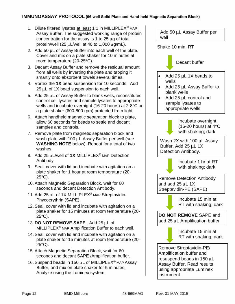
IMMUNOASSAY PROTOCOL (96-well Solid Plate and Hand-held Magnetic Separation Block)
1. Dilute filtered lysates at least 1:1 in MILLIPLEX® MAP
Assay Buffer. The suggested working range of protein
concentration for the assay is 1 to 25 g of total
protein/well (25 L/well at 40 to 1,000 g/mL).
2. Add 50 µL of Assay Buffer into each well of the plate. Cover and mix on a plate shaker for 10 minutes at
room temperature (20-25C).
3. Decant Assay Buffer and remove the residual amount from all wells by inverting the plate and tapping it smartly onto absorbent towels several times.
4. Vortex the 1X bead suspension for 10 seconds. Add
25 L of 1X bead suspension to each well.
5. Add 25 L of Assay Buffer to blank wells, reconstituted control cell lysates and sample lysates to appropriate wells and incubate overnight (16-20 hours) at 2-8°C on a plate shaker (600-800 rpm) protected from light.
Shake 10 min, RT
6. Attach handheld magnetic separation block to plate, allow 60 seconds for beads to settle and decant samples and controls.
7. Remove plate from magnetic separation block and
wash plate with 100 L Assay Buffer per well (see WASHING NOTE below). Repeat for a total of two washes.
8. Add 25 L/well of 1X MILLIPLEX® MAP Detection Antibody.
9. Seal, cover with lid and incubate with agitation on a plate shaker for 1 hour at room temperature (20-25°C).
10. Attach Magnetic Separation Block, wait for 60 seconds and decant Detection Antibody.
11. Add 25 L of 1X MILLIPLEX® MAP Streptavidin-Phycoerythrin (SAPE).
12. Seal, cover with lid and incubate with agitation on a plate shaker for 15 minutes at room temperature (20-25°C).
13. DO NOT REMOVE SAPE. Add 25 L of
MILLIPLEX® MAP Amplification Buffer to each well.
14. Seal, cover with lid and incubate with agitation on a plate shaker for 15 minutes at room temperature (20-25°C).
15. Attach Magnetic Separation Block, wait for 60 seconds and decant SAPE /Amplification buffer.
16. Suspend beads in 150 L of MILLIPLEX® MAP Assay Buffer, and mix on plate shaker for 5 minutes, Analyze using the Luminex system.
Wash 2X with 100 L Assay Buffer. Add 25 µL 1X Detection Antibody.
Remove Detection Antibody
and add 25 L 1X Streptavidin-PE (SAPE)
Incubate 1 hr at RT with shaking; dark
Incubate 15 min at RT with shaking; dark
DO NOT REMOVE SAPE and
add 25 L Amplification buffer
Incubate 15 min at RT with shaking; dark
Remove Streptavidin-PE/ Amplification buffer and
resuspend beads in 150 L Assay Buffer. Read results using appropriate Luminex instrument.
Add 25 µL 1X beads to wells
Add 25 µL Assay Buffer to blank wells
Add 25 µL control and sample lysates to appropriate wells
Incubate overnight (16-20 hours) at 4°C with shaking; dark
Add 50 µL Assay Buffer per well
Decant buffer

Page 13 EMD Millipore 48-669MAG Rev. 31 MAY 2015
WASHING NOTE: For hand-held magnet, rest plate on magnet for 60 seconds to allow complete settling of magnetic beads. Remove well contents by gently decanting the plate in an appropriate waste receptacle and gently tapping on absorbent pads to remove residual liquid. Wash plate with 100 µL of Assay Buffer by removing plate from magnet, adding Assay Buffer, shaking for 30 seconds, reattaching to magnet, letting beads settle for 60 seconds and removing well contents as previously described after each wash. Repeat wash steps as recommended in Assay Procedure. INSTRUMENT SETTINGS Luminex 200TM, HTS, FLEXMAP 3D® and MAGPIX® with xPONENT® software:
These specifications are for the Luminex 200™, Luminex HTS, Luminex FLEXMAP 3D® and Luminex MAGPIX® with xPONENT® software. Luminex instruments with other software (e.g. MasterPlex®, STarStation, LiquiChip, Bio-Plex Manager™, LABScan™ 100) would need to follow instrument instructions for gate settings and additional specifications from the vendors for reading Luminex Magnetic Beads. For magnetic bead assays, the Luminex 200™ and HTS instruments must be calibrated with the xPONENT® 3.1 compatible Calibration Kit (EMD Millipore Catalog #40-275) and performance verified with the Performance Verification Kit (EMD Millipore Catalog #40-276). The Luminex FLEXMAP 3D® instrument must be calibrated with the FLEXMAP 3D® Calibrator Kit (EMD Millipore Catalog #40-028) and performance verified with the FLEXMAP 3D® Performance Verification Kit (EMD Millipore Catalog #40-029). The Luminex MAGPIX® instrument must be calibrated with the MAGPIX® Calibration Kit (EMD Millipore Catalog #40-049) and performance verified with the MAGPIX® Performance Verification Kit (EMD Millipore Catalog #40-050). NOTE: These assays cannot be performed on any instruments running Luminex IS 2.3
or Luminex 1.7 software. The Luminex probe height must be adjusted to the plate provided in the kit. Please use EMD Millipore Catalog #MAG-PLATE, if additional plates are required for this purpose.
Events: 50 per bead
Sample Size: 100 L
Gate Settings: 8,000 to 15,000
Reporter Gain: Default (Low PMT)
Time Out: 60 seconds
Bead Region: JNK 18 Bad 20 Bcl-2 22 Akt 47 Caspase-9 51 p53 53 Caspase-8 78

Page 14 EMD Millipore 48-669MAG Rev. 31 MAY 2015
SUPPLEMENTAL PROTOCOLS
A. Analysis of viscous cell lysates Some cell lysates may not flow through the filter plate efficiently due to high viscosity or the formation of particulate matter from long-term storage. For these samples, the initial capture and wash steps can be done in microcentrifuge tubes. The beads are then transferred into 96-well filter plates for the rest of the assay.
Add 25 L/assay point of 1X beads to a 500 L centrifuge tube.
Next, add lysate diluted in MILLIPLEX® MAP Assay Buffer 2 to a final volume of
100 L or higher.
Vortex the mixture at high speed for 15 seconds then sonicate for an additional 15 seconds.
Rotate the mixture overnight at 2-8C, protected from light.
Centrifuge the beads for 1 min at 2,000 x g and carefully remove the supernatant to minimize bead loss.
Resuspend the pelleted beads in 25 L/assay point of MILLIPLEX® MAP Assay Buffer 2.
Transfer 25 L of the bead mixture to pre-wet filter plate wells and proceed to step 4 of the Immunoassay protocol.
B. Filter Plate Immunoassay Protocol
NOTE: This protocol requires the use of the included 96-well Filter plate and a Vacuum Manifold (EMD Millipore Vacuum Manifold Catalog #MSVMHTS00 or equivalent with EMD Millipore Vacuum Pump Catalog #WP6111560).
1. Dilute filtered lysates at least 1:1 in MILLIPLEX®
MAP Assay Buffer. The suggested working range of protein concentration for the assay is 1 to 25
g of total protein/well (25 L/well at 40 to 1,000
g/mL).
2. Pre-wet filter plate with 50 L/well of MILLIPLEX® MAP Assay Buffer. Remove by vacuum filtration by placing the filter plate over a vacuum manifold and gently applying vacuum. Gently blot the bottom of the filter plate on a paper towel to remove excess liquid.
3. Vortex the 1X bead suspension for 10 seconds.
Add 25 L of 1X bead suspension to each well.
4. Add 25 L of Assay Buffer to blank wells, 25 L reconstituted control cell lysates and sample lysates to appropriate wells and incubate overnight (16-20 hours) at 2-8°C. Seal, cover with lid and incubate with agitation on a plate shaker at 600-800 rpm.
5. Remove the lysate by vacuum filtration.
Add 25 µL 1X beads to wells
Add 25 µL Assay Buffer to blank wells
Add 25 µL control and sample lysates to appropriate wells
Remove buffer by vacuum
Incubate overnight (16-20 hours) at 4°C with shaking; dark 2 hour at RT with shaking; dark
Add 50 µL Assay Buffer per well

Page 15 EMD Millipore 48-669MAG Rev. 31 MAY 2015
6. Add 100 L/well of MILLIPLEX® MAP Assay Buffer. Remove buffer by vacuum filtration and gently blot the bottom of the filter plate on a paper towel. Repeat this step again for a total of two washes.
7. Add 25 L/well of 1X MILLIPLEX® MAP Detection Antibody.
8. Seal, cover with lid and incubate with agitation on a plate shaker for 1 hour at room temperature (20-25°C).
9. Remove Detection Antibody by vacuum and gently blot the bottom of the filter plate on a paper towel.
10. Add 25 L of 1X MILLIPLEX® MAP Streptavidin-Phycoerythrin (SAPE).
11. Seal, cover with lid and incubate with agitation on a plate shaker for 15 min at room temperature (20-25°C).
12. DO NOT REMOVE SAPE. Add 25 L of MILLIPLEX® MAP Amplification Buffer to each well.
13. Seal, cover with lid and incubate with agitation on a plate shaker for 15 min at room temperature (20-25°C).
14. Remove MILLIPLEX® MAP SAPE /Amplification buffer by vacuum filtration and gently blot the bottom of the filter plate on a paper towel.
15. Resuspend beads in 150 L of MILLIPLEX® MAP
Assay Buffer, and mix on plate shaker for 5 minutes.
16. Analyze using the Luminex system.
C. Plate Washer Use The use of a plate washer is not a recommended method of washing for cell signaling assays. Deterioration of assay performance and well-to-well variability have been noted when using plate washers. If desired, MPEQ-AB may be purchased and used as a general wash buffer with plate washers. MPEQ-AB should be diluted to 1X for use in plate washers. Follow standard protocol wash instructions when using a plate washer (2 washes after sample incubation). Contact EMD Millipore Tech Service if additional instructions are required.
Remove Detection Antibody
and add 25 L 1X Streptavidin-PE (SAPE)
Incubate 1 hr at RT with shaking; dark
Incubate 15 min at RT with shaking; dark
DO NOT REMOVE SAPE and
add 25 L Amplification buffer
Incubate 15 min at RT with shaking; dark
Remove Streptavidin-PE/ Amplification buffer and
resuspend beads in 150 L Assay Buffer. Read results using appropriate Luminex instrument.
Wash 2X with 100 L Assay Buffer. Add 25 µL 1X Detection Antibody.

Page 16 EMD Millipore 48-669MAG Rev. 31 MAY 2015
TROUBLESHOOTING GUIDE
Problem Probable Cause Solution
Insufficient Bead Count
Bead mix prepared inappropriately
Sonicate bead vials and vortex just prior to adding to bead mix bottle according to protocol. Agitate bead mix intermittently in reservoir while pipetting this into the plate.
Samples cause interference due to particulate matter or viscosity
See above. Also sample probe may need to be cleaned with Alcohol flush, Back flush and washes; or if needed probe should be removed and sonicated.
Probe height not adjusted correctly
When reading the assay on Luminex 200™, adjust probe height to the kit solid plate or to the recommended Millipore filter plates using 3 alignment discs. When reading the assay on
MAGPIX®, adjust probe height to the kit
solid plate or to the recommended Millipore filter plates using 2 alignment discs. When reading the assay on
FLEXMAP 3D®, adjust probe height to
the kit solid plate using 1 alignment disc.
For FLEXMAP 3D® when using the
solid plate in the kit, the final suspension should be in 150ul and 75ul should be aspirated.
Background is too high
Background wells were contaminated
Avoid cross-well contamination by using sealer appropriately, and pipeting with Multichannel pipets without touching reagent in plate.
Insufficient washes Increase number of washes.
Beads not in region or gate
Luminex not calibrated correctly or recently
Calibrate Luminex based on Instrument Manufacturer’s instructions, at least once a week or if temperature has changed by >3oC.
Gate Settings not adjusted correctly
Some Luminex instruments (e.g. Bio-
Plex®) require different gate settings
than those described in the Kit protocol. Use Instrument default settings.
Wrong bead regions in protocol template
Check kit protocol for correct bead regions or analyte selection.
Incorrect sample type used Samples containing organic solvents or if highly viscous should be diluted or dialyzed as required.
Instrument not washed or primed
Prime the Luminex 4 times to rid of air bubbles, wash 4 times with sheath fluid or water if there is any remnant alcohol or sanitizing liquid.

Page 17 EMD Millipore 48-669MAG Rev. 31 MAY 2015
Beads were exposed to light
Keep plate and bead mix covered with dark lid or aluminum foil during all incubation steps.
Signal for whole plate is same as background
Incorrect or no Detection Antibody was added
Add appropriate Detection Antibody and continue.
Streptavidin-Phycoerythrin was not added
Add Streptavidin-Phycoerythrin according to protocol. If Detection Antibody has already been removed, sensitivity may be low.
Signals too high Calibration target value set too high
With some Luminex Instrument (e.g.
Bio-Plex®) Default target setting for RP1
calibrator is set at High PMT. Use low target value for calibration and reanalyze plate.
Plate incubation was too long with samples
Use shorter incubation time.
Sample readings are out of range
Samples contain no or below detectable levels of analyte
If below detectable levels, it may be possible to use higher sample volume. Check with tech support for appropriate protocol modifications.
High Variation in samples
Multichannel pipet may not be calibrated
Calibrate pipets.
Plate washing was not uniform Confirm all reagents are removed completely in all wash steps.
Samples may have high particulate matter or other interfering substances
See above.
Plate agitation was insufficient Plate should be agitated during all incubation steps using a vertical plate shaker at a speed where beads are in constant motion without causing splashing.
Cross well contamination Check when reusing plate sealer that no reagent has touched sealer.
Care should be taken when using same pipet tips that are used for reagent additions and that pipet tip does not touch reagent in plate.
FOR FILTER PLATES ONLY
Filter plate will not vacuum
Vacuum pressure is insufficient Increase vacuum pressure such that 0.2 mL buffer can be suctioned in 3-5 seconds.
Samples have insoluble particles
Centrifuge samples just prior to assay setup and use supernatant.
High lipid concentration After centrifugation, remove lipid layer and use supernatant.
Plate leaked Vacuum Pressure too high Adjust vacuum pressure such that 0.2 mL buffer can be suctioned in 3-5 seconds. May need to transfer contents to a new (blocked) plate and continue.

Page 18 EMD Millipore 48-669MAG Rev. 31 MAY 2015
Plate set directly on table or absorbent towels during incubations or reagent additions
Set plate on plate holder or raised edge so bottom of filter is not touching any surface.
Insufficient blotting of filter plate bottom causing wicking
Blot the bottom of the filter plate well with absorbent towels after each wash step.
Pipette touching plate filter during additions
Pipette to the side of plate.
Probe height not adjusted correctly
Adjust probe to 3 alignment discs in well H6.
Sample too viscous May need to dilute sample.
REPLACEMENT REAGENTS
REPLACEMENT REAGENTS CATALOG #
MILLIPLEX® MAP 7-plex Early Apoptosis - Magnetic Beads (20X)
42-669MAG
MILLIPLEX® MAP 7-plex Early Apoptosis, Biotin (20X) (Detection Antibody)
44-669KMG
MILLIPLEX® MAP Lysis Buffer (1X) 43-040
MILLIPLEX® MAP Assay Buffer 2 (1X) 43-041
MILLIPLEX® MAP HeLa Cell Lysate: Lambda Phosphatase
47-229
MILLIPLEX® MAP Jurkat Cell Lysate: Anisomycin 47-207
MILLIPLEX® MAP Jurkat Cell Lysate: Paclitaxel 47-220
MILLIPLEX® MAP A549 Cell Lysate: Camptothecin 47-218
MILLIPLEX® MAP Streptavidin-Phycoerythrin (25X) 45-001H
MILLIPLEX® MAP Amplification Buffer (1X) 43-024A
Set of two MILLIPLEX® MAP 96-well Plates with sealers
MAG-PLATE
Set of two MILLIPLEX® MAP 96-well Filter Plates with sealers
MX-PLATE

Page 19 EMD Millipore 48-669MAG Rev. 31 MAY 2015
REPRESENTATIVE DATA
7-Plex Early Apoptosis Magnetic Bead Kit Analysis 7-Plex Early Apoptosis Magnetic Bead Kit Analysis of Anisomycin-treated Jurkat Cells of Paclitaxel-treated Jurkat Cells
7-Plex Early Apoptosis Magnetic Bead Kit Analysis of Camptothecin-treated Jurkat Cells
Figure 1. Multiplex analysis of Jurkat and A549 cells treated with anisomycin, paclitaxel, or camptothecin. HeLa cells treated with lambda phosphatase (negative control), Jurkat cells stimulated with 25
M anisomycin (4 hours) or 5 M paclitaxel (overnight), and A549 cells stimulated with 5 M camptothecin
(overnight) were assayed. The cells were lysed in MILLIPLEX® MAP Lysis Buffer containing protease inhibitors.
20 g total protein of each lysate diluted in MILLIPLEX® MAP Assay Buffer 2 were analyzed according the Assay
protocol (lysate incubation at 4ºC overnight). The Median Fluorescence Intensity (MFI) was measured with the Luminex system. The figures represent the average and standard deviation of three replicate wells. NT Stim NT Stim NT Stim NT Stim NT Stim NT Stim NT Stim
JNK Bad Bcl-2 Akt Caspase-9 p53 Caspase-8
Figure 2. Immunoprecipitation/Western Blot analysis of multiplexed analytes in various cells.
Cell lysates (10 to 50 g) were mixed with capture antibodies to immunoprecipitate each respective protein. The immunoprecipitated proteins were separated on SDS-PAGE, transferred to nitrocellulose, and probed with biotin labeled phospho-specific detection antibodies. The proteins were imaged using Streptavidin-HRP and chemiluminescent substrate. NT-non-treated lysate control, Stim- stimulated lysate control.

Page 20 EMD Millipore 48-669MAG Rev. 31 MAY 2015
ORDERING INFORMATION
To place an order:
To assure the clarity of your custom kit order, please FAX the following information to our customer service department:
Include:
Your name, telephone and/or fax number
Customer account number
Shipping and billing address
Purchase order number
Catalog number and description of product
Quantity of kits
Selection of MILLIPLEX® Analytes
FAX: (636) 441-8050
Toll-Free US: 1-800-MILLIPORE 781-533-8870
Mail Orders: EMD Millipore Corporation
6 Research Park Drive
St. Charles, Missouri 63304 U.S.A.
For European Customers:
To best serve our European customers in placing an order or obtaining additional information about MILLIPLEX® MAP products, please contact your multiplex specialist or sales representative or email our European Customer Service at:
Austria [email protected]
Belgium [email protected]
Denmark [email protected]
France [email protected]
Finland [email protected]
Germany [email protected]
Ireland [email protected]
Italy [email protected]
Netherlands [email protected]
Norway [email protected]
Spain [email protected]
Sweden [email protected]
Switzerland [email protected]

Page 21 EMD Millipore 48-669MAG Rev. 31 MAY 2015
ORDERING INFORMATION (continued)
Conditions of Sale
For Research Use Only. Not for Use in Diagnostic Procedures.
Material Safety Data Sheets (MSDS)
Material Safety Data Sheets for EMD Millipore products may be ordered by fax or phone or through our website at www.emdmillipore.com/techlibrary/index.do.
Technical Services
http://www.emdmillipore.com/techservices
To contact by phone
For North America: Toll-Free US: 1-(800) 221-1975 or 1-(781) 533-8045
Outside North America, contact your local office http://www.emdmillipore.com/offices

Page 22 EMD Millipore 48-669MAG 31 MAY 2015
WELL MAP
1 2 3 4 5 6 7 8 9 10 11 12
A
Assay Buffer 2 Blank
A549: Camptothecin positive control
B
Assay Buffer 2 Blank
A549: Camptothecin positive control
C HeLa: Lamda Phosphatase
negative control Sample 1
D HeLa: Lamda Phosphatase
negative control Sample 1
E Jurkat:
Anisomycin positive control
Sample 2
F Jurkat:
Anisomycin positive control
Sample 2
G Jurkat:
Paclitaxel positive control
Etc.
H Jurkat:
Paclitaxel positive control
Etc.