Embed Size (px)
Citation preview
7127CAN A 3.1 MM STAND-ALONE BATTERY POWEREDESOPHAGOSCOPE (BPE) SCREEN THE ESOPHAGUS FORESOPHAGITIS AND BARRETT’S ? A PROSPECTIVE BLINDEDCOMPARISON WITH A STANDARD VIDEOENDOSCOPE (SVE).Mahesh S. Mokhashi, Tammy Glenn, Christian Jost, Michael B. Wallace,Christopher Y. Kim, Yuko Y. Palesch, Peter B. Cotton, Robert H. Hawes,Med Univ of South Carolina, Charleston, SC.Intro: There exist several indications (varices, Barrett’s, GERD) where anesophagoscopy alone would suffice rather than a complete endoscopy.Feasibility of esophagoscopy using a prototype battery powered flexiblefiberoptic esophagoscope (Olympus XEF-DP) with an outer diameter of 3.1mm has been reported (GI Endo 1999;49: AB157). Aim: In a prospectiveblinded study, compare esophageal visualization between the BPE & theSVE. Methods: 95 consecutive pts. underwent sedated esophagoscopy withthe BPE foll. by SVE, done by 2 endoscopists, each blinded to the findingsof the other. On a visual analogue scale, the 2 endoscopists rated pt. toler-ance & instrument performance. Results: 89 (M 57) pts. (mean age 56 yr.)were analyzed. Mean duration of esophagoscopy was 4.4 mins (range 2-10).Sensitivity : specificity (%) with BPE was 94 : 96 (Barrett’s) & 87 : 94 (alllesions). Measures of pt. tolerance were (BPE : SVE, p value): intubationease 96 : 93, 0.06; gagging 98 : 91, 0.0012; coughing 98 : 96, 0.08; belching89 : 92, 0.02 (higher scores indicate better rating). Measures of scope per-formance were (BPE : SEV, p value): visibility 90 : 98, 0.0001; air insuffla-tion 92 : 98, 0.001; maneuverability 87 : 99, 0.001. Concl: The esophaguscan be accurately visualized in under 5 min with the BPE. Inter-observervariability may account for some of the disagreements. Being a stand-alone instrument, not needing a light source, processor or a monitor, itcould facilitate esophagoscopy outside of the conventional GI lab setting.Its tolerance unsedated, use transnasally & potential in a true screeningenvironment are being studied.
Esophageal findings
Lesions SVE BPE
<5 mm Barrett’s 2 1 false positiveShort seg Barrett’s 15 2 false positive;
1 false negLong seg Barrett’s 1 1Tumor 1 1Erosions 12 2 false negMisc. 14 3 false neg
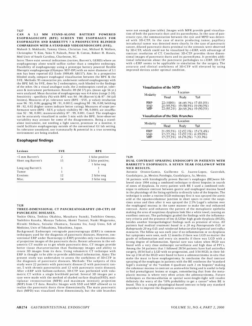
7128THREE-DIMENSIONAL CT PANCREATOGRAPHY (3D-CTP) OFPANCREATIC DISEASES.Yoshio Okita, Toshiya Okahisa, Masaharu Suzuki, Toshihiro Omoya,Yoshihiro Kusaka, Masaya Tadatsu, Akemi Tsutsui, Naoki Muguruma,Hiroshi Shibata, Seisuke Okamura, Susumu Ito, Junji Ueno, Sch ofMedicine, Univ of Tokushima, Tokushima, Japan.Background: Endoscopic retrograde pancreatograpy (ERP) is commontechniques used for the diagnosis of pancreatic diseases. However, con-ventional ERP under fluoroscopy (c-ERP) provides only two-dimension-al projection images of the pancreatic ducts. Recent advances in the vol-umetric CT enable us to get whole pancreatic deta. CT images providebetter tissue characterization than fluoloscopy images and ability tocreate 3D images from its data. Using volumetric CT technique withERP is thought to be able to provide additional information. Aims: Thepresent study was undertaken to assess the usefulness of 3D-CTP inthe diagnosis of pancreatic diseases. Methods: The subjects of thisstudy were 22 patients with pancreatic disease (5 with pancreatic can-cer, 5 with chronic pancreatitis and 12 with pancreatic cystic disease).After c-ERP with balloon-catheter, 3D-CTP was performed with volu-metric CT within a single brethhold period. Several 3D images per acase were made with the method of shaded surface display(SSD), mul-tiplanar reconstruction (MPR) and maximum intensity projection(MIP) from CT data. Results: Images with SSD and MIP allowed us torealize the pancreatic ducts three dimensionally. The main pancreaticduct (MPD) was visualized three dimensionaly, but the side branches
were not enough (see table) .Images with MPR provide good visualiza-tion of both the pancreatic duct and its parenchyma. In the case of pan-creatic cyst, the communication between the cyst and MPD was detect-ed with 3D-CTP. In the case of mutin producing tumor, papillaryintraductal tumor was detected more clearly. In the case of pancreaticcancer, dilated pancreatic ducts proximal to the stenosis were observedby 3D-CTP, which could not be visualized by c-ERP, with advantage ofcontrast resolution of CT. Conclusion: 3D-CTP provides three dimen-sional images of pancreatic ducts and its parenchyma. It provides addi-tional information about the pancreatic pathologies to c-ERP. 3D-CTPwith c-ERP seems to be applicable to simulation for the surgery. Theaccuracy and clinical usefulness of 3D-CTP will elevated by usingimproved devices under optimal condition.
7129DUAL CONTRAST SPRAYING ENDOSCOPY IN PATIENTS WITHBARRETT´S ESOPHAGUS. A SEVEN YEAR FOLLOW-UP WITHNICE RESULTS.Antonio Orozco-Gamiz, Guillermo G. Juarez-Lopez, Gastrolab,Guadalajara, ja, Mexico; Patologia, Guadalajara, Ja, Mexico.34 patients with histologically proven Barrett´s esophagus (BE)were fol-lowed since 1994 using a combined technique to direct biopsies in searchof zones of dysplasia. In every patient with BE I used a combined tech-nique to enhance contrast between gastric and esophageal mucosa basedin the physiology of the lining epithelia to directly take of the biopsies. Thetechnique is under a routine EGD endoscopy first it was sprayed the aceticacid at the squamo-columnar junction in short spurs to cover the suspi-cious areas and then after it was sprayed the 2.5% Lugol´s solution overthe esophageal mucosa in the same manner to make the real enhancedcontrast. Acetic acid enhances the pattern of the metaplastic epitheliamaking the area of suspicious dysplasia look like a white towel, causing anexcellent contrast. The pathologist graded the findings with the inflamma-tory criteria and the presence of low (LGD)or high grade dysplasia (HGD),besides another histopathological findings such as presence of virus. Allpatients had medical treatment based in a 20 mg Pantoprazole O.D. orRabeprazole 20 mg O.D. and reinforced behavior-diet-higienical anti-refluxmeasures. The follow up was each year if no inflammation or no dysplasiabut symptoms were seen, each 12 months if there was LGD no matter thegrade of inflammation and every six months if there was LGD with astrong degree of inflammation. Special care was taken when HGD wasfound with a very close endoscopic surveillance and high dose of PPI´s.Among the 34 patients that I followed 28/34 patients have had antirefluxsurgery, 18/34 had a LGD with no progression, and 3/34 HGD, in their fol-low up 2/34 of the HGD were found to have a adenocarcinoma in-situ thatmake the must to have esophagectomy. In conclusion the dual contrastspraying of the esophagus in patients with the BE reinforces the “standardlugol´s staining” or the “random 4 quadrant each 2 cm biopsies” makingthe columnar mucosa to contrast as aceto-white lining and is useful to helpto find premalignant lesions or stages, remembering that from the meta-plastic mucosa is where very often arises the adenocarcinoma. Futuretechniques as thermo-ablation or special wave-lenght-light will resolvedefinitely the worry of the “10% probability to get a cancer” when BE isfound. This is a simple physiological-based contrast to help any standardprocedure to improve the diagnostic accuracy.
AB274 GASTROINTESTINAL ENDOSCOPY VOLUME 51, NO. 4, PART 2, 2000