Embed Size (px)
Citation preview
CENTER FOR DRUG EVALUATION AND RESEARCH
APPLICATION NUMBER:
761121Orig1s000
NON-CLINICAL REVIEW(S)
MEMORANDUM
Date: May 23, 2019
From: Haleh Saber, PhDDeputy Director, Division of Hematology Oncology Toxicology (DHOT)Office of Hematology and Oncology Products (OHOP)CDER/ FDA
To: File for BLA 761121
Product: POLIVY (polatuzumab vedotin)
I have examined pharmacology/ toxicology review conducted by Dr. Gudi and Dr. Simpson and secondary memorandum provided by Dr. Christopher Sheth. I concur with Dr. Sheth’s conclusion that from a nonclinical perspective, POLIVY may be approved for the proposed indication of relapsed or refractory diffuse large B-cell lymphoma (DLBCL) and that no additional nonclinical studies are needed for this indication. The labeling negotiation is currently ongoing.
Reference ID: 4437894Reference ID: 4446194

--------------------------------------------------------------------------------------------This is a representation of an electronic record that was signedelectronically. Following this are manifestations of any and allelectronic signatures for this electronic record.--------------------------------------------------------------------------------------------/s/------------------------------------------------------------
HALEH SABER05/23/2019 02:30:24 PM
Signature Page 1 of 1
Reference ID: 4437894Reference ID: 4446194

MEMORANDUM
Date: May 23, 2019From: Christopher Sheth, PhD
Division of Hematology Oncology Toxicology (DHOT)Office of Hematology and Oncology Products (OHOP)
Re: Approvability for Pharmacology and ToxicologyBLA: 761121Drug: Polivy (polatuzumab vedotin)Indication: Relapsed or refractory diffuse large B-cell lymphoma (DLBCL)Applicant: Roche/Genentech
Polatuzumab vedotin is a CD79b-directed antibody-drug conjugate consisting of the anti-mitotic agent monomethyl auristatin E (MMAE) linked to a humanized IgG1 antibody for the treatment of patients with diffuse large B-cell lymphoma (DLBCL). Dr. Ramadevi Gudi and Dr. Natalie Simpson reviewed the pharmacology and toxicology studies submitted in support of the BLA. The BLA contains an appropriate complement of nonclinical studies characterizing the primary pharmacodynamics, genotoxicity, safety pharmacology, repeat dose toxicology, and embryo-fetal developmental toxicity of polatuzumab vedotin. The nonclinical findings are summarized in the “Executive Summary” of the BLA review and reflected in the product label. I concur with the pharmacology/toxicology reviewers that from a nonclinical perspective, Polivy may be approved and that no additional nonclinical studies are needed to support approval of Polivy for DLBCL.
Reference ID: 4437654Reference ID: 4446194

--------------------------------------------------------------------------------------------This is a representation of an electronic record that was signedelectronically. Following this are manifestations of any and allelectronic signatures for this electronic record.--------------------------------------------------------------------------------------------/s/------------------------------------------------------------
CHRISTOPHER M SHETH05/23/2019 10:25:10 AM
Signature Page 1 of 1
Reference ID: 4437654Reference ID: 4446194
1
DEPARTMENT OF HEALTH AND HUMAN SERVICESPUBLIC HEALTH SERVICE
FOOD AND DRUG ADMINISTRATIONCENTER FOR DRUG EVALUATION AND RESEARCH
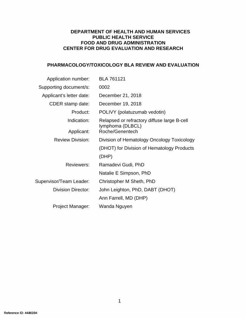
PHARMACOLOGY/TOXICOLOGY BLA REVIEW AND EVALUATION
Application number: BLA 761121
Supporting document/s: 0002
Applicant’s letter date: December 21, 2018
CDER stamp date: December 19, 2018
Product: POLIVY (polatuzumab vedotin)
Indication: Relapsed or refractory diffuse large B-cell lymphoma (DLBCL)
Applicant: Roche/Genentech
Review Division: Division of Hematology Oncology Toxicology
(DHOT) for Division of Hematology Products
(DHP)
Reviewers: Ramadevi Gudi, PhD
Natalie E Simpson, PhD
Supervisor/Team Leader: Christopher M Sheth, PhD
Division Director: John Leighton, PhD, DABT (DHOT)
Ann Farrell, MD (DHP)
Project Manager: Wanda Nguyen
Reference ID: 4436234Reference ID: 4446194
BLA # 761121 Gudi and Simpson
2
TABLE OF CONTENTS
1 EXECUTIVE SUMMARY...........................................................................................51.1 INTRODUCTION .....................................................................................................51.2 BRIEF DISCUSSION OF NONCLINICAL FINDINGS .......................................................51.3 RECOMMENDATIONS .............................................................................................8
2 DRUG INFORMATION..............................................................................................82.1 DRUG ..................................................................................................................82.2 RELEVANT INDS, NDAS, BLAS AND DMFS............................................................92.3 DRUG FORMULATION ..........................................................................................102.4 COMMENTS ON NOVEL EXCIPIENTS ......................................................................102.5 COMMENTS ON IMPURITIES/DEGRADANTS OF CONCERN ........................................102.6 PROPOSED CLINICAL POPULATION AND DOSING REGIMEN .....................................102.7 REGULATORY BACKGROUND ...............................................................................103.3 PREVIOUS REVIEWS REFERENCED.......................................................................10
4 PHARMACOLOGY .................................................................................................114.1 PRIMARY PHARMACOLOGY ..................................................................................114.2 SECONDARY PHARMACOLOGY .............................................................................204.3 SAFETY PHARMACOLOGY ....................................................................................21
5 PHARMACOKINETICS/ADME/TOXICOKINETICS ...............................................21
6 GENERAL TOXICOLOGY......................................................................................266.1 SINGLE-DOSE TOXICITY ......................................................................................266.2 REPEAT-DOSE TOXICITY .....................................................................................26
7 GENETIC TOXICOLOGY........................................................................................367.1 IN VITRO REVERSE MUTATION ASSAY IN BACTERIAL CELLS (AMES) .......................367.2 IN VITRO ASSAYS IN MAMMALIAN CELLS ...............................................................377.3 IN VIVO CLASTOGENICITY ASSAY IN RODENT (MICRONUCLEUS ASSAY) ..................387.4 OTHER GENETIC TOXICITY STUDIES.....................................................................39
8 CARCINOGENICITY...............................................................................................39
9 REPRODUCTIVE AND DEVELOPMENTAL TOXICOLOGY.................................399.1 FERTILITY AND EARLY EMBRYONIC DEVELOPMENT................................................399.2 EMBRYONIC FETAL DEVELOPMENT.......................................................................409.3 PRENATAL AND POSTNATAL DEVELOPMENT ..........................................................43
10 SPECIAL TOXICOLOGY STUDIES....................................................................43
11 INTEGRATED SUMMARY AND SAFETY EVALUATION..................................44
12 APPENDIX/ATTACHMENTS ..............................................................................44
Reference ID: 4436234Reference ID: 4446194
BLA # 761121 Gudi and Simpson
3
Table of Tables
Table 1: Composition of Polatuzumab Vedotin Drug Product .......................................10Table 2: Target Binding Using a Competitive Radioligand Cell Binding Assay .............13Table 3: EC50 Data of Antibodies from Individual Binding Experiments with FcƴRs......14Table 4: IC50 for Polatuzumab Vedotin in Ramos and Jurkat Cells ...............................16Table 5: Response of WSU-DLCL2 Xenograft Tumors to a Single Dose of Polatuzumab Antibody (MCDS4409A), Non-binding control ADC (CNJ1135), or Polatuzumab Vedotin (DCDS4501A).................................................................................................................18Table 6: Anti-DLBCL Tumor Activity of Polatuzumab Vedotin.......................................19Table 7: Anti-Tumor Activity of Polatuzumab in Vedotin in WSU-DLCL2 DLBCL .........20Table 8: Microscopic Findings at Terminal Necropsy in Rats........................................28Table 9: Microscopic Findings at Recovery Necropsy in Rats.......................................30Table 10: Toxicokinetics for Polatuzumab Vedotin and MMAE in Rat Serum...............31Table 11: Maternal Rat Serum Toxicokinetics ...............................................................43Table 12: Ratio of SGD-1010 Concentration in Amniotic Fluid and Fetal Serum to Maternal Serum on GD 18..............................................................................................43
Reference ID: 4436234Reference ID: 4446194
BLA # 761121 Gudi and Simpson
4
Table of Figures
Figure 1: CD79b Protein Sequence Alignment Across Species....................................11Figure 2: Binding Specificity Across Species.................................................................12Figure 3: ADCC and CDC Activity for Polatuzumab Vedotin and Antibody...................15Figure 5: Anti-Proliferative Activities of the Polatuzumab Antibody and MMAE ............16Figure 6: Growth Inhibition of BJAB-PD.cyCD79b.E3 Xenograft Tumors in Response to a Single Dose of Polatuzumab Vedotin and other Test Articles .....................................17
Reference ID: 4436234Reference ID: 4446194
BLA # 761121 Gudi and Simpson
5
1 Executive Summary
1.1 IntroductionPolatuzumab vedotin is an antibody-drug conjugate (ADC) that consists of the anti-mitotic agent (monomethyl auristatin E [MMAE]) covalently conjugated to a CD79b-directed humanized immunoglobulin (Ig) G1 monoclonal antibody through a protease-cleavable linker, maleimidocaproyl-valine-citrulline-p-aminobenzyloxycarbonyl (mc-vc-PAB) with a drug-to-antibody ratio (DAR) of 3.5. The established pharmacological class will be a CD79b-directed antibody-drug conjugate. CD79b is a signaling component of the B-cell receptor (BCR) and is located on the surface of B-cells, including normal and malignant B-cells (e.g., diffuse large B-cell lymphoma (DLBCL)). The proposed clinical dose of 1.8 mg/kg is to be administered as an intravenous infusion over 90 minutes every 21-days for 6 cycles. Nonclinical pharmacology, pharmacokinetic, and toxicology studies have been submitted to support the approval of polatuzumab vedotin for the treatment of patients with relapsed or refractory DLBCL.
1.2 Brief Discussion of Nonclinical FindingsPolatuzumab vedotin binds to human CD79b, is internalized, cleaved by lysosomal proteases, thereby delivering MMAE to malignant B-cells. The released MMAE binds to microtubules and kills dividing cells by inhibiting cell division and inducing apoptosis. Polatuzumab does not bind to its target epitope on species typically used in toxicology studies. Both polatuzumab vedotin and a surrogate ADC that binds monkey CD79b were used in the toxicology studies. The surrogate ADC carries a targeted average DAR of 3.5. The binding of polatuzumab vedotin to human CD79b-expressing cells, and of the surrogate ADC to monkey CD79b-expressing cells, was characterized with Kds of 1.83 and 1.51 nM, respectively. Polatuzumab vedotin and the surrogate ADC bound FcγIA comparably (~ 10 ng/mL or nM), but polatuzumab vedotin binding was 2- to 3-fold lower than the surrogate ADC and the positive control (rituximab) for other Fcγ receptors (IIA allotypes R131 and H131, IIB, or IIIA allotypes F158 and V158).
In cell-based assays, polatuzumab vedotin antibody-dependent cell-mediated cytotoxicity (ADCC) was one order of magnitude lower than the positive control (rituximab) and no complement-dependent cytotoxicity (CDC) was observed. Polatuzumab vedotin demonstrated anti-tumor activity in vitro in CD79b expressing human Burkitt lymphoma (Ramos) cells (IC50 = 0.071 nM) and in vivo in DLBCL and human B-cell lymphoma mouse xenograft models. A single IV dose of 12 mg/kg induced durable complete responses (i.e., no measurable tumor) in 75% of mice in a DLBCL xenograft model. The addition of obinutuzumab or rituximab and bendamustine or CHP [cyclophosphamide (C), doxorubicin (H), prednisone (P)] chemotherapy to 2 mg/kg polatuzumab vedotin reduced the time to tumor doubling.
Safety pharmacology assessments (neurobehavioral, cardiovascular, and respiratory) that were incorporated into the repeat-dose toxicology studies showed no polatuzumab vedotin- or surrogate ADC-induced adverse effects in monkeys. MMAE alone did not
Reference ID: 4436234Reference ID: 4446194
BLA # 761121 Gudi and Simpson
6
appreciably inhibit the human ether-à-go-go-related gene (hERG) channel (IC50 > 100 µM) in voltage-clamped human embryonic kidney cells.
In an in vitro stability study in human and animal plasma, total antibody concentrations for polatuzumab decreased by 25% over 96 hours at 37oC. The relative stability of polatuzumab vedotin and the surrogate ADC were further confirmed in toxicology studies through pharmacokinetic (PK) data. Studies showed the PK of polatuzumab vedotin and the surrogate ADC were generally comparable. An in vivo pharmacokinetic (PK) study in the same mouse species as the xenograft studies demonstrated that polatuzumab vedotin had a short distribution phase and a long elimination phase, as expected for a monoclonal antibody-based therapeutic. The Cmax and AUC for a 5 mg/kg dose was approximately 100 µg/mL and 1000 day*µg/mL, respectively, with a half-life of 12 days. In a distribution study in femoral vein-cannulated female Sprague-Dawley rats, polatuzumab vedotin localized in a non-specific manner to multiple highly perfused tissues, including liver, lungs, heart, kidneys, spleen, adrenal gland, ovaries, and bone marrow, with levels peaking 2 hours post-dose. Polatuzumab vedotin underwent catabolism in these tissues, and in a designated excretion study in bile duct cannulated female Sprague-Dawley rats, the major catabolites of polatuzumab vedotin were unconjugated MMAE, O-demethylated MMAE, and amide hydrolysis and N-demethylation with hydroxylation. In a combined distribution and elimination study, MMAE and MMAE-related catabolites of polatuzumab vedotin were primarily eliminated through the hepatobiliary pathway in rats: >95% in feces, ~5% in urine.
MMAE is known to be an in vitro substrate for CYP3A4/5 and P-gp and is not extensively metabolized in vitro or in vivo. In vitro data suggest a low potential for MMAE to result in clinically significant drug-drug interactions (DDIs) with other medications as a perpetrator.
In the repeat-dose rat study, polatuzumab vedotin was administered by intravenous (IV) injection once weekly for 4 weeks at doses of 2, 6, and 10 mg/kg. The adverse effects were observed at all dose levels and included bone marrow hypocellularity with associated hematology effects and liver toxicity, including increased apoptosis/mitoses of hepatocytes, multifocal hepatic necrosis with higher serum liver transaminases, and total bilirubin. Male reproductive organ toxicities included testicular seminiferous tubule degeneration with consequent abnormal lumen contents in the epididymis. Microscopic findings in many tissues were consistent with the known effects of MMAE on inducing mitotic arrest (due to inhibition of tubulin formation), particularly in cells/tissues with a higher background mitotic rate.
In the repeat-dose monkey toxicology study, polatuzumab vedotin or surrogate ADC were administered once every 3 weeks (Days 1, 22, 43, 64) by IV injection resulting in doses of 1, 3, and 5 mg/kg or 3 and 5 mg/kg, respectively. Toxicities related to polatuzumab vedotin and surrogate ADC treatment included, reversible dose-dependent bone marrow hypocellularity with corresponding myelosuppression at 3 and 5 mg/kg. The surrogate ADC induced decreases in circulating B-lymphocytes (CD20+) and absence of lymphoid follicular germinal centers in the spleen at 3 and 5 mg/kg,
Reference ID: 4436234Reference ID: 4446194
BLA # 761121 Gudi and Simpson
7
consistent with expected pharmacologic effects. Anti-drug-antibodies to polatuzumab vedotin or the surrogate ADC did not appear to impact exposure at any dose level and the toxicokinetic (TK) profiles were similar between ADA-positive and ADA-negative animals.
Acute and longer-term effects of MMAE administration evaluated in rats and cynomolgus monkeys included bone marrow toxicity (characterized by decreased peripheral platelets, red blood cell [RBC] and white blood cell [WBC] parameters, and decreased bone marrow cellularity), liver toxicity (characterized by elevated peripheral liver indices and hepatocellular apoptosis, necrosis, and increased mitosis), and lymphoid organ toxicity (characterized by decreased lymphoid cellularity in the thymus and spleen).
Taken together, the primary polatuzumab vedotin target organs identified by the repeat- dose toxicity studies were the bone marrow, hematolymphopoietic tissues, liver and the male reproductive organs consistent with the expected activity of MMAE.
Carcinogenicity studies in animals have not been performed with polatuzumab vedotin or MMAE and are not warranted for the proposed indication. A designated fertility study was not conducted with polatuzumab vedotin or MMAE and is not needed for the proposed indication. However, results of repeat-dose toxicity studies in rats indicate the potential for polatuzumab vedotin to impair fertility in males.
MMAE was genotoxic in the rat bone marrow micronucleus study through an aneugenic mechanism. Based on positive genotoxicity and results of general toxicology studies showing adverse effects on rapidly dividing cells, a dedicated embryofetal developmental (EFD) study with the ADC or the MMAE is not necessary, per recommendations in ICH S9. Despite this, results of EFD studies were submitted. In an embryo-fetal developmental study, pregnant rats were given 2 intravenous doses of 0.2 mg/kg MMAE during the period of organogenesis on gestation Days 6 and 13. MMAE-related toxicities included pre-/post-implantation loss and embryo-fetal lethality (early resorptions, pre-implantation and post-implantation loss, decreased numbers of live fetuses, and malformations). The fetal malformations included protruding tongue, malformed mandible corresponding to agnathia, malrotated limbs, and gastroschisis. These effects were observed at doses below the therapeutic AUC in patients who received the recommended dose of 1.8 mg/kg POLIVY every 21 days.
Based on the mechanism of action and findings from animal studies, the Warning and Precaution section of the product label will have a statement for embryo-fetal toxicity. Females of reproductive potential, and males with female partners of reproductive potential, will be advised to use effective contraception during treatment and for at least 3 and 5 months after the last dose, respectively. The recommendations for the duration for contraception are based on the FDA guidance, Oncology Pharmaceuticals: Reproductive Toxicity Testing and Labeling Recommendations1, for drugs that are
1 https://www.fda.gov/media/124829/download
Reference ID: 4436234Reference ID: 4446194
BLA # 761121 Gudi and Simpson
8
aneugenic. Based on the toxicity profile of the ADC, the label will also advise women not to breastfeed during treatment and for at least 2 months after the last dose.
1.3 Recommendations
1.3.1 ApprovabilityFrom a Pharmacology/Toxicology perspective, approval of POLIVY is recommended for the proposed indication.1.3.2 Additional Non Clinical RecommendationsNone1.3.3 LabelingThe recommendations to the Applicant’s proposed labeling were discussed internally and communicated to the Applicant. Information in the nonclinical sections of the label reflect findings from studies reviewed within this document and applicable guidance documents.
2 Drug Information
2.1 DrugCAS Registry Number 1313206-42-6Unique Ingredient Identifier KG6VO684Z6
Generic Name Polatuzumab vedotin Code Name ADC: DCDS4501A, DCDS4501S, RO5541077, pola
Polatuzumab antibody intermediate: MCDS4409ASGD-1010 for the small molecule, monomethyl auristatin E (MMAE)
Other names FCU2711, GNT-038, aCD79b-vcMMAE, MCDS4409A-VC-MMAE
Chemical Name Immunoglobulin G1, anti-(human antigen CD79b) (human-Mus musculus monoclonal MCDS4409A heavy chain), disulfide with human-Mus musculus monoclonal MCDS4409A k-chain, dimer, thioether with maleimidocaproyl-valinecitrulline-p-aminobenzyloxycarbonyl monomethylauristatin E
Molecular Formula/ Molecular Weight 150 kDa
Structure or Biochemical Description
Polatuzumab vedotin is a CD79b-directed antibody-drug conjugate (ADC) consisting of three components:
1) the humanized immunoglobulin G1 (IgG1) monoclonal antibody specific for human CD79b
Reference ID: 4436234Reference ID: 4446194
(b) (4)
BLA # 761121 Gudi and Simpson
9
2) the small molecule anti-mitotic agent MMAE3) a protease-cleavable linker maleimidocaproyl-
valine-citrulline-p-aminobenzyloxycarbonyl (mc-vc-PAB) that covalently attaches MMAE to the polatuzumab antibody.
Pharmacologic class CD79b-directed antibody-drug conjugate (ADC)
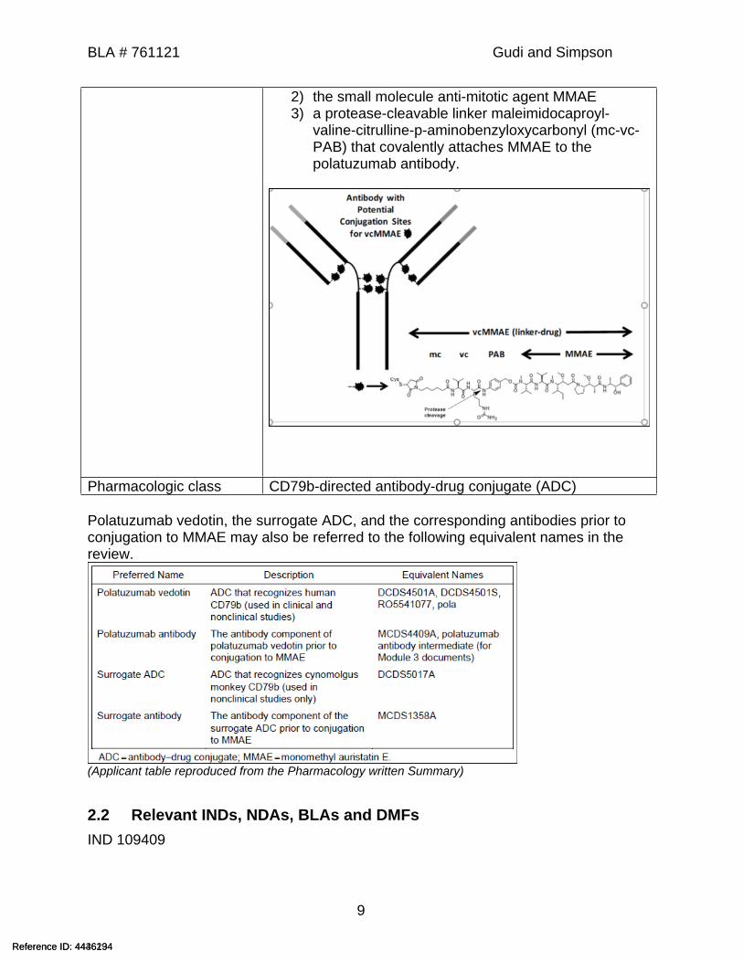
Polatuzumab vedotin, the surrogate ADC, and the corresponding antibodies prior to conjugation to MMAE may also be referred to the following equivalent names in the review.
(Applicant table reproduced from the Pharmacology written Summary)
2.2 Relevant INDs, NDAs, BLAs and DMFsIND 109409
Reference ID: 4436234Reference ID: 4446194
BLA # 761121 Gudi and Simpson
10
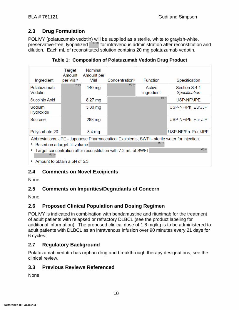
2.3 Drug FormulationPOLIVY (polatuzumab vedotin) will be supplied as a sterile, white to grayish-white, preservative-free, lyophilized for intravenous administration after reconstitution and dilution. Each mL of reconstituted solution contains 20 mg polatuzumab vedotin.
Table 1: Composition of Polatuzumab Vedotin Drug Product
2.4 Comments on Novel ExcipientsNone
2.5 Comments on Impurities/Degradants of ConcernNone
2.6 Proposed Clinical Population and Dosing RegimenPOLIVY is indicated in combination with bendamustine and rituximab for the treatment of adult patients with relapsed or refractory DLBCL (see the product labeling for additional information). The proposed clinical dose of 1.8 mg/kg is to be administered to adult patients with DLBCL as an intravenous infusion over 90 minutes every 21 days for 6 cycles.
2.7 Regulatory BackgroundPolatuzumab vedotin has orphan drug and breakthrough therapy designations; see the clinical review.
3.3 Previous Reviews ReferencedNone
Reference ID: 4436234Reference ID: 4446194
(b) (4)
(b) (4)
(b) (4)
(b) (4)
(b) (4)
(b) (4)
(b) (4)
BLA # 761121 Gudi and Simpson
11
4 Pharmacology
4.1 Primary PharmacologyA. In Vitro StudiesMechanism of Action Polatuzumab vedotin is an ADC that binds to human CD79b and delivers the anti-mitotic agent (monomethyl auristatin E [MMAE]) to B-cells. Upon binding CD79b, polatuzumab vedotin (also referred to as DCDS4501A or the “clinical ADC” in this review) is internalized within one hour.2 It is known that ADC linkers are cleaved by lysosomal proteases, leading to intracellular release of conjugated small molecule toxicant (i.e., MMAE). The released MMAE binds to microtubules and kills dividing cells by inhibiting cell division and inducing apoptosis.
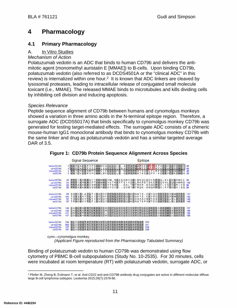
Species RelevancePeptide sequence alignment of CD79b between humans and cynomolgus monkeys showed a variation in three amino acids in the N-terminal epitope region. Therefore, a surrogate ADC (DCDS5017A) that binds specifically to cynomolgus monkey CD79b was generated for testing target-mediated effects. The surrogate ADC consists of a chimeric mouse-human IgG1 monoclonal antibody that binds to cynomolgus monkey CD79b with the same linker and drug as polatuzumab vedotin and has a similar targeted average DAR of 3.5.
Figure 1: CD79b Protein Sequence Alignment Across Species
(Applicant Figure reproduced from the Pharmacology Tabulated Summary)
Binding of polatuzumab vedotin to human CD79b was demonstrated using flow cytometry of PBMC B-cell subpopulations (Study No. 10-2535). For 30 minutes, cells were incubated at room temperature (RT) with polatuzumab vedotin, surrogate ADC, or
2 Pfeifer M, Zheng B, Erdmann T, et al. Anti-CD22 and anti-CD79B antibody drug conjugates are active in different molecular diffuse large B-cell lymphoma subtypes. Leukemia 2015;29(7):1578-86.
Reference ID: 4436234Reference ID: 4446194
BLA # 761121 Gudi and Simpson
12
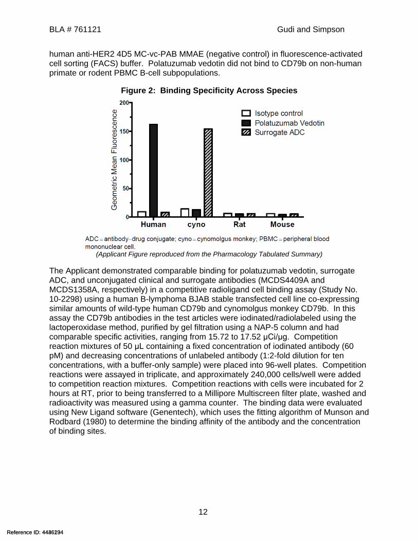
human anti-HER2 4D5 MC-vc-PAB MMAE (negative control) in fluorescence-activated cell sorting (FACS) buffer. Polatuzumab vedotin did not bind to CD79b on non-human primate or rodent PBMC B-cell subpopulations.
Figure 2: Binding Specificity Across Species
(Applicant Figure reproduced from the Pharmacology Tabulated Summary)
The Applicant demonstrated comparable binding for polatuzumab vedotin, surrogate ADC, and unconjugated clinical and surrogate antibodies (MCDS4409A and MCDS1358A, respectively) in a competitive radioligand cell binding assay (Study No. 10-2298) using a human B-lymphoma BJAB stable transfected cell line co-expressing similar amounts of wild-type human CD79b and cynomolgus monkey CD79b. In this assay the CD79b antibodies in the test articles were iodinated/radiolabeled using the lactoperoxidase method, purified by gel filtration using a NAP-5 column and had comparable specific activities, ranging from 15.72 to 17.52 μCi/μg. Competition reaction mixtures of 50 μL containing a fixed concentration of iodinated antibody (60 pM) and decreasing concentrations of unlabeled antibody (1:2-fold dilution for ten concentrations, with a buffer-only sample) were placed into 96-well plates. Competition reactions were assayed in triplicate, and approximately 240,000 cells/well were added to competition reaction mixtures. Competition reactions with cells were incubated for 2 hours at RT, prior to being transferred to a Millipore Multiscreen filter plate, washed and radioactivity was measured using a gamma counter. The binding data were evaluated using New Ligand software (Genentech), which uses the fitting algorithm of Munson and Rodbard (1980) to determine the binding affinity of the antibody and the concentration of binding sites.
Reference ID: 4436234Reference ID: 4446194
BLA # 761121 Gudi and Simpson
13
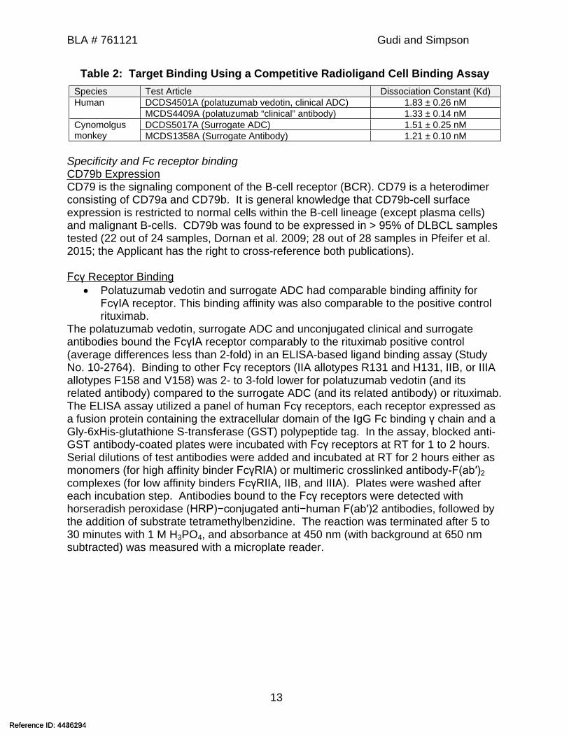
Table 2: Target Binding Using a Competitive Radioligand Cell Binding AssaySpecies Test Article Dissociation Constant (Kd)
DCDS4501A (polatuzumab vedotin, clinical ADC) 1.83 ± 0.26 nMHumanMCDS4409A (polatuzumab “clinical” antibody) 1.33 ± 0.14 nMDCDS5017A (Surrogate ADC) 1.51 ± 0.25 nMCynomolgus
monkey MCDS1358A (Surrogate Antibody) 1.21 ± 0.10 nM
Specificity and Fc receptor bindingCD79b ExpressionCD79 is the signaling component of the B-cell receptor (BCR). CD79 is a heterodimer consisting of CD79a and CD79b. It is general knowledge that CD79b-cell surface expression is restricted to normal cells within the B-cell lineage (except plasma cells) and malignant B-cells. CD79b was found to be expressed in > 95% of DLBCL samples tested (22 out of 24 samples, Dornan et al. 2009; 28 out of 28 samples in Pfeifer et al. 2015; the Applicant has the right to cross-reference both publications).
Fcγ Receptor Binding Polatuzumab vedotin and surrogate ADC had comparable binding affinity for
FcγIA receptor. This binding affinity was also comparable to the positive control rituximab.
The polatuzumab vedotin, surrogate ADC and unconjugated clinical and surrogate antibodies bound the FcγIA receptor comparably to the rituximab positive control (average differences less than 2-fold) in an ELISA-based ligand binding assay (Study No. 10-2764). Binding to other Fcγ receptors (IIA allotypes R131 and H131, IIB, or IIIA allotypes F158 and V158) was 2- to 3-fold lower for polatuzumab vedotin (and its related antibody) compared to the surrogate ADC (and its related antibody) or rituximab. The ELISA assay utilized a panel of human Fcγ receptors, each receptor expressed as a fusion protein containing the extracellular domain of the IgG Fc binding γ chain and a Gly-6xHis-glutathione S-transferase (GST) polypeptide tag. In the assay, blocked anti-GST antibody-coated plates were incubated with Fcγ receptors at RT for 1 to 2 hours. Serial dilutions of test antibodies were added and incubated at RT for 2 hours either as monomers (for high affinity binder FcγRIA) or multimeric crosslinked antibody-F(ab′)2 complexes (for low affinity binders FcγRIIA, IIB, and IIIA). Plates were washed after each incubation step. Antibodies bound to the Fcγ receptors were detected with horseradish peroxidase (HRP)−conjugated anti−human F(ab′)2 antibodies, followed by the addition of substrate tetramethylbenzidine. The reaction was terminated after 5 to 30 minutes with 1 M H3PO4, and absorbance at 450 nm (with background at 650 nm subtracted) was measured with a microplate reader.
Reference ID: 4436234Reference ID: 4446194

BLA # 761121 Gudi and Simpson
14
Table 3: EC50 Data of Antibodies from Individual Binding Experiments with FcƴRs
(Applicant Figure reproduced from the Pharmacology Tabulated Summary)
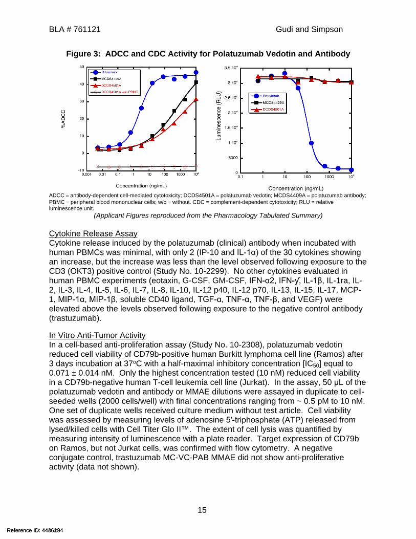
ADCC and CDC Activity Polatuzumab vedotin had no CDC activity. It had ADCC activity that was
substantially lower than that of the positive control, rituximab. The surrogate ADC was not tested for ADCC or CDC activity.
The ADCC activity of polatuzumab vedotin (and its antibody) was assessed using target cells and purified effector PBMCs from healthy human donors. The ADCC activity was one order of magnitude (1000-fold) lower for polatuzumab vedotin and its antibody compared with rituximab. The half minimal effective concentration (EC50) for rituximab was approximately 5 ng/mL (Study No. 10-2764). In the ADCC assay, PBMCs were verified as viable and blood donors were limited to those carrying the heterozygous FcγRIIIA genotype (F/V158). In this assay, polatuzumab vedotin or unconjugated clinical antibody were added at concentrations ranging from 10,000 to 0.0051 ng/mL in 50 μL to pre-seeded wells (4 × 10⁴ target BJAB cells) and incubated at 37o C for 30 minutes. After the incubation, 1.0 × 10⁶ PBMC effector cells (isolated by density gradient centrifugation) in 100 μL of assay medium were added to each well to give a ratio of 25:1 effector-to-target cells, and the plates were incubated for an additional 4 hours. The plates were centrifuged at the end of incubation and the readout for cytotoxicity in the ADCC assay was lactate dehydrogenase activity in the supernatants. The reaction was terminated after 15 minutes. In addition, no CDC was observed in BJAB cells (105 cells/well), incubated for 2 hours at 37o C with polatuzumab vedotin or unconjugated clinical antibody (1 to 10,000 ng/mL) with complement derived from rabbit serum (diluted 1:3 in assay medium). The readout for cytotoxicity was the same as in the cell proliferation assay with Cell Titer Glo II™ (see below). CDC was observed with the positive control, rituximab. The surrogate molecules (MCDS1358A and DCDS5017A) were not tested for ADCC or CDC activity due to unavailability of cynomolgus monkey cell lines.
Reference ID: 4436234Reference ID: 4446194
BLA # 761121 Gudi and Simpson
15
Figure 3: ADCC and CDC Activity for Polatuzumab Vedotin and Antibody
ADCC antibody-dependent cell-mediated cytotoxicity; DCDS4501A polatuzumab vedotin; MCDS4409A polatuzumab antibody; PBMC peripheral blood mononuclear cells; w/o without. CDC = complement-dependent cytotoxicity; RLU = relative luminescence unit.
(Applicant Figures reproduced from the Pharmacology Tabulated Summary)
Cytokine Release AssayCytokine release induced by the polatuzumab (clinical) antibody when incubated with human PBMCs was minimal, with only 2 (IP-10 and IL-1α) of the 30 cytokines showing an increase, but the increase was less than the level observed following exposure to the CD3 (OKT3) positive control (Study No. 10-2299). No other cytokines evaluated in human PBMC experiments (eotaxin, G-CSF, GM-CSF, IFN-α2, IFN-ƴ, IL-1β, IL-1ra, IL-2, IL-3, IL-4, IL-5, IL-6, IL-7, IL-8, IL-10, IL-12 p40, IL-12 p70, IL-13, IL-15, IL-17, MCP-1, MIP-1α, MIP-1β, soluble CD40 ligand, TGF-α, TNF-α, TNF-β, and VEGF) were elevated above the levels observed following exposure to the negative control antibody (trastuzumab).
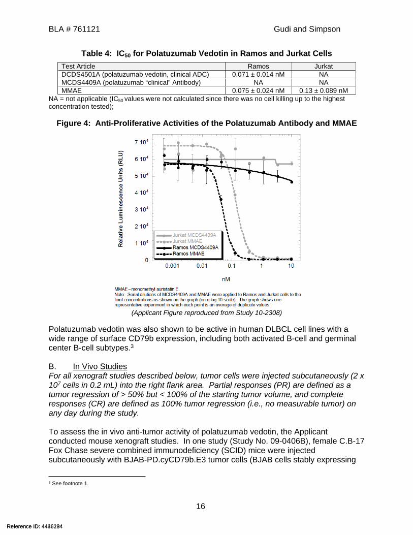
In Vitro Anti-Tumor Activity In a cell-based anti-proliferation assay (Study No. 10-2308), polatuzumab vedotin reduced cell viability of CD79b-positive human Burkitt lymphoma cell line (Ramos) after 3 days incubation at 37oC with a half-maximal inhibitory concentration [IC50] equal to 0.071 ± 0.014 nM. Only the highest concentration tested (10 nM) reduced cell viability in a CD79b-negative human T-cell leukemia cell line (Jurkat). In the assay, 50 μL of the polatuzumab vedotin and antibody or MMAE dilutions were assayed in duplicate to cell-seeded wells (2000 cells/well) with final concentrations ranging from ~ 0.5 pM to 10 nM. One set of duplicate wells received culture medium without test article. Cell viability was assessed by measuring levels of adenosine 5′-triphosphate (ATP) released from lysed/killed cells with Cell Titer Glo II™. The extent of cell lysis was quantified by measuring intensity of luminescence with a plate reader. Target expression of CD79b on Ramos, but not Jurkat cells, was confirmed with flow cytometry. A negative conjugate control, trastuzumab MC-VC-PAB MMAE did not show anti-proliferative activity (data not shown).
Reference ID: 4436234Reference ID: 4446194
BLA # 761121 Gudi and Simpson
16
Table 4: IC50 for Polatuzumab Vedotin in Ramos and Jurkat CellsTest Article Ramos JurkatDCDS4501A (polatuzumab vedotin, clinical ADC) 0.071 ± 0.014 nM NAMCDS4409A (polatuzumab “clinical” Antibody) NA NAMMAE 0.075 ± 0.024 nM 0.13 ± 0.089 nM
NA = not applicable (IC50 values were not calculated since there was no cell killing up to the highest concentration tested);
Figure 4: Anti-Proliferative Activities of the Polatuzumab Antibody and MMAE
(Applicant Figure reproduced from Study 10-2308)
Polatuzumab vedotin was also shown to be active in human DLBCL cell lines with a wide range of surface CD79b expression, including both activated B-cell and germinal center B-cell subtypes.3
B. In Vivo StudiesFor all xenograft studies described below, tumor cells were injected subcutaneously (2 x 10⁷ cells in 0.2 mL) into the right flank area. Partial responses (PR) are defined as a tumor regression of > 50% but < 100% of the starting tumor volume, and complete responses (CR) are defined as 100% tumor regression (i.e., no measurable tumor) on any day during the study.
To assess the in vivo anti-tumor activity of polatuzumab vedotin, the Applicant conducted mouse xenograft studies. In one study (Study No. 09-0406B), female C.B-17 Fox Chase severe combined immunodeficiency (SCID) mice were injected subcutaneously with BJAB-PD.cyCD79b.E3 tumor cells (BJAB cells stably expressing
3 See footnote 1.
Reference ID: 4436234Reference ID: 4446194
BLA # 761121 Gudi and Simpson
17
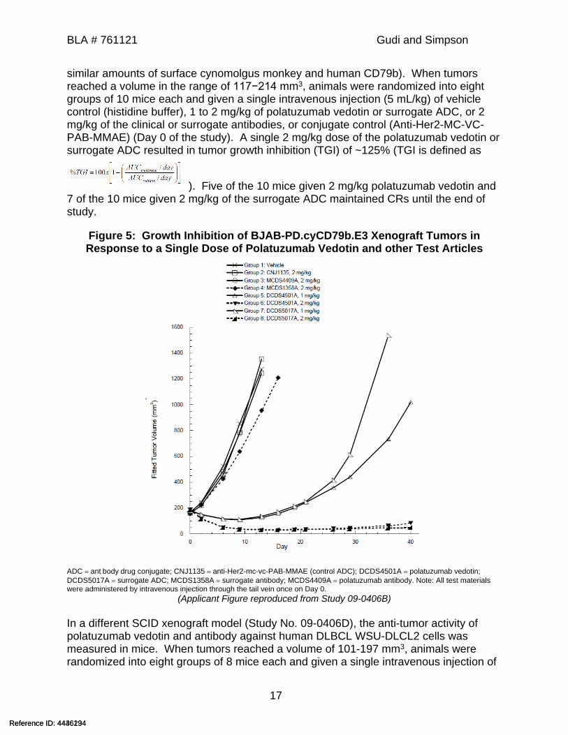
similar amounts of surface cynomolgus monkey and human CD79b). When tumors reached a volume in the range of 117−214 mm3, animals were randomized into eight groups of 10 mice each and given a single intravenous injection (5 mL/kg) of vehicle control (histidine buffer), 1 to 2 mg/kg of polatuzumab vedotin or surrogate ADC, or 2 mg/kg of the clinical or surrogate antibodies, or conjugate control (Anti-Her2-MC-VC-PAB-MMAE) (Day 0 of the study). A single 2 mg/kg dose of the polatuzumab vedotin or surrogate ADC resulted in tumor growth inhibition (TGI) of ~125% (TGI is defined as
). Five of the 10 mice given 2 mg/kg polatuzumab vedotin and 7 of the 10 mice given 2 mg/kg of the surrogate ADC maintained CRs until the end of study.
Figure 5: Growth Inhibition of BJAB-PD.cyCD79b.E3 Xenograft Tumors in Response to a Single Dose of Polatuzumab Vedotin and other Test Articles
ADC ant body drug conjugate; CNJ1135 anti-Her2-mc-vc-PAB-MMAE (control ADC); DCDS4501A polatuzumab vedotin; DCDS5017A surrogate ADC; MCDS1358A surrogate antibody; MCDS4409A polatuzumab antibody. Note: All test materials were administered by intravenous injection through the tail vein once on Day 0.
(Applicant Figure reproduced from Study 09-0406B)
In a different SCID xenograft model (Study No. 09-0406D), the anti-tumor activity of polatuzumab vedotin and antibody against human DLBCL WSU-DLCL2 cells was measured in mice. When tumors reached a volume of 101-197 mm3, animals were randomized into eight groups of 8 mice each and given a single intravenous injection of
Reference ID: 4436234Reference ID: 4446194

BLA # 761121 Gudi and Simpson
18
test materials (Day 0 of the study). Group 1 received histidine buffer as a vehicle control, Group 2 received 12 mg/kg anti-Her2-mc-vc-PAB-MMAE (non-binding control ADC), and Group 3 received 12 mg/kg polatuzumab antibody (adminstration was IV with a dose volume of 5 mL/kg in all groups). Groups 4-8 received 0.3, 1, 3, 6, or 12 mg/kg polatuzumab vedotin, respectively. At doses of 6 and 12 mg/kg polatuzumab vedotin, respectively, CRs were observed for 3/8 (~38%) and 7/8 (~88%) of mice. Six out of 8 of the 12 mg/kg polatuzumab vedotin mice maintained CRs through the end of the study (Day 49). The mean maximum plasma concentration at 12 mg/kg polatuzumab vedotin, was 100 µg/mL.
Table 5: Response of WSU-DLCL2 Xenograft Tumors to a Single Dose of Polatuzumab Antibody (MCDS4409A), Non-binding control ADC (CNJ1135), or
Polatuzumab Vedotin (DCDS4501A)
(Applicant Table reproduced from Study 09-0406D)
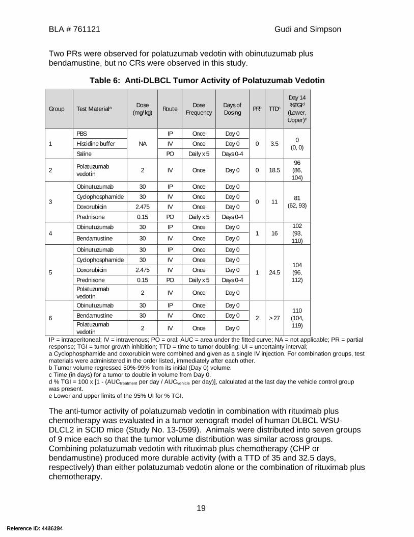
In a xenograft study (Study No. 14-3262B), SCID mice were injected with human DLBCL WSU-DLCL2 cells using similar methods as the other studies. When tumors reached an average size of 117 mm3 in volume, animals were randomized into six groups of 7 mice each and given polatuzumab vedotin, obinutuzumab, cyclophosphamide (C), doxorubicin (H), prednisone (P), and bendamustine according to the table below. While all test articles resulted in TGI nearing 100%, combining polatuzumab vedotin with obinutuzumab plus chemotherapy produced more durable activity than either alone with a time to tumor doubling of 24.5 days plus CHP and >27 days plus bendamustine compared to 3.5 days for vehicle controls (see Table below).
Reference ID: 4436234Reference ID: 4446194
BLA # 761121 Gudi and Simpson
19
Two PRs were observed for polatuzumab vedotin with obinutuzumab plus bendamustine, but no CRs were observed in this study.
Table 6: Anti-DLBCL Tumor Activity of Polatuzumab Vedotin
Group Test Materiala Dose (mg/kg) Route Dose
FrequencyDays of Dosing PRb TTDc
Day 14 %TGId
(Lower, Upper)e
PBS IP Once Day 0
Histidine buffer IV Once Day 01
Saline
NA
PO Daily x 5 Days 0-4
0 3.5 0 (0, 0)
2 Polatuzumab vedotin 2 IV Once Day 0 0 18.5
96 (86, 104)
Obinutuzumab 30 IP Once Day 0
Cyclophosphamide 30 IV Once Day 0
Doxorubicin 2.475 IV Once Day 03
Prednisone 0.15 PO Daily x 5 Days 0-4
0 11 81 (62, 93)
Obinutuzumab 30 IP Once Day 04
Bendamustine 30 IV Once Day 01 16
102 (93, 110)
Obinutuzumab 30 IP Once Day 0
Cyclophosphamide 30 IV Once Day 0
Doxorubicin 2.475 IV Once Day 0
Prednisone 0.15 PO Daily x 5 Days 0-45
Polatuzumab vedotin 2 IV Once Day 0
1 24.5104 (96, 112)
Obinutuzumab 30 IP Once Day 0
Bendamustine 30 IV Once Day 06Polatuzumab vedotin 2 IV Once Day 0
2 > 27110
(104, 119)
IP = intraperitoneal; IV = intravenous; PO = oral; AUC = area under the fitted curve; NA = not applicable; PR = partial response; TGI = tumor growth inhibition; TTD = time to tumor doubling; UI = uncertainty interval; a Cyclophosphamide and doxorubicin were combined and given as a single IV injection. For combination groups, test materials were administered in the order listed, immediately after each other.b Tumor volume regressed 50%-99% from its initial (Day 0) volume.c Time (in days) for a tumor to double in volume from Day 0.d % TGI = 100 x [1 - (AUCtreatment per day / AUCvehicle per day)], calculated at the last day the vehicle control group was present.e Lower and upper limits of the 95% UI for % TGI.
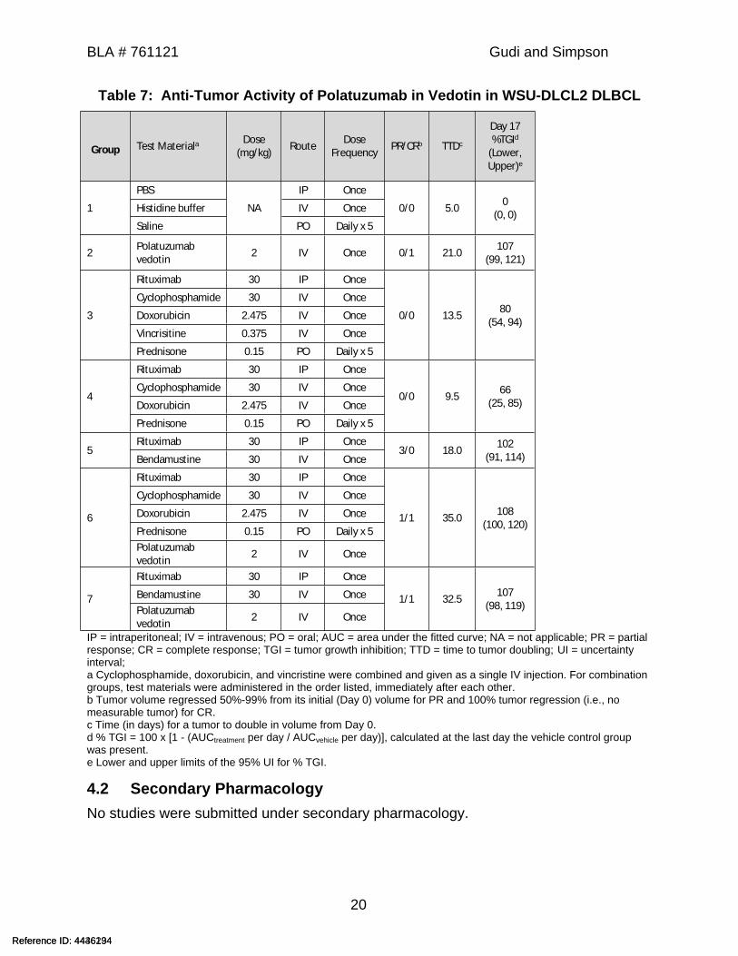
The anti-tumor activity of polatuzumab vedotin in combination with rituximab plus chemotherapy was evaluated in a tumor xenograft model of human DLBCL WSU-DLCL2 in SCID mice (Study No. 13-0599). Animals were distributed into seven groups of 9 mice each so that the tumor volume distribution was similar across groups. Combining polatuzumab vedotin with rituximab plus chemotherapy (CHP or bendamustine) produced more durable activity (with a TTD of 35 and 32.5 days, respectively) than either polatuzumab vedotin alone or the combination of rituximab plus chemotherapy.
Reference ID: 4436234Reference ID: 4446194
BLA # 761121 Gudi and Simpson
20
Table 7: Anti-Tumor Activity of Polatuzumab in Vedotin in WSU-DLCL2 DLBCL
Group Test Materiala Dose (mg/kg) Route Dose
Frequency PR/CRb TTDc
Day 17 %TGId
(Lower, Upper)e
PBS IP Once
Histidine buffer IV Once1
Saline
NA
PO Daily x 5
0/0 5.0 0 (0, 0)
2 Polatuzumab vedotin 2 IV Once 0/1 21.0 107
(99, 121)
Rituximab 30 IP Once
Cyclophosphamide 30 IV Once
Doxorubicin 2.475 IV Once
Vincrisitine 0.375 IV Once
3
Prednisone 0.15 PO Daily x 5
0/0 13.5 80 (54, 94)
Rituximab 30 IP Once
Cyclophosphamide 30 IV Once
Doxorubicin 2.475 IV Once4
Prednisone 0.15 PO Daily x 5
0/0 9.5 66 (25, 85)
Rituximab 30 IP Once5
Bendamustine 30 IV Once3/0 18.0 102
(91, 114)
Rituximab 30 IP Once
Cyclophosphamide 30 IV Once
Doxorubicin 2.475 IV Once
Prednisone 0.15 PO Daily x 56
Polatuzumab vedotin 2 IV Once
1/1 35.0 108 (100, 120)
Rituximab 30 IP Once
Bendamustine 30 IV Once7Polatuzumab vedotin 2 IV Once
1/1 32.5 107 (98, 119)
IP = intraperitoneal; IV = intravenous; PO = oral; AUC = area under the fitted curve; NA = not applicable; PR = partial response; CR = complete response; TGI = tumor growth inhibition; TTD = time to tumor doubling; UI = uncertainty interval; a Cyclophosphamide, doxorubicin, and vincristine were combined and given as a single IV injection. For combination groups, test materials were administered in the order listed, immediately after each other.b Tumor volume regressed 50%-99% from its initial (Day 0) volume for PR and 100% tumor regression (i.e., no measurable tumor) for CR.c Time (in days) for a tumor to double in volume from Day 0.d % TGI = 100 x [1 - (AUCtreatment per day / AUCvehicle per day)], calculated at the last day the vehicle control group was present.e Lower and upper limits of the 95% UI for % TGI.
4.2 Secondary PharmacologyNo studies were submitted under secondary pharmacology.
Reference ID: 4436234Reference ID: 4446194
BLA # 761121 Gudi and Simpson
21
4.3 Safety PharmacologyMMAE alone did not appreciably inhibit the human ether-à-go-go-related gene (hERG) channel (IC50 > 100 µM) in voltage-clamped human embryonic kidney cells (Study No. 07-0611; GLP-compliant). The positive control (60 nM terfenadine) inhibited hERG potassium current by 84.0 ± 0.4% (Mean ± SD; n = 2).
Neurobehavioral, motor activity, and ophthalmic assessments were included in the 4-week repeat-dose toxicology study in rats, using polatuzumab vedotin (Study 10-0898). Cardiovascular, respiratory, and neurologic assessments were included in the 10-week repeat-dose toxicology study in cynomolgus monkeys, using polatuzumab vedotin or the surrogate ADC (10-0044). These studies are reviewed below.
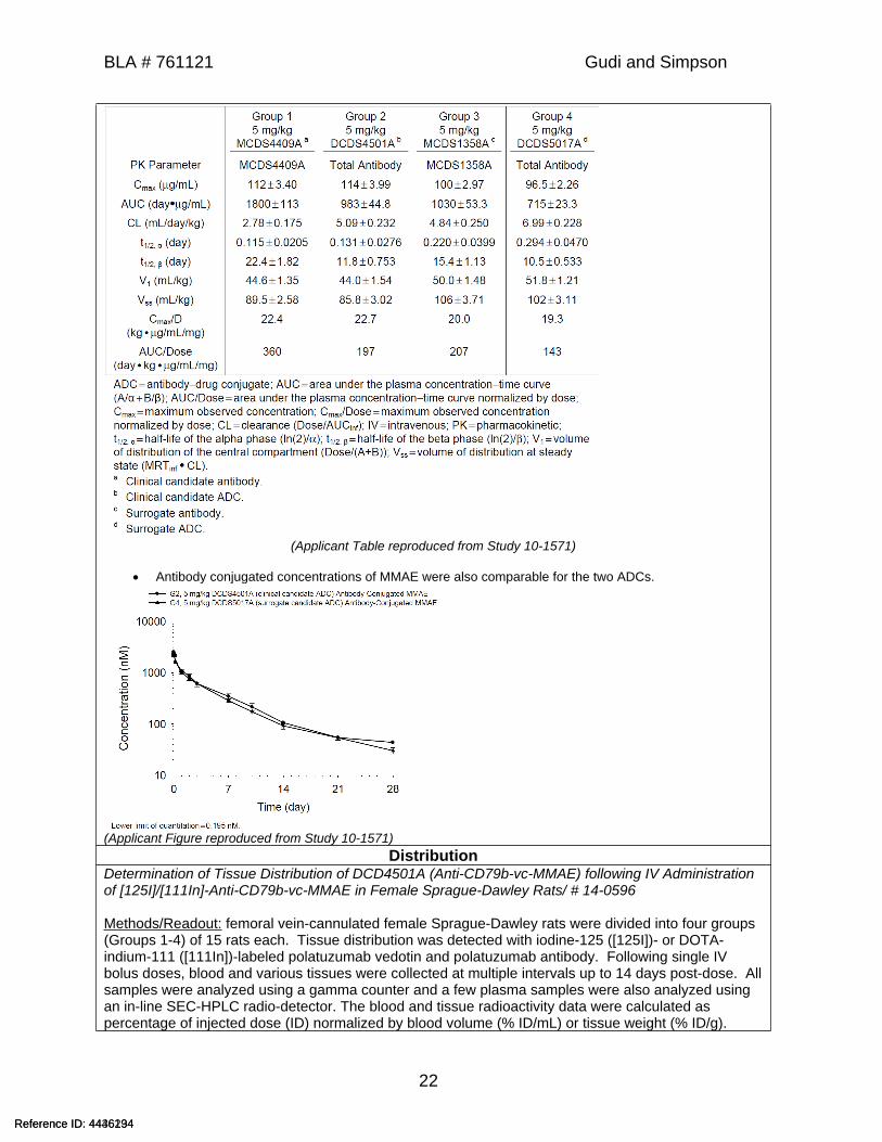
5 Pharmacokinetics/ADME/ToxicokineticsAbsorption
Characterization of the Pharmacokinetics of Naked SN8 (MCDS4409A), SN8-vc-MMAE (DCDS4501A), Naked 10D10 (MCDS1358A), and 10D10-vc-MMAE(DCDS5017A) in Female SCID Mice/ # 10-1571
Methods/Readout: 33 female mice/group. Antibody levels (conjugated and unconjugated) were measured with a semi-homogeneous assay (SHA) using a human (Groups 1 and 2) or cynomolgus monkey (Groups 3 and 4) 15-amino acid peptide for capture and a biotinylated goat anti-human Fc antibody conjugated with horseradish peroxidase (HRP) for detection. The ADCs were detected with ELISA. Antibody-Conjugated MMAE was measured using affinity capture from plasma and followed by enzyme-mediated release of MMAE and electrospray ionization liquid chromatography tandem mass spectrometry (LC/MS/MS) for detection.
Results: The PK profiles of total antibody following single IV administration of polatuzumab vedotin or the surrogate ADC were similar between the two ADCs and were characterized by a short distribution phase and a long elimination phase, as expected for a monoclonal antibody-based therapeutic.
Reference ID: 4436234Reference ID: 4446194
(b) (4)
BLA # 761121 Gudi and Simpson
22
(Applicant Table reproduced from Study 10-1571)
Antibody conjugated concentrations of MMAE were also comparable for the two ADCs.
(Applicant Figure reproduced from Study 10-1571)Distribution
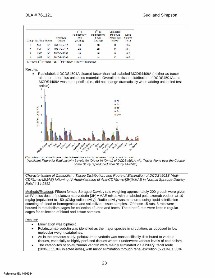
Determination of Tissue Distribution of DCD4501A (Anti-CD79b-vc-MMAE) following IV Administration of [125I]/[111In]-Anti-CD79b-vc-MMAE in Female Sprague-Dawley Rats/ # 14-0596
Methods/Readout: femoral vein-cannulated female Sprague-Dawley rats were divided into four groups (Groups 1-4) of 15 rats each. Tissue distribution was detected with iodine-125 ([125I])- or DOTA-indium-111 ([111In])-labeled polatuzumab vedotin and polatuzumab antibody. Following single IV bolus doses, blood and various tissues were collected at multiple intervals up to 14 days post-dose. All samples were analyzed using a gamma counter and a few plasma samples were also analyzed using an in-line SEC-HPLC radio-detector. The blood and tissue radioactivity data were calculated as percentage of injected dose (ID) normalized by blood volume (% ID/mL) or tissue weight (% ID/g).
Reference ID: 4436234Reference ID: 4446194
BLA # 761121 Gudi and Simpson
23
Results: Radiolabeled DCDS4501A cleared faster than radiolabeled MCDS4409A ( either as tracer
alone or tracer plus unlabeled materials. Overall, the tissue distribution of DCDS4501A and MCDS4409A was non-specific (i.e., did not change dramatically when adding unlabeled test article).
(Applicant Figure for Radioactivity Levels (% ID/g or % ID/mL) of DCDS4501A with Tracer Alone over the Course of the Study reproduced from Study 14-0596)
.Characterization of Catabolism, Tissue Distribution, and Route of Elimination of DCDS4501S (Anti-CD79b-vc-MMAE) following IV Administration of Anti-CD79b-vc-[3H]MMAE in Normal Sprague-DawleyRats/ # 14-2852
Methods/Readout: Fifteen female Sprague-Dawley rats weighing approximately 200 g each were given an IV bolus dose of polatuzumab vedotin-[3H]MMAE mixed with unlabeled polatuzumab vedotin at 10 mg/kg (equivalent to 150 µCi/kg radioactivity). Radioactivity was measured using liquid scintillation counting of blood or homogenized and solubilized tissue samples. Of those 15 rats, 6 rats were housed in metabolism cages for collection of urine and feces. The other 9 rats were kept in regular cages for collection of blood and tissue samples.
Results: Elimination was biphasic. Polatuzumab vedotin was identified as the major species in circulation, as opposed to low
molecular weight catabolites. As in the previous study, polatuzumab vedotin was nonspecifically distributed to various
tissues, especially to highly perfused tissues where it underwent various levels of catabolism. The catabolites of polatuzumab vedotin were mainly eliminated via a biliary−fecal route
(103%± 11.8% injected dose), with minor elimination through renal excretion (5.21%± 1.03%
Reference ID: 4436234Reference ID: 4446194
BLA # 761121 Gudi and Simpson
24
injected dose). Mass balance was achieved, indicating that the elimination was complete in rats over the 14-
day study period.Metabolism
In Vitro Plasma Stability Evaluation of DCDS4501A and DCDS5017A in Human and Animal Plasma/ # 10-1636
Methods/Readout: DCDS4501A , DCDS5017A, MCDS1358A , and MCDS4409A at 100 μg/mL were incubated at 37°C with vehicle, or human, cynomolgus monkey, rat, or mouse plasma for 0, 6, 24, 48, or 96 hours. Total antibody levels and free MMAE levels were measured.
Results:Apart from mouse samples (in which the surrogate ADC was less stable than in other species), the total antibody concentration was roughly similar for both conjugates in all samples, with all samples other than vehicle decreasing ~25% by 96 hours. Free MMAE at 96 hours was similar for both conjugates in all species with cynomolgus monkey and human samples showing no significant increase, 5x increase of MMAE in rat plasma compared to control for the clinical and nonclinical surrogate antibodies, and approximately 66x increase of MMAE in mouse plasma (~25% of theoretical MMAE present in the conjugate).
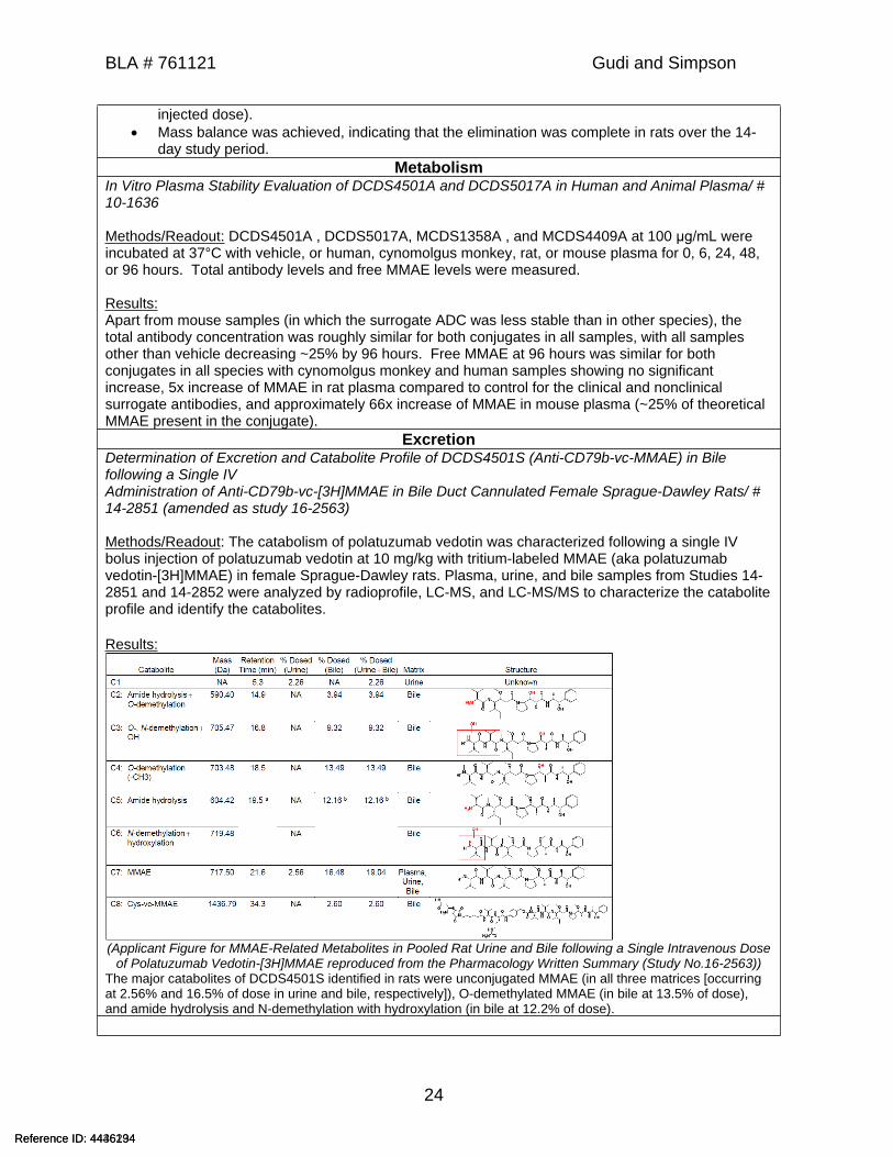
ExcretionDetermination of Excretion and Catabolite Profile of DCDS4501S (Anti-CD79b-vc-MMAE) in Bile following a Single IVAdministration of Anti-CD79b-vc-[3H]MMAE in Bile Duct Cannulated Female Sprague-Dawley Rats/ # 14-2851 (amended as study 16-2563)
Methods/Readout: The catabolism of polatuzumab vedotin was characterized following a single IV bolus injection of polatuzumab vedotin at 10 mg/kg with tritium-labeled MMAE (aka polatuzumab vedotin-[3H]MMAE) in female Sprague-Dawley rats. Plasma, urine, and bile samples from Studies 14-2851 and 14-2852 were analyzed by radioprofile, LC-MS, and LC-MS/MS to characterize the catabolite profile and identify the catabolites.
Results:
(Applicant Figure for MMAE-Related Metabolites in Pooled Rat Urine and Bile following a Single Intravenous Dose of Polatuzumab Vedotin-[3H]MMAE reproduced from the Pharmacology Written Summary (Study No.16-2563))
The major catabolites of DCDS4501S identified in rats were unconjugated MMAE (in all three matrices [occurring at 2.56% and 16.5% of dose in urine and bile, respectively]), O-demethylated MMAE (in bile at 13.5% of dose), and amide hydrolysis and N-demethylation with hydroxylation (in bile at 12.2% of dose).
Reference ID: 4436234Reference ID: 4446194

BLA # 761121 Gudi and Simpson
25
OtherPharmacokinetic Comparability of Polatuzumab Vedotin (v0.1-Derived Drug Product and v1.0-Derived Drug Product) in Female Sprague Dawley Rats/ # 18-0268
Methods/Readout: Female Sprague Dawley rats (15 weeks old and weighing approximately 236-279 g each) were assigned to two groups. Rats were administered a single IV dose of polatuzumab vedotin v0.1, clear colorless liquid (lot no. 669260)- or v1.0, white to greyish white lyophilized powder (lot no. 3217347)-derived materials at a nominal dose of 6 mg/kg (concentration of 1.2 mg/mL in dose volume of 5 mL/kg) (n = 25 rats/group). Rat plasma was sampled for systemic PK up to Day 28 and concentrations of both total antibody and conjugated MMAE in rat plasma were determined using qualified immunoassay LC-MS/MS methods. Concentration-time data for each treatment group (v0.1 and v1.0) were analyzed by noncompartmental PK analysis to determine key PK parameters; bioequivalence (BE) analysis was performed using the v0.1-derived drug product as the reference material.
Results: The PK profiles of total antibody and MMAE were comparable between the two drug products.
The two materials, v0.1 and v1.0, were bioequivalent (BE criteria of 0.8-1.25 with 90% CI) in rats with respect to the AUClast and Cmax for both total antibody and MMAE.
(Applicant Table reproduced from Study No. 18-0268)
Reference ID: 4436234Reference ID: 4446194

BLA # 761121 Gudi and Simpson
26
6 General Toxicology
6.1 Single-Dose ToxicityThe acute toxicologic effects of MMAE administered by a single intravenous bolus tail-vein injection were assessed in rats at 0.206 mg/kg (Study 03-0202) or 0.516 mg/kg (Study 03-0315) and monkeys at 0.116 mg/kg (SNBL.163.19) and at 0.03 and 0.063 mg/kg at (Study 07-0609).
Rats Bone marrow toxicity was associated with mortality and morbidity in males at
0.516 mg/kg. Dose-dependent bone marrow toxicity (decreased total WBCs and platelets,
decreased bone marrow cellularity), liver toxicity (increased AST, ALT, GGT, and total bilirubin, as well as increased mitosis, apoptosis, and necrosis), and lymphoid organ toxicity (decreased lymphocyte cellularity in the thymus and spleen).
Monkeys Bone marrow toxicity was associated with mortality and related opportunistic
infection (lung abscess) in males at 0.063 mg/kg. Significant reductions in WBCs, erythrocytes, hemoglobin, hematocrit, and
reticulocytes, albumin and slight elevations in AST. Decreased bone marrow cellularity and decreased lymphocyte cellularity and
necrosis in the thymus, spleen, and rectal gut-associated lymphoid tissue (GALT).
6.2 Repeat-Dose ToxicityStudy Title/number: Multiple-Dose Toxicity and Toxicokinetic Study of DCD4501A Administered Intravenously to Sprague-Dawley Rats Once Weekly for Four Doses Followed by a 6-Week Recovery Period/ 10-0898
Key findings: Toxicities were observed mainly in the hematopoietic/ lymphoid system, liver,
skin, GI tract, and reproductive organs.
Sprague-Dawley rats (15M/15F) were given 2, 6, and 10 mg/kg polatuzumab vedotin (DCDS4501A) administered weekly for a total of 4 doses with a 6-week recovery period to assess the antigen-independent toxicity/toxicokinetics.
Results: One 10 mg/kg male was euthanized in moribund condition during recovery due to
marked anemia with corresponding microscopic findings of decreased bone marrow cellularity associated with hematology changes; and, hepatic centrilobular degeneration associated with increases in aspartate and alanine aminotransferase.
Reference ID: 4436234Reference ID: 4446194
BLA # 761121 Gudi and Simpson
27
Increased incidence of low arena locomotor activity was noted during the dosing phase in males (4/10 versus 1/10 controls) at 10 mg/kg (measured by functional observational battery). These animals recovered and were consistent with controls in the recovery phase.
No visible ophthalmic lesions were noted in the study. Slightly lower (statistically significant) mean motor activity was noted in males at 10
mg/kg and at 2 mg/kg with no dose-response trend. Microscopic findings in many tissues were consistent with the known effects of
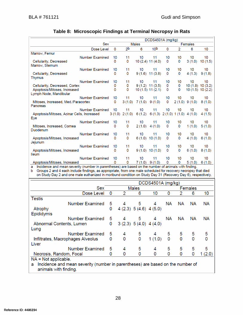
MMAE on inducing mitotic arrest (due to inhibition of tubulin formation), particularly in cells/tissues with a higher background mitotic rate.
At >6 mg/kg microscopic findings included decreased cellularity in bone marrow characterized by decreases in red cell mass, reticulocyte count, and absolute neutrophil count in the peripheral blood; decreased cortical cellularity and increased apoptosis/mitoses in thymus correlated with decrease of lymphocytes; liver toxicity characterized by effects on serum liver transaminases, total bilirubin, and microscopically by increased apoptosis/mitoses of hepatocytes, sinusoidal cells (endothelial and Kupffer cells), and bile duct epithelium and multifocal hepatic necrosis; skin effects consisted of a minimal increase in apoptosis/atrophy of the adnexa epithelial cells (sebaceous gland/hair follicle) and a minimal increase in mitoses in the epidermis and, increased apoptosis/mitoses (minimal to slight severity) in the epididymis duct, These findings exhibited reversibility after a 6-week recovery period.
At ≥ 2 mg/kg microscopic findings in the lung consisted of a macrophage infiltrate in the alveolus and hyperplasia/hypertrophy of Type 2 pneumocytes of males administered 10 mg/kg; testes toxicity was characterized seminiferous tubule degeneration with consequent abnormal lumen contents in the epididymis. This toxicity did not reverse at the end of the 6-week recovery.
Reference ID: 4436234Reference ID: 4446194
BLA # 761121 Gudi and Simpson
28
Table 8: Microscopic Findings at Terminal Necropsy in Rats
Reference ID: 4436234Reference ID: 4446194
BLA # 761121 Gudi and Simpson
29
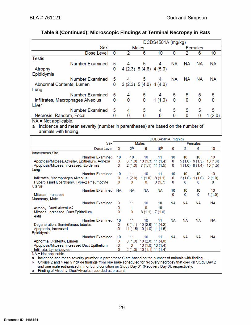
Table 8 (Continued): Microscopic Findings at Terminal Necropsy in Rats
Reference ID: 4436234Reference ID: 4446194

BLA # 761121 Gudi and Simpson
30
Table 9: Microscopic Findings at Recovery Necropsy in Rats
Reference ID: 4436234Reference ID: 4446194
BLA # 761121 Gudi and Simpson
31
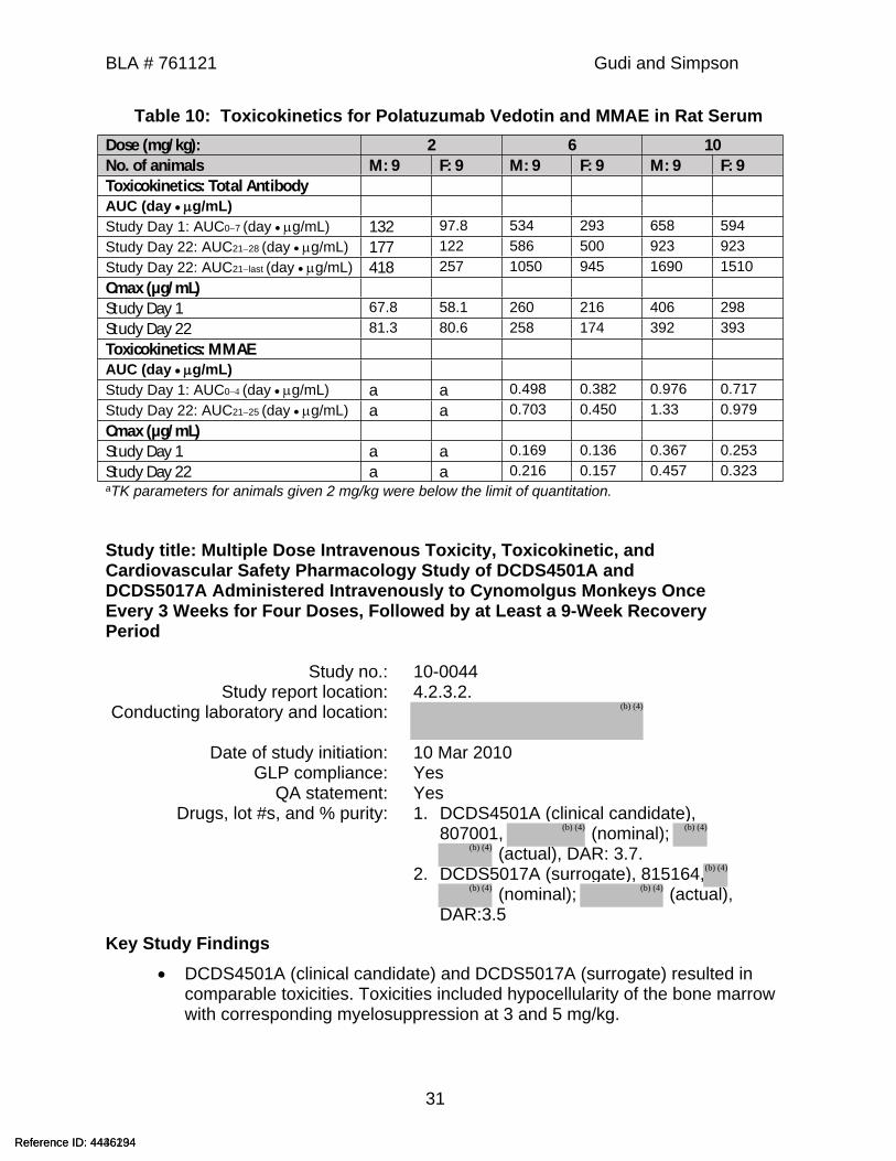
Table 10: Toxicokinetics for Polatuzumab Vedotin and MMAE in Rat SerumDose (mg/kg): 2 6 10No. of animals M: 9 F: 9 M: 9 F: 9 M: 9 F: 9Toxicokinetics: Total AntibodyAUC (day g/mL)Study Day 1: AUC07 (day g/mL) 132 97.8 534 293 658 594Study Day 22: AUC2128 (day g/mL) 177 122 586 500 923 923Study Day 22: AUC21last (day g/mL) 418 257 1050 945 1690 1510Cmax (µg/mL)Study Day 1 67.8 58.1 260 216 406 298Study Day 22 81.3 80.6 258 174 392 393Toxicokinetics: MMAEAUC (day g/mL)Study Day 1: AUC0 (day g/mL) a a 0.498 0.382 0.976 0.717Study Day 22: AUC2125 (day g/mL) a a 0.703 0.450 1.33 0.979Cmax (µg/mL)Study Day 1 a a 0.169 0.136 0.367 0.253Study Day 22 a a 0.216 0.157 0.457 0.323aTK parameters for animals given 2 mg/kg were below the limit of quantitation.
Study title: Multiple Dose Intravenous Toxicity, Toxicokinetic, and Cardiovascular Safety Pharmacology Study of DCDS4501A and DCDS5017A Administered Intravenously to Cynomolgus Monkeys Once Every 3 Weeks for Four Doses, Followed by at Least a 9-Week Recovery Period
Study no.: 10-0044Study report location: 4.2.3.2.
Conducting laboratory and location:
Date of study initiation: 10 Mar 2010GLP compliance: Yes
QA statement: YesDrugs, lot #s, and % purity: 1. DCDS4501A (clinical candidate),
807001, (nominal); (actual), DAR: 3.7.
2. DCDS5017A (surrogate), 815164, (nominal); (actual),
DAR:3.5Key Study Findings
DCDS4501A (clinical candidate) and DCDS5017A (surrogate) resulted in comparable toxicities. Toxicities included hypocellularity of the bone marrow with corresponding myelosuppression at 3 and 5 mg/kg.
Reference ID: 4436234Reference ID: 4446194
(b) (4)
(b) (4) (b) (4)
(b) (4)
(b) (4)
(b) (4) (b) (4)

BLA # 761121 Gudi and Simpson
32
DCDS5017A at 3 and 5 mg/kg induced decreases in circulating B-lymphocytes and depletion of lymphoid follicular germinal centers in the spleen in all animals.
Anti-therapeutic antibodies (ATA) were detected in several animals given DCDS4501A and DCDS5017A but did not impact the exposure.
MethodsDoses: Vehicle, 1, 3, and 5 mg/kg DCDS4501A (clinical
candidate); 3 and 5 mg/kg DCDS5017A (surrogate ADC)
Frequency of dosing: Once every 3 weeks (days 1, 22, 43, 64).Route of administration: Bolus intravenous (IV) injection
Dose volume:Formulation/Vehicle: DCDS4501A vehicle ( histidine acetate,
pH 5.5, sucrose, polysorbate 20). Note: this vehicle was used as a diluent for both test articles, DCDS4501A and DCDS5017A
Species/Strain: Cynomolgus monkeyNumber/Sex/Group: 5/sex/group (2/sex used for recovery)
Age: 3.0-3.7 yr for males, 2.8-3.83 yr for femalesWeight: Males 2.3 kg to 3.7 kg
Females 2.1 kg to 3.3 kg Deviation from study protocol: None that impact the study outcome
Observations and ResultsParameters Major findingsMortality One male treated with high dose DCDS5017A died on Day 53 prior to moribund
humane sacrifice. The clinical signs included decreased activity, hunched appearance, and poor coordination. Cause of death was bacterial endocarditis with corresponding material accumulation on inner surface of right ventricle and red or white pinpoint foci on the lungs. Bacteria or signs of bacterial infection where seen in other organs. Bacterial infection was likely secondary to myelosuppression due to study drug.One female treated with high dose DCDS4501A was moribund and sacrificed on Day 55. Clinical pathology findings included increased neutrophil count, and decreased lymphocyte and eosinophil counts, and minimally decreased indicators of RBC mass (Hb, Hct, and RBC count). Coagulation changes included decreased albumin and increased globulin and fibrinogen with no macroscopic or microscopic evidence of thrombosis.
Clinical Signs UnremarkableBody Weights UnremarkableRespiration Rates (respiration rates and pulse oximetry)
Unremarkable
Ophthalmoscopy UnremarkableECG (including blood pressure, heart rates, and ECGs)
Unremarkable
Neurological Reduced patellar reflexes over weeks 2 and 10 with no dose response:
Reference ID: 4436234Reference ID: 4446194
(b) (4)(b) (4)
(b) (4)
BLA # 761121 Gudi and Simpson
33
DCDS4501A:1 mg/kg -1 male and 1 female 3 mg/kg - 3 females 5 mg/kg -1 femaleDCDS5017A3 mg/kg - 1 male and 1 female 5 mg/kg - 6 females.
Hematology Both DCDS4501A and DCDS5017A had similar hematologic findings that were consistent with bone marrow toxicity and associated peripheral blood cell effects due to suppression of myeloid and erythroid cells, attributable to the pharmacologic activity of monomethyl auristatin E (MMAE).
Drug DCDS4501A DCDS5017ADose mg/kg 1 3 5 3 5Reticulocytes (day 8) -9 -37 -50 -33 -71Reticulocytes (day 71) - -45 -49 -52 -48Lymphocytes (day 8) -26 -25 -54 -65 -50Lymphocytes (day 71) -17 -29 -42 -52 -57Neutrophils (day 8) -9 -21 -30 -57 -78Monocytes (day 8) - - -38 -26 -64
Values in table represent percent change from control mean values (pooled sexes). ‘–‘ no change or not toxicologically relevant.
Findings were reversible.
Flow cytometry analysis showed decreases in B-lymphocytes (CD20+) up to 79% and 84% in 3 and 5 mg/kg DCDS5017A groups, respectively, consistent with pharmacodynamic activity.
Clinical Chemistry Globulins were decreased ~25% (pooled sexes) during the main study period in DCDS5017A and albumin:globulin ratios were correspondingly higher.Globulins were still decreased 24% by the end of recovery.
Anti-drug antibodies Anti-drug antibodies were found in 43% and 20% of DCDS4501 and DCDS5017A-treated animals, respectively.DCDS4501A:1 mg/kg: 5/103 mg/kg: 5/105 mg/kg: 3/10DCDS5017A3 mg/kg: 3/105 mg/kg: 1/10
Urinalysis UnremarkableGross Pathology UnremarkableOrgan Weights UnremarkableHistopathology
Adequate battery: Yes
The lymphoid follicular germinal centers in the spleen were absent in all DCDS5017A-treated animals. Recovery sacrifice findings were unremarkable.
Males FemalesDose mg/kg 0 1a 3a 5a 3b 5b 0 1a 3a 5a 3b 5b
# of Animals 3 3 3 3 3 2 3 3 3 3 3 3
BONE MARROW, STERNUMHypocellularitymild 0 0 2 1 1 0 0 0 0 1 2 2
Reference ID: 4436234Reference ID: 4446194
BLA # 761121 Gudi and Simpson
34
moderate 0 0 0 1 0 0 0 0 0 0 0 0HypercellularityMyeloid mild 0 0 1 1 2 2 0 0 3 2 1 1
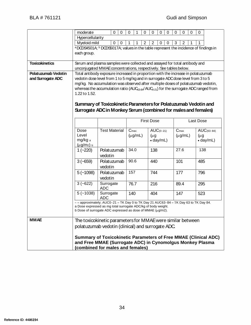
a DCDS4501A; b DCDS5017A; values in the table represent the incidence of findings in each group.
Toxicokinetics Serum and plasma samples were collected and assayed for total antibody and unconjugated MMAE concentrations, respectively. See tables below.
Polatuzumab Vedotin and Surrogate ADC
Total antibody exposure increased in proportion with the increase in polatuzumab vedotin dose level from 1 to 5 mg/kg and in surrogate ADC dose level from 3 to 5 mg/kg. No accumulation was observed after multiple doses of polatuzumab vedotin, whereas the accumulation ratio (AUC63-84/AUC0-21) for the surrogate ADC ranged from 1.22 to 1.52.
Summary of Toxicokinetic Parameters for Polatuzumab Vedotin and Surrogate ADC in Monkey Serum (combined for males and females)
First Dose Last Dose
Dose Levelmg/kg a(g/m2) b
Test Material Cmax
(g/mL)AUC(021)
(g day/mL)
Cmax
(g/mL)AUC(6384)
(g day/mL)
1 (~220) Polatuzumab vedotin
34.0 138 27.6 138
3 (~659) Polatuzumab vedotin
90.6 440 101 485
5 (~1098) Polatuzumab vedotin
157 744 177 796
3 (~622) Surrogate ADC
76.7 216 89.4 295
5 (~1038) Surrogate ADC
140 404 147 523
approximately; AUC021 TK Day 0 to TK Day 21 AUC6384 K Day 63 to TK Day 84.a Dose expressed as mg total surrogate ADC/kg of body weight.b Dose of surrogate ADC expressed as dose of MMAE (g/m2).
MMAE The toxicokinetic parameters for MMAE were similar between polatuzumab vedotin (clinical) and surrogate ADC.
Summary of Toxicokinetic Parameters of Free MMAE (Clinical ADC) and Free MMAE (Surrogate ADC) in Cynomolgus Monkey Plasma (combined for males and females)
Reference ID: 4436234Reference ID: 4446194
BLA # 761121 Gudi and Simpson
35
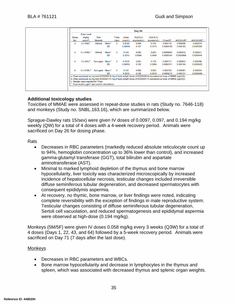
Additional toxicology studiesToxicities of MMAE were assessed in repeat-dose studies in rats (Study no. 7646-118) and monkeys (Study no. SNBL.163.16), which are summarized below.
Sprague-Dawley rats 15/sex) were given IV doses of 0.0097, 0.097, and 0.194 mg/kg weekly (QW) for a total of 4 doses with a 4-week recovery period. Animals were sacrificed on Day 26 for dosing phase.
Rats Decreases in RBC parameters (markedly reduced absolute reticulocyte count up
to 94%, hemoglobin concentration up to 36% lower than control), and increased gamma-glutamyl transferase (GGT), total bilirubin and aspartate aminotransferase (AST).
Minimal to marked lymphoid depletion of the thymus and bone marrow hypocellularity, liver toxicity was characterized microscopically by increased incidence of hepatocellular necrosis, testicular changes included irreversible diffuse seminiferous tubular degeneration, and decreased spermatocytes with consequent epididymis aspermia.
At recovery, no thymic, bone marrow, or liver findings were noted, indicating complete reversibility with the exception of findings in male reproductive system. Testicular changes consisting of diffuse seminiferous tubular degeneration, Sertoli cell vacuolation, and reduced spermatogenesis and epididymal aspermia were observed at high-dose (0.194 mg/kg).
Monkeys (5M/5F) were given IV doses 0.058 mg/kg every 3 weeks (Q3W) for a total of 4 doses (Days 1, 22, 43, and 64) followed by a 5-week recovery period. Animals were sacrificed on Day 71 (7 days after the last dose).
Monkeys
Decreases in RBC parameters and WBCs. Bone marrow hypocellularity and decrease in lymphocytes in the thymus and
spleen, which was associated with decreased thymus and splenic organ weights.
Reference ID: 4436234Reference ID: 4446194
BLA # 761121 Gudi and Simpson
36
7 Genetic Toxicology
7.1 In Vitro Reverse Mutation Assay in Bacterial Cells (Ames)
Study title: Bacterial Reverse Mutation AssayStudy no.: AA66EH.503.BTL
Study report location: 4.2.3.3.1.Conducting laboratory and location:
Date of study initiation: 03 October 2002GLP compliance: Yes
QA statement: YesDrug, lot #, and % purity: SGD-001010, -0-01, 98.7%
Key Study Findings
SGD-001010 (MMAE or SGD-1010) was not cytotoxic (growth inhibition) to the test system up to 5000 μg/plate.
SGD-001010 was negative in bacterial reverse mutation test with or without metabolic activation up to 5000 μg/plate.
MethodsStrains: Salmonella typhimurium (TA98, TA100,
TA1535, and TA1537) and Escherichia coli WP2uvrA
Concentrations in definitive study: 75, 200, 600, 1800 and 5000 µg per plateBasis of concentration selection: Neither precipitate nor toxicity were
observed in the initial toxicity-mutation assay
Negative control: DMSOPositive control: -S9:
Sodium azide for TA1535, TA1009-aminoacridine for TA15372-Nitrofluorene for TA98Methyl methanesulfonate for WP2 uvrA+S9:2-Aminoanthracene for all strains
Formulation/Vehicle: DMSOIncubation & sampling time: Plate incorporation method: 48 to 72 hours
at 37±2°C.
Study Validity
Selection of the tester strains was adequate based upon Guideline for Industry: Specific Aspects of Regulatory Genotoxicity Tests for Pharmaceuticals (ICHS2A, April 1996).
A minimum of three non-toxic doses were included for each strain.
Reference ID: 4436234Reference ID: 4446194
(b) (4)
BLA # 761121 Gudi and Simpson
37
Tester strain culture titers were greater than or equal to 0.3x10⁹ cells/mL. The vehicle control values were within the laboratory historical ranges. The positive control compounds (± S9 mix) produced increases in the number of
revertant colonies (at least 3x increase in the number of revertants over the mean value of the respective negative control).
Results
No positive mutagenic responses were observed with any of the tester strains in either the presence or absence of S9 activation.
7.2 In Vitro Assays in Mammalian CellsStudy title: L5178Y TK+/- Mouse Lymphoma Forward Mutation Assay with a Confirmatory Assay
Study no.: 8204155Study report location: 4.2.2.3.1.
Conducting laboratory and location:
Date of study initiation: 20 April 2009GLP compliance: Yes
QA statement: YesDrug, lot #, and % purity: SGD-1010, 2002E, 92.8%
Key Study Findings
SGD-1010 (MMAE) was cytotoxic and was negative in the L5178Y TK+/- mouse lymphoma forward mutation assay.
MethodsCell line: Mouse lymphoma L5178Y cell line,
Concentrations in definitive study: 4-hour treatment with S9: 0.05, 0.1, 0.25, 0.5, 1.0, 2.5, 5, 10, 20, 30, 40, 50, 60, and 70 ng/mL.4-hour treatment without S9: 0.005, 0.01, 0.05, 0.1, 0.25, 0.5, 0.75, 1, 2.5, 5, 7.5, 10, 12.5, and 15 ng/mL. 24-hour treatment without S9: 0.001, 0.005, 0.01, 0.05, 0.1, 0.25, 0.5, 0.75, 1, 1.5, 2, 2.5, 3, 4, 5, and 6 ng/mL.
Basis of concentration selection: Cytotoxicity (decreases in relative suspension growth)
Negative control: 0.01N HCl/0.9% salinePositive control: With S9: Methylcholanthrene
Without S9: Methyl methanesulfonateFormulation/Vehicle: 0.01N HCl/0.9% saline
Reference ID: 4436234Reference ID: 4446194
(b) (4)
BLA # 761121 Gudi and Simpson
38
Incubation & sampling time: 4-hour treatment with and without S9, and a 24-hour treatment without S9.
Study ValidityThe study validity was evaluated using criteria recommended by the Mouse Lymphoma Assay Workgroup of the International Workshop on Genotoxicity testing for assay acceptance criteria, positive controls and data evaluation (Moore et al., 2006, 2007). All criteria for a valid assay were met.
Results: SGD-1010 (MMAE) was cytotoxic (decreases in relative total growth, RTG) to the
test system under all three (4-hour treatment with and without S9, and a 24-hour treatment without S9) treatment conditions.
SGD-1010 (MMAE) was negative in the L5178Y TK+/- mouse lymphoma forward mutation assay with a confirmatory assay up to 10 to 20% RTG.
7.3 In Vivo Clastogenicity Assay in Rodent (Micronucleus Assay)Study title: In Vivo Rat Bone Marrow Micronucleus Assay
Study no: 8204151Study report location: 4.2.3.3.2.
Conducting laboratory and location:
Date of study initiation: 28 April 2009GLP compliance: Yes
QA statement: YesDrug, lot #, and % purity: SGD-1010 (MMAE), 2002E, 92.8%
Key Study Findings
SGD-1010 was positive in the rat bone marrow micronucleus assay. SGD-1010 induced micronuclei via an aneugenic mode of action based on 60-
76% positive micronuclei for kinetochore staining.
Reference ID: 4436234Reference ID: 4446194
(b) (4)
BLA # 761121 Gudi and Simpson
39
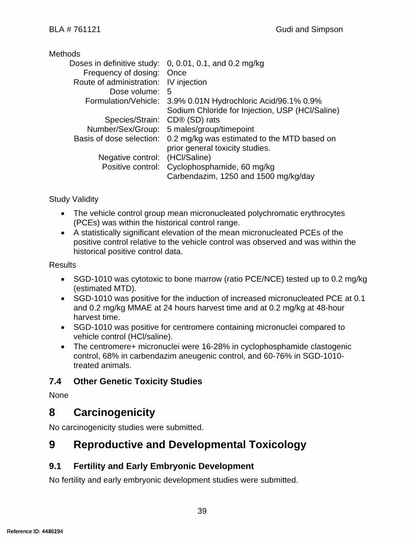
MethodsDoses in definitive study: 0, 0.01, 0.1, and 0.2 mg/kg
Frequency of dosing: OnceRoute of administration: IV injection
Dose volume: 5Formulation/Vehicle: 3.9% 0.01N Hydrochloric Acid/96.1% 0.9%
Sodium Chloride for Injection, USP (HCl/Saline)Species/Strain: CD® (SD) rats
Number/Sex/Group: 5 males/group/timepointBasis of dose selection: 0.2 mg/kg was estimated to the MTD based on
prior general toxicity studies.Negative control: (HCl/Saline)Positive control: Cyclophosphamide, 60 mg/kg
Carbendazim, 1250 and 1500 mg/kg/day
Study Validity
The vehicle control group mean micronucleated polychromatic erythrocytes (PCEs) was within the historical control range.
A statistically significant elevation of the mean micronucleated PCEs of the positive control relative to the vehicle control was observed and was within the historical positive control data.
Results
SGD-1010 was cytotoxic to bone marrow (ratio PCE/NCE) tested up to 0.2 mg/kg (estimated MTD).
SGD-1010 was positive for the induction of increased micronucleated PCE at 0.1 and 0.2 mg/kg MMAE at 24 hours harvest time and at 0.2 mg/kg at 48-hour harvest time.
SGD-1010 was positive for centromere containing micronuclei compared to vehicle control (HCl/saline).
The centromere+ micronuclei were 16-28% in cyclophosphamide clastogenic control, 68% in carbendazim aneugenic control, and 60-76% in SGD-1010-treated animals.
7.4 Other Genetic Toxicity StudiesNone
8 CarcinogenicityNo carcinogenicity studies were submitted.
9 Reproductive and Developmental Toxicology
9.1 Fertility and Early Embryonic DevelopmentNo fertility and early embryonic development studies were submitted.
Reference ID: 4436234Reference ID: 4446194
BLA # 761121 Gudi and Simpson
40
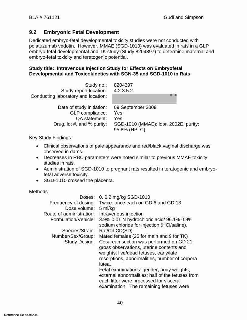
9.2 Embryonic Fetal DevelopmentDedicated embryo-fetal developmental toxicity studies were not conducted with polatuzumab vedotin. However, MMAE (SGD-1010) was evaluated in rats in a GLP embryo-fetal developmental and TK study (Study 8204397) to determine maternal and embryo-fetal toxicity and teratogenic potential.
Study title: Intravenous Injection Study for Effects on Embryofetal Developmental and Toxicokinetics with SGN-35 and SGD-1010 in Rats
Study no.: 8204397Study report location: 4.2.3.5.2.
Conducting laboratory and location:
Date of study initiation: 09 September 2009GLP compliance: Yes
QA statement: YesDrug, lot #, and % purity: SGD-1010 (MMAE); lot#, 2002E, purity:
95.8% (HPLC)Key Study Findings
Clinical observations of pale appearance and red/black vaginal discharge was observed in dams.
Decreases in RBC parameters were noted similar to previous MMAE toxicity studies in rats.
Administration of SGD-1010 to pregnant rats resulted in teratogenic and embryo-fetal adverse toxicity.
SGD-1010 crossed the placenta.
MethodsDoses: 0, 0.2 mg/kg SGD-1010
Frequency of dosing: Twice: once each on GD 6 and GD 13Dose volume: 5 ml/kg
Route of administration: Intravenous injectionFormulation/Vehicle: 3.9% 0.01 N hydrochloric acid/ 96.1% 0.9%
sodium chloride for injection (HCl/saline).Species/Strain: Rat/Crl:CD(SD)
Number/Sex/Group: Mated females (25 for main and 9 for TK) Study Design: Cesarean section was performed on GD 21:
gross observations, uterine contents and weights, live/dead fetuses, early/late resorptions, abnormalities, number of corpora lutea.Fetal examinations: gender, body weights, external abnormalities; half of the fetuses from each litter were processed for visceral examination. The remaining fetuses were
Reference ID: 4436234Reference ID: 4446194
(b) (4)

BLA # 761121 Gudi and Simpson
41
processed for skeletal examination.Deviation from study protocol: None that impacted the outcome of the study
Observations and ResultsParameters Major findingsMortality All animals survived until scheduled sacrifice except one dam in the
SGD-1010 group, who delivered on GD 21 and was sacrificed.Clinical Signs SGD-1010-related paleness (both ears and entire body), red/black
vaginal discharge, and red fluid in cage pan were noted.Body Weights SGD-1010-related decrease in mean maternal body weight change of
20% was noted during the study period GD 6 through GD 21 compared to controls.
Food Consumption Food consumption decreased by 12% from GD 6 through 21.Hematology Percent changes in the SGD1010 compared to mean control
mg/kg RBC HGB HCT WBC NEUT EOS LUC PRET
0 6.29 12.2 34.7 10.74 3.83 0.1 0.09 2.8
0.2 -35 -31 -29 -19 -41 -70 44 396
Fetal Weights (g) covariate adjusted There was no significant change in the SGD-1010 treated group mean total (M and F) fetal body weights compared to control (-4% change).
Necropsy findings Summary Cesarean Section DataTreatments Control SGD-1010
Dose mg/kg 0 0.2
Gravid Uterine weights (g) (mean) 97.73 74.2
Females Mated (N) 25 25
Pregnant (N) 25 24
Aborted (N) 0 0
Died (N) 0 0
Delivered Early (N) NL 0 1
Pregnant at C-section (N) 25 24
Dams with Viable Fetuses (N) 25 23
Dams with no Viable Fetuses (N) 0 1
Corpora Lutea (mean) 13.8 14.2
Implantation Sites (mean) 12.8 12.8
Preimplantation Loss (mean%) 6.8 9.3*
Resorptions:
Total (mean%) 1 27.4*
Early (mean%) 1 22.2*
Late (mean%) 0 5.2*
Dead Fetuses Total 0 0
Post implantation Loss (mean%) 1 27.4*
Live Fetuses (mean%) 99 72.6
% Male fetuses 52.2 50
Mean fetal weights (grams, M+F) 5.62 5.4
* values are outside of the Historical Control (HC) Range
Reference ID: 4436234Reference ID: 4446194
BLA # 761121 Gudi and Simpson
42
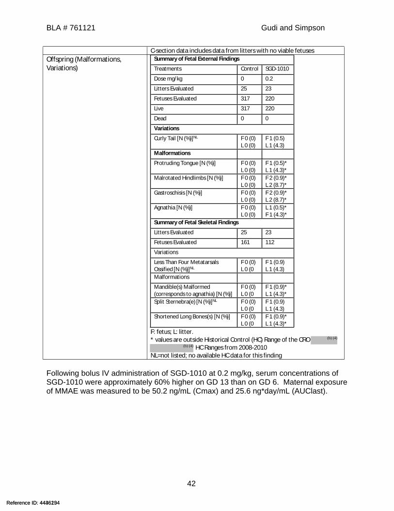
C-section data includes data from litters with no viable fetusesOffspring (Malformations, Variations)
Summary of Fetal External Findings Treatments Control SGD-1010
Dose mg/kg 0 0.2 Litters Evaluated 25 23 Fetuses Evaluated 317 220 Live 317 220 Dead 0 0 Variations
Curly Tail [N (%)]NL F 0 (0)L 0 (0)
F 1 (0.5)
L 1 (4.3)
Malformations
Protruding Tongue [N (%)] F 0 (0)L 0 (0)
F 1 (0.5)*L 1 (4.3)*
Malrotated Hindlimbs [N (%)] F 0 (0)L 0 (0)
F 2 (0.9)*L 2 (8.7)*
Gastroschisis [N (%)] F 0 (0)L 0 (0)
F 2 (0.9)*L 2 (8.7)*
Agnathia [N (%)] F 0 (0)L 0 (0)
L 1 (0.5)*F 1 (4.3)*
Summary of Fetal Skeletal Findings Litters Evaluated 25 23 Fetuses Evaluated 161 112 Variations
Less Than Four Metatarsals Ossified [N (%)]NL
F 0 (0)L 0 (0
F 1 (0.9)L 1 (4.3)
Malformations
Mandible(s) Malformed (corresponds to agnathia) [N (%)]
F 0 (0)L 0 (0
F 1 (0.9)*L 1 (4.3)*
Split Sternebra(e) [N (%)]NL F 0 (0)L 0 (0
F 1 (0.9)L 1 (4.3)
Shortened Long Bones(s) [N (%)] F 0 (0)L 0 (0
F 1 (0.9)*L 1 (4.3)*
F: fetus; L: litter.* values are outside Historical Control (HC) Range of the CRO
HC Ranges from 2008-2010 NL=not listed; no available HC data for this finding
Following bolus IV administration of SGD-1010 at 0.2 mg/kg, serum concentrations of SGD-1010 were approximately 60% higher on GD 13 than on GD 6. Maternal exposure of MMAE was measured to be 50.2 ng/mL (Cmax) and 25.6 ng*day/mL (AUClast).
Reference ID: 4436234Reference ID: 4446194
(b) (4)
(b) (4)
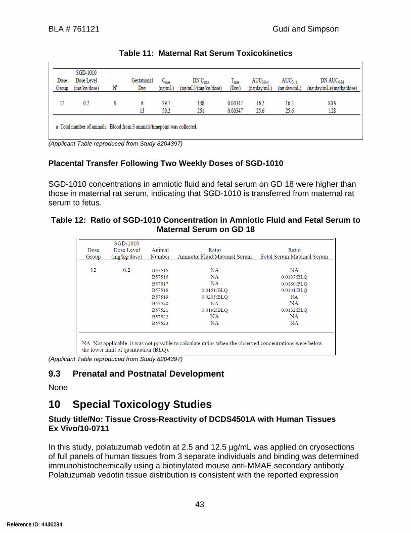
BLA # 761121 Gudi and Simpson
43
Table 11: Maternal Rat Serum Toxicokinetics
(Applicant Table reproduced from Study 8204397)
Placental Transfer Following Two Weekly Doses of SGD-1010
SGD-1010 concentrations in amniotic fluid and fetal serum on GD 18 were higher than those in maternal rat serum, indicating that SGD-1010 is transferred from maternal rat serum to fetus.
Table 12: Ratio of SGD-1010 Concentration in Amniotic Fluid and Fetal Serum to Maternal Serum on GD 18
(Applicant Table reproduced from Study 8204397)
9.3 Prenatal and Postnatal DevelopmentNone
10 Special Toxicology StudiesStudy title/No: Tissue Cross-Reactivity of DCDS4501A with Human TissuesEx Vivo/10-0711
In this study, polatuzumab vedotin at 2.5 and 12.5 μg/mL was applied on cryosections of full panels of human tissues from 3 separate individuals and binding was determined immunohistochemically using a biotinylated mouse anti-MMAE secondary antibody. Polatuzumab vedotin tissue distribution is consistent with the reported expression
Reference ID: 4436234Reference ID: 4446194
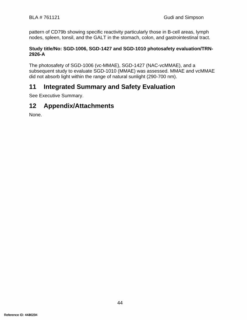
BLA # 761121 Gudi and Simpson
44
pattern of CD79b showing specific reactivity particularly those in B-cell areas, lymph nodes, spleen, tonsil, and the GALT in the stomach, colon, and gastrointestinal tract.
Study title/No: SGD-1006, SGD-1427 and SGD-1010 photosafety evaluation/TRN-2926-A
The photosafety of SGD-1006 (vc-MMAE), SGD-1427 (NAC-vcMMAE), and a subsequent study to evaluate SGD-1010 (MMAE) was assessed. MMAE and vcMMAE did not absorb light within the range of natural sunlight (290-700 nm).
11 Integrated Summary and Safety EvaluationSee Executive Summary.
12 Appendix/AttachmentsNone.
Reference ID: 4436234Reference ID: 4446194
--------------------------------------------------------------------------------------------This is a representation of an electronic record that was signedelectronically. Following this are manifestations of any and allelectronic signatures for this electronic record.--------------------------------------------------------------------------------------------/s/------------------------------------------------------------
RAMADEVI GUDI05/21/2019 08:34:28 AM
NATALIE E SIMPSON05/21/2019 08:38:47 AM
CHRISTOPHER M SHETH05/21/2019 08:39:55 AMI concur
Signature Page 1 of 1
Reference ID: 4436234Reference ID: 4446194