Embed Size (px)
Citation preview

The eyelids 79
be induced by forced lid closure. It is seen most commonly in elderly patients where the orbicularis muscle is weakened. It may also be caused by conjunc-tival scarring, drawing the lid inwards ( cicatricial entropion ). The inturned lashes abrade the cornea and cause marked irritation of the eye. The eye may be red. Short - term treatment includes the application of lubricants to the eye or taping of the lid to turn the lashes away from the globe. The condition can be alleviated for a period by the injection of botulinum toxin into the palpebral part of the orbicularis muscle of the lower lid, or cured permanently by surgery.
Ectropion Here there is an eversion of the lid away from the globe (Figure 5.3 ). Usual causes include:
• age - related orbicularis muscle laxity; • scarring of the periorbital skin; • seventh nerve palsy.
The malposition of the lids everts the puncta and prevents drainage of the tears, leading to epiphora. It also exposes the conjunctiva and lower globe to dehydration (see Chapter 6 ). Ectropion causes an irritable eye. Surgical treat-ment is again an effective treatment.
Infl ammations of the e yelids Blepharitis This is a very common, chronic infl ammation of the lid margins (Figure 5.4 ). In anterior blepharitis infl ammation of the lid margin is concentrated in
Figure 5.3 Ectropion.

80 The eyelids
the lash line and accompanied by squamous debris around the eyelashes. The conjunctiva becomes injected. It is sometimes associated with a chronic staphylococcal infection. In severe disease the cornea is affected ( blepharokeratitis ). Small infi ltrates or ulcers may form in the peripheral cornea ( marginal keratitis ) due to an immune complex response to staphylococcal exotoxins.
In posterior blepharitis (or meibomian gland dysfunction ) the meibomian glands are usually obstructed by squamous debris. The two forms may occur independently.
Figure 5.4 Blepharitis. (a) A diagram showing the signs. (b) The clinical appearance of the lid margin. Note (1) the scales on the lashes, (2) dilated blood vessels on the lid margin and (3) plugging of the meibomian glands.
Injection of thelid margin
Collarette formationaround lashes
Meibomian glandplugging
Cloudy meibomiangland secretion
Scales
(a)
(b)
The eyelids 81
Symptoms
These include:
• tired, itchy, sore eyes, worse in the morning; • crusting of the lid margins in anterior blepharitis and redness in both.
Signs
In anterior blepharitis there may be:
• redness and scaling of the lid margins; some lash bases may be ulcerated – a sign of staphylococcal infection;
• debris in the form of a collarette around the eyelashes (cylindrical dan-druff). This may indicate an infestation of the lash roots by Demodex folliculorum .
• a reduction in the number of eyelashes.
In posterior blepharitis there may be:
• obstruction and plugging of the meibomian orifi ces; • thickened, cloudy, expressed meibomian secretions; • injection of the lid margin and conjunctiva; • tear fi lm abnormalities and punctate keratitis.
Both forms of blepharitis are strongly associated with seborrhoeic dermati-tis, atopic eczema and acne rosacea. In rosacea there is hyperaemia and tel-angiectasia of the facial skin and a rhinophyma (a bulbous irregular swelling of the nose with hypertrophy of the sebaceous glands).
Treatment
This is often diffi cult and must be long - term for these chronic conditions. For anterior blepharitis, lid toilet with a cotton bud wetted with bicarbonate
solution or diluted baby shampoo helps to remove squamous debris from the lash line. Topical steroids can reduce infl ammation but must be used infre-quently, to avoid steroid complications. Staphylococcal lid disease may also require therapy with topical antibiotics (e.g. fusidic acid gel), and occasionally with systemic antibiotics. Demodex infestation responds to the application of ‘ tea tree oil ’ .
For meibomian gland dysfunction, abnormal secretions can be expressed by lid massage after hot bathing through the closed lids. If this treatment fails, then there may be a place for topical azithromycin drops. Alternatively, meibo-mian gland function can be improved by short courses of oral tetracycline. Where meibomian gland obstruction is extensive, the absence of an oily layer on the tear fi lm can induce an evaporative dry eye, which requires treatment with artifi cial tears.

Learning o bjectives To understand:
✓ The symptoms, signs, causes and treatment of con-junctival disease.
✓ The symptoms, signs, causes and treatment of corneal disease.
✓ The difference between episcleritis and scleritis.
7 Conjunctiva, c ornea and s clera
Introduction Disorders of the conjunctiva and cornea are a common cause of symptoms. The ocular surface is regularly exposed to the external environment and subject to trauma, infection and allergic reactions – which account for the majority of diseases in these tissues. Degenerative and structural abnormalities account for a minority of problems.
Symptoms Patients may complain of the following:
1 Pain and irritation. Conjunctivitis alone is seldom associated with anything more than mild discomfort. Pain signifi es something more serious such as corneal injury or infection. This symptom helps differentiate conjunctivitis from corneal disease.
Ophthalmology Lecture Notes, Eleventh Edition. Bruce James, Anthony Bron. © 2011 Bruce James and Anthony Bron. Published 2011 by Blackwell Publishing Ltd.

98 Conjunctiva, cornea and sclera
2 Redness. In conjunctivitis the entire conjunctival surface including that cov-ering the tarsal plates is involved. If the redness is localized to the limbus ( a ciliary fl ush ) the following should be considered: a keratitis (an infl ammation of the cornea); b uveitis (see Chapter 9); c acute glaucoma (see Chapter10).
3 Discharge. Purulent discharge suggests a bacterial conjunctivitis. Viral con-junctivitis is associated mainly with a more watery discharge.
4 Visual loss which is not cleared by blinking. This occurs only when the central cornea is affected. Loss of vision is thus an important symptom requiring urgent action.
5 Patients with corneal disease may also complain of photophobia.
Signs The following features may be seen in conjunctival disease:
• Papillae. These are raised lesions on the upper tarsal conjunctiva, about 1 mm or more in diameter with a central vascular core. They are a non - specifi c sign of chronic infl ammation. They result from infl ammatory infi ltrates within the conjunctiva, constrained by the presence of multiple, tiny fi brous septa. Giant papillae are typical of allergic eye disease and are formed by the coa-lescence of papillae (see Figure 7.4 ). They are also seen as a reaction to contact lens wear.
• Follicles (Figure 7.1 ). These are raised, gelatinous, oval lesions about 1 mm in diameter, found usually in the lower tarsal conjunctiva and upper tarsal border, and occasionally at the limbus. Each follicle represents a lymphoid collection with its own germinal centre. Unlike papillae, the causes of follicles are more specifi c (e.g. viral and chlamydial infections) and they are therefore a clue to aetiology.
• Dilation of the conjunctival vasculature (termed injection ).
Figure 7.1 The clinical appearance of follicles.

Conjunctiva, cornea and sclera 99
• Subconjunctival haemorrhage, often bright red in colour because it is fully oxygenated by the ambient air, through the conjunctiva.
The features of corneal disease are different, and include the following:
• Epithelial and stromal oedema, causing clouding of the cornea. • Cellular infi ltrate in the stroma causing focal, granular white spots. • Deposits of cells on the corneal endothelium (termed keratic precipitates or
KPs , neutrophils with fi ne KPs and lymphocytes or macrophages with coarse ( ‘ mutton fat ’ ) KPs; see Chapter 9 ).
• Chronic keratitis may stimulate new blood vessels superfi cially, under the epithelium ( pannus ; Figure 7.2 ) or deeper in the stroma. Stromal oedema, which causes swelling and separates the collagen lamellae, facilitates vessel invasion.
• Punctate epithelial erosions (PEE) are points of superfi cial epithelial cell loss or dysfunction which may be isolated or scattered, or confl uent. On the cornea they are best detected using fl uorescein dye, viewed with a blue light. Similar PEE occur on the conjunctiva and are best stained by lissamine green. More extensive epithelial loss, due to chemical or physical trauma, is referred to as an abrasion.
Conjunctiva Infl ammatory d iseases of the c onjunctiva
Bacterial c onjunctivitis
Patients present with:
• redness of the eye; • discharge; • ocular irritation.
Figure 7.2 Pannus.
100 Conjunctiva, cornea and sclera
The commonest causative organisms are Staphylococcus , Streptococcus , Pneumococcus and Haemophilus . The condition is usually self - limiting, although a broad - spectrum antibiotic eye drop will hasten resolution. Conjunctival swabs for culture are indicated in severe disease or if the condi-tion fails to resolve.
Antibiotics
Some of the antibiotics available for topical ophthalmic use. Chloramphenicol is an effective broad - spectrum agent; an unproven risk of bone marrow aplasia is a moot point. • Azithromycin. • Ceftazidine. • Chloramphenicol. • Ciprofl oxacin. • Fusidic acid. • Gentamicin. • Neomycin. • Tetracycline. • Ofl oxacin.
Ophthalmia n eonatorum
Ophthalmia neonatorum, refers to any conjunctivitis that occurs in the fi rst 28 days of neonatal life and is a notifi able disease requiring urgent treatment. Swabs for culture are mandatory. It is also important that the cornea is exam-ined to exclude any ulceration.
The commonest causative agents are:
• Bacterial conjunctivitis (usually Gram - positive). • Neisseria gonorrhoeae . In severe cases this can cause corneal perforation.
Systemic complications include rhinitis, stomatitis, arthritis, meningitis and septicaemia. Due to increasing resistance to penicillin a systemic, third - generation cephalosporin (ceftriaxone) is used to treat the condition. The eye must be kept clean. Topical bacitracin ointment can also be given but sys-temic treatment is the most important. Refer parents to a sexually transmit-ted diseases clinic.
• Herpes simplex, which can cause corneal scarring. Topical antivirals are used to treat the condition.
• Chlamydia . This may be responsible for a chronic conjunctivitis and cause sight - threatening corneal scarring. Topical tetracycline ointment and sys-temic erythromycin are used to treat the local and systemic disease respec-tively. Refer parents to a sexually transmitted diseases clinic.

Conjunctiva, cornea and sclera 101
Viral c onjunctivitis
This is distinguished from bacterial conjunctivitis by:
• a watery and limited purulent discharge; • the presence of conjunctival follicles (hence follicular conjunctivitis ). Pre -
auricular lymph nodes are also enlarged; • there may also be lid oedema and excessive lacrimation.
The commonest causative agent is adenovirus, and to a much lesser extent Coxsackie and picornavirus. Adenovirus conjunctivitis is self - limiting but highly contagious and frequently occurs in epidemics. There is a risk of hospital - acquired infection, which can arise where there is failure to hand wash and disinfect equipment when managing a patient with a red eye and conjunctivi-tis. Certain adenovirus serotypes also cause a troublesome punctate keratocon-junctivitis, in which vision is affected, and which may have visual sequelae. Adenoviruses can also cause a conjunctivitis associated with the formation of a pseudomembrane across the conjunctiva. Patients must be given hygiene instruction to minimize the spread of infection in the home (e.g. frequent hand washing; using separate towels). Treatment of keratoconjunctivitis is contro-versial. No effective commercial antiviral is available. Antibacterial therapy is not indicated unless there is a secondary bacterial infection The use of topical steroids damps down symptoms and causes corneal opacities to resolve, but rebound infl ammation is common when the steroid is stopped, and the corneal opacities reappear.
Chlamydial i nfections
Different serotypes of the obligate intracellular organism Chlamydia trachoma-tis are responsible for two forms of ocular infections.
Inclusion k eratoconjunctivitis This is a sexually transmitted disease and may take a chronic course (up to 18 months) unless adequately treated. Patients present with a mucopurulent follicular conjunctivitis and develop a micropannus (superfi cial peripheral corneal vascularization and scarring) associated with subepithelial scarring. Urethritis or cervicitis is common. Diagnosis is confi rmed by detection of chlamydial antigens, using immunofl uorescence, or by identifi cation of typical inclusion bodies by Giemsa staining in conjunctival swab or scrape specimens.
Inclusion conjunctivitis is treated with topical and systemic tetracycline. The patient should be referred to a sexually transmitted diseases clinic.
Trachoma This is the commonest infective cause of blindness in the world, although it is uncommon in developed countries (more details will be found in Chapter 17 ). The housefl y acts as a vector, and the disease is encouraged by poor hygiene
102 Conjunctiva, cornea and sclera
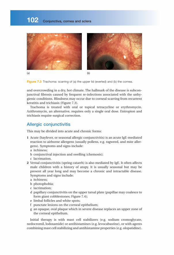
and overcrowding in a dry, hot climate. The hallmark of the disease is subcon-junctival fi brosis caused by frequent re - infections associated with the unhy-gienic conditions. Blindness may occur due to corneal scarring from recurrent keratitis and trichiasis (Figure 7.3 ).
Trachoma is treated with oral or topical tetracycline or erythromycin. Azithromycin, an alternative, requires only a single oral dose. Entropion and trichiasis require surgical correction.
Allergic c onjunctivitis
This may be divided into acute and chronic forms:
1 Acute (hayfever, or seasonal allergic conjunctivitis) is an acute IgE - mediated reaction to airborne allergens (usually pollens, e.g. ragweed, and mite aller-gens). Symptoms and signs include: a itchiness; b conjunctival injection and swelling (chemosis); c lacrimation.
2 Vernal conjunctivitis (spring catarrh) is also mediated by IgE. It often affects male children with a history of atopy. It is usually seasonal but may be present all year long and may become a chronic and intractable disease. Symptoms and signs include: a itchiness; b photophobia; c lacrimation; d papillary conjunctivitis on the upper tarsal plate (papillae may coalesce to
form giant cobblestones; Figure 7.4 ); e limbal follicles and white spots; f punctate lesions on the corneal epithelium; g an opaque, oval plaque which in severe disease replaces an upper zone of
the corneal epithelium.
Initial therapy is with mast cell stabilizers (e.g. sodium cromoglycate, nedocromil, lodoxamide) or antihistamines (e.g. levocabastine), or with agents combining mast cell stabilizing and antihistamine properties (e.g. olopatidine).
Figure 7.3 Trachoma: scarring of (a) the upper lid (everted) and (b) the cornea.
(b)(a)

Conjunctiva, cornea and sclera 103
Topical steroids are required in severe cases but long - term use is avoided if possible because of the risks of steroid - induced glaucoma or cataract. Mucolytics (acetlycysteine) may be required to help dissolve the corneal plaque but surgery may be required.
Contact lens wearers may develop an allergic reaction to their lenses or to lens cleaning materials, leading to a giant papillary conjunctivitis ( GPC ) with a mucoid discharge. Whilst this may respond to topical treatment with mast cell stabilizers, it is often necessary to stop lens wear for a period, or even perma-nently if symptoms recur.
Conjunctival d egenerations Cysts are common in the conjunctiva. They rarely cause symptoms, but if necessary can be removed. Pingueculae and pterygia are found on the inter-palpebral bulbar conjunctiva (Figure 7.5 ). They are thought to result from excessive exposure to the refl ected or direct ultraviolet component of sunlight. Histologically the collagen structure is altered. Pingueculae are small, elevated yellowish paralimbal lesions that never impinge on the cornea. Pterygia are
Figure 7.4 The appearance of giant (cobblestone) papillae in vernal conjunctivitis.
Figure 7.5 The clinical appearance of (a) a pinguecula; (b) a pterygium.
(b)(a)

104 Conjunctiva, cornea and sclera
wing - shaped and located nasally, with the apex towards the cornea, onto which they progressively extend. They may cause irritation and, if extensive, may encroach onto the visual axis. They can be excised but may recur.
Conjunctival t umours
These are rare. They include:
• Squamous cell carcinoma. An irregular raised area of conjunctiva which may invade the deeper tissues.
• Malignant melanoma. The differential diagnosis from benign pigmented lesions (for example a naevus or melanosis) may be diffi cult. Review is neces-sary to assess whether the lesion is increasing in size. Biopsy, to achieve a defi nitive diagnosis, may be required.
Cornea Infective c orneal l esions
Herpes s implex k eratitis
Type 1 herpes simplex (HSV) is a common and important cause of ocular disease. Type 2, which causes genital disease, may occasionally cause keratitis and infantile chorioretinitis. Primary infection by HSV1 is usually acquired early in life by close contact such as kissing. It may be asymptomatic, but oth-erwise is accompanied by:
• fever; • vesicular lid lesions; • follicular conjunctivitis; • pre - auricular lymphadenopathy.
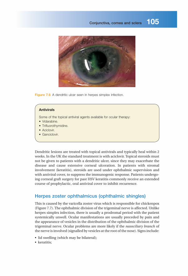
Primary infection may cause a conjunctivitis, with or without punctate kera-titis. It is followed by resolution and latency of the virus in the trigeminal gan-glion. ‘ Recurrent ’ infection involves reactivation of the latent virus, which travels centrifugally to nerve terminals in the corneal epithelium to cause an epithelial keratitis. There may be no past clinical history. The risk of reactivation is increased if the patient is debilitated (e.g. systemic illness, immunosuppression). The pathognomonic appearance is of a dendritic ulcer (Figure 7.6 ). These epithelial ulcers may heal without a scar but they may progress to a stromal keratitis, asso-ciated with an infl ammatory infi ltration and oedema, and ultimately a loss of corneal transparency and permanent scarring. This stage of the disease repre-sents an immunogenic response to the viral antigen. If corneal scarring is severe, a corneal graft may be required to restore vision. Uveitis and glaucoma may accompany the disease. Disciform keratitis is another immunogenic reaction to herpes antigen in the stroma and presents as disc - or ring - shaped stromal oedema and clouding without ulceration, often associated with iritis.
Conjunctiva, cornea and sclera 105
Figure 7.6 A dendritic ulcer seen in herpes simplex infection.
Antivirals
Some of the topical antiviral agents available for ocular therapy: • Vidarabine. • Trifl uorothymidine. • Aciclovir. • Ganciclovir.
Dendritic lesions are treated with topical antivirals and typically heal within 2 weeks. In the UK the standard treatment is with aciclovir. Topical steroids must not be given to patients with a dendritic ulcer, since they may exacerbate the disease and cause extensive corneal ulceration. In patients with stromal involvement (keratitis), steroids are used under ophthalmic supervision and with antiviral cover, to suppress the immunogenic response. Patients undergo-ing corneal graft surgery for past HSV keratitis commonly receive an extended course of prophylactic, oral antiviral cover to inhibit recurrence.
Herpes z oster o phthalmicus ( o phthalmic s hingles)
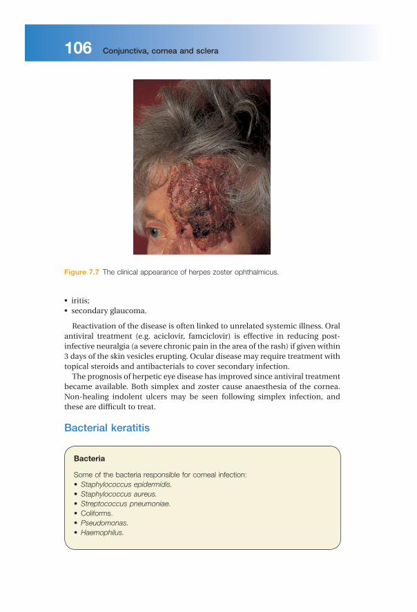
This is caused by the varicella zoster virus which is responsible for chickenpox (Figure 7.7 ). The ophthalmic division of the trigeminal nerve is affected. Unlike herpes simplex infection, there is usually a prodromal period with the patient systemically unwell. Ocular manifestations are usually preceded by pain and the appearance of vesicles in the distribution of the ophthalmic division of the trigeminal nerve. Ocular problems are more likely if the nasociliary branch of the nerve is involved (signalled by vesicles at the root of the nose). Signs include:
• lid swelling (which may be bilateral); • keratitis;
106 Conjunctiva, cornea and sclera
• iritis; • secondary glaucoma.
Reactivation of the disease is often linked to unrelated systemic illness. Oral antiviral treatment (e.g. aciclovir, famciclovir) is effective in reducing post - infective neuralgia (a severe chronic pain in the area of the rash) if given within 3 days of the skin vesicles erupting. Ocular disease may require treatment with topical steroids and antibacterials to cover secondary infection.
The prognosis of herpetic eye disease has improved since antiviral treatment became available. Both simplex and zoster cause anaesthesia of the cornea. Non - healing indolent ulcers may be seen following simplex infection, and these are diffi cult to treat.
Bacterial k eratitis
Bacteria
Some of the bacteria responsible for corneal infection: • Staphylococcus epidermidis. • Staphylococcus aureus. • Streptococcus pneumoniae. • Coliforms. • Pseudomonas. • Haemophilus.
Figure 7.7 The clinical appearance of herpes zoster ophthalmicus.
Conjunctiva, cornea and sclera 107
Pathogenesis A host of bacteria may infect the cornea. Some are found on the lid margin as part of the normal fl ora. The conjunctiva and cornea are protected against infection by:
• blinking; • washing away of debris by the fl ow of tears; • entrapment of foreign particles by mucus; • the antibacterial properties of the tears; • the barrier function of the corneal epithelium ( Neisseria gonorrhoeae is the
only organism that can penetrate the intact epithelium).
Predisposing causes of bacterial keratitis include:
• keratoconjunctivitis sicca (dry eye); • a breach in the corneal epithelium (e.g. following surgery or trauma); • contact lens wear; • prolonged use of topical steroids.
Symptoms and s igns These include:
• pain, usually severe unless the cornea is anaesthetic; • purulent discharge; • ciliary injection; • visual loss (severe if the visual axis is involved); • hypopyon – sometimes (a mass of white cells collected in the anterior
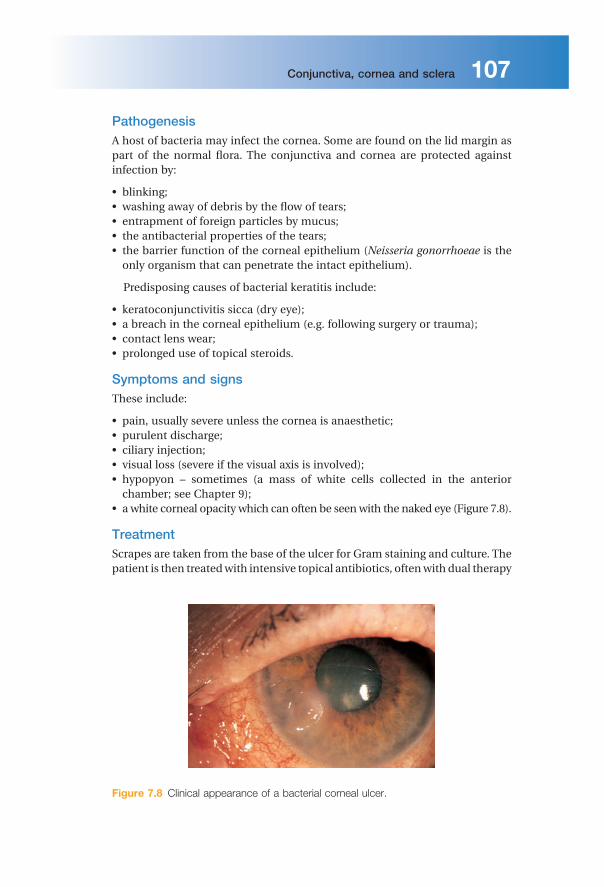
chamber; see Chapter 9 ); • a white corneal opacity which can often be seen with the naked eye (Figure 7.8 ).
Treatment Scrapes are taken from the base of the ulcer for Gram staining and culture. The patient is then treated with intensive topical antibiotics, often with dual therapy
Figure 7.8 Clinical appearance of a bacterial corneal ulcer.

108 Conjunctiva, cornea and sclera
using specially formulated, concentrated, combined preparations to cover most organisms (e.g. cefuroxime against Gram - positive bacteria and gen-tamicin for Gram - negative bacteria). The use of fl uoroquinolones (e.g. cipro-fl oxacin, ofl oxacin, available commercially) as a monotherapy is gaining popularity. The drops are given hourly, day and night, for the fi rst couple of days and are reduced in frequency as clinical improvement occurs. In severe or unresponsive disease the cornea may perforate. This can be treated initially with tissue adhesives (cyanoacrylate glue) and a subsequent corneal graft. A persistent scar may also require a corneal graft to restore vision.
Acanthamoeba k eratitis
This freshwater amoeba is responsible for infective keratitis (Figure 7.9 ). The infection has become more common with the increasing use of soft contact lenses. A painful keratitis with prominent, infi ltrated corneal nerves results. The amoeba can be isolated from the cornea (and from the contact lens case) with a scrape and cultured on special plates impregnated with Escherichia coli . Topical chlorhexidine, polyhexamethylene biguanide (PHMB) and propami-dine are used to treat the condition.
Fungal k eratitis
This is unusual in the UK but more common in warmer climates such as the southern USA. In India it accounts for 30 – 50% of infective keratitis. It should be considered in:
• lack of response to antibacterial therapy in corneal ulceration; • cases of trauma with vegetable matter; • cases associated with the prolonged use of steroids.
Figure 7.9 The clinical appearance of acanthamoeba keratitis. Arrows indicate neurokeratitis.

Conjunctiva, cornea and sclera 109
The corneal opacity appears fl uffy, and satellite lesions may be present. Liquid and solid Sabouraud ’ s medium is used to grow the fungi. Incubation may need to be prolonged. Treatment requires topical antifungal drops such as pimaricin (natamycin) 5%.
Interstitial k eratitis
This term is used for any vascular keratitis that affects the corneal stroma without epithelial involvement. Classically the most common cause was con-genital syphilis, leaving a midstromal scar interlaced with the empty ( ‘ ghost ’ ) blood vessels. Corneal grafting may be required when the opacity is marked and visual acuity reduced.
Corneal d ystrophies These are rare inherited disorders. They affect different layers of the cornea and often affect corneal transparency (Figure 7.10 ). They may be divided into:
• Anterior dystrophies involving the epithelium. These may present with recur-rent corneal erosion.
• Stromal dystrophies presenting with visual loss. If very anterior they may cause corneal erosion and pain.
• Posterior dystrophies which affect the endothelium and cause gradual loss of vision due to corneal oedema. They may also cause pain due to epithelial erosion.
Disorders of s hape
Keratoconus
This is usually a sporadic disorder but may occasionally be inherited. Thinning of the centre of the cornea leads to an ectatic, conical corneal shape, with
Figure 7.10 Example of a corneal dystrophy (granular dystrophy).
Learning o bjectives To understand:
✓ The defi nition of uveitis and the ocular structures involved.
✓ The symptoms, signs, causes and treatment of uveitis.
9 Uveitis
Introduction Infl ammation of the uveal tract (the iris, ciliary body and choroid) has many causes and is termed uveitis (Figure 9.1 ). It is usual for structures adjacent to the infl amed uveal tissue to become involved in the infl ammatory process. It may be classifi ed anatomically:
• Infl ammation of the iris, accompanied by increased vascular permeability, is termed iritis or anterior uveitis (Figure 9.2 ). White cells circulating in the aqueous humour of the anterior chamber can be seen with a slit lamp. Protein, which also leaks into the anterior chamber from the blood vessels, is picked out by its light - scattering properties in the beam of the slit lamp as a ‘ fl are ’ .
• An infl ammation of the ciliary body is termed cyclitis , of the pars plana is pars planitis and of the vitreous is vitritis . As a group these are termed inter-mediate uveitis .
• Infl ammation of the posterior uvea is termed posterior uveitis and may involve the choroid ( choroiditis) , the retina ( retinitis) or both ( chorioretinitis ).
• A panuveitis is present when infl ammatory changes affect the anterior chamber, vitreous and retina and/or the choroid.
Ophthalmology Lecture Notes, Eleventh Edition. Bruce James, Anthony Bron. © 2011 Bruce James, Anthony Bron. Published 2011 by Blackwell Publishing Ltd.

Uveitis 131
Epidemiology The incidence of uveitis is about 15 per 100 000 people. About 75% of these are anterior uveitis.
About 50% of patients with uveitis have an associated systemic disease.
History
The patient may complain of:
• ocular pain (less frequent with posterior uveitis or choroiditis); • photophobia; • blurring of vision; • redness of the eye.
Posterior uveitis may, however, not be painful. The patient must be questioned about other relevant symptoms that may
help determine whether or not there is an associated systemic disease:
• Respiratory symptoms such as shortness of breath, cough, and the nature of any sputum produced (associated sarcoidosis or tuberculosis).
• Skin problems. Erythema nodosum (painful raised red lesions on the arms and shins) may be present in granulomatous diseases such as sarcoidosis and Beh ç et ’ s disease. Patients with Beh ç et ’ s may also have thrombophlebitis, der-matographia and oral and genital ulceration. Psoriasis (in association with arthritis) may be accompanied by uveitis.
• Joint disease. Ankylosing spondylitis with back pain is associated with acute anterior uveitis. In children juvenile chronic arthritis may be associated with uveitis. Reiter ’ s disease (classically urethritis, conjunctivitis and a seronega-tive arthritis) may also be associated with anterior uveitis.
Figure 9.1 External ocular appearance in a patient with anterior uveitis. Note the infl ammatory response at the limbus.
132 Uveitis
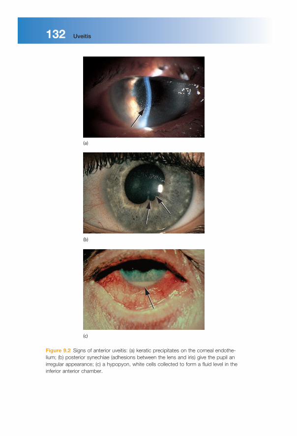
Figure 9.2 Signs of anterior uveitis: (a) keratic precipitates on the corneal endothe-lium; (b) posterior synechiae (adhesions between the lens and iris) give the pupil an irregular appearance; (c) a hypopyon, white cells collected to form a fl uid level in the inferior anterior chamber.
(a)
(b)
(c)
Uveitis 133
• Bowel disease. Occasionally uveitis may be associated with infl ammatory bowel diseases such as ulcerative colitis, Crohn ’ s disease and Whipple ’ s disease.
• Infectious disease. Syphilis with its protean manifestations can cause uveitis (particularly posterior choroiditis). Herpetic disease (herpes simplex and zoster) may also cause uveitis. Cytomegalovirus (CMV) may cause uveitis, particularly in patients with AIDS. Fungal infections and metastatic infec-tions may also cause uveitis, usually in immunocompromised patients.
Signs
On examination:
• The visual acuity may be reduced. • The eye will be infl amed, mostly around the limbus ( ciliary injection ).
Anterior uveitis • Infl ammatory cells may be visible clumped together on the endothelium of
the cornea, particularly inferiorly ( keratitic precipitates or KPs ). • Slit - lamp examination will reveal aqueous cells and a fl are due to exuded
protein. If the infl ammation is severe there may be suffi cient white cells to collect as a fl uid level inferiorly ( hypopyon ).
• The vessels on the iris may be dilated. • The iris may adhere to the lens and bind down the pupil ( posterior synechiae
or PS ). Peripheral anterior synechiae (PAS) between the iris and the trabecu-lar meshwork or cornea may occlude the drainage angle.
• The intraocular pressure may be elevated by PAS or increased aqueous protein.
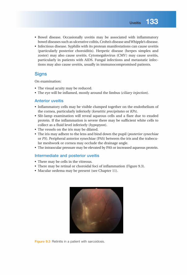
Intermediate and posterior uveitis • There may be cells in the vitreous. • There may be retinal or choroidal foci of infl ammation (Figure 9.3 ). • Macular oedema may be present (see Chapter 11 ).
Figure 9.3 Retinitis in a patient with sarcoidosis.

134 Uveitis
Investigations
These are aimed at determining a systemic association and are directed in part by the type of uveitis present. An anterior uveitis is more likely to be associated with ankylosing spondylitis, and human leucocyte antigen (HLA) typing may help confi rm the diagnosis. The presence of large KPs and possibly nodules on the iris may suggest sarcoidosis: a chest radiograph, serum calcium and serum angiotensin - converting enzyme levels would be appropriate. In toxoplasmic retinochoroiditis the focus of infl ammation often occurs at the margin of an old infl ammatory choroidal scar. A posterior uveitis may have an infectious or systemic infl ammatory cause. Some diseases such as CMV infections in HIV - positive patients have a characteristic appearance, and with an appropriate history may require no further diagnostic tests. Associated symptoms may also point towards a systemic disease (e.g. fever, diarrhoea, weight loss). Not all cases of anterior uveitis require investigation at fi rst presentation unless associated systemic symptoms are present.
Treatment
This is aimed at:
• suppressing infl ammation in the eye, and relieving pain in anterior uveitis; • preventing damage to ocular structures, particularly to the macula and the
optic nerve, which may lead to permanent visual loss.
Steroid therapy is the mainstay of treatment. In anterior uveitis this is deliv-ered by eye drops. However, topical steroids do not effectively penetrate to the posterior segment. Posterior uveitis is therefore treated with systemic steroids, or with steroids injected onto the orbital fl oor or into the sub - Tenon ’ s space (Figure 9.4 ).
In anterior uveitis, dilating the pupil relieves the pain from ciliary spasm and prevents the formation of posterior synechiae by separating it from the anterior lens capsule. Synechiae otherwise interfere with normal dilation of the pupil. Dilation is achieved with mydriatics, e.g. cyclopentolate or atropine drops. Atropine has a prolonged action lasting weeks. An attempt to break any syn-echiae that have formed should be made with initial intensive cyclopentolate and phenylephrine drops. A subconjunctival injection of mydriatics may help to break resistant synechiae.
In posterior uveitis/retinitis visual loss may occur either from destructive processes caused by the retinitis itself (e.g. in toxoplasmosis or CMV infection) or from fl uid accumulation in the layers of the macula (macular oedema). Apart from systemic or injected steroids, specifi c antiviral or antibiotic medication may also be required. Some rare but severe forms of uveitis, e.g. that associated with Beh ç et ’ s disease, may require treatment with other systemic immunosup-pressive drugs such as azathioprine or ciclosporin. Long - term treatment may be necessary.

Uveitis 135
Specifi c c onditions a ssociated with u veitis There is a large number of systemic diseases associated with uveitis. A few of the more common ones are outlined in Table 9.1 .
Ankylosing s pondylitis This is a seronegative (rheumatoid factor negative) spondyloarthropathy causing infl ammation of the tendons, ligaments and joints of the spine. Untreated, the condition leads to a disabling, gross rigidity and fl exion deform-ity of the spine. Genetic factors are involved in the disease. Ninety per cent of patients with uveitis have the tissue type HLA B27, although the prevalence of the disease in people in general with HLA B27 is only 1%. Approximately 20%
Figure 9.4 The principle of a sub - Tenon ’ s injection. A cannula is placed in the potential space between the sclera and Tenon ’ s capsule. Injection of steroid separates the two layers and the steroid surrounds the eye.
Conjunctiva
Cannula
Tenon’s capsule

Learning o bjectives To understand:
✓ The nature of glaucoma.
✓ The difference between primary and secondary glaucoma; open and closed angle glaucoma.
✓ The different symptoms and signs of open and closed angle glaucoma.
✓ The three major forms of glaucoma therapy.
10 Glaucoma
Introduction The glaucomas are a group of diseases causing damage to the optic nerve ( optic neuropathy ) by the effects of raised ocular pressure on the optic nerve head. Independent ischaemia of the optic nerve head may also be important. Axon loss results in visual fi eld defects and a loss of visual acuity if the central visual fi eld is involved.
Pathophysiology The intraocular pressure level is determined by a balance between production and removal of aqueous humour (Figure 10.1 ). Aqueous is actively secreted into
Ophthalmology Lecture Notes, Eleventh Edition. Bruce James, Anthony Bron. © 2011 Bruce James and Anthony Bron. Published 2011 by Blackwell Publishing Ltd.

Glaucoma 147
the posterior chamber by the ciliary processes, by a combination of active transport and ultrafi ltration. It then passes through the pupil into the anterior chamber and leaves the eye, predominantly, via the trabecular meshwork, Schlemm ’ s canal and the episcleral veins to reach the bloodstream ( the conven-tional pathway ). A small but important proportion of the aqueous (4%) drains across the ciliary body into the supra - choroidal space and is absorbed into the venous circulation ( the uveoscleral pathway ).
Two theories have been advanced to explain how elevated intraocular pres-sure, acting at the nerve head, damages the optic nerve fi bres:
1 Raised intraocular pressure causes mechanical damage to the axons. 2 Raised intraocular pressure causes ischaemia of the nerve axons by reducing
blood fl ow at the nerve head. The pathophysiology of glaucoma is probably multifactorial and both mech-
anisms are important.
Classifi cation The mechanism by which aqueous drainage is reduced provides a means to classify the glaucomas. Classifi cation of the primary glaucomas (Figure 10.2 ) is based on whether or not the peripheral iris is:
• clear of the trabecular meshwork ( open angle glaucoma); • covering the meshwork ( closed angle glaucoma).
Figure 10.1 Diagram of the drainage angle, showing routes taken by aqueous from production to absorption.
Schlemm's canal
Trabecular meshworkconventional or major pathway
Uveoscleraloutflowpathway
Ciliary epithelium(aqueous production)

148 Glaucoma
Figure 10.2 Diagram showing the difference between open and closed angle glaucoma. Outfl ow resistance is increased in each case. In open angle glaucoma the obstruction is due to structural changes in the trabecular meshwork. In closed angle glaucoma the peripheral iris blocks the meshwork.
Trabecular meshwork
Trabecular meshwork(covered by iris)
Open angle
Closed angle
Iris
Iris
Classifi cation of the g laucomas
1 Primary glaucoma: • Chronic open angle. • Acute and chronic closed angle.
2 Congenital glaucoma: • Primary. • Secondary to maternal rubella infection. • Secondary to inherited ocular disorders (e.g. aniridia – absence of the iris).
3 Secondary glaucoma (causes): • Trauma. • Ocular surgery. • Associated with other ocular disease (e.g. uveitis). • Raised episcleral venous pressure. • Steroid induced.

Glaucoma 149
Pathogenesis Primary o pen a ngle g laucoma A special contact lens applied to the cornea (a gonioscopy lens) provides a view of the iridocorneal angle with the slit lamp. In open angle glaucoma the trabec-ular meshwork appears normal on gonioscopy but functionally, it offers an increased resistance to the outfl ow of aqueous. This results in an elevated ocular pressure. The causes of outfl ow obstruction include:
• thickening of the trabecular lamellae, which reduces pore size; • reduction in the number of lining trabecular cells; • increased extracellular material in the trabecular meshwork spaces.
A form of glaucoma also exists in which glaucomatous fi eld loss and cupping of the optic disc occurs even though the intraocular pressure is not raised ( normal tension or low tension glaucoma ). It is thought that the optic nerve head in these patients is unusually susceptible to the intraocular pressure and/or has an intrinsically low blood fl ow (Figure 10.3 ).
Conversely, intraocular pressure may be raised without evidence of visual damage or pathological optic disc cupping ( ocular hypertension ). These sub-jects may represent the extreme end of the normal range of intraocular pres-sure; however, a small proportion (about 1% per year) will subsequently develop glaucoma.
Closed a ngle g laucoma This condition occurs in small eyes (i.e. often hypermetropic) which therefore have shallow anterior chambers. In the normal eye the point of contact between
Figure 10.3 The distribution of intraocular pressure in a normal and glaucomatous population.
Perc
enta
ge
10 15 20Intraocular pressure (mmHg)
Skewed distribution in non-glaucomatous population
Wide distribution inglaucomatous population
25 30
Glaucoma 159
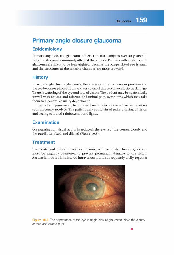
Primary a ngle c losure g laucoma Epidemiology Primary angle closure glaucoma affects 1 in 1000 subjects over 40 years old, with females more commonly affected than males. Patients with angle closure glaucoma are likely to be long - sighted, because the long - sighted eye is small and the structures of the anterior chamber are more crowded.
History In acute angle closure glaucoma, there is an abrupt increase in pressure and the eye becomes photophobic and very painful due to ischaemic tissue damage. There is watering of the eye and loss of vision. The patient may be systemically unwell with nausea and referred abdominal pain, symptoms which may take them to a general casualty department.
Intermittent primary angle closure glaucoma occurs when an acute attack spontaneously resolves. The patient may complain of pain, blurring of vision and seeing coloured rainbows around lights.
Examination On examination visual acuity is reduced, the eye red, the cornea cloudy and the pupil oval, fi xed and dilated (Figure 10.9 ).
Treatment The acute and dramatic rise in pressure seen in angle closure glaucoma must be urgently countered to prevent permanent damage to the vision. Acetazolamide is administered intravenously and subsequently orally, together
Figure 10.9 The appearance of the eye in angle closure glaucoma. Note the cloudy cornea and dilated pupil.
160 Glaucoma
with topical pilocarpine and beta - blockers. Pilocarpine constricts the pupil and draws the peripheral iris out of the angle; the acetazolamide and beta - blocker reduce aqueous secretion and the pressure gradient across the iris. These measures often break the attack and lower intraocular pressure. Subsequent management requires that a small hole ( iridotomy or iridectomy ) be made in the peripheral iris to prevent further attacks. This provides an alternative pathway for fl uid to fl ow from the posterior to the anterior chamber, bypassing the pupil and thus reducing the pressure gradient across the iris. This can be done with a YAG laser or surgically.
If the pressure has been raised for some days the iris becomes adherent to the peripheral cornea ( peripheral anterior synechiae or PAS ). The iridocorneal angle is damaged, and additional medical or surgical measures may be required to lower the ocular pressure. In some patients with cataract, lens extraction with implantation of an intraocular lens may help open the irido-corneal angle.
Secondary g laucoma Secondary glaucomas are much rarer than the primary glaucomas. The symp-toms and signs depend on the rate at which intraocular pressure rises; most are again symptomless. Treatment broadly follows the lines of the primary disease. In secondary glaucoma it is important to treat any underlying cause, e.g. uveitis, which may be responsible for the glaucoma.
In particularly diffi cult cases it may be necessary to selectively ablate the ciliary processes in order to reduce aqueous production. This is done by appli-cation of a laser or cryoprobe to the sclera overlying the processes. Endoscopic techniques are also under development.
Congenital g laucoma This covers a diverse range of disease. It may present at birth or within the fi rst year. Symptoms and signs include:
• excessive tearing, photophobia and blepharospasm; • an increased corneal diameter and enlargement of the globe ( buphthalmos ),
resulting in progressive myopia; • a cloudy cornea due to epithelial and stromal oedema; • splits in Descemet ’ s membrane.
Congenital glaucoma is usually treated surgically. An incision is made into the trabecular meshwork ( goniotomy ) to increase aqueous drainage, or a direct passage between Schlemm ’ s canal and the anterior chamber is created ( trabeculotomy ).

Tropical ophthalmology: eye diseases in the developing world 271
As with all health programmes, it is important fi rst to understand the epide-miology of the problem so that appropriate resources can be determined. There has been increasing emphasis on training local healthcare workers to run pro-grammes, with outside aid providing equipment, consumables and training rather than direct surgical input. Additionally, assessment of the outcome of surgery is emphasized, to maximize the quality of care provided and match that in the developed world.
Tropical d iseases Trachoma Trachoma causes blindness by corneal scarring and is due to infection by Chlamydia trachomatis . It was fi rst described in Egypt in the sixteenth century. Active trachoma has not been seen in Europe since the early twentieth century but was present in America until the 1960s. Trachoma is endemic in 48 coun-tries in Latin America, Africa, the Middle East, Asia and Australasia. It affects more than 84 million people worldwide and 8 million are irreversibly visually impaired. Ten per cent of the world population are at risk, mostly in dry, hot parts of the developing world (Figure 17.2 ). It is responsible for 3% of world blindness. Some 6 million were blind from trachoma in 1990, falling to 2 million in 2003. In some developing communities some 60% of children are affected by active disease. It is endemic in rural communities, in areas of water shortage, where living conditions are crowded and sanitation and hygiene are poor.
Figure 17.2 The worldwide distribution of trachoma.
Widespread active trachomaPockets of active trachoma

272 Tropical ophthalmology: eye diseases in the developing world
Transmission is by the transfer of the elementary bodies of the bacteria, from one person to another, usually from the nasal and ocular secretions of an infected child. This may involve eye to eye transmission with the fi ngers, the sharing of towels, handkerchiefs and bedclothes, coughing and sneezing. In addition, eye - seeking fl ies (e.g. Musca sorbens ) act as important vectors, carry-ing the infective particles from eye or nose, to other individuals. The fi rst infec-tion does not cause serious disease.
Initially, follicles (collections of lymphocytes) appear in the superior tarsal conjunctiva (Figure 17.3 ), followed by papillae. Then the limbal cornea is invaded by superfi cial vessels ( pannu s) with the development of peripheral scarring (Figure 17.4 ). Repeated episodes of infection with C. trachomatis lead to extensive upper tarsal conjunctival scarring, lid deformity and trichiasis, which together exacerbate the corneal changes. Continued re - infection leads to chronic corneal scarring and blindness. This results from:
• Dry eye. Conjunctival scarring blocks lacrimal and meibomian gland duct orifi ces, leading to reduced tear production and excessive evaporative water loss (Figure 17.5 ).
• Reduced lubrication through tear defi ciency and loss of goblet cell mucin. • Corneal trauma, due to cicatricial entropion (in - turning of the lid margins by
tarsal scarring; Figure 17.6 ) and trichiasis, where aberrant lashes abrade the
Figure 17.3 Follicles on the upper tarsus of a patient with trachoma.
Figure 17.4 Peripheral corneal scarring in trachoma.

Tropical ophthalmology: eye diseases in the developing world 273
cornea. This also predisposes to secondary bacterial and fungal corneal infection and scarring.
Public - health intervention has led to the development of the SAFE strategy, which involves:
• S urgery, for those with entropion, everting the lid to move the lashes away from the globe.
• A ntibiotic therapy. A single dose of oral azithromycin is effective in treating infected individuals, but the disease must be eradicated from all individuals in a community to prevent re - infection, and treatment must be repeated yearly. This is expensive, and the disease will recur unless changes are made to the environment in which the organism prospers. This is much harder to achieve.
• F ace washing, which reduces transmission of the disease. The fl ies are less likely to be attracted to the child and the child is less likely to spread Chlamydia by direct contact.
• E nvironmental change. A clean environment reduces the fl y population. Improving the water supply and sanitation, with bore holes and pit latrines, is important in this respect.
Figure 17.5 Scarring of the tarsal conjunctiva in trachoma.
Figure 17.6 Upper lid entropion and corneal scarring in trachoma.