Embed Size (px)
Citation preview

1
Autoimmune Endocrine Disorders: Who and How to EvaluateA Case Based Discussion
Jennifer M. Barker MDPediatric Endocrinology
Children’s Hospital ColoradoUniversity of Colorado Anschutz Medical Campus
Objectives
• Describe the screening guidelines forautoimmune endocrine disorders in:– Type 1 diabetes
– Down syndrome
– Turner syndrome
• Discuss laboratory evaluation for autoimmuneendocrine disorders
• Identify clues in the history and physicalexamination that would lead to further testingfor autoimmune endocrine disorders.
Outline
• Brief overview of the autoimmune disorders
• Overview of autoimmune endocrine diseasesin:
– Type 1 diabetes
– Down syndrome
– Turner syndrome
• Case discussion
• Summary

2
BRIEF OVERVIEW OF AUTOIMMUNE DISORDERS
Thyroid disease
Hypothalamic/pituitary/thyroid axis
Hypothalamus
Pituitary
Thyroid
TRH
TSH
T4 (80‐85%), T3 (15‐20%)
Negative feedback
Hypothyroidism
• Symptoms – Decreased linear growth velocity– Many others!
• Physical examination– Goiter– Decreased relaxation phase of reflexes– Sallow complextion– Round face
• Laboratory evaluation– Low TT4, TT3– Elevated TSH – Autoantibodies:
• anti‐thyroid peroxidase (TPO)• anti‐thyroglobulin (TG)

3
Hyperthyroidism
• Symptoms– Weight loss, heat intolerance, palpitations, etc.– Can be difficult to assess in a patient with developmental disability (Down
syndrome)– But you have to ask!
• Physical examination– Increased heart rate and blood pressure– Hyperdynamic precordium– Increased reflexes
• Laboratory evaluation– Elevated TT4, TT3, FreeT4– Suppressed TSH (less than 0.01)– Autoantibodies:
• Thyroid stimulating immunoglobulin (TSI)• Thyroid receptor antibody (TRab)
Celiac Disease (CD)
• Enteropathy
• Triggered by ingestion of gluten in a susceptibleindividual
• Leads to malabsorption and growth failure inchildren
• Excellent review of Celiac Disease: Fasano A, CatassiC. N Engl J Med 2012;367:2419‐2426
Epidemiology of celiac disease
• 0.6‐1% of the population has celiac disease
• 20% of those with disease are diagnosed
• Increased in:
– Type 1 diabetes (3‐16%)
– Hashimoto’s thyroiditis (5%)
– Down syndrome (5%)
– Turner syndrome (3%)
– IgA deficiency (9%)
– Those with a first degree relative with CD (10‐15%)

4
Clinical Presentation of celiac disease
• Classic –– diarrhea– malnutrition– failure to thrive
• Atypical –– poor growth, delayed puberty– evidence for malnutrition– neurologic and psychiatric abnormalities
• Physical examination – May be normal– Can have specific rashes
• Dermatitis Herpetiformis ‐ severe, itchy, blistering skin rash– 15‐25% of people with celiac disease will have it
Testing forCeliac Disease
• Tissue transglutaminase (IgA)– 95% sensitive and specific– First line screening test– Get with IgA level to rule out IgA deficiency
• Antiendomesial antibodies (IgA)– 90% sensitive, 98% specific– Useful with an uncertain diagnosis
• Deamidated gliadin peptides (IgG)– IgA deficiency and young children
• Tissue transglutaminase (IgG)– IgA defiency
• HLA‐DQ2 or HLA‐DQ 8 (genetic testing)– High negative predictive value
Diagnosis of celiac disease
• High index of clinical suspicion
• Screening labs may indicate malabsorption
– Anemia
– Vitamin D deficiency
• Antibody screen
• Confirmatory small intestinal biopsy
• Improvement in antibody levels and biopsy ongluten free diet
5
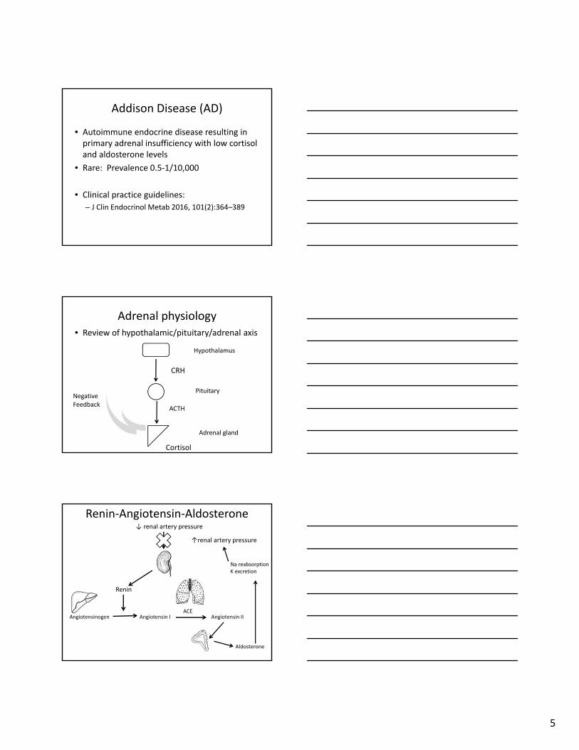
Addison Disease (AD)
• Autoimmune endocrine disease resulting inprimary adrenal insufficiency with low cortisoland aldosterone levels
• Rare: Prevalence 0.5‐1/10,000
• Clinical practice guidelines:
– J Clin Endocrinol Metab 2016, 101(2):364–389
Adrenal physiology• Review of hypothalamic/pituitary/adrenal axis
Hypothalamus
Pituitary
Adrenal gland
CRH
ACTH
Cortisol
Negative Feedback
Renin‐Angiotensin‐Aldosterone
Renin
Angiotensinogen Angiotensin I Angiotensin II
Aldosterone
Na reabsorptionK excretion
ACE
↓ renal artery pressure
↑renal artery pressure

6
Clinical Presentation of AD
• Non‐specific symptoms:– fatigue, weight loss, nausea, pre‐syncope, syncope
• Increased pigment• Salt craving
• PE with orthostatic hypotension,increased pigment
Note: symptoms may exist for years before the diagnosis is considered
AD diagnosis
• Abnormalities of electrolytes:– Hyponatremia
– Hyperkalemia
– Acidosis
• Elevated ACTH and PRA
• Positive 21‐OH antibody
• May require high dose ACTH stimulation testingto diagnose:– Expect cortisol level 60 minutes after cosyntropin tobe greater than 18‐20 mcg/dL
TYPE 1 DIABETES

7
Type 1 diabetes (T1D)
• T1D is an autoimmune disease resulting indestruction of the pancreatic β‐ cells
• T1D is associated with:
– Autoimmune thyroid disease
• Hypothyroidism > Graves disease
– Celiac disease
– Addison’s disease
– Other?
Thyroid disease and T1D
• ≈ 20‐30% will have + TPO/TG antibodies
• ↑ in females vs. males
• ↑ by age and diabetes dura on
• Risk for thyroid disease in those with +antibodies ↑ overtime
– 80% with + antibodies have hypothyroidism after 20 years of follow‐up
Screening for thyroid disease in T1D• ADA (2016 Guidelines)
– Consider screening for TPO and TG antibodies (at diagnosis)
– Measuring TSH concentrations soon after diagnosis of T1D,after metabolic control has been established, is reasonable.
– Consider rechecking TSH every 1‐2 years.
– Monitor for and screen TSH with:
• symptoms of thyroid dysfunction,
• thyromegaly
• abnormal growth rate
• ISPAD (2014 Guidelines)– Screen TSH and TPO or TG antibodies at diagnosis
– Monitor TSH every 2 year in antibodies negative patients
– Monitor TSH more frequently (? How much more frequently) in antibody positive patients

8
• Overlapping genetic risk– HLA DQ2/DQ8
• Multiple populations with T1D screened with TTgantibody – show prevalence of 3‐16% positive– Biopsy positive in approximately 50‐75% of those biopsied
– Many of those positive are asymptomatic
• Most cases are identified within the first 5 yearsof diagnosis of T1D
• Risk has been related to ↓ age of onset ofdiabetes
Celiac disease and T1D
Celiac disease and T1D
• Children identified by screening are oftenasymptomatic
• No increased risk for severe hypoglycemia or DKA
• Pa ents with T1D and CD ↑ risk for thyroid disease• Diabetes Care. 2016 Mar;39(3):371‐5
• CD and T1D is associated with ↓ BMD but no ↑infracture rate
• J Pediatr. 2016 Feb;169:44‐48
• J Pediatr. 2016 Feb;169:49‐54
• Treatment adds financial and logistical burden tofamilies already burdened with diabetes management
• ADA (2016 guidelines)– “Consider” screening TTg, IgA and/or deaminated gliadin
antibodies at diagnosis– “Consider screening in children who have a first‐degree relative
with celiac disease, growth failure, weight loss, failure to gain weight, diarrhea, flatulence, abdominal pain, or signs of malabsorption or in children with frequent unexplained hypoglycemia or deterioration in glycemic control”
• ISPAD (2014 guidelines)– Screen at diagnosis with TTg or EMA and IgA– Screen every 1‐2 years – Screen sooner with symptoms, growth concerns, glycemic
variability
T1D and celiac disease –recommendations for screening

9
Addison’s disease and T1D
• 0.5‐1% of patients with T1D have AD
• 1.5‐2% of patients with T1D have positive 21‐OH antibodies
• Patients with diabetes with AD can presentwith symptoms of decreased insulin dose,decreased hemoglobin A1c, increasedhypoglycemia
• No recommended routine screening
DOWN SYNDROME
Down syndrome and thyroid disease
• Congenital hypothyroidism 1–7%
• Acquired primary hypothyroidism 0.3–3%
• Subclinical hypothyroidism 12.5–33%
• Hyperthyroidism up to 2%
• ↑ risk for acquired primary hypothyroidismwith + TPO antibodies

10
Recommendations for screening thyroid function in DS
• AAP guidelines (2011)
– Newborn screen for thyroid function
– Measure TSH at 6 months, 12 months and thenannually
• Risk for celiac disease in patients with Downsyndrome 5‐10% across multiple populations
• No difference noted in HLA predisposition inpatients with celiac disease and Downsyndrome compared with patients with celiacdisease and no Down syndrome
• Symptoms may be difficult to assess
Down syndrome and celiac disease
• AAP guidelines (2011)
– Screen for symptoms
– Check TTg with symptoms
• North American Society for PediatricGastroenterology, Hepatology and Nutrition
– At age 3 or after at least 1 year of gluten exposure
– Repeat screening at some interval
Down syndrome and celiac disease screening recommendations

11
TURNER SYNDROME
Turner syndrome and thyroid disease
• Primary hypothyroidism 20%
– Can be diagnosed as a young child
• Graves disease 1‐2%
• ↑ risk of overt hypothyroidism with + TPOantibodies in those with subclinicalhypothyroidism
Recommendations for screening for thyroid disease in TS
• Turner syndrome consensus study group(2007)
– Screen all girls with TSH, T4 annually starting at age 4 years
• Guidelines are consistent across groups

12
• Girls and women with Turner syndrome have aknown increase in hypothyroidism
• Risk for celiac disease 2‐5% across differentpopulations
• Commonly diagnosed when adult women withTS re‐present as adults for comprehensivecare
• Similar HLA predisposition in the populationwith TS
Turner syndrome and celiac disease
• Turner syndrome consensus study group– At diagnosis (> age 4 years)
– Screen with TTg
– Repeat screening for symptoms of celiac disease andevery 2‐5 years
• North American Society for PediatricGastroenterology, Hepatology and Nutrition– Screen at diagnosis, if diagnosis is at age 3 or after atleast 1 year of gluten exposure
– Repeat screening at some interval
Recommendations for screening
BONUS: THYROID AND CELIAC

13
• Overlapping genetic risk– DQ2
• Risk for celiac disease is increased in patientswith both hypo and hyperthyroidism
• Patients with celiac disease are at an increasedrisk for autoimmune thyroid disease
• Patients with celiac disease and hypothyroidismhave increased requirements for thyroidhormone replacement which improved with GFD
• Risk ranges from 2‐5%
Autoimmune thyroid disease and celiac disease
CASES
Patient with type 1 diabetes
• Age 13 months presented in severe DKA atDx, pH 6.89, HCO3 <5.
• Labs at diagnosis of T1D– IgA TTG Latest Range: ‐0.2‐0.050 ‐0.007
– HYD21 Latest Range: ‐0.2‐0.150 ‐0.027
– mIAA Latest Range: ‐0.2‐0.010 0.022 (A)
– GADA Latest Range: 0.0‐20 DK 218 (A)
– IA2 Latest Range: 0.0‐5 DK 0
– ZNT8RW Latest Range: ‐0.2‐0.030 0.007
Age 19 monthsWeight loss, vomiting, failure to thrive
Ref. Range 2/9/2012 06:26Immunoglobulin A Latest Range: 15‐111 mg/dL 253 (H)IgA TTG Index Value No range found 172.1IgA TTG Antibody Latest Range: NEGATIVE POSITIVE (H)

14
FR
• CC: fatigue and easy bruising
• HPI: 13 year old female with
– Bruising X 2 weeks
– Fatigue X 2‐4 weeks
– Diffuse abdominal pain X 4‐5 months ‐ increasing
– Weight loss (7‐8 lbs)
FR continued
• PMH: seasonal allergies; MCL tear
• FH: MOC with hypothyroidism and vitiligo
• PE: skin with bruising/purpura (not noted tobe hyperpigmented); T2 breast development;remainder normal
Laboratory evaluation
• Day PTA Na = 121 mEq/L; glu = 412 mg/dL; Plt = 19
• ED: Na = 124 mEq/L; glu = 364 mg/dL; Plt = 11
• Hospital course – Diagnosis of diabetes confirmed when high BG persisted
– Etiology of hyponatremia unclear
• ? Chronic hyperglycemia
• ? Renal losses
– Na remained low (at 126 mEq/L) at d/c
– Platelets remained low
– Thyroid function normal
• What else do you want to know?

15
Further follow‐up
• Laboratory Analysis
– Random cortisol of 23 ug/dL
– Aldosterone 3.6 ng/dL (supine 2‐22; upright 4‐48)
– Plasma renin activity 12814 ng/dL/hr (50‐300)
• Positive 21‐OH antibody
• Hyponatremia resolved (1/7/08 Na = 135)
• Continued muscle pain, fatigue
• ACTH stim testing 2/08
Take home message
• Consider AD in patients with other autoimmuneendocrine disoders
• Marked by presence of 21‐hydroxylase antibodies
• Clinically:
– Hyponatremia not responsive to fluid resuscitation
– In a known diabetic, increased hypoglycemia, decreasing insulin dose, weight loss, decreasing A1c
– Non‐specific complaints of abdominal pain, muscle pain,increased pigmentation
– Can have a very insidious onset

16
Patient with Turner syndrome and Hypothyroidism
11 year old male with celiac diseas
16 year old male with Down syndrome, T1D
• Developed progressive muscle weakness with refusal to walk• Laboratory evaluation revealed:
– Calcium = 4.3 mg/dL (normal 8.9‐10.7)– Phos = 2.8 mg/dL (normal 3‐5.2 )– Alk Phos = 1063 U/L (normal 58‐237)
• Laboratory evaluation suggestive of vitamin D deficiency – But why?• Further laboratory evaluation confirmed multiple vitamin
deficiencies including vitamin D• TTg was markedly elevated = 4879 (greater than 30 is strongly
positive!)• Treatment with gluten free diet and dietary supplementation of
vitamin D and calcium resulted in improved calcium and Vitamin Dand ability to walk

17
SUMMARY
Historical clues
• Screen patients with T1D, Down syndrome and Turner syndrome for symptoms:– Thyroid disease
• Symptoms can be non‐specific
– Celiac disease• Children may be asymptomatic
– Addison’s disease• Unique symptoms in T1D: increased hypoglycemia, decreased insulin requirement and A1c
• Growth chart can provide valuable information– Decreased height (hypothyroidism)– Weight loss (celiac disease, Addison’s disease,hyperthyroidism)
Physical examination findings
• Vitals
– Increased heart rate and blood pressure –hyperthyroidism
– Orthostatic hypotension – Addison disease
• Thyroid enlargement
• Skin
– Pigment – Addison disease
– Dermatitis Herpetiformis – Celiac disease

18
Disorder Thyroid Celiac
Type 1 diabetes Screen annually Screen at onset and withsymptomsSome recommend screening every several years
Down syndrome Screen with NBS, 6 months 12 months of age and then annually
Screen once eating gluten and after age 3 yearsScreen every several years
Turner syndrome Screen annually: starting at age 4 years
Screen once eating gluten and after age 3 yearsScreen every several years
No recommendations for screening for thyroid in patients with celiacNo recommendations for screening for celiac in patients with thyroidNo recommendations for screening for Addison’s disease in patients with T1D