Embed Size (px)
Citation preview
OX THE VAQUS NERVES OF HYRAX CAPENSIS. 14Y
8. On tlie Vayus and Syriipatlietic Nerves of Hyra.e cupensis. By CHARLES F. SONNTAG, M.D., F.Z.E., Anatoniist to the Society.
[Received Jariwy 23, 1821 : llend February 21, 1022.1
(Text -figures 6-8.)
The present paper is based on the examiiiation of several speci- mens of Ifyiuz capensis, both immature and fully adult, which died in the Society's Gardens. Variations were observed in the branches of their vagus and sympathetic nerves ; and the con- ditions present in some of the animals are more complex than those already described in my papers on the Marsupialia (3, 4) and Edentatn (5). They are also more complex than those in many animals belonging to other orders.
The anterior cervical parts of the nerves are placed deeply, and it is necessary to remove the wide ascending rami of the mandible to gain access to them and their branches.
I n nll examples the cervical parts of t,he vagus and sympathetic nerves are fused on both sides to form vago-sympathetic cords, the union taking place a t the level of the middle of the thyroid cartilage. The cords are resolved again into their component element's at the root of the neck, but the separation is ueunlly higher on the left side. After they have parted, however, branches of communicsztion inay run between them.
THE VAGUS NERVES. Course :-As it emerges from the foraiiieir Incerum posticnui
each ragus has the usual relations to the glosso-pli:~ryngeal,spinal accessory and hypoglossal nerves. and it communicates with theni anrl the superior cervical ganglion by well-marked branches (text-fig. 6. CIX, CXI, CXII and S.C.G). No ganglion nodosum is present in the neck, as in Tarnmdua tetrudmtyh (d), or within the foramen lacerum posticum. It then course5 postero-mesially, gives off communicating branches to the cervical plexus (C.C.P) anrl unites with the cervical sympathetic to form the vago- syiupathetic cord (V-S) at the level of the middle of the thyroid cnhlage. And the nerve np to this point is internal to the mandible. The vago-sympathetic cords break up again i n tlie posterior third of the neck; tlie vagus half runs still postero- mesially and enters the thorax. Owing to the high level reacliecl by the heart and aorta the first part of the intra-thoracic courbe is short, but the calibre of the left nerve remains thicker than that of the right one.
Hehind the roots of the lungs thc vagi are united by a sinuous cord which is as tl.ick as the left vagus (text-fig. 7, bv), and tlie nerves emerge again from its extremities, so the arrangement is H-shaped. The left vagw is replaced by iiglit and left branclies.

150 DR. C. F. 80h”TAG ON THE VAGUS AND
The former runs along the rentrnl border of the esophagus, passes through the esophageal opening in the diaphragm and ramifies over the anterior and ventral aspects of the body a i d fiindus of the etoniach. The latter runs along the dorsal border of the oesophagus and unites with the riglit vagus which describes n wide curve to meet it j and the combined trunk ends on the dorsal aspect of the stomach. Numerous communicating branches link the vngi to one another and to the celiac plexus, and some go to the duodenum.
Text-figure 6.
The Cervical and Upper Thoracic Parts *of tbe Vagus atid Sjmpathetic Nerves V : vagiis ; S : s~.mpsthc~tic; Mn : line of mandible. of Hymx capensis.
Other letters in text.
B ~ f f i n c l ~ s :--Numerous branclies of communication and distri- hution are again given off, many of which are very complex.
The pharyngeal n c m e s (text-fig. 6, a) are given off from the trunk of the vagus aucl the loop (C) connecting its superior and recurrent laryngeal branches. They form :t plexus with branches of the glosso-pharyngeal nerve and sympntlietic. I n some speci- mens the laryngeal nerves do not form a loop which gives off pharyngeal branches.

SYMPATHETIC NERVES OF HYRAX CAPENSIS. 151
The sicperior lrcryngeol aerwe (h) is replaced by a strong cord which unites with one of the divisions of the recurrent nerve. This cord gives off internal laryngeal (i.Zn), two external laryn- geal (e.Z.n), two thyroid ( t .y .n) , and three external pharyngeal (a) nerves. Small branches ( w m ) accompany the common carotid artery. The internal laryngeal branch enters the larynx through the thyro-hyoid interval.
The lefb reczci-rent nerve ( e ) is giren off in the common positiori. It encircles the aortic arch and runs antero-1atern.lly t,hrough the neck. 111 the middle of the neck i t shows a fusiform expansion (E) and divides into a mesial nerve (R.L.N), wliicli is the trite inferior lnryngeal nerve, and a. lateral cord (C) which unites with the superior laryngeal nerve to form a loop, whose upper part has been described above. Nunierous tracheal (TN) and cesopha- geal nerves (ON) are given off. They form plexuses of which tlie tracheal one is the more superficial. And the esophageal plexus anastomoses with the cesophn.gea1 branches of the Tight ~ecurrent nerwe (d). The lntt,er n.lso forms a loop with the right superior Iarynged nerve, but there is no fusiform expansion. And botli recurrent nerves give off cardiac branches (C.N) which are thicker arid more numeiwis on the left side.
I n some specimens these loops are absent, and there is no external communication between the superior mid recurrent laryngeal nerves.
‘I’he existence of anastomoses between the laryngeal nerves ill Man has long been known. And Landois atid Stirling (2) suni- marked our knon-ledge as follows :-“ A connecting branch rwls from the superior litryngeal to the inferior (the nnastoniosis of (Men), which occasionally gives oft’ sensory branches to the upper half of the trachea (sometimes to the larynx 2) ; perhaps also to the esophagus (Longet), and sensory fibres (?) for the muscles of the larynx supplied by the recurrent laryngeal. According to Franpois Frank, sensory fibres pass by this anastomosis from the recurrent into t,lie superior laryngeal. According to Waller and Burcklitird, the motor fibres of both laryngeal nerves are a.11 de- rived from the accessorius, while Chauvenu maintains that the crico-thyroid is an exception.” In a recent paper Dilwortli (1) described anti figured a well- marked branch running betis-een the recurrent n.nd internal laryngeal nerves, and says: ‘‘ . . . . tlie lni-yngeal nerves are really a plexus of nerves. Jus t as the vagus hre:iks up into its various plexuses in the body, it does the same in tlie larynx. It is a highly modified plexus. I would further suggest tliat it arose by the 1:wynx separating n strand of fibres from tmlie vagus-that this strand is represented by the coiltinuow nerve joining the internal arid recurrent laryngeal, n.ntl that the separation from this straud of further fibres forms the various iierves of the larnyx.” The conditioris sliown in text-fig. 6 sup- port, nilworth’s views, and I woultl extend his views of tlie origin of the Inryngen.1 nerves from the continuous strand to account for the origin of the tracheal, msopha.gea1, n.nd thyroid nerves.
152 DR. C. F. SOSXTAQ ON TLIE VAQUS AND
The Cardiac J w v e s (text-fig. G , f ) arise on the left side from the vagus distal to the origin of the left recurrent nerve, but the right ones arise from the vagus und its recnrrent branch. They rnn to the cardiac plexus wherein they become associated with the sympathetic ( c . B . ~ ) .
The mophapal nerves (text-figs. 6, ON, 7, ON, ant1 8, VB) arise in the neck from the cords nniting the laryngeal nerves. I n the upper part of the thorax they are branches of the right vag'us, but they come from the two divisions of the left ragus distal to the roots of the lung&
The tracheal icerwes (text-figs. 6, T.N and 7, T.N) arise from the laryngeal cords in the neck, but its terminal part and the main bronchi receive their nerve-supply from the thick bridge between the vagi (b).
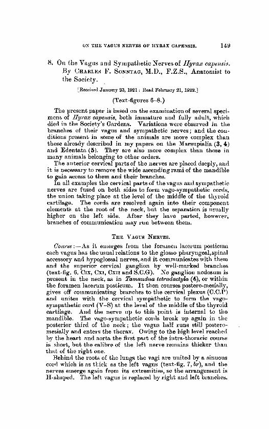
Text-figure 7.
The Lower Thoracic and Abdoinind parts of the Vngus Nerves of Hyjvx capensis. L.V: parts of left rngns; R.V: right vngos. Other letters in test.
The pzslmoizary pleauses (test-fig. 7, g):--The right one is formed by two branches from the right vngus and communi- cating toigs from the deep cardiac plexns. But the left one consists of many braiicheh fiom the bridge between the two vagi. The latter sends a 1)ranc.h to the no?-tic p1c.m~ (A.P), which receives inany twigs froin the left vagus. ,4nd no ganglia were fomitl in the pnlmonary and aortic plexnses.
The Gastric ,Verws (text-fig. 7 ) :-The left 7 ngus reaches the stoniach along the ventral boi der of the ci>sophagus and breaks "p into two branches. The first gives off twigs which run along

SYMPAFHETIC NERVES OF HYRAX CAPESSIS. 153
the lesser curvature, arid the latter supplies the fundus. Both groups anastoniose with branches from the right vagus, antl with the celiac plexus. The cord formed by the right vagus antl pait of the left; oiie supplies the dorsal aspect of tlie stomach alniost to the pylorns. I t s branches anastornose with those of the left vagus, and t,migs from the celiac plexus. A well-marked branch (splanchnic?) runs to the left sympathetic cord (A). Many of the cesophageal nerves run posteriorly and anastomose with both vagi, so there is a close network round the lower end of tlie tcsophagus and over the left part of the stomach.
THE SYMPATHETIC NERVES.
The AS'qierioi- C'erakol GuiEgIion (text-fig. 6. S.C.G) is round or oval, and flat. It gives off an internal carntid nerve of con- siderable length (I.c.N) which acconipmies tlie internal carotid artery into the skull. The nerve communicates with the glosso- pharyngeal and hypoglossal nei'res, and the ganglion is connected to the vagus, cervical plexus, and loop between the superior and recurrent laryngeal nerves. But no connection exists between either and the spinal accessory nerve. From the posterior pole of the ganglion the sympathetic cord runs laterally and joins the vagus a t tlie level of the middle of tlie thyroid cartilage to form the vago-symp:ttlietic cord.
I n IIyruz, as in all aniinals possessing the vago-sympathetic cord, no middle cervical ganglion is present, and 110 direct ranii coinniunicantes run to the middle cei vical nerves.
The sympnthetic separates again in the posterior part of the iieck, but communicates with the vagus after the partition. It exhibits a well-marked hLferior c w v i c d ganglion, (I.C.0) on the left side, but none on the right. From the ganglion branches of ro in inkhi t ion run to the brachial plexus, but I was unable to cletect any coriiniunication between tlie right sympathetic arid brachial nerves, or between either cord and phrenic nerves. The right sympathetic gives off H. branch wliicli acconipanies the vertebral artery (V.A.N), and a loop connects it to the right recurrent nerve. And as no branch runs directly to the cztrdinc plexus, the right recurrent and right vagus nerves conduct all the right sympathetic filaments to the heart.
The left inferior cervical ganglion gives off n medium-sizetl cardiac I,ranch (c.B.s) which runs almost parallel to the left recurrent iierve, passes to tlie dorsal aspect of the aortic arch and ends in the deep c:wdiac plexus.
The 811111~1218 qf Vieussevs (A.V) is present only on the left hide, arid the right sympathetic cord passes in front of the right subclavian aitery.
The left gmpmthetic thomcic cord (text-fig. 8, L.S) has fern g:tiiglia in its hnterior part, and it forms 100)~. A t the level of the middle of the root of the left lung it divides into Iateial and mesial divisions. The former possesses a loop and a small ganglion,

154 DR. C. F. SONPI’TAQ ON THE VAOUS AHD
and is continued as a thick nerve which ends in the cteliac grng- lion (text-fig. 8, c.G). The latter ends in a large ganglion whence two nerves emerge. One joins the lateral division and the other becomes the garigliated cord of the sympathetic. In the thorax it gives branches to the aortic plexus(A.P) and along branch runs to the coeliac ganglion. The lateral division of the cord, and the coeliac branch of its mesial half constitute splanchnic nerves (8p.N).
Text-figure 8.
Tlw Thoracic and Abdominal Sympathetic of H y a s capejrsis. V.B : aortic Other nnd u?sq)hngcal branclics of the vngi; L.D : line of diaphragm.
letters iii test.
The abdominal p:wt of the left sympathetic (text-figs. 7 and 8) possesses four gnnglia. It gives offtwo groups of lateral branches to the semilunar ganglion, ranii cornmuiiicnntes to the lumbar nerves, and branches t o the aortic plexus. It lias a great ten- dency to suldivision.
Tlie tlioyacic pcwt of the Tight sympathetic (text-fig. 8, R.S) h:ts few ganglia in tlie anterior part, and its posterior psrt is very coiiiplicatetl, but not so mucli as the left cord. It divides into mesial and lateral parts and these are united a t intervals by coiiinioii ganglia or communicating hmnclies. The lateml division ultimately continues the cord back to t,he sacrum. Tlie mesial division is thick and strong. Branches of the cord run to
SSYPATHETIC NERVES OF UYRAX CAPENSIS. 155
tlie aortic plexus (A.P), vena azygos major ( A d ) , thoracic nerves, antl left synipatlietic.
I n tlie abdomen the mesial division divides into two, and the following are the fibres of distribution :-
1. To the ctrliktc ganglion (A). 2. Hepatic plexus (contained in A). 3. Phrenic plexus (contained in A). 4. Fibres to the right renal plexus (R.E.P). 5. Fibres to Meckel’s Tract and Duodenum (b1.B). 6. Fibres to the colon (C.B). The abdominal part lies close to the left sympathetic and fibises
connect them. It gives OK the right renal plexus (E.R.P), right spermatic plexus ( R S P ) , and filament,s to the aortic plexus.
The Cwdinc Plaxt~s consists alinost entirely of the deep part, the snperficid plexus consisting only of a few filaments derived from it. Tlie nerves entering into it are :-
1. Three branclies of the left vagns. 2. A twig from the left recurrent nerve. 3. A branch from the inferior cervical ganglion of the left
4. Two branches froiii the riglit recurrent nerve. 5. Two branches from the riglit va.gns. No ganglia are present, nor nre there sepa.rat;e depressor nerves ;
but tliose may be contained in the loops uniting the snperior antl recurrent laryngeal nerves. OEshoots of the plexus can be traced into the pulrnonary and tfiwheal plexuses.
The Aortic Plexus (A.P) derives its fibres from the two syni1i:t- thetic cords, the bridge between the vagi iii the tliorax, m d the solar plexus.
The S’olar Plrxus (test-fig. 7):-In the upper p r t of the abdomen tlorsad of the stomach there is :I. bro:itl band of fibres with a large reddish-brown aeinilunar gnnglion nt its left extreniity. Tlie fibres are closely packed and hare wide con- nections as follows :-
1. The splnnchiiic nerves froin the left sympathetic cortl entering the anterior pole of the ganglion (A).
2. Two bnntlles of fibres froiii the left synipRtlietic cortl entering the niesial border of the pnglion (B).
3. Fibres from the right v n p s to tlie large left splanclinir nerve (C). 4. Filwes froin the right mgus to tlie semilunar ganglion (I)). 5. Fibres from the left vngus to the semilunar gaiigliou (E). 6. k’ilres from the riglit vagus rnnning into the plexus aiid
7. Fibres froin the semilun:w ga.nglion to the splenic plesiis (G). 8. Branclies fi-oni tlie solar to the splenic plexus (H). 9. Hranclies from the right sympathetic to the semiluii;~r
10. Hepatic plexus (J) giving off pyloro-duodenal nerves (K). 11. Diaplimgina.tic plexus (L).
sj-mpnthet’ic.
turning down to the splenic plexns (F).
ganglion (I).

1 56 ON THE VAGGS NERVES OF IIYBAX CAPEXSIS.
SUMMARY AXL) CONCLUSIONS. 1 . I n all examples of Hyrax capensis the cervical parts of the
vngus and symp~thetic nerves are fused. And the ganglion nodosum is frequently absent in the neck.
2. The recurrent and superior laryngeal nerves are frequently, hit not always, connectetl by a loop.
3. There is no separate depressor nerve. 4. The internal carotid nerve has a very long cervical course. 5. The right recurrent nerve has well-marked cardiac branches. 6. The left sympathetic alone has an inferior cervical ganglion
7. The posterior thoracic parts of the vagi have a complicated
8. The thoracic sympathetic cords have few ganglia, and there
9. The right sympttlietic is distributed to the colon, and the
and an Annulus of Vieussens.
arrangement.
is only one semilunar ganglion.
left one and coeliac plexus supply tlie small intestines.
BIBLIOGRAPHY.
1. l)ILwORT,U, T. F. M.--Journal of Anatomy, 1021, pp. 48-52. 2. LAXDOIS and STIRLING.-A Text-book of Human Physio-
3. SOSSTAG, 0. F.-Proc. Zool. SOC. London, 1921, pp. 572-575.
5. -, Proc. Zool. SOC. London, 1922, pi?. 99-180.
logy, vol. ii.
4. , Proc. Zool. SOC. London, 1921, pp. 873-876.