Embed Size (px)
Citation preview
7/23/2016
1
“I Can’t Breathe” Air Leak Syndromes
Pulmonary Hypertension
Cor pulmonale
Cheryl Herrmann, APN, CCRN, CCNS-CSC-CMC
Cardiac Patient Care Problems (47%)
Other Patient Problems (21%)
A. Acute Coronary Syndrome
B. Dysrhythmias
C. Heart Failure
D. Other Cardiac Issues◦ Cardiomyopathies
◦ Pulmonary Hypertension
E. Vascular Issues
A. Acute Pulmonary Embolus
B. Acute Respiratory FailureC. Acute Lung Injury
(ALI/ARDS)
D. Cor PulmonaleE. PneumothoraxF. Hemothorax
� Air Leak Syndromes
� Pulmonary Vasodilators
Pneumothorax
Pneumopericardium
Pneumomediastinum
4
5
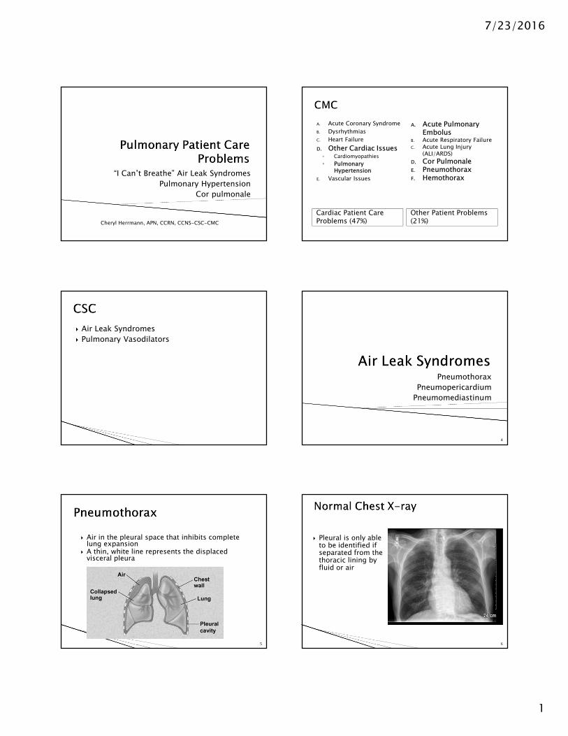
� Air in the pleural space that inhibits complete lung expansion
� A thin, white line represents the displaced visceral pleura
6
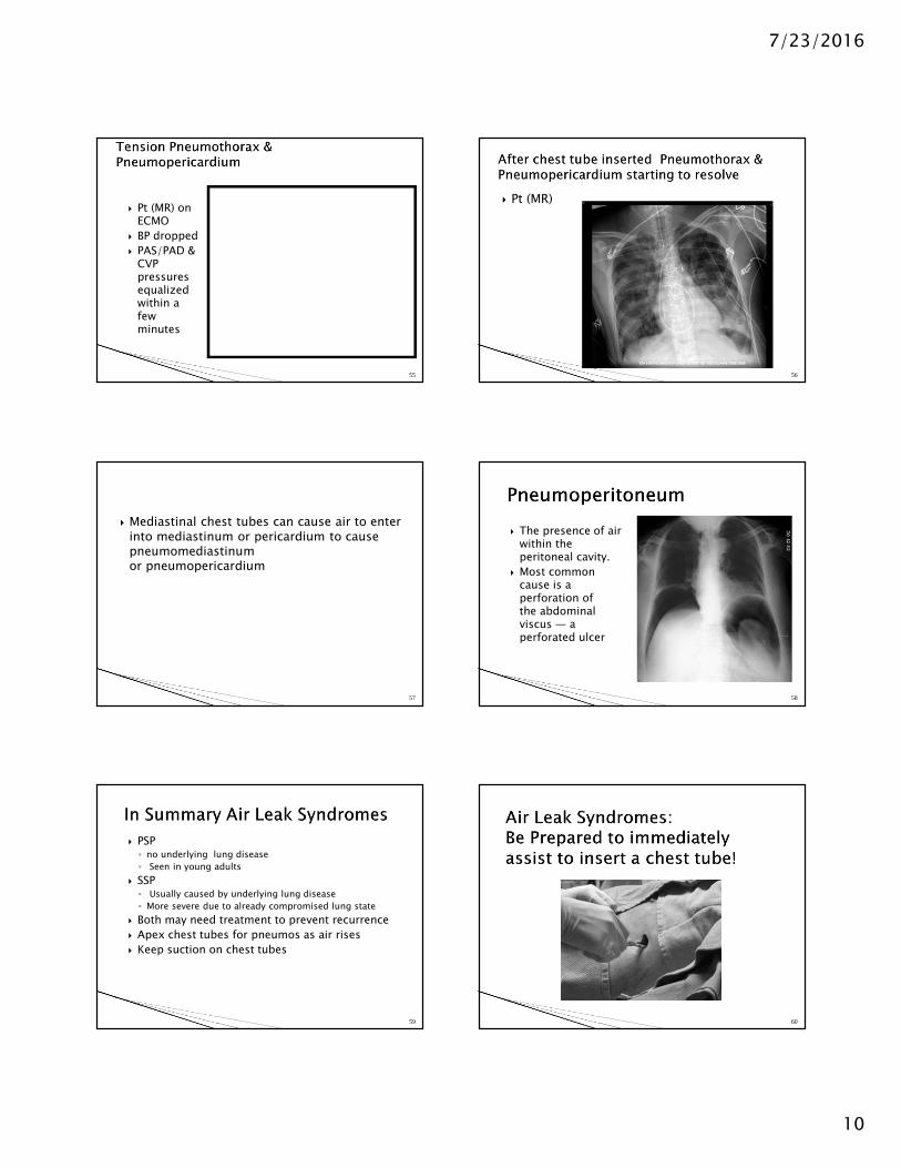
� Pleural is only able to be identified if separated from the thoracic lining by fluid or air

7/23/2016
2
7
� Left Pneumothorax on CT scan
� 7-16 AK
� Diminished or absent lung sounds over the affected lung
� Dyspnea
� Tachypnea
� Acute pain on affected side of the chest
� Decreased Sp02 & p02
� Subcutaneous emphysema
� Black area over lung field with no lung markings on CXR
9
� Causes:◦ Direct injury to the lung during surgery◦ Line insertion causing tear in lung ◦ Baratrauma during positive pressure
ventilation◦ Occurs more on left due to LIMA disection
(CABG pt)
� Treatment:◦ Chest tube insertion if greater than 10 – 15 %◦ If tension pneumothorax ---- it is a medical
EMERGENCY and needs immediate needle decompression
10
� Distended neck veins
� Hypotension
� Tracheal deviation
Note compressed swan ganz
1. Primary pneumothorax
2. Secondary pneumothorax
3. Iatrogenic pneumothorax
4. Pneumomediastinum
5. Pneumopericardium
6. Hydropneumothorax
11
� Occurs without a precipitating event in a person who does not have lung disease
� Actually, most individuals with PSP have unrecognized lung disease
12

7/23/2016
3
� Incidence◦ 7.4 per 100,000
◦ Greater in men than women
� Risk Factors◦ Smoking
◦ Family History
◦ Marfan’s Syndrome
◦ Homocystinuria
◦ Thoracic endometriosis
13
� Usually occurs at rest
� Sudden onset of dyspnea and pleuritic chest pain
� Symptoms related to the volume of air in the pleural space
� Hypoxemia
� Rarely hypercapnia – no underlying lung disease
� Acute respiratory alkalosis if pain, anxiety and hypoxemia
� Age = early 20’s, rare after 40
14
� Initial◦ Removal of air from the pleural space
� Needle aspiration, if small
� Chest tube, if large
◦ Supplemental oxygen
� Subsequent◦ Preventing reoccurrence
◦ Reoccurance is 35 - 54%
15
� If after 6 hours the pneumothorax reabsorbs, patient may be sent home
� Needs to live close to emergency medical center if d/c in 6 hours.
16
� Air in the pleural space is reabsorbed when the communication between the alveoli and the pleural space (air leak) closes.
� Supplemental oxygen markedly increases the rate of reabsorption
17
1. Heimlich valve
2. Infusing autologous blood into the pleural space
3. Video-Assisted Thoracoscopy (VAT) to oversew the area of the leak and perform pleurodesis
18

7/23/2016
4
� One way valve
� Can be discharged
� Call 911 if sudden sharp chest pain and severe shortness of breathe
19
� Pleurodesis: ◦ Mechanical or chemical irritation between the
parietal and the visceral layers of the pleura to close off the space between them and prevent further air or fluid from accumulating
20
� Mechanical◦ Parietal pleurectomy
◦ Laser abrasion of the parietal pleura
◦ Pleural abrasion with dry gauze
� Chemical◦ Intrapleural instillation of a chemical irritant –
usually tetracycline derivative or talc
21
� 18 y/o female walking up a hill and felt a “pop” in chest
� Abruptly becomes SOB and severe stabbing pain in left chest area
� Couldn’t take deep breaths
� Pain eventually subsided and whole lung area felt weak and bruised
22
� Walking on college campus and had to stop 2 – 3 times during the walk
� Breathing was labored and pain was stabbing.
� Came to ED
23
� 90% collapse of left lung
� Chest tube inserted
� Resolved after several days
� No family history
24

7/23/2016
5
� Looking back as a senior was running sprints on a really cold windy day. I felt something “pop” in my chest and couldn’t take deep breaths.
� Stopped running, went home, rested. Just felt “tight/bruised” feeling.
� Now questions if it was a small pneumothorax.
� Had a few more of these episodes in HS
25
� Found underlying asthma
26
� Walking , Abruptly becomes SOB and severe right chest pain
� Dx: spontaneous right pneumothorax (90%)
� Chest tube inserted
� Took 10 days to resolve
� “There was just a moment when I just knew that it had closed”
27
� Tubing in the ocean waves
� Sudden stabbing pain in left lung
� Xray: 10% pneumothorax that resolved on it’s own.
28
� Inhalers for asthma and steroid inhaler for next 10 years
� Kinesiologist: natural supplements to boost the adrenal system
� Now at age 42, off inhalers and has not had any further episodes
29
� A pneumothorax that occurs as a complication of an underlying lung disease
� Can be a complication of any lung disease. Most often occurs with:◦ COPD
◦ Pneumocystis jirovecii infection
◦ Cystic fibrosis
◦ Tuberculosis
30

7/23/2016
6
� C/O of dyspnea and chest pain on the same side as the pneumothorax
� Symptoms more severe than with PSP as SSP patients have less pulmonary reserve due to the underlying lung disease.
� Persistent air leaks are more common and tend to persist longer than PSP
31
� Should be hospitalized: diminished pulmonary reserve increases their risk for adverse outcomes.
� Initial Treatment◦ Chest tube insertion
◦ Chest tube should remain in place until a procedure if performed to prevent recurrent SSP
32
� Video-Assisted Thoracoscopy (VAT) with stapling of blebs and pleural abrasion.
� Chemical pleurodesis
� Pleural Blood Patch
� Heimlich valve
33 34
� Woke up “feeling weird” and felt very SOB
� The left sided chest pain, which does not radiate, started when the SOB started.
� The pain is mildly sharp and stabbing in quality
35
◦ COPD – wears continuous oxygen at home
◦ CHF
◦ AAA repair
◦ Hx PE
◦ PVD
◦ Idiopathic thrombocytopenia purpura
◦ Antiphopholipid antibody syndrome
◦ Recurrent small bowel syndrome
36
� BP 136/77
� HR 134, regular
� RR 32
� Temp 97 oral
� SpO2 91% on 15 liters nonrebreather
� Pain 7/10

7/23/2016
7
37
� Patient did not go to surgery for decoritication due to pulumonary hypertension – poor surgical candidate
� Sent home with Heimlich valve
� Patients with resolving pneumothorax should be cautioned not to fly until intrapleural air has completely resolved.
� Deep sea diving should be avoided unless thoracotomy or pleurodesis has been performed
38
� Ms Syncope came to the ED because of an episode of lightheadedness today that caused her to fall to the ground. There was no actual LOC.
� She was working in the garden at the time and also had a mild pressure sensation over her chest which is still present in ED.
� Diagnosis: Tachybrady Syndrome
� Treatment : Pacermaker insertion
� It is 6 hours post Ms Syncope’s pacemaker insertion via the left subclavian.
� She is complaining of dyspnea and pain on left side of shest
� No lung sounds on left side
� CXR ordered
41 42

7/23/2016
8
43
� Left pneumo from pacer insertion
� Medical procedure resulting in traumatic pneumothorax
44
� Transthoracic needle aspiration procedures
� Subclavian and supraclavicular needle sticks
� Thoracentesis
� Mechanical ventilation related to peak airway pressures
� Pleural biopsy
� Transbronchial lung biopsy
� CPR
� Tracheostomy
45
� Blunt trauma from motor vehicle accident, falls, blows to chest, penetrating chest trauma, or blast injuries results in tear in pleura and causes pneumothorax
46
� Needle Aspiration
� Chest Tube insertion
� Recurrence is not usually a factor
47
� Also called Sucking Chest Wound
� Air enters the intrapleural space through the chest wall
� Cause: Penetrating trauma
48

7/23/2016
9
49
� Patient became severely dyspnic after CXR.
� CT was accidentally disconnected from bottle during CXR.
� Air in the mediastinal soft tissues
� Pneumothorax may occur secondary to pneumomediastinum
50
� Rupture of alveoli
� Acute production of high intrathoracic pressures (inhalational drug use)
� Smoking marijuana
� Inhalation of cocaine
� Asthma
� Respiratory tract infection
� Vomiting or severe coughing
� Mechanical ventilation
� Trauma or surgical disruption of the oropharyngeal, esophageal, or respiratory mucous
51
� May or may not have symptoms
� SQ emphysema
� Hammas sign◦ Precordial crunching noise synchronous with
the heart beat
� Severe chest pain below the sternum that may radiate to the neck or arms
� Hypotension may occur due to compression of the veins from the air.
52
53
� Air in the pericardial sac
� Same hemodynamic instability as tamponade
54
7/23/2016
10
55
� Pt (MR) on ECMO
� BP dropped
� PAS/PAD & CVP pressures equalized within a few minutes
56
� Pt (MR)
� Mediastinal chest tubes can cause air to enter into mediastinum or pericardium to cause pneumomediastinum or pneumopericardium
57
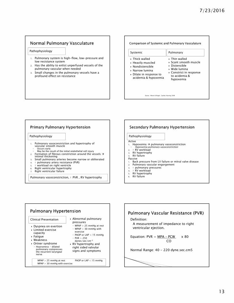
� The presence of air within the peritoneal cavity.
� Most common cause is a perforation of the abdominal viscus — a perforated ulcer
58
� PSP◦ no underlying lung disease
◦ Seen in young adults
� SSP◦ Usually caused by underlying lung disease
◦ More severe due to already compromised lung state
� Both may need treatment to prevent recurrence
� Apex chest tubes for pneumos as air rises
� Keep suction on chest tubes
59 60

7/23/2016
11
� Bubbling in the water seal chamber indicates air leak
� If suction is ordered for PSP or SSP, keep suction going even when ambulating!
61
� Patients with resolving pneumothorax should be cautioned not to fly until intrapleural air has completely resolved.
� Deep sea diving should be avoided unless thoracotomy or pleurodesis has been performed
62
� Bubbling in the water seal chamber indicates air leak
� If suction is ordered for PSP or SSP, keep suction going even when ambulating!
63
� Patients with resolving pneumothorax should be cautioned not to fly until intrapleural air has completely resolved.
� Deep sea diving should be avoided unless thoracotomy or pleurodesis has been performed
64
� Enlargement of the right ventricle (either dilatation or hypertrophy) from pulmonary pathology◦ Diseases of the lung like
COPD
◦ Diseases of the pulmonary circulation
� Pulmonary hypertension
� Thromboembolic disease

7/23/2016
12
Pathophysiology
1. Increase in pulmonary vascular resistance
2. Causes increase in pulmonary pressures
3. Results in increased RV workload
4. RV increases
Enlargement of RV from ↑ pulmonary resistance
Clinical Presentation
Right sided failure symptoms
� Right sided Heart Failure◦ JVD
◦ Hepatomegaly◦ Peripheral edema
� Jugular venous palpitation◦ Associated with prominent
“a” wave secondary to ↓ RV compliance
� Prominent V wave on right atrial tracing from tricuspid regurgitation
� Heart Sounds◦ S4
◦ Palpable left parasternal lift
◦ Murmurs if tricuspid or pulmonic insufficiency
� Echo◦ Right sided abnormalities
� EKG◦ Right axis deviation
◦ Right atrial enlargement –tall P waves
◦ RBBB
◦ Right precordial T wave inversion
Clinical Management
� Oxygen – pulmonary vasodilator
◦ ↓ PVR and ↑ RV stroke volume
� Diuretics – if congested
� Inotropes may be used with vasodilators
� Phlebotomy if polycythemia (HCT > 60%)
Pulmonary specific vasodilators� IV◦ Nitroglycerin◦ Sodium nitroprusside
(Nipride)◦ Prostaglandins (PGE1, PGI2)◦ PDE1 (phosphodiesterase
enzyme)� Inhaled◦ Any of the above IV
medications◦ Nitric oxide◦ Prostacyclin (PGI1,
Epoprosternol, Flolan) or derivative Iloprost
Pulmonary specific vasodilators
� Polycythemia may result from an increased erythropoietin (EPO) production in response to chronic hypoxia ◦ COPD, HF, pulmonary
hypertension, sleep apnea
� Treatment◦ Phlebotomy
� High blood pressure in the arteries that supply lungs and right side of the heart◦ MPAP > 25 mmHg at rest◦ MPAP > 30 mmHg with
exercise◦ PAOP or LAP < 15 mmHg
� One of the most serious, progressive, and potentially life threatening condition of the pulmonary vascular.
UnknownUnknown
� Primary◦ A rare disease that affects one
to two people per million in the USA every year.
◦ Most likely seen in women between the ages of 21 and 40
� Oral contraceptives a risk factor
� Secondary◦ Arises as a result of some other
underlying disease or factor
� COPD, PE, MS, Tricuspid regurgitation, HIV. congenital defects – ASD/VSD
Comorbidity for Cardiac
Surgery!
7/23/2016
13
Pathophysiology
1. Pulmonary system is high-flow, low-pressure and low resistance system
2. Has the ability to enlist unperfused vessels of the pulmonary vascular when needed
3. Small changes in the pulmonary vessels have a profound effect on resistance
Systemic Pulmonary
� Thick walled
� Heavily muscled
� Nondistensible
� Narrow lumina
� Dilate in response to acidemia & hypoxemia
� Thin walled� Scant smooth muscle� Distensible� Wide lumina� Constrict in response
to acidemia & hypoxemia
Source: Moser & Riegel, Cardiac Nursing 2008
Pathophysiology
1. Pulmonary vasoconstriction and hypertrophy of vascular smooth muscle◦ Occurs early◦ May be the result of the initial endothelial cell injury
2. Formation of fibrous constriction around the vessels �intimal thickening
3. Small pulmonary arteries become narrow or obliterated4. ↑ pulmonary artery resistance (PVR)5. ↑ workload on right ventricle6. Right ventricular hypertrophy7. Right ventricular failure
Pulmonary vasoconstriction, ↑ PVR , RV hypertrophy
Pathophysiology
Active1. Hypoxemia � pulmonary vasoconstriction◦ Hypoxemia pulmonary vasoconstriction
2. ↑ RV workload3. RV hypertrophy4. RV failurePassive1. Back pressure from LV failure or mitral valve disease2. Pulmonary vascular engorgement3. ↑ pulmonary pressures4. ↑ RV workload5. RV hypertrophy6. RV failure
Clinical Presentation
MPAP > 25 mmHg at rest PAOP or LAP < 15 mmHg
MPAP > 30 mmHg with exercise
� Dyspnea on exertion
� Limited exercise capacity
� Fatigue� Weakness
� Ortner syndrome◦ Hoarseness – dilated
pulmonary compresses the recurrent laryngeal nerve
� Abnormal pulmonary pressures◦ MPAP > 25 mmHg at rest
◦ MPAP > 30 mmHg with exercise
◦ PAOP or LAP < 15 mmHg
◦ PVR > 250 dynes/sec/cm-5
� RV hypertrophy and right sided valvular signs and symptoms
Definition:
A measurement of impedance to right ventricular ejection.
Equation: PVR = MPA – PCW x 80
CO
Normal Range: 40 - 220 dyne.sec.cm5

7/23/2016
14
Parameter Normal Values
Cardiac Output (CO) 4 - 8 l/min
Cardiac Index (CI) 2.5 – 4.2 l/min/m2
Right atrial pressure (CVP) 0 – 8 mmHg
Pulmonary artery pressure (PAS/PAD)
15 - 30/6 -12 mmHg
Pulmonary artery occlusive pressure
4 – 12 mmHg
Systemic vascular resistance (SVR) 770 – 1500 dyne/sec/cm5
Pulmonary vascular resistance (PVR)
20 – 120 dyne/sec/cm5
Stroke Volume (SV) 60 -130 mL/beat
Stroke Volume Index (SVI) 30 – 65 mL/beat/m2
Arterial oxygenation saturation 95 – 100 %
Venous oxygenation saturation 60 – 80 %
Source: Sited in Cardiac Surgery Essentials, page 148
Clinical Management
� Reverse or inhibit the three primary abnormalities of vasoconstriction, smooth muscle proliferation, and vascular remodeling
� ↓ PAP and PVR
� Improve RV function
� Energy conservation methods� Moderate exercise to avoid
overexertion
↓ PAP and PVR
Pharmacologic Agents•Oxygen•Isoproterenol•Aminophylline•Calcium channel blocking agents•Nitrous Oxide
Humoral Substances•Acetylcholine•Bradykinin•Prostaglandin E•Prostacyclin•Sildenafil (Viagra)
PE – sudden onset
� Symptoms depend on severity
� Dyspnea/Tachypnea- use of accessary muscles
� Tachycardia� Pallor or cyanosis � Sharp, pleuritic chest pain ..
worse with deep inspiration� Anxiety – feeling of
impending doom
Major PE – one causing hemodynamic instability is an ominous emergency!
� Create your study plan…