Embed Size (px)
Citation preview
04/19/23 1
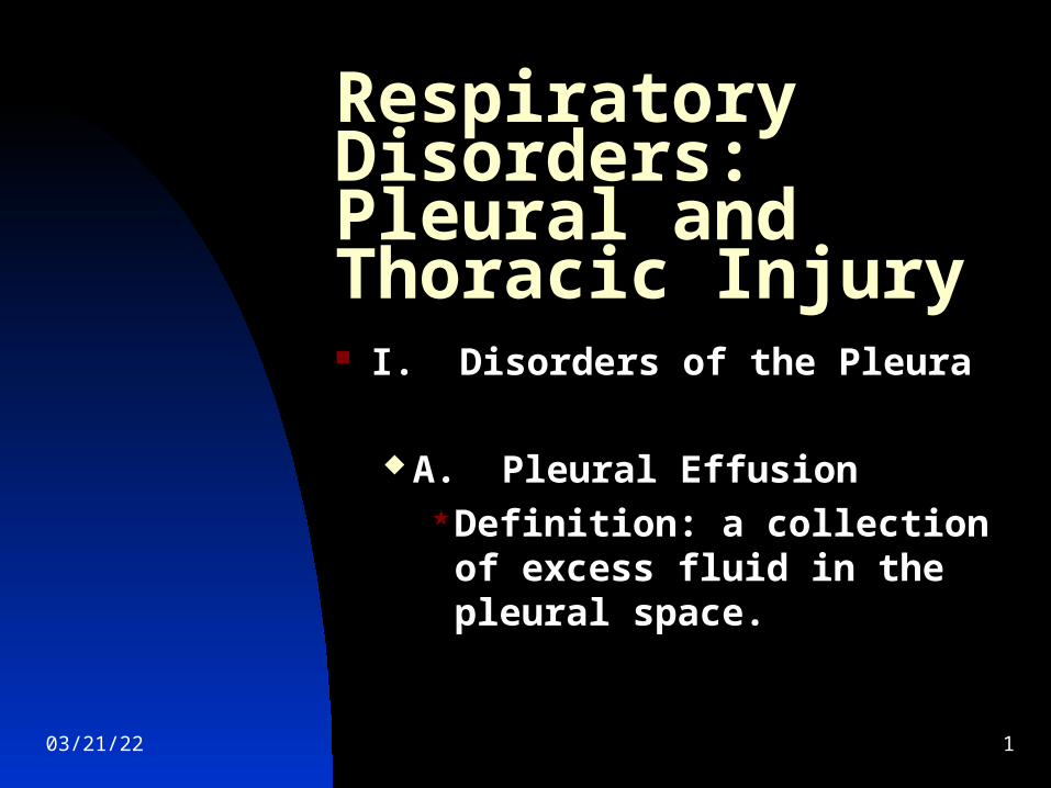
Respiratory Disorders: Pleural and Thoracic Injury
I. Disorders of the Pleura
A. Pleural EffusionDefinition: a collection of
excess fluid in the pleural space.
04/19/23 2
04/19/23 3
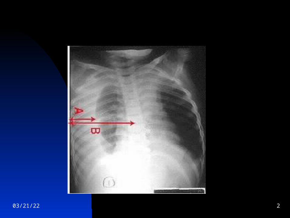
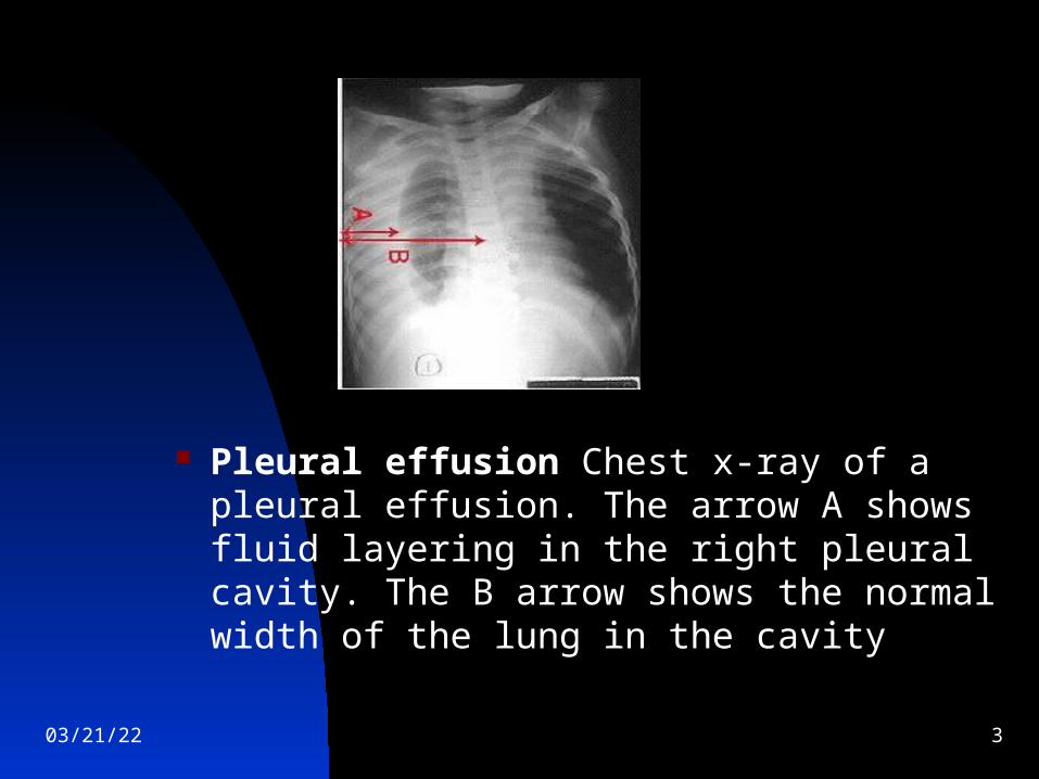
Pleural effusion Chest x-ray of a pleural effusion. The arrow A shows fluid layering in the right pleural cavity. The B arrow shows the normal width of the lung in the cavity
04/19/23 4
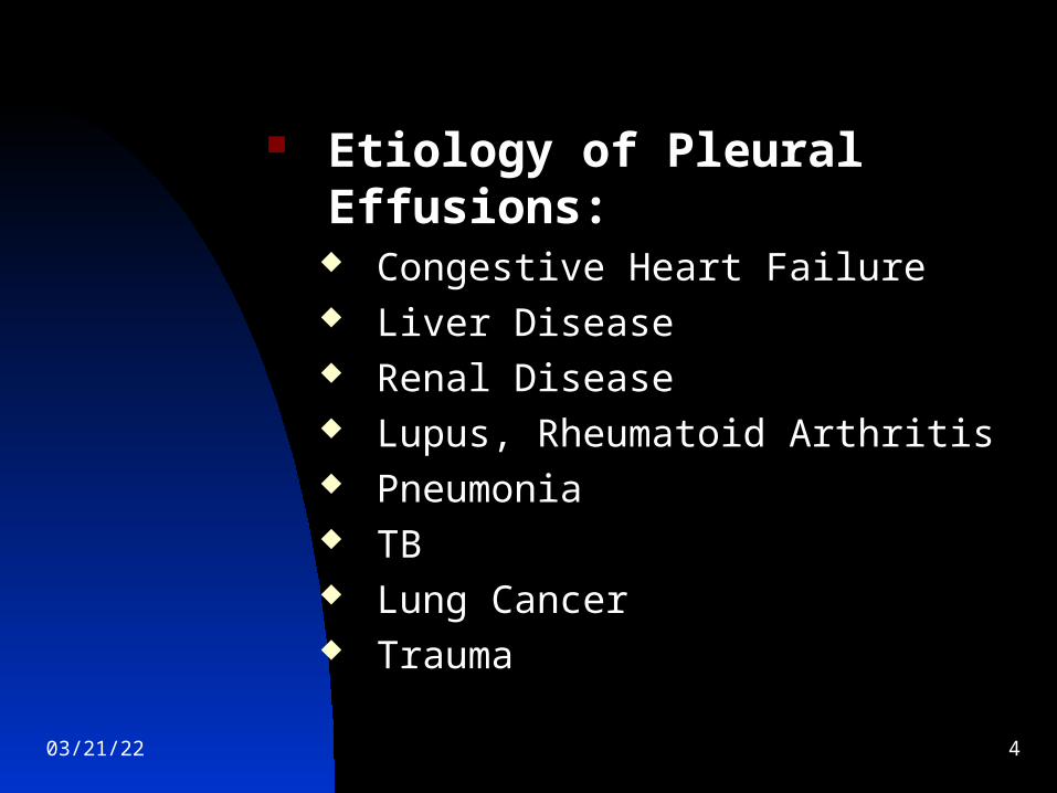
Etiology of Pleural Effusions: Congestive Heart Failure Liver Disease Renal Disease Lupus, Rheumatoid Arthritis Pneumonia TB Lung Cancer Trauma
04/19/23 5
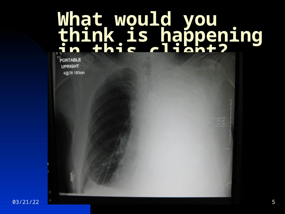
What would you think is happening in this client?
04/19/23 6
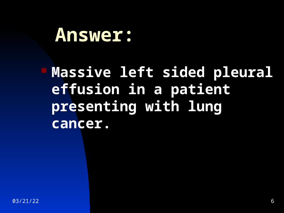
Answer:
Massive left sided pleural effusion in a patient presenting with lung cancer.
04/19/23 7
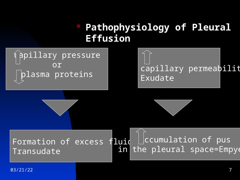
Pathophysiology of Pleural Effusion
capillary pressureor
plasma proteins
Formation of excess fluid=Transudate
capillary permeability=Exudate
Accumulation of pusin the pleural space=Empyema
04/19/23 8
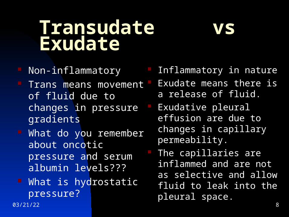
Transudate vs Exudate
Non-inflammatory Trans means movement of
fluid due to changes in pressure gradients
What do you remember about oncotic pressure and serum albumin levels???
What is hydrostatic pressure?
Inflammatory in nature Exudate means there is a
release of fluid. Exudative pleural effusion
are due to changes in capillary permeability.
The capillaries are inflammed and are not as selective and allow fluid to leak into the pleural space.
04/19/23 9
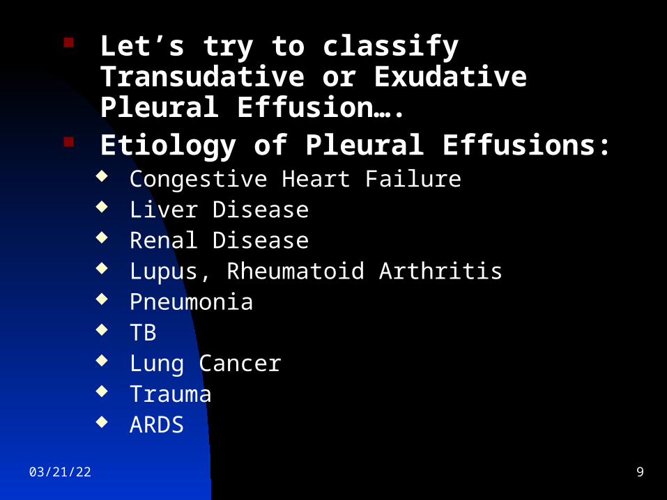
Let’s try to classify Transudative or Exudative Pleural Effusion….
Etiology of Pleural Effusions: Congestive Heart Failure Liver Disease Renal Disease Lupus, Rheumatoid Arthritis Pneumonia TB Lung Cancer Trauma ARDS
04/19/23 10
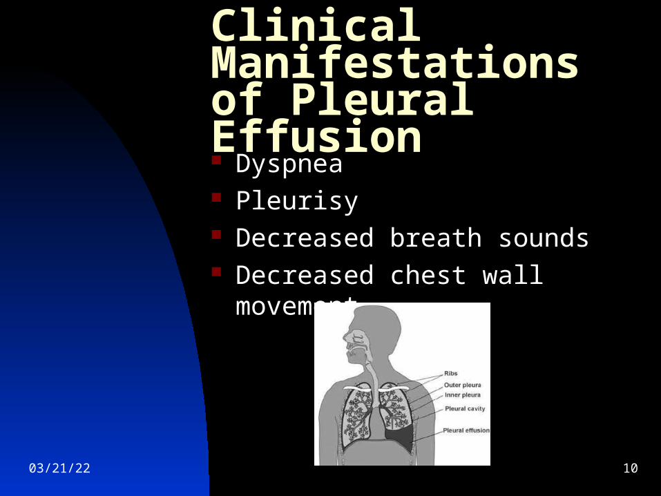
Clinical Manifestationsof Pleural Effusion Dyspnea Pleurisy Decreased breath sounds Decreased chest wall movement
04/19/23 11
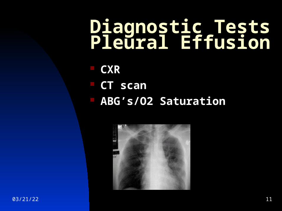
Diagnostic Tests Pleural Effusion CXR CT scan ABG’s/O2 Saturation
04/19/23 12
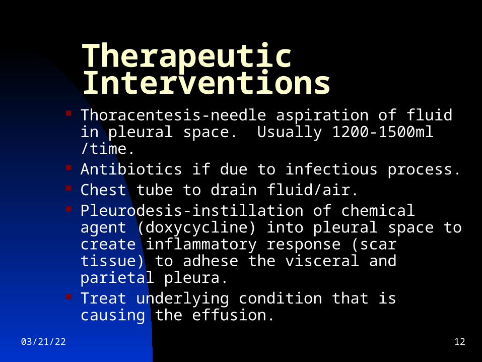
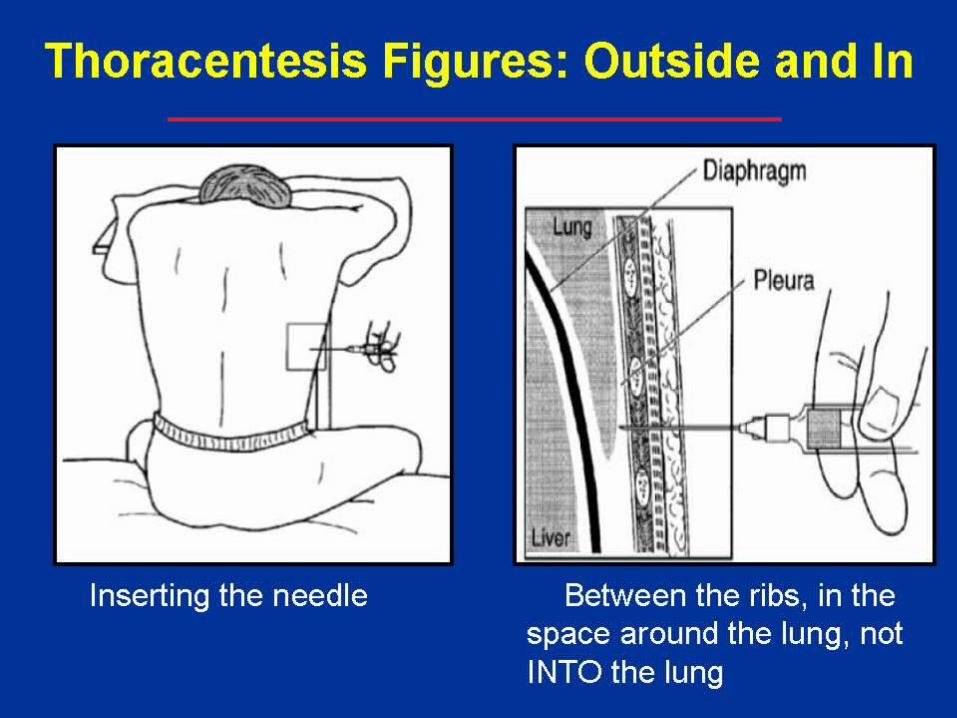
Therapeutic Interventions Thoracentesis-needle aspiration of fluid in
pleural space. Usually 1200-1500ml /time. Antibiotics if due to infectious process. Chest tube to drain fluid/air. Pleurodesis-instillation of chemical agent
(doxycycline) into pleural space to create inflammatory response (scar tissue) to adhese the visceral and parietal pleura.
Treat underlying condition that is causing the effusion.
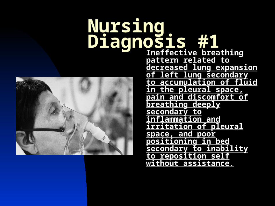
Nursing Diagnosis #1Ineffective breathing pattern related to decreased lung expansion of left lung secondary to accumulation of fluid in the pleural space, pain and discomfort of breathing deeply secondary to inflammation and irritation of pleural space, and poor positioning in bed secondary to inability to reposition self without assistance.
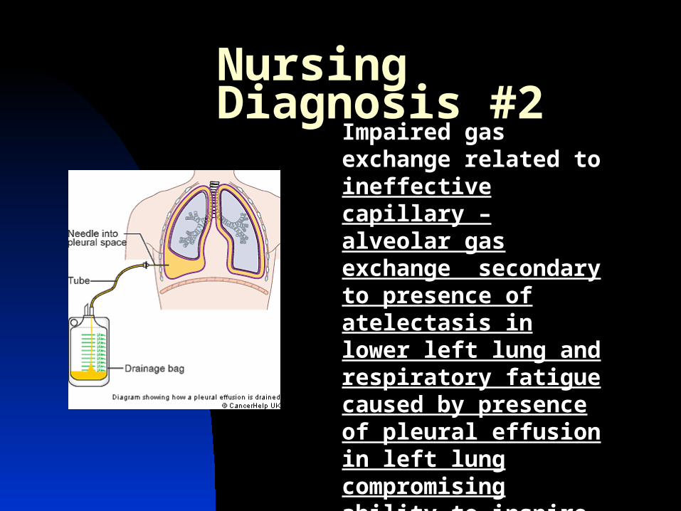
Nursing Diagnosis #2Impaired gas exchange related to ineffective capillary – alveolar gas exchange secondary to presence of atelectasis in lower left lung and respiratory fatigue caused by presence of pleural effusion in left lung compromising ability to inspire deeply and causing pain.
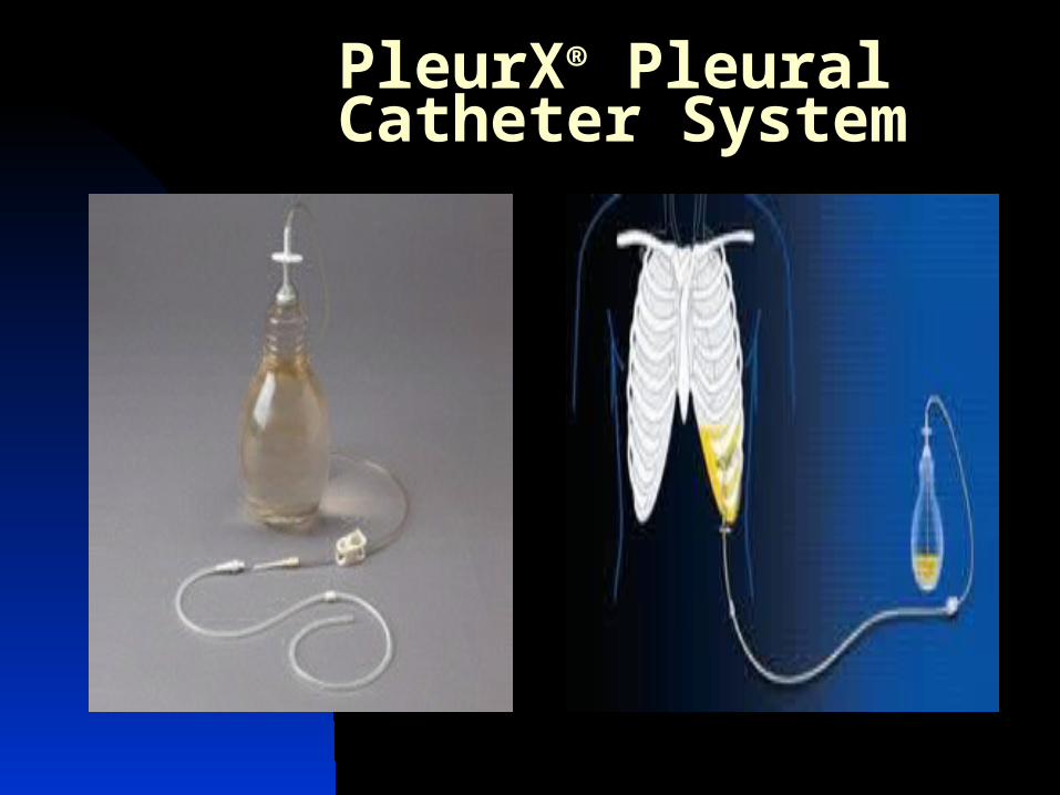
PleurX® Pleural Catheter System
04/19/23 17
B. Spontaneous PneumothoraxDefinition-accumulation of air in the
pleural spacePathophysiology
Rupture of bleb on the lung surface allows air into the pleural space
• Primary pneumothorax- affects previously healthy individuals
• Secondary pneumothorax-affects individuals with preexisting lung disease
– Which diseases can you think of???
04/19/23 18
Clinical Manifestations of Spontaneous Pnemo Abrupt onset Pleuritic chest pain SOB, dyspnea respiratory rate, tachycardia Unequal chest excursion Decreased breath sounds on
affected side
04/19/23 19
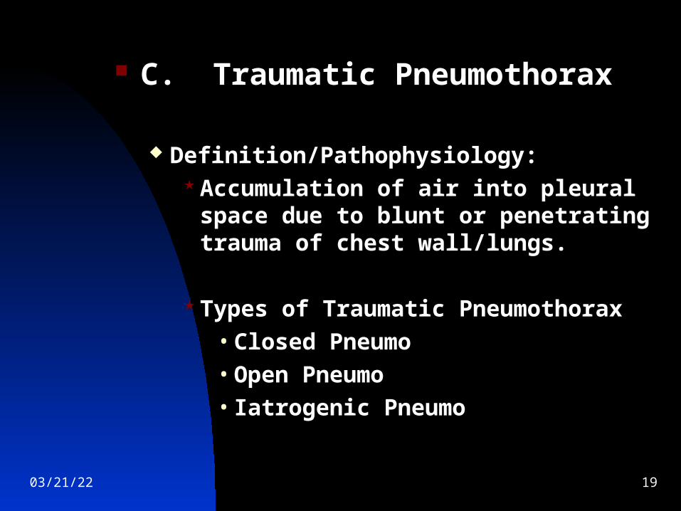
C. Traumatic Pneumothorax
Definition/Pathophysiology: Accumulation of air into pleural space
due to blunt or penetrating trauma of chest wall/lungs.
Types of Traumatic Pneumothorax• Closed Pneumo• Open Pneumo• Iatrogenic Pneumo
04/19/23 20
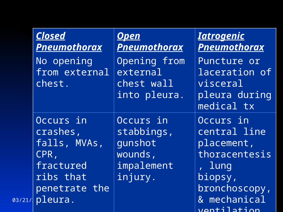
Closed Pneumothorax
No opening from external chest.
Open Pneumothorax
Opening from external chest wall into pleura.
Iatrogenic Pneumothorax
Puncture or laceration of visceral pleura during medical tx
Occurs in crashes, falls, MVAs, CPR, fractured ribs that penetrate the pleura.
Occurs in stabbings, gunshot wounds, impalement injury.
Occurs in central line placement, thoracentesis, lung biopsy, bronchoscopy, & mechanical ventilation
04/19/23 21
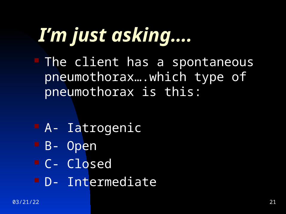
I’m just asking…. The client has a spontaneous
pneumothorax….which type of pneumothorax is this:
A- Iatrogenic B- Open C- Closed D- Intermediate
04/19/23 22
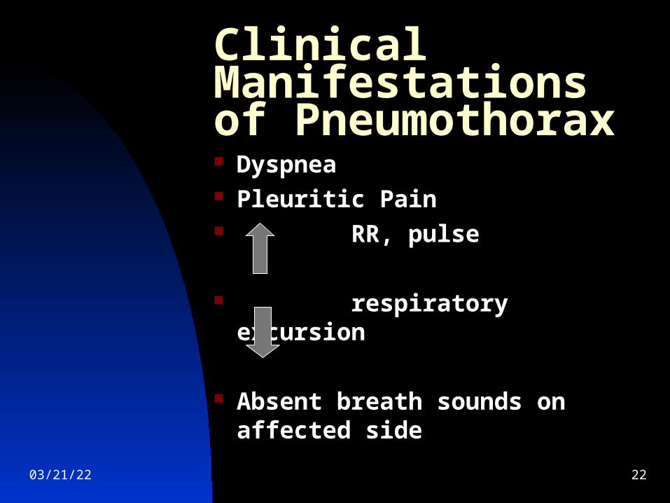
Clinical Manifestations of Pneumothorax Dyspnea Pleuritic Pain RR, pulse
respiratory excursion
Absent breath sounds on affected side
04/19/23 23
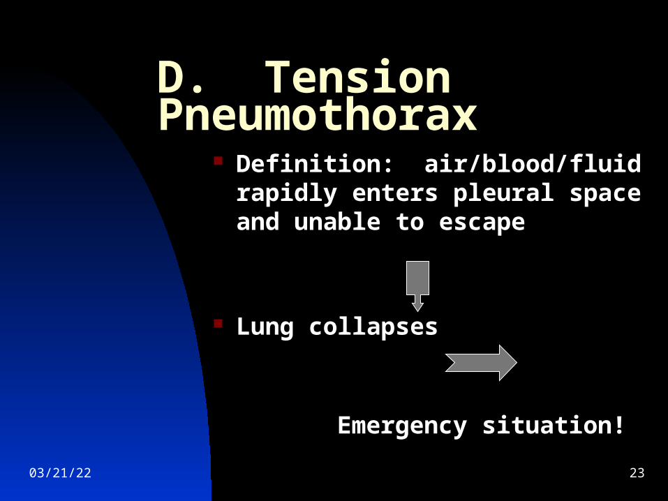
D. Tension Pneumothorax Definition: air/blood/fluid
rapidly enters pleural space and unable to escape
Lung collapses
Emergency situation!
04/19/23 24
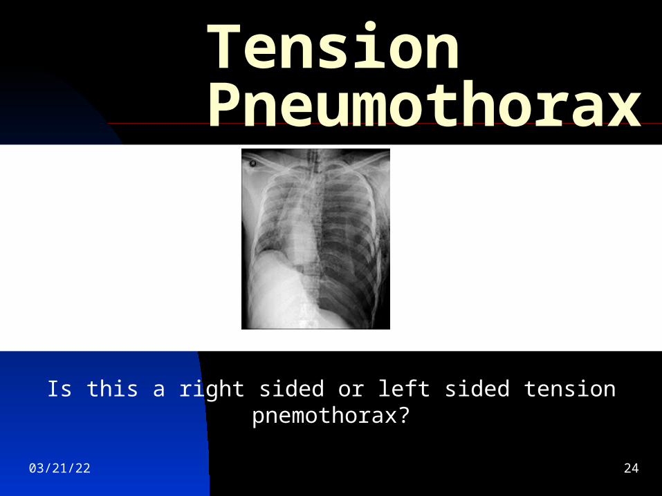
Tension Pneumothorax
Is this a right sided or left sided tension pnemothorax?
04/19/23 25
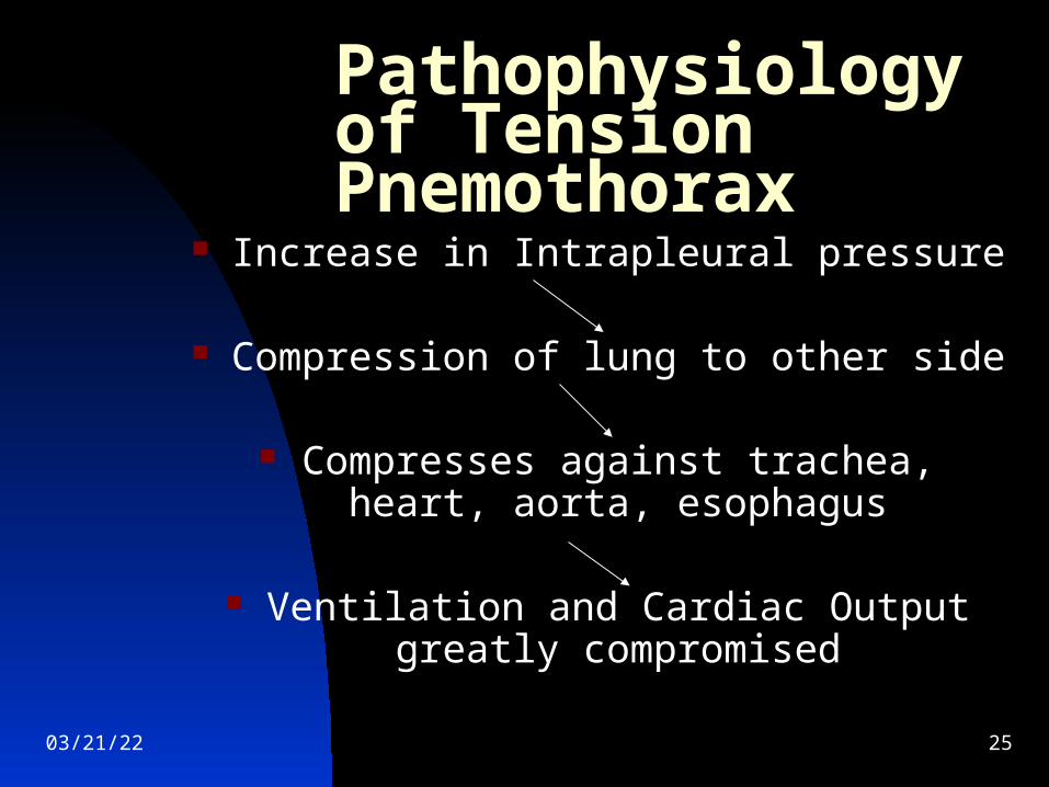
Pathophysiology of Tension Pnemothorax
Increase in Intrapleural pressure
Compression of lung to other side
Compresses against trachea, heart, aorta, esophagus
Ventilation and Cardiac Output greatly compromised
04/19/23 26
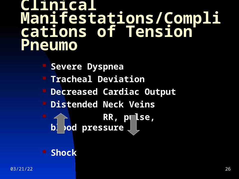
Clinical Manifestations/Complications of Tension Pneumo
Severe Dyspnea Tracheal Deviation Decreased Cardiac Output Distended Neck Veins RR, pulse, blood pressure
Shock
04/19/23 27
Therapeutic Interventions for Pneumothorax
High Fowlers position O2 as ordered Rest to decrease O2 demand Chest tube insertion Pleurodesis Surgery: Thoracotomy to remove blebs,
partial excision of parietal pleura done using VATS (video assisted thorascopic surgery)
04/19/23 28
II. Trauma of the Chest/Lung Chest injury is the leading cause of death
from trauma May involve chest wall, lungs, heart, great
vessels, esophagus Life threatening chest injuries include:
Airway obstruction Tension pneumo, open pneumo, massive
hemothorax Flail chest with pulmonary contusion
04/19/23 29
A. Rib Fracture
Simple rib fracture in an at risk client may lead to pneumonia, atelectasis, respiratory failure
Displaced rib fractures can result in pnemo/hemothorax, intrathoracic vessel tears, liver or spleen injury
04/19/23 30
Clinical Manifestations of Rib Fractures Pain on inspiration/coughing Voluntary splinting Rapid, shallow respirations Decreased breath sounds Crepitus on palpation Signs/symptoms of
pneumo/hemothorax
04/19/23 31
B. Flail Chest Etiology/Pathophysiology
Occurs when 2+ consecutive ribs are fractured in multiple places
Segment of chest wall becomes “free-floating” or flail
Flail segment of chest wall is sucked in during inspiration and moves outward with expiration
04/19/23 32
The client presents in the ED: Chest trauma client http://www.youtube.com/watch?v=PyDcGB-
i7OQ&feature=related
What did you note in this client? What would you do 1st? 2nd?
04/19/23 33
Clinical Manifestations of Flail Chest Dyspnea Pain especially on
inspiration Palpable crepitus Decreased breath sounds Unequal Chest expansion
04/19/23 34
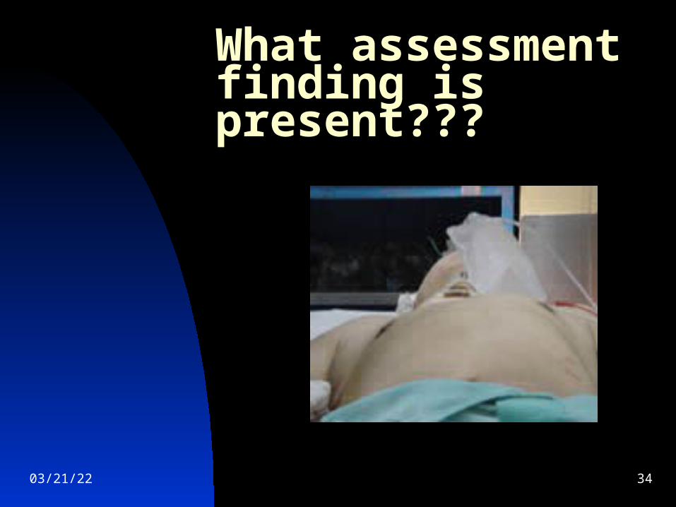
What assessment finding is present???
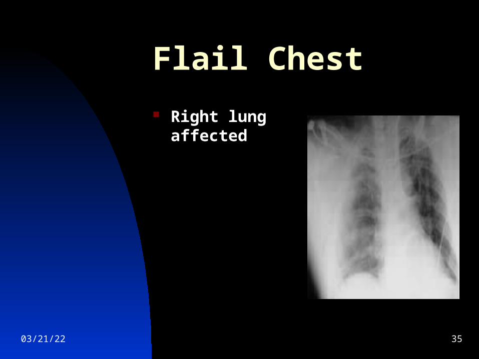
04/19/23 35
Flail Chest
Right lung affected
04/19/23 36
Therapeutic Interventions Flail Chest
O2 as ordered Elevate HOB Intercostal nerve block or epidural
analgesia to decrease pain Suction as ordered Splint affected area Preferred treatment= Intubation and
positive pressure ventilation
04/19/23 37
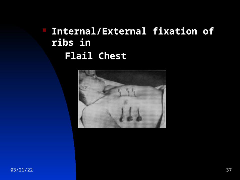
Internal/External fixation of ribs in
Flail Chest
04/19/23 38
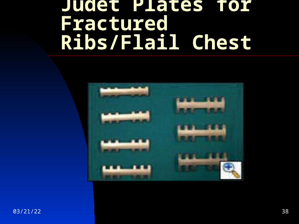
Judet Plates for Fractured Ribs/Flail Chest

04/19/23 39
Sanchez Plates for Fractured Ribs/Flail Chest