Embed Size (px)
Citation preview
86th Annual Meeting of the ATAEndocrine Neck Advanced Ultrasound
Course – September 21, 2016
DIFFUSE THYROID CONDITIONS
Stephanie L. Lee, M.D., Ph.D.Director of the BMC Thyroid Nodule and Cancer Center
Section of Endocrinology, Diabetes and NutritionBoston Medical Center
86th Annual Meeting of the American Thyroid Association, September 21, 2016, Denver, Colorado NOT FOR REPRODUCTION – FOR ATA ULTRASOUND COURSE USE ONLY

Normal Thyroid Anatomy• Each lobe has a rounded
superior pole and an elongated inferior pole
• Sagittal length can vary from 4-6 cm
• Usually lobe AP thickness is < 2 cm
• Isthmus AP thickness is <0.5 cm
• Normal adult thyroid volume is ~15 cc
superior
inferior
<2 cm
<5mm
86th Annual Meeting of the American Thyroid Association, September 21, 2016, Denver, Colorado NOT FOR REPRODUCTION – FOR ATA ULTRASOUND COURSE USE ONLY
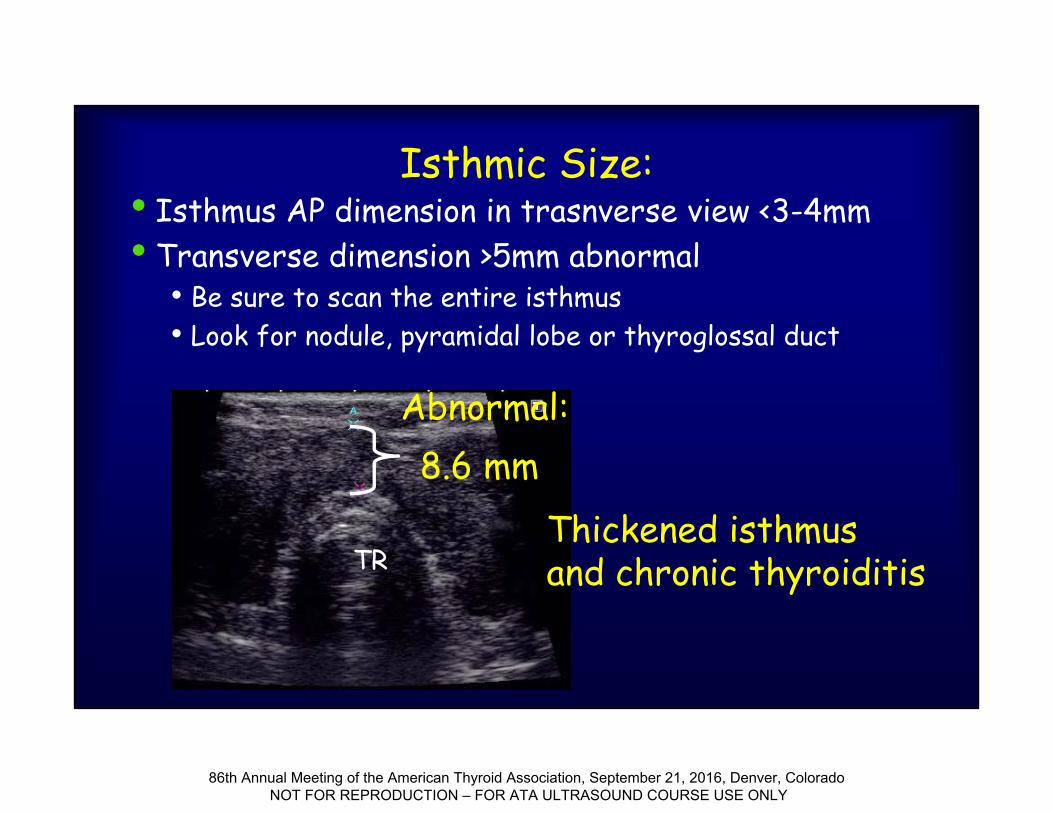
Isthmic Size:• Isthmus AP dimension in trasnverse view <3-4mm• Transverse dimension >5mm abnormal
• Be sure to scan the entire isthmus• Look for nodule, pyramidal lobe or thyroglossal duct
Abnormal:8.6 mm
TRThickened isthmusand chronic thyroiditis
86th Annual Meeting of the American Thyroid Association, September 21, 2016, Denver, Colorado NOT FOR REPRODUCTION – FOR ATA ULTRASOUND COURSE USE ONLY
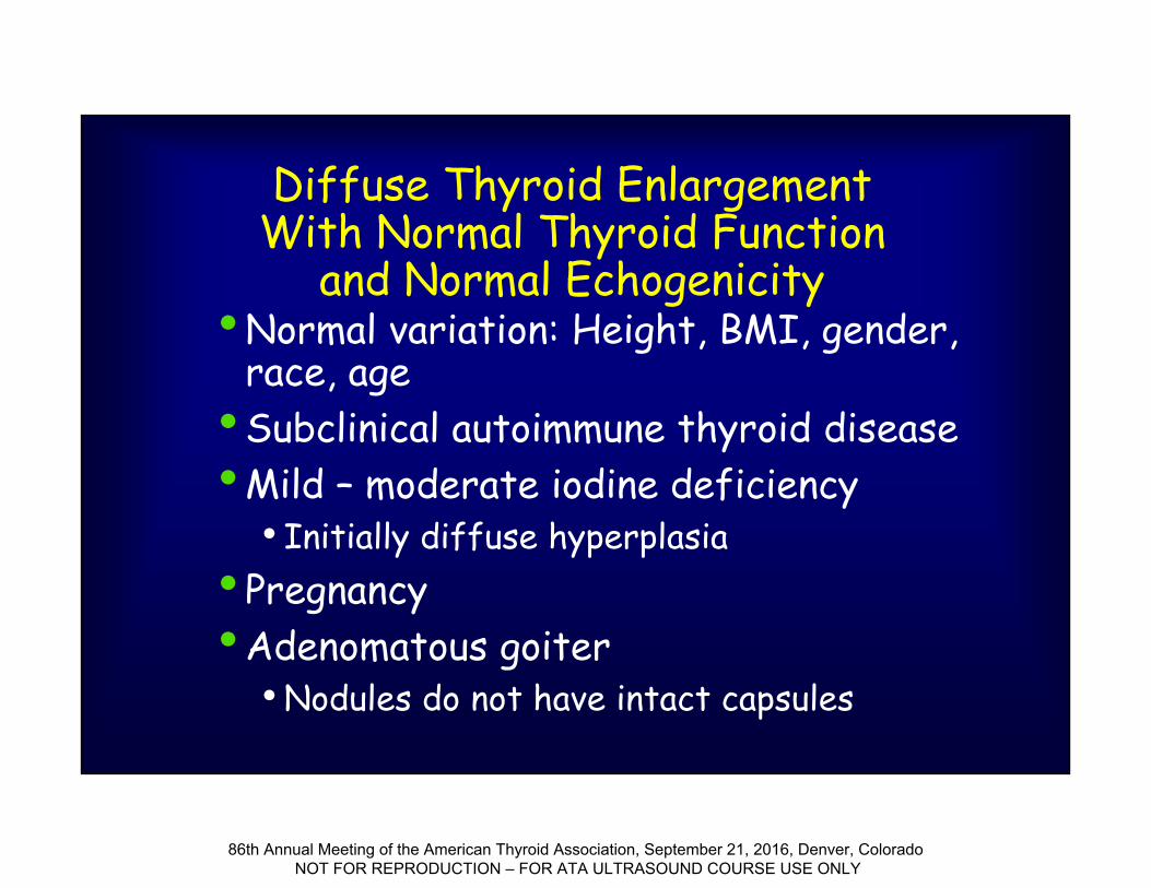
Diffuse Thyroid Enlargement With Normal Thyroid Function
and Normal Echogenicity•Normal variation: Height, BMI, gender,
race, age•Subclinical autoimmune thyroid disease•Mild – moderate iodine deficiency
• Initially diffuse hyperplasia• Pregnancy•Adenomatous goiter
• Nodules do not have intact capsules
86th Annual Meeting of the American Thyroid Association, September 21, 2016, Denver, Colorado NOT FOR REPRODUCTION – FOR ATA ULTRASOUND COURSE USE ONLY
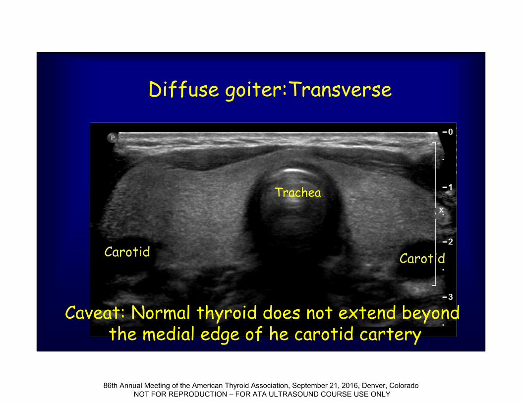
Diffuse goiter:Transverse
Trachea
Carotid Carotid
Caveat: Normal thyroid does not extend beyondthe medial edge of he carotid cartery
86th Annual Meeting of the American Thyroid Association, September 21, 2016, Denver, Colorado NOT FOR REPRODUCTION – FOR ATA ULTRASOUND COURSE USE ONLY
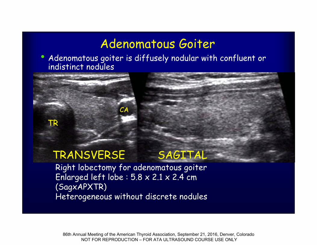
Adenomatous Goiter• Adenomatous goiter is diffusely nodular with confluent or
indistinct nodules
TRCA
Right lobectomy for adenomatous goiterEnlarged left lobe : 5.8 x 2.1 x 2.4 cm (SagxAPXTR)Heterogeneous without discrete nodules
TRANSVERSE SAGITAL
86th Annual Meeting of the American Thyroid Association, September 21, 2016, Denver, Colorado NOT FOR REPRODUCTION – FOR ATA ULTRASOUND COURSE USE ONLY
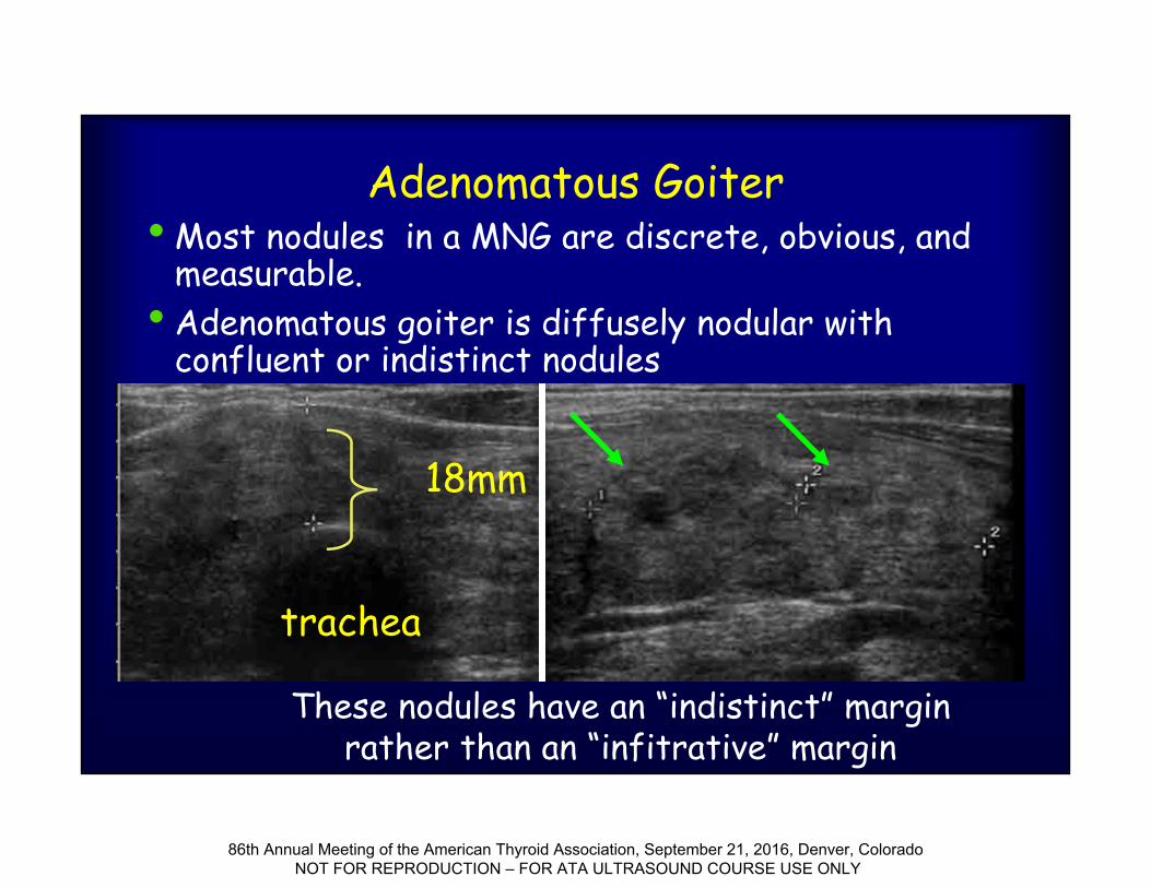
Adenomatous Goiter• Most nodules in a MNG are discrete, obvious, and
measurable.• Adenomatous goiter is diffusely nodular with
confluent or indistinct nodules
18mm
trachea
These nodules have an “indistinct” margin rather than an “infitrative” margin
86th Annual Meeting of the American Thyroid Association, September 21, 2016, Denver, Colorado NOT FOR REPRODUCTION – FOR ATA ULTRASOUND COURSE USE ONLY
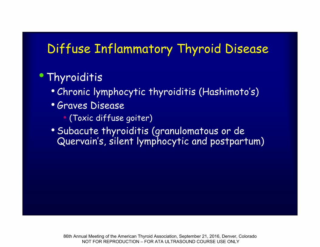
Diffuse Inflammatory Thyroid Disease
•Thyroiditis• Chronic lymphocytic thyroiditis (Hashimoto’s)• Graves Disease
• (Toxic diffuse goiter)• Subacute thyroiditis (granulomatous or de
Quervain’s, silent lymphocytic and postpartum)
86th Annual Meeting of the American Thyroid Association, September 21, 2016, Denver, Colorado NOT FOR REPRODUCTION – FOR ATA ULTRASOUND COURSE USE ONLY
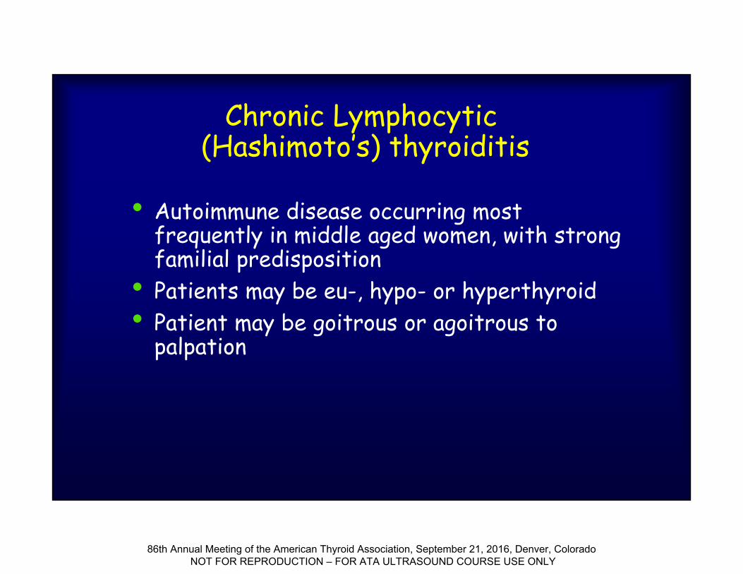
Chronic Lymphocytic(Hashimoto’s) thyroiditis
• Autoimmune disease occurring most frequently in middle aged women, with strong familial predisposition
• Patients may be eu-, hypo- or hyperthyroid• Patient may be goitrous or agoitrous to
palpation
86th Annual Meeting of the American Thyroid Association, September 21, 2016, Denver, Colorado NOT FOR REPRODUCTION – FOR ATA ULTRASOUND COURSE USE ONLY
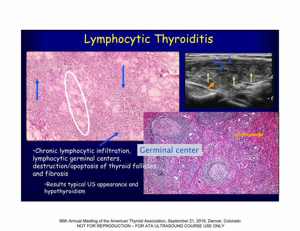
Lymphocytic Thyroiditis
Germinal center•Chronic lymphocytic infiltration, lymphocytic germinal centers, destruction/apoptosis of thyroid follicles and fibrosis
•Results typical US appearance and hypothyroidism
86th Annual Meeting of the American Thyroid Association, September 21, 2016, Denver, Colorado NOT FOR REPRODUCTION – FOR ATA ULTRASOUND COURSE USE ONLY
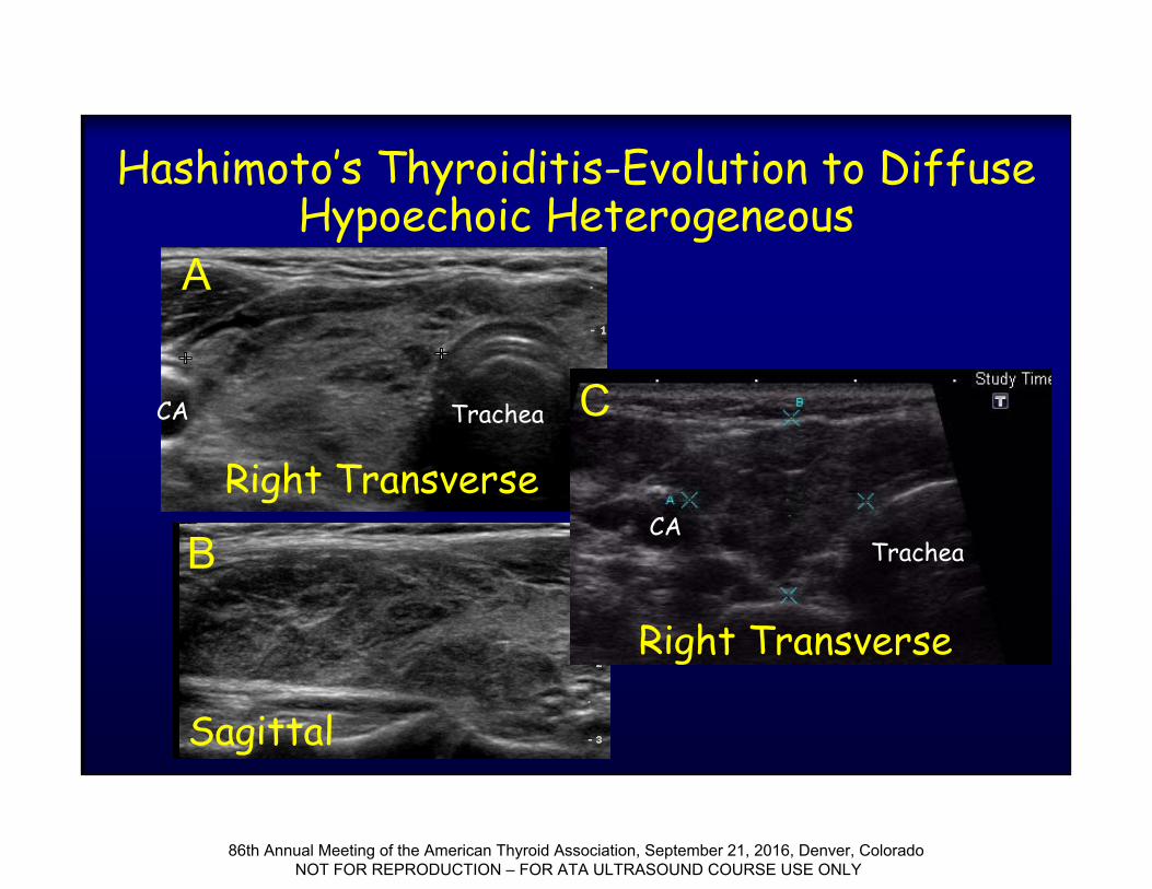
Hashimoto’s Thyroiditis-Evolution to Diffuse Hypoechoic Heterogeneous
Right Transverse
Right Transverse
Sagittal
A
B
CTrachea
TracheaCA
CA
86th Annual Meeting of the American Thyroid Association, September 21, 2016, Denver, Colorado NOT FOR REPRODUCTION – FOR ATA ULTRASOUND COURSE USE ONLY
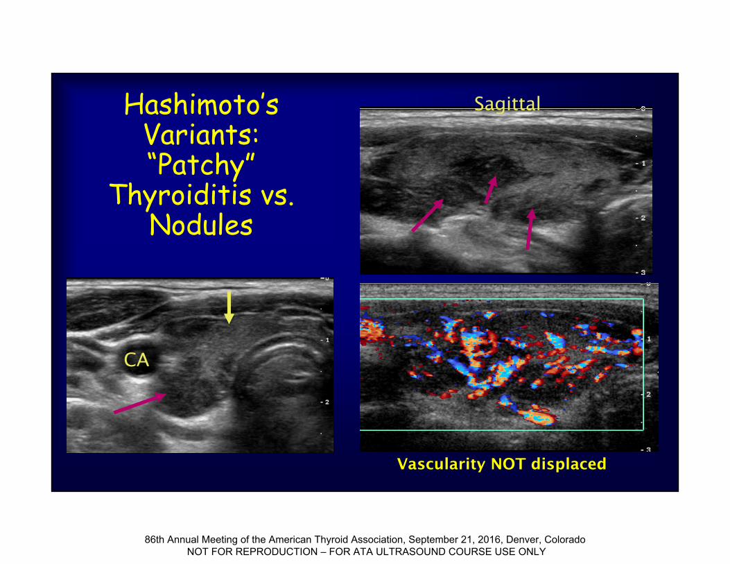
Hashimoto’s Variants: “Patchy”
Thyroiditis vs. Nodules
Sagittal
CA
Vascularity NOT displaced
86th Annual Meeting of the American Thyroid Association, September 21, 2016, Denver, Colorado NOT FOR REPRODUCTION – FOR ATA ULTRASOUND COURSE USE ONLY
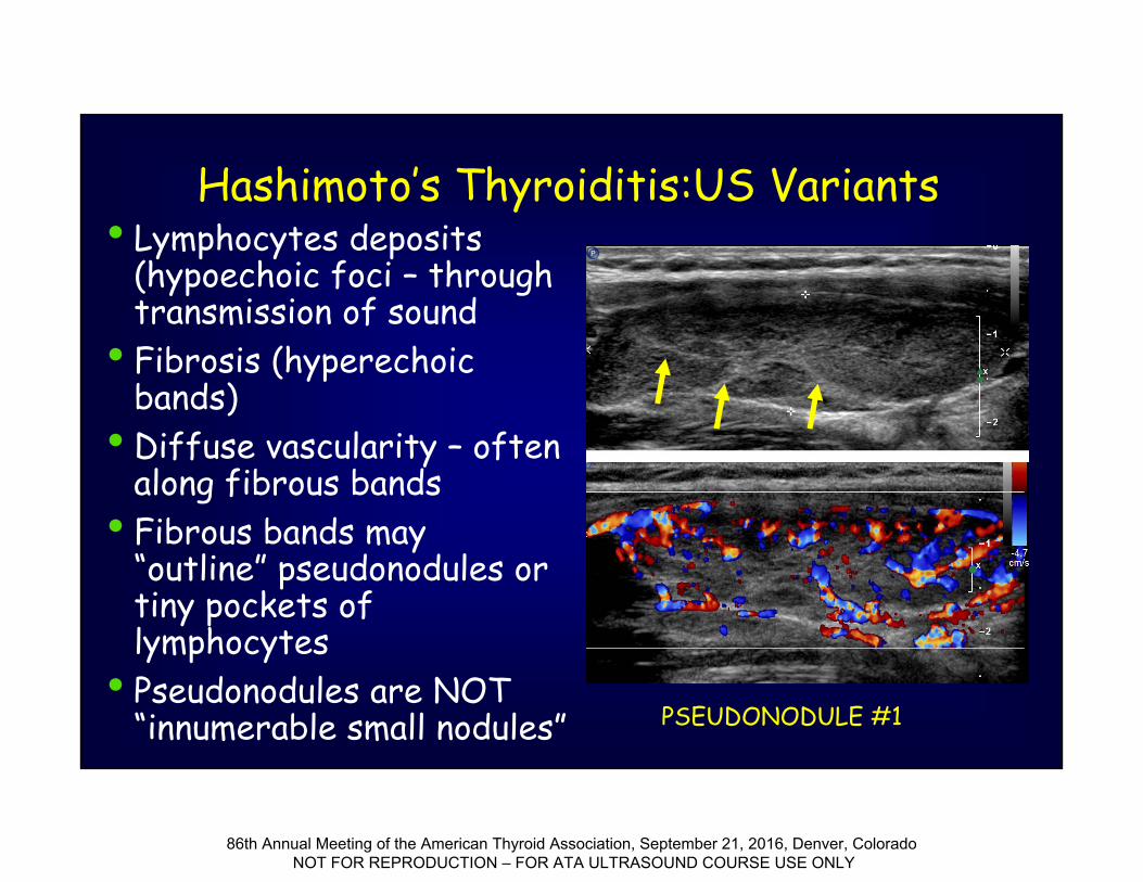
• Lymphocytes deposits (hypoechoic foci – through transmission of sound
• Fibrosis (hyperechoic bands)
• Diffuse vascularity – often along fibrous bands
• Fibrous bands may “outline” pseudonodules or tiny pockets of lymphocytes
• Pseudonodules are NOT “innumerable small nodules”
Hashimoto’s Thyroiditis:US Variants
PSEUDONODULE #1
86th Annual Meeting of the American Thyroid Association, September 21, 2016, Denver, Colorado NOT FOR REPRODUCTION – FOR ATA ULTRASOUND COURSE USE ONLY
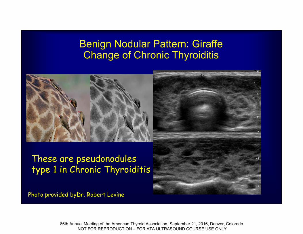
Benign Nodular Pattern: Giraffe Change of Chronic Thyroiditis
Photo provided byDr. Robert Levine
These are pseudonodules type 1 in Chronic Thyroiditis
86th Annual Meeting of the American Thyroid Association, September 21, 2016, Denver, Colorado NOT FOR REPRODUCTION – FOR ATA ULTRASOUND COURSE USE ONLY
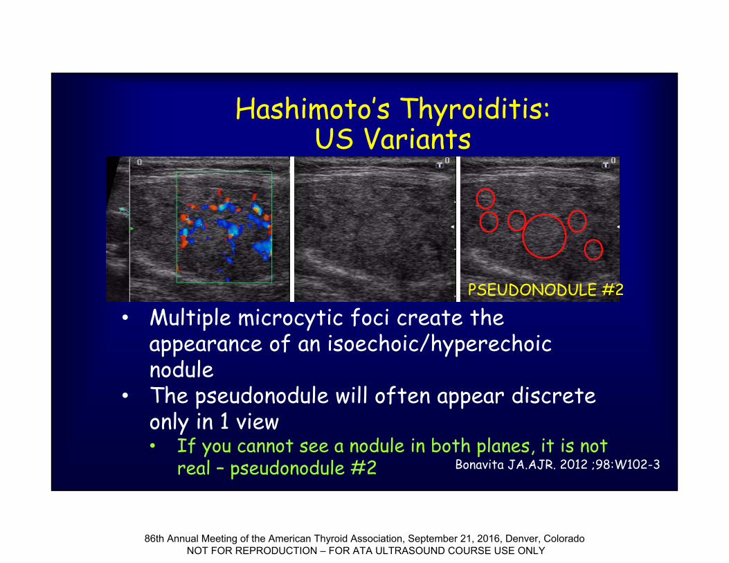
• Multiple microcytic foci create the appearance of an isoechoic/hyperechoic nodule
• The pseudonodule will often appear discrete only in 1 view• If you cannot see a nodule in both planes, it is not
real – pseudonodule #2
Hashimoto’s Thyroiditis:US Variants
Bonavita JA.AJR. 2012 ;98:W102-3
PSEUDONODULE #2
86th Annual Meeting of the American Thyroid Association, September 21, 2016, Denver, Colorado NOT FOR REPRODUCTION – FOR ATA ULTRASOUND COURSE USE ONLY
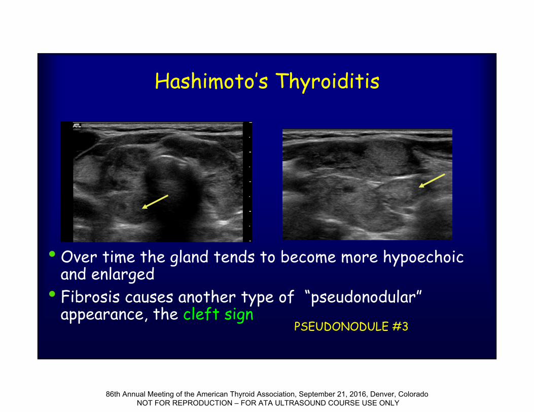
Hashimoto’s Thyroiditis
• Over time the gland tends to become more hypoechoic and enlarged
• Fibrosis causes another type of “pseudonodular” appearance, the cleft sign
PSEUDONODULE #3
86th Annual Meeting of the American Thyroid Association, September 21, 2016, Denver, Colorado NOT FOR REPRODUCTION – FOR ATA ULTRASOUND COURSE USE ONLY
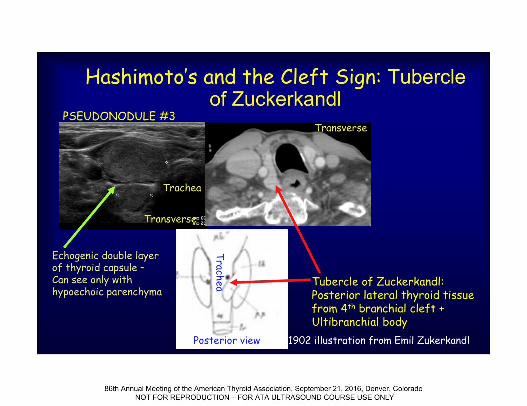
Hashimoto’s and the Cleft Sign: Tubercle of Zuckerkandl
1902 illustration from Emil Zukerkandl
Trachea
Transverse
PSEUDONODULE #3
Tubercle of Zuckerkandl: Posterior lateral thyroid tissue from 4th branchial cleft + Ultibranchial body
Transverse
Echogenic double layerof thyroid capsule –Can see only with hypoechoic parenchyma
Posterior view
Trachea
86th Annual Meeting of the American Thyroid Association, September 21, 2016, Denver, Colorado NOT FOR REPRODUCTION – FOR ATA ULTRASOUND COURSE USE ONLY
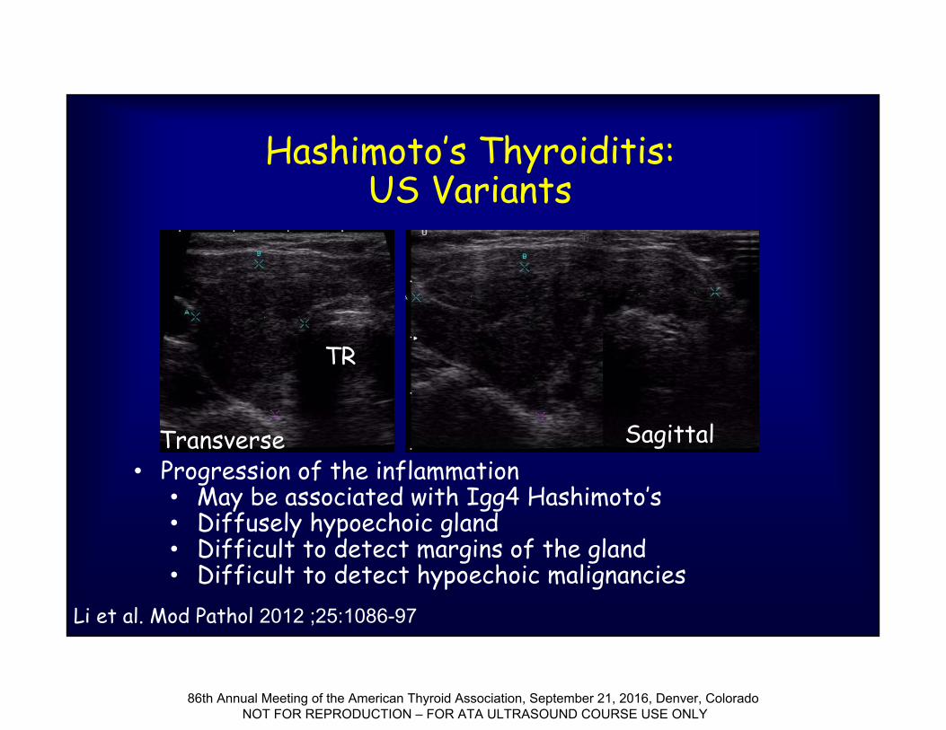
Hashimoto’s Thyroiditis:US Variants
• Progression of the inflammation• May be associated with Igg4 Hashimoto’s• Diffusely hypoechoic gland• Difficult to detect margins of the gland• Difficult to detect hypoechoic malignancies
TR
Transverse Sagittal
Li et al. Mod Pathol 2012 ;25:1086-97
86th Annual Meeting of the American Thyroid Association, September 21, 2016, Denver, Colorado NOT FOR REPRODUCTION – FOR ATA ULTRASOUND COURSE USE ONLY
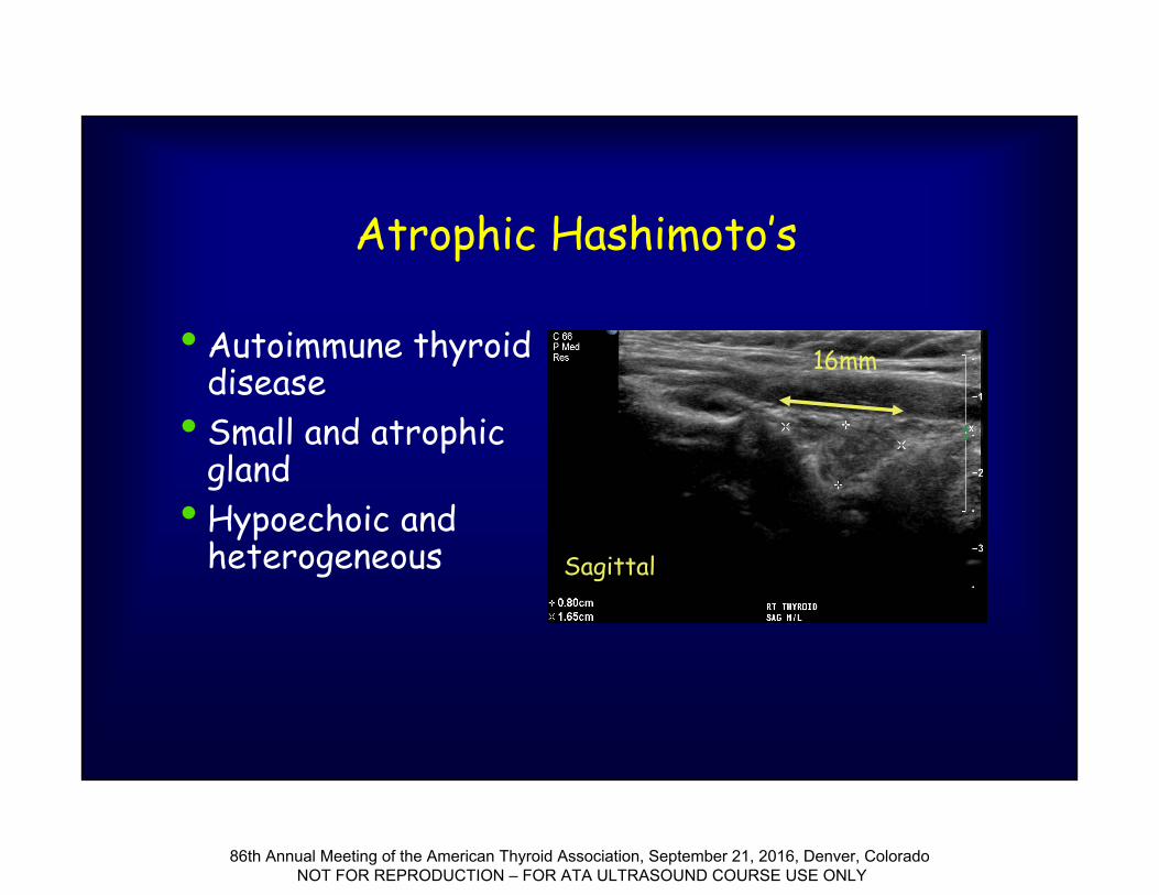
Atrophic Hashimoto’s
• Autoimmune thyroid disease
• Small and atrophic gland
• Hypoechoic and heterogeneous
16mm
Sagittal
86th Annual Meeting of the American Thyroid Association, September 21, 2016, Denver, Colorado NOT FOR REPRODUCTION – FOR ATA ULTRASOUND COURSE USE ONLY
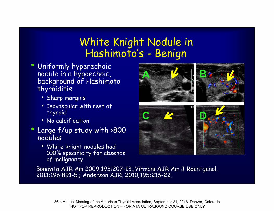
White Knight Nodule in Hashimoto’s - Benign
• Uniformly hyperechoic nodule in a hypoechoic, background of Hashimoto thyroiditis• Sharp margins• Isovascular with rest of
thyroid• No calcification
• Large f/up study with >800 nodules• White knight nodules had
100% specificity for absence of malignancy
A B
C D
Bonavita AJR Am 2009;193:207-13.;Virmani AJR Am J Roentgenol. 2011;196:891-5.; Anderson AJR. 2010;195:216-22.
86th Annual Meeting of the American Thyroid Association, September 21, 2016, Denver, Colorado NOT FOR REPRODUCTION – FOR ATA ULTRASOUND COURSE USE ONLY
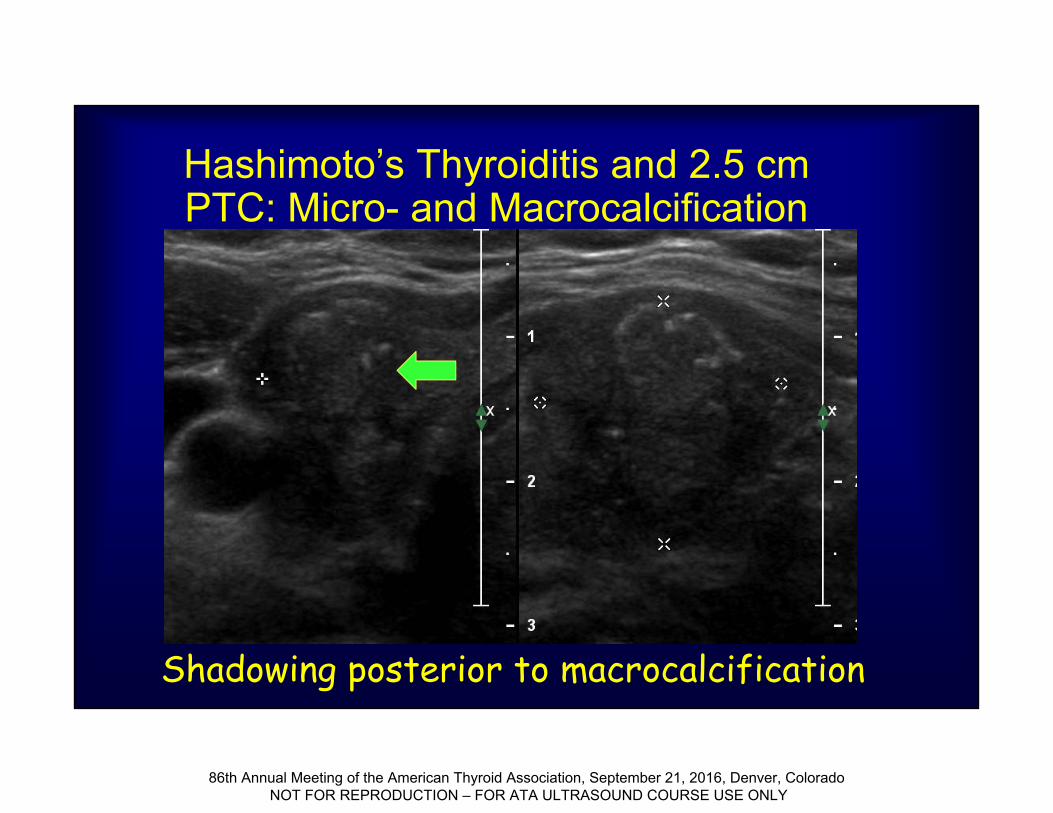
Hashimoto’s Thyroiditis and 2.5 cm PTC: Micro- and Macrocalcification
Shadowing posterior to macrocalcification
86th Annual Meeting of the American Thyroid Association, September 21, 2016, Denver, Colorado NOT FOR REPRODUCTION – FOR ATA ULTRASOUND COURSE USE ONLY
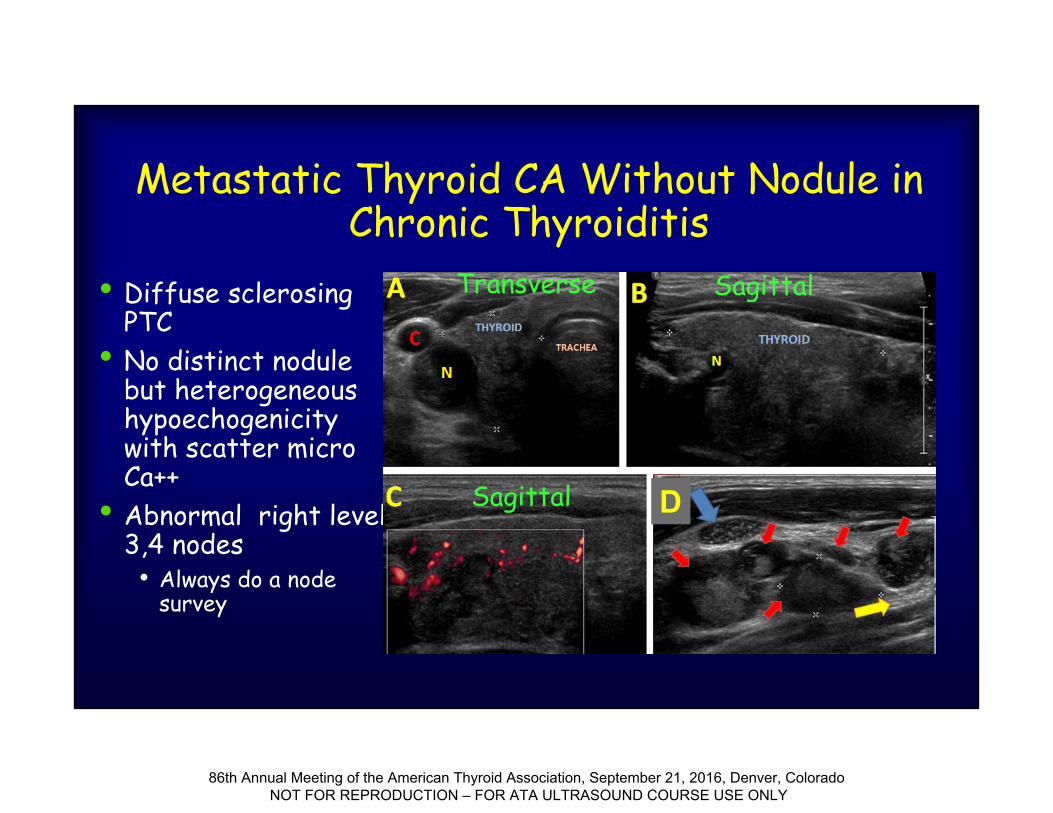
Metastatic Thyroid CA Without Nodule in Chronic Thyroiditis
• Diffuse sclerosing PTC
• No distinct nodule but heterogeneous hypoechogenicity with scatter micro Ca++
• Abnormal right level 3,4 nodes• Always do a node
survey
D
Transverse Sagittal
Sagittal
86th Annual Meeting of the American Thyroid Association, September 21, 2016, Denver, Colorado NOT FOR REPRODUCTION – FOR ATA ULTRASOUND COURSE USE ONLY
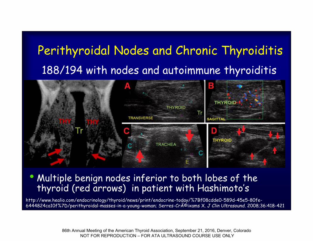
Perithyroidal Nodes and Chronic Thyroiditis
• Multiple benign nodes inferior to both lobes of the thyroid (red arrows) in patient with Hashimoto’s
http://www.healio.com/endocrinology/thyroid/news/print/endocrine-today/%7Bf08cdde0-589d-45e5-80fe-6444824ca10f%7D/perithyroidal-masses-in-a-young-woman; Serres-Créixams X. J Clin Ultrasound. 2008;36:418-421
188/194 with nodes and autoimmune thyroiditis
86th Annual Meeting of the American Thyroid Association, September 21, 2016, Denver, Colorado NOT FOR REPRODUCTION – FOR ATA ULTRASOUND COURSE USE ONLY
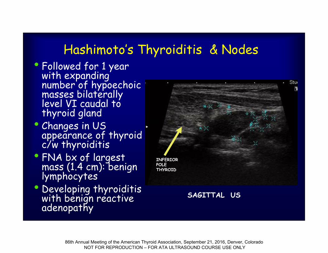
Hashimoto’s Thyroiditis & Nodes• Followed for 1 year
with expanding number of hypoechoic masses bilaterally level VI caudal to thyroid gland
• Changes in US appearance of thyroid c/w thyroiditis
• FNA bx of largest mass (1.4 cm): benign lymphocytes
• Developing thyroiditis with benign reactive adenopathy
SAGITTAL US
INFERIOR POLETHYROID
86th Annual Meeting of the American Thyroid Association, September 21, 2016, Denver, Colorado NOT FOR REPRODUCTION – FOR ATA ULTRASOUND COURSE USE ONLY
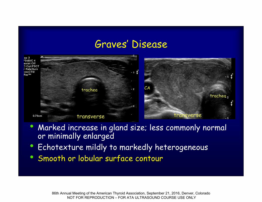
Graves’ Disease
• Marked increase in gland size; less commonly normal or minimally enlarged
• Echotexture mildly to markedly heterogeneous• Smooth or lobular surface contour
transverse transverse
tracheatrachea
CA
86th Annual Meeting of the American Thyroid Association, September 21, 2016, Denver, Colorado NOT FOR REPRODUCTION – FOR ATA ULTRASOUND COURSE USE ONLY
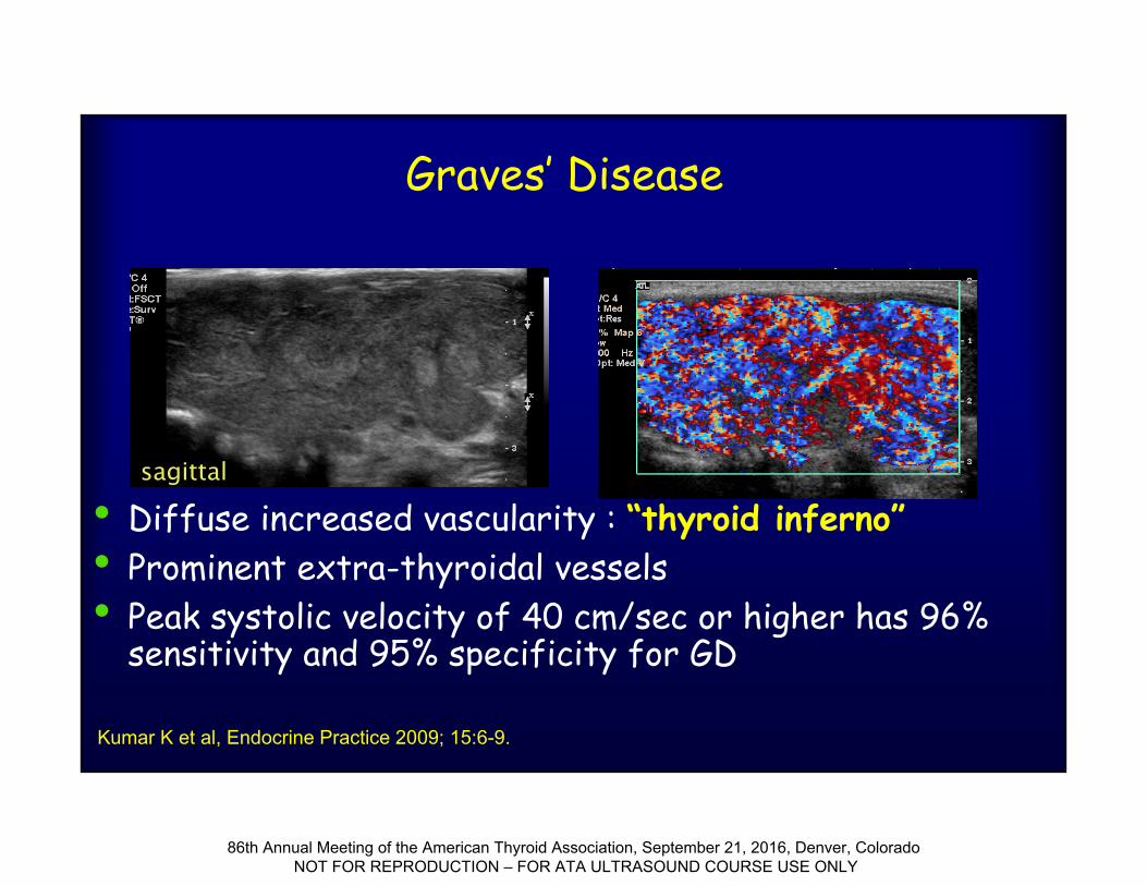
Graves’ Disease
• Diffuse increased vascularity : “thyroid inferno”• Prominent extra-thyroidal vessels• Peak systolic velocity of 40 cm/sec or higher has 96%
sensitivity and 95% specificity for GD
Kumar K et al, Endocrine Practice 2009; 15:6-9.
sagittal
86th Annual Meeting of the American Thyroid Association, September 21, 2016, Denver, Colorado NOT FOR REPRODUCTION – FOR ATA ULTRASOUND COURSE USE ONLY
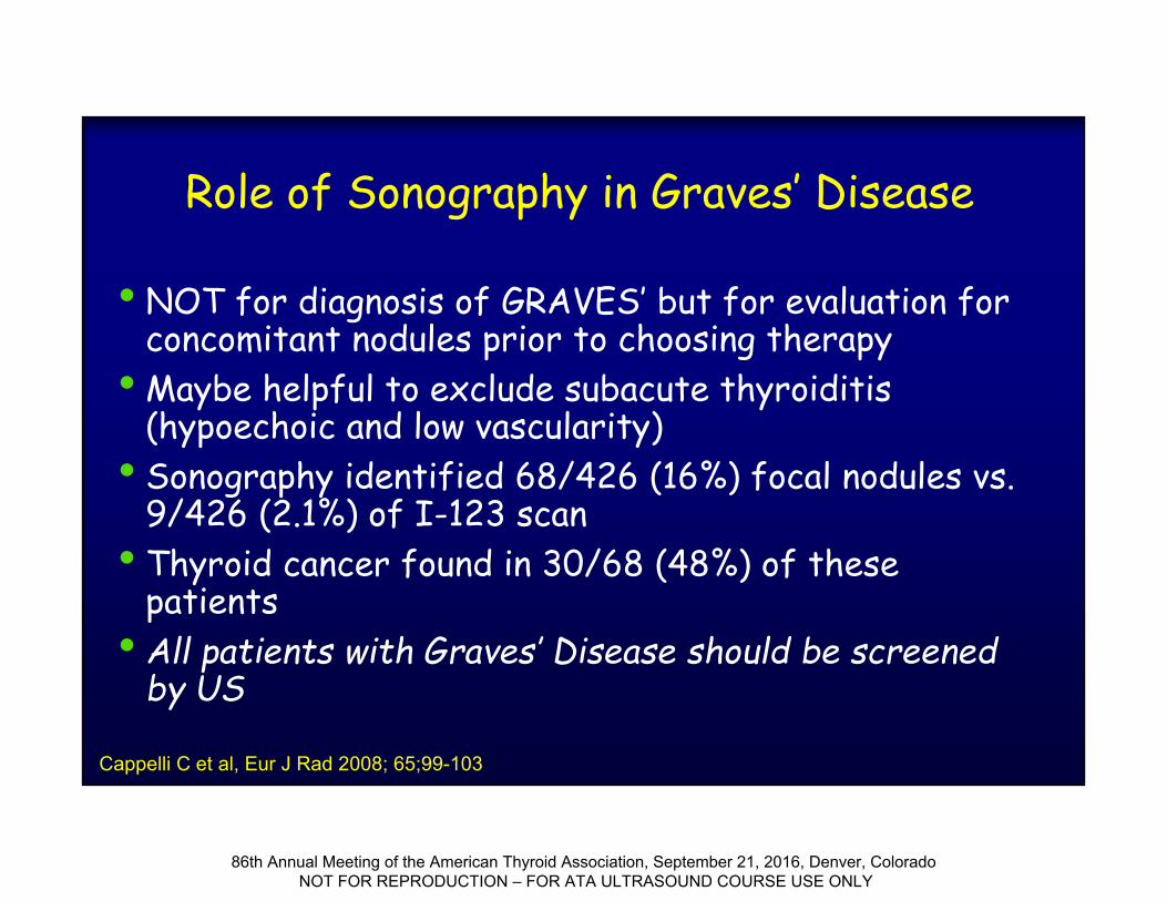
Role of Sonography in Graves’ Disease
• NOT for diagnosis of GRAVES’ but for evaluation for concomitant nodules prior to choosing therapy
• Maybe helpful to exclude subacute thyroiditis (hypoechoic and low vascularity)
• Sonography identified 68/426 (16%) focal nodules vs. 9/426 (2.1%) of I-123 scan
• Thyroid cancer found in 30/68 (48%) of these patients
• All patients with Graves’ Disease should be screened by US
Cappelli C et al, Eur J Rad 2008; 65;99-103
86th Annual Meeting of the American Thyroid Association, September 21, 2016, Denver, Colorado NOT FOR REPRODUCTION – FOR ATA ULTRASOUND COURSE USE ONLY
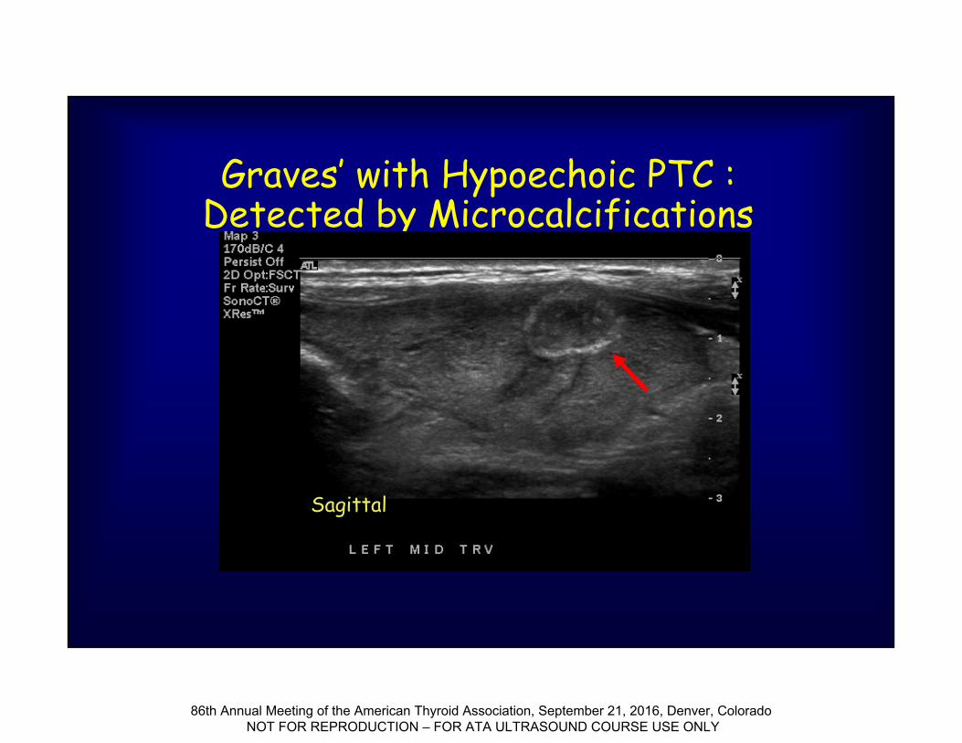
Graves’ with Hypoechoic PTC : Detected by Microcalcifications
Sagittal
86th Annual Meeting of the American Thyroid Association, September 21, 2016, Denver, Colorado NOT FOR REPRODUCTION – FOR ATA ULTRASOUND COURSE USE ONLY
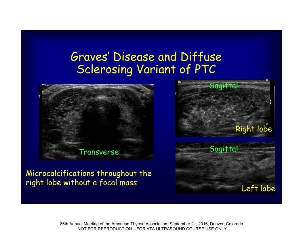
Graves’ Disease and Diffuse Sclerosing Variant of PTC
Microcalcifications throughout the right lobe without a focal mass
Left lobe
Right lobe
Sagittal
SagittalTransverse
86th Annual Meeting of the American Thyroid Association, September 21, 2016, Denver, Colorado NOT FOR REPRODUCTION – FOR ATA ULTRASOUND COURSE USE ONLY
Subacute Thyroiditis-“De Quervains”
• 0.16- 0.36% of thyroid disease• Usually after an upper respiratory viral infection• Presents NOT with signs of thyroid dysfunction but
with thyroid tenderness, systemic systems • May have thyrotoxicosis or be euthyroid• Hypoechoic patchy or nodular areas that resolve• Variable vascularity—generally NOT vascular
86th Annual Meeting of the American Thyroid Association, September 21, 2016, Denver, Colorado NOT FOR REPRODUCTION – FOR ATA ULTRASOUND COURSE USE ONLY
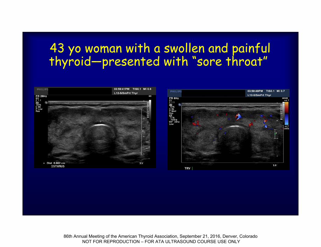
43 yo woman with a swollen and painful thyroid—presented with “sore throat”
86th Annual Meeting of the American Thyroid Association, September 21, 2016, Denver, Colorado NOT FOR REPRODUCTION – FOR ATA ULTRASOUND COURSE USE ONLY
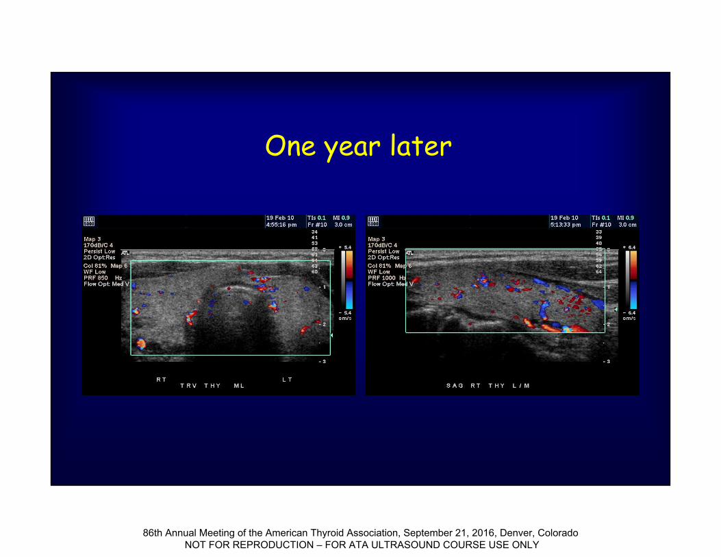
One year later
86th Annual Meeting of the American Thyroid Association, September 21, 2016, Denver, Colorado NOT FOR REPRODUCTION – FOR ATA ULTRASOUND COURSE USE ONLY
Other (rare) Causes of Diffuse Thyroid Abnormalities
• Malignancy • Lymphoma; Anaplastic carcinoma
• Infiltrative Disorders• Amyloid, Iron, etc.
• Reidel’s thyroiditis • ? others
86th Annual Meeting of the American Thyroid Association, September 21, 2016, Denver, Colorado NOT FOR REPRODUCTION – FOR ATA ULTRASOUND COURSE USE ONLY
Malignant Lymphoma
•Usually occurs in a Hashimoto’s gland• 1-2% of all thyroid malignancies•Nodular pattern
• Homogeneously hypoechoic with lobulated but well defined border; enhanced through transmission
•Diffuse OR asymmetric enlargement
Ito Y et al, World J Surg 2010; 34:1171-80
86th Annual Meeting of the American Thyroid Association, September 21, 2016, Denver, Colorado NOT FOR REPRODUCTION – FOR ATA ULTRASOUND COURSE USE ONLY
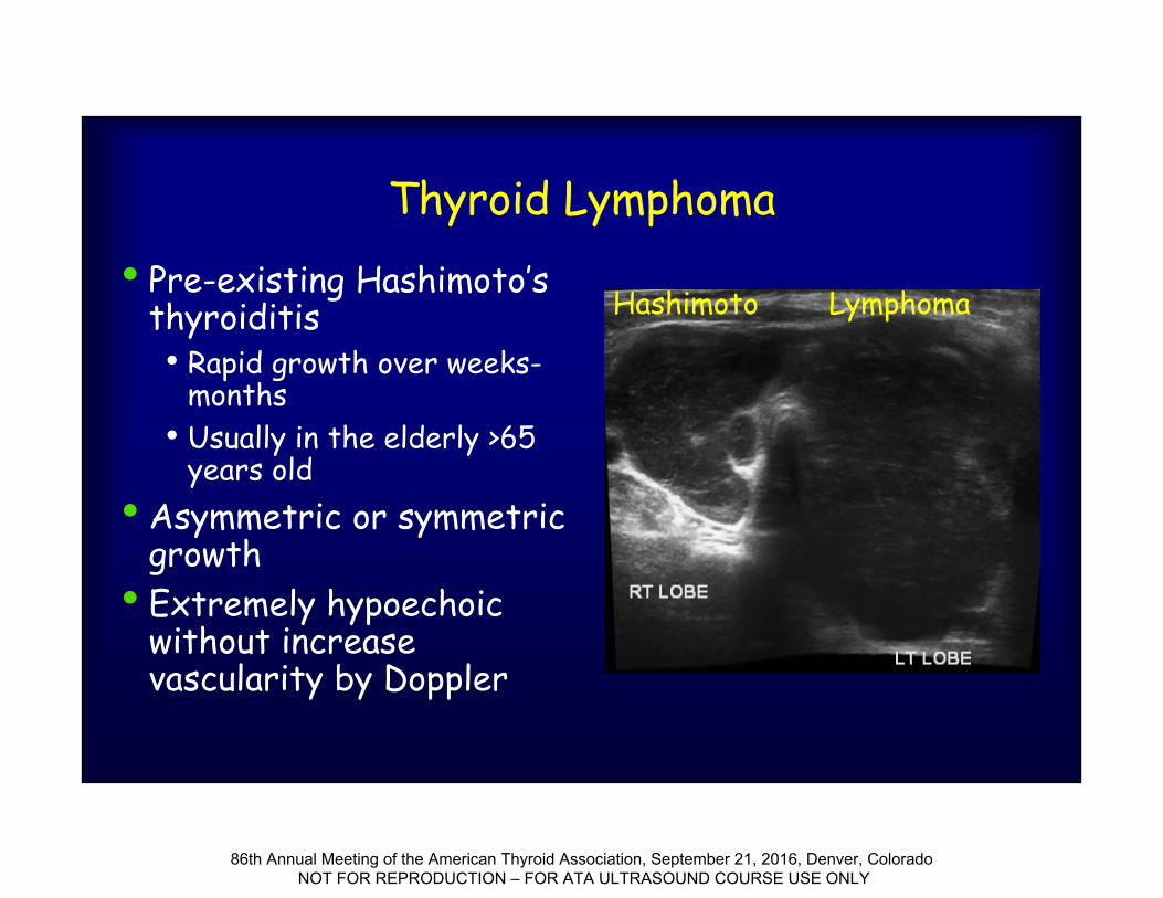
Thyroid Lymphoma• Pre-existing Hashimoto’s
thyroiditis• Rapid growth over weeks-
months• Usually in the elderly >65
years old• Asymmetric or symmetric
growth• Extremely hypoechoic
without increase vascularity by Doppler
Hashimoto Lymphoma
86th Annual Meeting of the American Thyroid Association, September 21, 2016, Denver, Colorado NOT FOR REPRODUCTION – FOR ATA ULTRASOUND COURSE USE ONLY
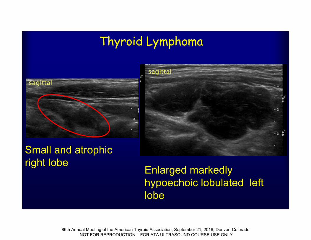
Thyroid Lymphoma
Small and atrophic right lobe
Enlarged markedly hypoechoic lobulated left lobe
sagittal
sagittal
86th Annual Meeting of the American Thyroid Association, September 21, 2016, Denver, Colorado NOT FOR REPRODUCTION – FOR ATA ULTRASOUND COURSE USE ONLY
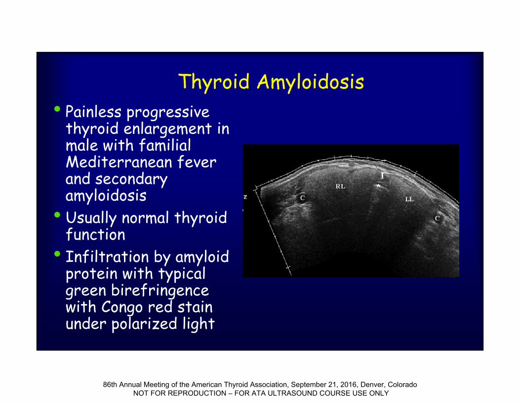
Thyroid Amyloidosis • Painless progressive
thyroid enlargement in male with familial Mediterranean fever and secondary amyloidosis
• Usually normal thyroid function
• Infiltration by amyloid protein with typical green birefringence with Congo red stain under polarized light
86th Annual Meeting of the American Thyroid Association, September 21, 2016, Denver, Colorado NOT FOR REPRODUCTION – FOR ATA ULTRASOUND COURSE USE ONLY
Conclusions•Sonographic markers of autoimmune thyroid
disease include enlarged size, heterogeneous echotexture, increased vascularity
•Clinical information is key•Differentiation of “pseudo-nodules” from true
nodules and tumors may be challenging• Asymmetric calcifications• Vascular pattern• Unilateral large LNS
86th Annual Meeting of the American Thyroid Association, September 21, 2016, Denver, Colorado NOT FOR REPRODUCTION – FOR ATA ULTRASOUND COURSE USE ONLY
Thank you for your attention!
QUESTIONS?
86th Annual Meeting of the American Thyroid Association, September 21, 2016, Denver, Colorado NOT FOR REPRODUCTION – FOR ATA ULTRASOUND COURSE USE ONLY


![Endocrine system [Head & Neck]cfd.mc.ntu.edu.tw/uploads/asset/data...regio 組織 組織 Female Reproductive System (I) Lab Anterior triangle of neck and Submandibular n Female Reproductive](https://img.pdfslide.net/doc/110x75/609a3f903b6608265c2b2e3f/endocrine-system-head-neckcfdmcntuedutwuploadsassetdata-regio.jpg)