Embed Size (px)
Citation preview
I-I
Ultraviolet Radiation Effectson the Corneal Epithelium
ITI
®REL EC TEMAR 141991,9 By
D Morris R. Lattimore
Sensory Research Division
February 1991
Approved for public releas; dlstrbuilo unlimited
91 3 1216 060United States Army Aeromedical Research Laboratory
Fort Rucker, Alabama 36362-5292
Notice
Oualified reauesters
Qualified requesters may obtain copies from the DefenseTechnical Information Center (DTIC), Cameron Station, Alexandria,Virginia 22314. Orders will be expedited if placed through thelibrarian or other person designated to request documents fromDTIC.
Change of address
Organizations receiving reports from the U.S. ArmyAeromedical Research Laboratory on automatic mailing lists shouldconfirm correct address when corresponding about laboratoryreports.
Animal use
In conducting the research described in this report, theinvestigators adhered to the Guide for the care and use oflaboratory animals, al promulgated by the Committee on Care andUse of Laboratory Anikals, National Academy of Sciences-NationalResearch Council.
Disposition
Destroy this report when it is no longer needed. Do notreturn to the originator.
Disclaimer
The views, opinions, and/or findings contained in thisreport are those of the authors and should not be construed as anofficial Department of the Army position, policy, or decisionunless so designated by other official documentation. Citationof trade names in this report does not constitute an officialDepartment of the Army endorsement or approval of the use of suchcommercial items.
Review.,&:.
THOMAS L. __ME_--U
LTC, MSDirector, Sensory'ResearchDivision Released for publication:
RM W. WI Wl, 0. D. , Ph. D. DAVID H. KARNEYChairman, Scientific Colonel, MC, SFSReview Committee Commanding
UnclassifiedSECURITY CLASSIFICATION OF THIS PAGE
SForm Approved
REPORT DOCUMENTATION PAGE OMNo. 07040 188la. REPORT SECURITY CLASSIFICATION lb RESTRICTIVE MARKINGS
Unclassified2a. SECURITY CLASSIFICATION AUTHORITY 3 DISTRIBUTION /AVAILABILITY OF REPORT
2b. DECLASSIFICATION/DOWNGRADING SCHEDULE Approved for public release; distributionunlimited
4. PERFORMING ORGANIZATION REPORT NUMBER(S) 5. MONITORING ORGANIZATION REPORT NUMBER(S)
USAARL Report No. 91-106a- NAME OF PERFORMING ORGANIZATION 6b. OFFICE SYMBOL 7a. NAME OF MONITORING ORGANIZATION
U.S. Army Aeromedical Research (if applicable) U.S. Army Medical Research and DevelopmentLaboratory SGRD-UAS-VS Command
6c. ADDRESS (City, State, and ZIP Code) 7b. ADDRESS (City, State, and ZIP Code)
Fort DetrickFort Rucker, AL 36362-5292 Frederick, MD 21702-5012
Ba. NAME OF FUNDING/SPONSORING 8b. OFFICE SYMBOL 9. PROCUREMENT INSTRUMENT IDENTIFICATION NUMBERORGANIZATION (if applicable)
8c. ADDRESS (City, State, and ZIP Code) 10. SOURCE OF FUNDING NUMBERS
PROGRAM PROJECT TASK WORK UNITELEMENT NO. NO. NO. ACCESSION NO.
62787A 3M162787A7 19 BG 16811. TITLE (Include Security Classification)
Ultraviolet Radiation Effects on the Corneal Epithelium (U)
12. PERSONAL AUTHOR(S)
Morris R. Lattimore13a. TYPE OF REPORT 13b. TIME COVERED 14 DATE OF REPORT (Year, Month, Day) 15. PAGE COUNT
FROM TO , 1991 FThruOy_ 2016. SUPPLEMENTARY NOTATION Presented at the AGARD Aerospace Medical Panel Symposium on OcularHazards in Flight and Remedial Measures, 22-26 October 1990, London, UK.
17. COSATI CODES 18. SUBJECT TERMS (Continue on reverse if necessary and identify by block number)
FIELD GROUP SUB-GROUP
06 p 04 UVR, corneal epithelium, metabolism, glucose, oxygen20 06
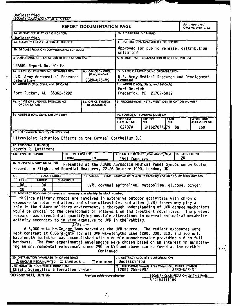
19. ABSTRACT (Continue on reverse if necessary and identify by block number)- Since military troops are involved in extensive outdoor activities with chronicexposure to solar radiation, and since ultraviolet radiation (UVR) lasers may play arole in the future military environment, a thorough understanding of UVR damage mechanismswould be crucial to the development of intervention and treatment modalities. The presentresearch was directed at quantifying possible alterations in corneal epithelial metabolicactivity secondary to in vivo exposure to UVR in tbt'rabbitz.
A 5,000 watt Hg-Xe ar- lamp served as the UVR source. The radiant exposures werekept constant at 0.05/ 1J.cnm for all UVR wavelengths used (290, 300, 310, and 360 nm).Wavelength isolation was accomplished with a double monochromator providing a 6 nm fullbandpass. The four experimental wavelengths were chosen based on an interest in maintain-ing an environmental relevance,sine 290 nm UVR and above can be found at the earth's
Continued
20. DISTRIBUTION/AVAILABILITY OF ABSTRACT 21. ABSTRACT SECURITY CLASSIFICATIONI0IUNCLASSIFIED/UNLIMITED 0 SAME AS RPT. 03 DTIC USERS Unclassified
22a. NAME OF RESPONSIBLE INDIVIDUAL 22b. TELEPHONE (Include Area Code) 22c. OFFICE SYMBOLChief, Scientific Information Center (205) 255-6907 SGRD-UAX-SI
DD Form 1473, JUN 86 Previous editions are obsolete. SECURITY CLASSIFICATION OF THIS PAGE
Unclassified
19. ABSTRACT (Continued)surface. Micropolarographic measurement of corneal oxygen uptake ratesserved as an in vivo index of UVR-induced effects on oxidative metabolism.Microfluorometric analyses of key epithelial energy metabolites (glucose,glycogen, ATP, and PCr) were used as an in vitro index of UVR-inducedeffects on overall metabolic activity. A paired difference analysis ofthe oxygen uptake rate data demonstrated a decrease in relative cornealoxidative metabolic activity that was wavelength-dependent. These sameexperimental UVR exposure conditions served to significantly increaseepithelial glucose and glycogen concentrations. Although the epithelialATP concentrations were unchanged, the epithelial PCr concentrations (a
_ high energy phosphate bond reservoir) decreased as a result of UVR exposure.
These _ata demonstratesa decrease in corneal epithelial oxidativemetabolic activity as a result of UVR exposure, and infer an adverse effecton glycolytic metabolism, as well. It is suggested that immediate UVR-induced metabolic inhibitory effects can be responsible for the pattern ofepithelial cell loss seen in photokeratitis.
I-,!
Table of contents
Introduction .................................... 3Materials and Methods. . .................. . ............ 4
Experimental animals................................. 4Oxygen electrode. . . ............................... 4Calibration procedure. . ............. .. .. ........... 5In vivo micropolarographic application .....................5In vitro processsing procedu re........................ 6Underlying principles . ............................ 6Source measurement. . .. .. .......................... . 6
Summiary' .. ............................. .............. 15References ............................................. 17
List of figures
Figure 1, UVR-altered corneal 0, uptake rate:extrapolated projection to baseline....................... 9
Figure 2, TVR-alteration of cornealepithelial metabolites.............................. 10
Figure 3, Corneal 0 z uptake vs epithelialenergy metabolites. .............................. 11
Figure 4, Corneal 02 uptake vs epithelialmetabolite accumulation................................... 12
Figure 5, Altered 02 uptake vs exposureduration. ......... :. .......... ................. 14
Figure 6, Direct comparison of UVR-altered 02consumption to metabolite changes .................... 16
F AcesonForNTIS CA&IJDT IC TAB -
B y ..... . ..... ...... .. .... .
Av: .r.- r1 Dist
This page intentionally left blank.
2
Introductioni
Ultraviolet radiation (UVR) has been implicated withinpublic health arenas as a potential stimulus for degenerativeocular changes since the time of the ancient Greeks.2 Recentspeculation concerning changes in the nature of the atmosphericozone layer has led to increased interest in adverse effects ofUVR exposure. The advent of UVR lasers have further spurredresearch interest concerning the determination of possiblemechanisms of action, since the development of a full spectrum ofapplications is dependent upon a thorough understanding ofoperant mechanisms.
The ideal tissue for investigating the in vivo effects ofUVR is the corneal epithelium. As the most anterior tissue layerof the eye, the corneal epithelium is subject to a directinteraction with incident radiation. The tissue is uncomplicatedby spurious absorbers, thermal effects, or pigmentaryphotochemistry. In addition, its avascularity and accessabilityenable response gathering uninfluenced by circulatory systemfactors remotely external to the tissue. Finally, the cornealepithelium is exposed to UVR on a daily basis, so exposurestudies are not subjecting the tissue to a completely unnaturalcondition.
Many investigations into the effects of UVR on the cornealepithelium have concentrated on morphological evaluationsutilizing the biomicroscope, the light microscope, and/or theelectron microscope (Verhoeff and Bell, 1916; Cogan and Kinsey,1946; Pitts and Tredici, 1971; Ringvold, 1980 and 1983). Suchstudies have provided detailed information concerning the delayedstructural changes characteristic of UVR damage that occur 4 to12 hours after exposure. As a result of this histologicaldetection delay, information concerning either immediate orfunctional effects of UVR cannot be probed by such methods.
Millodot and Earlam (1984), seeking to evaluate this damage-delay phenomenon, revealed the presence of a period of decreasedcorneal sensitivity immediately following exposure to UVR. Theirfinding appears to signify an immediate effect of UVR upon thesensory neurons subserving the corneal epithelium. If such isthe case, and knowing that these axons appear deep within the
1 Presented at the AGARD Aerospace Medical Panel Symposium on
Ocular Hazards in Flight and Remedial Measures, 22-26 October,1990. London, UK.
2 Xenophan's treatise "Anabasis" discusses the condition of
"snowblindness."
3
basal cell layer of the corneal epithelium and within theanterior stroma, it would be reasonable to assume that theremight also be an immediate effect of UVR on the cornealepithelium itself. Therefore, the purpose of this study was toevaluate the possible immediate effect of exposure to UVR on themetabolism of the corneal epithelium in the rabbit.
Selection of the exposure wavelengths (290, 300, 310, and360 nm) was based on an interest in maintaining an environmentalrelevance. An additional factor was the intention of creating adistinctive span of effects by maintaining a constant level ofradiant exposure at 0.05 J-cm - with varied wavelengthchallenges. Measurement of the corneal oxygen uptake rate servedas an in vivo assessment of epithelial metabolic activity, whilemicro-fluorometric metabolite analyses served as an in vitroindex of epithelial metabolic activity.
Materials and methods
Experimental animals
Healthy, adult, Dutch-belted, pigmented rabbits were used asthe experimental animals. The animals were housed in NIH-approved quarters under controlled, normal lighting conditions.The animals were maintained, and the experiments were conductedin accordance with procedures outlined in the "Guide forLaboratory Animals Facilities and Care" of the National Academyof Sciences-National Research Council. Anesthesia was maintainedthroughout the course of the experiment with intramuscularinjections of Ketalar (10 mg/kg) and Rompun (5 mg/kg). The UVR-exposed animals were sacrificed by cervical dislocation 2 minutesafter exposure was discontinued. The eyes were then immediatelyremoved and immersed in liquid nitrogen in order to preventsignificant change in metabolite levels. Control animals, aftera mock-exposure period under the same level of anesthesia as theexperimental animals, underwent the same tissue preparationprocedures.
Oxygen electrode
The micropolarographic oxygen probe consisted of a platinumcathode (25-um diameter) and a silver anode embedded in a plasticcarrier. A potassium chloride (KCl) solution served as anelectrolyte bridge between the cathode and the anode. Anoxygen-permeable polyethylene membrane, 25 um thick, effectivelysealed the entire electrode-KCl assembly into one operating unit.The micropolarographic system was similar to that used byBenjamin and Hill (1986).
4
In general terms, when a voltage is imposed across the KClbridge from the anode to the cathode, oxygen present at thesensor tip undergoes an electrolytic reduction at the platinumcathode, with the reverse reaction occuring at the anode. Thecurrent generated across the cathode/anode gap is directlyproportional to the oxygen concentration of the solution when thesystem is properly calibrated. The generated current isamplified by a blood gas analyzer and recorded vs time on aphysiograph.
Calibration procedure
The probe was calibrated by first placing it in anair-saturated saline solution of 155 mm Hg partial pressure ofoxygen. The probe was then placed in a nitrogen-saturated salinesolution of 0 mm Hg partial pressure of oxygen. The timerequired for the recorded oxygen tension to then drop from 140 mmHg to 40 mm Hg provided the time constant of themicropolarographic system. The experimental procedure involvedapplying the probe to the anterior surface of the cornealepithelium of the living, anesthetized rabbit. The sensor, whenapplied to the eye, provided a limited reservoir of oxygen forthe underlying tissue. The average rate of oxygen depletion fromthe sensor reservoir, between recordings of 140 and 40 mm Hg, andafter correction for the time constant, became the measure ofcorneal oxygen uptake rate, which in turn represented a relativemeasure of the aerobic requirement of the corneal epithelium.Since the aerobic metabolic rate of a tissue directly reflectsmitochondrial function (Graymore, 1969), a micropolarographicoxygen electrode, when used to record the rate of oxygen uptakeby the anterior surface of the intact cornea, would primarilyserve as a relative index of corneal mitochondrial actility.
In vivo micropolarographic application
The eyes of 32 rabbits were exposed in vivo to specificradiant exposure levels of UVR. There were 8 rabbits used in the290 nm experimental group, 12 rabbits used in the 300 nmexperimental group, 7 rabbits used in the 310 nm experimentalgroup, and 5 rabbits used in the 360 nm experimental group.Prior to UVR exposure, five baseline oxygen uptake rates weredetermined for each eye using a micropolarographic oxygen sensorthat was attached to a pH/blood gas monitor in the fashion ofBenjamin and Hill (1986). Oxygen uptake rates again weredetermined 2 minutes post-UVR exposure, enabling the experimenterto compare the change in oxygen uptake rate as a result of theUVR exposure. The uptake rate values were subjected to apaired-difference analysis. A two-way analysis of variance(ANOVA) was performed to examine both within and betweenexperimental groups, and to determine the presence or absence ofan overall effect of UVR on the measured oxygen uptake rates.
5
In vitro processing procedure
The rabbit eyes were transferred from the liquid nitrogencontainer into a -80° C freezer for storage until tissueprocessing could be accomplished. The cornea was removed fromthe globe by dissection under -30" C conditions in a wedeen-typecryostat. The isolated cornea was cut into halves, which weremounted on a sectioning button by immersion in a dry ice-cooledhexane solution. The corneal button-mount then was transferredto a cryostatic microtome where tissue sectioning was performed.The resulting central cornea cross sections were approximately 20micrometers (gm) in thickness. The sectioned tissue samples wereplaced in a metal tissue holder, covered with glass slides, andinserted into a vacuum tube. The tube was placed in a -200 Cfreezer and attached to a vacuum pump. The tissue then wasfreeze-dried for a 24 hour period. After the freeze-dryingprocess was completed the tissue was kept at -200 C untilassayed.
Samples needed for assay were thawed under vacuum for 1 hourto prevent condensation-stimulated enzyme action. The differentlayers of the cornea were defined clearly which permitted easyisolation of the corneal epithelium under a 3X binoculardissecting microscope. Tissue size was determined by dry weight,rather than by tissue section dimensions, which permitted theanalysis of very small and irregularly shaped specimens. Thetissue samples immediately were weighed on a quartz fiberfishpole balance possessing gg sensitivity. After weighing andrecovery, the samples were placed in an oil well rack forspecific metabolite assay.
Underlying principles
The cycling system contains several enzymes which catalyzespecific interrelated reactions yielding a "net reaction." Abyproduct of this multi-step reaction is reduced nicotinamideadenine dinucleotide phosphate (NADPH), which fluoresces light of460 nm wavelength when excited with UVR of 340 nm wavelength. Bymeasuring the amount of this reduced pyridine nucleotidefluorescence, the original concentration of the assayedmetabolite can be inferred by calculation (Lowry and Passonneau,1972). Appropriate blanks and standards were employed to monitorthe reliability of the assays. Individual enzymatic cycling andincubating techniques permitted isolation of the specificmetabolite being analyzed.
Source measurement
Source calibration and radiometric quantification followedthe procedures described by Pitts, Cullen, and Hacker (1977).The UVR source was a 5,000 watt xenon-mercury (Xe-Hg) highpressure arc lamp, powered by a 10 kilowatt (kw) direct current
6
power supply regulated to +/. 0.5 percent and capable ofdelivering from 0 to 80 amps at 25 to 65 volts to the arcelectrodes. The lamp housing was cooled by a double-blowersystem. The radiation from the source was focused at amonochromator entrance slit by the housing optics. A 10 cmquartz-enclosed water chamber was placed between the focusinglenses and the monochromator in order to remove infraredradiation. The desired UVR waveband was obtained with a doublegrating monochromator with gratings blazed at 300 nm and groovedwith 1,180 grooves/mm, allowing approximately a 5 nm bandpass.The linear dispersion equated to a value of 0.82 nm/mm.Entrance, intermediate, and exit slits were set to pass a nominalfull bandpass of 6.6 nm. The system was aligned with a helium-neon laser and the wavelength counter was calibrated with amercury source. Exposure durations were set with an electronicshutter controlled by a preset counter. The preset counterallowed exposure durations of any desired length with millisecondaccuracy.
The exit optical beam was focused by a quartz lens with abeam size of 1.6 cm by 1.8 cm at the plane of the experimentalanimal's cornea. A 16-junction thermopile, traceable to aNational Bureau of Standards (NBS) standard source, was used tocharacterize the spectral irradiance of the UVR source. Whentaking the spectral irradiance readings, the thermopile wasplaced in the same position relative to the monochromator exitport as the rabbit's cornea was to be situated during UVRexposure. The irradiance (Ee), in watts per square centimeter(W-cm'2), incident on the thermopile was determined by thefollowing relationship:
Ee = kVt.
The value "k" represents the thermopile calibration constant inmicrowatts per square centimeter per microvolt (gW.cm
2 -V'1),
while the value "V," represents the average (mean) of threethermopile-voltmeter readings in microvolts (AV). Thecalibration constant for the thermopile used in this experimentwas 5.131 pW-cm'2-V "1. The radiant exposure (H), in Joules persquare centimeter (J-cm2), was calculated by the formula:
H = Eet.
The value "t" is simply time in seconds; it should be kept inmind that a Joule is a watt-second. Therefore, for a givenirradiance "Ee" the exposure duration "t" can be varied to obtaindifferent values of radiant exposure "H" as desired. Conversely,a radiant exposure can be kept constant, even though wavelengthirradiance may differ, by varying the time of exposure. Thismeans of output characterization and source calibration wasestimated to have a +/_10 percent accuracy.
7
The variation of "t" in order to obtain a constant ",H"I, inthe context of the wavelengths used in this experiment, createsan outcome that is somewhat dependent upon the validity of theprinciple of reciprocity (i.e., the biological effects orendpoints are independent of exposure time and irradiance).Corneal effects of a krypton-ion laser, with simultaneous outputat 350.7 and 356.4 nm (3:1 ratio), illustrates that the productof threshold intensity and the pulsewidth is a constant; thethresholds for multi-pulse exposures have been shown to be inagreement with those for single-pulse exposures (Zuclich andConnolly, 1976). A similar corneal damage pattern can beelicited from helium-cadmium laser data at 325 nm (Ebbers andSears, 1975). Based on the literature, it would not beunreasonable to assume that reciprocity holds for all four UVRwavelengths utilized in this experiment.
Results
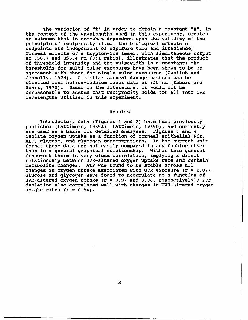
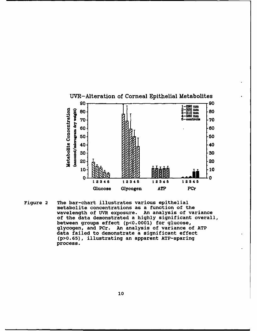
Introductory data (Figures 1 and 2) have been previouslypublished (Lattimore, 1989a; Lattimore, 1989b), and currentlyare used as a basis for detailed analyses. Figures 3 and 4isolate oxygen uptake as a function of corneal epithelial PCr,ATP, glucose, and glycogen concentrations. In the current unitformat these data are not easily compared in any fashion otherthan in a general graphical relationship. Within this generalframework there is very close correlation, implying a directrelationship between UVR-altered oxygen uptake rate and certainmetabolite changes. ATP was found to be stable across allchanges in oxygen uptake associated with UVR exposure (r = 0.07).Glucose and glycogen were found to accumulate as a function ofUVR-altered oxygen uptake (r = 0.97 and 0.98, respectively); PCrdepletion also correlated well with changes in UVR-altered oxygenuptake rates (r = 0.84).
8
UVR-Altered Corneal 02 Uptake Rate:Extrapolated Projection to Baseline
00
S-.0.5 J/om2
6) 0-0. 10 i/cm2
S-..15 j/=m2
(D-'0.20 i/cm2
--0.25 ,/cm
-51280 300 320 340 360 380 400 420 440 460
Wavelength (rn)
Figure 1 By plotting the UVR-altered corneal oxygen uptake ratedata as a function of wavelength, and by makingseparate datasets for each radiant exposure, a"family" of plots is obtained. A two-way analysis ofvariance demonstrated an overall significantbetween-groups difference (p < 0.0001), as well asrevealed an interactive effect between wavelength anddose (p < 0.005). Unexposed eyes exhibited nosignificant change in corneal oxygen uptake rates overthe course of the experiment.
9
UVR-Alteration of Corneal Epithelial Metabolites90 1 1-200 am 90
2-300 am80-3-310 an -804-380 nm
70--OnPB 7
~60- 60
0I 50 50404
o 30. 30
20 - 20
10 100 0
12345 12345 12345 12345
Glucose Glycogen ATP PCr
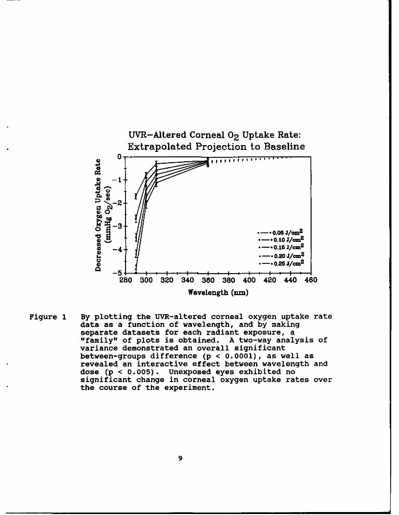
Figure 2 The bar-chart illustrates various epithelialmetabolite concentrations as a function of thewavelength of UVR exposure. An analysis of varianceof the data demonstrated a highly significant overall,between groups effect (p<0.0001) for glucose,glycogen, and PCr. An analysis of variance of ATPdata failed to demonstrate a significant effect(p>0.65), illustrating an apparent ATP-sparingprocess.
10
Corneal 02 Uptake vsEpithelial Energy Metabolites
r...0.0
CQ 0oe A..O84Pu0
r ~ .. o J/.m 2
4 8 8 10 1W1
10 =OU
UVR-Altered Metabolite Concentration(nmol/ugram dry weight)
Figure 3 Plotted decreases in the oxygen uptake rate as afunction of PCr and ATP, illustrate ATP-sparing at theapparent expense of PCr.
... . ......1
Corneal 02 Uptake vsEpithelial Metabolite Accumulation
T S glcgenS R vaL ..... 0.979
STope...-O.OSZ
2-0.20 =
0.30 310
CQOV -0.80 Sooa
-170 RaL 0974 T am
SlopO...-O.084 Radiant Uposure...0.05 J/cm2
6 . . .. i '--0 . . . . . . i -4 0- - . . .. . .,0 80 1020 30 40 0 0 70o 80
UVR-Altered Metabolite Concentration(nmol/uram dry weight)
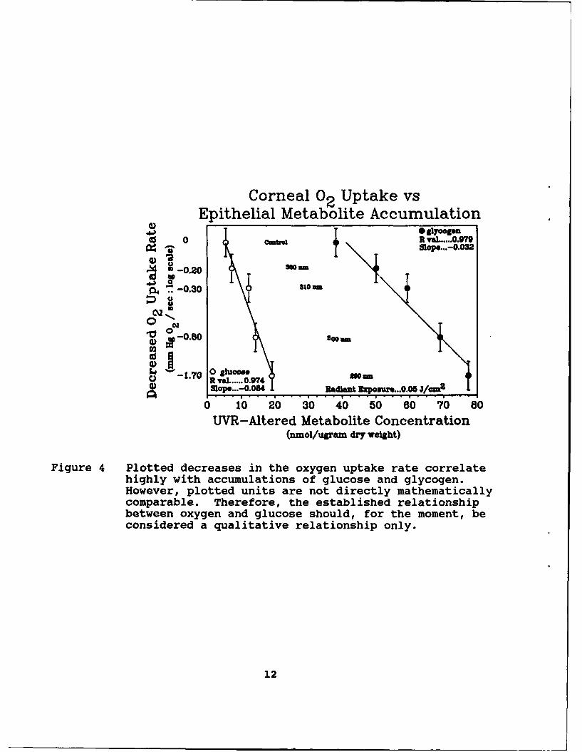
Figure 4 Plotted decreases in the oxygen uptake rate correlatehighly with accumulations of glucose and glycogen.However, plotted units are not directly mathematicallycomparable. Therefore, the established relationshipbetween oxygen and glucose should, for the moment, beconsidered a qualitative relationship only.
12
Discussion
The corneal epithelium is known to conduct both aerobic andanaerobic metabolic activity concurrently (Kinoshita and Masurat,1959). According to some estimates, the rabbit cornealepithelium routinely consumes up to 85 percent of availableglucose in anaerobic channels, with the remaining 15 percent usedvia aerobic channels (Riley, 1969). When anaerobic conditionsare artificially imposed upon a cornea (i.e., by the applicationof a thick contact lens), the tissue response has been portrayedto be increased anaerobic activity, inferred by the depletion ofepithelial glycogen stores (Uniacke and Hill, 1972). Yet, underthe UVR exposure conditions of this experiment, decreased oxygenutilization was evidenced with a contradictory accumulation ofboth glucose and glycogen, rather than the expected depletion.This paradox led to the examination of oxygen uptake changes as adependent variable of key metabolites.
The results indicate: a decrease in oxygen consumption, anapparent decrease in glucose utilization, a stabilization of ATP,and a decrement in PCr. The manifested close relationshipbetween the research variables point toward an alteration ofoxidative or mitochondrial activity resulting from UVR exposure.In addition, paradoxical glucose and glycogen accumulationssuggest a secondary underlying effect on the anaerobic orglycolytic chain. A global enzyme inactivation can be excludedbecause glycogen and ATP storing are mediated enzymatically.Therefore, it can be concluded that observed UVR effects are theresult of more than one damage mechanism. However, this closecorrelation stems from a superficial view; translation of oxygendata to units more directly comparable could provide a greaterinsight into operant mechanisms of UVR damage.
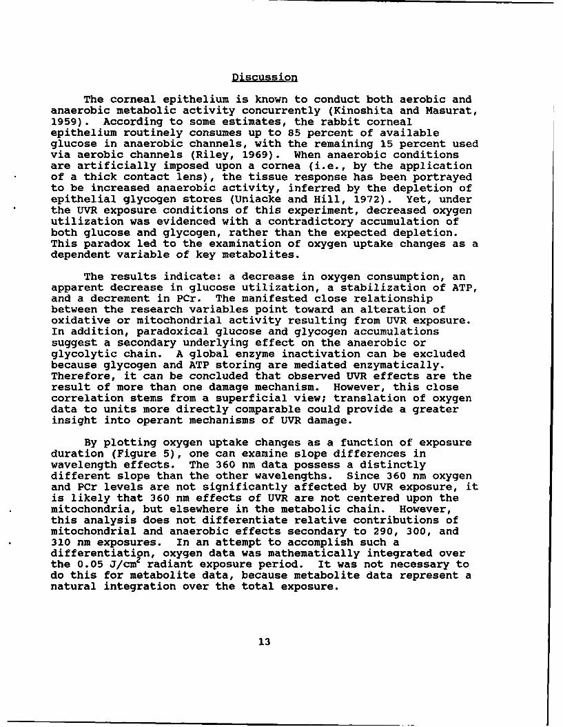
By plotting oxygen uptake changes as a function of exposureduration (Figure 5), one can examine slope differences inwavelength effects. The 360 nm data possess a distinctlydifferent slope than the other wavelengths. Since 360 nm oxygenand PCr levels are not significantly affected by UVR exposure, itis likely that 360 nm effects of UVR are not centered upon themitochondria, but elsewhere in the metabolic chain. However,this analysis does not differentiate relative contributions ofmitochondrial and anaerobic effects secondary to 290, 300, and310 nm exposures. In an attempt to accomplish such adifferentiation, oxygen data was mathematically integrated overthe 0.05 J/cm2 radiant exposure period. It was not necessary todo this for metabolite data, because metabolite data represent anatural integration over the total exposure.
13
Altered 02 Uptake vs Exposure Duration
- 3-000
41 -2-2
0 3 -30I -4, o290nrum -4•~ 300 rum -
-5 %310 nm-6 '38°"" -8-7i -7Cj, Radiant Zzpomve: 0.05 - 0.28 1/cm2-8 1:1t ::11aI-f5: & .. -8
0 2000 4000 6000 8000 10000
Time (sec)
Figure 5 Comparison of the oxygen-exposure time slope datahighlight 360 nm effects to be independent of otherwavelength mechanisms of action. The combined absenceof oxygen effects accompanied by glucose accumulationsuggest the presence of a damage mechanism isolatedwithin the anaerobic stages of metabolism.
14
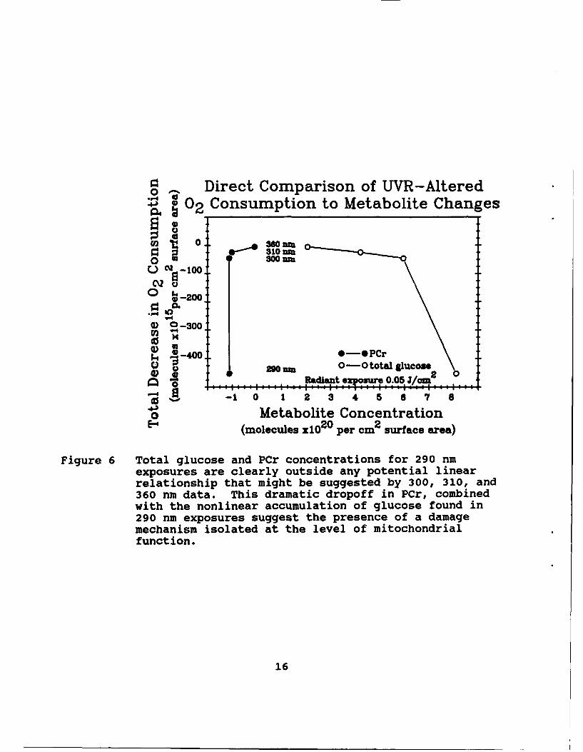
Since oxy en data are theoretically translatable from mm HgO2/sec to ul/cm, and metabolite data can be translated fromnmol/ug to nmol/cm2 , a directly comparable unit correlation maybe obtained. This method of data presentation (Figure 6)highlights the relative relationships between mitochondrial andanaerobic metabolism, with UVR wavelength. Short wavelength UVR(i.e., 290 nm and possibly shorter) are shown to possesspredominantly an adverse mitochondrial effect on the cornealepithelium, since PCr dramatically falls off compared to theother wavelength exposures, and total glucose accumulations aremuch less than a standard, linear model would predict. As theexposure wavelength increases, total glucose accumulation and PCrdepletion conform to a linear representation when compared toequivalent oxygen decrements. Therefore, it can be concludedthat observed UVR effects on the corneal epithelium are theresult of more than one damage mechanism. UVR exposures at ornear 360 nm will produce effects predominantly by way ofdisruption of anaerobic/glycolytic metabolic pathways. UVRexposures at and possibly below 290 nm will produce effectspredominantly by way of disruption of aerobic/mitochondrialmetabolic pathways. UVR exposures at intermediate wavelengthswill produce compound effects on the corneal epithelium involvingboth damage mechanisms.
Summary
Corneal epithelial metabolism is affected adversely by adual mechanism of UVR damage. This duality differentiallypresents itself dependent upon exposure wavelength. Whileconsiderable overlap is possible, clearly short wavelength UVR ispredominantly toxic to the mitochondrial system, while longerwavelength UVR predominantly affects the anaerobic metabolicpathways. Dual damage mechanisms, with overlapping actionspectra could complicate the development of intervention andtreatment modalities. Specific enzymatic analyses will benecessary to fully elicit wavelength specificities and potentialtreatment options.
15
0 A Direct Comparison of UVR-AlteredS02 Consumption to Metabolite Changes
0 -200
0 0-300
4),-400 0-0 PCr290 0-0 total glucose
datftt 0re.05Jl/aA-10 1 2 3 4 7 8
0 Metabolite ConcentrationE--, (molecules X10 20 per cm2 surface area)
Figure 6 Total glucose and PCr concentrations for 290 nmexposures are clearly outside any potential linearrelationship that might be suggested by 300, 310, and360 nm data. This dramatic dropoff in PCr, combinedwith the nonlinear accumulation of glucose found in290 nm exposures suggest the presence of a damagemechanism isolated at the level of mitochondrialfunction.
16
References
Benjamin, W. J., and Hill, R. M. 1986. Human cornealoxygen demand: the closed eye interval. Graefes archivesof clinical and experimental ophthalmology.224(3) :291-294.
Cogan, D. G., and Kinsey, V. E. 1946. Action spectrum ofkeratitis produced by ultraviolet radiation.Archives of oohthalmology. 35:670-677.
Ebbers, R. W., and Sears, D. 1975. Ocular effects of a 325nm ultraviolet laser. American journal of optometry andRhysiological optics. 52:216-219.
Graymore, C. N. 1969. "Biochemistry of the eye." AcademicPress. London, New York.
Kinoshita, J. H., and Masurat, A. B. 1959. Aerobicpathways of glucose metabolism in bovine cornealepithelium. American journal of ovhthalmolov.48:47-52.
Lattimore, M. R. 1989a. Effects of ultraviolet radiationon the oxygen uptake rate of the rabbit cornea.Optometry and vision science 66:117-122.
Lattimore, M. R. 1989b. Effect of ultraviolet radiation onthe energy metabolism of the corneal epithelium of therabbit. Photochemistry and Rhotobiology. 49:175-180.
Lowry, 0. H., and Passonneau, J. V. 1972. "A flexiblesystem of enzymatic analysis." Academic Press, New York.
Millodot, M., and Earlam, R. A. 1984. Sensitivity of thecornea after exposure to ultraviolet light. Ophthalmicresearch. 16:325-328.
Pitts, D. G., and Tredici, T. J. 1971. The effects ofultraviolet on the eye. American industrial hygieneassociation lournal. 32:235-246.
Pitts, D. G., Cullen, A. P., and Hacker, P. D. 1977.Ocular effects of ultraviolet radiation from 295 to365 nm. Investigative oDhthalmolouy and visual science.16:932-939.
17
Riley, M4. V. 1969. Glucose and oxygen utilization by therabbit cornea. Experimental eye research 8:193-19?.
Ringvold, A. 1980. Cornea and ultraviolet radiation.Acta oohthalmologica. 58:63-68.
Ringvold, A. 1983. Damage of the corneal epitheliumu causedby ultraviolet radiation. Acta ophthalmologica.61:898-907.
Uniacke, C. A., and Hill, R. M4. 1972. The depletion courseof epithelial glycogen with corneal anoxia. Arve~qophthalmology. 87:56-59.
Verhoeff, F. H., and Bell, L. 1916. The pathologicaleffects of radiant energy on the eye. Proceedings of theAnerican academy of arts and sciences. 61:630-818.
Zuclich, 3. A., and Connolly, J. S. 1976. Ocular damageinduced by near-ultraviolet laser radiation.Investigative ophthalmology and visual science.15:760-764.
Initial distribution
Commander, U.S. Army Natick Research, U.S. Army Avionics ResearchDevelopment and Evaluation Center and Development Activity
ATTN: STRNC-MIL (Documents ATTN: SAVAA-P-TPLibrarian) Fort Monmouth, NJ 07703-5401Natick, MA 01760-5040
U.S. Army Communications-ElectronicsNaval Submarine Medical Command
Research Laboratory ATTN: AMSEL-RD-ESA-DMedical Library, Naval Sub Base Fort Monmouth, NJ 07703Box 900Groton, CT 06340 Library
Naval Submarine Medical Research LabCommander/Director Box 900, Naval Sub BaseU.S. Army Combat Surveillance Groton, CT 06349-5900
and Target Acquisition LabATTN: DELCS-D CommanderFort Monmouth, NJ 07703-5304 Man-Machine Integration System
Code 602Commander Naval Air Development Center10th Medical Laboratory Warminster, PA 18974ATTN: AudiologistAPO New York 09180 Commander
Naval Air Development CenterNaval Air Development Center ATTN: Code 602-B (Mr. Brindle)Technical Information Division Warminster, PA 18974Technical Support DetachmentWarminster, PA 18974 Commanding Officer
Harry G. Armstrong AerospaceCommanding Officer, Naval Medical Medical Research Laboratory
Research and Development Command Wright-PattersonNational Naval Medical Center Air Force Base, OH 45433Bethesda, MD 20814-5044
DirectorDeputy Director, Defense Research Army Audiology and Speech Center
and Engineering Walter Reed Army Medical CenterATTN: Military Assistant Washington, DC 20307-5001
for Medical and Life SciencesWashington, DC 20301-3080 Commander, U.S. Army Institute
of Dental ResearchCommander, U.S. Army Research ATTN: Jean A. Setterstrom, Ph. D.
Institute of Environmental Medicine Walter Reed Army Medical CenterNatick, MA 01760 Washington, DC 20307-5300
19
Naval Air Systems Command Naval Research LaboratoryTechnical Air Library 950D Library Code 1433Room 278, Jefferson Plaza II Washington, DC 20375Department of the NavyWashington, DC 20361 Harry Diamond Laboratories
ATTN: Technical Information BranchNaval Research Laboratory Library 2800 Powder Mill RoadShock and Vibration Adelphi, MD 20783-1197
Information Center, Code 5804Washington, DC 20375 U.S. Army Materiel Systems
Analysis AgencyDirector, U.S. Army Human ATTN: AMXSY-PA (Reports Processing)
Engineering Laboratory Aberdeen Proving GroundATIN: Technical Library MD 21005-5071Aberdeen Proving Ground, MD 21005
U.S. Army Ordnance CenterCommander, U.S. Army Test and School library
and Evaluation Command Simpson Hall, Building 3071ATTN: AMSTE-AD-H Aberdeen Proving Ground, MD 21005Aberdeen Proving Ground, MD 21005
U.S. Army EnvironmentalDirector Hygiene AgencyU.S. Army Ballistic Building E2100
Research Laboratory Aberdeen Proving Ground, MD 21010ATTN: DRXBR-OD-ST Tech ReportsAberdeen Proving Ground, MD 21005 Technical Library Chemical Research
and Development CenterCommander Aberdeen Proving Ground, MDU.S. Army Medical Research 21010--5423
Institute of Chemical DefenseATTN: SGRD-UV-AO CommanderAberdeen Proving Ground, U.S. Army Medical ResearchM0 21010-5425 Institute of Infectious Disease
SGRD-UIZ-CCommander, U.S. Army Medical Fort Detrick, Frederick, MD 21702Research and Development CommandATTN: SGRD-RMS (Ms. Madigan) Director, BiologicalFort Detrick, Frederick, MD 21702-5012 Sciences Division
Office of Naval ResearchDirector 600 North Quincy StreetWalter Reed Army Institute of Research Arlington, VA 22217Washington, DC 20307-5100
CommanderU.S. Army Materiel Command
HQ DA (DASG-PSP-O) ATTN: AMCDE-XS5109 Leesburg Pike 5001 Eisenhower AvenueFalls Church, VA 22041-3258 Alexandria, VA 22333
20
Commandant CommanderU.S. Army Aviation U.S. Army Aviation Systems Command
Logistics School ATIN: ATSQ-TDN ATTN: SGRD-UAX-AL (MAJ Gillette)Fort Eustis, VA 23604 4300 Goodfellow Blvd., Building 105
St. Louis, MO 63120Headquarters (ATMD)U.S. Army Training U.S. Army Aviation Systems Command
and Doctrine Command Library and Information Center BranchFort Monroe, VA 23651 ATTN: AMSAV-DIL
4300 Goodfellow BoulevardStructures Laboratory Library St. Louis, MO 63120USARTL-AVSCOMNASA Langley Research Center Federal Aviation AdministrationMail Stop 266 Civil Aeromedical InstituteHampton, VA 23665 Library AAM-400A
P.O. Box 25082Naval Aerospace Medical Oklahoma City, OK 73125
Institute LibraryBuilding 1953, Code 03L CommanderPensacola, FL 32508-5600 U.S. Army Academy
of Health SciencesCommand Surgeon ATTN: LibraryHQ USCENTCOM (CCSG) Fort Sam Houston, TX 78234U.S. Central CommandMacDill Air Force Base FL 33608 Commander
U.S. Army Institute of Surgical ResearchAir University Library ATTN: SGRD-USM (Jan Duke)(AUL/LSE) Fort Sam Houston, TX 78234-6200Maxwell Air Fore Base, AL 36112
AAMRL/HEXU.S. Air Force Institute Wright-Patterson
of Technology (AFIT/LDEE) Air Force Base, OH 45433Building 640, Area BWright-Patterson University of MichiganAir Force Base, OH 45433 NASA Center of Excellence in Man-
Systems ResearchHenry L. Taylor ATTN: R. G. Snyder, DirectorDirector, Institute of Aviation Ann Arbor, MI 48109University of Illinois-Willard AirportSavoy, IL 61874 John A. Dellinger,
Southwest Research InstituteChief, Nation Guard Bureau P. 0. Box 28510ATIN: NGB-ARS (COL Urbauer) San Antonio, TX 78284Room 410, Park Center 44501 Ford AvenueAlexandria, VA 22302-1451
21

Product Manager CommanderAviation Life Support Equipment Code 3431ATTN: AMCPM-ALSE Naval Weapons Center4300 Goodfellow Boulevard China Lake, CA 93555St. Louis, MO 63120-1798
Aeromechanics LaboratoryCommander U.S. Army Research and Technical LabsU.S. Army Aviation Ames Research Center, M/S 215-1
Systems Command Moffett Field, CA 94035ATTN: AMSAV-ED4300 Goodfellow Boulevard Sixth U.S. ArmySt. Louis, MO 63120 ATTN: SMA
Presidio of San Francisco, CA 94129Commanding OfficerNaval Biodynamics Laboratory CommanderP.O. Box 24907 U.S. Army Aeromedical CenterNew Orleans, LA 70189-0407 Fort Rucker, AL 36362
Assistant Commandant U.S. Air Force SchoolU.S. Army Field Artillery School of Aerospace MedicineATN: Morris Swott Technical Library Strughold Aeromedical Library TechnicalFort Sill, OK 73503-0312 Reports Section (TSKD)
Brooks Air Force Base, TX 78235-5301CommanderU.S. Army Health Services Command Dr. Diane DamosATTN: HSOP-SO Department of Human FactorsFort Sam Houston, TX 78234-6000 ISSM, USC
Los Angeles, CA 90089-0021Director of Professional ServicesHQ USAF/SGDT U.S. Army White SandsBoiling Air Force Base, DC 20332-6188 Missile Range
ATTN: STEWS-IM-STU.S. Army Dugway Proving Ground White Sands Missile Range, NM 88002Technical Library, Building 5330Dugway, UT 84022 U.S. Army Aviation Engineering
Flight ActivityU.S. Army Yuma Proving Ground ATTN: SAVTE-M (Tech Lib) Stop 217Technical Library Edwards Air Force Base, CA 93523-5000Yuma, AZ 85364
Ms. Sandra G. HartAFFTC Technical Library Ames Research Center6510 TW/TSTL MS 262-3Edwards Air Force Base, Moffett Field, CA 94035CA 93523-5000
22
Commander, Letterman Army Institute Netherlands Army Liaison Officeof Research Building 602
ATT: Medical Research Library Fort Rucker, AL 36362Presidio of San Francisco, CA 94129
British Army Liaison OfficeMr. Frank J. Stagnaro, ME Building 602Rush Franklin Publishing Fort Rucker, AL 36362300 Orchard City DriveCampbell, CA 95008 Italian Army Liaison Office
Building 602Commander Fort Rucker, AL 36362U.S. Army Medical Materiel
Development Activity Directorate of Training DevelopmentFort Detrick, Frederick, MD 21702-5009 Building 502
Fort Rucker, AL 36362CommanderU.S. Army Aviation Center ChiefDirectorate of Combat Developments USAHEL/USAAVNC Field OfficeBuilding 507 P. 0. Box 716Fort Rucker, AL 36362 Fort Rucker, AL 36362-5349
U. S. Army Research Institute Commander U.S. Army Aviation CenterAviation R&D Activity and Fort RuckerATIN: PERI-IR ATTN: ATZQ-CGFort Rucker, AL 36362 Fort Rucker, AL 36362
Commander Commander/PresidentU.S. Army Safety Center TEXCOM Aviation BoardFort Rucker, AL 36362 Cairns Army Air Field
Fort Rucker, AL 36362U.S. Army Aircraft Development
Test Activity Dr. William E. McLeanA'ITN: STEBG-MP-P Human Engineering LaboratoryCairns Army Air Field ATTN: SLCHE-BRFort Rucker, AL 36362 Aberdeen Proving Ground,
MD 21005-5001Commander U.S. Army Medical Research
and Development Command Canadian Army Liaison OfficeATTN: SGRD-PLC (COL Sedge) Building 602Fort Detrick, Frederick, MD 21702 Fort Rucker, AL 36362
MAJ John Wilson German Army Liaison OfficeTRADOC Aviation LO Building 602Embassy of the United States Fort Rucker, AL 36362APO New York 09777
23
LTC Patrick Laparra Aviation Medicine ClinicFrench Army Liaison Office TMC #22, SAAFUSAAVNC (Building 602) Fort Bragg, NC 28305Fort Rucker, AL 36362-5021
U.S. Air Force ArmamentAustralian Army Liaison Office Development and Test CenterBuilding 602 Eglin Air Force Base, FL 32542Fort Rucker, AL 36362
Commander, U.S. Army MissileDr. Garrison Rapmund Command6 Burning Tree Court Redstone Scientific Information CenterBethesda, MD 20817 ATITN: AMSMI-RD-CS-R/ILL
Documents Redstone Arsenal, AL 35898Commandant Royal Air ForceInstitute of Aviation Medicine U.S. Army Research and TechnologyFarnborough Hampshire GU14 65Z UK Laboratories (AVSCOM)
Propulsion Laboratory MS 302-2Dr. A. Kornfield, President NASA Lewis Research CenterBiosearch Company Cleveland, OH 441353016 Revere RoadDrexel Hill, PA 29026 Dr. H. Dix Christensen
Bio-Medical Science Building, Room 753Commander Post Office Box 26901U.S. Army Biomedical Research Oklahoma City, OK 73190
and Development LaboratoryAITN: SGRD-UBZ-l Col. Otto Schramm FilhoFort Detrick, Frederick, MD 21702 c/o Brazilian Army Commission
Office-CEBWDefense Technical Information Center 4632 Wisconsin Avenue NWCameron Station Washington, DC 20016Alexandra, VA 22313
Dr. Christine SchlichtingCommander, U.S. Army Foreign Science Behavioral Sciences Department
and Technology Center Box 900, NAVUBASE NLONAIFRTA (Davis) Groton, CT 06349-5900220 7th Street, NECharlottesville, VA 22901-5396 COL Eugene S. Channing, O.D.
Brooke Army Medical CenterDirector, ATTN: HSHE-EAH-OApplied Technology Laboratory Fort Sam Houston, TX 78234-6200USARTL-AVSCOMATTN: Library, Building 401Fort Eustis, VA 23604
U.S. Army Trainingand Doctrine Command
ATTN: SurgeonFort Monroe, VA 23651-5000
24


![Strauss xenophon's anabasis [integral]](https://img.pdfslide.net/doc/110x75/5790572e1a28ab900c9c516d/strauss-xenophons-anabasis-integral.jpg)