Embed Size (px)
Citation preview
Rat Barrel Cortex Principles of Sensory Processing
Prof. Kay Tye 9.17
3-11-2013
1
Last week: Information theory, mutual info between stimulus and neural activity, rate coding. “What does this cell's activity tell us about the world?” The somatosensory and visual systems. Somatotopic/retinotopic organization, cortical magnification, and the homunculus. Adaptation and lateral inhibition. Rat barrel cortex.
2
Last week: Information theory, mutual info between stimulus and neural activity, rate coding. The somatosensory and visual systems. Somatotopic/retinotopic organization, cortical magnification, and the homunculus. Adaptation and lateral inhibition. Rat barrel cortex.
3
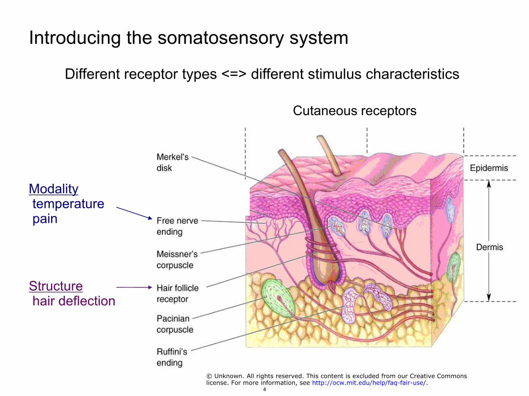
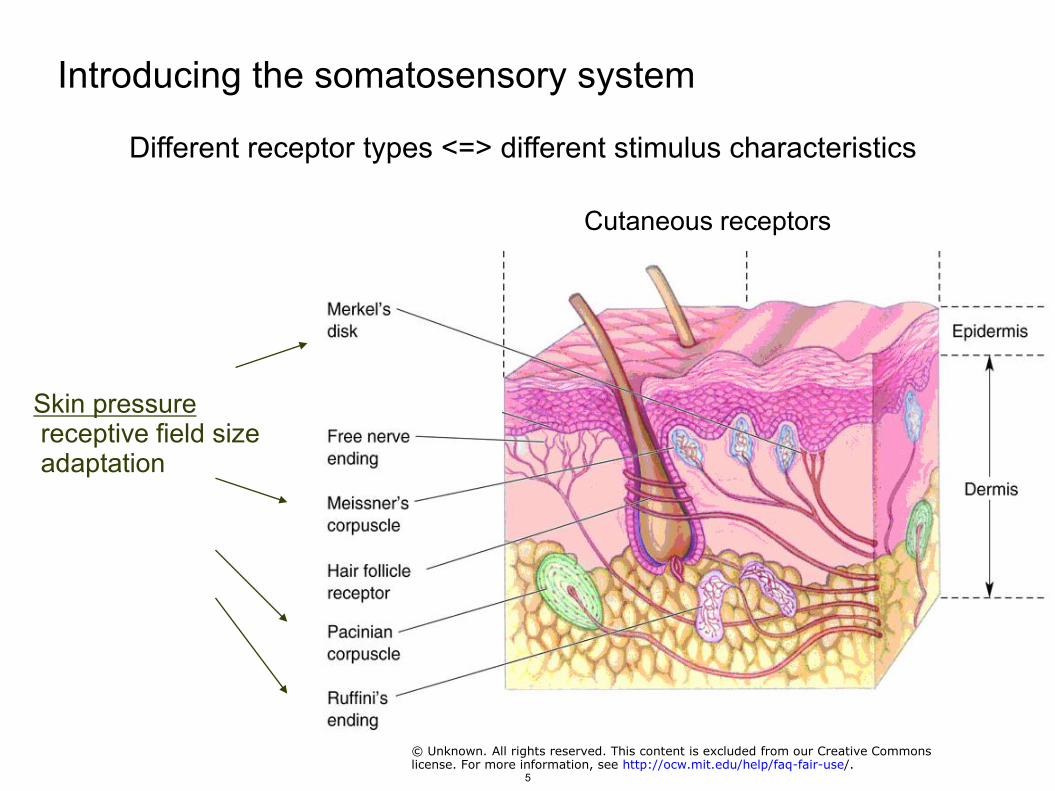
Introducing the somatosensory system
Different receptor types <=> different stimulus characteristics
Cutaneous receptors
Modality temperature pain
Structure hair deflection
© Unknown. All rights reserved. This content is excluded from our Creative Commonslicense. For more information, see http://ocw.mit.edu/help/faq-fair-use/.
4
Introducing the somatosensory system
Different receptor types <=> different stimulus characteristics
Cutaneous receptors
Skin pressure receptive field size adaptation
© Unknown. All rights reserved. This content is excluded from our Creative Commonslicense. For more information, see http://ocw.mit.edu/help/faq-fair-use/.
5
Receptive field sizes
"Figure 22-3 Mechanoreceptors in glabrous skin vary in the size and structure of their receptive fields" removed due to copyrightrestrictions. See Garner, Esther P., John H. Martin, and Thomas M. Jessel. "The Bodily Senses." Chapter 22 in Principles of Neural
Science. Edited by Eric R. Kandel, James H. Schwartz, and Thomas M. Jessell. 4th ed, MGraw-Hill Companies, 2000. pp. 434.
6
Adaptation in cutaneous receptors
"Figure 21-1 The sensory systems encode four elementary attributes of stimuli—modality, location, intensity, and timing—which are manifested in sensation."removed due to copyright restrictions. See Garner, Esther P., and John H. Martin. "Coding of Sensory Information." Chapter 21 in Principles of NeuralScience. Edited by Eric R. Kandel, James H. Schwartz, and Thomas M. Jessell. 4th ed, MGraw-Hill Companies, 2000, pp. 412.
7
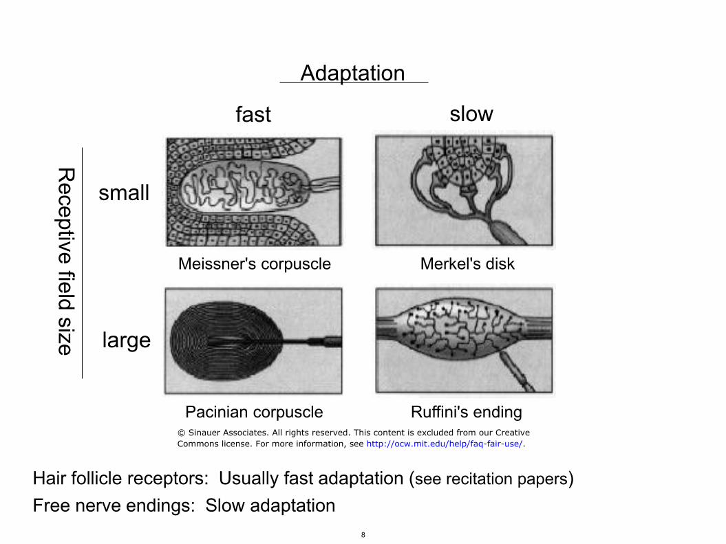
Adaptation
fast slow
small
large
Receptive field size
Meissner's corpuscle Merkel's disk
Pacinian corpuscle Ruffini's ending
Hair follicle receptors: Usually fast adaptation (see recitation papers) Free nerve endings: Slow adaptation
© Sinauer Associates. All rights reserved. This content is excluded from our CreativeCommons license. For more information, see http://ocw.mit.edu/help/faq-fair-use/.
8
Proprioception
muscle stretch
tendon tension
joint angle
Generally slowly adapting (like the roach?)
Images removed due to copyright restrictions. See http://neurobiography.info/teaching.php?mode=view&lectureid=67&slide=1
9
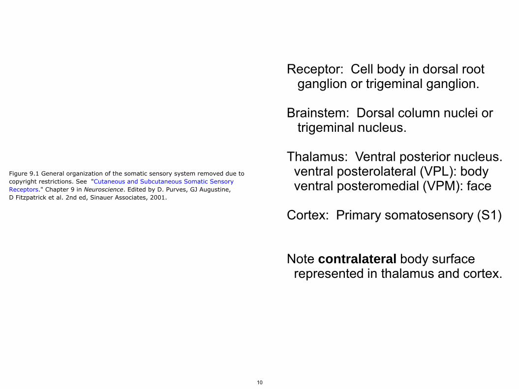
Receptor: Cell body in dorsal root ganglion or trigeminal ganglion. Brainstem: Dorsal column nuclei or trigeminal nucleus. Thalamus: Ventral posterior nucleus. ventral posterolateral (VPL): body ventral posteromedial (VPM): face Cortex: Primary somatosensory (S1) Note contralateral body surface represented in thalamus and cortex.
Figure 9.1 General organization of the somatic sensory system removed due tocopyright restrictions. See "Cutaneous and Subcutaneous Somatic SensoryReceptors." Chapter 9 in Neuroscience. Edited by D. Purves, GJ Augustine,D Fitzpatrick et al. 2nd ed, Sinauer Associates, 2001.
10
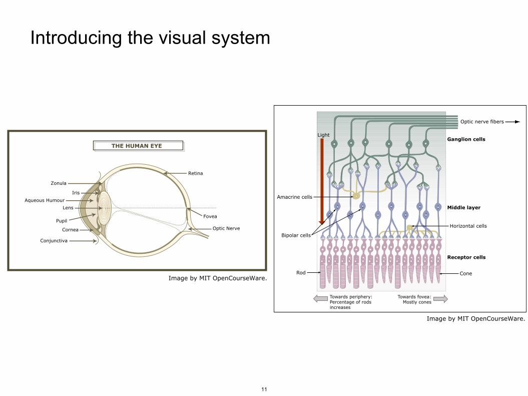
Introducing the visual system
11
THE HUMAN EYE
Retina
Zonula
Iris
Aqueous Humour
Lens
FoveaPupil
Cornea Optic Nerve
Conjunctiva
Image by MIT OpenCourseWare.
Ganglion cells
Middle layer
Receptor cells
Rod Cone
Towards periphery:Percentage of rods increases
Towards fovea:Mostly cones
Bipolar cells
Horizontal cells
Amacrine cells
Optic nerve fibers
Light
Image by MIT OpenCourseWare.
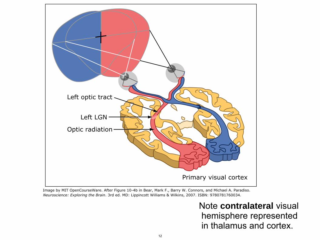
Note contralateral visual hemisphere represented in thalamus and cortex.
12
Primary visual cortex
Optic radiation
Left LGN
Left optic tract
Image by MIT OpenCourseWare. After Figure 10-4b in Bear, Mark F., Barry W. Connors, and Michael A. Paradiso. Neuroscience: Exploring the Brain. 3rd ed. MD: Lippincott Williams & Wilkins, 2007. ISBN: 9780781760034.
Last week: Information theory, mutual info between stimulus and neural activity, rate coding. The somatosensory and visual systems. Somatotopic/retinotopic organization, cortical magnification, and the homunculus. Adaptation and lateral inhibition. Rat barrel cortex.
13
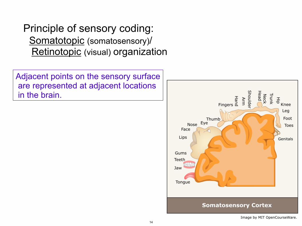
Principle of sensory coding: Somatotopic (somatosensory)/ Retinotopic (visual) organization
Adjacent points on the sensory surface are represented at adjacent locations in the brain.
14
Genitals
Toes
Foot
Leg
Knee
Hip
Trunk
Neck
Head
Should
erArm
Han
dFingers
ThumbEyeNose
Face
Face
Lips
Gums
Teeth
Jaw
Tongue
Somatosensory Cortex
Image by MIT OpenCourseWare.
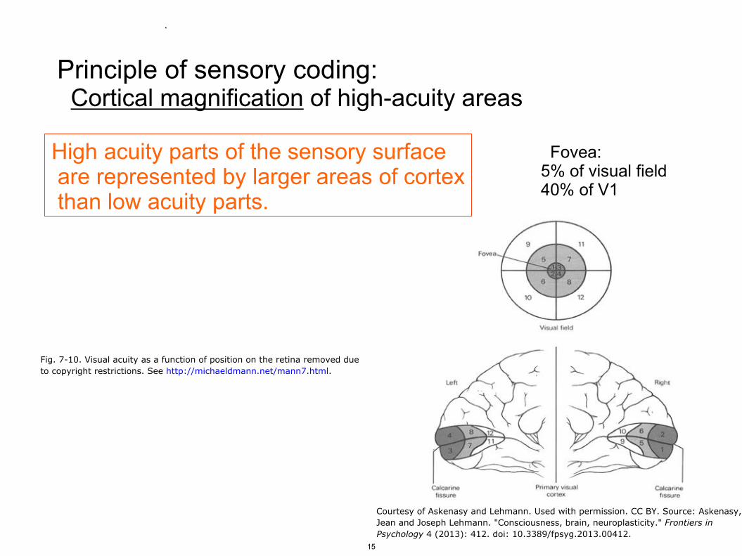
Fovea: 5% of visual field 40% of V1
Principle of sensory coding: Cortical magnification of high-acuity areas
High acuity parts of the sensory surface are represented by larger areas of cortex than low acuity parts.
Fig. 7-10. Visual acuity as a function of position on the retina removed dueto copyright restrictions. See http://michaeldmann.net/mann7.html.
Courtesy of Askenasy and Lehmann. Used with permission. CC BY. Source: Askenasy,Jean and Joseph Lehmann.
.
"Consciousness, brain, neuroplasticity." Frontiers inPsychology 4 (2013): 412. doi: 10.3389/fpsyg.2013.00412.
15
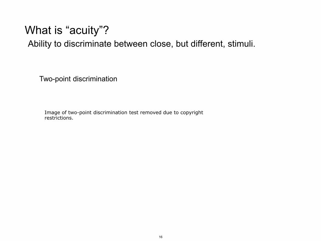
What is “acuity”? Ability to discriminate between close, but different, stimuli.
Two-point discrimination
Image of two-point discrimination test removed due to copyright restrictions.
16
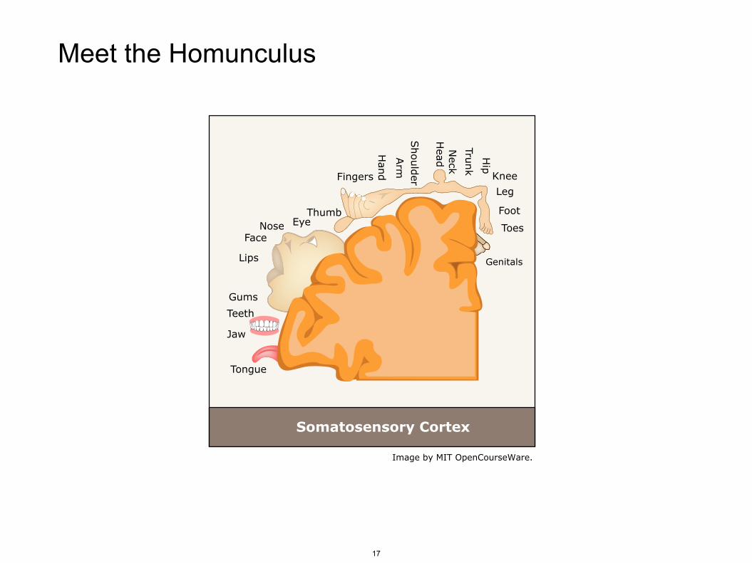
Meet the Homunculus
17
Genitals
Toes
Foot
Leg
Knee
Hip
Trunk
Neck
Head
Should
erArm
Han
dFingers
ThumbEyeNose
Face
Face
Lips
Gums
Teeth
Jaw
Tongue
Somatosensory Cortex
Image by MIT OpenCourseWare.
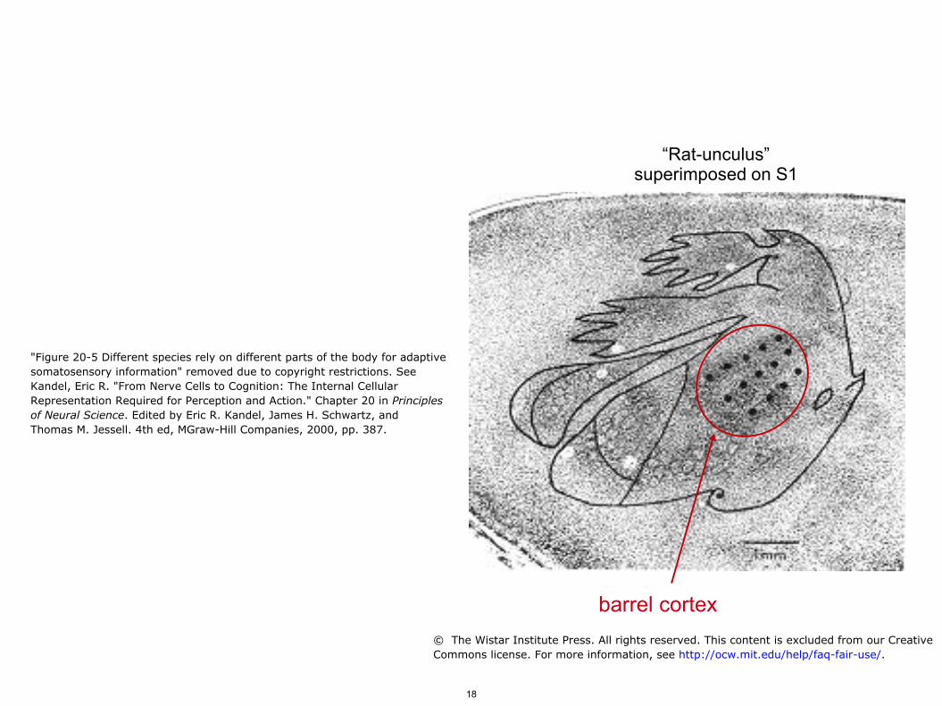
“Rat-unculus” superimposed on S1
barrel cortex
"Figure 20-5 Different species rely on different parts of the body for adaptivesomatosensory information" removed due to copyright restrictions. SeeKandel, Eric R. "From Nerve Cells to Cognition: The Internal CellularRepresentation Required for Perception and Action." Chapter 20 in Principlesof Neural Science. Edited by Eric R. Kandel, James H. Schwartz, andThomas M. Jessell. 4th ed, MGraw-Hill Companies, 2000, pp. 387.
© The Wistar Institute Press. All rights reserved. This content is excluded from our CreativeCommons license. For more information, see http://ocw.mit.edu/help/faq-fair-use/.
18
Last week: Information theory, mutual info between stimulus and neural activity, rate coding. The somatosensory and visual systems. Somatotopic/retinotopic organization, cortical magnification, and the homunculus. Adaptation and lateral inhibition. Rat barrel cortex.
19

Adaptation, more generally: Detect novelty.
Rachel E. Locke and
Jeanne M. Nerbonne, 1997
visual cortex current injection
Fig. 3. Two distinct firing patterns are evident in CP neurons A and B removed due to copyright restrictions. See Locke, Rachel E.,and Jeanne M. Nerbonne. "Role of Voltage-Gated K+ Currents in Mediating the Regular-Spiking Phenotype of Callosal-ProjectingRat Visual Cortical Neurons." Journal of Neurophysiology 78, no. 5 (1997): 2321-35.
20
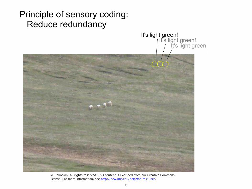
Principle of sensory coding: Reduce redundancy
It's light green! It's light green
!
It's light green!
© Unknown. All rights reserved. This content is excluded from our Creative Commonslicense. For more information, see http://ocw.mit.edu/help/faq-fair-use/.
21
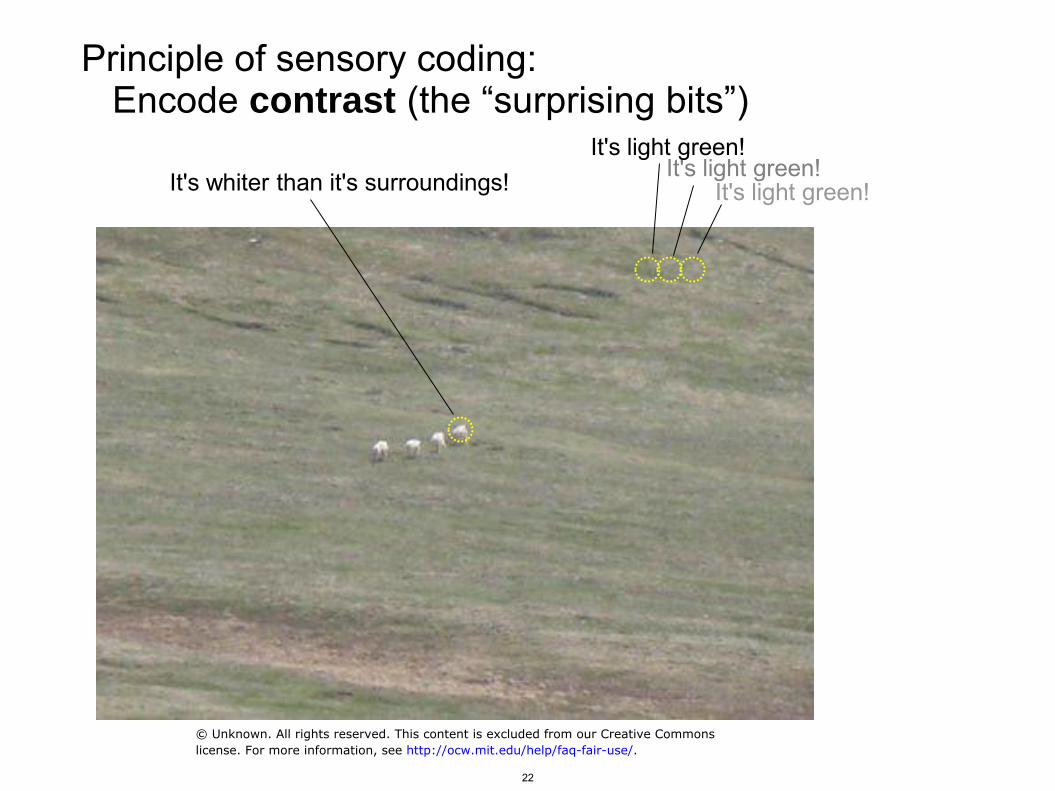
Principle of sensory coding: Encode contrast (the “surprising bits”)
It's light green! It's light green!
It's light green! It's whiter than it's surroundings!
© Unknown. All rights reserved. This content is excluded from our Creative Commonslicense. For more information, see http://ocw.mit.edu/help/faq-fair-use/.
22
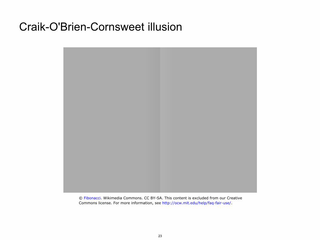
Craik-O'Brien-Cornsweet illusion
© Fibonacci. Wikimedia Commons. CC BY-SA. This content is excluded from our CreativeCommons license. For more information, see http://ocw.mit.edu/help/faq-fair-use/.
23
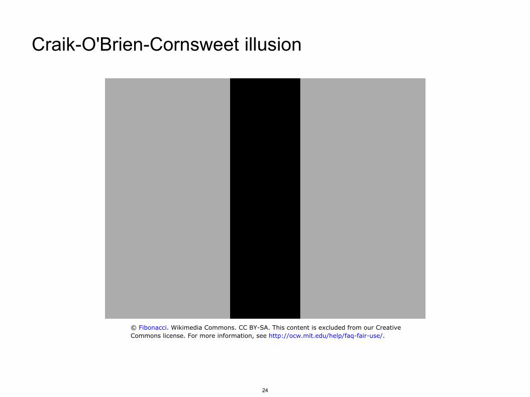
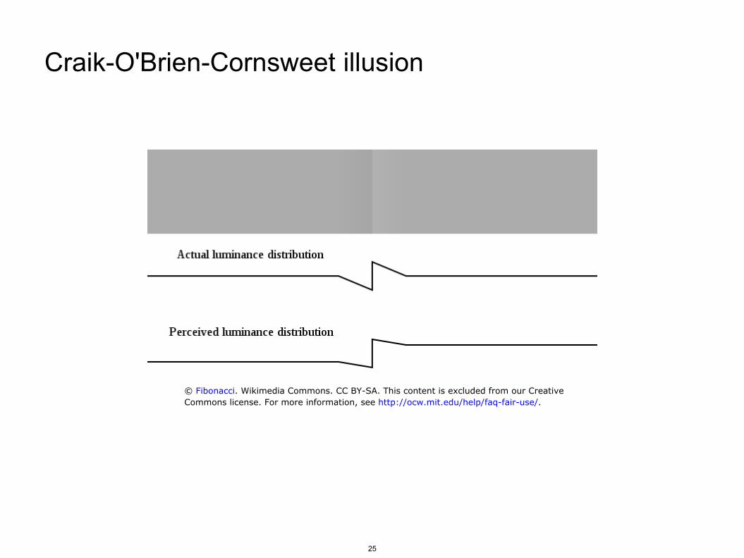
Craik-O'Brien-Cornsweet illusion
© Fibonacci. Wikimedia Commons. CC BY-SA. This content is excluded from our CreativeCommons license. For more information, see http://ocw.mit.edu/help/faq-fair-use/.
24
Craik-O'Brien-Cornsweet illusion
© Fibonacci. Wikimedia Commons. CC BY-SA. This content is excluded from our CreativeCommons license. For more information, see http://ocw.mit.edu/help/faq-fair-use/.
25
Last week: Information theory, mutual info between stimulus and neural activity, rate coding. The somatosensory and visual systems. Somatotopic/retinotopic organization, cortical magnification, and the homunculus. Rat barrel cortex.
26
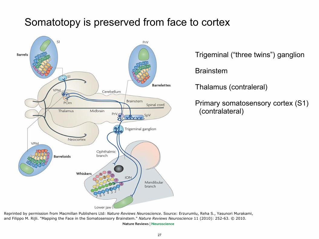
Somatotopy is preserved from face to cortex
Trigeminal (“three twins”) ganglion Brainstem Thalamus (contraleral) Primary somatosensory cortex (S1) (contralateral)
Reprinted by permission from Macmillan Publishers Ltd: Nature Reviews Neuroscience. Source: Erzurumlu, Reha S., Yasunori Murakami,and Filippo M. Rijli. "Mapping the Face in the Somatosensory Brainstem." Nature Reviews Neuroscience 11 (2010): 252-63. © 2010.
27
Rats “whisk” to actively sense their environment
5 – 10 Hz Tracked by high-speed video
Movie screenshot removed due to copyright restrictions. See Supplemental Movie. "Tracking Whisker and HeadMovements in Unrestrained Behaving Rodents." Journal of Neurophysiology 93, no. 4 (2005): 2294-301.
28
The barrel cortex of the rat
Know this! Rows A through E (dorsal to ventral) Whiskers numbered posterior to anterior Posterior inter-row whiskers with special names, or Greek characters
"Figure 23-9 The representation of whiskers in the somatosensory cortex of the rat" removed due to copyright restrictions.See Gardner, Esther P., amd Eric R. Kandel. "Touch." Chapter 23 in Principles of Neural Science. Edited byKandel, Eric R., James H. Schwartz, and Thomas M. Jessell. 4th ed, MGraw-Hill Companies, 2000. pp. 462.
29
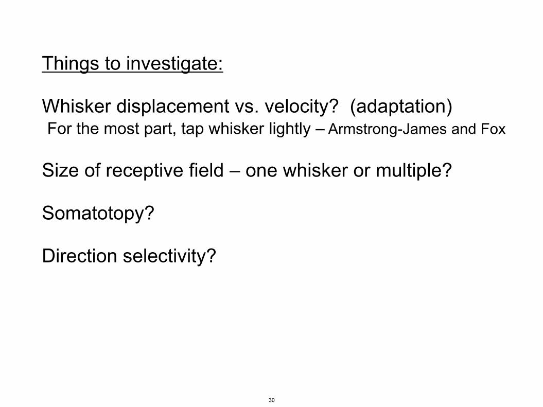
Things to investigate: Whisker displacement vs. velocity? (adaptation) For the most part, tap whisker lightly – Armstrong-James and Fox Size of receptive field – one whisker or multiple? Somatotopy? Direction selectivity?
30
MIT OpenCourseWarehttp://ocw.mit.edu
9.17 Systems Neuroscience LabSpring 2013 For information about citing these materials or our Terms of Use, visit: http://ocw.mit.edu/terms.