Embed Size (px)
Citation preview
U.O.No. 9103/2014/Admn Dated, Calicut University.P.O, 19.09.2014
File Ref.No.38933/GA - IV - J2/2013/CU
UNIVERSITY OF CALICUT
AbstractMSc programme in Radiation Physics-University Teaching Department-under Choice based Credit Semester
System (PG)- Typographical errors in the Scheme and Syllabus for 2012 admissions corrected-approved --
Corrigendum issued.
G & A - IV - J
Read:-1. U.O.No.GA IV/J1/1373/08 dated 01.07.2008.
2. U.O.No. 1027/2013/CU dated, 01.04.2013(Ref.no. 4863/GA IV E2/2012/CU)
3. Item no. 4 of the minutes of the Board of Studies in Radiation Physics held on 30.11.2013.
4. Remarks of the Dean Faculty of Science dated 16.12.2013
5. U.O.No. 6918/2013/CU Dated, 20.12.2013.
6. U.O.Note no. 1016/EX-II-ASST-3/2014/ P.B, dated: 26.08.2014.
7. Clarification from Chairman Board of Studies in Radiation Physics (PG)
8. Orders of the Registrar on 16.09.2014 in the file of even no.
ORDER
The Choice based Credit Semester System was implemented in all Regular PG programmes in University
Teaching Departments of the University with effect from 2008 admissions, as per paper read as (1) above.
The Regulations Scheme and Syllabus of MSc programme in Radiation Physics was implemented as per paper
read as (2)
The Board of Studies in Radiation Physics, vide paper read as (3), resolved to approve the Modified Syllabus of
having assigned Course Codes in the syllabus of MSc programme in Radiation Physics 2012.
The Dean, Faculty of Science recommended to implement the resolution of the Board vide paper read as (4)
The Vice-Chancellor,Considering the exigency, exercising the powers of the Academic Council, has approved the
item regarding the implementation of the Modified Syllabus of having assigned Course Codes in the syllabus of
MSc programme in Radiation Physics 2012, subject to ratification by the Academic Council and was implemented
vide paper read as (5).
Vide paper read as (6), the Pareeksha Bhavan of the University cited some anomalies in title of the Courses in
the Scheme and in the Syllabus of the MSc programme in Radiation Physics 2012. And vide paper read as (7) the
Chairman has clarified that it is typographical error and the Scheme shall be corrected as follows.
1. RPH4C20, Quality assurance and Commissioning Radiotherapy equipment (4 credits) shall be correctedas Quality control, acceptance testing and calibration of radiation system
2. RPH4C22. Modern trends in Radiation therapy (4 credits) shall be corrected as Modern trends in Radiology and Radiotherapy; and
3. RPH4C24 Practicals in Q/A and calibration of radiological equipment (2 credits) shall be correctedas Practicals in Q/A testing and calibration of radiological equipment.
The Registrar has approved to implement the clarification given by the Chairman vide paper read as (8)

Lalitha K.P
Assistant Registrar
Forwarded / By Order
Section Officer
Sanction has, therefore, been accorded to implement the clarifications given by the Chairman, Board of Studies
in Radiation Physics and to correct the typographical errors crept in the Scheme of MSc. programme in Radiation
Physics 2012, cited by the Pareeksha Bhavan, as follows.
RPH4C20, Quality assurance and Commissioning Radiotherapy equipment (4 credits) shall be correctedas Quality control, acceptance testing and calibration of radiation system.
RPH4C22. Modern trends in Radiation therapy (4 credits) shall be corrected as Modern trends in Radiology and Radiotherapy; and
RPH4C24 Practicals in Q/A and calibration of radiological equipment (2 credits) shall be correctedas Practicals in Q/A testing and calibration of radiological equipment.
Therefore the Scheme and Syllabus implemented vide paper read as (5) stands corrected to this effect.
Corrigendum is issued accordingly.
To
1.The Pareeksha Bhavan, University of Calicut.
2.The Dept. of Physics.
3. Board of Studies in Radiation Physics PG.
--'"'/-.---i-A.!-/7(,\
email:mm [email protected], [email protected] Ph-04942407475, (o), 9745509190 (m)
Ref. No. CR/RP/BOS/2013 20.08.2074
To
The Registrar
University of Calicut
UNIVERSITY OF CALICUTDr. M.M.MUSTIIAFA
Coordinato4 M.Sc. Radiation Physics course
Calicut University P.O.
Kerala INDIA 673 63
Sir,
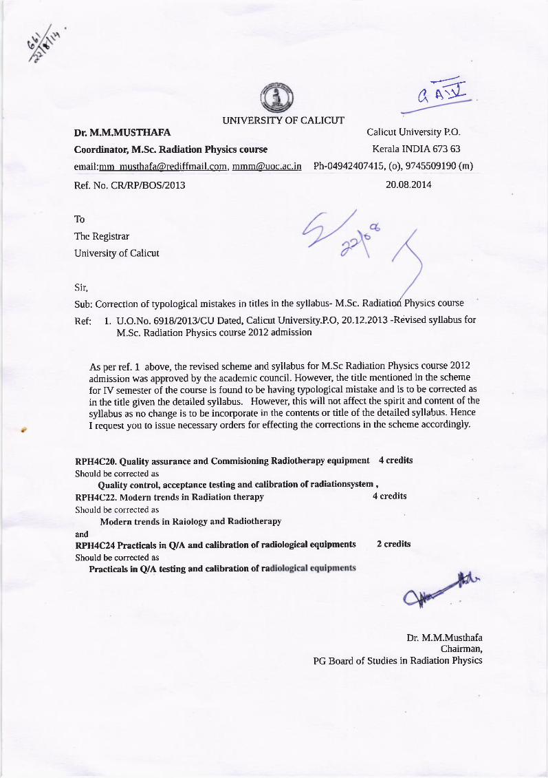
Sub: Correction of typological mistakes in titles in the syllabus- M.Sc. Radiatiori Physics course
Ref: 1. U.O.No. 69f 8/2013/CU Dated, Calicut Universiry.P.O, 20.12.2013 -Revised syllabus forM.Sc. Radiation Physics course 2012 admission
As per ref. 1 above, the revised scheme and syllabus for M.Sc Radiation Physics course 2012
admission was approved by the academic council. However, the tide mentioned in the scheme
for IV semester of the course is found to be having typological mistake and is to be corrected as
in the title given rhe detailed syllabus. However, this will not affect the spirit and content of the
syllabus as no change is to be incorporate in the contents or title of the detailed syllabus. Hence
I request you to issue necessary orders for effecting the corrections in the scheme accordingly.
RPH4C20, Quality assurance and Commisioning Radiotherapy equipment 4 credits
Should be corrected as
Quality controt, acceptance testing and calibration of radiationsystem ,
RPH4C22. Modern trends in Radiation therapy
Should be corrected as
Modern trends in Raiology and Radiotherapy
and
RPH4C24 Practicals in Q/A and calibration of radiological equipments 2 crtditsShould be corrected as
Practicals in Q/A testing and c{libration of ra
Dr. M.M.MusthafaChairman,
PG Board of Studies in Radiation Physics
4 credits
U.O.No. 6918/2013/CU Dated, Calicut University.P.O, 20.12.2013
Muhammed S
Deputy Registrar
Forwarded / By Order
Section Officer
File Ref.No.38933/GA - IV - J2/2013/CU
UNIVERSITY OF CALICUT
AbstractMSc programme in Radiation Physics-University Teaching Department-under Choice based Credit Semester
System (PG)-Modified Syllabus for 2012 admissions-approved -implemented-Orders issued.
G & A - IV - J
Read:-1.U.O.No.GA IV/J1/1373/08 dated 01.07.2008.
2.U.O.No. 1027/2013/CU dated, 01.04.2013(Ref.no. 4863/GA IV E2/2012/CU)
3.Item no. 4 of the minutes of the Board of Studies in Radiation Physics held on
30.11.2013.
4.Remarks of the Dean Faculty of Science dated 16.12.2013
5.Orders of the Vice Chancellor on 16-12-13.
ORDER
The Choice based Credit Semester System was implemented in all Regular PG programmes in University
Teaching Departments of the University with effect from 2008 admissions, as per paper read as (1) above.
The Regulations Scheme and Syllabus of MSc programme in Radiation Physics was implemented as per paper
read as (2)
The Board of Studies in Radiation Physics, vide paper read as (3), resolved to approve the Modified Syllabus of
having assigned Course Codes in the syllabus of MSc programme in Radiation Physics 2012.
The Dean, Faculty of Science recommended to implement the resolution of the Board vide paper read as (4)
The Vice-Chancellor,Considering the exigency, excercising the powers of the Academic Council, has approved the
item regarding the implementation of the Modified Syllabus of having assigned Course Codes in the syllabus of
MSc programme in Radiation Physics 2012, subject to ratification by the Academic Council vide paper read as (5)
Sanction has, therefore, been accorded for implementing the modifications of having assigned course codes in
the syllabus of MSc programme in Radiation Physics 2012 under CCSS (PG) in the University Teaching
Department.
Orders are issued accordingly.
(The Syllabus is available in the Official website of the University:universityofcalicut.info)
To
1.The Pareeksha Bhavan, University of Calicut.
2.The Dept. of Physics

ANNUXURE C
RULES AND REGULATIONS
FOR THE
M.Sc. RADIATION PHYSICS COURSE(Corrected for 2012 Admission)
UNIVERSITY OF CALICUT
1
UNIVERSITY OF CALICUT
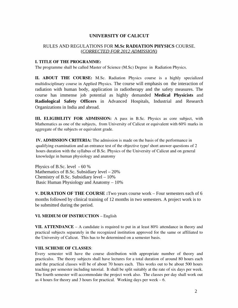
RULES AND REGULATIONS FOR M.Sc RADIATION PHYSICS COURSE.(CORRECTED FOR 2012 ADMISSION)
I. TITLE OF THE PROGRAMME: The programme shall be called Master of Science (M.Sc) Degree in Radiation Physics.
II. ABOUT THE COURSE: M.Sc. Radiation Physics course is a highly specialized multidisciplinary course in Applied Physics. The course will emphasis on the interaction of radiation with human body, application in radiotherapy and the safety measures. The course has immense job potential as highly demanded Medical Physicists and Radiological Safety Officers in Advanced Hospitals, Industrial and Research Organizations in India and abroad.
III. ELIGIBILITY FOR ADMISSION: A pass in B.Sc. Physics as core subject, with Mathematics as one of the subjects, from University of Calicut or equivalent with 60% marks in aggregate of the subjects or equivalent grade.
IV. ADMISSION CRITERIA: The admission is made on the basis of the performance in qualifying examination and an entrance test of the objective type/ short answer questions of 2 hours duration with the syllabus of B.Sc. Physics of the University of Calicut and on general knowledge in human physiology and anatomy
Physics of B.Sc. level - 60 %Mathematics of B.Sc. Subsidiary level – 20%Chemistry of B.Sc. Subsidiary level – 10%Basic Human Physiology and Anatomy – 10%
V. DURATION OF THE COURSE :Two years course work – Four semesters each of 6 months followed by clinical training of 12 months in two semesters. A project work is to be submitted during the period.
VI. MEDIUM OF INSTRUCTION – English
VII. ATTENDANCE – A candidate is required to put in at least 80% attendance in theory and practical subjects separately in the recognized institution approved for the same or affiliated to the University of Calicut. This has to be determined on a semester basis.
VIII. SCHEME OF CLASSES:Every semester will have the course distribution with appropriate number of theory and practicalss. The theory subjects shall have lectures for a total duration of around 80 hours each and the practical classes will be of about 70 hours each. This works out to be about 500 hours teaching per semester including tutorial. It shall be split suitably at the rate of six days per week. The fourth semester will accommodate the project work also. The classes per day shall work out as 4 hours for theory and 3 hours for practical. Working days per week – 6.
2

Failure in Semester:A candidate who has failed in the I semester shall be promoted to II and III semester but will not be allowed to attend the IV semester classes until he/she cleared the 1st semester subjects.
Discontinuation: No discontinuation is allowed in normal basis. However if a student has to discontinue the course in any semester due to the reasons of not his own, and he has paid the fee for the semester, he can be readmitted to the semester in later time, if the coordinator is fully satisfied with the reason. In such cases he has to complete the course work as per the regulations of the newly admitted batch he is readmitted and appear for the examinations accordingly. This provision is conditional on the availability of seats and facility.
IX. PROJECT WORK:Every candidate must do a project work under an approved supervisor (approved by the Coordinator) in a topic having relevance to the application of radiation in medicine, industry, agriculture and research in the 4th semester. The project thesis should be submitted to the University. The supervisor should certify about the satisfactory completion of the project. Students must present their project work before a committee constituted by the course coordinator. Project Report must be submitted within two months from the last working day of the final semester
X. FIELD TRAINING:Total duration of the training will be 1 year (as prescribed by the AERB). It should be done under the supervision of a designated academic staff member of recognized institute. The supervisor must certify to the adequacy of the field training on the basis of the thesis report submitted by the candidate. The students should necessarily present at least one seminar on the basis of the field training and the record of the field training must be duly certified by the designated officer in the centre and the Course Coordinator. (The students should pay the charges for clinical training as required by the institution).
XI. RADIOLOGICAL SAFETY OFFICER (RSO) approval by AERB:The University shall initiate steps to get Radiological Safety Officer (Level III Medical) certification for all candidates. The examination for the same shall be conducted by Radiological Physics and Advisory Division (RPAD) of DAE as per the regulations of the Atomic Energy Regulator Board (AERB). Students qualifying this examination will be eligible for RSO. Candidate completing one year clinical training are eligible for this examination. Student should attend and qualify the RSO examination at their own risk
XII. SCHEME OF EXAMINATION:Theory papers: Each paper is of three hours duration – Maximum marks 100 End semester examination 70 marks Continuous valuation 30 marks* Practical Examination: Four hours duration – Maximum marks 100 End semester: 70 marks
Continuous evaluation: 30 marks* * Continuous evaluation is based on the regular performance in attendance 4 marks, internal tests (best 2 out of 3) 12 marks, Assignments 8 marks, seminars (6 marks).
3
Vivavoce: There will be an end semester vivavoce, distributed on all papers of the semester , for all the four semester. Viva carries a maximum marks of 50.Project work: Total marks: 150
Project Record : 100Presentation and viva: 50
Total Marks: 2700 (650+650+750+750)
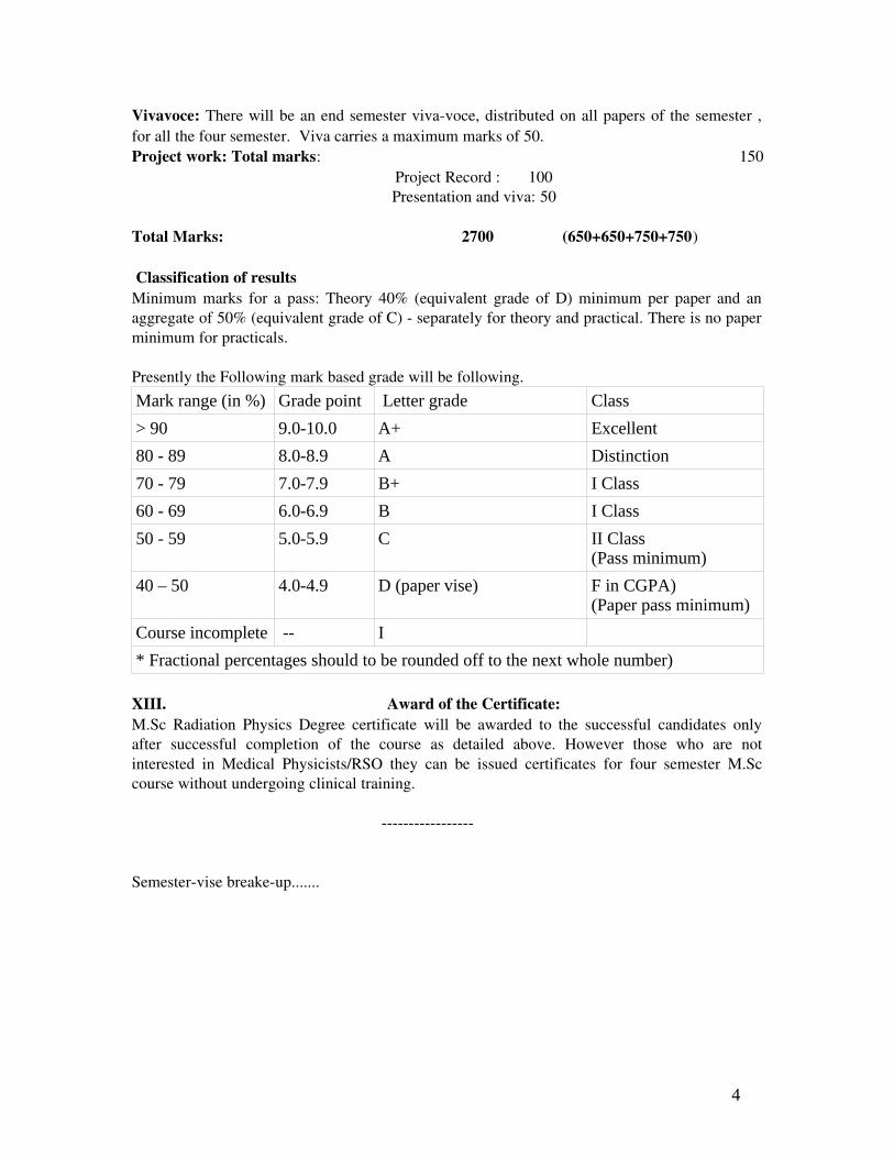
Classification of resultsMinimum marks for a pass: Theory 40% (equivalent grade of D) minimum per paper and an aggregate of 50% (equivalent grade of C) separately for theory and practical. There is no paper minimum for practicals.
Presently the Following mark based grade will be following.
Mark range (in %) Grade point Letter grade Class
> 90 9.0-10.0 A+ Excellent
80 - 89 8.0-8.9 A Distinction
70 - 79 7.0-7.9 B+ I Class
60 - 69 6.0-6.9 B I Class
50 - 59 5.0-5.9 C II Class(Pass minimum)
40 – 50 4.0-4.9 D (paper vise) F in CGPA)(Paper pass minimum)
Course incomplete -- I
* Fractional percentages should to be rounded off to the next whole number)
XIII. Award of the Certificate:M.Sc Radiation Physics Degree certificate will be awarded to the successful candidates only after successful completion of the course as detailed above. However those who are not interested in Medical Physicists/RSO they can be issued certificates for four semester M.Sc course without undergoing clinical training.
Semestervise breakeup.......
4
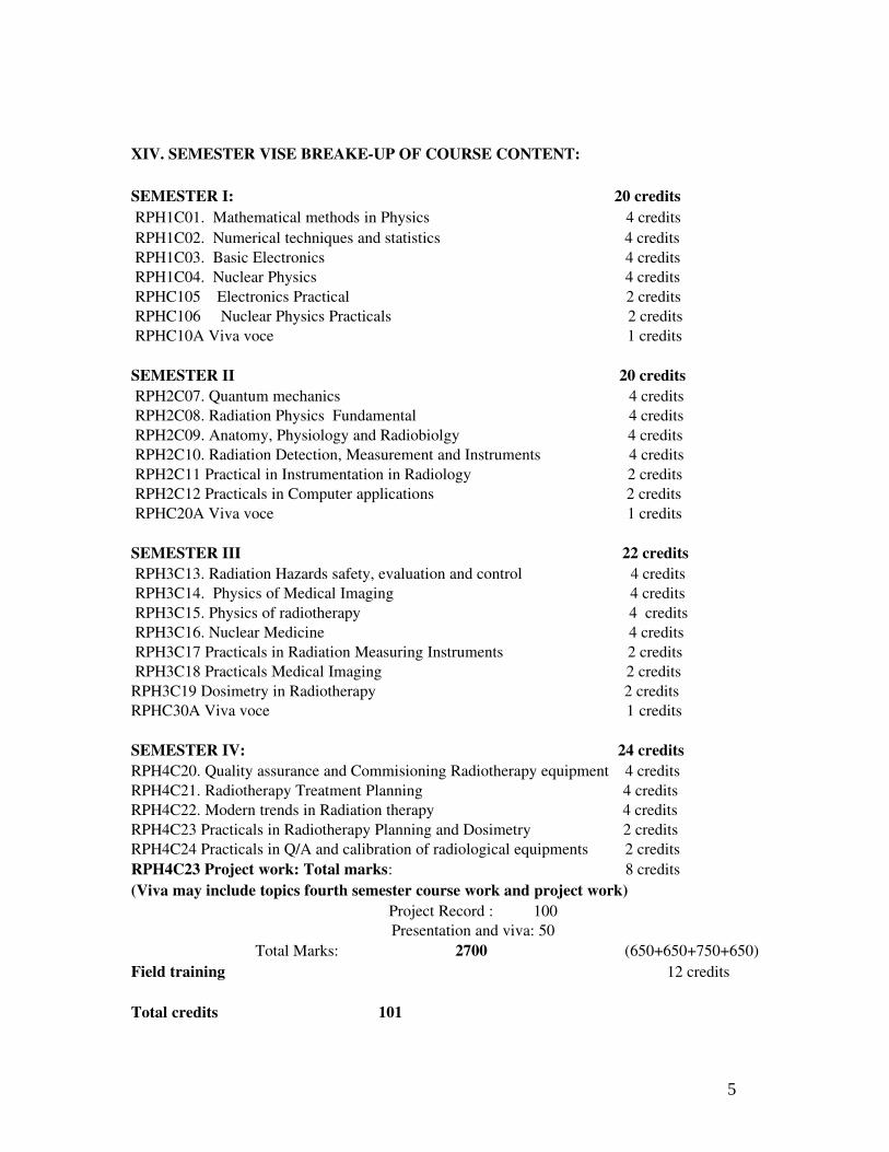
XIV. SEMESTER VISE BREAKEUP OF COURSE CONTENT:
SEMESTER I: 20 credits RPH1C01. Mathematical methods in Physics 4 credits RPH1C02. Numerical techniques and statistics 4 credits RPH1C03. Basic Electronics 4 credits RPH1C04. Nuclear Physics 4 credits RPHC105 Electronics Practical 2 credits RPHC106 Nuclear Physics Practicals 2 credits RPHC10A Viva voce 1 credits
SEMESTER II 20 credits RPH2C07. Quantum mechanics 4 credits RPH2C08. Radiation Physics Fundamental 4 credits RPH2C09. Anatomy, Physiology and Radiobiolgy 4 credits RPH2C10. Radiation Detection, Measurement and Instruments 4 credits RPH2C11 Practical in Instrumentation in Radiology 2 credits RPH2C12 Practicals in Computer applications 2 credits RPHC20A Viva voce 1 credits
SEMESTER III 22 credits RPH3C13. Radiation Hazards safety, evaluation and control 4 credits RPH3C14. Physics of Medical Imaging 4 credits RPH3C15. Physics of radiotherapy 4 credits RPH3C16. Nuclear Medicine 4 credits RPH3C17 Practicals in Radiation Measuring Instruments 2 credits RPH3C18 Practicals Medical Imaging 2 creditsRPH3C19 Dosimetry in Radiotherapy 2 creditsRPHC30A Viva voce 1 credits
SEMESTER IV: 24 credits RPH4C20. Quality assurance and Commisioning Radiotherapy equipment 4 creditsRPH4C21. Radiotherapy Treatment Planning 4 creditsRPH4C22. Modern trends in Radiation therapy 4 creditsRPH4C23 Practicals in Radiotherapy Planning and Dosimetry 2 creditsRPH4C24 Practicals in Q/A and calibration of radiological equipments 2 creditsRPH4C23 Project work: Total marks: 8 credits(Viva may include topics fourth semester course work and project work)
Project Record : 100 Presentation and viva: 50
Total Marks: 2700 (650+650+750+650)Field training 12 credits
Total credits 101
5
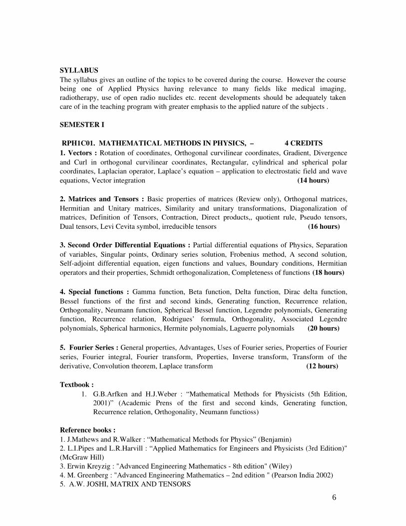
SYLLABUSThe syllabus gives an outline of the topics to be covered during the course. However the course being one of Applied Physics having relevance to many fields like medical imaging, radiotherapy, use of open radio nuclides etc. recent developments should be adequately taken care of in the teaching program with greater emphasis to the applied nature of the subjects .
SEMESTER I
RPH1C01. MATHEMATICAL METHODS IN PHYSICS, – 4 CREDITS1. Vectors : Rotation of coordinates, Orthogonal curvilinear coordinates, Gradient, Divergence and Curl in orthogonal curvilinear coordinates, Rectangular, cylindrical and spherical polar coordinates, Laplacian operator, Laplace’s equation – application to electrostatic field and wave equations, Vector integration (14 hours)
2. Matrices and Tensors : Basic properties of matrices (Review only), Orthogonal matrices, Hermitian and Unitary matrices, Similarity and unitary transformations, Diagonalization of matrices, Definition of Tensors, Contraction, Direct products,, quotient rule, Pseudo tensors, Dual tensors, Levi Cevita symbol, irreducible tensors (16 hours)
3. Second Order Differential Equations : Partial differential equations of Physics, Separation of variables, Singular points, Ordinary series solution, Frobenius method, A second solution, Selfadjoint differential equation, eigen functions and values, Boundary conditions, Hermitian operators and their properties, Schmidt orthogonalization, Completeness of functions (18 hours)
4. Special functions : Gamma function, Beta function, Delta function, Dirac delta function, Bessel functions of the first and second kinds, Generating function, Recurrence relation, Orthogonality, Neumann function, Spherical Bessel function, Legendre polynomials, Generating function, Recurrence relation, Rodrigues’ formula, Orthogonality, Associated Legendre polynomials, Spherical harmonics, Hermite polynomials, Laguerre polynomials (20 hours)
5. Fourier Series : General properties, Advantages, Uses of Fourier series, Properties of Fourier series, Fourier integral, Fourier transform, Properties, Inverse transform, Transform of the derivative, Convolution theorem, Laplace transform (12 hours)
Textbook : 1. G.B.Arfken and H.J.Weber : “Mathematical Methods for Physicists (5th Edition,
2001)” (Academic Prens of the first and second kinds, Generating function, Recurrence relation, Orthogonality, Neumann functioss)
Reference books : 1. J.Mathews and R.Walker : “Mathematical Methods for Physics” (Benjamin) 2. L.I.Pipes and L.R.Harvill : “Applied Mathematics for Engineers and Physicists (3rd Edition)" (McGraw Hill) 3. Erwin Kreyzig : "Advanced Engineering Mathematics 8th edition" (Wiley) 4. M. Greenberg : "Advanced Engineering Mathematics – 2nd edition " (Pearson India 2002)5. A.W. JOSHI, MATRIX AND TENSORS
6
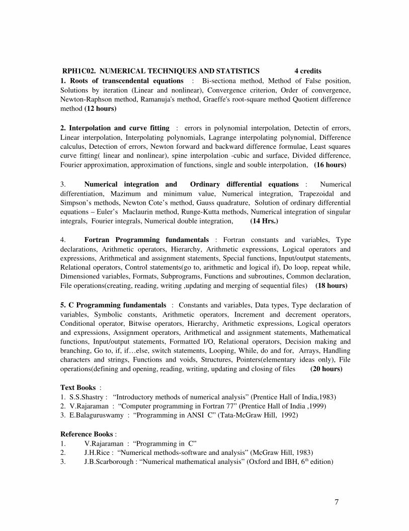
RPH1C02. NUMERICAL TECHNIQUES AND STATISTICS 4 credits1. Roots of transcendental equations : Bisectiona method, Method of False position, Solutions by iteration (Linear and nonlinear), Convergence criterion, Order of convergence, NewtonRaphson method, Ramanuja's method, Graeffe's rootsquare method Quotient difference method (12 hours)
2. Interpolation and curve fitting : errors in polynomial interpolation, Detectin of errors, Linear interpolation, Interpolating polynomials, Lagrange interpolating polynomial, Difference calculus, Detection of errors, Newton forward and backward difference formulae, Least squares curve fitting( linear and nonlinear), spine interpolation cubic and surface, Divided difference, Fourier approximation, approximation of functions, single and souble interpolation, (16 hours)
3. Numerical integration and Ordinary differential equations : Numerical differentiation, Mazimum and minimum value, Numerical integration, Trapezoidal and Simpson’s methods, Newton Cote’s method, Gauss quadrature, Solution of ordinary differential equations – Euler’s Maclaurin method, RungeKutta methods, Numerical integration of singular integrals, Fourier integrals, Numerical double integration, (14 Hrs.) 4. Fortran Programming fundamentals : Fortran constants and variables, Type declarations, Arithmetic operators, Hierarchy, Arithmetic expressions, Logical operators and expressions, Arithmetical and assignment statements, Special functions, Input/output statements, Relational operators, Control statements(go to, arithmetic and logical if), Do loop, repeat while, Dimensioned variables, Formats, Subprograms, Functions and subroutines, Common declaration, File operations(creating, reading, writing ,updating and merging of sequential files) (18 hours)
5. C Programming fundamentals : Constants and variables, Data types, Type declaration of variables, Symbolic constants, Arithmetic operators, Increment and decrement operators, Conditional operator, Bitwise operators, Hierarchy, Arithmetic expressions, Logical operators and expressions, Assignment operators, Arithmetical and assignment statements, Mathematical functions, Input/output statements, Formatted I/O, Relational operators, Decision making and branching, Go to, if, if…else, switch statements, Looping, While, do and for, Arrays, Handling characters and strings, Functions and voids, Structures, Pointers(elementary ideas only), File operations(defining and opening, reading, writing, updating and closing of files (20 hours)
Text Books :1. S.S.Shastry : “Introductory methods of numerical analysis” (Prentice Hall of India,1983)2. V.Rajaraman : “Computer programming in Fortran 77” (Prentice Hall of India ,1999)3. E.Balaguruswamy : “Programming in ANSI C” (TataMcGraw Hill, 1992)
Reference Books :1. V.Rajaraman : “Programming in C”2. J.H.Rice : “Numerical methodssoftware and analysis” (McGraw Hill, 1983)3. J.B.Scarborough : “Numerical mathematical analysis” (Oxford and IBH, 6 th edition)
7

RPH1C03. BASIC ELECTRONICS – 4 CREDITS1. Basic Electronic components Semiconductor devices, diode, transistor, oscillator– Electrical measurements – Galvanometer and its applications, Multimeter, Universal Bridge, VTVM and Cathode ray oscilloscope – Special tubes – Electrometer tubes, Photomultiplier tubes and decatron tubes. (16 hours)
2. Basic amplifier principles: Pre amplifier circuits noise, linear pulse amplifier, pulse shaping, –– D.C. amplifier – Power, amplifier – Distortion in amplifiers – Feedback amplifiers – emitter follower – Types of oscillators – Power supplies – Rectifiers, filter circuits Regulated HT and EHT supplies – RF power supplies. analogue to digital converters, coincidence and anticoincidence circuits, single and multi channel analyzers voltage doubler circuits (14 hours)
3. Amplifiers and Oscillators: – Power amplifier design – class B pushpull amplifier – emitter follower – Darlington pair operational amplifier characteristics – OPAMP amplifier and its frequency response – Instrumentation amplifier – Differentiating and integrating circuits. Solving differential equation – RC phase shift oscillator – Blocking oscillator – A/D and D/A converter – Frequency to voltage converter – Digital voltmeter (Ramp type). (20 hours)
4. Trigger circuits, Digital electronics, Logic systems::– Multivibrator and univibrator – Discriminator – Scale of two Scale of ten– Coincidence and anticoincidence circuits – Amplitude analyzer and counting rate meters – Small current electrometers – Principles of Servomechanism and control. Logic gates: Flip flops RS, Clocked RS, D, JK, MSJK and T flip flops Synchronous and asynchronous counters, Mod 3,5 and 10 counters. (16 hours)
5. Transducers and electronic devices: LVDT – A.C and D.C Tachometers – Capacitance transducers – Thermistor based thermometers – Strain gauge – Ultrasonic transducers and their electrical equivalent circuits. Principles of filters and their application in instrumentation. Strip chart recorder – Magnetic recording – CRO – Phosphors – LED – LCD Plasma display – Seven segment – dot matrix system – Guest Host effect. (14 hours)
BOOKS FOR STUDY1. A.K.Sawhney, Electrical and Electronic Measurements and Instrumentation, Dhanapat Rai
and Sons, New Delhi – 1982.2. Lal Kishore K., Electronic Devices and Circuit Analysis, B.S.Publications3. Malvino and Leech, Digital Principles and Applications, Tata McGrow Hills (1978)4. H.S.Kalsi, electronics Indsturmentation, Indian soceity for Technical Education5. J.Millman and C.Halkias. Integrated Circuits, McGraw Hill, 1979.STANDARD BOOKS FOR REFERENCES1. J.D.Ryder, Electronics Fundamentals and Applications, Prentice Hall of India, New Delhi. 2. W.Cooper, Electronic Instrumentation and Measurement Techn., Prentice Hall of India. 19703. R.P.Jain, Modern Digital Electronics, Tata McGrow Hills
RPH1C04. INTRODUCTORY NUCLEAR PHYSICS 4 CREDITS1. Nuclear Forces:Four basic forces Gravitational, Electromagnetic, Weak and Strong Relative strengths, Properties of the nucleus, size, binding energy, angular momentum, The deuteron and
8

twonucleon scattering experimental data, Simple theory of the deuteron structure, Low energy np scattering, characteristics of nuclear forces, Spin dependence, Tensor force, Scattering cross sections, Partial waves, Phase shift, Singlet and triplet potentials, Effective range theory, pp scattering. (16 hours)
2. Nuclear Decay:Basics of alpha decay and theory of alpha emission. Beta decay, Energetics of beta decay, Fermi theory of beta decay, Comparative halflife, Allowed and forbidden transitions, Selection rules, Parity violation in beta decay. Neutrino. Energetics of Gamma Decay, Multipole moments, Decay rate, Angular momentum and parity selection rules, Internal conversion, Lifetimes.
(16 hours)3. Nuclear Models, Fission and FusionShell model potential, Spinorbit potential, Magnetic dipole moments, Electric quadruple moments, Valence Nucleons, Collective structure, Nuclear vibrations, Nuclear rotations, Liquid drop Model, Semiempirical Mass formula, Energetics of Fission process, Controlled Fission reactions. Fusion process, Characteristics of fusion, solar fusion, Controlled fusion reactors. Critical conditions, four factor formula (16 hours)
4. Nuclear Radiation Detectors Modes of Energy Deposition in the Detector, Gas detectors – Ionization chamber, Proportional counter and G M counter, Multiwire Proportion counter, Parallel Plate Avalanche Counter, Scintillation detector, Photo Multiplier Tube (PMT), Semiconductor detectors – Ge(Li), Si(Li) and HPGe detectors, surface barrier detectors, Detection of XRays with a Si(Li) Detector. Liquid scintillators (16 hours)
5. Nuclear Electronics and techniquesPreamplifiers, Amplifiers, Single channel analyzers, Multichannel analyzers, counting statistics, energy measurements, introduction to spectroscopy , Definition of Energy Spectra, Measurement of an Integral Spectrum and Differential Spectrum, Energy Resolution of a Detection System, Multichannel Analyzer (MCA) ,Calibration of a Multichannel Analyzer , , chargedparticle spectroscopy, Energy Straggling, The TimeofFlight Spectrometer, Detector Telescopes (E d E / h Detectors), PositionSensitive Detectors. ( 16 hours)
Books for study and reference: 1. H.Enge : “Introduction to Nuclear Physics” (Addison Wesley)2. H.S.Hans : “Nuclear Physics – Experimental and theoretical” (New Age
International – 2001.3. G.F.Knoll : “Nuclear Radiation Detectors” (Willyi nternational, New york)4. K. Muraleedhara Varier : “Nuclear Radiation Detection, Measurements and
Analysis” (Narosa) 5. G.D.Couoghlan and J.E.Dodd. “The ideas of particle physics an introduction for
scientists”, (Cambridge Press)6. david griffiths – “introduction to elementary particles” – wiley (1989)7. S.B.Patel, An introduction to nulcear Physics, (New Age Inerantional Pulishing,
9

PRACTICALS:RPH1C05. Electronics – Minimum expts. to be carried out 8 (70 Hours) 2 CREDITS
1. Measurement of L, C and R by Universal bridge2. Series resonance and Q of a coil3. Two stage RC coupled amplifier – frequency response4. Construction of a voltage multiplier5. Characteristics of a regulated power pack6. DC voltage regulator using transistors7. Feedback amplifier8. Construction of an oscillator9. Free running multivibrator10. Construction of a scale of two.11. OPAMP circuits – Inverting and non inverting amplifiers12. Integrator and differentiator circuit using OPAMP13. Simple D/A converter – Ladder type14. Coincidence and anticoincidence circuits15. Pulse shaping circuits
RPH1C06. Nuclear Physics – Minimum expts. to be carried out 8. (70 Hours) 2 CREDITS1. GM counter – characteristics – plateau and variation of pulse height with applied voltage2. GM counter – Statistics of counting3. GM counter – Feather Analysis – end point energy4. Gamma ray spectroscopy using NaI(Tl) characteristics – plateau and variation of pulse
height with applied voltage5. Scintillation spectrometer – Calibration and determination of unknown energy6. Ge detectors calibration and determination of unknown energy 7. Study of absorption of beta rays 8. Measurement of linear and mass attenuation coefficients of an Xray beam9. Measurement of linear and mass attenuation coefficients for a gamma ray beam using
GM counter10. Measurement of range of Beta rays (1) in air (2) in material like aluminum and
calculation of absorption coefficients11. Determination of K40 half life12. Estimation of resolving time of a G.M counter
SEMESTER II
RPH2C07. QUANTUM MECHANICS– 4 CREDITS1. The Formulation of Quantum Mechanics : Vector spaces, The Hilbert space, Dimensions and basis, Operators and properties, Representation of vectors and operators, Commutator, Functions of operators, Eigen values and eigen vectors, Matrix representation of bras, kets and operators, Coordinate and momentum representations and their connection, The fundamental postulates Probability density, Superposition principle, Observables and operators, The uncertainty principle (18 hours)
2. Quantum Dynamics : The equation of motion, Schrodinger, Heisenberg and the Interaction pictures of time development, The linear harmonic oscillator in the Schroedinger and Heisenberg pictures, Hydrogen atom (14 hours)
10

3. Theory of Angular Momentum : Angular momentum operators, Matrix representation of angular momentum operators, Pauli spin matrices, Orbital angular momentum, The hydrogen atom, Addition of angular momenta, ClebshGordon coefficients, Simple examples
(14 hours)4. Symmetry and Conservation Laws : Spacetime symmetries, Space translation and conservation of linear momentum, Time translation and conservation of energy, Space rotation and conservation of angular momentum, Space inversion and time reversal, Identical particles, Construction of symmetric and anti symmetric wave functions, Slater determinant, Pauli exclusion principle, Bosons and Fermions, Spin wave functions for two electrons, The ground state of He atom, Scattering of identical particles (18 hours)
5. Scattering : Scattering cross section and scattering amplitude, Low energy scattering by a central potential, The method of partial waves, Phase shifts, Optical theorem, Convergence of partial wave series, Scattering by a rigid sphere, Scattering by a square well potential, High energy scattering, Scattering integral equation and Born approximation (16 hours) Textbook : N. Zettili, “Quantum Mechanics – Concepts and applications’ (John Wiley & Sons, 2004)Reference books :1. V.K.Thankappan : “Quantum Mechanics” (Wiley Eastern)2. L.I. Schiff : “Quantum Mechanics” (McGraw Hill)3. P.M.Mathews and K.Venkatesan : “A Textbook of Quantum Mechanics" (TataMcGraw Hill4. A.Messiah : “Quantum Mechanics”5. J.J.Sakurai : “Modern Quantum Mechanics” (Addison Wesley)6. Stephen Gasiorowics : “Quantum Physics”7. A.Ghatak and S.Lokanathan : “Quantum Mechanics” (Macmillan)
RPH2C08. RADIATION PHYSICS FUNDAMENTAL 4 CREDITSUnit 1: Interaction of electromagnetic radiation with matterThomson scattering Photoelectric absorption – Angular distribution of photoelectrons – Compton effect, Compton process – Klein Nishina crosssection – Scattering coefficients – angular distribution of compton electrons – Pair production – Annihilation radiation, electrons – energy momentum conservation, Photo nuclear reactions,– Attenuation – Linear, mass attenuation coefficients Total absorption coefficients. Absorption and scattering coefficients and cross sections (14 hours)
Unit 2: Interaction of electrons and heacy charged particles with Matter Classical theory of inelastic collisions with atomic electrons, energy loss per ion pair by primary and secondary ionization, Cerenkov radiation, Electron, Absorption, Scattering, Excitation and Ionization. Values of w in different media, radiactive collision– Radiation energy loss (bremsstrahlung)– Range of beta particles, Range straggling, absorption of beta particles and back scattering, self absorption . Interaction of heavy charged particles with matter –Energy loss by collision, maximum energy loss in a single collision, range energy relation Bragg curve, specific ionization, BetheBloch formula for collision, stopping power and radiation stopping power (16 hours)
11
Unit 3: Interaction of Neutrons with Matter Neutron capture Neutron sources, properties, energy classification – Elastic and inelastic scattering coefficients and cross sections– Energy transfer and logarithmic energy decrement – Inelastic scattering, Nuclear reaction, Dependence on E and Z – (n, p), (n, 2n), (n, f) and other reaction – Neutron activation., Radio isotope production. (10 hours)
Unit 4: Radiation Quantities and UnitsCharacteristics of Electromagnetic Radiation, Radiation of Energy from an Atom, Radiation quantities and units – Radiometry – Particle flux and fluence – Energy flux and fluence – Cross section – Mass energy transfer and mass absorption coefficients – LET Radiation chemical yield – W value – Dosimetry – Energy imparted –Absorbed dose Radiation and tissue weighting factors, equivalent dose, effective dose, committed equivalent dose, committed effected dose, Concepts of collective dose – KERMACEMA, Exposure, Air kerma rate constant – Charged particle equilibrium (CPE) – Relationship between kerma, absorbed dose and exposure under CPE, Dose equivalent, Ambient and directional dose equivalents [(H*(d) and H’(d)], individual dose equivalent penetrating Hp(d), Individual dose equivalent superficial Hs(d). (20 hours)
Unit 5: Standardization of X and Gamma Rays BeamsStandards – Primary and Secondary Standards, Traceability, Uncertainly in measurement. Bragggray principle and air wall chamber, Standardization of Xray, electrons and gamma ray beams: Determination of exposure and air kerma, conditions for the realization of exposure, ionization chamber for low, medium and high energy xrays and gamma rays, determination of absorbed dose, Bragg Gray theory and its validity, Burlin’s theory for measurement for radiation quantities, design of free air chambers(FAIC), Standardization of electron beams used in radiotherapycalibration of secondary standards, details of IAEA and other protocols for dosimetry of photon and electron beams, standardization of brachytherapy sources and sealed sources in terms of their radiation output, calibration of protection level dosimeters in terms of dose equivalent units. . General definition of calibration factor – Nx, Nk, ND, air, ND, w. IAEA TRS277: Various steps to arrive at the expression for Dw starting from Nx. TRS398: ND, w, Q : ND, W : KQ,Q0 : KQ, Derivation of an expression for KQ,Q0. Calorimetric standards –AAPM TG 51 and other dosimetric protocols (20 hours)
STANDARD BOOKS FOR STUDY AND REFERENCES1. K.S.Krane, "Introductory Nuclear Physics", (John Wiley & Sons)2. Practical Applications of Radioactivity and Nuclear Radiations, G.C.Lowental and
P.L.Airey, Cambridge Universiyt Press, U.K., 20013. F.M Khan : “Physics of Radiation Therapy” Fourth Edition.4. J.R Greening, Medical Physics, North Holland publishing Co, New York, 1981.5. H.E.Jones, J.R.Cunnigham, “The Physics of Radiology” Charles C.Thomas, NY, 1980.6. W.J.Meredith and J.B.Massey “Fundamental Physics of Radiology” John Wright and
sons, UK, 1989.7. W.R.Hendee, “Medical Radiation Physics”, Year Book – Medical Publishers Inc.
London, 1981.8. E.J.Hall Radiobiology for Radiologists J.B.Lippincott Company, Philadelphia 1987.
12
RPH2C09. ANATOMY, PHYSIOLOGY AND RADIOBIOLGY – 4 CREDITSUnit 1: Basic Anatomy and Physiology Introduction to the Human Body , Cellular Chemistry , Cell Structure and Function, Tissues , Integumentary System , Skeletal System, Muscle Tissue and Mode of contraction , Muscular System , Nervous Tissue, Central Nervous System , Peripheral and Autonomic Nervous Systems, Sensory Organs ,Endocrine System , Cardiovascular System: Blood , Cardiovascular System:The Heart , Cardiovascular System: Vessels and Blood Circulation , Lymphatic System and Body Immunity , Respiratory System , Digestive System, Metabolism, Nutrition, and Temperature Regulation , Urinary System , Water and Electrolyte Balance , Reproductive System (16 hours)
Unit 2: Clinical Aspects of Radiation OncologyAnatomy and Physiology applied to Radiology and Radiation Therapy, Xray anatomy –CT/MRI anatomysurface anatomy applied to RD and RT –introduction to the nature of diseases and traumainflammation and infection. Radiation therapy, Surgery, Chemotherapy, Hormone Therapy, Immunotherapy & Radionuclide therapy, Benign and malignant disease, Methods of spread of malignant disease, Staging and grading systems, Treatment intent – Curative & Palliative, Cancer prevention and public education and Early detection & Screening. Patient management on treatment – side effects related to radiation and dose – Acute & Late – Monitoring and common management of side effects – Information and communication
(12 hours)Unit 3: Radiation Chemistry and Cell KineticsElements of cell biology, Action of radiation on living cells –– Direct and indirect action ,effects of ionizing radiation at Molecular, Radiation Chemistry, Stochastic Nature Of Energy TransfersCellular Levels, Radiation Chemistry Of Water , G Value: Expression Of Yield In Radiation Chemistry, Products Of Radiolysis , Fricke Dosimeter Direct And Indirect Action , Recombination, Restitution, And Repair, Reactions Of The Products Of Water Radiolysis, Dna Structure And Radiation Damage, Theories And Models For Cell Survival, Clonogenic Survival, Biological Survival Curves, Development Of The Target Theory Model, Multitarget SingleHit Survival, Molecular Models For Cell Death, Molecular Theory Of Radiation Action, Survival Curve And Its Significance, Significance Of The Shoulder On The Survival Curve, Repair Of Sublethal Damage, Repair Of Potentially Lethal Damage, (18 hours)
Unit 4: Radiobiological ConceptsLinear energy transfer and its effect, Tumor lethal dose, tissue tolerance dose and therapeutic ratio Radiobiological effectiveness (RBE), Oxygen effect, Oxygen enhancement ratio (OER), , Five R's of Radiobiology, Tissue structure and radiation effect, Radiation effect on the fetus, Fractionation and its effect, TCP / NTCP Time dose fraction (TDF) basis for dose fractionation in radiotherapy Concept of nominal standard dose (NSD), Linear Quadratic models, Alpha Beta concepts. Sensitizers and Protectors, Reduction of side effects. Somatic effects of radiation – Acute radiation sickness – LD 50 dose – Effect of radiation on skin – Blood changes – Sterility – Cataract formation – Effects of chronic exposure to radiation Doubling dose and its effect on genetic equilibrium. Effects of radiation on different systems in man, dependence on dose and dose rate, tolerance limits for various systems, acute radiation syndrome, effects of low level irradiation, effects relevant to women, fetus and children (20 hours)
Unit 5: Modifiers and Dose limitsModification Of The Radiation Response, Role Of Water Temperature And Radiation Damage Oxygen Effect, Modification Of The Radiation Response, Hypoxia And Radiosensitivity In
13
Tumor Cells, Late Effects Of Radiation On Normal Tissues: Nonstochastic/ Deterministic Effects Stochastic Effects, Fractionation And Protraction Of Exposure In The Modification Of Late Radiation Injury, Stochastic Effects Radiation Carcinogenesis, Biological Modifiers, Cell Kinetics, Cell Cycle Control Mechanisms (14 hours)
Reference:1. Delmar’s “Fundamentals of Anatomy & Physiology”, Thomson LearningUSA, 20012. A LANGE medical book “Basic Radiology” 2nd Edition, The McGrawHill 20113. Edward L. Alphen, “Radiation Biophysics” Academic Press, Second Edition.4. EJ Hall, “ Radiobiology for Radiologist”5. IAEA, AERB , NCRP PUBLICATIONS ON DOSE LIMITS.
RPH2C10. RADIATION DETECTION, MEASUREMENT AND INSTRUMENTS 4 CREDITSUnit 1: Introduction to Radiation Measurements and Detector Electronics Radiation, Statistical Nature of Radiation Emission. The Errors and Accuracy and Precision of Measurements Types of Errors. Nuclear Instrumentation Introduction ,The Detector, The NIM Concept, The HighVoltage Power Supply, The Preamplifier The Amplifier The Oscilloscope, The Discriminator or SingleChannel Analyzer (SCA), The Timer The Multichannel Analyzer Differentiating Circuit Integrating Circuit Delay Lines Pulse Shaping Timing CoincidenceAnticoincidence Measurements, PulseShape Discrimination, AnalogtoDigital Converters (ADC). (16 hours)
Unit 2: Gas filled , Scintillation Detectors:Relationship Between High Voltage and Charge Collected, Different Types of GasFilled Detectors, Ionization Chambers, Proportional Counters, GeigerMuiller Counters, GasFlow Counters, Rate Meters Scintillation detectors, Inorganic (Crystal) Scintillators , Organic Scintillators , Gaseous Scintillators , The Relationship Between Pulse Height and Energy and Type of Incident Particle , The Photomultiplier Tube, Assembly of a Scintillation Counter and the Role of Light Pipes , Dead Time of Scintillation Counters , Sources of Background in a Scintillation Counter , Resolving time of detectors Resolving power of detector (16 hours)
Unit 3: Semiconductor, Thermo luminescent DosimetryThe Different Types of Semiconductor Detectors SurfaceBarrier Detectors DiffusedJunction Detectors Silicon LithiumDrifted [Si(Li)] Detectors Germanium LithiumDrifted [Ge(Li)] Detectors germanium (Ge) Detectors CdTe and HgI, Detectors Radiation Damage to Semiconductor Detectors , relative and absolute measurements, Geometry Effects, Source Effects, Detector Effects, Counting Rate and Source Strength. Thermo luminescent dosimetry; process and properties, glow curve and dose response, photon energy dependence, fading, physical form of TL material, residual TL and annealing for reuse, repeated read out of TLDs, TLs instrumentation, instruments of personal monitoring, beta, gamma extremity dosimetry, ultra thin TLDs, graphite/boron carbide mixed TLDs, glow curve analysis, common TLD materials, their characteristics, energy dependence and method of use. (18 hours)
Unit 4: Neutron Detectors, Spectroscopy:The BF3 Counter , 6Li Counters , 3 He Fission Chambers , Neutron Detection by Foil Activation (Activation counter), Detection of Fast Neutrons Using Threshold Activation
14

Reactions (Threshold Detector) , The TimeofFlight Method , Bubble chambers, Sources of Radiation , Irradiation of the Sample , Counting of the Sample , Analysis of the Results , Advantages and Disadvantages of the Activation Analysis Method ,Measurement of neutron flux – Activation and absorption methods , CR 39 films, SSNTD, Albedo Dosimeter, Manganese Bath, Precision long Counter. (16 hours)
Unit 5: Miscellaneous Detectors: Chemical dosimetery, Organic and inorganic systems Ferrousferric Fricke dosimeter – FBX dosimeter – Free radical dosimeter – Ceric sulphate dosimeter – Other high and low level dosimeters – Applications of chemical dosimeters in Radiotherapy and industrial irradiators.and cericcerous systems – Glass dosimetry – Calorimetry , Instruments of personal monitoring, films, digital pocket dosimeters ,Radiation survey meter – Contamination monitors ,gamma ray spectrometer, whole body monitor etc. – Thermal and fast neutron survey meters (14 hours)
BOOKS FOR STUDY AND REFERENCES1. Nicholas Tsoulfanidis Measurement and Detection of Radiation, second edition2. W.E. Burcham & M. Jobes – Nuclear and Particle Physics – Longman (1995)3. 3. G.F.Knoll, Radiation detection and measurements4. Thermoluninescense Dosimetry, Mcknlay, A.F., Bristol, Adam Hilger (Medical Physics
Handbook 5)5. W.J.Meredith and J.B.Massey “Fundamental Physics of Radiology” John Wright and
sons, UK, 1989.6. J.R.Greening “Fundamentals of Radiation Dosimetry”, Medical Physics Hand Book
Series No.6 Adam Hilger Ltd., Bristol 1981.7. Practical Applications of Radioactivity and Nuclear Radiations, G.C.Lowental and
P.L.Airey, Cambridge Universiyt Press, U.K., 2001
RPH2C11 INSTRUMENTATION IN RADIOLOGY Minimum expts. to be carried out 8. (70 Hours) 2 CREDITS
1. Absorption of Gamma rays from different isotopes energy dependance2. Absorption of Gamma rays by different materials – Z dependence3. Voltage current characteristics of an ion chamber4. Gamma Spectrometer Compton scattering5. Experiments with solid state nuclear track detector(SSNTD)6. Resolution of Scintillation counter at various energies7. Resolution of Ge/Si detector at various energies8. GM counter – Inverse square law properties9. Determination of half life of a short lived isotope10. Dependence of radiation intensity from a source on time, distance and shielding.11. Study of working of ionization chamber12. Voltage current characteristics of an ion chamber13. Characteristics of a flow type proportional counter14. Measurement of radioactivity using an isotope calibrator
15
RPH2C12 COMPUTER APPLICATIONS Minimum expts. to be carried out 8. (70 Hours) 2 CREDITS
1. Microprocessor experiments (Addition, subtraction, division and multiplication – 8 bit using 8085) Fortran Programmes :
2. Solution of quadratic equation3. Least squares fitting4. Numerical interpolation5. Numerical Integration(Simpson’s method)6. Numerical solution of first order differential equation by Runge_Kutta method
Simulation (BASIC / C)7. Quantum mechanical particle in a box8. Bouncing ball9. Phase space plots for damped and undamped oscillator10. Transmission coefficient for a potential barrierL BARRIER
SEMESTER III RPH3C13. RADIATION HAZARD, SAFETY, EVALUATION AND CONTROL 4 CREDITS 1: Radiation Hazard Radiation Hazard external, internal hazard, Radiation Hazard Evaluation by Calculation and measurement. Calculation of specific gamma constant. RHM, RMM, Area monitoring, personal monitoring Internal Hazard Evaluation by Calculation and measurement – inhalation, ingestion, and Absorption, Physical Decay, Biological Decay. Bioassay, Whole Body counter. Internal Radiation hazard Evaluation and Control, contamination on work surfaces, person and samples – Internal radiation hazards – Radio toxicity of different radio nuclides and the classifications of laboratories – General requirements of class A, class B and class C laboratories – Basic Principles for control of contamination, Methods of decontamination.
Effects of distance, time and shielding – Shielding calculations, Alpha, Beta, Neutron Shielding, Shielding thickness calculation, Narrow Beam/ good geometry, Broad beam geometry, HVT, TVT , relation between TVT and HVT (18 hours)
Unit 2: Transport of Radioactive Material:Introduction, Regulatory aspects, Objective of the regulations, Radioactive Material, Special form Radioactive Material, A1, A2 values, Determination of A1/ A2 values of radionuclides, Contamination, Exclusive Use, Low specific activity material, Surface Contaminated object, Shipment under special arrangement, Package Excepted package, Industrial (IP1, IP2, IP3) package, Type A package, Type B package, type B(U) /(M), Type C package. Contents limit for package, General requirements for all types of packages, Additional requirements for packages transported by Air, Requirements for Type A , B(U), B(M), C packages,
Test Procedures: Test for special form radioactive material, Tests for different types of packages Type A, Type B(U),B(M) and Type C. Approval and Administrative Requirements, Contamination level for packages, Categories of packages, Transport Index, Radiation level on Surface, Marking , Labeling and Placarding. Conginors Responsibility, Emergency Response Requirements on transport accidents. (16 hours)
16
Unit 3: Radiation waste Disposal:Disposal of radioactive wastes – Sources of radioactive waste – Classification of wastes –Permissible levels and authorization – Disposal of liquid wastes – Treatment techniques –for solid, liquid and gaseous effluents – permissible limits for disposal of wastes, Sampling technique for water, air and solid, ecological considerations, general methods of disposal, management of radioactive waste in hospital and research establishments – Meteorological parameters. Emergency preparedness, emergency handling, graded approach, site emergency.Safe custody of sources procedures for issue for applications methods of eventual disposal. (14 hours)Unit 4: Administrative and legislative aspects of radiation protection Aims of Radiological Protection, need for protection, System of Radiological Protection, Justification, Optimization, Dose Limit,Types Of Radiation Exposure Fetus Dose, Radiation trainee Dose limit, external and internal exposure, additive risk model and multiplicative risk model, risk coefficients, Emergency/ Interventions, ICRP and AERB recommendations, Atomic Energy Act, Radiation Protection Rules (RPR). Applicable Safety Codes, Standards, Guides and Manuals. Regulatory Control – Licensing, Inspection And Enforcement. Responsibilities of Employers, Licensees, Radiological Safety Officers And Radiation Workers (16 hours)
Unit 5: Safety Concern on Therapy/ Diagnostic / Brachytherpay Room Planning: Shielding materials, Site selection, Area requirements, Parameters used for shielding calculations, Use factor, work load, Occupancy Factor, TVT, HVT, Radiation dose Permissible limits, Calculation of Shielding thickness for the walls and ceiling primary wall, secondary wall, Maze wall and its importance, Width of Primary barrier, Calculation of secondary thickness, scattered radiation, leakage radiation, Radiation at Door level. Neutron dose shielding in high energy Linac. Workload of xray machine, Shielding calculation for diagnostic Xray rooms, Dose due to primary, leakage, scattered radiations, Lead lining of the Door. Brachytherapy Room calculation for Manual after loading, Remote After loading and HDR. (16 hours)
STANDARD BOOKS FOR STUDY AND REFERENCES1. Practical Applications of Radioactivity and Nuclear Radiations, G.C.Lowental andP.L.Airey, Cambridge Universiyt Press, U.K., 20012. S.P.Yaremonenko, “Radiobiology of Humans and Animals”, MIR Publishers, Moscow, 1988.3. R.F. Mold “Radiation Protection in Hospitals” Adam Hilger Ltd. Bristol, 1985.4. A.Martin and S.A.Harbisor, An Introduction to Radiation Protection, John Willey &Sons, Inc. New York, 1981.5.NCRP, ICRP, ICRU, IAEA, AERB Publications.6.Herman Cember. “Introduction to Health Physics”
RPH3C14. PHYSICS OF MEDICAL IMAGING 4 CREDITS Unit 1: XRay Production, XRay Tubes and GeneratorsDiscovery of Xrays, Production and properties of xrays ,Xray tubes, Xray tube insert, tube housing filtration and collimation, Xray generator function and components, xray generator circuit design, Timers in radiography. Factors affecting xray emission Power rating and heat loading, xray exposure rating charts. Nature of Cooling, Safety devices in Xray tubesMammography Xray tube design, Xray generator and phototimer system, compression scattered radiation and magnification, screenfilm cassettes and film processing, Ancillary procedures, radiation dosimetry, (16 hours)
17
Unit 2: ScreenFilm Radiography and Film Processing Basic geometric principles of radiographic image, Latent image, screenfilm system, construction and Characteristics, optical density, contrast, speed and latitude, Types of films, intensifying screens –construction and action, Types of screensrare earth, Fluoroscopic ,Film exposure, Radiographic grids. Film processing, Automatic Film Processing, artefacts, Processor QA, Contrast and dose in radiography, scattered radiation in projection Radiography, reduction of patient dose, patient dose measurement, dose level for diagnostic procedures, methods to reduce patient dose. Image Quality – Unsharpness, Spatial resolution, Contrast, contrast agents, Image Noise, Image distortion and artefacts, detective quantity efficiency, sampling and aliasing in digital images, contrastdetail curves, (16 hours)
Unit 3: Computed Tomography and Other Xray techniquesBasic principles, Historical development, Detectors and detector arrays , Details of acquisition, Reconstruction algorithms, Digital image display, scan motions, xray sources, collimation, Xray detectors, viewing system, Radiation Dose, Image quality , Artefacts, Fluoroscopy, image intensification, Digital fluoroscopy, Automatic Brightness Control, Cine fluorography, Xeroradiography. Digital Radiography ThermographyBasic principles, scanning techniques, radiation dose to patients, Radiography of weldscasting and forgings, Microradiography, Autoradiography, Flash radiography, Xray diffraction analysis. (16 hours)
Unit 4: UltrasoundBasic principles, Characteristics of sound, nature and production of ultrasound, interaction of ultrasound with matter, Transducers and their design, Piezoelectric effect, frequency response of transducers, various types of transducers, Ultrasound beam properties, Image data acquisition, Dynamic range, Different scan modesA,B,M modes, TwoDimensional image display and storage Real time scanning, Principles of Grayscale imaging, significance of gain and gain compensation, pulse rate and its significance, Resolution and frequency and depth and frequency, Image quality and artefacts, Doppler techniques and principles of colour Doppler ,System performance and QA, Acoustic power and biological effect of ultrasound (16 hours)
Unit 5: Nuclear Magnetic Resonance (NMR) and MRIMagnetisation properties, Generation and detection of magnetic resonance signals, Interaction of nuclei with a static magnetic field, Rotation and precession, Interaction of nuclei with radiofrequency wave, induction of a magnetic resonance signal in a coil, Quantum mechanical interpretation, Bulk magnetisation, relaxation processes:T1 and T2, Relaxation times (T1 and T2) for biologic materials. Pulse sequences, spin echo, Inversion recovery, Gradient recalled echo, signal from flow, perfusion and diffusion contrast, Magnetisation transfer contrast,Principles of MRI, Localisation of MR signal, kspace data acquisition and image reconstruction, 3d Fourier transform image acquisition, image characteristics, angiography and magnetisation transfer contrast, artefacts, instrumentation, safety and bioeffets. (16 hours)
STANDARD BOOKS FOR STUDY AND REFERENCES1. “The Essential Physics of Medical Imaging” Jerrold T Bushberg, Second Edition
2002, LWW.
18
2. “Introduction to Medical Imaging Physics, Engineering and Clinical Applications” N. Smith and A. Webb 2011,Cambridge University Press
3. W.J. Meredith and J.B. Massey “Fundamental Physics of Radiology” John Wright and Sons, UK, 1989
4. Christensen ‘Physics of Diagnostic Radiology’ Lea and Febiger – Philadelphia (1990).
5. W.R. Hendee, “Medical Radiation Physics”, Year Book – Medical Publishers Inc. London, 1981
6. P. Sprawls, Magnetic Resonance Imaging: Principles, Methods and Techniques, Medical Physics Publishing, Madison (2000)
RPH3C15. PHYSICS OF RADIOTHERAPY 4 CREDITS Unit 1: Basic Therapy PhysicsElectromagnetic Radiation, Nuclear Transformation, Decay Process, Radioactive Equilibrium, Modes of Radioactive Decay, Nuclear Reactions, Activation of Nuclides, Nuclear reactors, physics of Xray Production Bremsstrahlung, Characteristic, Xray Spectra, Clinical Radiation generators Kilovolage units, Grenz Ray, Superficial therapy, Deep therapy, Megavoltage, Vande Graff, Medical linear accelerators and its components, Magnetron, Klystron, Betatron, Microtron, Cyclotron, Co60 teletherapy unit, Source description, Source Housing, Penumbra, Heavy particle beam, (16 hours)
Unit 2: Interaction and Measurement of Ionizing Radiation Ionization, Photon beam attenuation, coefficients, Relative importance of various interactions, interaction of Charged Particles, Interaction of Neutrons, comparative beam characteristic, Measurement units, Roentgen, Exposure, FAIC, thimble chamber, chamber calibration, Electrometers, Parallel plate chambers, Ion collection, Polarity effect, Measurement of exposure, Radiation absorbed Dose, Kerma, Relation between Dose, Kerma and exposure. Calculation of Absorbed Dose from Exposure. Effective point of measurement. Calibration of megavolatage beam. (12 hours)
Unit 3: Dose Distribution and Dosimetric CalculationsPhantom, Depth Dose Distribution, PDD, factors effecting PDD, Mayneord factor, TAR, Dose Calculation, SAR, TPR, Collimator factor(Output factor) , Phantom scatter factor, TMR, Accelerator calculations SSD technique and SAD technique. SPR, SMR, Isodose chart, Measurements of Isodose curves, Parameters of Isodose curves, Wedges Filters, Combination of Radiation fields, Parallel opposed Field, Integral Dose, Multiple Fields, Isocentric, Rotation Therapy, Wedged field technique, Wedge Angle, ICRU 23/ 60 target volumes, ICRU reference Points. (16 hours)
Unit 4: Treatment Planning Acquisition of patient data, Body Contours, Localization of Internal Structures, Treatment simulation, Treatment Verification, Electronic Portal Imaging, Correction for contour irregularities, Corrections for Tissue Inhomgenities, Absorbed Dose within Inhomogenities, Tissue Compensators, 2D Compensators, 3D Compensators, Patient positioning, XYZ isocenter
19
Setup. Field Blocking, Custom Blocking, Multi Leaf Collimators, Skin Dose, Separation of Adjacent Fields, Guidelines for field matching. (16 hours)
Unit 5: Electron therapy, Brachytherapy.Electron Interaction, Rate of energy loss, Electron Scattering, Depth Dose Curve, Absorbed Dose determination, Use of Films, Solid Phantoms, Central Axis Depth Dose Curves, Isodose Carves, Field Flatness and symmetry, Electron source, Xray Contamination, Choice of Energy Treatment Planning, Use of bolus, External shielding, Total Skin Irradiation, Large Field Techniques, Treatment Planning Algorithums,
Radioative Sorces in Bracytherapy, criteria for source selection Use of Radium and radiumsubstitutes, Co60, Ta82 ,Cs137 Ir192, I125 and Au198, Calibration of Bracytheapy Sources, Isodose Curves, Systems of Implant dosimetry, Computer Dosimetry, implantation techniques, Dose specification : Cancer of Cervix. Dose rate considerations and classification of brachytheraphy techniques – Low dose rate (LDR), high dose rate (HDR) and pulsed dose rate (PDR) Intracavity, interstitial, intraluminal and surface mould Afterloading techniques – Advantages and disadvantages of manual and remote afterloading techniques temporary and permanent implants (20 hours)
STANDARD BOOKS FOR STUDY AND REFERENCES1. F.A.Attix “Radiation Dosimetry” Vol IIII, Academic press New York, 1985.2. F.M. Khan “ Physics of Radiation Therapy” 2010 Fourth edition. 3. H.E.Jones, J.R.Cunnigham, “The Physics of Radiology” Charles C.Thomas, NY, 1980.4. W.R.Hendee, “Medical Radiation Physics”, Year Book – Medical Publishers Inc
London, 1981. 5. R.F.Mould, “Radiotherapy Treatment Planning Medical Physics Hand book series No.7, Adam Hilger Ltd, Bristol, 1981. 6..S.C.Klevenhagen “Physics of Electron Beam Therapy” Medical Physics Hand Book Series No.6 Adam Hilger Ltd, Bristol, 1981.
7. J.R.Greening “Fundamentals of Radiation Dosimetry”, Medical Physics Hand Book,ADAM Hildre, 1981
RPH3C16. NUCLEAR MEDICINE 4 CREDITS) 1: Introduction to Nuclear MedicineNuclides and Radioactive Processes, Nuclides and Their Classification, Nuclear Structure and Excited States of a Nuclide, Radionuclide and Stability of Nuclides, Radioactive Processes and Conservation Laws, Radioactivity: Law of Decay, Calculation of the Mass of a Radioactive sample, Specific Activity, Problems on Radioactive Decay, Average Life (T av), Biological HalfLife, Effective HalfLife, Statistics of Radioactive Decay , Mean, Standard Deviation, Random error, Systematic error, Accuracy, Variance, Poisson distribution, standard deviation, probable error, resolving time and loss of counts, sample counting procedures. (16 hours)
2: Production Of Radio Nuclides and Detectors usedUse of unsealed sources in diagnosis and treatment, details of radionuclide including decay schemes, method of preparation, storage and handling, nature of pharmaceutical preparations. Isotopes, Selection of Isotopes, career free, Radiopharmaceuticals , Production of Radionuclide, Radionuclide generator. Instruments used in radiation detection and measurement in nuclear medicine, GM systems, liquid scintillators, solid scintillators, and electronic circuits for a
20

scintillation detector, single and multi channel anlysers, Pulse height spectroscopy. In Vitro radiation detectionWell type NaI (TL) Scintillation detectors, 4 coincidence counting ,π β γ Routine sample measurements with radioisotopes – reentrant chamber methods, Liquid counters – Windowless counting of liquid samples ,statistics of isotopes counting, (16 hours)
3: In vitro In vivo ProceduresUptake studies, thyroid uptake, details of instruments used, method of uptake measurement, determination of plasma volume using a well counter, time dependence studies like life of erythrocytes, Studies with radioactive tracers, uses of isotopes like C14, P32, Cr51, Co57, Co58,Gallium 67, Tc99m, I123, I131, Xenon 133, 111In, Au198, Thallium 201. uses ofwhole body counters, – Circulation studies with Na24 iron physical principles of isotopes, dilution analysis, multiple compartment system, measurement of circulation time, renal, liver, lung, cerebral function studies, Invitro procedures, RIA kit, Treatment of thyrotoxicosis, thyroid cancer with Iodine, use of phosphorus 32 for therapy, Treatment of Polycythaemia vera and leukemia with P32, patient doses. Use of colloidal gold and chromic phosphate in the treatment of malignant effusions – Calculation of treatment doses. (16 hours)
4: Emission TomographyImaging using radio nuclides, rectilinear scanner, the Anger Camera – Principles of construction, use and maintenance. Different types of Collimators. Basic principles and Problems, Focal planeTomography, Emission Computed Tomography, Single Photon Emission Computed Tomography. Various Image Reconstruction Techniques SPECT, Positron emission tomography (PET), principles of PET imaging, clinical applications. Working of Medical Cyclotron, Radioisotopes produced and their characteristic. (16 hours)
5: QA of Nuclear Medicine equipments, Room Design and Safety concern QA in the preparation of radio pharmaceuticals, QA in imaging, flood phantom. QA of Gamma Camera Spatial Resolution (intrinsic resolution, collimator resolution, Scatter resolution), geometric efficiency. Handling of radioactive materials, radiation units, permissible radiation exposures, ALARA, radiation protection measures, nuclear medicine special laboratory procedures.Planning of nuclear laboratories for diagnostic and therapeutic procedures Categories of Nuclear Medicine laboratories (category 1, 2, 3, 4), Equipments and accessories, Staff requirements, shielding requirements in diagnostic and therapy nuclear medicine laboratories. Delay tank system. Site planning for cyclotron – PET/CT facility. (16 hours)
STANDARD BOOKS FOR STUDY AND REFERENCES1. W.H.Blahd, “Nuclear Medicine”, McGraw Hill Co., New Delhi, 1980.2. H.N.Wagner, “Principles of Nuclear Medicine”, W.B.Saunders Co, London, 1970.3. Herbert (John) & D.A.Rocha, Text Book of Nuclear Medicine, Vol 2 & 6, Lea and
Febiger, Philadelphia, 1984.4. Ramesh Chandra, “ Nuclear Medicine Physics The Basics Nuclear Medicine Physics:
The Basics, 6th Edition” ©2004 5. 6. Safety Report Series No. 40 “ Applying Radiation Safety Standards in Nuclear
Medicine “ – IAEA6. “ Nuclear Medicine Resources Manual” INTERNATIONAL ATOMIC ENERGY
AGENCY VIENNA, 2006
21
RPH3C17 RADIATION DETECTION AND MEASURING INSTRUMENTS Minimum expts. to be carried out 8. (70 Hours) 2 CREDITS
1. Efficiency of G.M. Counter2. Efficiency of NaI(Tl) and semiconductor detectors3. Measurement of exposure time of Xray units using spinning top.4. Study of dependence of exposure on factors like Kv, mA, time and distance.5. Measurement of HVL of an Xray beam6. Liquid scintillation counter7. Use of a large volume ion chamber for monitoring8. Thermo luminescent dosimeter9. Measurement of contamination level and methods of decontamination.10. Auto radiography of discrete sources11. Contamination monitoring of discrete sources12. Use of isotope calibrator
RPH3C18 MEDICAL IMAGING Minimum expts. to be carried out 8. (70 Hours)2 CREDITS
1. Standard procedures for processing of an exposed film2. Study of safe light and light proof nature of dark room3. Study of speed of an intensifying screen4. Latitude of a film screen combination5. Testing of collimator and field congruence6. Measurements of KVp, mAS for an Xray unit7. Study of radiation level around an Xray tube head8. Working of automatic processing systems9. Patient dose measurements in diagnostic radiology10. Study of effectiveness of filters11. Preparation of processing chemicals12. Use of a sensitometer
RPH3C19 DOSIMETRY IN RADIOTHERAPY Minimum expts. to be carried out 8. (70 Hours) 2 CREDITS
1. Calibration of a cobalt therapy unit2. Acceptance testing of a cobalt therapy unit3. Measurement of central axis percent depth dose4. Measurement of TAR and BSF5. Measurement of TPR6. Use of a large volume ion chamber as an isotope calibrator7. Calibration of a survey meter using a standard source8. Fabrication of beam direction shells9. Plotting of combined isodose curves for parallel pair fields with various IFDs10. Plotting of combined isodose curves for parallel pair fields for various energies photon
beams11. Combined isodose curves for oblique fields12. Wedge fields planning13. Dosimetry of brachytherapy / Conventional14. Orthogonal films and calculations15. Use of optical densitometer for field profile determination
22

SEMESTER IV
RPH4C20. QUALITY CONTROL, ACCEPTANCE TESTING AND CALIBRATION OF RADIATION SYSTEM 4 CREDITS 1: Need for Quality Assurance Need for quality assurance, Goals of QA, Physics Staffing, Personnel Requirements for Clinical Radiation Therapy, Roles and Responsibilities. Documentation And Quality Assurance, Definition Of Terms Quality Control, Quality Assurance, dvantages of a Code of Practice based on standards of absorbed dose to water, Expression of uncertainties, The International Measurement System, The IAEA/WHO network of SSDLs, Standards of absorbed dose to water, (14 hours)
2: Dosimetric Protocols and QA for Radiation TherapyDifferent Protocols For Dosimetry TRS 277, TRS398, TG51, TG43 ND,WBASED FORMALISM, Correction For The Radiation Quality Of The Beam, Kq,Qo Ionization Chambers, Phantoms, Calibration Of Ionization Chambers, Determination Of Absorbed Dose To Water Electric check, Mechanical Checks, Dosimetric Checks, Protection Checks for Co 60 and Linacs, field size, alignment of radiation and optical fields, safety system and warning lights, symmetry and parallelism of collimation jaws, energy stability, Daily Checks in C0 60 and LINAC. Brachy therapy – QA of sources, Leakage and Contamination, Source Strength Verification, Uniformity of Activity, QC of Applicator, Dwell Position Verification, QC of treatment Unit, Radiation Safety, HDR Source Transport, Type A package, Source Transfer Process and safety Concern in HDR. (16 hours)
3: Quality Assurance tests in Diagnostic Radiology.Details of medical diagnostic Xray equipment and manufacturer, Mechanical Characteristics and Display Indicators, QA tests for diagnostic Xray machine, Air kerma rate at table top, Resolution of the imaging system, Radiation leakage levels from XRay tube housing & Collimator at 1m from the focus, Radiation protection survey. Darkroom QC , Intensifying Screen Cleaning Procedure, Darkroom Integrity or Fog Test Mechanical tests for CT Alignment of table to gantry, Gantry tilt, Positioning of the patient support, patient positioning accuracy, Collimation test, Accuracy of kV, mA Irradiation Time (t), Reproducibility, Radiation Dose test, Noise, Mean CT number and Uniformity, Low contrast resolution, Calibration methods, duration of dose delivery, documentation of physical parameters, , quality indices, Magnetic resonance imaging (MRI), Phantom materials, resonance frequency, signal to noise ratio, image uniformity, spatial linearity, high contract spatial resolution, slice thickness, slice position/ separation, image artifacts, (18 hours)
4: Quality Assurance tests for TPSAAPM Task Group 53 Quality Assurance for Clinical Radiotherapy Treatment Planning (1998) and IAEA Technical Report Series No. 430 Commissioning and Quality Assurance of Computerized Treatment Planning Systems for Radiation Treatment of Cancer (2005). Digitizer Accuracy, Image Acquisition and Display, Hardcopy Output Accuracy, Monitor Unit Check – Open and Wedge Fields, Isodose Checks, Clinical Isodose/Monitor Unit Check, Electron Monitor Unit and PDD Check, Operating Consistency of
23
IMRT Dose Optimization Software, HDR Treatment Planning QA, Prostate Seed Treatment Planning QA (14 hours)
5: Commissioning, Acceptance tests and Decommissioning Procedures Acceptance / QA tests for Co60 Teletherapy, Medical linear Accelerators and Brachytherapy.Mechanical Tests, Electrical Tests, Photon Beam Characteristics, Electron Beam Characteristic, Dose Monitoring System, Treatment table, Radiation Leakage, Protection Survey, essential Equipments for Commission and Decommission. Decommissioning Process for Radioactive Sources, Depleted Uranium, Medical Linacs Brachytherpay , Equipments required, Pocket dosimeters and others , Transport of Sources, Survey for Contamination. (18 hours)
STANDARD BOOKS FOR STUDY1. F.A.Attix “Radiation Dosimetry” Vol IIII, Academic press New York, 1985.2. Treatment Planning in Radiation Oncology, Faiz M.Khan Roger A.Potish3. NCRP, ICRP, ICRU, IAEA, AERB Publications on QA.4. TRS398, TRS 277, TRS 430 IAEA Technical Series5. TG 51, TG 21, TG 43, TG 53 AAPM Task Group
RPH4C201 RADIOTHERAPY TREATMENT PLANNING 4 CREDITS1. Dosimetric Parameters, Dose Distribution and Scatter AnalysisTarget Volume Definition and Dose Prescription Criteria (ICRU 50, ICRU 62 and ICRU 83), Gross tumor volume (GTV),Clinical target volume (CTV), Planning target volume (PTV) etc Dose prescription point, isodose line, or isodose surface, Photon Beams: Dose Modeling and Treatment Planning , Singlefield dose distribution, Parameters influencing isodose curves and isodose surfaces, Combination of fields, Wedged and angled fields, Corrections for SSD, missing tissue, and inhomogeneities, Dose specification and normalization. (14 hours)
2. Manual and Computerized Treatment Planning MethodsPhoton Beams: Treatment Planning, Acquisition of isodose data, Computer hardware Common algorithms: Convolution, superposition, pencil beam, Dimensionality (2D, 2.5D, and 3D treatment plans), Noncoplanar plans, Treatment planning with asymmetric collimators, Treatment planning with wedges (hard, dynamic, and virtual), Treatment planning with multileaf collimators (MLCs), Compensator design, 3D treatment planning, Inverse planning objectives and techniques. Optimization methods, Treatment planning with Monte Carlo techniques, Biological modifiers/optimization, Clinical Photon Beams: Patient Application, Patient data acquisition, Contours, Contouring Images from CR, CT, MRI, US, PET, Fusion Techniques Conventional simulator techniques, Positioning/immobilization, Use of contrast, markers, Image parameters/optimization, Block cutting, Compensators, Bolus, CTsimulator techniques, Scout view images, Virtual simulation Digitally reconstructed radiographs (DRRs), CT number and (electron) density relation and calibration, Special considerations, Skin dose, Field matching, Integral dose, Dosevolume histograms (DVHs): Differential and integral (18 hours)
3. Electron Beam TherapyTreatment Planning and Dose CalculationEffects of patient and beam geometry Air gap, Beam obliquity, Irregular patient surface, Internal heterogeneities: bone, fat, lung, air, Dose algorithms, Analytical algorithms (e.g., FermiEyges based pencil beam), Monte Carlo algorithms, Clinical commissioning, Quality
24

assurance of treatment plans, Treatment planning techniques Energy and field size selection, Bolus: Constant thickness and shape, Collimation: Inserts, skin, internal, Field abutment techniques Photonelectron mixed beams, Special electron treatment techniques, Total skin irradiation, Total limb irradiation, Electron arc therapy, Intraoperative electron therapy, Total scalp irradiation, Craniospinal irradiation, Conformal therapy (16 hours)
4. Techniques of Radiotherapy Planning for all The Malignancies in The BodyConcept of therapeutic ratio, TCP, NTCP, organs at risk, TD50/5 table and doses, Individual organ tolerances, Patient care before and during radiotherapy Indications and RT techniques for tumors Skin, Head and Neck, Oral cavity, Pharynx, CNS, Thyroid, Lung, Breast, lymphomas, Esophagus and stomach, Pancreas and liver, Rectum, Anus, Prostate, pediatric tumors, Indications for systemic irradiation, Curative /Palliative RT, Nodular treatment, Dosefractionation, Hypo and hyper, Accelerated radiotherapy, Concomitant radiotherapy and chemotherapy, Concomitant radiotherapy and hypoxic sensitizers, Gaps in treatment delivery, Interfractional motion, (16 hours)
5. Brachytherpay Treatment Planning and Dose CalculationPurposes of Brachytherapy Treatment Planning, Prescription points for vaginal cylinder, T&O, esophagus, endobronchial , and bile duct treatments, Treatment site, disease, prescribed doses, isodoseline /prescription points, isotopes, applicators used , Contrast, markers, skin wires, Target and critical organs , Applicator insertion , T&O implants ,total source strength and exposure time (or dose), Seedsalone vs. boost prostate implants, indexer lengths , Catheter numbering in interstitial implants, Use of spacers, ICRU Report 58 Quantities, RTOG 9517 , Accelerated Partial Breast Brachytherapy, Differential DVH for Optimized Plan, Simplified analytical solutions(unfiltered line source Sievertintegral), Use of classical implant systems(Manchester, Quimby, Paris) for interstitial implants (16 hours)
STANDARD BOOKS FOR STUDY AND REFERENCES1. R.F.Mould, “Radiotherapy Treatment Planning Medical Physics Hand book series No.7,
Adam Hilger Ltd, Bristol, 1981.2. S.C.Klevenhagen “Physics of Electron Beam Therapy” Medical Physics Hand Book
Series No.6 Adam Hilger Ltd, Bristol, 1981.3. F.A.Attix “Radiation Dosimetry” Vol IIII, Academic press New York, 1985.4. 4. Fahiz M.Khan, Treatment Planning in Radiation Oncology, LWW publication,
Second Edition5. Ann Barrett, Jane Dobbs, Stephen Morris and Tom Roques. “Practical Radiotherapy
Planning” Fourth Edition 2009
RPH4C22. MODERN TRENDS IN RADIOLOGY AND RADIOTHERAPY 4 CREDITS1: Imaging in RadiotherapyRecent advances in Diagnostic Radiology, Digital Radiography, Digitally Subtracted Radio Graph (DRR), Xero Radiography, Mammography, New Generation Of CTScanners and Their Applications – CVCT, Spiral CT, CBCT, 4DCT Etc , Image Reconstruction, CT Simulation and Simulator CT, MRI and PET Imaging, Image Fusion In Radiotherapy, Portal Imaging With Gas Filled And Solid State Detectors, KV and MV Imaging. (16 hours)
25
Unit 2: Introduction to Treatment planning system and Dose Calculation algorithmTreatment Planning System (TPS), Computers In Radiotherapy, Hard Ware and Software Requirements, Development Of 3D TPS, Physics Of Treatment Planning and Various Steps In Treatment PlanningDose Calculation Algorithms Correction Based and Model Based Algorithms, Convolution, Super Position and Montecarlo Methods, PBC, AAA And Collapsed Cone Algorithms and Dose Calculation. Quality Assurance of Treatment Planning System. (16 hours)
Unit 3: Multileaf collimators and Conformal RadiotherapyPhysical and Clinical Aspects of Multileaf Collimators, Quality Assurance for Multi Leaf Collimators, Dose Volume Specifications In Radiotherapy, Concepts Of GTV, CTV, ITV and PTV, ICRU50 And ICRU62 Guidelines, Beams Eye View, Planning Optimization Methods, Plan Evaluation, Dose Volume Histogram, DMLC, MMLC, QA Of 3DCRT Plans (14 hours)
Unit 4: Advancements in Conformal RadiotherapyIntensity Modulated Radiotherapy (IMRT)Physical and Clinical Aspects, Treatment Planning, Optimization and Delivery Methods, Quality Assurance Of IMRT Machine Specific And Patient Specific QA in IMRT. Image Guided Radiotherapy (IGRT)Physical and Clinical Aspects, Gated IGRT, Technical Requirements for IGRT. Volumetric Arc TherapyPhysical and Clinical Aspects and Quality Assurance. Tomotherapy, Particle BeamProton, Neutron and Heavy Ion Therapy and Adaptive Radiotherapy,
Stereotactic Radio Surgery(SRS) and Stereotactic Radiotherapy ( SRT) Cranial and Extra Cranial Surgery SystemsGamma Knife , XKnife and Cyber knife Systems, Clinical Applications Of Cranial and Extra Cranial Radio Surgery, Concept Of SBRTStereotactic Body Radiotherapy. (18 hours)
Unit 5: Advancements in BrachytherapyCT Based 3D Planning Systems In Brachytherapy, Use Of New Radionuclides in Brachytherapy, LDR, HDR and PDR Systems, IntraOperative Radiotherapy, Intraluminal Radiotherapy, Prostate Implants and Ultra Sounded Guided Implants, Perineal Templates, Iodine Seeds For Prostate Cancer, Intra Vascular Brachytherapy, Dose Prescription and Plan Evaluation In Brachytherapy, QA In Brachytherapy. (16 hours)
REFERENCES:1. “3D Conformal and Intensity Modulated Radiation Therapy Physics and Clinical
Applications” by James A Pondy2. “Contemporary IMRT Developing Physics and Clinical Implementation”, S. Webb3. New Technologies in Radiation Oncology” W. Schlegel ・ T. Bortfeld ・ A. L. Grosu 4. “The Physics of Conformal Therapy Advances in Technology” by S.Webb5. A Practial Guide to CT simulation”, by Lowrence Coy6. 5. The Physics of Medical Imaging, S. Webb, Medical Science Series, Adam Hilger,
Bristol,1984.7. Therapeutic Applications of Monte Carlo Calculations in Nuclear Medicine” Habib
Zaidi, George Sgouros IOP, Institute of Physics Publishing, Bristol and Philadelphia
RPH4C23 RADIOTHERAPY PLANNING AND DOSIMETRY
26
Minimum expts. to be carried out 8. (70 Hours) 2 CREDITS1. Planning and dosimetry for single field photon irradiation2. Planning and dosimetry for multiple fields3. Planning of a three field isocentric treatment4. Use of TAR and TPR in practical situations5. Study of advantages of longer SSD using isodose curves in parallel pair irradiation6. Dosimetry for rotational treatment7. Dosimetry for four field isocentric irradiation8. Determination of critical organ doses in typical multi field techniques9. Measurement of entrance and exit doses and evaluation10. Exit dose measurement and evaluation of deep tissues in homogeneities11. Dosimetry for a case of irradiation of maxillary antrum12. Use of computerized treatment planning system13. Management of emergencies in a cobalt therapy unit14. Management of emergencies in a brachytherapy unit15. Tracing a missing source16. Preparation of a surface applicator and its dosimetry17. Dosimetry of irregular fields18. Dosimetry of a linear arrangement of brachytherapy sources19. Dosimetry for single plane and double plane implants20. Dosimetry for a cylindrical mould
RPH4C24 Q/A TESTING AND CALIBRATION OF RADIOLOGICAL EQUIPMENT Minimum expts. to be carried out 8. (70 Hours) 2 CREDITS
1. Calibration of radiation monitor 2. Q.A testing of a simple diagnostic Xray unit I3. Q.A testing of a simple diagnostic Xray unit II4. Q.A testing of brachytherapy systems5. Q.A testing of C.T units6. Q.A testing of telecoblat therapy units7. Routine testing of a Linac system8. Room planning of a radiotherapy installation9. Radiation monitoring around a teletherapy installation10. Study of the effectiveness of protective systems in diagnostic radiology11. Study of the effectiveness of shielding blocks in radiotherapy
Chairman PG Board of Studies in Radiation Physics
University of Calicut
27