Embed Size (px)
Citation preview
RESEARCH ARTICLE
A binding-block ion selective mechanismrevealed by a Na/K selective channel
Jie Yu1, Bing Zhang2,4,5, Yixiao Zhang1, Cong-qiao Xu3, Wei Zhuo1, Jingpeng Ge1, Jun Li3, Ning Gao1&,Yang Li2&, Maojun Yang1&
1 Ministry of Education Key Laboratory of Protein Science, School of Life Sciences, Tsinghua-Peking Joint Center for LifeSciences, Beijing Advanced Innovation Center for Structural Biology, Tsinghua University, Beijing 100084, China
2 Key Laboratory of Receptor Research, Shanghai Institute of Materia Medica, Chinese Academy of Sciences, Shanghai201203, China
3 Department of Chemistry and Key Laboratory of Organic Optoelectronics and Molecular Engineering of the Ministry ofEducation, Tsinghua University, Beijing 100084, China
4 University of Chinese Academy of Sciences, Beijing 100049, China5 Anesthesiology, Shanghai First Maternity and Infant Hospital, Tongji University school of Medicine, Shanghai 201203, China& Correspondence: [email protected] (N. Gao), [email protected] (Y. Li), [email protected] (M. Yang)
Received July 2, 2017 Accepted August 2, 2017
ABSTRACT
Mechanosensitive (MS) channels are extensively stud-ied membrane protein for maintaining intracellularhomeostasis through translocating solutes and ionsacross the membrane, but its mechanisms of channelgating and ion selectivity are largely unknown. Here, weidentified the YnaI channel as the Na+/K+ cation-selec-tive MS channel and solved its structure at 3.8 Å by cryo-EM single-particle method. YnaI exhibits low conduc-tance among the family of MS channels in E. coli, andshares a similar overall heptamer structure fold withpreviously studied MscS channels. By combiningstructural based mutagenesis, quantum mechanical andelectrophysiological characterizations, we revealed thation selective filter formed by seven hydrophobicmethionine (YnaIMet158) in the transmembrane poredetermined ion selectivity, and both ion selectivity andgating of YnaI channel were affected by accompanyinganions in solution. Further quantum simulation andfunctional validation support that the distinct bindingenergies with various anions to YnaIMet158 facilitateNa+/K+ pass through, which was defined as binding-
block mechanism. Our structural and functional studiesprovided a new perspective for understanding themechanism of how MS channels select ions driven bymechanical force.
KEYWORDS cryo-EM, MscS, Na+/K+ selective channel
INTRODUCTION
Substantial progresses have been achieved in addressingthe fundamental question of gating and ion selectivity in ionchannels studies. The molecular mechanisms of how theions are selected have been widely explored, such as thecation-selective potassium (Brohawn et al., 2012; Fox andRichards, 1982; Hite et al., 2015; Kawate et al., 2009;Valiyaveetil et al., 2006; Zhou et al., 2001), sodium (Ba-conguis et al., 2014; McCusker et al., 2012; Payandeh et al.,2012; Payandeh et al., 2011; Zhang et al., 2012a), or calciumchannels (Hou et al., 2012; Jiang et al., 2002; Van Petegemet al., 2004; Wagenknecht et al., 1989; Wu et al., 2015),anion-selective channels (Kane Dickson et al., 2014; Zhanget al., 2012b), and mechanosensitive (MS) channels (Basset al., 2002; Bottcher et al., 2015; Chang et al., 1998; Geet al., 2015; Perozo et al., 2002; Wang et al., 2008; Zhanget al., 2012b). Mechanosensitive channel of small conduc-tance (MscS) has an indispensable role in the protection ofbacterial cells when the cells experience a transfer from ahigh osmolarity medium to a low osmolarity environment.MscS have been investigated on structural and functional
Jie Yu, Bing Zhang, and Yixiao Zhang contributed equally to thiswork.
Electronic supplementary material The online version of thisarticle (doi:10.1007/s13238-017-0465-8) contains supplementary
material, which is available to authorized users.
© The Author(s) 2017. This article is an open access publication
Protein Cell 2018, 9(7):629–639https://doi.org/10.1007/s13238-017-0465-8 Protein&Cell
Protein
&Cell
levels and displayed different ion selectivities. Previousstudies demonstrated that MscS from E. coli (EcMscS) hada slight anion preference (PCl/PK ∼1.5–3), whereas MscSfrom Thermoanaerobacter tengcongensis (TtMscS) had astrong anion preference (PCl/PK ∼39). The underlyingmolecular mechanism for anion-selective MscS has beenelucidated deeply, but for MscS with cation selectivitiesremains elusive.
In the present study, we characterize a Na+/K+ cation-selective MS channel, YnaI, which exhibits small conduc-tance among the family of MS channels in E. coli (Bottcheret al., 2015). We solved the structure of YnaI at 3.8 Å res-olution by single-particle cryo-EM. Guided by structuralinformation, mutant channels were constructed and recon-stituted into liposomes for electrophysiological characteri-zation. Functional results indicated that the cation selectivityof YnaI channel was affected by accompanying anions insolution. Our data revealed that YnaIMet158 in the trans-membrane pore was instrumental in determination of ionselectivity. Further simulation and mutagenesis validationsupported that seven methionine residues formed a circleand bound anions with different binding energy, leading toelegant modulation of further passing of cations. Our studydefines a novel role of the transmembrane region in ionselection of a Na+/K+-selective MscS channel and provides anew venue for understanding the selectivity and gatingmechanism of ion channels.
RESULTS
YnaI is a Na+/K+ selective mechanosensitive channel
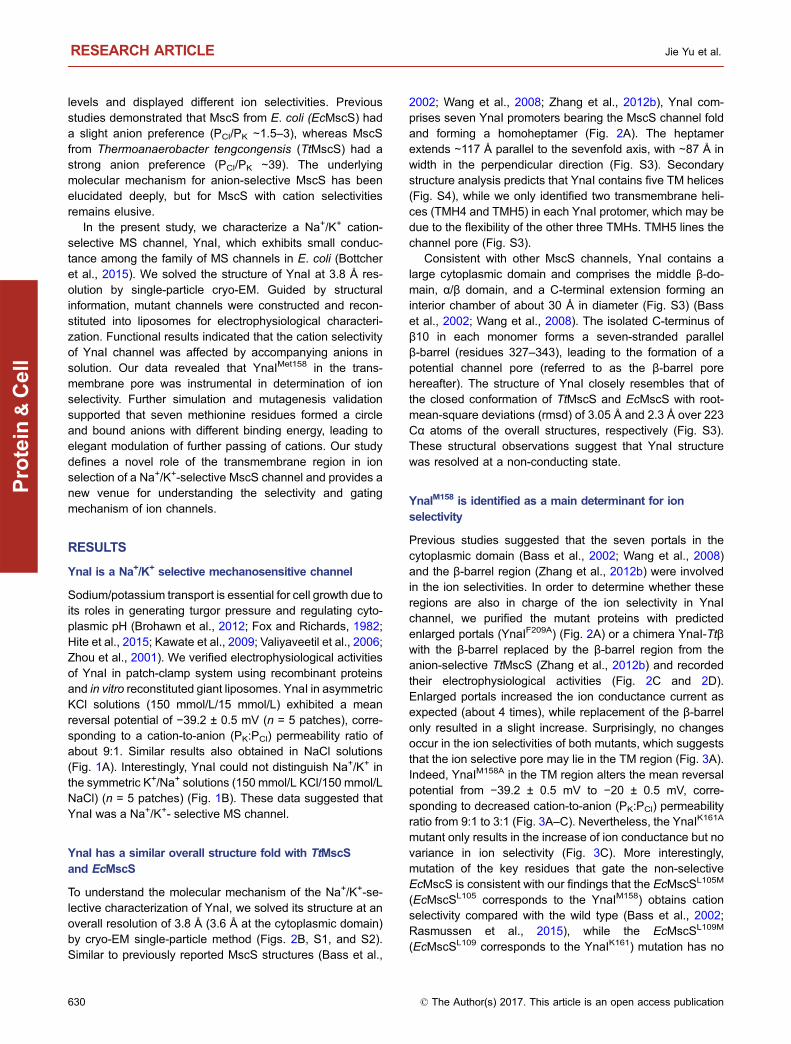
Sodium/potassium transport is essential for cell growth due toits roles in generating turgor pressure and regulating cyto-plasmic pH (Brohawn et al., 2012; Fox and Richards, 1982;Hite et al., 2015; Kawate et al., 2009; Valiyaveetil et al., 2006;Zhou et al., 2001). We verified electrophysiological activitiesof YnaI in patch-clamp system using recombinant proteinsand in vitro reconstituted giant liposomes. YnaI in asymmetricKCl solutions (150 mmol/L/15 mmol/L) exhibited a meanreversal potential of −39.2 ± 0.5 mV (n = 5 patches), corre-sponding to a cation-to-anion (PK:PCl) permeability ratio ofabout 9:1. Similar results also obtained in NaCl solutions(Fig. 1A). Interestingly, YnaI could not distinguish Na+/K+ inthe symmetric K+/Na+ solutions (150 mmol/L KCl/150 mmol/LNaCl) (n = 5 patches) (Fig. 1B). These data suggested thatYnaI was a Na+/K+- selective MS channel.
YnaI has a similar overall structure fold with TtMscSand EcMscS
To understand the molecular mechanism of the Na+/K+-se-lective characterization of YnaI, we solved its structure at anoverall resolution of 3.8 Å (3.6 Å at the cytoplasmic domain)by cryo-EM single-particle method (Figs. 2B, S1, and S2).Similar to previously reported MscS structures (Bass et al.,
2002; Wang et al., 2008; Zhang et al., 2012b), YnaI com-prises seven YnaI promoters bearing the MscS channel foldand forming a homoheptamer (Fig. 2A). The heptamerextends ∼117 Å parallel to the sevenfold axis, with ∼87 Å inwidth in the perpendicular direction (Fig. S3). Secondarystructure analysis predicts that YnaI contains five TM helices(Fig. S4), while we only identified two transmembrane heli-ces (TMH4 and TMH5) in each YnaI protomer, which may bedue to the flexibility of the other three TMHs. TMH5 lines thechannel pore (Fig. S3).
Consistent with other MscS channels, YnaI contains alarge cytoplasmic domain and comprises the middle β-do-main, α/β domain, and a C-terminal extension forming aninterior chamber of about 30 Å in diameter (Fig. S3) (Basset al., 2002; Wang et al., 2008). The isolated C-terminus ofβ10 in each monomer forms a seven-stranded parallelβ-barrel (residues 327–343), leading to the formation of apotential channel pore (referred to as the β-barrel porehereafter). The structure of YnaI closely resembles that ofthe closed conformation of TtMscS and EcMscS with root-mean-square deviations (rmsd) of 3.05 Å and 2.3 Å over 223Cα atoms of the overall structures, respectively (Fig. S3).These structural observations suggest that YnaI structurewas resolved at a non-conducting state.
YnaIM158 is identified as a main determinant for ionselectivity
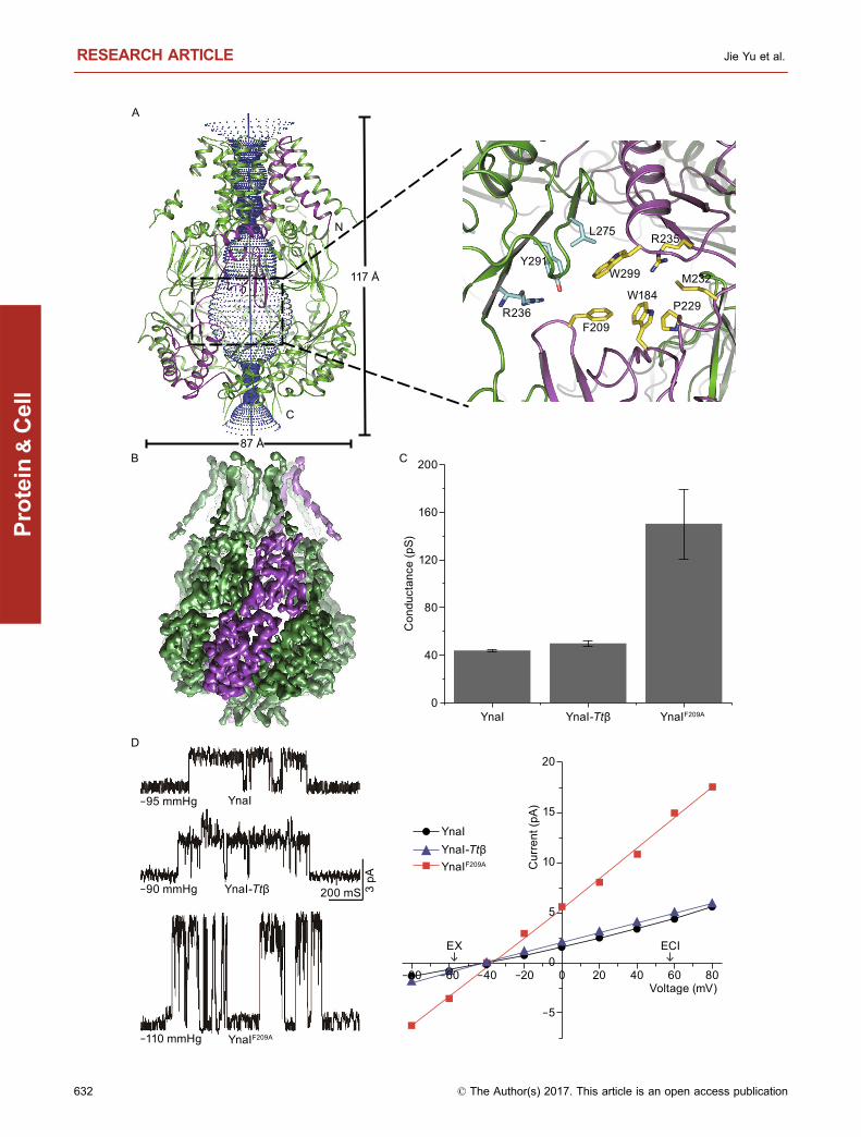
Previous studies suggested that the seven portals in thecytoplasmic domain (Bass et al., 2002; Wang et al., 2008)and the β-barrel region (Zhang et al., 2012b) were involvedin the ion selectivities. In order to determine whether theseregions are also in charge of the ion selectivity in YnaIchannel, we purified the mutant proteins with predictedenlarged portals (YnaIF209A) (Fig. 2A) or a chimera YnaI-Ttβwith the β-barrel replaced by the β-barrel region from theanion-selective TtMscS (Zhang et al., 2012b) and recordedtheir electrophysiological activities (Fig. 2C and 2D).Enlarged portals increased the ion conductance current asexpected (about 4 times), while replacement of the β-barrelonly resulted in a slight increase. Surprisingly, no changesoccur in the ion selectivities of both mutants, which suggeststhat the ion selective pore may lie in the TM region (Fig. 3A).Indeed, YnaIM158A in the TM region alters the mean reversalpotential from −39.2 ± 0.5 mV to −20 ± 0.5 mV, corre-sponding to decreased cation-to-anion (PK:PCl) permeabilityratio from 9:1 to 3:1 (Fig. 3A–C). Nevertheless, the YnaIK161A
mutant only results in the increase of ion conductance but novariance in ion selectivity (Fig. 3C). More interestingly,mutation of the key residues that gate the non-selectiveEcMscS is consistent with our findings that the EcMscSL105M
(EcMscSL105 corresponds to the YnaIM158) obtains cationselectivity compared with the wild type (Bass et al., 2002;Rasmussen et al., 2015), while the EcMscSL109M
(EcMscSL109 corresponds to the YnaIK161) mutation has no
RESEARCH ARTICLE Jie Yu et al.
630 © The Author(s) 2017. This article is an open access publication
Protein
&Cell

effect on the ion selectivity (Figs. 3D and S5). Collectively,these data provide strong evidence that a circular pore(SMC, seven-methionine circle) formed by the sevenYnaIM158 residues could be an important structural determi-nant for ion selectivity in YnaI.
YnaIM158 binding various anion with distinct bindingenergies facilitates Na+/K+ pass through
The critical role of a hydrophobic residue methionine incation selectivity has intrigued us to pursue further
mechanistic insights. Although it has been observed previ-ously that a methionine-formed circle element contributed tocation selectivity in ion channels but the mechanism has notbeen described clearly, such as TRP (Liao et al., 2013;Zubcevic et al., 2016), TPC (Guo et al., 2016) and Slo2.2(Hite et al., 2015). To elucidate the nature of Na+/K+ selec-tivity determined by the SMC element in YnaI, we performedsystematic quantum chemical investigations of the chemicalbonding, charge distribution, and binding energy (Gibbs freeenergy, ΔG) of the protein with a series of biological relevantcations and anions (M = Na+, K+, F−, Cl−, and NO3
−) (Fig. 4A
A
B
40 mVKCl
NaCl
500 mS 3 pA
-130 mmHg
-125 mmHg
40 mV
20 mV
0 mV
-40 mV
-20 mV
200 mS 3 pA
-127 mmHg
-115 mmHg
-120 mmHg
-118 mmHg
-109 mmHg
40 mV
KClNaCl
lCEXE
Cur
rent
(pA)
Voltage (mV)
8
6
4
2
0
-2
-80 -60 -40 -20 200 40 60 80
Voltage (mV)
150 mmolNa+
150 mmolK+
Cur
rent
(pA)
6
4
2
0
-2
-4
-6
-80 -60 -40 -20 0 20 40 60 80
Figure 1. YnaI is a Na+/K+ selective mechanosensitive channel. (A) Left: single-channel traces were recorded by patch-clamp
system from giant liposomes in KCl solution (upper) or NaCl solution (lower) at +40 mV. YnaI mutants were recorded in only KCl
solution by the same method. The numbers under the single-channel traces represent the negative pressure applied to the patch
during the event. Right: I–V curves for YnaI channel at a 10:1 salt gradient (150 mmol/L/15 mmol/L, KCl or NaCl); the reversal
potentials of an ideal anion or cation-selective channel with Erev = +58 mV or −58 mVaccording to the Nernst equation are indicated.
The reversal potentials for YnaI at KCl or NaCl solution are −39.2 ± 1.1 mV (n = 5, mean ± SE) and −40 ± 1.0 mV (n = 6, mean ± SE),
respectively. (B) Left: single-channel traces of YnaI at different voltages (0 mV, ±20 mV, ±40 mV), with recording solutions filled by
150 mmol/L NaCl in the pipette and 150 mmol/L KCl in bath solutions, respectively. Right: I–V curve for YnaI channel at the condition
that had described in left panel. The reversal potential was changed to 0 mV under 150 mmol/L NaCl/150 mmol/L KCl condition.
Binding-block ion selective mechanism RESEARCH ARTICLE
© The Author(s) 2017. This article is an open access publication 631
Protein
&Cell
F209 P229
W184 M232
R235
W299
L275
Y291
R236
-95 mmHg
-90 mmHg
-110 mmHg
YnaI
YnaI-Ttβ
YnaIF209A
200 mS 3 pA
C
D
N
C
117 Å
87 Å
A
B
Voltage (mV)
Cur
rent
(pA)
EX ECI
20
15
10
5
0
-5
-80 -60 -40 -20 200 40 60 80
YnaI-TtβYnaI
YnaIF209A
YnaI
Con
duct
ance
(pS)
YnaI-Ttβ YnaIF209A
200
160
120
80
40
0
RESEARCH ARTICLE Jie Yu et al.
632 © The Author(s) 2017. This article is an open access publication
Protein
&Cell
and Table S1). To our surprise, the anions (F−, Cl−, andNO3
−) represent much higher binding energies than that ofthe cations (Na+, ΔG = −0.87 kcal·mol−1 and K+, ΔG =3.51 kcal·mol−1). Among these three anions, F− has thelargest binding energy (−23.31 kcal·mol−1) with the calcu-lated structure elements, while NO3
− has the lowest bindingenergy (−13.83 kcal·mol−1), with Cl− in the middle(−16.56 kcal·mol−1). Therefore, the occupation of the anionsin YanI probably hampers the binding of Na+/K+ to SMCelement. The data led us to hypothesize that ion bindingenergy could be relevant to the abilities of the anions to blockcation transportation (F− > Cl− > NO3
−) and even the ionselectivity.
Our model predicts that higher pressure would be neededto overcome the energy barrier to open the channel in KFsolutions, due to the tight interaction of F− with the SMCelement; while in KNO3 solutions lesser pressure is required.To test this hypothesis, we measured the electrophysiologi-cal activities of YnaI in the solutions of K+ in combination withdifferent anions using patch-clamp system and in vitroreconstituted giant liposomes. Indeed, in KF solutions, rela-tively high pressure is required to obtain detectable currentsof the YnaI channel, while in KNO3 solutions, the YnaIchannel could open spontaneously with much higher con-ductance even in the absence of any pressure (Fig. 4B).Surprisingly, YanI in KF solution shows a lower meanreversal potentials (−19.2 ± 1.6 mV, n = 4 patches), while inKNO3 solution displays a much negative mean reversalpotentials (−52.3 ± 0.4 mV, n = 4 patches) in comparison withthat of in KCl solution. Correspondingly, the cation-to-anionpermeability ratio decreased to about 2.6:1 in KF solutions(PK:PF), while raised to about 39.6:1 in KNO3 solutions (PK:PNO3) (Fig. 4B and Table S2). In addition, mutant YanIM158A
could open spontaneously without applied any pressure withalmost the same ion selectivity in above three differentsolutions, which implies that selectivities influenced by theanions almost vanish, further highlights the important role ofSMC element in ion selectivity (Fig. 4C and Table S2). These
data provide strong evidence that the ion selectivity andtransmittance of YnaI could be determined by the differentanions.
Previous studies demonstrated that the TM1 and TM2may be responsible for gating the channel, and truncation ofthese two helixes would induce the channel to open spon-taneously and present the typical gain-of-function beha-viours when expressed in the living cells (Bottcher et al.,2015). Consistently, electrophysiological studies of singlechannel with the YnaIΔ2−63 mutant clearly show that thechannel can open spontaneously in KCl solutions withoutany external pressure (Fig. S6). Apparently, the YnaIΔ2−63
mutant also opens spontaneously in KNO3 solutions.External pressure is still needed to open the truncation in KFsolutions albeit at much smaller level. The ion conductanceand ion selectivity of YnaIΔ2−63 are almost same as that ofwild type YnaI in these three solutions (Fig. 4D andTable S2). These data strengthen the conclusion that TM1and TM2 play roles in the gating of YnaI, but do not changethe influence of anions on selectivity and gating manner ofYnaI, which further support the critical role of YanIM158 andSMC in the TM region.
DISCUSSION
Based on the results, we proposed a new binding-blockmodel for the molecular mechanism of how the ions wereselected by the ion channels and how the mechanosensitivechannels were gated by the ions. In the case of YnaIchannel, different binding affinities of disparate anions to theion selective filter (SMC) due to chemical interactions(Fig. 4A and Table S1), leads to different degree blockade ofthe channel gating. Among the cases we studied, the F− ionhas the strongest binding affinity with SMC, so the channelneeds the highest pressure to overcome the energy barrierto open the channel. When the channels are forced to openat high pressure, K+ could pass through the SMC, and the F−
might move together with K+, causing low ion selectivity andcurrents in KF solutions. While in the KNO3 solutions, NO3
−
binds to the SMC with the lowest affinity, therefore thechannels could open spontaneously without pressureapplied. At the same time, the lowest affinity of NO3
− bindingto the SMC prevents the transportation of NO3
− which maylead to very high cation selectivity (PK:PNO3 = 39.6:1). Mostimportant of all, as the most vital and abundant anion in livingorganisms, Cl− has just the right binding affinity with the SMCto gate the channel, and the precise regulation has chosenmethionine residues as key determination possibly after longcourse evolution upon high selection power.
Together, our structural, biological, biochemical, quantummechanical, and electrophysiological results provided strongevidence that the MscS-like YnaI channel selected Na+/K+
cations specifically through the SMC element at the TM poreand the ion permeability and selectivity were determined bythe anions present in the circumstances. These results notonly explain why the YnaI is highly selective, but also lead to
Figure 2. The cytoplasmic equatorial portals of YnaI con
tribute to ion conductance. (A) Right: overall structure of YnaI
homoheptamer. One protomer is colored in purple, and the
others are colored in green. The channel passage is shown in
blue dots along a blue axis. Left: ribbon diagram of close views
of one of the seven portals in YnaI. Residues lining the portals
are shown in yellow and cyan sticks. (B) Cryo-EM density map
of YnaI, with one of the seven promoters highlighted in dark
purple. (C) YnaIF209A mutant showed a higher conductance
comparing with wide-type YnaI and YnaI mutant substituted
with the TtMscS β-barrel region (YnaI-Ttβ) (n = 4, mean ± SE).
(D) Left: single-channel traces of YnaI, YnaI-Ttβ and YnaIF209A
mutant were recorded at +40 mV. Right: I–V curves for YnaI,
YnaI-Ttβ, and YnaIF209A mutant. Both YnaI-Ttβ and YnaIF209A
mutants shared a similar reversal potential with YnaI. YnaIF209A
displayed an obviously higher conductance.
b
Binding-block ion selective mechanism RESEARCH ARTICLE
© The Author(s) 2017. This article is an open access publication 633
Protein
&Cell
C
D
BA
M158M158K161
-100KT +100KT
-120 mmHg
-100 mmHg
YnaIM158A
YnaIK161A
-110 mmHg YnaI
200 mS 5 pA
-87 mmHg
-95 mmHg
-90 mmHg
EcMscS
EcMscSL105M
EcMscSL105M
200 mS 5 pAL105M
EClEk
Cur
rent
(pA)
Voltage (mV) -80 -60 -40 -20 200 40 60 80
12
8
4
0
-4
-8
-12
EcMscSEcMscSL105M
EcMscSL105M
Voltage (mV)
Cur
rent
(pA)
Ek ECl
-80 -60 -40 -20 200 40 60 80
12
8
4
0
-4
-8
YnaIM158A
YnaIK161A
YnaI
RESEARCH ARTICLE Jie Yu et al.
634 © The Author(s) 2017. This article is an open access publication
Protein
&Cell
the novel binding-block mechanism of the gating and ionselectivities of ion channels.
MATERIALS AND METHODS
Protein expression and purification
Gene YnaI was cloned from E. coli into pET-21b vector (Novagen)
with a C-terminal 6× His tag. Overexpression of YnaI was induced
in E. coli strain BL21 (DE3) by 0.5 mmol/L isopropyl-β-D-thio-
galactoside when the cell density reached OD600 = 1.0. After
growth for 4 h at 37°C, the cells were collected, resuspended in
buffer containing 20 mmol/L Tris pH 8.0, 200 mmol/L NaCl, and
lysed by sonication. Cell debris was removed by centrifugation at
15,422 ×g for 15 min. The supernatant containing membrane was
applied to ultracentrifugation at 173,021 ×g for 1 h. The mem-
brane fraction was collected and incubated with 1.5% (w/v)
n-dodecyl-β-D-maltopyranoside (DDM; Anatrace) for 3 h with slow
stirring at 4°C. After additional ultracentrifugation at 173,021 ×g
for 30 min, the supernatant was collected and loaded onto
Ni2+-nitrilotriacetate affinity resin (Ni-NTA; Qiagen). The resin was
then washed with buffer A containing 25 mmol/L Tris pH 8.0, 20
mmol/L imidazole, 500 mmol/L NaCl, and 0.02% DDM. Followed
by eluted from affinity resin with buffer A supplemented with 300
mmol/L imidazole, the protein was concentrated and applied to a
gel-filtration resin (Superdex-200 HR 10/30; GE Healthcare), pre-
viously equilibrated with buffer containing 20 mmol/L Mes pH 6.5,
200 mmol/L NaCl, 5 mmol/L DTT (Dithiothreitol), and 0.02% DDM.
The peak fractions were collected for cryo-EM and electrophysi-
ology studies. Various YnaI mutants followed the same
procedures.
For cryo-EM study, the protein was mixed with amphipols (Ana-
trace) at 1:3 (w/w) for 5 h with slow stirring at 4°C. Detergent was
removed with Bio-Beads SM-2. After separation from Bio-beads, the
protein was loaded to Superdex 200 again with buffer containing
20 mmol/L Mes pH 6.5, 200 mmol/L NaCl, 5 mmol/L DTT. The peak
fractions were collected for analysis by cryo-EM.
Preparation of giant liposomes and electrical recording
All lipids used in reconstitution were purchased from Avanti Polar
Lipids. The wild-type YnaI and the mutant proteins were reconsti-
tuted into lipid vesicles composed of 1-palmitoyl-2-oleoyl-phos-
phatidylethanolamine (POPE, 7.5 mg/mL) and 1-palmitoyl-2-oleoyl-
phosphatidylglycerol (POPG, 2.5 mg/mL) as previously described
method (Li et al., 2007). The giant liposomes were obtained by
regular dehydrate and hydrate processes. The patch-clamp
recording of YnaI were performed in asymmetrical conditions with
15 mmol/L KCl or NaCl, 500 mmol/L sucrose, 5 mmol/L K-Hepes
(pH 7.0) in bath solution, and 150 mmol/L KCl or NaCl, 500 mmol/L
sucrose, 5 mmol/L K-Hepes (pH 7.0) in pipette solution. YnaI and
YnaI Δ2−62 were performed in asymmetrical KCl, KF and KNO3
solutions, but other mutants were performed only in asymmetrical
KCl solution. Concentrations of KF and KNO3 used in asymmetrical
conditions are same as KCl. After attained a gigohm seal (the
resistance was about 3–8 GΩ), the current was recorded by using an
Axopatch 200B amplifier with a Digidata 1322A analogue-to-digital
converter (Axon Instruments). The mechanical pressure was mea-
sured by a pressure monitor (PM015D, WPI). Permeability ratios
were calculated by using Nernst equation as following:
Erev =RTF
ln(PK [ K ] o +PCl [ Cl ] iPK [ K ] i +PCl [ Cl ] o
)
where [X]o and [X]i are ion concentration on extracellular (cis-side)
and intracellular sides (trans-side), respectively.
EM sample preparation and data collection
The homogeneity of purified YnaI in amphipols were examined by
negative staining with 2% uranyl acetate. Images were recorded
using a 4 k × 4 k CCD camera (UltraScan 4000, Gatan) in an FEI
T12 microscope operated at 120 kV. For cryo-grid preparation, the
Quantifoil 1.2/1.3 holy carbon grids were baked at 50°C in an oven
for 2 weeks. This treatment of grids would allow better distribution of
particles into the carbon holes. 4 μL aliquots of freshly purified
sample (0.1 mg/mL) were loaded on the pretreated and glow-dis-
charged grids. Blotting and sample freezing were performed in an
FEI Mark IV Vitrobot (4°C and 85% humidity). Images were recorded
using a K2 Summit direct electron detector (Gatan) in super reso-
lution counting mode at a nominal magnification of 22,500× (ren-
dering a pixel size of 1.32 Å) on an FEI Titan Krios electron
microscope at 300 kV. All images were collected by UCSF-image4
(X. Li and Y. Cheng, UCSF) with defocus ranging from −1 ∼ −2.2 µm.
The total exposure time is 8 s (32 frames), with a dose rate of ∼8.2counts per physical pixel per second.
Image processing and analyses
The initial model was calculated using EMAN2 (Ludtke et al., 1999)
with the 2D class averages from negatively stained particles. For
cryo-EM data, the motion correction was performed with MOTION-
CORR (Li et al., 2013) at micrograph level, and the estimation of
contrast transfer function parameters was performed with
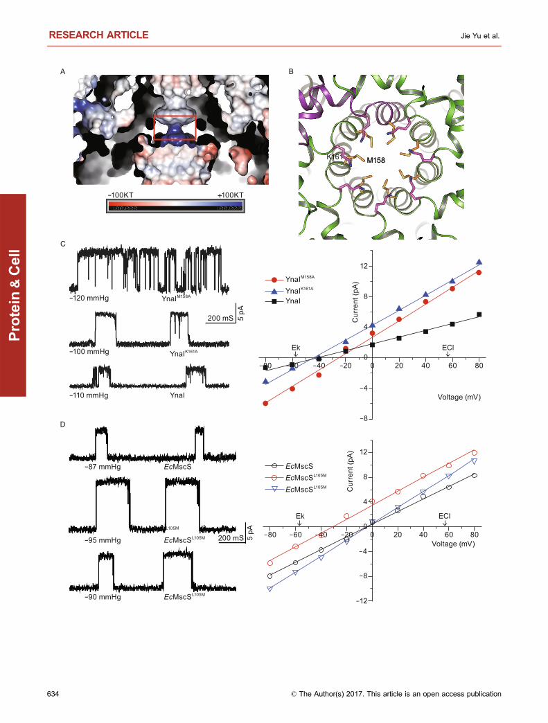
Figure 3. YnaIM158 located at transmembrane region deter-
mines the cation selectivity. (A) Electrostatic potentials
around the transmembrane pore inner surface of YnaI. Narrowest
region where M158 and K161 located is indicated by red square.
(B) Ribbon diagram of TM helices surrounding restriction site
viewed alongmembrane bilayer fromextracellular side.M158 and
K161 are shown in yellow and purple sticks, respectively.
(C) Mutation in the transmembrane region decreased the cation
selectivity of YnaI. Left: single-channel traces of YnaI, YnaIM158A,
and YnaIK161A were recorded at +40 mV. Right: I–V curves for
YnaI, YnaIM158A, and YnaIK161A. The reversal potential of M158A
shifted right (−26 ± 1.5 mV, n = 3, mean ± SE), representing an
attenuation of cation selectivity. (D) Mutation of the key residues in
gating ion endowed EcMscS channel cation selectivity. Left:
single-channel traces of EcMscS, EcMscSL105M, and
EcMscSL109M mutants were recorded at +40 mV. Right: I–Vcurves forEcMscS,EcMscSL105M, andEcMscSL109Mmutants. The
reversal potential of EcMscSL105M shifted right (−31.1 ± 0.6 mV,
n = 6, mean ± SE), representing an increased cation selectivity.
b
Binding-block ion selective mechanism RESEARCH ARTICLE
© The Author(s) 2017. This article is an open access publication 635
Protein
&Cell
B
A
C
200 mS 5 pA
-150 mmHg
0 mmHg
-130 mmHg
KNO3
KCl
KF C
urre
nt (p
A)Voltage (mV)
Voltage (mV)
Cur
rent
(pA)
0 mmHg
0 mmHg
-90 mmHg
KNO3
KCl
KF
200 mS 5 pA
Cur
rent
(pA)
Voltage (mV)
D
200 mS
KNO3
KCl
0 mmHg
0 mmHg
KF0 mmHg
5 pA
-80-60 -40 -20 200 40 60 80
-5
-10
-15
-20
-2530
25
20
15
10
5
0
-5
YnaI-KNO3
YnaI-KClYnaI-KF
YnaIM158A -KNO3
YnaIM158A -KClYnaIM158A -KF
YnaIΔ2-63 -KNO3
YnaIΔ2-63 -KClYnaIΔ2-63 -KF
15
10
5
0
-5
-10
-80 -60 -40 -20 200 40 60 80
20
16
12
8
4
0
-4-80 -60 -40 -20 200 40 60 80
Na+ K+ F- Cl- NO3-
ΔG(k
cal•m
ol- 1)
5
0
RESEARCH ARTICLE Jie Yu et al.
636 © The Author(s) 2017. This article is an open access publication
Protein
&Cell
CTFFIND3 (Mindell and Grigorieff, 2003). Micrograph screening,
automatic particle picking, and particle normalization were per-
formed with SPIDER (Shaikh et al., 2008) software packages. The
2D classification, 3D classification and refinement were performed
with RELION (Scheres, 2012). A total of 550,000 particles (window
size 144 × 144) were automatically picked from 2,100 micrographs.
Based on the results of 2D classification, 90% of the particles were
grouped into top view classes, 8% of the particles were in tilted
views, and less than 2% of the particles were in standard side views.
Although the tilted- and side-view particles (42,000 particles) only
contribute to a minor portion of the total particles, they are essential
in determining correct reconstruction. We have tried to mix different
numbers of top-view particles with these tilted and side-view parti-
cles in several rounds of 3D refinement, and we found that if we
omitted all top-view particles, we could get a better map in both the
nominal resolution and map quality. Therefore, after region-based
3D classification and refinement, a final data set of 42,000 nontop-
view particles resulted in a 3.8 Å map (gold-standard FSC 0.143).
The density of the transmembrane region was relatively weak, and
could not be improved after global or local 3D classification. To
improve the resolution in the region of the intracellular domain, we
added a soft mask around the intracellular domain in the refinement
and obtained a 3.6 Å map. To further improve the map quality, we
used the dose-reduced particles summed from frames of 3–18, andthe overall resolution of the map has been improved to 3.6 Å. The
local resolution maps were calculated by ResMap (Kucukelbir et al.,
2014).
Model building and refinement
The homologue crystal structure of Thermoanaerobacter tengcon-
gensis MscS (PDB 3UDC) was docked into the density map of YnaI
heptamer as a start model in Chimera (Pettersen et al., 2004).
Sequence alignment of YnaI with the crystal template was per-
formed by BLAST (Mount, 2007). The atomic model of the intra-
cellular domain was manually built in coot (Emsley et al., 2010)
based on the start model with the Mutate and Renumber tools.
Sequence assignment was mainly guided by the clearly resolved
bulky residues (such as Phe, Tyr, Trp, and Arg). This model was
refined by real-space refinement (phenix.real_space_refine) in
Phenix (Adams et al., 2010), with stereochemical and secondary
structure constraints applied. The refined model was examined by
cross-validation according to previously described procedures (Li
et al., 2015). Specifically, the coordinates of the refined model were
randomly shifted by 0.2 Å using Phenix PDB tool. The shifted model
was then refined with half1 map in Phenix. The new refined model
(with half1 map) was converted to mrc map, and then compared with
half1 map, half2 map and combined map to calculate the FSC
curves, respectively. These curves indicated that the model was not
overfitted. The model of transmembrane domain was built in MDFF
(Trabuco et al., 2009). We deleted all the side chains in this region
due to the resolution limitation.
Quantum chemical methods and computational details
The geometries of the model systems (H3CSCH3)7 and A@
(H3CSCH3)7 (A = Na+, K+, F−, Cl−, NO3−) were optimized via con-
strained energy minimization at the level of density functional theory
(DFT). All the atomic coordinates are optimized except the positions
of the inner ring formed by C atoms and all the S atoms that were
fixed at the experimental structures. The B3LYP hybrid exchange-
correlation function (Becke, 1993; Lee et al., 1988) was used with
6–31 + G* basis sets (Francl et al., 1982) for all the elements. The
vibrational frequencies were calculated with the harmonic approxi-
mation. The thermodynamic properties were calculated at ambient
temperature and pressure using statistical mechanics. All the cal-
culations were carried out by using Gaussian 09 program (Frisch, M.
J. et al. Gaussian 09. Revision C.01 (Gaussian, 2010).)
ACKNOWLEDGEMENTS
We thank the Tsinghua University Branch of China National Center
for Protein Sciences (Beijing) for providing the facility support. The
computation was completed on the “Explorer 100” cluster system of
Tsinghua National Laboratory for Information Science and Technol-
ogy. We also thank Samantha Miller (University of Aberdeen, UK) for
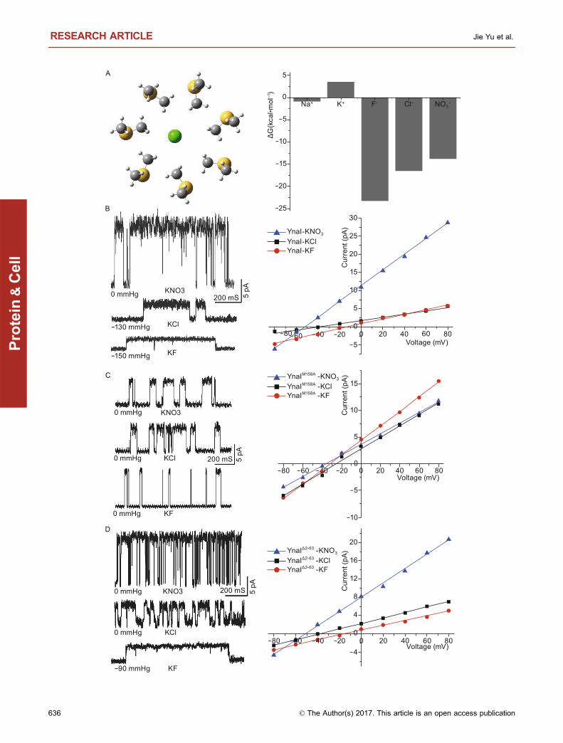
Figure 4. Different anions affect the ion selectivity and
transmittance of YnaI. (A) Optimized structures of
(H3CSCH3)7 and Cl@(H3CSCH3)7, where van der Waals radii
were used in the later to illustrate the size of the cavity. Relative
binding energies for A + (H3CSCH3)7 → A@(H3CSCH3)7 (A =
Na+, K+, F−, Cl−, Br−). (B) Left: single-channel traces of YnaI
were recorded at +40 mV in different asymmetric potassium salt
solutions. The required negative pressures for opening the
channel were different in those solutions. In KF, the required
negative pressure was −150 ± 13 mmHg (n = 4, mean ± SE). In
KCl, the required negative pressure was −130 ± 11 mmHg
(n = 5, mean ± SE). In KNO3, YnaI channel opened sponta-
neously without pressure applied (n = 4). Right: I–V curves for
YnaI at different asymmetric potassium salt solutions. The
reversal potentials of YnaI varied in different solutions. In KF,
KCl and KNO3, the reversal potentials were −19.2 ± 1.6 (n = 4,
mean ± SE), −39.2 ± 0.5 (n = 5, mean ± SE), −52.3 ± 0.4 (n = 4,
mean ± SE) respectively. (C) Left: single-channel traces of
mutant YnaIM158A were recorded at +40 mV in different
asymmetric potassium salt solutions. YnaIM158A opened spon-
taneously without pressure applied to the pipette in asymmetric
KCl, KF, and KNO3 solutions. Right: I–V curves for YnaIM158A at
different asymmetric potassium salt solutions. In KCl, KF
and KNO3, the reversal potentials were −28.2 ± 2.6 (n = 6,
mean ± SE), −26.5 ± 1.9 (n = 6, mean ± SE), −35.1 ± 0.5 (n = 7,
mean ± SE), respectively. (D) Left: single-channel traces of
mutant YnaIΔ2−63 were recorded at +40 mV in different
asymmetric potassium salt solutions. The required negative
pressures for opening the channel were different in those
solutions. In KF, the required negative pressure was −60 ± 9
mmHg (n = 4, mean ± SE). In KCl and KNO3, YnaIΔ2−63 opened
spontaneously without pressure applied (n = 5, n = 4,
respectively). Right: I–V curves for YnaIΔ2−63 at different
asymmetric potassium salt solutions. The reversal potentials
of YnaIΔ2−63 varied in different solutions. In KF, KCl and KNO3,
the reversal potentials were 14.4 ± 1.7 (n = 4, mean ± SE),
−39.2 ± 0.6 (n = 5, mean ± SE), 51.9 ± 0.8 (n = 4, mean ± SE),
respectively.
b
Binding-block ion selective mechanism RESEARCH ARTICLE
© The Author(s) 2017. This article is an open access publication 637
Protein
&Cell
helpful discussion. This work was supported by funds from the
Ministry of Science and Technology (2016YFA0501100 and
2017YFA0504600 to M.J., and 2016YFA0500700 and
2013CB910400 to N.G.), and the National Fund for Distinguished
Young Scholar (31625008 to M.Y.) and National Natural Science
Foundation of China (Grant Nos. 21532004 and 31570733 to M.Y.,
31422016 to N.G., 31371066 and 31671049 to Y.L., and 91426302
to J.L.)
ABBREVIATIONS
DDM, n-dodecyl-β-D-maltopyranoside; MS, mechanosensitive; MscS,
mechanosensitive small conductance; Ni-NTA, Ni2+-nitrilotriacetate
affinity resin; POPE, 1-palmitoyl-2-oleoyl-phosphatidylethanolamine;
POPG, 1-palmitoyl-2-oleoyl-phosphatidylglycerol
COMPLIANCE WITH ETHICS GUIDELINES
Jie Yu, Bing Zhang, Yixiao Zhang, Cong-qiao Xu, Wei Zhuo,
Jingpeng Ge, Jun Li, Ning Gao, Yang Li, and Maojun Yang declare
that they have no conflict of interest. This article does not contain
any studies with human or animal subjects performed by any of the
authors.
OPEN ACCESS
This article is distributed under the terms of the Creative Commons
Attribution 4.0 International License (http://creativecommons.org/
licenses/by/4.0/), which permits unrestricted use, distribution, and
reproduction in any medium, provided you give appropriate credit to
the original author(s) and the source, provide a link to the Creative
Commons license, and indicate if changes were made.
REFERENCES
Adams PD, Afonine PV, Bunkoczi G, Chen VB, Davis IW, Echols N,
Headd JJ, Hung LW, Kapral GJ, Grosse-Kunstleve RW et al
(2010) PHENIX: a comprehensive Python-based system for
macromolecular structure solution. Acta Crystallogr D Biol Crys-
tallogr 66:213–221Baconguis I, Bohlen CJ, Goehring A, Julius D, Gouaux E (2014)
X-ray structure of acid-sensing ion channel 1-snake toxin
complex reveals open state of a Na(+)-selective channel. Cell
156:717–729Bass RB, Strop P, Barclay M, Rees DC (2002) Crystal structure of
Escherichia coli MscS, a voltage-modulated and mechanosensi-
tive channel. Science 298:1582–1587Becke AD (1993) Density-functional thermochemistry. 3. The role of
exact exchange. J Chem Phys 98:5648–5652Bottcher B, Prazak V, Rasmussen A, Black SS, Rasmussen T
(2015) The structure of YnaI implies structural and mechanistic
conservation in the MscS family of mechanosensitive channels.
Structure 23:1705–1714Brohawn SG, del Marmol J, MacKinnon R (2012) Crystal structure of
the human K2P TRAAK, a lipid- and mechano-sensitive K+ ion
channel. Science 335:436–441
Chang G, Spencer RH, Lee AT, Barclay MT, Rees DC (1998)
Structure of the MscL homolog from Mycobacterium tuberculosis:
a gated mechanosensitive ion channel. Science 282:2220–2226Emsley P, Lohkamp B, Scott WG, Cowtan K (2010) Features and
development of Coot. Acta Crystallogr D Biol Crystallogr 66:486–501
Fox RO Jr, Richards FM (1982) A voltage-gated ion channel model
inferred from the crystal structure of alamethicin at 1.5-A
resolution. Nature 300:325–330Francl MM, Pietro WJ, Hehre WJ, Binkley JS, Gordon MS, Defrees
DJ, Pople JA (1982) Self-consistent molecular-orbital methods.
23. A polarization-type basis set for 2nd-row elements. J Chem
Phys 77:3654–3665Ge J, Li W, Zhao Q, Li N, Chen M, Zhi P, Li R, Gao N, Xiao B, Yang
M (2015) Architecture of the mammalian mechanosensitive
Piezo1 channel. Nature 527:64–69Guo J, Zeng W, Chen Q, Lee C, Chen L, Yang Y, Cang C, Ren D,
Jiang Y (2016) Structure of the voltage-gated two-pore channel
TPC1 from Arabidopsis thaliana. Nature 531:196–201Hite RK, Yuan P, Li Z, Hsuing Y, Walz T, MacKinnon R (2015) Cryo-
electron microscopy structure of the Slo2.2 Na(+)-activated K(+)
channel. Nature 527:198–203Hou X, Pedi L, Diver MM, Long SB (2012) Crystal structure of the
calcium release-activated calcium channel Orai. Science
338:1308–1313Jiang Y, Lee A, Chen J, Cadene M, Chait BT, MacKinnon R (2002)
Crystal structure and mechanism of a calcium-gated potassium
channel. Nature 417:515–522Kane Dickson V, Pedi L, Long SB (2014) Structure and insights into
the function of a Ca(2+)-activated Cl(−) channel. Nature
516:213–218Kawate T, Michel JC, Birdsong WT, Gouaux E (2009) Crystal
structure of the ATP-gated P2X(4) ion channel in the closed state.
Nature 460:592–598Kucukelbir A, Sigworth FJ, Tagare HD (2014) Quantifying the local
resolution of cryo-EM density maps. Nat Methods 11:63–65Lee CT, Yang WT, Parr RG (1988) Development of the Colle-Salvetti
Correlation-Energy Formula into a Functional of the Electron-
Density. Phys Rev B 37:785–789Li Y, Berke I, Chen L, Jiang Y (2007) Gating and inward rectifying
properties of the MthK K+ channel with and without the gating
ring. J Gen Physiol 129:109–120Li X, Mooney P, Zheng S, Booth CR, Braunfeld MB, Gubbens S,
Agard DA, Cheng Y (2013) Electron counting and beam-induced
motion correction enable near-atomic-resolution single-particle
cryo-EM. Nat Methods 10:584–590Li N, Zhai Y, Zhang Y, Li W, Yang M, Lei J, Tye BK, Gao N (2015)
Structure of the eukaryotic MCM complex at 3.8 A. Nature
524:186–191Liao M, Cao E, Julius D, Cheng Y (2013) Structure of the TRPV1 ion
channel determined by electron cryo-microscopy. Nature
504:107–112Ludtke SJ, Baldwin PR, Chiu W (1999) EMAN: semiautomated
software for high-resolution single-particle reconstructions.
J Struct Biol 128:82–97McCusker EC, Bagneris C, Naylor CE, Cole AR, D’Avanzo N,
Nichols CG, Wallace BA (2012) Structure of a bacterial voltage-
RESEARCH ARTICLE Jie Yu et al.
638 © The Author(s) 2017. This article is an open access publication
Protein
&Cell
gated sodium channel pore reveals mechanisms of opening and
closing. Nat Commun 3:1102
Mindell JA, Grigorieff N (2003) Accurate determination of local
defocus and specimen tilt in electron microscopy. J Struct Biol
142:334–347Mount DW (2007) Using the basic local alignment search tool
(BLAST). CSH protocols 2007, pdb top17
Payandeh J, Scheuer T, Zheng N, Catterall WA (2011) The crystal
structure of a voltage-gated sodium channel. Nature 475:353–358
Payandeh J, Gamal El-Din TM, Scheuer T, Zheng N, Catterall WA
(2012) Crystal structure of a voltage-gated sodium channel in two
potentially inactivated states. Nature 486:135–139Perozo E, Cortes DM, Sompornpisut P, Kloda A, Martinac B (2002)
Open channel structure of MscL and the gating mechanism of
mechanosensitive channels. Nature 418:942–948Pettersen EF, Goddard TD, Huang CC, Couch GS, Greenblatt DM,
Meng EC, Ferrin TE (2004) UCSF Chimera—a visualization
system for exploratory research and analysis. J Comput Chem
25:1605–1612Rasmussen T, Rasmussen A, Singh S, Galbiati H, Edwards MD,
Miller S, Booth IR (2015) Properties of the mechanosensitive
channel MscS pore revealed by tryptophan scanning mutagen-
esis. Biochemistry 54:4519–4530Scheres SH (2012) A Bayesian view on cryo-EM structure determi-
nation. J Mol Biol 415:406–418Shaikh TR, Gao H, Baxter WT, Asturias FJ, Boisset N, Leith A, Frank
J (2008) SPIDER image processing for single-particle recon-
struction of biological macromolecules from electron micro-
graphs. Nat Protoc 3:1941–1974Trabuco LG, Villa E, Schreiner E, Harrison CB, Schulten K (2009)
Molecular dynamics flexible fitting: a practical guide to combine
cryo-electron microscopy and X-ray crystallography. Methods
49:174–180Valiyaveetil FI, Leonetti M, Muir TW, Mackinnon R (2006) Ion
selectivity in a semisynthetic K+ channel locked in the conductive
conformation. Science 314:1004–1007Van Petegem F, Clark KA, Chatelain FC, Minor DL Jr (2004)
Structure of a complex between a voltage-gated calcium channel
beta-subunit and an alpha-subunit domain. Nature 429:671–675Wagenknecht T, Grassucci R, Frank J, Saito A, Inui M, Fleischer S
(1989) Three-dimensional architecture of the calcium chan-
nel/foot structure of sarcoplasmic reticulum. Nature 338:167–170Wang W, Black SS, Edwards MD, Miller S, Morrison EL, Bartlett W,
Dong C, Naismith JH, Booth IR (2008) The structure of an open
form of an E. coli mechanosensitive channel at 3.45 A resolution.
Science 321:1179–1183Wu J, Yan Z, Li Z, Yan C, Lu S, Dong M, Yan N (2015) Structure of
the voltage-gated calcium channel Cav1.1 complex. Science
350:aad2395
Zhang X, Ren W, DeCaen P, Yan C, Tao X, Tang L, Wang J,
Hasegawa K, Kumasaka T, He J et al (2012a) Crystal structure of
an orthologue of the NaChBac voltage-gated sodium channel.
Nature 486:130–134Zhang X, Wang J, Feng Y, Ge J, Li W, Sun W, Iscla I, Yu J, Blount P,
Li Y et al (2012b) Structure and molecular mechanism of an
anion-selective mechanosensitive channel of small conductance.
Proc Natl Acad Sci USA 109:18180–18185Zhou Y, Morais-Cabral JH, Kaufman A, MacKinnon R (2001)
Chemistry of ion coordination and hydration revealed by a K+
channel-Fab complex at 2.0 A resolution. Nature 414:43–48Zubcevic L, Herzik MA Jr, Chung BC, Liu Z, Lander GC, Lee SY
(2016) Cryo-electron microscopy structure of the TRPV2 ion
channel. Nat Struct Mol Biol 23:180–186
Binding-block ion selective mechanism RESEARCH ARTICLE
© The Author(s) 2017. This article is an open access publication 639
Protein
&Cell


![Highly selective binding of methyl orange dye by cationic ... · Highly selective binding of methyl orange dye by cationic water-soluble pillar[5]arenes L. S. Yakimova, D. N. Shurpik,](https://img.pdfslide.net/doc/110x75/5f0a48517e708231d42ae57c/highly-selective-binding-of-methyl-orange-dye-by-cationic-highly-selective-binding.jpg)
![Selective Metal Binding by Vanabin2 from the Vanadium-rich ... · Cu(II), and Zn(II)] were examined for binding to Vanabin2. VOSO4•nH2O (n = 3-4), FeCl3, CoSO4•7H2O, NiCl2 •6H2O,](https://img.pdfslide.net/doc/110x75/5fae788c39da264765209717/selective-metal-binding-by-vanabin2-from-the-vanadium-rich-cuii-and-znii.jpg)


![Molecular Docking of Selective Binding Affinity of ... · sulfonamide derivatives with the glycolytic enzymes [31]. This blind docking was [30] used to map different binding pockets](https://img.pdfslide.net/doc/110x75/5f04ffa77e708231d410c119/molecular-docking-of-selective-binding-affinity-of-sulfonamide-derivatives-with.jpg)


![Techniques and strategies employing engineered …bleris/papers/2017-TALEs.pdfNCP [31,32]. Chromatin immunoprecipitation and sequencing (ChIP-seq) has revealed dCas9 binding from tens](https://img.pdfslide.net/doc/110x75/60accfbcf2c1682e39595fa9/techniques-and-strategies-employing-engineered-blerispapers2017-talespdf-ncp.jpg)