Embed Size (px)
Citation preview

J. Cell Set. 8, 93-109(1971) 93Printed in Great Britain
A BIOLOGICAL SYSTEM PRODUCING A
SELF-ASSEMBLING CHOLESTERIC PROTEIN
LIQUID CRYSTAL
A. C. NEVILLE AND B. M. LUKEA.R.C. Unit of Insect Physiology, Department of Zoology,South Parks Road, Oxford, England
SUMMARY
The protein in the oothecal glands of praying mantids (Sphodromantis tenuidentata, Mio-mantis monacha) exists in the form of lamellar liquid crystalline spherulites, which coalesce asthey flow out of a punctured gland tubule. Electron micrographs of sections of these spherulitesafter fixation show parabolic patterns of an electron-light component, set in a continuous matrixof protein. Such patterns arise in helicoidal systems (e.g. arthropod cuticle) and microdensito-metric scans of the matrix show a rhythmical electron-density variation consistent with heli-coidal structure. Double spiral patterns identical to those seen in liquid crystal spherulites areillustrated. These properties resemble those of cholesteric liquid crystals. The constructionalunits appear to be molecular rather than fibrillar as described by previous authors. Thehelicoidal architecture arises by self-assembly in the gland lumen. Lamellar surface structuresself-assembled spontaneously on glass coverslips when the protein was left to stand for severaldays. When heated to 55 °C, the birefringent liquid crystalline protein abruptly changes to anisotropic gel, with associated loss of parabolic patterning in electron micrographs and of therhythmical electron-density variation on microdensitometric scans. This behaviour is comparedto the formation of gelatin from collagen, in terms of the randomization of an originally orderedsecondary structure.
INTRODUCTION
Previous studies on mantid oothecal protein have shown that it is an a-protein,which can be converted into ribbons by treatment in vitro with calcium ions (Rudall,1956); also, within the colleterial glands it exists in spherulites, suitable sections ofwhich show a double birefringent spiral (Kenchington & Flower, 1969), like thoseseen in cholesteric liquid crystal spherulites (Robinson, 1966; Wilkins, 1963). Electronmicrographs of thin sections of such spherulites showed a parabolic pattern (Ken-chington & Flower, 1969), which can be derived from oblique sections through ahelicoidal structure (Bouligand, 1965). (The basis of helicoidal structure is shown inFig. 1, p. 95). Our own work, which has proceeded independently from that ofKenchington & Flower (1969) but which was inspired by a polarized-light micro-graph of a double spiral in oothecal protein from Sphodromantis centralis (Kenching-ton, 1965), supports the helicoidal interpretation of the protein spherulites; but ourinterpretation of the units which form such helicoids differs radically from that of theabove workers. Furthermore, we wish to demonstrate the self-assembly of suchhelicoids, and to emphasize their liquid crystalline nature.
94 A. C. Neville and B. M. Luke
MATERIAL AND METHODS
Protein samples were obtained from the left colleterial gland of adult female Sphodromantistenuidentata, reared from eggs in the laboratory by Mr C. W. Berg. Results were confirmed ona South African mantis, Miomantis monacha. Material for electron microscopy was preparedby fixing whole gland tubules for 2 h in 2-5 % glutaraldehyde in 0-05 M cacodylate buffer atpH 7-2 at 4 CC. It was washed in several changes of buffer at 4 °C for 0-5 h, then immersed in1 % aqueous osmium tetroxide for 1 h. The tubules were transferred directly to 70 % ethanol,dehydrated, transferred to propylene oxide, and embedded in Araldite. Thin sections werestained either in a saturated solution of uranyl acetate in 50 % ethanol, or in 2 % aqueouspotassium permanganate, followed in both cases by lead citrate. Sections were examined in anAEI EM 6 B electron microscope. Microdensitometric scans of electron micrographs were madeon a Joyce double-beam automatic recording microdensitometer. Polarization and phase-contrast microscopy were carried out with Zeiss polarizing and phase-contrast microscopes,and observations at controlled temperature were made using a Kofler hot microscope stage.
RESULTS
Spherulite ultrastructure
Fixed material was sectioned in situ in the oothecal gland. The system appearslamellar with a periodicity of 1-3 /tm, and double spirals of lamellae were frequentlyseen both in phase contrast (Fig. 3) and in the electron microscope (Fig. 4).
Observation of unfixed material in freshly killed mantids shows that the oothecalgland is packed with lamellar spherulites best observed between crossed polaroids(Fig. 5). A typical isolated spherulite is photographed between crossed polaroids inFig. 6. The distortion from truly spherical shape was typical in our material. Use ofa 550-nm retardation plate shows the orientation of construction units to be circum-ferential, birefringence being positive parallel to the lamellae. The single nature of theextinction rings (lamellae) shows the system to be uniaxial, as distinct from biaxial(double rings). The spherulites were stable for many weeks when mounted inglycerol. Electron micrographs (Fig. 14) show that the edges of spherulites appear'sticky', with strands of material perhaps forming by contact with neighbouringspherulites. Rudall (1956) described the globules, which he showed to be protein, ascorpuscles moving in the gland serum. We confirm this observation, but wish toextend it by distinguishing between the corpuscular mobility and the actual flow ofthe protein itself; the difference, which is an important one, is easily seen with apolarizing microscope. A fresh colleterial gland tubule was punctured, and thespherulites seen to coalesce as they flowed out through the puncture, forming a systemwith preferred orientation in the direction of flow. This system coiled backwards andforwards upon itself some distance outside the tubule. These observations show thatthe protein in the gland is in a liquid crystalline state.
The construction units of spherulites have been described by Rudall (1956) asfibrils 0-2-0-5 /tm in diameter, and by Kenchington & Flower (1969) as fibrils0-05-0-1 /tm in diameter. Such fibrils were never seen in any of oui electron micro-graphs. On the contrary, the oothecal protein forms a continuous phase which can betraced at high magnification (x 250000) with no discontinuities throughout a section,and which appears electron-dense after staining with potassium permanganate. The
Protein liquid crystal from a biological system 95
less electron-dense discontinuous phase seen in the micrographs (Figs. 4, 8, 9) hasnot yet been identified. It is probably pushed into a helicoidal array by the surroundingcontinuous phase protein. A comparable system is the moulding of pore canals intotwisted ribbons by the crystalline helicoidal array of microfibrils in insect cuticles(Neville & Luke, 1969a).
Evidence for helicoidal structure
The electron-light component forms a useful natural marker system, displaying theparabolic patterns typical of a helicoidal system (Fig. 8). Such patterns have beenanalysed for crustacean cuticle microfibrils (Bouligand, 1965), and for insect cuticlepore canals (Neville, Thomas & Zelazny, 1969).
Lamellar period
A
\
| \
Helicoidal pitch
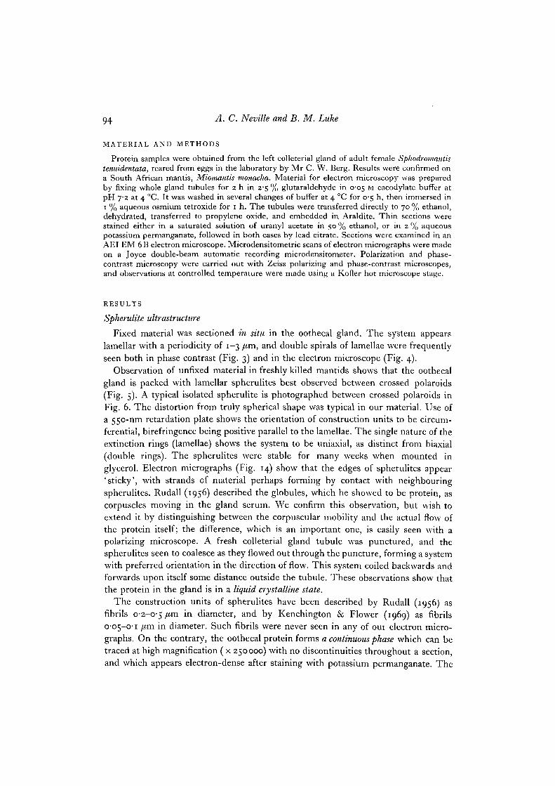
Fig. 1. Diagram illustrating the principle of helicoidal structure. Representativeplanes (drawn as circles) of anisometric and parallel construction units are drawn forevery 45 ° of rotation about the axis running through the centres of all the circles.Orientation of the units is shown by arrows. Between crossed polaroids the units whichlie in the plane of the page would show maximal birefringence, appearing as lamellae.Hence the lamellar period is half the true pitch of the helicoid.
The electron-density variation across such micrographs as Fig. 8 was measured witha microdensitometer. The rhythmical variation (Fig. 2 A) is consistent with the steadyrotation (theoretically sinusoidal) of the units in a helicoidal structure as in Fig. 1.Superimposed upon this are seen the troughs caused by the electron-light discon-tinuous phase (arrows in Fig. 2 A). The presence of a double spiral at the centre of aspherulite (Fig. 4) also supports a helicoidal explanation for this structure by com-parison with light-microscopical observations on cholesteric liquid crystals, and isdiscussed below. The evidence supports the hypothesis for the helicoidal nature ofcholesteric liquid crystals.
Evidence for self-assembly
Our electron micrographs show that helicoidal structure arises extracellularly(Fig. 11), no parabolic patterns appearing in the cells. Extensive areas of parabolicpatterning are restricted to the lumen of the gland, occurring at a distance of 3 /tmfrom the apical border of the gland cells and 1 /tm from the luminal cells in which the

9.6 A. C. Neville and B. M. Luke
gland cells are embedded. The intervening space is filled with the so-called serum(Fig. 13). The amount of secretion present within the end-apparatus of the gland cellsis insufficient to enable us to determine whether it is already helicoidal. Since thereare no discontinuities over extensive volumes of the final spherulites, the productsextruding from the gland cells must be capable of assembling on to the materialwhich has already been secreted. Thus the formation of helicoidal architecture occursby an extracellular self-assembly process in the gland lumen.
180° 360°
1 /im
Fig. 2. Microdensitometric scans of electron-density variation across electron-micro-scope plates of (A) helicoidal mantis oothecal protein like that shown in Fig. 8, and(B) the same after gel formation at 55 °C, as shown in Fig. 9. The density variation seenin A varies with lamellar periodicity, reflecting the helicoidal rotation of componentunits in the densely stained matrix. This variation had disappeared in B, but theelectron-light component (arrows) is still present. Angle of rotation of helicoid isindicated in A, with 0° and 3600 lying in the plane of the page.
Lamellar surface structures formed in vitro by self-assembly on a glass coverslipor on the surface of a glass tube, when oothecal protein was left in locust saline forseveral days. These surface structures (Fig. 7) closely resemble those formed on glasssurfaces by solutions of poly-y-benzyl D-glutamate and poly-y-benzyl L-glutamate indioxan (Robinson, 1958). They provide further evidence for the self-assemblingproperties of mantis oothecal protein.
Gel formation
When extracted oothecal protein is heated on a microscope slide with a Kofler hotstage, during continuous observation between crossed polaroids, a dramatic change isseen at a critical temperature of 55 °C. The previously flowing and birefringent liquidcrystalline phase abruptly changes into a static and isotropic gel. Lowering thetemperature showed the change to be irreversible. The system has changed in physicalstate from a liquid crystal to a hydrated rubber-like gel of low tensile strength, whichdevelops cracks on deformation, and shows reversible strain birefringence when

Protein liquid crystal from a biological system 97
stresses lower than that causing tensile failure are applied. Identical results wereobtained with protein from both species of mantid tested.
The above procedure was repeated and the resulting gel fixed for electron micro-scopy. Thin sections showed that the helicoidal pattern had disappeared, leaving arandom matrix with the electron-light discontinuous phase still present (Fig. 9).(Gelling prior to fixation resulted in harder material causing the scratches in Fig. 9.)Microdensitometiic scanning confirmed the abolition of the rhythmical variation inelectron density, but the electron-light component was still represented by troughs(arrows, Fig. 2B).
Cytology
The general histological appearance of the secretory cells at the electron-microscopelevel has been described for the closely similar cockroach left colleterial gland in apioneer paper on insect gland cell ultrastructure (Mercer & Brunet, 1959). Theequivalent details of the mantid left colleterial gland have been given by Kenchington& Flower (1969). Whilst confirming the fundamentals of these descriptions, we wishto add the following details.
Gland cells. Prior to secretion, the oothecal protein occurs as vesicles in the cells(Fig. 12), which, by contrast with normal epidermal cells secreting cuticle, are veryrich in rough endoplasmic reticulum, suggesting that the protein is synthesized forexport in the gland cells themselves. (We note this feature because epidermal cells ingeneral may obtain some of the proteins which they subsequently secrete from else-where in the body via the haemocoel. This may be deduced from the electrophoresisresults of Fox & Mills, 1969.) The microvillate end-apparatus through which secre-tion of the mantis oothecal protein occurs is typical of insect gland cells in general(Mercer & Brunet, 1959; Gupta & Smith, 1969). As in the colleterial glands ofSaturniid moths (Berry, 1968), several of the mantid colleterial gland cells containcytolysomes with myelin-like figures.
Lumen cells. Colleterial gland cells are set in an epithelium otherwise composed ofso-called ' chitinogenous' cells. Whilst agreeing with this general layout, we disagreewith previous workers (Mercer & Brunet, 1959; Kenchington & Flower, 1969) in thenaming of these cells. The structure bordering the lumen of the organ resembles anepicuticle in ultrastructure and thickness (Figs. 10, 11, 13). Since epicuticle does notcontain chitin, we therefore propose to call the cells responsible for this structure'lumen cells'. In the mantids they contain numerous microtubules oriented parallelto the surface of the lumen. This has previously been noted by Berry (1968) inSaturniid moth colleterial glands.
DISCUSSION
Helicoidal ultrastructure
The above ultrastructural evidence supports the theory, based upon optical pro-perties, of the helicoidal structure of cholesteric liquid crystals (Friedel, 1922). Themantis oothecal protein appears potentially useful for building chemical and archi-
7 CEL 8

98 A. C. Neville and B. M. Luke
tectural models of cuticle, and for experiments on helicoid self-assembly. It emergesthat protein can form a helicoid in the absence of chitin (Hackman & Goldberg,i960, have shown that chitin is absent from the oothecal protein of a mantid, Orthoderaministralis), but this does not necessarily imply that protein is the prime factorgoverning assembly of helicoids in arthropod cuticle.
We have suggested that the helicoidal structure of arthropod cuticle in generalmight arise by subsequent stabilization of a self-assembling cholesteric liquid crystal-line deposition zone present as a thin region next to the cuticle-secreting epidermalcells (Neville & Luke, 19690,6; Neville & Caveney, 1969). Electron-microscopeimages show evidence of helicoidal structure in this deposition zone. The fact thatwe have shown above that it is possible to fix and visualize a cholesteric liquid crystal-line system in the electron microscope, does not detract from the hypothesis.
Significance of the double spiral pattern
Kenchington & Flower (1969) briefly mention the similarity between the doublespiral seen in light-microscope preparations of mantis spherulites, with that ofpolypeptide spherulites (Robinson, 1966). We wish to extend the comparison toinclude the double spiral patterns in transfer RNA spherulites (Wilkins, 1963), andto stress that such spiral patterns arise because of geometrical reasons. The mathe-matical derivation of double spirals in helicoidal spherulites is discussed by Pryce &Frank in Robinson, Ward & Beevers (1958). They show that sections through ahelicoidal spherulite will always contain a double spiral pattern except in the planeof the single radial line of disinclination, which is also a geometrical consequence oftheir construction. Double spirals have also been seen in sections of tubercles in crabcuticle and their origin is beautifully demonstrated in diagrams by Bouligand (1965).They also occur in sections of corneal lenses of some arthropod eyes (Horridge, 1969;S. Caveney, unpublished), where they arise from a hemisphere of cuticle withhelicoidal construction.
Spherulite construction units
With regard to the units from which the helicoids are built, we disagree with theinterpretation of Kenchington & Flower (1969). They described the units as electron-densely staining 'fibrils' arranged at angles of 18 ° to each other, for which they havespecifically constructed a perspex model. By contrast, we regard the constructionunits as unresolvable by electron microscopy of thin sections. A more likely candidatefor a unit could be the twin-coiled a-helices postulated by Rudall (1956) on thebasis of X-ray diffraction of artificial fibres pulled out from the viscous protein in thegland. It is significant that the building units of other helicoidal systems are alsoasymmetrical (cholesteryl derivatives), often with helical components (transfer RNA)or even totally helical (synthetic polypeptides and DNA: Robinson, 1961).
We find no discontinuities in the electron densely-staining matrix, and this issupported by the gelling experiments in which a continuous isotropic gel was formed.

Protein liquid crystal from a biological system 99
One common feature of all the helicoidal systems we have worked with is that theyare capable of showing a very wide range of pitch (see Fig. 1, p. 95), reflecting a widevariation of angle between successive planes of units.
Gel formation
The formation of a gel from mantis oothecal protein clearly differs from theshrinkage of collagen, since the latter phenomenon is readily reversible. A closeranalogy is the formation of gelatin from collagen (Harkness, 1961), which involvesthe irreversible denaturation of the secondary structure, and the formation of a newrandom configuration with higher entropy. We appear to have converted an orderedliquid crystal into a random gel, perhaps by the breakdown of the H-bonds in thesecondary structure of the original units, followed by the formation of a new randomsecondary structure. The breaking of the original H-bonding (for instance in thetwin a-helices postulated by Rudall, 1956), could explain the dramatically suddendisappearance of the helicoidal architecture.
We wish to thank Mr C. W. Berg for rearing the mantids and Mr S. Caveney for obtainingMiomantis monacha. We thank Prof. J. W. S. Pringle, F.R.S., for his comments on the manu-script. Finally, our grateful thanks to the Agricultural Research Council for full financialsupport.
REFERENCES
BERRY, S. J. (1968). The fine structure of the colleterial glands of Hyalophora cecropia (Lepi-doptera). J. Morpli. 125, 259-280.
BOULIGAND, Y. (1965). Sur une architecture torsadde r^pandue dans de nombreuses cuticulesd'arthropodes. C. r. hebd. Se'anc. Acad. Sci., Paris 261, 3665-3668.
Fox, F. R. & MILLS, R. R. (1969). Changes in haemolymph and cuticle proteins during themoulting process in the American cockroach. Comp. Biochem. Physiol. 29, 1187-1195.
FRIEDEL, M. G. (1922). Les etats mesomorphes de la matiere. Annls Phys. 18, 273-474.GUPTA, B. L. & SMITH, D. S. (1969). Fine structural organization of the spermatheca in the
cockroach, Periplaneta americana. Tissue & Cell 1, 295-324.HACKMAN, R. H. & GOLDBERG, M. (i960). Composition of the oothecae of three Orthoptera.
J. Insect Physiol. 5, 73-78.HARKNESS, R. D. (1961). Biological functions of collagen. Biol. Rev. 36, 399-463.HORRIDGE, G. A. (1969). The eye of the firefly, Photuris. Proc. R. Soc. B 171, 445-463.KENCHINCTON, W. (1965). The Structure and Function of Protein Secreting and Associated
Glands in Insects. Ph.D. Thesis, University of Leeds.KENCHINCTON, W. & FLOWER, N. E. (1969). Studies on insect fibrous proteins: the structural
protein of the ootheca in the praying mantis, Sphodromantis centralis Rehn. J. Microscopy89, 263-281.
MERCER, E. H. & BRUNET, P. C. J. (1959). The electron microscopy of the left colleterial glandof the cockroach. J. biophys. biochem. Cytol. 5, 257-262.
NEVILLE, A. C. & CAVENEY, S. (1969). Scarabaeid beetle exocuticle as an optical analogue ofcholesteric liquid crystals. Biol. Rev. 44, 531-562.
NEVILLE, A. C. & LUKE, B. M. (1969a). Molecular architecture of adult locust cuticle at theelectron microscope level. Tissue & Cell 1, 355-366.
NEVILLE, A. C. & LUKE, B. M. (19696). A two-system model for chitin-protein complexes ininsect cuticles. Tissue & Cell 1, 689-707.
NEVILLE, A. C , THOMAS, M. G. & ZELAZNY, B. (1969). Pore canal shape related to moleculararchitecture of arthropod cuticle. Tissue & Cell 1, 183-200.
7-2

ioo A. C. Neville and B. M. Luke
PRYCE, M. H. L. & FRANK, F. C. (1958). The spherulitic texture. Appendix to Robinson, C,Ward, J. C. & Beevers, R. B. (1958). Liquid crystalline structure in polypeptide solutions.Discuss. Faraday Soc. 25, 29-42.
ROBINSON, C. (1958). Surface structures in liquid crystals. In Surface Phenomena in Chemistryand Biology. London: Pergamon Press.
ROBINSON, C. (1961). Liquid-crystalline structures in polypeptide solutions. Tetrahedron13, 219-234.
ROBINSON, C. (1966). The cholesteric phase in polypeptide solutions and biological structures.Molecular Crystals 1, 467-494.
RUDALL, K. M. (1956). Protein ribbons and sheets. Led. scient. Basis Med. 5, 217-230.WILKINS, M. H. F. (1963). X-ray diffraction studies on the molecular configuration of nucleic
acids. In Aspects of Protein Structure (ed. G. N. Ramachandran), pp. 23-27. London:Academic Press.
{Received id June 1970)
Fig. 3. Phase-contrast micrograph of i-/tm section through oothecal protein fixedin situ in the colleterial gland of M. monacha. The lamellation is due to helicoidalstructure.Fig. 4. Electron micrograph through centre of liquid crystalline spherulite of oothecalgland protein of S. tenuidentata, fixed in situ in colleterial gland. The lamellae,between which runs a parabolic pattern of obliquely sectioned electron-light com-ponents, themselves appear to coil round in a double spiral typical of the centre of ahelicoidal spherulite. x 6500.

Protein liquid crystal from a biological system 1 0 1

102 A. C. Neville and B. M. Luke
Fig. 5. Photomicrograph of spherulites of M. monacha oothecal protein in situbetween crossed polaroids.Fig. 6. As for Fig. 5 but a single spherulite extracted.Fig. 7. Phase-contrast micrograph of M. monacha oothecal protein surface structureswhich have reassembled on a glass surface in vitro.

Protein liquid crystal from a biological system 103
I
104 A. C. Neville and B. M. Luke
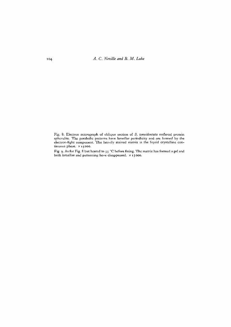
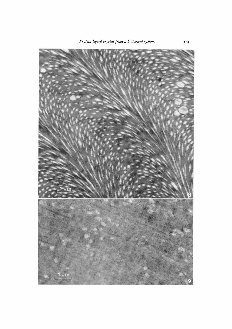
Fig. 8. Electron micrograph of oblique section of 5. tenuidentata oothecal proteinspherulite. The parabolic patterns have lamellar periodicity and are formed by theelectron-light component. The heavily stained matrix is the liquid crystalline con-tinuous phase, x 15000.Fig. 9. As for Fig. 8 but heated to 55 °C before fixing. The matrix has formed a gel andboth lamellae and patterning have disappeared, x 15000.
Protein liquid crystal from a biological system 105
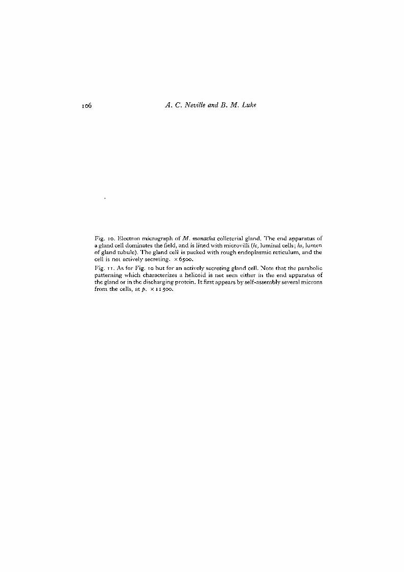
io6 A. C. Neville and B. M. Luke
Fig. io. Electron micrograph of M. monacha colleterial gland. The end apparatus ofa gland cell dominates the field, and is lined with microvilli (/c, luminal cells; hi, lumenof gland tubule). The gland cell is packed with rough endoplasniic reticulum, and thecell is not actively secreting, x 6500.Fig. 11. As for Fig. 10 but for an actively secreting gland cell. Note that the parabolicpatterning which characterizes a helicoid is not seen either in the end apparatus ofthe gland or in the discharging protein. It first appears by self-assembly several micronsfrom the cells, at p. x 11 500.

Protein liquid crystal from a biological system 107
11

io8 A.C. Neville and B. M. Luke
Fig. 12. Electron micrograph of colleterial gland cell cytoplasm of M. monacha. Muchribosome-studded endoplasmic reticulum is present, correlated with the synthesis oflarge volumes of oothecal protein for export. Vesicles, v, are thought to contain theprotein en route to the end apparatus, ea. x 19000.Fig. 13. Electron micrograph of inside edge of wall of colleterial gland from M. monacha.Note the absence of a deposition zone by contrast with insect cuticle deposition,(e, epicuticle lining of luminal cell; p, parabolic patterning in liquid crystalline proteinwithin lumen. Arrows indicate convoluted cell membrane separating luminal cell,Ic (containing microtubules), from gland cell, gc (containing rough endoplasmic reti-culum).) x 27000.Fig. 14. Electron micrograph of the edges of oothecal protein spherulites fromM. monacha. x 8500.

Protein liquid crystal from a biological system 109
12
13


![Large Colloids in Cholesteric Liquid Crystals · Large Colloids in Cholesteric Liquid Crystals 1499 the rotation of molecules by shear flow [3]. The right hand side ensures the relaxation](https://img.pdfslide.net/doc/110x75/5e54bbc32d2cd701df71bc52/large-colloids-in-cholesteric-liquid-crystals-large-colloids-in-cholesteric-liquid.jpg)