Embed Size (px)
Citation preview
A cancer cell metalloprotease triadregulates the basement membranetransmigration programKevin Hotary, Xiao-Yan Li, Edward Allen, Susan L. Stevens, and Stephen J. Weiss1
Division of Molecular Medicine and Genetics, Department of Internal Medicine, Life Sciences Institute, University ofMichigan, Ann Arbor, Michigan 48109, USA
Carcinoma cells initiate the metastatic cascade by inserting invasive pseudopodia through breaches in thebasement membrane (BM), a specialized barrier of cross-linked, extracellular matrix macromolecules thatunderlies epithelial cells and ensheaths blood vessels. While BM invasion is the sine qua non of the malignantphenotype, the molecular programs that underlie this process remain undefined. To identify genes that directBM remodeling and transmigration, we coupled high-resolution electron microscopy with an ex vivo model ofinvasion that phenocopies the major steps observed during the transition of carcinoma in situ to frankmalignancy. Herein, a triad of membrane-anchored proteases, termed membrane type-1, type-2, and type-3metalloproteinases, are identified as the triggering agents that independently confer cancer cells with theability to proteolytically efface the BM scaffolding, initiate the assembly of invasive pseudopodia, andpropagate transmigration. These studies characterize the first series of gene products capable of orchestratingthe entire BM remodeling program that distinguishes the carcinomatous phenotype.
[Keywords: Basement membrane; type IV collagen; matrix metalloproteinases; MT-MMPs; cancer; invasion]
Supplemental material is available at http://www.genesdev.org.
Received May 24, 2006; revised version accepted August 4, 2006.
In all forms of cancer, a hallmark of the malignant pro-cess is the acquisition of an invasive phenotype that al-lows neoplastic cells to penetrate the basement mem-brane (BM), a specialized form of extracellular matrixthat underlies all epithelial cells and ensheaths bloodvessels, nerves, and muscle as well as fat (Hanahan andWeinberg 2000; Kalluri 2003). Comprised of >50 distinctmacromolecular components, the BM is a thin (∼100 nmthick), but mechanically strong structure dominated by ascaffolding of cross-linked type IV collagen moleculescointertwined with a network of polymeric laminin(Kalluri 2003). Consistent with the critical role that BMsassume in regulating cell adhesion, migration, differen-tiation, and survival, developmental defects in its assem-bly or composition result in embryonically lethal phe-notypes (Poschl et al. 2004). Nonetheless, BMs regularlyundergo focal remodeling during normal growth and de-velopment in organisms ranging from Caenorhabditiselegans to mammals (Sherwood et al. 2005). While themolecular machinery that allows trafficking cells to dis-assemble and transmigrate BMs under controlled cir-cumstances remains undefined, current evidence sug-gests that similar, if not identical, gene programs are
inappropriately marshalled by malignant cells in cancer(Friedl and Wolf 2003; Sherwood et al. 2005).
In vivo, BMs are stabilized by a structurally complexmix of covalent and noncovalent forces that coalesce thenetwork of interacting molecules into a molecular sievethat assumes an effective pore size on the order of 50 nm(Abrams et al. 2000; Kalluri 2003; Than et al. 2005; Va-nacore et al. 2005). As such, it has long been assumedthat cancer cells would, by necessity, either mobilizetheir own proteolytic enzymes or recruit them from ac-cessory cell populations (e.g., fibroblasts or leukocytes)in an effort to degrade key BM components as a preludeto invasion (Friedl and Wolf 2003). Indeed, in the in vivosetting, malignant cells can often be found in the act ofinserting invasive pseudopodial-like extensions throughthe BM wall while establishing adhesive interactionswith underlying stromal structures (Friedl and Wolf2003). However, no single protease, or set of proteases,has yet been identified that can confer neoplastic cellswith the ability to degrade or traverse intact BMs. Con-sistent with this theme, more recent studies have con-cluded that neoplastic cells may adopt amoeboid pheno-types to traffic across BM barriers by exerting physicaland mechanical forces that distort matrix architectureby nonproteolytic means alone (Friedl and Wolf 2003;Evan-Ram and Yamada 2005).
In part, efforts to characterize the mechanisms direct-
1Corresponding author.E-MAIL [email protected]; FAX (734) 647-7950.Article published online ahead of print. Article and publication date areonline at http://www.genesdev.org/cgi/doi/10.1101/gad.1451806.
GENES & DEVELOPMENT 20:2673–2686 © 2006 by Cold Spring Harbor Laboratory Press ISSN 0890-9369/06; www.genesdev.org 2673
Cold Spring Harbor Laboratory Press on June 15, 2018 - Published by genesdev.cshlp.orgDownloaded from Cold Spring Harbor Laboratory Press on June 15, 2018 - Published by genesdev.cshlp.orgDownloaded from Cold Spring Harbor Laboratory Press on June 15, 2018 - Published by genesdev.cshlp.orgDownloaded from
ing BM invasion have been stymied by difficulties inher-ent in recapitulating the dynamic interactions that de-fine tumor cell–matrix interactions in the in vivo set-ting. To this end, we now define an ex vivo modelwherein cancer cell-mediated BM disassembly and trans-migration can be tracked visually by high-resolutionelectron microscopic techniques. Using this system,neoplastic cells are shown to irreversibly remodel intactBM barriers while activating a cell-autonomous invasionprogram that is directed by any one of three distinct,membrane-anchored metalloproteinases. The identifica-tion of a proteolytic triad that arms human cancer cellswith the ability to transmigrate native BM barriers couldhave important implications for the design of a new gen-eration of selective, therapeutic interventions operativeat the cell–matrix interface.
Results
Cancer cell-mediated BM perforationand transmigration
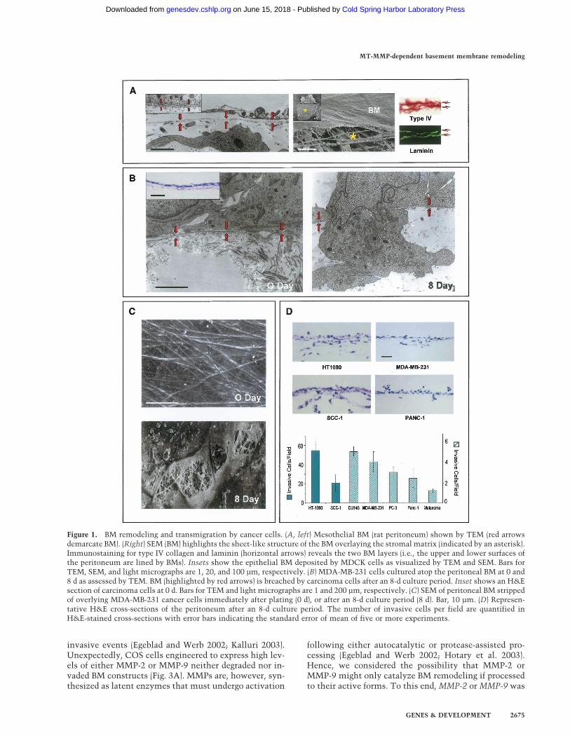
BMs recovered intact from in vivo tissues (i.e., the peri-toneum) or assembled in long-term, three-dimensionalculture by immortalized epithelial cells appear as con-tinuous and organized ∼80–100-nm-thick sheets of typeIV collagen- and laminin- rich ECM (Fig. 1A). Gentlemechanical disruption of either BM construct allows forthe direct visualization of the underlying stromal matrixthat is dominated by interstitial collagen fibrils (Fig. 1A).To determine the BM-invasive potential of human can-cer cell lines, tumor cells of either epithelial, mesenchy-mal, or neural crest origin were cultured atop denudedBMs. Following a 2-h incubation period, all tumor cellsare tightly apposed to the exposed surface of the BM andcome to lie within 100 nm or less of the underlying ma-trix (Fig. 1B). Over the course of an 8-d culture period,cancer cells begin to actively perforate the underlyingBM within 24 h by extending microfilament-rich ∼0.8-µm diameter invadopodia into the subjacent intersti-tium in a fashion that recapitulates the in vivo invasionprogram (Fig. 1B). Scanning electron micrographs of tu-mor cell-traversed BMs reveal a pockmarked landscapedecorated with excavated pits in which the cable-likestructure of the underlying stromal matrix can be ob-served (Fig. 1C). Cancer cell-mediated BM remodelingallows each of the cell lines studied access to underlyingtissues (Fig. 1D) with similar results obtained when liveperitoneal explants are used in place of the cell-free ex-plant (data not shown).
BM transmigration is drivenby a metalloproteinase-dependent process
Of the >500 proteases expressed in the mammalian ge-nome, most can be categorized as members of either theserine-, aspartyl-, cysteinyl-, or metallo-proteinase fami-lies (Puente et al. 2003). In the presence of high concen-trations of potent, broad-spectrum inhibitors used com-monly to block serine-, aspartyl-, or cysteinyl-protein-
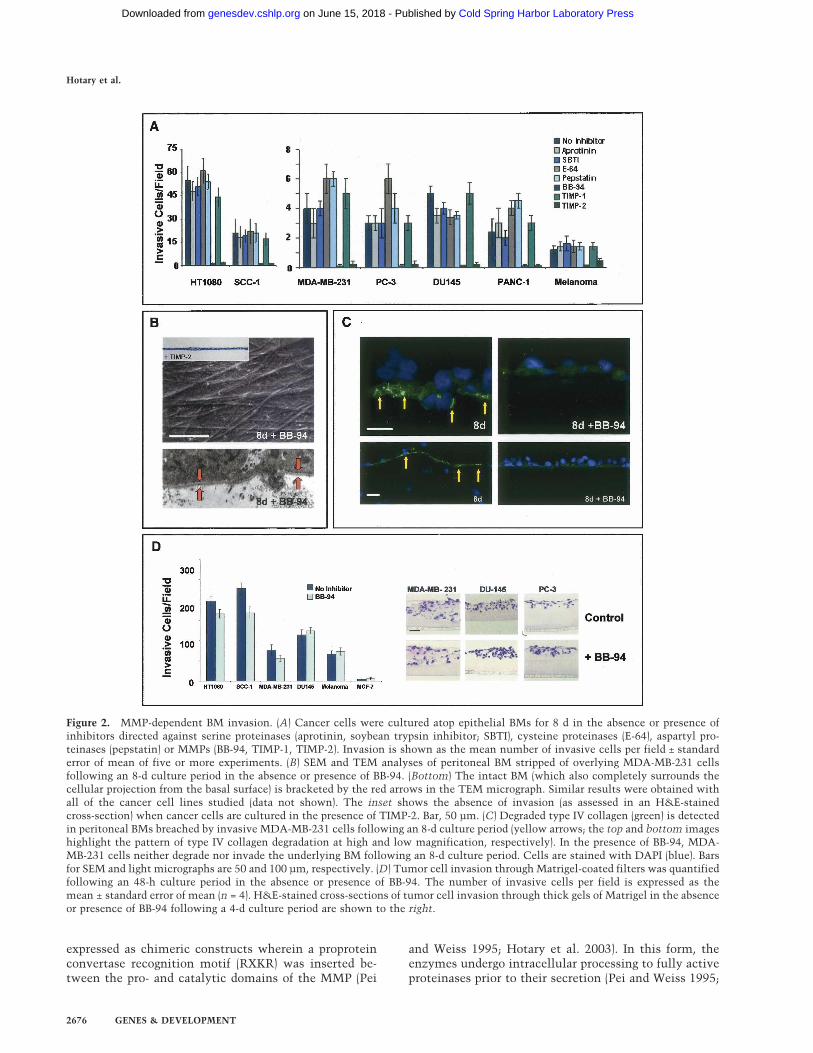
ases (Hotary et al. 2003; Filippov et al. 2005), BMinvasive activity is unaffected and all cancer cell linesexamined efficiently transmigrate the ECM barrier(Fig. 2A).
A subgroup of metalloproteinases belonging to the ma-trix metalloproteinase (MMP) gene family are uniformlyup-regulated in invasive carcinomas (Egeblad and Werb2002; Kalluri 2003). In the presence of either a peptido-mimetic MMP inhibitor (i.e., BB-94) or the endogenousMMP inhibitor, TIMP-2 (Hotary et al. 2003), cancer cellinvasion is blocked completely over an 8-d culture pe-riod (Fig. 2A) and recovered BMs are entirely defect free(Fig. 2B). Neither inhibitor affects cell proliferation orviability under these conditions (Hotary et al. 2003; Sa-beh et al. 2004). Further, whereas type IV collagen deg-radation products are immunodetected in associationwith both the perforated BM and transmigrated carci-noma cells (Xu et al. 2001), type IV collagen maintainsits native structure in the presence of BB-94 (Fig. 2C).While potential roles for MMPs in cancer cell invasionhave been largely dismissed given the paucity of signifi-cant clinical responses (Coussens et al. 2002), tumor celltransmigration across intact BMs is no longer inhibitedex vivo (data not shown) when the BB-94 concentrationis lowered 25-fold to levels similar to those obtained inthe in vivo setting (Denis and Verweij 1997). Further,though carcinoma cells have been reported to adopt anamoeboid-like phenotype in order to negotiate ECM bar-riers by nonproteolytic processes (Friedl and Wolf 2003),none of the cancer cell populations studied are able tomount BM invasive activity in the presence of broad-spectrum MMP inhibitors. Of note, however, neither BB-94 nor TIMP-2 are able to block cancer cell invasionthrough a gel-like mixture of type IV collagen, laminin,and proteoglycan (i.e., Matrigel), an artificial matrix thatdoes not assemble into the ordered, covalently cross-linked structure that characterizes the native BM (Fig.2D; Kalluri 2003; Evan-Ram and Yamada 2005). Hence,while tumor cells may not require MMP activity to tra-verse BM-like barriers whose structural characteristicsare defined by noncovalent forces, proteolytic systemsmust be engaged in order to penetrate the intact BMbarrier.
A subfamily of MT-MMPs confer BM invasive activity
The MMP family encompasses a group of >20 secreted ormembrane-anchored enzymes that alone, or in a combi-nation, can degrade a multiplicity of ECM components(Egeblad and Werb 2002). However, no MMP has beenshown to confer cells with the ability to traverse theintact BM with its multicomposite amalgam of type IVcollagen, laminin, nidogen, and complex proteoglycans.To this end, we first sought to identify individual MMPsthat might endow invasion-null COS cells with the BMdegradative activities necessary to support transmigra-tion.
Originally characterized as type IV collagenases,MMP-2 (gelatinase A) and MMP-9 (gelatinase B) havelong been postulated to play a dominant role in BM-
Hotary et al.
2674 GENES & DEVELOPMENT
Cold Spring Harbor Laboratory Press on June 15, 2018 - Published by genesdev.cshlp.orgDownloaded from
invasive events (Egeblad and Werb 2002; Kalluri 2003).Unexpectedly, COS cells engineered to express high lev-els of either MMP-2 or MMP-9 neither degraded nor in-vaded BM constructs (Fig. 3A). MMPs are, however, syn-thesized as latent enzymes that must undergo activation
following either autocatalytic or protease-assisted pro-cessing (Egeblad and Werb 2002; Hotary et al. 2003).Hence, we considered the possibility that MMP-2 orMMP-9 might only catalyze BM remodeling if processedto their active forms. To this end, MMP-2 or MMP-9 was
Figure 1. BM remodeling and transmigration by cancer cells. (A, left) Mesothelial BM (rat peritoneum) shown by TEM (red arrowsdemarcate BM). (Right) SEM (BM) highlights the sheet-like structure of the BM overlaying the stromal matrix (indicated by an asterisk).Immunostaining for type IV collagen and laminin (horizontal arrows) reveals the two BM layers (i.e., the upper and lower surfaces ofthe peritoneum are lined by BMs). Insets show the epithelial BM deposited by MDCK cells as visualized by TEM and SEM. Bars forTEM, SEM, and light micrographs are 1, 20, and 100 µm, respectively. (B) MDA-MB-231 cells cultured atop the peritoneal BM at 0 and8 d as assessed by TEM. BM (highlighted by red arrows) is breached by carcinoma cells after an 8-d culture period. Inset shows an H&Esection of carcinoma cells at 0 d. Bars for TEM and light micrographs are 1 and 200 µm, respectively. (C) SEM of peritoneal BM strippedof overlying MDA-MB-231 cancer cells immediately after plating (0 d), or after an 8-d culture period (8 d). Bar, 10 µm. (D) Represen-tative H&E cross-sections of the peritoneum after an 8-d culture period. The number of invasive cells per field are quantified inH&E-stained cross-sections with error bars indicating the standard error of mean of five or more experiments.
MT-MMP-dependent basement membrane remodeling
GENES & DEVELOPMENT 2675
Cold Spring Harbor Laboratory Press on June 15, 2018 - Published by genesdev.cshlp.orgDownloaded from
expressed as chimeric constructs wherein a proproteinconvertase recognition motif (RXKR) was inserted be-tween the pro- and catalytic domains of the MMP (Pei
and Weiss 1995; Hotary et al. 2003). In this form, theenzymes undergo intracellular processing to fully activeproteinases prior to their secretion (Pei and Weiss 1995;
Figure 2. MMP-dependent BM invasion. (A) Cancer cells were cultured atop epithelial BMs for 8 d in the absence or presence ofinhibitors directed against serine proteinases (aprotinin, soybean trypsin inhibitor; SBTI), cysteine proteinases (E-64), aspartyl pro-teinases (pepstatin) or MMPs (BB-94, TIMP-1, TIMP-2). Invasion is shown as the mean number of invasive cells per field ± standarderror of mean of five or more experiments. (B) SEM and TEM analyses of peritoneal BM stripped of overlying MDA-MB-231 cellsfollowing an 8-d culture period in the absence or presence of BB-94. (Bottom) The intact BM (which also completely surrounds thecellular projection from the basal surface) is bracketed by the red arrows in the TEM micrograph. Similar results were obtained withall of the cancer cell lines studied (data not shown). The inset shows the absence of invasion (as assessed in an H&E-stainedcross-section) when cancer cells are cultured in the presence of TIMP-2. Bar, 50 µm. (C) Degraded type IV collagen (green) is detectedin peritoneal BMs breached by invasive MDA-MB-231 cells following an 8-d culture period (yellow arrows; the top and bottom imageshighlight the pattern of type IV collagen degradation at high and low magnification, respectively). In the presence of BB-94, MDA-MB-231 cells neither degrade nor invade the underlying BM following an 8-d culture period. Cells are stained with DAPI (blue). Barsfor SEM and light micrographs are 50 and 100 µm, respectively. (D) Tumor cell invasion through Matrigel-coated filters was quantifiedfollowing an 48-h culture period in the absence or presence of BB-94. The number of invasive cells per field is expressed as themean ± standard error of mean (n = 4). H&E-stained cross-sections of tumor cell invasion through thick gels of Matrigel in the absenceor presence of BB-94 following a 4-d culture period are shown to the right.
Hotary et al.
2676 GENES & DEVELOPMENT
Cold Spring Harbor Laboratory Press on June 15, 2018 - Published by genesdev.cshlp.orgDownloaded from
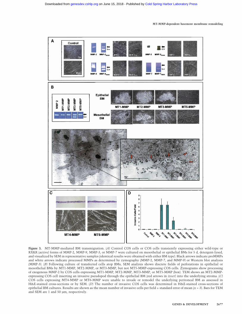
Figure 3. MT-MMP-mediated BM transmigration. (A) Control COS cells or COS cells transiently expressing either wild-type orRXKR (active) forms of MMP-2, MMP-9, MMP-3, or MMP-7 were cultured on mesothelial or epithelial BMs for 5 d, detergent-lysed,and visualized by SEM in representative samples (identical results were obtained with either BM type). Black arrows indicate proMMPsand white arrows indicate processed MMPs as determined by zymography (MMP-2, MMP-7, and MMP-9) or Western blot analysis(MMP-3). (B) Following culture of transfected cells atop BMs, SEM analysis shows discrete fields of perforations in epithelial ormesothelial BMs by MT1-MMP, MT2-MMP, or MT3-MMP, but not MT5-MMP-expressing COS cells. Zymograms show processingof exogenous MMP-2 by COS cells expressing MT1-MMP, MT2-MMP, MT3-MMP, or MT5-MMP (box). TEM shows an MT2-MMP-expressing COS cell inserting an invasive pseudopod through the epithelial BM (red arrows in inset) into the underlying stroma. (C)COS cells expressing MT4-MMP or MT6-MMP were unable to invade or remodel the underlying peritoneal BM as assessed inH&E-stained cross-sections or by SEM. (D) The number of invasive COS cells was determined in H&E-stained cross-sections ofepithelial BM cultures. Results are shown as the mean number of invasive cells per field ± standard error of mean (n = 3). Bars for TEMand SEM are 1 and 50 µm, respectively.
MT-MMP-dependent basement membrane remodeling
GENES & DEVELOPMENT 2677
Cold Spring Harbor Laboratory Press on June 15, 2018 - Published by genesdev.cshlp.orgDownloaded from
Hotary et al. 2003). Nonetheless, while COS cells trans-fected with MMP-2/RXKR or MMP-9/RXKR expressionvectors secrete high concentrations of the active protein-ases as assessed by zymography, the active enzymes areunable to support BM degradation or COS cell transmi-gration (Fig. 3A). Similarly, though indirect evidence haslinked the expression of MMP-3 (stromelysin-1), MMP-7(matrilysin), as well as MMP-11 (stromelysin-3) to BMremodeling activity (Egeblad and Werb 2002), COS cellsexpressing neither the latent nor active forms of the re-spective proteinases degrade or traverse the BM barrier(Fig. 3A). Attempts to establish a potential role forMMP-1 (collagenase-1), MMP-13 (collagenase-3), orMMP-19 in BM remodeling or invasion were likewiseunsuccessful (Supplementary Fig. 1). The inability of se-creted MMPs to support BM invasion is further sup-ported by the fact that high concentrations of TIMP-1, anendogenous inhibitor that preferentially targets secretedMMPs (Hotary et al. 2003; Overall and Kleifeld 2006),does not inhibit BM remodeling by any of the cancer celllines studied (Fig. 2A).
Unlike TIMP-1, the metalloproteinase inhibitors, BB-94 and TIMP-2, not only inhibit secreted MMPs, but alsothe subclass of type I transmembrane MMPs (i.e., MT1-MMP, MT2-MMP, MT3-MMP, and MT5-MMP) (Hotaryet al. 2003). Remarkably, COS cells expressing eitherMT1-MMP, MT2-MMP, or MT3-MMP acquire the abil-ity to both perforate and transmigrate peritoneal or epi-thelial BMs (Fig. 3B–D). Scanning electron microscopy(SEM) and transmission electron microscopy (TEM)analyses demonstrate that MT-MMP-expressing COScells actively remodel the subjacent matrices during theearly stages of invasion by inserting pseudopodia directlythrough the BM into the underlying interstitium (Fig.3B). Using high-magnification TEM, the sharply cutedges of the perforated BM can be seen to curl away fromthe body of the invasive membrane protrusions extendedby the MT-MMP-transfected COS cells (Fig. 3B, inset).COS cells expressing MT1-MMP, MT2-MMP, or MT3-MMP do not display increased motility when culturedatop gelatin-coated Transwell filters or increased two-dimensional migratory activity when cultured atop thesurface of denuded BMs (data not shown). Though MT5-MMP, as well as the glycophosphatidylinositol-anchoredMMPs, MT4-MMP and MT6-MMP, have been assignedlimited matrix-degrading activity and postulated to playa role in regulating invasive activity (Itoh and Seiki2006), none of these MT-MMP family members dis-played BM remodeling activity (Fig. 3B–D). Hence, onlyMT1-MMP, MT2-MMP, or MT3-MMP can serve as di-rect-acting proteases that are capable of dissolving theintervening BM while simultaneously inducing transmi-gration.
Structural determinants of MT-MMP-dependentBM transmigration
Following proteolytic processing, the N-terminal pro-peptide regions of the MT-MMPs are shed, and the activeproteases displayed at the cell surface (Yana and Weiss
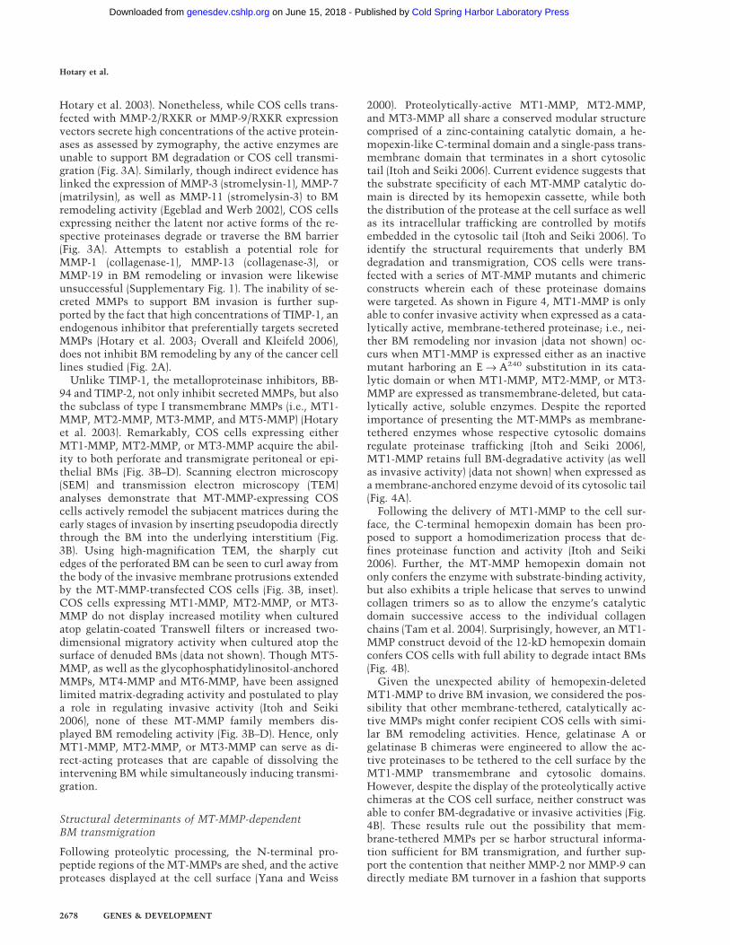
2000). Proteolytically-active MT1-MMP, MT2-MMP,and MT3-MMP all share a conserved modular structurecomprised of a zinc-containing catalytic domain, a he-mopexin-like C-terminal domain and a single-pass trans-membrane domain that terminates in a short cytosolictail (Itoh and Seiki 2006). Current evidence suggests thatthe substrate specificity of each MT-MMP catalytic do-main is directed by its hemopexin cassette, while boththe distribution of the protease at the cell surface as wellas its intracellular trafficking are controlled by motifsembedded in the cytosolic tail (Itoh and Seiki 2006). Toidentify the structural requirements that underly BMdegradation and transmigration, COS cells were trans-fected with a series of MT-MMP mutants and chimericconstructs wherein each of these proteinase domainswere targeted. As shown in Figure 4, MT1-MMP is onlyable to confer invasive activity when expressed as a cata-lytically active, membrane-tethered proteinase; i.e., nei-ther BM remodeling nor invasion (data not shown) oc-curs when MT1-MMP is expressed either as an inactivemutant harboring an E → A240 substitution in its cata-lytic domain or when MT1-MMP, MT2-MMP, or MT3-MMP are expressed as transmembrane-deleted, but cata-lytically active, soluble enzymes. Despite the reportedimportance of presenting the MT-MMPs as membrane-tethered enzymes whose respective cytosolic domainsregulate proteinase trafficking (Itoh and Seiki 2006),MT1-MMP retains full BM-degradative activity (as wellas invasive activity) (data not shown) when expressed asa membrane-anchored enzyme devoid of its cytosolic tail(Fig. 4A).
Following the delivery of MT1-MMP to the cell sur-face, the C-terminal hemopexin domain has been pro-posed to support a homodimerization process that de-fines proteinase function and activity (Itoh and Seiki2006). Further, the MT-MMP hemopexin domain notonly confers the enzyme with substrate-binding activity,but also exhibits a triple helicase that serves to unwindcollagen trimers so as to allow the enzyme’s catalyticdomain successive access to the individual collagenchains (Tam et al. 2004). Surprisingly, however, an MT1-MMP construct devoid of the 12-kD hemopexin domainconfers COS cells with full ability to degrade intact BMs(Fig. 4B).
Given the unexpected ability of hemopexin-deletedMT1-MMP to drive BM invasion, we considered the pos-sibility that other membrane-tethered, catalytically ac-tive MMPs might confer recipient COS cells with simi-lar BM remodeling activities. Hence, gelatinase A orgelatinase B chimeras were engineered to allow the ac-tive proteinases to be tethered to the cell surface by theMT1-MMP transmembrane and cytosolic domains.However, despite the display of the proteolytically activechimeras at the COS cell surface, neither construct wasable to confer BM-degradative or invasive activities (Fig.4B). These results rule out the possibility that mem-brane-tethered MMPs per se harbor structural informa-tion sufficient for BM transmigration, and further sup-port the contention that neither MMP-2 nor MMP-9 candirectly mediate BM turnover in a fashion that supports
Hotary et al.
2678 GENES & DEVELOPMENT
Cold Spring Harbor Laboratory Press on June 15, 2018 - Published by genesdev.cshlp.orgDownloaded from

the invasion program. MT-MMPs are, however, able toprocess MMP-2 to its active form, which in turn caninitiate an MMP-9 activation cascade (Itoh and Seiki2006). As such, MT1-MMP could conceivably remodelthe BM only in collaboration with MMP-2 or MMP-9. AsMMP-2 and MMP-9 not only circulate in plasma, butmay also be embedded in the BM or its surrounding tis-sues, MT1-MMP-expressing COS cells were culturedatop BMs isolated from either MMP-2−/− or MMP-9−/−
mice and suspended in the presence of the respectivenull sera. Despite the complete absence of MMP-2 orMMP-9 under these conditions, neither BM remodeling(Fig. 4C) nor invasion (data not shown) are affected.
MT-MMPs are both necessary and sufficient for BMinvasion by human cancer cells
Among the carcinoma cell lines studied, the phenotypicbehavior of the breast cancer cell lines MDA-MB-231and MCF-7 are diametrically opposed in vitro and in vivo(Fujita et al. 2003; Hotary et al. 2003). Akin to aggressivebreast carcinoma lesions in vivo (Ueno et al. 1997),MDA-MB-231 cells are highly invasive and express pre-dominately MT1-MMP and MT2-MMP (MT3-MMP isonly intermittently detected) (Fig. 5; data not shown). Incontrast, MCF-7 display a more indolent behavior invivo, and express little, if any, of the BM invasive MT-
Figure 4. Structure–function analysis ofMT-MMP-mediated BM proteolysis. (A)COS cells were transfected with MT1-MMP; catalytically inactive MT1-MMP(MT1-MMPE → A
240); cytoplasmic tail-de-leted MT1-MMP (MT1-MMPCT); orsoluble, transmembrane-deleted forms ofMT1-MMP, MT2-MMP, or MT3-MMP(�MT1-MMP, �MT2-MMP, or �MT3-MMP) and cultured atop epithelial BMs fora 5-d culture period. Cells were removedand the underlying BM was assessed bySEM. Insets show representative H&E-stained cross-sections. (B) COS cells weretransfected with an MT1-MMP constructwherein the hemopexin domain was de-leted (MT1-MMPPexDel) or, alternatively,with chimeric forms of either membrane-anchored, active MMP-2 or MMP-9 (TM-MMP-2RXKR or TM-MMP-9RXKR, respec-tively). Following a 5-d culture period, BMstructure was assessed by SEM. Insetsdemonstrate the ability of the membrane-anchored gelatinases to degrade a subja-cent bed of fluorescent gelatin. (C) Meso-thelial BMs isolated from wild-type,MMP-2−/−, or MMP-9−/− mice were visual-ized by SEM after a 5-d culture period inthe respective null serum with control orMT1-MMP-transfected COS cells. Bars forSEM and light micrographs are 10 and 100µm, respectively.
MT-MMP-dependent basement membrane remodeling
GENES & DEVELOPMENT 2679
Cold Spring Harbor Laboratory Press on June 15, 2018 - Published by genesdev.cshlp.orgDownloaded from
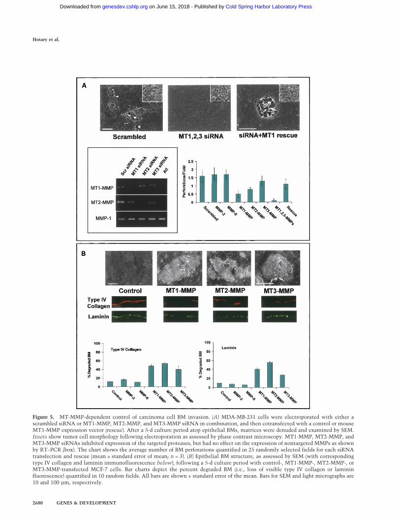
Figure 5. MT-MMP-dependent control of carcinoma cell BM invasion. (A) MDA-MB-231 cells were electroporated with either ascrambled siRNA or MT1-MMP, MT2-MMP, and MT3-MMP siRNA in combination, and then cotransfected with a control or mouseMT1-MMP expression vector (rescue). After a 5-d culture period atop epithelial BMs, matrices were denuded and examined by SEM.Insets show tumor cell morphology following electroporation as assessed by phase contrast microscopy. MT1-MMP, MT2-MMP, andMT3-MMP siRNAs inhibited expression of the targeted proteases, but had no effect on the expression of nontargeted MMPs as shownby RT–PCR (box). The chart shows the average number of BM perforations quantified in 25 randomly selected fields for each siRNAtransfection and rescue (mean ± standard error of mean; n = 3). (B) Epithelial BM structure, as assessed by SEM (with correspondingtype IV collagen and laminin immunofluorescence below), following a 5-d culture period with control-, MT1-MMP-, MT2-MMP-, orMT3-MMP-transfected MCF-7 cells. Bar charts depict the percent degraded BM (i.e., loss of visible type IV collagen or lamininfluorescence) quantified in 10 random fields. All bars are shown ± standard error of the mean. Bars for SEM and light micrographs are10 and 100 µm, respectively.
Hotary et al.
2680 GENES & DEVELOPMENT
Cold Spring Harbor Laboratory Press on June 15, 2018 - Published by genesdev.cshlp.orgDownloaded from
MMPs (Hotary et al. 2003). Hence, we sought to charac-terize (1) the impact of silencing MT1-MMP, MT2-MMP,and MT3-MMP on the BM remodeling potential of MDA-MB-231 cells and, conversely, (2) the ability of eitherMT1-MMP, MT2-MMP, or MT3-MMP to endow inva-sion-null MCF-7 cells with a BM-invasive phenotype.
Following electroporation with a scrambled small in-terfering RNA (siRNA) construct, MDA-MB-231 cells re-tain the ability to perforate and transmigrate the intactBM (Fig. 5A). Further, as predicted, siRNAs directedagainst either MMP-2 or MMP-9 are unable to interferewith BM remodeling (Fig. 5A). In contrast, while the spe-cific siRNA-mediated silencing of either MT1-MMP,MT2-MMP, or MT3-MMP did not affect MDA-MB-231cell morphology or adhesion, BM perforation and inva-sion are inhibited by as much as 70% (Fig. 5A; data notshown). These findings are not restricted to BMs recov-ered from animal species, as MT-MMPs similarly confercancer cells with the ability to perforate and traverseBMs isolated from human tissue (Supplementary Fig. 2).Moreover, when all three MT-MMPs are silenced, MDA-MB-231 cells (as well as HT-1080 cells) (data not shown)are completely unable to either remodel or transmigratethe underlying BM (Fig. 5A). Following re-expression of amouse MT1-MMP ortholog that escapes siRNA targetingdirected against the human proteinases, BM remodelingand transmigration activities are reconstituted in MDA-MB-231 cells that no longer express their endogenouscomplement of human MT1-MMP, MT2-MMP, or MT3-MMP (Fig. 5A).
In contradistinction to MDA-MB-231 cancer cells, theinvasive activity of MCF-7 cells is normally held incheck by estrogen receptor-regulated transcriptional co-repressors (Fujita et al. 2003). To finally determinewhether regulatory checkpoints might be bypassed bydownstream-acting MMPs, MCF-7 cells were transfectedtransiently with either MT1-MMP, MT2-MMP, or MT3-MMP expression vectors and BM remodeling activitymonitored. As expected, control MCF-7 cells are com-pletely unable to traverse (0 ± 0 invasive cells/field;mean ± standard error of mean, n = 6) or remodel the un-derlying BM as assessed by SEM (Fig. 5B). Importantly,MT1-MMP, MT2-MMP, or MT3-MMP were each able toconfer MCF-7 cells with the ability to perforate andtransmigrate the BM (Fig. 5B; Supplementary Fig. 3).Hence, MT1-MMP, MT2-MMP, and MT3-MMP are notonly necessary, but sufficient for driving cancer cell-me-diated BM remodeling and invasion.
Discussion
As tumor cells undergo the transition from a benign tocancerous state, gene programs are accessed that armneoplastic cells with the ability to perforate abuttingBMs (Hanahan and Weinberg 2000). While BMs compriseonly a small part of the overall ECM mass, the cova-lently cross-linked, interlocking network of type IV col-lagen and associated macromolecules serves as the majorstructural barrier to advancing tumor cells (Kalluri
2003). Indeed, within the primary neoplastic nest, cancercells confined by an as-yet-intact BM—i.e., the so-calledcarcinoma in situ—harbor a favorable clinical prognosis.In contrast, cancer cells that acquire invasive activityand perforate the intervening BM portend a more direoutcome (Hanahan and Weinberg 2000). Despite the factthat BM transmigration serves as a rate-limiting step inthe mobilization of the malignant phenotype, the mo-lecular machinery underlying this specialized form ofECM remodeling has remained undefined (Sherwood2006). Herein, we identify a triad of membrane-anchoredmetalloproteases that are embedded with all of the struc-tural information necessary to directly confer humancancer cells with the ability to proteolyze, perforate, andtransmigrate BM barriers.
BM remodeling: a MMP-dependent process
To date, multiple classes of proteolytic enzymes havebeen proposed to act as key participants in BM transmi-gration (e.g., Mignatti et al. 1986; Kalluri 2003). How-ever, studies implicating serine, cysteine, or aspartyl pro-teinases have relied almost entirely on (1) the use of ar-tificial BM constructs that do not recapitulate accuratelythe structural characteristics of the in vivo or ex vivo-assembled matrix or (2) in vivo systems that fail to di-rectly track the dynamic interactions that occur betweenadvancing cancer cells and the surrounding BM (Evan-Ram and Yamada 2005). While acellular BM sheets havebeen used periodically in earlier studies (e.g., Mignatti etal. 1986), these constructs frequently contain pores thatcan permit transmigration to proceed independently ofproteolysis (Aplin et al. 1985; Howat et al. 2001). In oursystem, we developed an ex vivo model of invasion,wherein cancer cell lines are held in juxtaposition withintact BMs in long-term culture, and changes in BM ar-chitecture evaluated directly. Under these conditions,neoplastic cells proteolytically remodel the subjacentnetworks of BM macromolecules, assemble and insertpseudopodia through the associated perforations, andtransmigrate the BM defects in a fashion that recapitu-lates each of the major steps involved in cancer cell in-vasion in vivo (Sherwood 2006). Further, we find thatcancer cell-mediated BM remodeling and invasion pro-ceed via a MMP-dependent process. At a glance, theseobservations appear to contradict at least three majorbodies of literature. First, in clinical trials, MMP inhibi-tors have failed to exert the predicted beneficial effects(Coussens et al. 2002; Overall and Kleifeld 2006). Theseresults have lent strength to the contention that themultiplicity of proteinases operative in cancer cells pre-clude their reliance on any single proteolytic system(Coussens et al. 2002; Egeblad and Werb 2002; Overalland Kleifeld 2006). Interestingly, however, our data sug-gest a more trivial explanation for the apparent failureof MMP inhibitor-based interventions—i.e., the peakplasma concentrations of synthetic inhibitors reached inthe clinical setting (e.g., Denis and Verweij 1997) fall farbelow those needed to quench critical proteolytic eventsthat occur within the sequestered microenvironments
MT-MMP-dependent basement membrane remodeling
GENES & DEVELOPMENT 2681
Cold Spring Harbor Laboratory Press on June 15, 2018 - Published by genesdev.cshlp.orgDownloaded from

that exist at the tightly apposed, tumor cell–BM inter-face. Second, other studies have documented roles forother proteolytic systems in tumorigenesis, particularlyin the in vivo setting (e.g., Gocheva et al. 2005; Laurent-Matha et al. 2005). However, caution need be exercisedin assigning a given proteinase’s role to the direct regu-lation of invasive activity. Indeed, an increasing body ofwork has demonstrated that proteinases indirectly affectcell behavior by generating chemokines, motility, andgrowth factors or regulating cell–cell and cell–matrix ad-hesive interactions (Egeblad and Werb 2002; Radisky etal. 2005; Itoh and Seiki 2006). In our ex vivo studies,potent inhibitors previously demonstrated to effectivelytarget serine, cysteine, or aspartyl proteinases in intactcell systems (e.g., Rosenthal et al. 1998; Filippov et al.2003, 2005) fail to affect cancer cell–BM interactions ortransmigration. Third, recent reports have stressed theability of tumor cells to adopt an amoeboid phenotype inorder to negotiate ECM barriers by protease-independentschemes (Friedl and Wolf 2003). However, such conclu-sions were reached using reconstituted BM extracts (i.e.,Matrigel) as the barrier construct. In contrast to authen-tic BMs, wherein structural integrity is assigned to a typeIV collagen polymer that is cross-linked by intermolecu-lar disulfide, aldimine, and hydroxylysine–methioninebonds, the macromolecular network found in Matrigel isdominated by noncovalent forces alone (Evan-Ram andYamada 2005; Than et al. 2005; Vanacore et al. 2005).Apparently, proteinase-independent schemes can onlybe mobilized under conditions in which BM cross-linkshave been disrupted. Though it is conceivable that typeIV collagen cross-links may be dissociated reversiblyduring invasive events (e.g., intermolecular disulfidebonds arrangements can be reshuffled by protein disul-fide isomerases), none of the eight cancer cells we exam-ined traverse intact BMs without mobilizing MMP ac-tivity.
An MT-MMP triad directs BM proteolysis
In human cancers, a diverse array of human MMP familymembers have been found to be expressed within themass of advancing tumor tissue (Overall and Kleifeld2006). Yet, the role that specific MMPs play in BM re-modeling has remained unresolved. Presumably, to in-vest a given cell population with a BM-remodeling ac-tivity, a proteolytic activity must be envisioned that canefface the >50 matrix molecules that are noncovalentlyassociated with a scaffolding of cross-linked type IV col-lagen. In turn, the type IV polymer must be cleaved in aproductive fashion that allows adhesive ligands to eitherbe retained, or newly generated, which then serve to sup-port the forward propulsive movement of the invadingcell. Consistent with these multiple requirements fortransmigration, MMP family members display omnivo-rous, ECM-degrading activities (Egeblad and Werb 2002;Overall and Kleifeld 2006). Further, the type IV collagentriple helix itself is interrupted by no fewer than 22 pro-tease-sensitive, nonhelical domains (Kalluri 2003). Assuch, the previously reported abilities of secreted
MMPs to hydrolyze soluble type IV collagen under cell-free conditions would seem to support the contentionthat these enzymes could play a direct role in BM inva-sion. However, in contrast to the isolated type IV colla-gen molecules that are standardly analyzed as solution-phase substrate targets, the cross-linked network of typeIV collagen heterotrimers encountered in situ assumes afar more stable and protease-resistant conformation (Lin-senmayer et al. 1984; Eble et al. 1996). Indeed, COS cellsengineered to express high concentrations of variousmembers of the secreted MMP family that have beenlinked indirectly to BM remodeling events such as,MMP-2, MMP-3, MMP-9, MMP-11, or MMP-13 did notacquire BM-invasive activity (Hanahan and Weinberg2000; Kalluri 2003; Overall and Kleifeld 2006). Given thebroad substrate repertoire of these MMPs, it seems un-likely that no BM-associated components were cleaved(e.g., Mott et al. 1997). Nevertheless, these MMPs wereunable to remodel the BM to a degree where focal defectsmaterialized or invasive activity was promoted. Whilecontroversial, these findings are entirely consistent withan increasing body of work demonstrating that mice har-boring inactivating mutations in secreted MMPs exhibitlargely unaffected invasive phenotypes (e.g., Baluk et al.2004; McCawley et al. 2004; Hartenstein et al. 2006).Nevertheless, in contrast to the inability of the secretedMMPs to support BM remodeling, three members of themembrane-anchored subfamily of MMPs have beenidentified that can invest host cells with the ability totraverse BMs in a manner that phenocopies the behaviorof invasive cancer cell populations. Despite the distinctstructure of each of their respective catalytic domains(Itoh and Seiki 2006; Overall and Kleifeld 2006), MT1-MMP, MT2-MMP, and MT3-MMP each display indistin-guishable BM remodeling and proinvasive behaviors.Like other MMP family members, MT-MMPs can de-grade a variety of BM-associated macromolecules (Itohand Seiki 2006). Though MT1-MMP, MT2-MMP, orMT3-MMP have not been reported previously to degradeisolated type IV collagen, the ability of each of theseproteinases to completely efface the BM wall indicatesthat protease-sensitive sites are exposed in the type IVcollagen network in situ. Importantly, these activitiesare not generally shared by other MT-MMP family mem-bers (i.e., MT4-MMP, MT5-MMP, or 6-MMP) or theirartificially engineered counterparts; e.g., membrane-an-chored forms of MMP-2 or MMP-9. Apparently, the cata-lytic domains of MT1-MMP, MT2-MMP, and MT3-MMP have been designed specifically to accommodateeach of the major structural components required for BMremodeling. Interestingly, studies of BM removal duringanchor cell invasion in C. elegans have implicated anMT-MMP-like, GPI-anchored enzyme that potentiallysubserves functions similar to those we describe for can-cer cell MT-MMPs (Sherwood et al. 2005). Though si-lencing this specific metalloprotease exerted little effecton anchor cell BM invasion, C. elegans express at leastthree MT-MMP orthologs and may, like humans, use acomplement of these enzymes to drive the invasion pro-gram (Wada et al. 1998).
Hotary et al.
2682 GENES & DEVELOPMENT
Cold Spring Harbor Laboratory Press on June 15, 2018 - Published by genesdev.cshlp.orgDownloaded from

MT-MMPs as molecular motors
By fusing proteolytic activity to the cell surface, MT1-MMP, MT2-MMP, and MT3-MMP are ideally situated toensure that high concentrations of the active proteaseare apposed directly to the underlying substrate. Indeed,BM remodeling is no longer detected when MT-MMPsare expressed in a transmembrane-deleted, secreted formrather than in their wild-type, membrane-tethered for-mat. Presumably, the loss of subjacent proteolytic activ-ity reflects the expected decrease in the effective en-zyme:substrate ratio at the cell–BM interface. Moreover,in the absence of membrane tethering, active MT-MMPswould diffuse away from the cell surface and fall prey toinhibition by serum-borne antiproteinases (Weiss 1989).
While membrane tethering is critical to BM-remodel-ing activity, our findings stand in contrast to recent stud-ies that have suggested that the MT-MMP cytoplasmicdomain plays a necessary role in both directing the pro-tease to the cell–substratum interface and transducingintracellular signaling cascades (for review, see Itoh andSeiki 2006). As demonstrated, the tail-deleted MT-MMPmutants retain full degradative activity during BM re-modeling, trigger the assembly of invasive pseudopod-like structures at the cell surface, and propagate trans-migration as well. Likewise, while MT1-MMP has beenreported to induce rac-dependent motility independentof its proteolytic activity (Cao et al. 2004), we find thatcatalytically inactive MT1-MMP is unable to supportcell trafficking across BMs. Interestingly, a dual require-ment for membrane-anchoring and proteolytic activity isconsistent with reports that MMPs can “walk” over amatrix-coated substratum via a biased diffusion process,whereby the protease binds and then cleaves substratemolecules (Saffarian et al. 2004). As enzyme-type IV col-lagen-binding interactions are likely weakened followingproteolysis and the consequent thermal denaturation ofthe type IV collagen trimers, backward diffusion acrossthe denatured substratum is prohibited. In this scenario,the membrane-anchored protease may “pull” or guidethe cell forward by forming successive adhesive interac-tions with uncleaved substrate. We anticipated that thisactive diffusion process might require an intact MT-MMP hemopexin domain, as both triple helicase and col-lagen-binding activities have been associated with thisC-terminal portion of the protease (Cao et al. 2004; Tamet al. 2004). However, BM proteolytic, and invasive ac-tivities are retained in the hemopexin-deleted enzyme.Nonetheless, as the MT1-MMP catalytic domain maydisplay both hydrolytic and helicase activities (Pelmanet al. 2005), our results support a model wherein thetruncated, but catalytically active protease, contains suf-ficient structural information to effectively drive the BMremodeling and transmigration programs.
MT-MMPs in cancer
In normal tissues, the BM plays a key role in regulatingcell adhesion, migration, differentiation, and growth(Kalluri 2003). Cancer cell-dependent proteolysis of BM
components not only serves to disrupt these regulatoryinteractions, but also allows for the generation of a widerange of bioactive ECM fragments that can participate intumorigenesis by affecting events ranging from tumorcell motility to angiogenesis (Egeblad and Werb 2002;Kalluri 2003). Further, as migrating cancer cells cross thedegraded BM and gain access to interstitial matrix com-ponents, new ECM–receptor interactions are establishedthat activate gene programs that accelerate the loss ofdifferentiated phenotype (Brabletz et al. 2004). As MT1-MMP, MT2-MMP, and MT3-MMP are widely expressedin a wide range of human cancer cells—both at primaryand micrometastatic sites (Klein et al. 2002; Itoh andSeiki 2006)—we propose that this MMP triad is purpose-fully mobilized to initiate BM remodeling and drive theinvasive phenotype in vivo. While we have used MCF-7and MDA-MB-231 breast-cancer cells as “prototypical”carcinomas, it is important to note that each of the sevencancer cell lines used in our study rely on TIMP-2-sen-sitive MT-MMPs to negotiate authentic BM barriers.Though increased attention has focused on the ability ofstroma or leukocyte-derived proteases to participate intumor progression (Egeblad and Werb 2002), our findingsdemonstrate that the expression of either MT1-MMP,MT2-MMP, or MT3-MMP is sufficient to confer tumorcells with the ability to dismantle and transmigrate BMsindependently of any accessory cell population.
Having crossed the BM barrier, cancer cells find them-selves embedded in a structurally distinct, three-dimen-sional ECM composite (Hanahan and Weinberg 2000;Evan-Ram and Yamada 2005). As MT-MMPs can supporta series of matrix remodeling events necessary to accom-modate three-dimensional cell proliferation, tissue-inva-sive activity, and differentiation programs as well(Hiraoka et al. 1998; Hotary et al. 2000, 2002, 2003; Sa-beh et al. 2004; Chun et al. 2006), our findings suggestthat this specialized triad may serve as multipurposeregulators of the cancer cell phenotype.
Materials and methods
Cell lines
Cells were obtained from ATCC with the exception of the squa-mous cell carcinoma line, SCC-1 (T. Carey, University ofMichigan). All Cells were maintained in DMEM supplementedwith 10% FBS, L-glutamine (2 mM) and penicillin (100 U/mL)/streptomycin (100 µg/mL). MCF-7 cell medium was furthersupplemented with 10 µg/mL insulin (Hotary et al. 2003).
BMs
Peritoneal BM (Witz et al. 2001) was prepared by stripping theoverlying mesothelial cells from rat or mouse (wild type,MMP2−/−, or MMP9−/−) mesentery using 1 N ammonium hy-droxide and mounting the isolated mesentery on 6.5-mm diam-eter Transwells. Alternatively, BMs were assembled by a clonalMDCK epithelial cell line cultured atop type I collagen gels inthe upper wells of Transwell dishes (Ecay and Valentich 1992;Erickson and Couchman 2001). After a 3-wk culture period, thecells were stripped with deoxycholate in hypotonic buffer (10mM Tris HCl, 0.1% BSA, 0.1 mM CaCl2 at pH 7.5) followed by
MT-MMP-dependent basement membrane remodeling
GENES & DEVELOPMENT 2683
Cold Spring Harbor Laboratory Press on June 15, 2018 - Published by genesdev.cshlp.orgDownloaded from

0.5% NP-40 in hypotonic buffer. All experiments were run incomplete medium (above), unless otherwise indicated, in theabsence or presence of protease inhibitors (5 µM BB-94, 12.5µg/mL TIMP-1, 5 µg/mL TIMP-2, 200 µg/mL aprotinin, 100µg/mL soybean trypsin inhibitor, 100 µM E-64, or 50 µM pep-statin), which were added with each medium change at 48–72 h(Hotary et al. 2003). Mesothelial and epithelial BMs were usedinterchangeably with similar, if not identical, results obtainedon either matrix. In selected experiments, tumor cell invasionwas also assessed using Matrigel barriers (13 mg/mL; BD Bio-sciences) applied as either a thin (50-µL) or thick (500-µL) coatatop Transwell filters.
Expression vector construction and transfection
COS-1 and MCF-7 cells were transiently transfected with con-trol vector (PCR3.1-Uni) or expression vectors encoding humanMT1-MMP through MT6-MMP, transmembrane-deleted formsof MT1-MMP, MT2-MMP, MT3-MMP (�MT-MMP), MMP-1,MMP-2, MMP-3, MMP-7, MMP-9, MMP-11, MMP-19 (wild-type or proprotein convertase-activatable [MMPRXKR] forms), amembrane-anchored, proprotein convertase-activatable form ofMMP-2 (gift of S. Zucker and J. Cao, State University of NewYork at Stony Brook), or MMP-9 (TM-MMPRXKR), using Fu-gene6 (Koshikawa et al. 2000; Hotary et al. 2003). Cytoplasmictail-deleted MT1-MMP and hemopexin-deleted MT1-MMPwere prepared as described (Hotary et al. 2003; Wang et al. 2004).Expression levels were confirmed by Western blot and/or zy-mography as described previously (Hotary et al. 2003). MT-MMP activity was monitored as a function of the ability oftransfected cells to process exogenous MMP-2 as assessed byzymography (Hotary et al. 2003).
SiRNA electroporation
siRNAs were targeted against 21-nucleotide sequences of MT1-MMP (5�-AACAGGCAAAGCTGATGCAGA-3�; nucleotides228–248), MT2-MMP (5�-AAGGCCAAGTGGTCCGTGTGA-3�; nucleotides 650–670), MT3-MMP (5�-AAGCCAATCA-CAGTCTGGAAA-3�; nucleotides 1423–1443), MMP-2 (5�-AATACCATCGAGACCATGCGG-3�; nucleotides 274–294),or MMP-9 (5�-AAGGAGTACTCGACCTGTACC-3�; nucleo-tides 1066–1086). A control siRNA sequence was generatedfrom a scrambled MT1-MMP sequence (5�-AAGTGATCAAG-CACCGAAGAG-3�). Tumor cells were electroporated withsiRNA oligonucleotides (50–100 nM) using a nucleofector kit(Amaxa) with >90% transfection efficiency (Sabeh et al. 2004).Targeted protein expression was knocked down by 95% or morefor up to 72 h (Sabeh et al. 2004). For rescue experiments, tumorcells were cotransfected with mouse MT1-MMP (gift of M.Seiki, University of Tokyo). MMP expression and siRNAknockdown were confirmed by RT–PCR as described previously(Sabeh et al. 2004).
Light, fluorescence, and electron microscopy
For light microscopy, BM cultures were fixed in 4% paraform-aldehyde in PBS, paraffin-embedded, sectioned, and stainedwith hematoxylin and eosin (H&E) (Hotary et al. 2003; Sabeh etal. 2004). The number of invading cells per microscopic fieldwas determined as described (Hotary et al. 2000; Sabeh et al.2004). Immunofluorescence was performed on frozen sectionsfixed in 1% paraformaldehyde and incubated with monoclonal(type IV collagen; Oncogene Research Products) or polyclonal(Laminin; Sigma) antibodies and visualized with the appropriateTexas red- or fluorescein-labeled secondary antibodies. De-
graded type IV collagen was detected using monoclonal anti-body HUIV26 (Xu et al. 2001).
BM structure was visualized by SEM after cells were strippedfrom the BMs by either detergent or ammonium hydroxide ly-sis. For both SEM and TEM, cultures were fixed in 2% glutar-aldehyde/1.5% paraformaldehyde in 0.1 M cacodylate buffer,post-fixed in 1% osmium tetroxide, and dehydrated through agraded ethanol series as described (Hotary et al. 2003).
Acknowledgments
This work was supported by NIH grants R01 CA88308 and R01CA71699.
References
Abrams, G.A., Goodman, S.L., Nealey, P.F., Franco, M., andMurphy, C.J. 2000. Nanoscale topography of the basementmembrane underlying the corneal epithelium of the rhesusmacaque. Cell Tissue Res. 299: 39–46.
Aplin, J.D., Campbell, S., and Allen, T.D. 1985. The extracellu-lar matrix of human amniotic epithelium: Ultrastructure,composition and deposition. J. Cell Sci. 79: 119–136.
Baluk, P., Raymond, W.W., Ator, E., Coussens, L.M., McDon-ald, D.M., and Caughey, G.H. 2004. Matrix metalloprotein-ase-2 and -9 expression increases in mycoplasma-infectedairways but is not required for microvascular remodeling.Am. J. Physiol. Lung Cell. Mol. Physiol. 287: L307–L317.
Brabletz, T., Spaderna, S., Kolb, J., Hlubek, F., Faller, G., Bruns,C.J., Jung, A., Nentwich, J., Duluc, I., Domon-Dell, C., et al.2004. Down-regulation of the homeodomain factor Cdx2 incolorectal cancer by collagen type I: An active role for thetumor environment in malignant tumor progression. CancerRes. 64: 6973–6977.
Cao, J., Kozarekar, P., Pavlaki, M., Chiarelli, C., Bahou, W.F.,and Zucker, S. 2004. Distinct roles for the catalytic and he-mopexin domains of membrane type 1-matrix metallopro-teinase in substrate degradation and cell migration. J. Biol.Chem. 279: 14129–14139.
Chun, T.H., Hotary, K.B., Sabeh, F., Saltiel, A.R., Birkedal-Han-sen, H., Allen, E.D., and Weiss, S.J. 2006. A pericellular col-lagenase directs the 3-dimensional development of whiteadipose tissue. Cell 125: 577–591.
Coussens, L.M., Fingleton, B., and Matrisian, L.M. 2002. Matrixmetalloproteinase inhibitors and cancer: Trials and tribula-tions. Science 295: 2387–2392.
Denis, L.J. and Verweij, J. 1997. Matrix metalloproteinase in-hibitors: Present achievements and future prospects. Invest.New Drugs 15: 175–185.
Eble, J.A., Ries, A., Lichy, A., Mann, K., Stanton, H., Gavrilovic,J., Murphy, G., and Kuhn, K. 1996. The recognition sites ofthe integrins �1�1 and �2�1 within collagen IV are protectedagainst gelatinase A attack in the native protein. J. Biol.Chem. 271: 30964–30970.
Ecay, T.W. and Valentich, J.D. 1992. Basal lamina formation byepithelial cell lines correlates with laminin A chain synthe-sis and secretion. Exp. Cell Res. 203: 32–38.
Egeblad, M. and Werb, Z. 2002. New functions for the matrixmetalloproteinases in cancer progression. Nat. Rev. Cancer2: 161–174.
Erickson, A.C. and Couchman, J.R. 2001. Basement membraneand interstitial proteoglycans produced by MDCK cells cor-respond to those expressed in the kidney cortex. Matrix Biol.19: 769–778.
Hotary et al.
2684 GENES & DEVELOPMENT
Cold Spring Harbor Laboratory Press on June 15, 2018 - Published by genesdev.cshlp.orgDownloaded from
Evan-Ram, S. and Yamada, K.M. 2005. Cell migration in 3Dmatrix. Curr. Opin. Cell Biol. 17: 524–532.
Filippov, S., Caras, I., Murray, R., Matrisian, L.M., Chapman Jr.,H.A., Shaprio, S., and Weiss, S.J. 2003. Matrilysin-dependentelastolysis by human macrophages. J. Exp. Med. 198: 925–935.
Filippov, S., Koenig, G.C., Chun, T.H., Hotary, K.B., Ota, I.,Bugge, T.H., Roberts, J.D., Fay, W.P., Birkedal-Hansen, H.,Holmbeck, K., et al. 2005. MT1-matrix metalloproteinasedirects arterial wall invasion and neointima formation byvascular smooth muscle cells. J. Exp. Med. 202: 663–671.
Friedl, P. and Wolf, K. 2003. Tumour-cell invasion and migra-tion: Diversity and escape mechanisms. Nat. Rev. Cancer 3:362–374.
Fujita, N., Jaye, D.L., Kajita, M., Geigerman, C., Moreno, C.S.,and Wade, P.A. 2003. MTA3, a Mi-2/NuRD complex sub-unit, regulates an invasive growth pathway in breast cancer.Cell 113: 207–219.
Gocheva, V., Zeng, W., Ke, D., Klimstra, D., Reinheckel, T.,Peters, C., Hanahan, D., and Joyce, J.A. 2006. Distinct rolesfor cysteine cathepsin genes in multistage tumorigenesis.Genes & Dev. 20: 543–556.
Hanahan, D. and Weinberg, R.A. 2000. The hallmarks of cancer.Cell 100: 57–70.
Hartenstein, B., Dittrich, B.T., Stickens, D., Heyer, B., Vu, T.H.,Teurich, S., Schorpp-Kistner, M., Werb, Z., and Angel, P.2006. Epidermal development and wound healing in matrixmetalloproteinase 13-deficient mice. J. Invest. Dermatol.126: 486–496.
Hiraoka, N., Allen, E., Apel, I.J., Gyetko, M.R., and Weiss, S.J.1998. Matrix metalloproteinases regulate neovasculariza-tion by acting as pericellular fibrinolysins. Cell 95: 365–377.
Hotary, K., Allen, E., Punturieri, A., Yana, I., and Weiss, S.J.2000. Regulation of cell invasion and morphogenesis in athree-dimensional type I collagen matrix by membrane-typematrix metalloproteinases 1, 2, and 3. J. Cell. Biol. 149:1309–1323.
Hotary, K.B., Yana, I., Sabeh, F., Li, X.Y., Holmbeck, K.,Birkedal-Hansen, H., Allen, E.D., Hiraoka, N., and Weiss, S.J.2002. Matrix metalloproteinases regulate fibrin-invasive ac-tivity via MT1-MMP-dependent and independent processes.J. Exp. Med. 195: 295–308.
Hotary, K.B., Allen, E.D., Brooks, P.C., Datta, N.S., Long, M.S.,and Weiss, S.J. 2003. Membrane type-1 matrix metallopro-teinase usurps tumor growth control imposed by the three-dimensional extracellular matrix. Cell 114: 33–45.
Howat, W.J., Holmes, J.A., Holgate, S.T., and Lackie, P.M. 2001.Basement membrane pores in human bronchial epithelium.Am. J. Pathol. 158: 673–680.
Itoh, Y. and Seiki, M. 2006. MT1-MMP: A potent modifier ofpericellular microenvironment. J. Cell. Physiol. 206: 1–8.
Kalluri, R. 2003. Basement membranes: Structure, assemblyand role in tumour angiogenesis. Nat. Rev. Cancer 3: 422–433.
Klein, C.A., Seidl, S., Petat-Dutter, K., Offner, S., Geigl, J.B.,Schmidt-Kittler, O., Wendler, N., Passlick, B., Huber, R.M.,Schlimok, G., et al. 2002. Combined transcriptome and ge-nome analysis of single micrometastatic cells. Nat. Biotech-nol. 20: 387–392.
Koshikawa, N., Giannelli, G., Cirulli, V., Miyazaki, K., andQuaranta, V. 2000. Role of cell surface metalloproteaseMT1-MMP in epithelial cell migration over laminin-5. J.Cell Biol. 148: 615–624.
Laurent-Matha, V., Maruani-Herrmann, S., Prebois, C., Beau-jouin, M., Glondu, M., Noel, A., Alvarez-Gonzalez, M.L.,Blacher, S., Coopman, P., Baghdiguian, S., et al. 2005. Cata-
lytically inactive human cathepsin D triggers fibroblast in-vasive growth. J. Cell Biol. 168: 489–499.
Linsenmayer, T.F., Gibney, E., Fitch, J.M., Gross, J., and Mayne,R. 1984. Thermal stability of the helical structure of type IVcollagen within basement membranes in situ: Determina-tion with a conformation-dependent monoclonal antibody. J.Cell Biol. 99: 1405–1409.
McCawley, L.J., Crawford, H.C., King Jr., L.E., Mudgett, J., andMatrisian, L.M. 2004. A protective role for matrix metallo-proteinase-3 in squamous cell carcinoma. Cancer Res. 64:6965–6972.
Mignatti, P., Robbins, E., and Rifkin, D.B. 1986. Tumor invasionthrough the human amniotic membrane: Requirement for aproteinase cascade. Cell 47: 487–498.
Mott, J.D., Khalifah, R.G., Nagase, H., Shield III, C.F., Hudson,J.K., and Hudson, B.G. 1997. Nonenzymatic glycation oftype IV collagen and matrix metalloproteinase susceptibil-ity. Kidney Int. 52: 1302–1312.
Overall, C.M. and Kleifeld, O. 2006. Validating matrix metallo-proteinases as drug targets and anti-targets for cancertherapy. Nat. Rev. Cancer 6: 227–239.
Pei, D. and Weiss, S.J. 1995. Furin-dependent intracellular acti-vation of the human stromelysin-3 zymogen. Nature 375:244–247.
Pelman, G.W., Morrison, C.J., and Overall, C.M. 2005. Pivotalmolecular determinants of peptidic and collagen triple heli-case activities reside in the S3� subsite of matrix metallopro-teinase 8 (MMP-8). J. Biol. Chem. 280: 2370–2377.
Poschl, E., Schlotzer-Schrehardt, U., Brachvogel, B., Saito, K.,Ninomiya, Y., and Mayer, U. 2004. Collagen IV is essentialfor basement membrane stability but dispensable for initia-tion of its assembly during early development. Development131: 1619–1628.
Puente, X.S., Sanchez, L.M., Overall, C.M., and Lopez-Otin, C.2003. Human and mouse proteases: A comparative genomicapproach. Nat. Rev. Genet. 4: 544–558.
Radisky, D.C., Levy, D.D., Littlepage, L.E., Liu, H., Nelson,C.M., Fata, J.E., Leake, D., Godden, E.L., Albertson, D.G.,Nieto, M.A., et al. 2005. Rac1b and reactive oxygen speciesmediate MMP-3-induced EMT and genomic instability. Na-ture 436: 123–127.
Rosenthal, E.L., Johnson, T.M., Allen, E.D., Apel, I.J., Punturi-eri, A., and Weiss, S.J. 1998. Role of the plasminogen acti-vator and matrix metalloproteinase systems in epidermalgrowth factor- and scatter factor-stimulated invasion of car-cinoma cells. Cancer Res. 58: 5221–5230.
Sabeh, F., Ota, I., Holmbeck, K., Birkedal-Hansen, H., Soloway,P., Balbin, M., Lopez-Otin, C., Shapiro, S., Inada, M., Krane,S., et al. 2004. Tumor cell traffic through the extracellularmatrix is controlled by the membrane-anchored collagenase,MT1-MMP. J. Cell Biol. 167: 769–781.
Saffarian, S., Collier, I.E., Marmer, B.L., Elson, E.L., and Gold-berg, G. 2004. Interstitial collagenase is a Brownian ratchetdriven by proteolysis of collagen. Science 306: 108–111.
Sherwood, D.R. 2006. Cell invasion through basement mem-branes: An anchor of understanding. Trends Cell Biol. 16:250–256.
Sherwood, D.R., Butler, J.A., Kramer, J.M., and Sternberg, P.W.2005. FOS-1 promotes basement-membrane removal duringanchor-cell invasion in C. elegans. Cell 121: 951–962.
Tam, E.M., Moore, T.R., Butler, G.S., and Overall, C.M. 2004.Characterization of the distinct collagen binding, helicaseand cleavage mechanisms of matrix metalloproteinase 2 and14 (gelatinase A and MT1-MMP). J. Biol. Chem. 279: 43336–43344.
Than, M.E., Bourenkov, G.P., Henrich, S., Mann, K., and Bode,
MT-MMP-dependent basement membrane remodeling
GENES & DEVELOPMENT 2685
Cold Spring Harbor Laboratory Press on June 15, 2018 - Published by genesdev.cshlp.orgDownloaded from
W. 2005. The NC1 dimer of human placental basementmembrane collagen IV: Does a covalent crosslink exist? Biol.Chem. 386: 759–766.
Ueno, H., Nakamura, H., Inoue, M., Imai, K., Noguchi, M., Sato,H., Seiki, M., and Okada, Y. 1997. Expression and tissuelocalization of membrane-types 1, 2, and 3 matrix metallo-proteinases in human invasive breast carcinomas. CancerRes. 57: 2055–2060.
Vanacore, R.M., Friedman, D.B., Ham, A.L., Sundaramoorthy,M., and Hudson, B.G. 2005. Identification of S-hydroxylysyl-methionine as the covalent cross-link of the noncollagenous(NC1) hexamer of the �1�1�2 collagen IV network. J. Biol.Chem. 280: 29300–29310.
Wada, K., Sato, H., Kinoh, H., Kajita, M., Yamamoto, H., andSeiki, M. 1998. Cloning of three Caenorhabditis elegansgenes potentially encoding novel matrix metalloproteinases.Gene 211: 57–62.
Wang, P., Nie, J., and Pei, D. 2004. The hemopexin domain ofmembrane-type matrix metalloproteinase-1 (MT1-MMP) isnot required for its activation of proMMP2 on cell surfacebut is essential for MT1-MMP-mediated invasion in three-dimensional type I collagen. J. Biol. Chem. 279: 51148–51155.
Weiss, S.J. 1989. Tissue destruction by neutrophils. N. Engl. J.Med. 320: 365–376.
Witz, C.A., Montoya-Rodriguez, I.A., Cho, S., Centonze, V.E.,Bonewald, L.F., and Schenken, R.S. 2001. Composition ofthe extracellular matrix of the peritoneum. J. Soc. Gynecol.Investig. 8: 299–304.
Xu, J., Rodriguez, D., Petitclerc, E., Kim, J.J., Hangai, M., Yuen,S.M., Davis, G.E., and Brooks, P.C. 2001. Proteolytic expo-sure of a cryptic site within collagen type IV is required forangiogenesis and tumor growth in vivo. J. Cell Biol. 154:1069–1079.
Yana, I. and Weiss, S.J. 2000. Regulation of membrane type-1matrix metalloproteinase activation by proprotein conver-tase. Mol. Biol. Cell 11: 2387–2401.
Hotary et al.
2686 GENES & DEVELOPMENT
Cold Spring Harbor Laboratory Press on June 15, 2018 - Published by genesdev.cshlp.orgDownloaded from
Erratum
Genes & Development 20: 2673–2686 (2006)
A cancer cell metalloprotease triad regulates the basement membrane transmigration programKevin Hotary, Xiao-Yan Li, Edward Allen, Susan L. Stevens, and Stephen J. Weiss
The authors of the above-mentioned paper have discovered an error in the nucleotide sequence reported in theMaterials and Methods section on page 2684, left column, third paragraph under the heading SiRNAElectroporation. The sequence for MT2-MMP on lines 3 and 4 should read“(5�-AACAACCACCATCTGACCTTT-3�; nucleotides 418–438).” This correction alters none of the data orconclusions.
GENES & DEVELOPMENT 21:1139 © 2007 by Cold Spring Harbor Laboratory Press ISSN 0890-9369/07; www.genesdev.org 1139
10.1101/gad.1451806Access the most recent version at doi: originally published online September 18, 200620:2006, Genes Dev.
Kevin Hotary, Xiao-Yan Li, Edward Allen, et al. transmigration programA cancer cell metalloprotease triad regulates the basement membrane
Material
Supplemental
http://genesdev.cshlp.org/content/suppl/2006/09/19/gad.1451806.DC1
Related Content
Genes Dev. May , 2007 21: 1139
Erratum
References
http://genesdev.cshlp.org/content/20/19/2673.full.html#related-urls
Articles cited in:
http://genesdev.cshlp.org/content/20/19/2673.full.html#ref-list-1This article cites 55 articles, 25 of which can be accessed free at:
License
ServiceEmail Alerting
click here.right corner of the article or
Receive free email alerts when new articles cite this article - sign up in the box at the top
Copyright © 2006 RNA Society
Cold Spring Harbor Laboratory Press on June 15, 2018 - Published by genesdev.cshlp.orgDownloaded from