Embed Size (px)
Citation preview
26
INTRODUCTION
Suprasellar neoplasms include various types of tumors. The most common primary intracranial tumor involving the su-prasellar mass is pituitary adenoma, which account for 10–15% of intracranial tumors [1]. However, many other types of tumors can manifest as a suprasellar mass, including not only primary intracranial tumors, but also metastatic brain tumors. Suprasellar tumors present with a variety of neuro-logic or endocrine dysfunctions depending on their site of origin and mass effect on adjacent structures [1].
With the exception of some cases such as germ cell tumors with elevated tumor markers, histopathologic exam of tumor tissue is required for a definite diagnosis. However, biopsy of the sellar area has substantial risks, so it is a challenge for physi-
A Case of Langerhans Cell Histiocytosis Manifested as a Suprasellar MassJu Young Yoon1, Byung-Kiu Park1, Heon Yoo2, Sang Hyun Lee3, Eun Kyung Hong4, Weon Seo Park4, Young Joo Kwon1, Jong Hyung Yoon1, Hyeon Jin Park1
1Center for Pediatric Cancer, 2Neuro-Oncology Clinic, Departments of 3Diagnostic Radiology, 4Pathology, National Cancer Center, Goyang, Korea
Received December 8, 2015Revised January 29, 2016Accepted March 3, 2016
CorrespondenceHyeon Jin ParkCenter for Pediatric Cancer, National Cancer Center, 323 Ilsan-ro, Ilsandong-gu, Goyang 10408, KoreaTel: +82-31-920-1654Fax: +82-31-920-1244E-mail: [email protected]
Langerhans cell histiocytosis (LCH) has diverse clinical manifestations, including intracranial mass le-sions. We report a case of LCH that manifested as a suprasellar mass, and initially misdiagnosed as a germ cell tumor. A 29-year-old woman presented with polyuria, polydipsia and amenorrhea. Laborato-ry findings revealed hypopituitarism with central diabetes insipidus, and a suprasellar mass and a pine-al mass were observed on magnetic resonance imaging. Under the clinical impression of a germ cell tumor, the patient was treated with germ cell tumor chemotherapy (cisplatin and etoposide) and radia-tion therapy without biopsy. After initial shrinkage of the lesions, further growth of the tumor was ob-served and a biopsy was performed. The histopathology revealed LCH. After chemotherapy according to the LCH III protocol, the tumor disappeared. She is on regular follow up for 5 years without relapse. The present findings indicate that LCH should be included in the differential diagnosis of a suprasellar mass, even in adults, especially when it manifests with diabetes insipidus. This case also underscores the importance of a histopathologic diagnosis in patients with suprasellar tumors before the initiation of a specific therapy, even if the clinical findings are highly suggestive of a specific diagnosis.
Key Words Langerhans cell histiocytosis; Germinoma; Central nervous system neoplasms; Sella turcica; Diabetes insipidus.
cians to decide whether to perform a biopsy of suprasellar mass.Langerhans cell histiocytosis (LCH) is histiocytic disorder
derived from myeloid progenitor cells that express CD34 surface antigen [2]. The clinical presentations of LCH vary depending upon the sites and extent of involvement. Com-mon presenting symptoms include skin rash, dyspnea or tachypnea, polydipsia, and polyuria. LCH can involve nearly every organ, and commonly involved areas are bone, skin, and lymph nodes [3]. Because of its various manifestations, it is sometimes difficult to suspect LCH with only clinical findings.
Here we report a case of LCH, which manifested as a su-prasellar mass with hypopituitarism and diabetes insipidus (DI), and was initially suspected as an intracranial germino-ma. This case highlights the importance of histopathological diagnosis in patients with a suprasellar mass.
CASE REPORT
In June 2007, a 29-year-old woman presented with a 1-year history of polyuria and polydipsia. She also reported amen-
CASE REPORT Brain Tumor Res Treat 2016;4(1):26-29 / pISSN 2288-2405 / eISSN 2288-2413http://dx.doi.org/10.14791/btrt.2016.4.1.26
This is an Open Access article distributed under the terms of the Creative Commons Attribution Non-Commercial License (http://creativecommons.org/licenses/by-nc/3.0) which permits unrestricted non-commercial use, distribution, and reproduction in any medium, provided the original work is properly cited.Copyright © 2016 The Korean Brain Tumor Society, The Korean Society for Neuro-Oncology, and The Korean Society for Pediatric Neuro-Oncology
JY Yoon et al.
27
orrhea for 9 months. Her serum Na level was 139 mEq/L (nor-mal range: 135–145 mEq/L), serum osmol was 302 mOsm/kg (normal range: 289–302 mOsm/kg), urine osmol was 67 mOsm/kg (normal range: 300–900 mOsm/kg), and the urine specific gravity was 1.005 (normal range: 1.005–1.030).
On hormonal evaluation, her prolactin level was elevated to 43.3 ng/mL (normal range: 2.8–29.2 ng/mL). Serum levels of follicle stimulating hormone (0.67 mIU/mL), luteinizing hor-mone (1.3 mIU/mL), and estradiol (54.0 pmol/L) were nor-mal. Serum levels of free thyroxine was 3.6 pmol/L (normal range: 12–30 pmol/L) and thyroid stimulation hormone was 2.5 uIU/mL (normal range: 0.55–4.78 uIU/mL), suggestive of central hypothyroidism. She also showed adrenal insuffi-ciency on the low dose ACTH stimulation test, with a peak cortisol level of 262 nmol/L (normal response: above 500 nmol/L). She was started on desmopressin, synthyroid, and hydrocortisone.
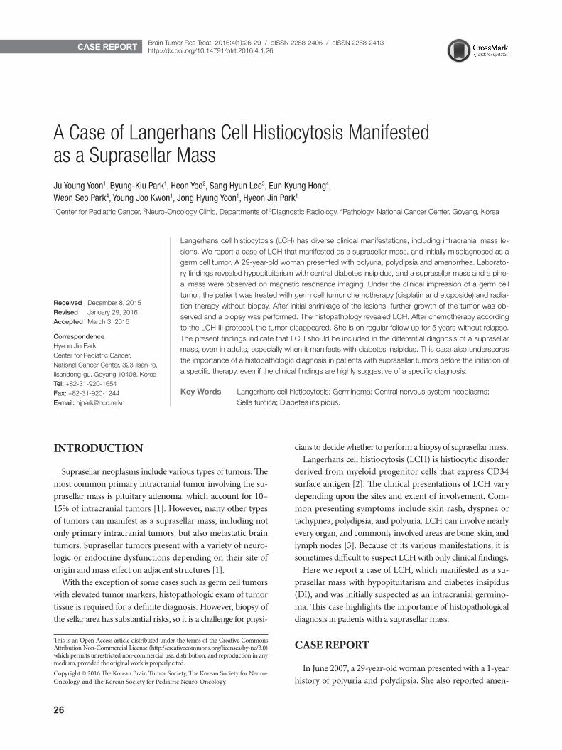
The brain magnetic resonance imaging (MRI) revealed a mass with a diameter of 2.3 cm on the suprasellar area, which showed strong enhancement. There was also a 1 cm-sized rim-
enhancing lesion in the pineal gland (Fig. 1A). Her spine MRI revealed no abnormal leptomeningeal enhancement, and the cerebrospinal fluid (CSF) exam revealed no malignant cells. Her serum tumor markers were as follows: alpha-fetoprotein 1.4 µg/L (normal range: 0–20 µg/L), carcinoembryonic anti-gen 0.61 µg/L (normal range: 0–6 µg/L), beta-human chori-onic gonadotropin 1.0 IU/L (normal range: 0–3 IU/L). CSF tumor markers were not examined.
A malignant intracranial tumor was suspected based on the above findings, and a biopsy of the brain lesion was rec-ommended, but the patient refused. With a clinical impression of a germ cell tumor, three cycles of chemotherapy for ad-vanced germ cell tumor (cisplatin+etoposide #3) was admin-istered from June 11, 2007–August 25, 2007. In each cycle, cisplatin (20 mg/m2×5 days) and etoposide 300 mg/m2 (60 mg/m2×5 days) were given. After the chemotherapy, the tu-mor showed partial response (Fig. 1B). Radiation therapy (whole ventricle 3,600 cGy/20 fractions, boost 900 cGy/5 fractions) was provided from October 11, 2007–November 4, 2007.
A
D
B
E
C
Fig. 1. Sagittal, T1-weighted images. A: Initial MRI on June 2007. Well enhanced mass with a diameter of 2.3 cm is observed around the pi-tuitary stalk (white arrow). Another 1 cm-sized rim-enhancing lesion is observed in the pineal gland (black arrow). B: MRI on September 2007. After 3 cycles of germ cell tumor chemotherapy, mass size decreased markedly, showing only linear enhancement. C: MRI on Sep-tember 2008. Increased size of enhancing mass in pituitary stalk and hypophysis areas observed (white arrow). D: MRI on December 2008. After Langerhans cell histiocytosis initial chemotherapy. Significant reduction in tumor size is observed. E: MRI on May 2015. Subtle residual enhancement in 3rd ventricle floor is observed. No other abnormal enhancing lesion is observed. Pituitary gland and stalk shows atrophy.
28 Brain Tumor Res Treat 2016;4(1):26-29
Suprasellar Langerhans Cell Histiocytosis
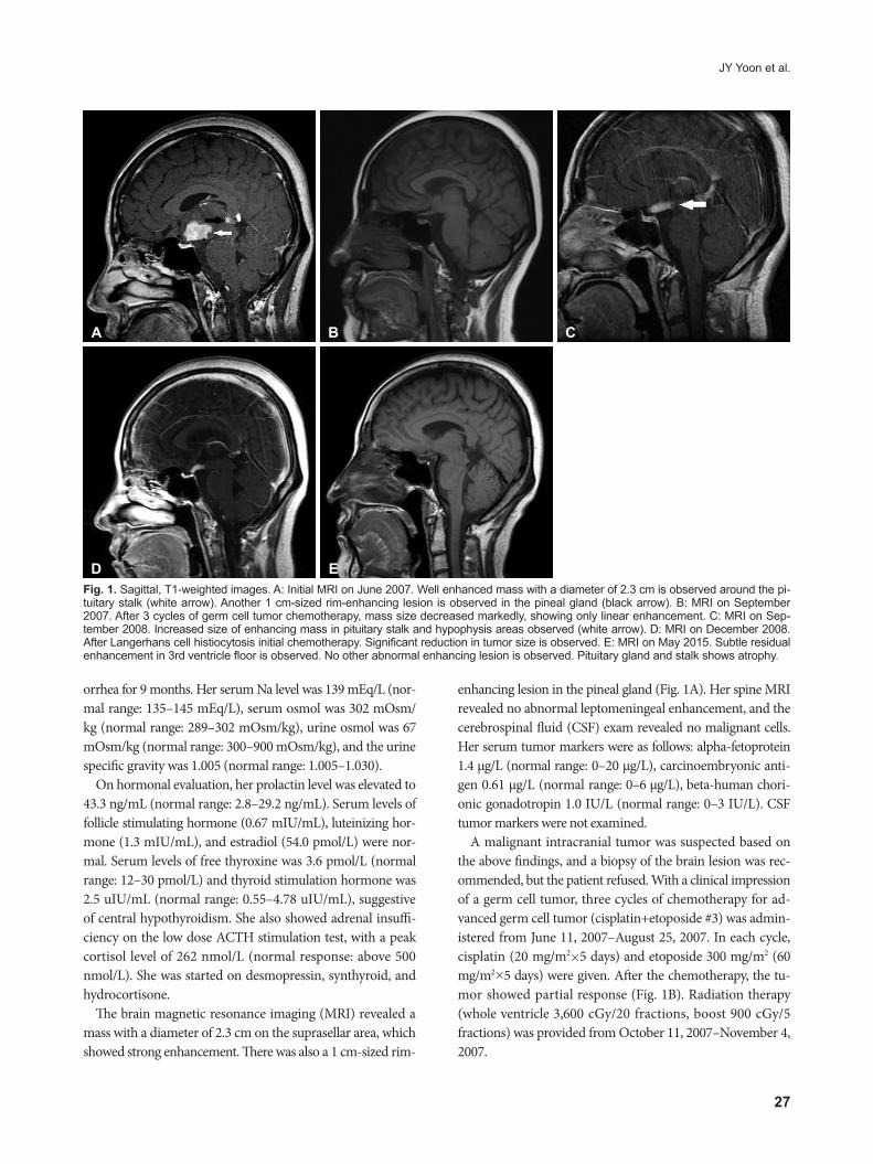
A follow up MRI on September 2008 showed increased mass size (Fig. 1C). She was transferred to our hospital for further evaluation and management. Excisional biopsy was performed and the biopsy result showed LCH (Fig. 2). Skele-tal X-rays and bone scan revealed no bony lesions. No extra-cranial lesions were observed on chest CT and liver ultraso-nography. Chemotherapy according to the LCH III protocol (for multifocal bone disease and special site involvement) was started from November 5, 2008. After the initial therapy (6 weeks with daily 40 mg/m2 oral prednisolone, and 6 mg/m2 i.v. vinblastine every 7 days), there was significant reduc-tion in tumor size (Fig. 1D). She received chemotherapy until July 15, 2010. She is now on regular follow up in our outpa-tient department. Due to panhypopituitarism and DI, she is now taking levothyroxine, hydrocortisone, and desmopressin, and we are considering starting sex hormone replacement therapy. She also has a visual field defect due to glaucoma and is taking glaucoma medication. Her visual acuity is only finger count possible for both eyes. She also complains of memory disturbance which inhibits normal social activities. The last brain MRI on June 2015 showed no new enhancing lesions or abnormal leptomeningeal enhancement, but atrophy of the pituitary gland was observed (Fig. 1E).
DISCUSSION
LCH is diagnosed in all age groups but is most common in children from one to three years old. Its incidence is about three to five cases per million in children, and one to two cases per million in adults [4]. In adults, lung involvement is more common than in children, which is partly attributable to sm-oking [5]. On the contrary, lymphoreticular organ involve-ment occurs less frequently in adults than in children [4].
LCH involves the central nervous system (CNS) in about 6 percent of patients [3]. The most common manifestations of CNS involvement are diabetes insipidus and neurodegenera-tion. Diabetes insipidus presents with polydipsia and polyuria, and occurs secondary to infiltration of the posterior pituitary. Neurodegeneration is characterized by symptoms such as ataxia and cognitive dysfunction, and is caused by lesions of the cerebellum or the basal ganglia. Though rare, LCH can manifest as an intracranial mass lesion, usually in the hypo-thalamic pituitary area or the pineal gland [6].
In this case, the tumor was initially treated as a germinoma by clinical suspicion without a biopsy. Among pediatric CNS tumors, germ cell tumors accounts for 0.4–3.4 percent in west-ern countries, and this incidence is higher and reported to be up to 11 percent of all pediatric brain tumors in Asian coun-
A
C
B
DFig. 2. Photograph of the surgical specimen. A: Many lymphohistiocytic infiltrations are noted in the low power view (HE staining, ×100). B: Scattered Langerhans cells are seen, with eosinophils and lymphocytes (HE staining, ×400). C and D: Immunohistochemical staining shows strong CD1 positivity (C) and weak S-100 positivity (D). HE, hematoxylin and eosin.
JY Yoon et al.
29
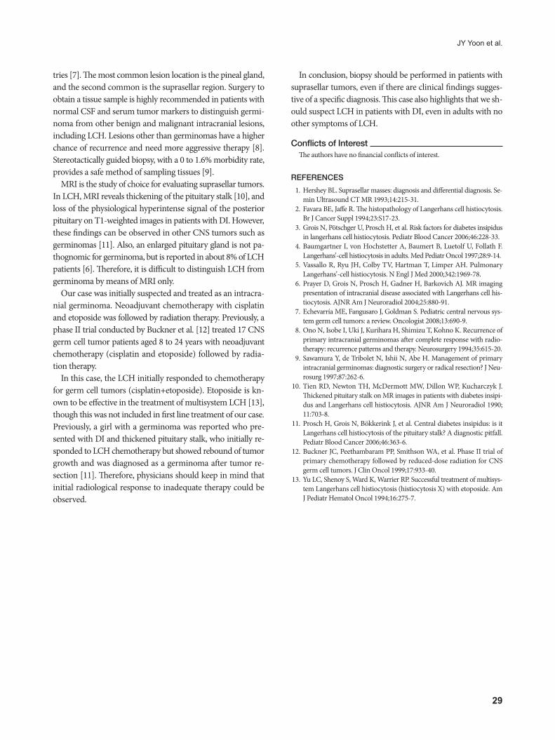
tries [7]. The most common lesion location is the pineal gland, and the second common is the suprasellar region. Surgery to obtain a tissue sample is highly recommended in patients with normal CSF and serum tumor markers to distinguish germi-noma from other benign and malignant intracranial lesions, including LCH. Lesions other than germinomas have a higher chance of recurrence and need more aggressive therapy [8]. Stereotactically guided biopsy, with a 0 to 1.6% morbidity rate, provides a safe method of sampling tissues [9].
MRI is the study of choice for evaluating suprasellar tumors. In LCH, MRI reveals thickening of the pituitary stalk [10], and loss of the physiological hyperintense signal of the posterior pituitary on T1-weighted images in patients with DI. However, these findings can be observed in other CNS tumors such as germinomas [11]. Also, an enlarged pituitary gland is not pa-thognomic for germinoma, but is reported in about 8% of LCH patients [6]. Therefore, it is difficult to distinguish LCH from germinoma by means of MRI only.
Our case was initially suspected and treated as an intracra-nial germinoma. Neoadjuvant chemotherapy with cisplatin and etoposide was followed by radiation therapy. Previously, a phase II trial conducted by Buckner et al. [12] treated 17 CNS germ cell tumor patients aged 8 to 24 years with neoadjuvant chemotherapy (cisplatin and etoposide) followed by radia-tion therapy.
In this case, the LCH initially responded to chemotherapy for germ cell tumors (cisplatin+etoposide). Etoposide is kn-own to be effective in the treatment of multisystem LCH [13], though this was not included in first line treatment of our case. Previously, a girl with a germinoma was reported who pre-sented with DI and thickened pituitary stalk, who initially re-sponded to LCH chemotherapy but showed rebound of tumor growth and was diagnosed as a germinoma after tumor re-section [11]. Therefore, physicians should keep in mind that initial radiological response to inadequate therapy could be observed.
In conclusion, biopsy should be performed in patients with suprasellar tumors, even if there are clinical findings sugges-tive of a specific diagnosis. This case also highlights that we sh-ould suspect LCH in patients with DI, even in adults with no other symptoms of LCH.
Conflicts of InterestThe authors have no financial conflicts of interest.
REFERENCES
1. Hershey BL. Suprasellar masses: diagnosis and differential diagnosis. Se-min Ultrasound CT MR 1993;14:215-31.
2. Favara BE, Jaffe R. The histopathology of Langerhans cell histiocytosis. Br J Cancer Suppl 1994;23:S17-23.
3. Grois N, Pötschger U, Prosch H, et al. Risk factors for diabetes insipidus in langerhans cell histiocytosis. Pediatr Blood Cancer 2006;46:228-33.
4. Baumgartner I, von Hochstetter A, Baumert B, Luetolf U, Follath F. Langerhans’-cell histiocytosis in adults. Med Pediatr Oncol 1997;28:9-14.
5. Vassallo R, Ryu JH, Colby TV, Hartman T, Limper AH. Pulmonary Langerhans’-cell histiocytosis. N Engl J Med 2000;342:1969-78.
6. Prayer D, Grois N, Prosch H, Gadner H, Barkovich AJ. MR imaging presentation of intracranial disease associated with Langerhans cell his-tiocytosis. AJNR Am J Neuroradiol 2004;25:880-91.
7. Echevarría ME, Fangusaro J, Goldman S. Pediatric central nervous sys-tem germ cell tumors: a review. Oncologist 2008;13:690-9.
8. Ono N, Isobe I, Uki J, Kurihara H, Shimizu T, Kohno K. Recurrence of primary intracranial germinomas after complete response with radio-therapy: recurrence patterns and therapy. Neurosurgery 1994;35:615-20.
9. Sawamura Y, de Tribolet N, Ishii N, Abe H. Management of primary intracranial germinomas: diagnostic surgery or radical resection? J Neu-rosurg 1997;87:262-6.
10. Tien RD, Newton TH, McDermott MW, Dillon WP, Kucharczyk J. Thickened pituitary stalk on MR images in patients with diabetes insipi-dus and Langerhans cell histiocytosis. AJNR Am J Neuroradiol 1990; 11:703-8.
11. Prosch H, Grois N, Bökkerink J, et al. Central diabetes insipidus: is it Langerhans cell histiocytosis of the pituitary stalk? A diagnostic pitfall. Pediatr Blood Cancer 2006;46:363-6.
12. Buckner JC, Peethambaram PP, Smithson WA, et al. Phase II trial of primary chemotherapy followed by reduced-dose radiation for CNS germ cell tumors. J Clin Oncol 1999;17:933-40.
13. Yu LC, Shenoy S, Ward K, Warrier RP. Successful treatment of multisys-tem Langerhans cell histiocytosis (histiocytosis X) with etoposide. Am J Pediatr Hematol Oncol 1994;16:275-7.