Embed Size (px)
Citation preview
Brief Report
S36 Ann Dermatol
Received September 3, 2018, Revised December 6, 2018, Accepted for publication January 10, 2019
Corresponding author: Min Kyung Shin, Department of Dermatology, School of Medicine, Kyung Hee University, 23 Kyungheedae-ro, Dongdaemun-gu,Seoul 02447, Korea. Tel: 82-2-958-8300, Fax: 82-2-969-6538, E-mail: [email protected]: https://orcid.org/0000-0001-9834-7931
This is an Open Access article distributed under the terms of the Creative Commons Attribution Non-Commercial License (http://creativecommons.org/licenses/by-nc/4.0) which permits unrestricted non-commercial use, distribution, and reproduction in any medium, provided the original work is properly cited.
Copyright © The Korean Dermatological Association and The Korean Society for Investigative Dermatology
pISSN 1013-9087ㆍeISSN 2005-3894Ann Dermatol Vol. 31, Suppl, 2019 https://doi.org/10.5021/ad.2019.31.S.S36
BRIEF REPORT
A Case of Post-Herpetic Nevoid Comedones
Jong-Kil Seo, Ki-Heon Jeong, Min Kyung Shin
Department of Dermatology, School of Medicine, Kyung Hee University, Seoul, Korea
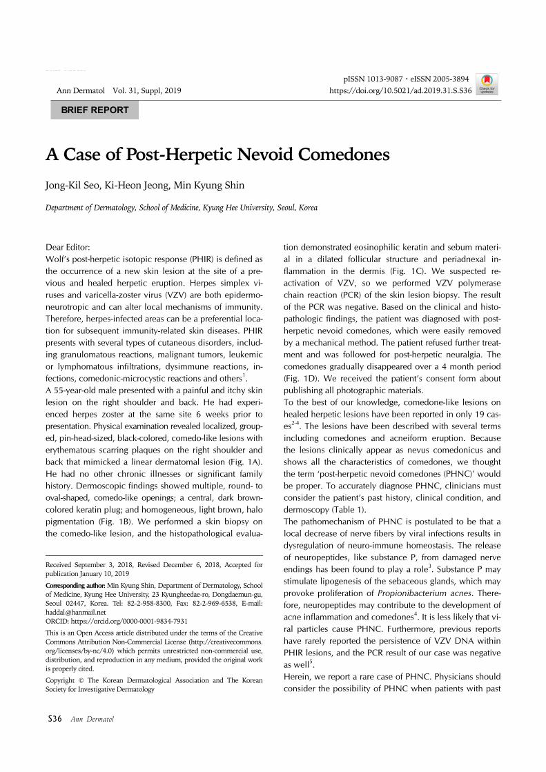
Dear Editor:Wolf’s post-herpetic isotopic response (PHIR) is defined as the occurrence of a new skin lesion at the site of a pre-vious and healed herpetic eruption. Herpes simplex vi-ruses and varicella-zoster virus (VZV) are both epidermo-neurotropic and can alter local mechanisms of immunity. Therefore, herpes-infected areas can be a preferential loca-tion for subsequent immunity-related skin diseases. PHIR presents with several types of cutaneous disorders, includ-ing granulomatous reactions, malignant tumors, leukemic or lymphomatous infiltrations, dysimmune reactions, in-fections, comedonic-microcystic reactions and others1. A 55-year-old male presented with a painful and itchy skin lesion on the right shoulder and back. He had experi-enced herpes zoster at the same site 6 weeks prior to presentation. Physical examination revealed localized, group-ed, pin-head-sized, black-colored, comedo-like lesions with erythematous scarring plaques on the right shoulder and back that mimicked a linear dermatomal lesion (Fig. 1A). He had no other chronic illnesses or significant family history. Dermoscopic findings showed multiple, round- to oval-shaped, comedo-like openings; a central, dark brown- colored keratin plug; and homogeneous, light brown, halo pigmentation (Fig. 1B). We performed a skin biopsy on the comedo-like lesion, and the histopathological evalua-
tion demonstrated eosinophilic keratin and sebum materi-al in a dilated follicular structure and periadnexal in-flammation in the dermis (Fig. 1C). We suspected re-activation of VZV, so we performed VZV polymerase chain reaction (PCR) of the skin lesion biopsy. The result of the PCR was negative. Based on the clinical and histo-pathologic findings, the patient was diagnosed with post- herpetic nevoid comedones, which were easily removed by a mechanical method. The patient refused further treat-ment and was followed for post-herpetic neuralgia. The comedones gradually disappeared over a 4 month period (Fig. 1D). We received the patient’s consent form about publishing all photographic materials.To the best of our knowledge, comedone-like lesions on healed herpetic lesions have been reported in only 19 cas-es2-4. The lesions have been described with several terms including comedones and acneiform eruption. Because the lesions clinically appear as nevus comedonicus and shows all the characteristics of comedones, we thought the term ‘post-herpetic nevoid comedones (PHNC)’ would be proper. To accurately diagnose PHNC, clinicians must consider the patient’s past history, clinical condition, and dermoscopy (Table 1).The pathomechanism of PHNC is postulated to be that a local decrease of nerve fibers by viral infections results in dysregulation of neuro-immune homeostasis. The release of neuropeptides, like substance P, from damaged nerve endings has been found to play a role3. Substance P may stimulate lipogenesis of the sebaceous glands, which may provoke proliferation of Propionibacterium acnes. There-fore, neuropeptides may contribute to the development of acne inflammation and comedones4. It is less likely that vi-ral particles cause PHNC. Furthermore, previous reports have rarely reported the persistence of VZV DNA within PHIR lesions, and the PCR result of our case was negative as well5. Herein, we report a rare case of PHNC. Physicians should consider the possibility of PHNC when patients with past
Brief Report
Vol. 31, Suppl, 2019 S37
Table 1. Differential diagnosis of post-herpetic nevoid comedones (PHNC)
Characteristic Comedones PHNC Nevus comedonicus (NC)
Clinical presentation Predilection site for acne Usually linear (Previous Herpes zoster site)
No cysts, fistulas, abscess, or scars
Usually linearHoneycomb patternNC syndrome (neurologic, orthopedic,
and ophthalmic abnormalities)Dermoscopic findings Numerous, homogenous,
dark-brown or black areas, usually circular in shape and located superficially in the epidermis
Like NC (smaller hyperkeratotic plug)
Numerous, circular and barrel-shaped, homogenous areas in light and dark-brown shades with remarkable keratin plugs
Histological Obligatory folliclesTrue comedones (keratin+sebum)
Similar to comedones Rudimentary folliclesPseudocomedones (keratin)
Mechanical removal Easy Easy Not easyTreatment and prognosis Good response to treatment Spontaneous remission Challenging
Fig. 1. (A) Localized, grouped, pin- head-sized, black-colored, comedo-like lesions with erythematous scar-ring plaques on the right shoulder and back, mimicking a linear der-matomal lesion. (B) Dermoscopic findings showed multiple, round- to oval-shaped, comedo-like open-ings with a central, dark brown- color keratin plug and homogene-ous, light brown, halo pigmenta-tion. (C) Histologic findings reveal-ed eosinophilic keratin and sebum material in a dilated follicular struc-ture in the dermis (H&E, ×40). (D) The comedo-like lesions gradually disappeared after 4 months.
history of herpes zoster complain of cutaneous lesions. Because PHNC can spontaneously disappear, disease pro-gress can be observed without unnecessary treatment.
CONFLICTS OF INTEREST
The authors have nothing to disclose.
ORCID
Jong-Kil Seo, https://orcid.org/0000-0001-8822-6466Ki-Heon Jeong, https://orcid.org/0000-0001-6908-0932
Min Kyung Shin, https://orcid.org/0000-0001-9834-7931
REFERENCES
1. Ruocco V, Ruocco E, Brunetti G, Russo T, Gambardella A,
Wolf R. Wolf's post-herpetic isotopic response: infections, tumors, and immune disorders arising on the site of healed
herpetic infection. Clin Dermatol 2014;32:561-568.
2. Dong H, Hu Y, Basude ME. Post-herpetic acneiform erup-tion: report of two cases with dermoscopic observations.
Australas J Dermatol 2015;56:310-311.
3. Wang B, Zheng J, Wang HW. Postherpetic comedones in

Brief Report
S38 Ann Dermatol
two Chinese Han patients. Chin Med J (Engl) 2017;130: 1615-1616.
4. Sanchez-Salas MP. Appearance of comedones at the site of
healed herpes zoster: Wolf's isotopic response. Int J Der-matol 2011;50:633-634.
5. Requena L, Kutzner H, Escalonilla P, Ortiz S, Schaller J, Rohwedder A. Cutaneous reactions at sites of herpes zoster
scars: an expanded spectrum. Br J Dermatol 1998;138:161-
168.


![H Control Technology of the Hydraulic System · With the rapid development of computer technology and PHNC (Power Hydraulic Numerical ... JLKZS[2014]05, NO. LH[2015]7043) and NO](https://img.pdfslide.net/doc/110x75/5ccc482988c993d2098bc7d6/h-control-technology-of-the-hydraulic-system-with-the-rapid-development-of-computer.jpg)