Embed Size (px)
Citation preview
Immunopaedia.org.zaImmunopaedia.org.za
A case of scaly annular plaquesA case of scaly annular plaques
Patient PresentationHistoryDifferential DiagnosisExaminationInvestigationsDiscussionTreatmentFinal OutcomeReferencesEvaluation - Questions & answersMCQ
Patient PresentationPatient Presentation
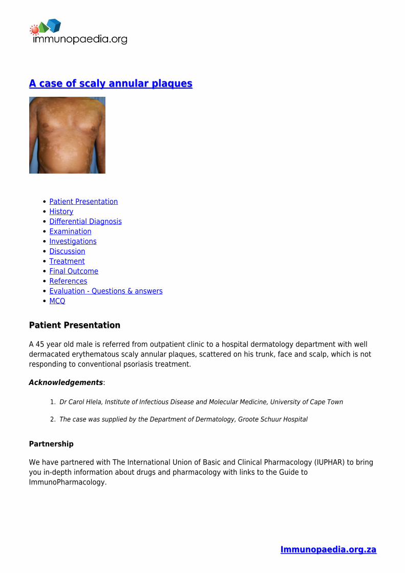
A 45 year old male is referred from outpatient clinic to a hospital dermatology department with welldermacated erythematous scaly annular plaques, scattered on his trunk, face and scalp, which is notresponding to conventional psoriasis treatment.
Acknowledgements:
Dr Carol Hlela, Institute of Infectious Disease and Molecular Medicine, University of Cape Town1.
The case was supplied by the Department of Dermatology, Groote Schuur Hospital2.
Partnership
We have partnered with The International Union of Basic and Clinical Pharmacology (IUPHAR) to bringyou in-depth information about drugs and pharmacology with links to the Guide toImmunoPharmacology.
Immunopaedia.org.zaImmunopaedia.org.za
HistoryHistory
History revealed that the patient had suffered from psoriasis for the past 6 years. During this time hewas under the care of a general practitioner who treated his psoriasis with oral and topical steriods-without any improvement. Following these 6 years of treatment without relief the patient was referredto specialist care. At the Dermatology outpatient department of a tertiary hospital the patient wasstarted on different topical therapies which included Dovonex ointment, a synthetic Vitamin D3derivative, and UV phototherapy. However, despite months of follow ups and treatment adjustmentshe still remained poorly controlled. Now at his 12 month visit he has been admitted to hospital due toa severe flare up of psoriasis. He has been initiated on topical Anthralin (dithranol) in addition to hiscurrent treatment while continuing UV phototherapy.
Past Medical History
No history of joint pain or stiffness.
Past Surgical History
Nil
Family History
Father- hypertension on treatmentMother- wellNo family history of psoriasis or arthritis
Allergies
None known
Medication
Dithranol (Anthralin), Salicylic acid, Dovonex and UV phototherapy
Social History
Non smokerNo alcohol or illicit drug use
Differential DiagnosisDifferential Diagnosis
PsoriasisNummular dermatitis
Immunopaedia.org.zaImmunopaedia.org.za
Lichen planusMycoses Fungoides (MF)
ExaminationExamination
ON ADMISSION:
Appearance:
Ambulatory male, awake, alert and co- operative
Immunopaedia.org.zaImmunopaedia.org.za
Vitals
Temperature: afebrileBlood pressure: 132/76Heart rate: 75Respiratory rate: 16
General
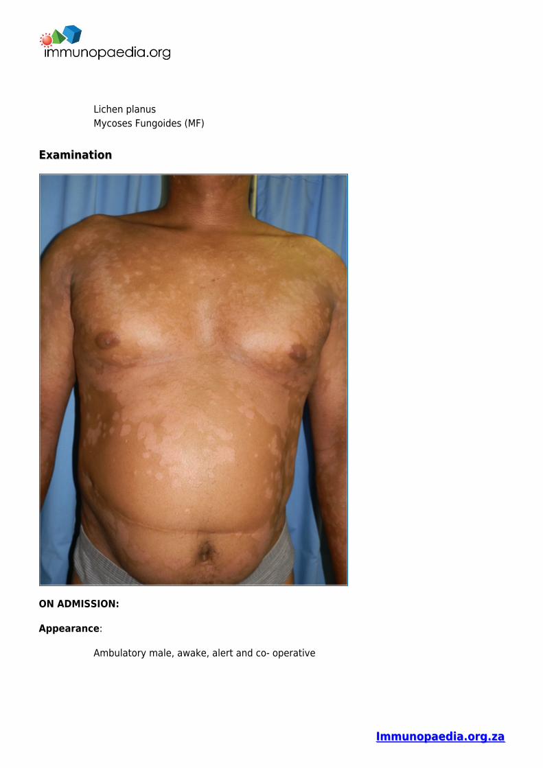
Scattered erythematous medium to large plaques on chest, upper back, lower limbs,thighs, scalp and face, with silvery scale.No palpable lymph nodes, jaundice, pallor or oedema
Chest
Chest clear
Cardiovascular
NormotensiveNo murmurs, no added heart sounds
Abdomen
No distension or tenderness. Bowel sounds present.
Neurological
Normal level of consciousness, alert and co-operativeGait, power, tone, sensation and reflexes intact and functioning within normal limits.
Musculoskeletal
No swellings, no effusions, no tendernessNo joint deformitiesNormal range of motion in all joints
Dermatological
Scattered erythematous medium-large plaques on chest, upper back, lower limbs, thighs,scalp and face, with silvery scaling. (See figure 1)Flexural involvement.Positive Auspitz sign (capillary bleeding occurring after overlying scale removed)
Immunopaedia.org.zaImmunopaedia.org.za
PASI score (psoriasis area severity index) was E3 S3I3, indicating that erythema, scale andinduration were all severe, with BSA (body surface area of >25%). The PASI is a measureof overall psoriasis severity and coverage that assesses body surface area, erythema andscaling.Scalp lesions extended to the face.Several lesions on the upper back were tumour-like
InvestigationsInvestigations
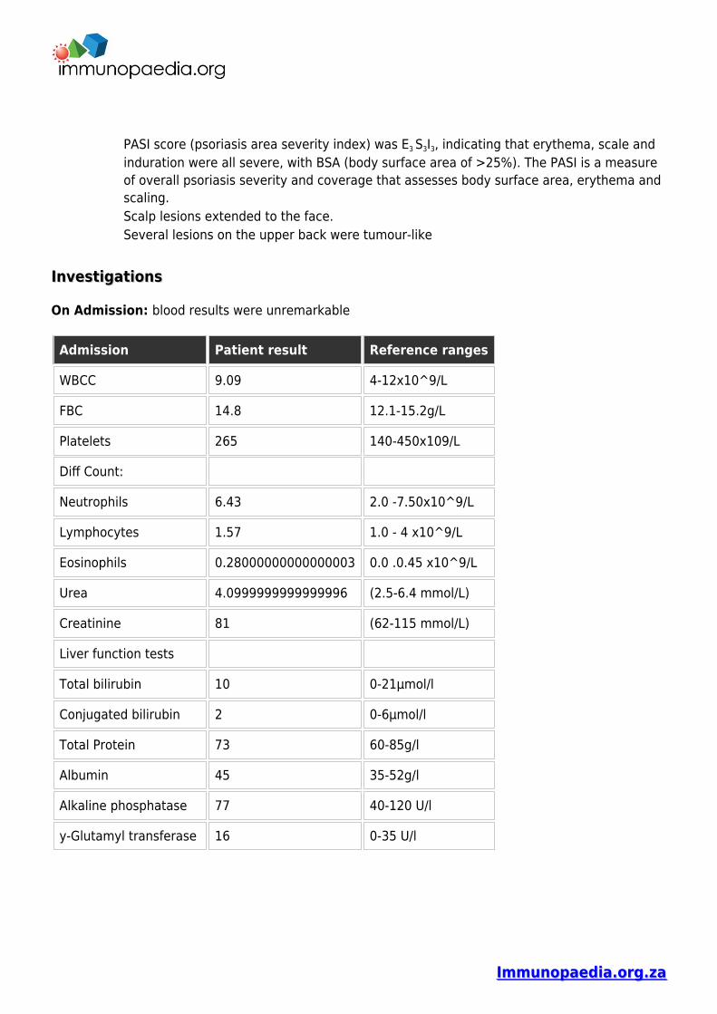
On Admission: blood results were unremarkable
Admission Patient result Reference ranges
WBCC 9.09 4-12x10^9/L
FBC 14.8 12.1-15.2g/L
Platelets 265 140-450x109/L
Diff Count:
Neutrophils 6.43 2.0 -7.50x10^9/L
Lymphocytes 1.57 1.0 - 4 x10^9/L
Eosinophils 0.28000000000000003 0.0 .0.45 x10^9/L
Urea 4.0999999999999996 (2.5-6.4 mmol/L)
Creatinine 81 (62-115 mmol/L)
Liver function tests
Total bilirubin 10 0-21µmol/l
Conjugated bilirubin 2 0-6µmol/l
Total Protein 73 60-85g/l
Albumin 45 35-52g/l
Alkaline phosphatase 77 40-120 U/l
y-Glutamyl transferase 16 0-35 U/l
Immunopaedia.org.zaImmunopaedia.org.za
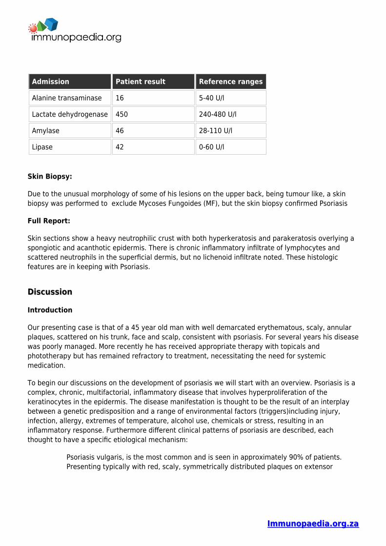
Admission Patient result Reference ranges
Alanine transaminase 16 5-40 U/l
Lactate dehydrogenase 450 240-480 U/l
Amylase 46 28-110 U/l
Lipase 42 0-60 U/l
Skin Biopsy:
Due to the unusual morphology of some of his lesions on the upper back, being tumour like, a skinbiopsy was performed to exclude Mycoses Fungoides (MF), but the skin biopsy confirmed Psoriasis
Full Report:
Skin sections show a heavy neutrophilic crust with both hyperkeratosis and parakeratosis overlying aspongiotic and acanthotic epidermis. There is chronic inflammatory infiltrate of lymphocytes andscattered neutrophils in the superficial dermis, but no lichenoid infiltrate noted. These histologicfeatures are in keeping with Psoriasis.
DiscussionDiscussion
Introduction
Our presenting case is that of a 45 year old man with well demarcated erythematous, scaly, annularplaques, scattered on his trunk, face and scalp, consistent with psoriasis. For several years his diseasewas poorly managed. More recently he has received appropriate therapy with topicals andphototherapy but has remained refractory to treatment, necessitating the need for systemicmedication.
To begin our discussions on the development of psoriasis we will start with an overview. Psoriasis is acomplex, chronic, multifactorial, inflammatory disease that involves hyperproliferation of thekeratinocytes in the epidermis. The disease manifestation is thought to be the result of an interplaybetween a genetic predisposition and a range of environmental factors (triggers)including injury,infection, allergy, extremes of temperature, alcohol use, chemicals or stress, resulting in aninflammatory response. Furthermore different clinical patterns of psoriasis are described, eachthought to have a specific etiological mechanism:
Psoriasis vulgaris, is the most common and is seen in approximately 90% of patients.Presenting typically with red, scaly, symmetrically distributed plaques on extensor
Immunopaedia.org.zaImmunopaedia.org.za
surfaces- as is seen in a severe form in our case study patient.Guttate psoriasis, is characterized by the eruption of small papules on the upper trunkand proximal extremities, often but not exclusively triggered by Group A streptococcalthroat infections.Inverse psoriasis, is characterised by lesionsin a flexural distribution with involvement ofaxillae, groin and perineum.Erythrodermic psoriasis represents a generalized form of disease that affects more than90% of body surface area.Pustular psoriaris, is characterised by predominatly pustular lesions.
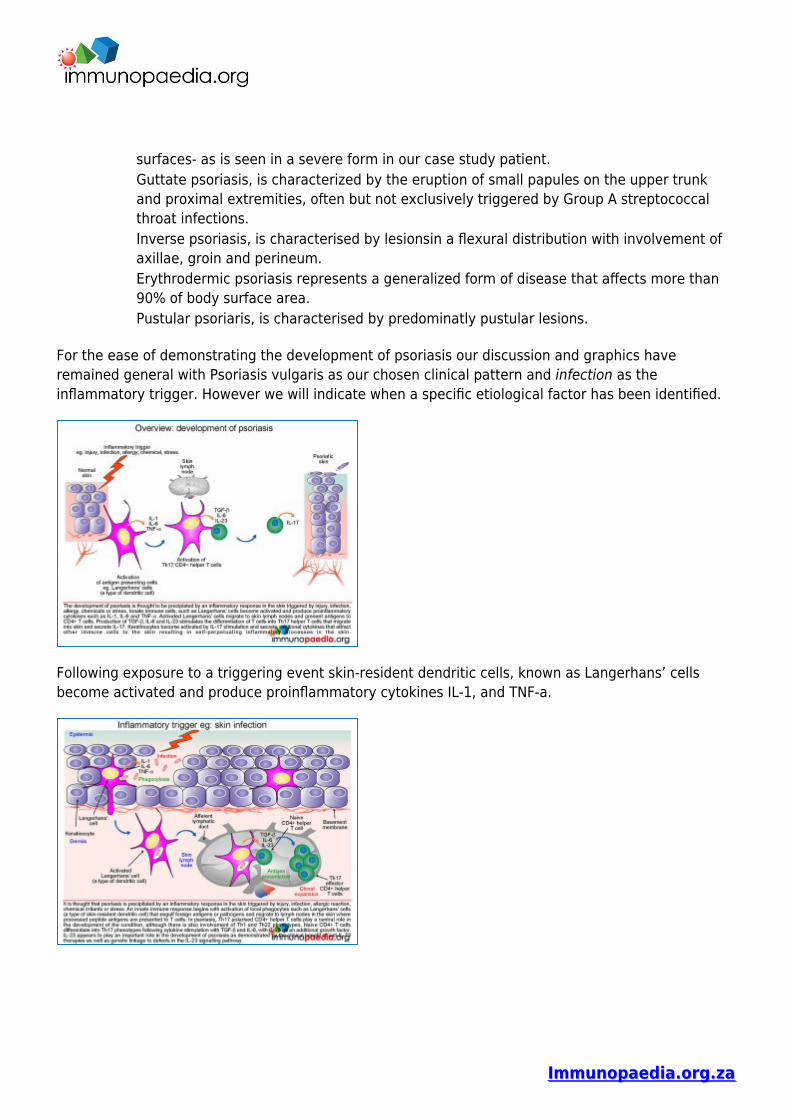
For the ease of demonstrating the development of psoriasis our discussion and graphics haveremained general with Psoriasis vulgaris as our chosen clinical pattern and infection as theinflammatory trigger. However we will indicate when a specific etiological factor has been identified.
Following exposure to a triggering event skin-resident dendritic cells, known as Langerhans’ cellsbecome activated and produce proinflammatory cytokines IL-1, and TNF-a.
Immunopaedia.org.zaImmunopaedia.org.za
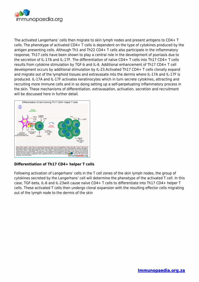
The activated Langerhans’ cells then migrate to skin lymph nodes and present antigens to CD4+ Tcells. The phenotype of activated CD4+ T cells is dependent on the type of cytokines produced by theantigen presenting cells. Although Th1 and Th22 CD4+ T cells also participate in the inflammatoryresponse, Th17 cells have been shown to play a central role in the development of psoriasis due tothe secretion of IL-17A and IL-17F. The differentiation of naïve CD4+ T cells into Th17 CD4+ T cellsresults from cytokine stimulation by TGF-b and IL-6. Additional enhancement of Th17 CD4+ T celldevelopment occurs by additional stimulation by IL-23.Activated Th17 CD4+ T cells clonally expandand migrate out of the lymphoid tissues and extravasate into the dermis where IL-17A and IL-17F isproduced. IL-17A and IL-17F activates keratinocytes which in turn secrete cytokines, attracting andrecruiting more immune cells and in so doing setting up a self-perpetuating inflammatory process inthe skin. These mechanisms of differentiation, extravasation, activation, secretion and recruitmentwill be discussed here in further detail.
Differentiation of Th17 CD4+ helper T cells
Following activation of Langerhans’ cells in the T cell zones of the skin lymph nodes, the group ofcytokines secreted by the Langerhans’ cell will determine the phenotype of the activated T cell. In thiscase, TGF-beta, IL-6 and IL-23will cause naïve CD4+ T cells to differentiate into Th17 CD4+ helper Tcells. These activated T cells then undergo clonal expansion with the resulting effector cells migratingout of the lymph node to the dermis of the skin
Immunopaedia.org.zaImmunopaedia.org.za
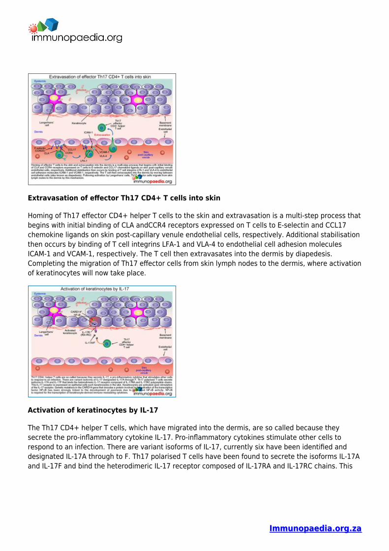
Extravasation of effector Th17 CD4+ T cells into skin
Homing of Th17 effector CD4+ helper T cells to the skin and extravasation is a multi-step process thatbegins with initial binding of CLA andCCR4 receptors expressed on T cells to E-selectin and CCL17chemokine ligands on skin post-capillary venule endothelial cells, respectively. Additional stabilisationthen occurs by binding of T cell integrins LFA-1 and VLA-4 to endothelial cell adhesion moleculesICAM-1 and VCAM-1, respectively. The T cell then extravasates into the dermis by diapedesis.Completing the migration of Th17 effector cells from skin lymph nodes to the dermis, where activationof keratinocytes will now take place.
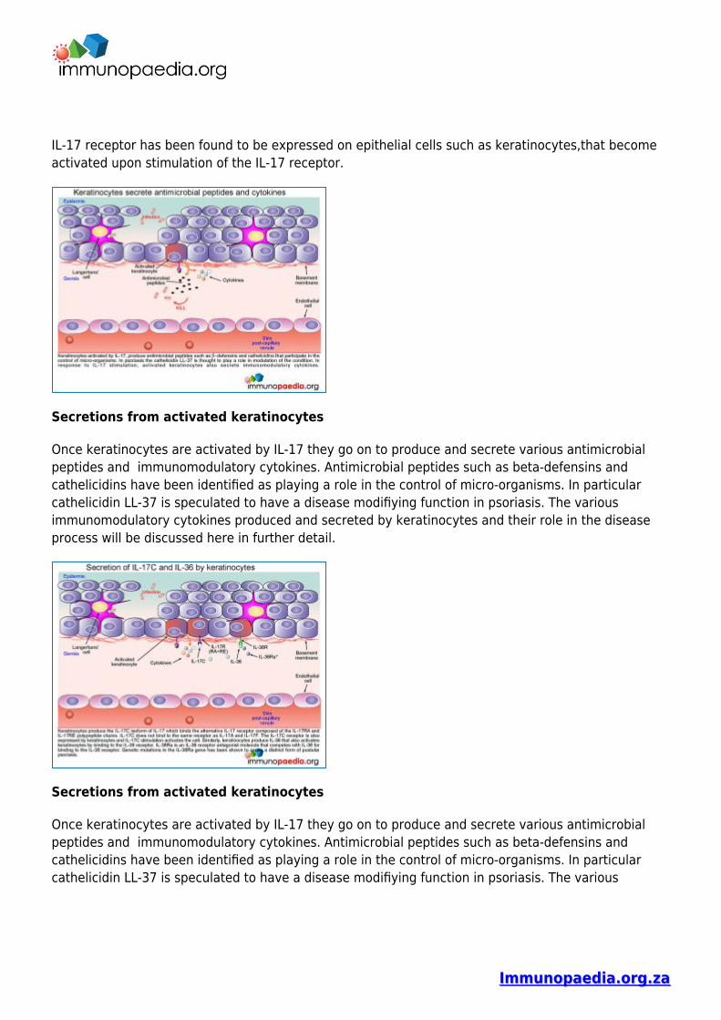
Activation of keratinocytes by IL-17
The Th17 CD4+ helper T cells, which have migrated into the dermis, are so called because theysecrete the pro-inflammatory cytokine IL-17. Pro-inflammatory cytokines stimulate other cells torespond to an infection. There are variant isoforms of IL-17, currently six have been identified anddesignated IL-17A through to F. Th17 polarised T cells have been found to secrete the isoforms IL-17Aand IL-17F and bind the heterodimeric IL-17 receptor composed of IL-17RA and IL-17RC chains. This
Immunopaedia.org.zaImmunopaedia.org.za
IL-17 receptor has been found to be expressed on epithelial cells such as keratinocytes,that becomeactivated upon stimulation of the IL-17 receptor.
Secretions from activated keratinocytes
Once keratinocytes are activated by IL-17 they go on to produce and secrete various antimicrobialpeptides and immunomodulatory cytokines. Antimicrobial peptides such as beta-defensins andcathelicidins have been identified as playing a role in the control of micro-organisms. In particularcathelicidin LL-37 is speculated to have a disease modifiying function in psoriasis. The variousimmunomodulatory cytokines produced and secreted by keratinocytes and their role in the diseaseprocess will be discussed here in further detail.
Secretions from activated keratinocytes
Once keratinocytes are activated by IL-17 they go on to produce and secrete various antimicrobialpeptides and immunomodulatory cytokines. Antimicrobial peptides such as beta-defensins andcathelicidins have been identified as playing a role in the control of micro-organisms. In particularcathelicidin LL-37 is speculated to have a disease modifiying function in psoriasis. The various
Immunopaedia.org.zaImmunopaedia.org.za
immunomodulatory cytokines produced and secreted by keratinocytes and their role in the diseaseprocess will be discussed here in further detail.
Secretion of IL-17C and IL-36 by keratinocytes
Keratinocytes produce the IL-17C isoform of IL-17 that binds the alternative IL-17 receptor composedof the IL-17RA andIL-17RE chain. IL-17C does not bind to the same receptor as IL-17A and IL-17F. TheIL-17C receptor is also expressed by keratinocytes, and binding activates the cell. Similarly,keratinocytes produce IL-36 that also activates keratinocytes by binding to the IL-36 receptor. IL-36Rais an IL-36 receptor antagonist molecule that competes with IL-36 for binding to the IL-36 receptor(and modulates/reduces excessive keratinocyte responses to IL-36). Genetic mutations in the IL-36Ragene that encode non-functional IL-36a proteins have been shown to cause pustular psoriasis due toincreased stimulation of the IL-36 receptors
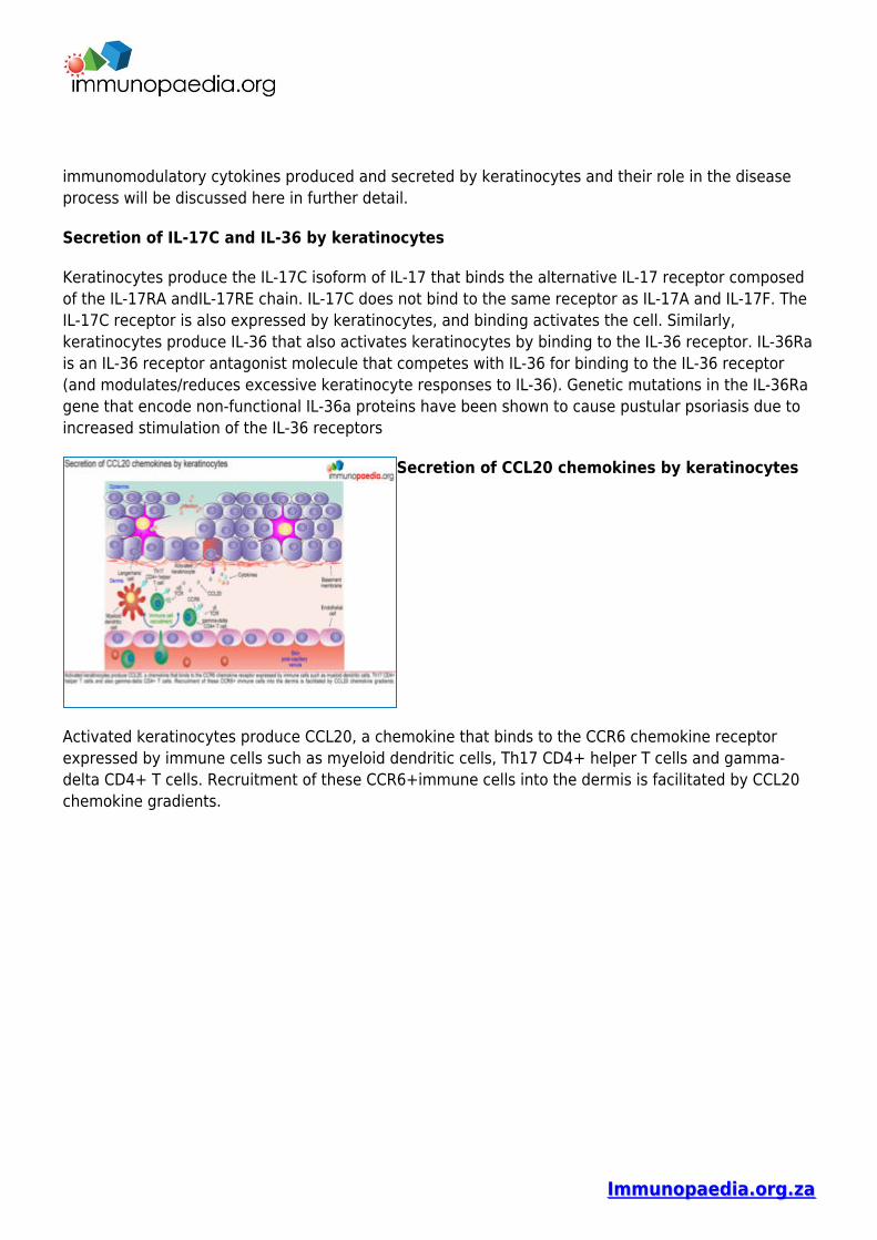
Secretion of CCL20 chemokines by keratinocytes
Activated keratinocytes produce CCL20, a chemokine that binds to the CCR6 chemokine receptorexpressed by immune cells such as myeloid dendritic cells, Th17 CD4+ helper T cells and gamma-delta CD4+ T cells. Recruitment of these CCR6+immune cells into the dermis is facilitated by CCL20chemokine gradients.
Immunopaedia.org.zaImmunopaedia.org.za
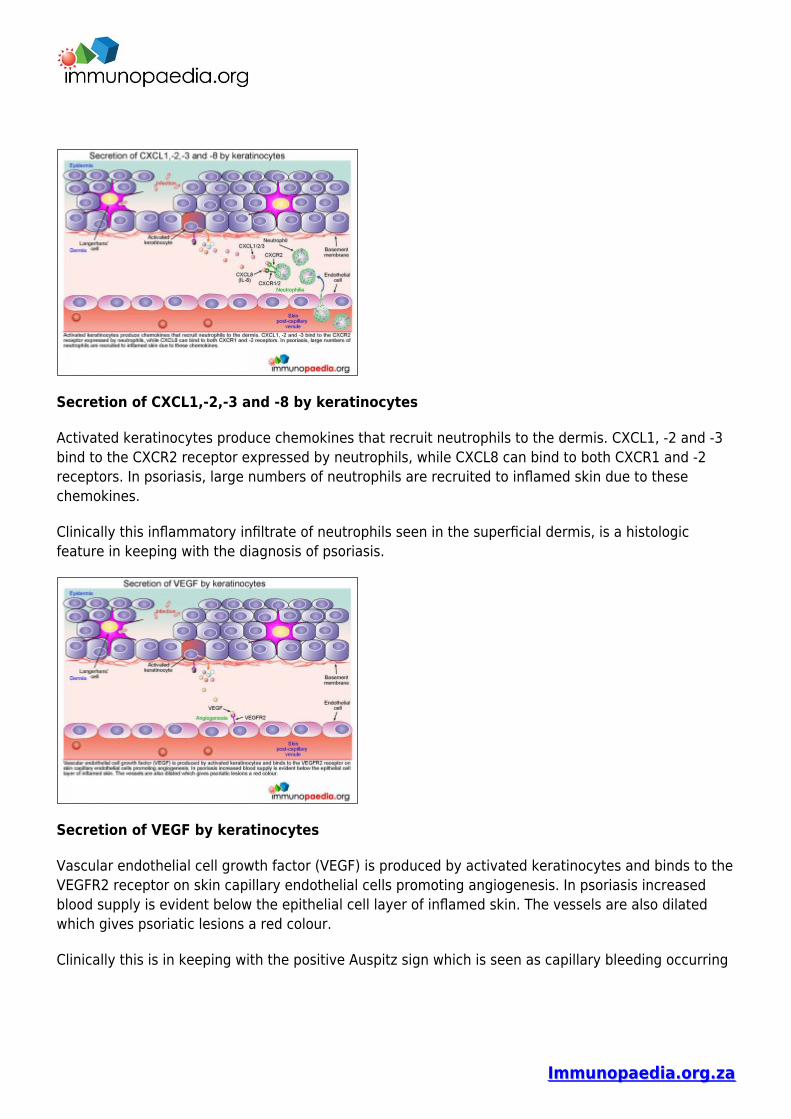
Secretion of CXCL1,-2,-3 and -8 by keratinocytes
Activated keratinocytes produce chemokines that recruit neutrophils to the dermis. CXCL1, -2 and -3bind to the CXCR2 receptor expressed by neutrophils, while CXCL8 can bind to both CXCR1 and -2receptors. In psoriasis, large numbers of neutrophils are recruited to inflamed skin due to thesechemokines.
Clinically this inflammatory infiltrate of neutrophils seen in the superficial dermis, is a histologicfeature in keeping with the diagnosis of psoriasis.
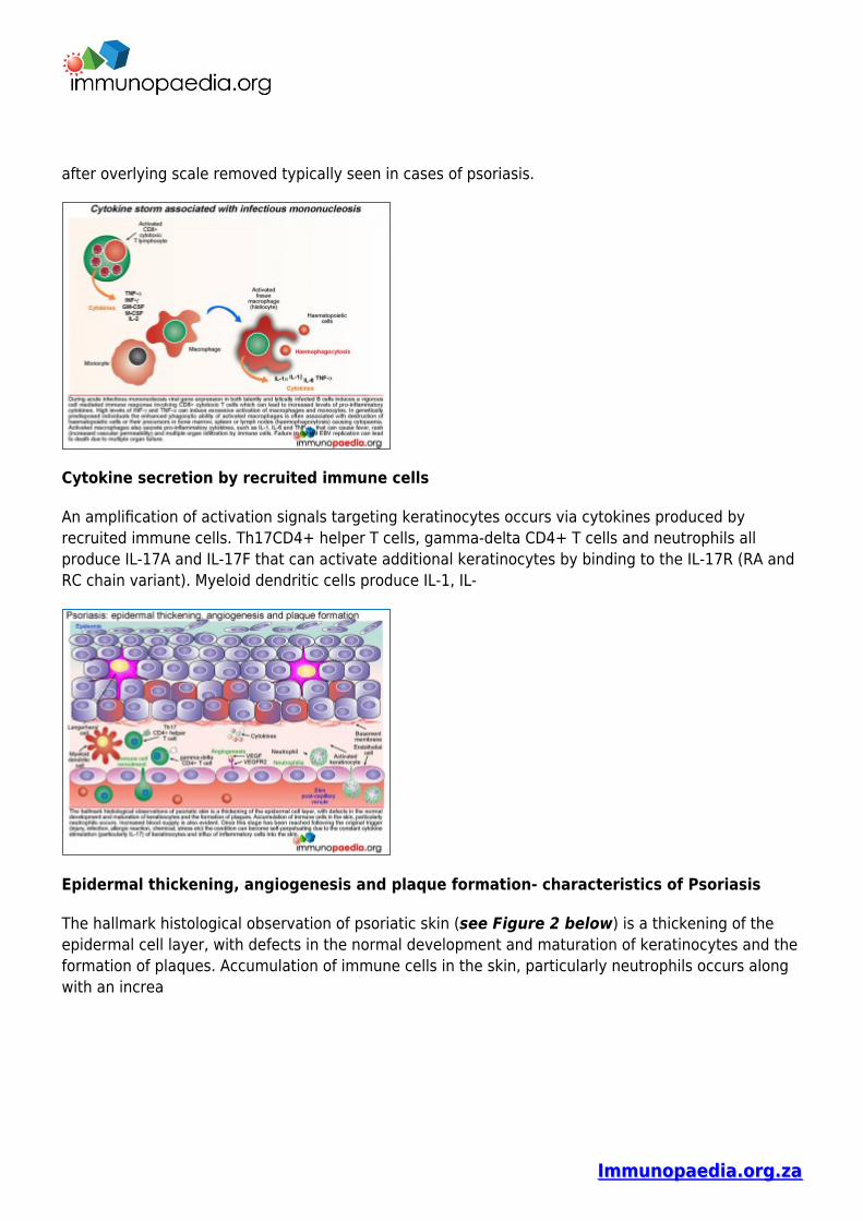
Secretion of VEGF by keratinocytes
Vascular endothelial cell growth factor (VEGF) is produced by activated keratinocytes and binds to theVEGFR2 receptor on skin capillary endothelial cells promoting angiogenesis. In psoriasis increasedblood supply is evident below the epithelial cell layer of inflamed skin. The vessels are also dilatedwhich gives psoriatic lesions a red colour.
Clinically this is in keeping with the positive Auspitz sign which is seen as capillary bleeding occurring
Immunopaedia.org.zaImmunopaedia.org.za
after overlying scale removed typically seen in cases of psoriasis.
Cytokine secretion by recruited immune cells
An amplification of activation signals targeting keratinocytes occurs via cytokines produced byrecruited immune cells. Th17CD4+ helper T cells, gamma-delta CD4+ T cells and neutrophils allproduce IL-17A and IL-17F that can activate additional keratinocytes by binding to the IL-17R (RA andRC chain variant). Myeloid dendritic cells produce IL-1, IL-
Epidermal thickening, angiogenesis and plaque formation- characteristics of Psoriasis
The hallmark histological observation of psoriatic skin (see Figure 2 below) is a thickening of theepidermal cell layer, with defects in the normal development and maturation of keratinocytes and theformation of plaques. Accumulation of immune cells in the skin, particularly neutrophils occurs alongwith an increa
Immunopaedia.org.zaImmunopaedia.org.za
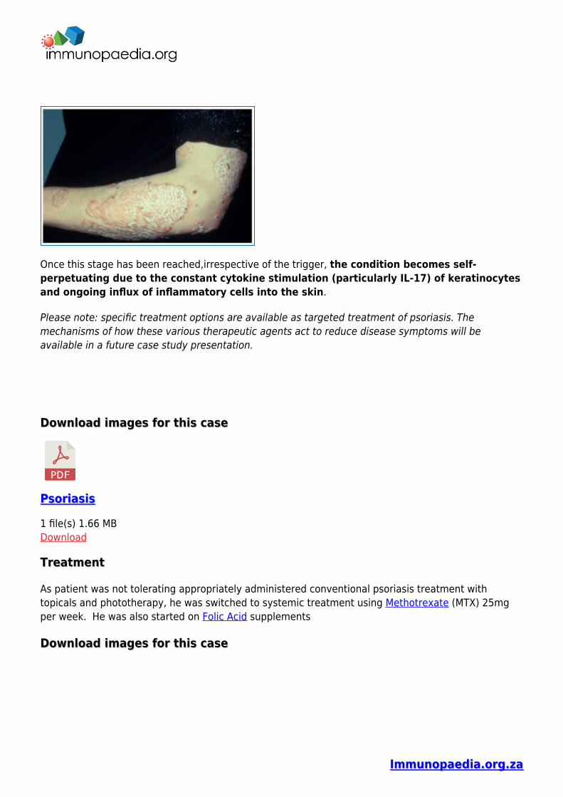
Once this stage has been reached,irrespective of the trigger, the condition becomes self-perpetuating due to the constant cytokine stimulation (particularly IL-17) of keratinocytesand ongoing influx of inflammatory cells into the skin.
Please note: specific treatment options are available as targeted treatment of psoriasis. Themechanisms of how these various therapeutic agents act to reduce disease symptoms will beavailable in a future case study presentation.
Download images for this caseDownload images for this case
PsoriasisPsoriasis
1 file(s) 1.66 MBDownload
TreatmentTreatment
As patient was not tolerating appropriately administered conventional psoriasis treatment withtopicals and phototherapy, he was switched to systemic treatment using Methotrexate (MTX) 25mgper week. He was also started on Folic Acid supplements
Download images for this caseDownload images for this case
Immunopaedia.org.zaImmunopaedia.org.za
PsoriasisPsoriasis
1 file(s) 1.66 MBDownload
Final outcomeFinal outcome
Good response on commencement of MTX. He was last seen, 6 months after starting MTX andcontinues to be notably improving, with a PASI E2S1I1, BSA5-10%
Download images for this caseDownload images for this case
PsoriasisPsoriasis
1 file(s) 1.66 MBDownload
ReferencesReferences
Menter A et al. (2008). Guidelines for the care and management of psoriasis and psoriatic arthritis. JAm Acad Dermatol ; 58: 826-850
Link to abstract
Girolomoni G et al. (2012) Psoriasis: rationale for targeting interleukin-17. Br J Dermatol.Oct;167(4):717-24
Link to abstract
Cai Y et al. (2013). Dermal yo-T cells – A new player in the pathogenesis of psoriasis. IntImmunopharmacol. Mar 13. pii: S1567-5769(13)00060-X
Link to abstract
Johnston A et al. (2013). Keratinocyte overexpression of IL-17C promotes psoriasiform skininflammation. J Immunol. Mar 1;190(5):2252-62.
Immunopaedia.org.zaImmunopaedia.org.za
Link to abstract
Jordan CT et al. (2012). Rare and common variants in CARD14, encoding an epidermal regulator ofNF-kappaB, in psoriasis. Am J Hum Genet. May 4;90(5):796-808.
Link to abstract
Download images for this caseDownload images for this case
PsoriasisPsoriasis
1 file(s) 1.66 MBDownload
Evaluation – Questions & answersEvaluation – Questions & answers
What was the final diagnosis?
Severe Psoriasis vulgaris, refractory to conventional treatment with topicals and phototherapy
Which innate immune cells become activated in the skin following some form ofenvironmental exposure?
Langerhans cells
In the development of psoriasis Th1 and Th22 CD4+ T cells are known to play a role in theinflammatory response. However which cells are central to the development of psoriasis?
Th17 cells play a central role in the development of psoriasis due to the secretion of IL-17A andIL-17F. IL-17A and F are produced from activated Th17 CD4+ T that have migrated into the dermis.Here IL-17A and F activate keratinocytes to secrete cytokines, which results in a self-perpetuatinginflammatory process as immune cells are attracted and recruited to the skin.
What determines the migration of Th17 CD4+ T cells to the dermis?
Homing of Th17 effector CD4+ helper T cells to the skin is determined by the CCL17 chemokinegradient. Once the cells have moved to the skin, these cells then bind to E-selectin and CCL17chemokine ligands on skin post-capillary venule endothelial cells using CLA andCCR4 receptorsexpressed on T cells, respectively.
What antimicrobial peptides are produced by keratinocytes after IL-17 stimulation?
Immunopaedia.org.zaImmunopaedia.org.za
Antimicrobial peptides such as beta-defensins and cathelicidins have been identified as playing a rolein the control of micro-organisms. In particular cathelicidin LL-37 is speculated to have a diseasemodifiying function in psoriasis
Which genetic mutation has been shown to cause a form of pustular psoriasis?
Genetic mutations in the IL-36Ra gene that encode non-functional IL-36a proteins leads to over-excessive IL-36 production and unregulated keratinocyte activation and ongoing inflammation
There was a heavy neutrophil infiltration seen on the patient, what causes the recruitmentof neutrophils to the inflammed skin
Activated keratinocytes produce CXCL1, -2, and -3 which bind to CXCR2 on the surface of neutrophils.
Download images for this caseDownload images for this case
PsoriasisPsoriasis
1 file(s) 1.66 MBDownload
Multiple Choice QuestionsMultiple Choice Questions
Earn 1 HPCSA or 0.25 SACNASP CPD Points – Earn 1 HPCSA or 0.25 SACNASP CPD Points – Online QuizOnline Quiz
Download images for this caseDownload images for this case
PsoriasisPsoriasis
1 file(s) 1.66 MBDownload