Embed Size (px)
Citation preview
Pediatric Anesthesia and Critical Care Journal 2015; 3(2):99-102 doi:10.14587/paccj.2015.20
Basantwani et al. Anaesthesia, d-TGA and craniotomy 99
Key points
Congenital cardiac anomalies pose many challenges during anesthesia due to anatomic and physiological alterations.
The inherent complications associated with these disorders necessitate vigilance for providing anesthesia to even
seemingly simple surgical intervention.
A case of uncorrected D-TGA for craniotomy in cerebral abscess: anaesthesia management
S. Basantwani, H. Karnik, B. Govardhane, B. Tendolkar
Department of Anaesthesia, Lokmanya Tilak Municipal Medical College and Hospital , Mumbai, India
Corresponding author: Department of Anaesthesia, Lokmanya Tilak Municipal Medcial College and Hospital, Mumbai, India. Email: [email protected]
Abstract
Transposition of Great Arteries (D-TGA) is one of the
most common cyanotic congenital heart defect in new-
born, having atrioventricular concordance with ventricu-
loarterial discordance where Aorta arises from right
ventricle and pulmonary artery from left ventricle. Pa-
tients with cyanotic congenital heart disease (cCHD) are
prone to develop frequent brain abscesses. Mortality rate
remains very high in these patients. Anesthetizing chil-
dren with cCHD and a brain abscess necessitates use of
an anesthesia regimen appropriate to both cCHD and
intracranial surgery. Here, we share our experience of
anesthesia management of uncorrected Dextro Transpo-
sition of Great Arteries (D TGA) for craniotomy and
intracerebral abscess drainage.
Keywords: Dextro transposition of great arteries; cere-
bral abscess
Introduction
Brain abscess is an uncommon and life threatening in-
tracranial infection characterised by purulence and in-
flammation in one or more localised areas within the
brain parenchyma. It results from spread of infection
from surrounding non-neuronal tissues eg middle ear,
fracture skull. Intracranial surgery, hematogenous
spread as in congenital heart disease with a right to left
shunt (5-18.7%) or a direct introduction into the brain.1
(D-TGA) accounts approximately 5% to 7% of all con-
genital heart diseases. There is discordance of the ven-
triculoarterial connection. Areas of mixing of oxygena-
ted and non oxygenated blood are vital for the survival
of the baby. Without mixing, the two circuits remain se-
parate, leading to death from systemic hypoxaemia and
acidosis. The possible locations for mixing are via a pa-
tent foramen ovale (PFO), atrial septal defect (ASD),
ventricular septal defect (VSD), patent ductus arteriosus
(PDA), or through bronchopulmonary collaterals. The
larger size of the communications between chambers
leads to more mixing of oxygenated and deoxygenated
blood resulting in higher oxygen saturation and better
heamodynamic stability.2
This intercirculatory mixing of blood leads to hypoxia
and its consequent polycythemia and hyperviscosity.
The latter results in sluggish blood flow in cerebral mi-
crocirculation, microthrombi formation and direct entry
of organisms, emboli, infected seed to cerebral circula-
tion forming cerebral abscess. Also, abscess related pro-
blems like vomiting, dehydration, acid base and elec-
Pediatric Anesthesia and Critical Care Journal 2015; 3(2):99-102 doi:10.14587/paccj.2015.20
Basantwani et al. Anaesthesia, d-TGA and craniotomy 100
trolyte imbalance, raised intracranial pressure (ICP),
seizures, make them high-risk candidates for abscess
excision under GA.2There is a paucity of anesthesia lite-
rature on the coexistence of these two conditions.We
report a case of 6 years old child of TGA with cerebral
abscess for drainage.
Case report
A 6 years old male child weighing 12 kg, diagnosed ca-
se of a D-TGA presented with fever, vomiting and gene-
ralized seizures since 20 days. General examination re-
vealed an afebrile patient, central cyanosis with on air
O2 saturation (SpO2) of 71%, grade 4 clubbing of digits
in all four limbs,no peripheral signs of infective endo-
carditis. An ejection systolic murmur in pulmonary area
was heard in cardiac examination. Neurologically he
was irritable with Glasgow coma scale (GCS) of
13(E4M5V4). An upper motor neuron type of facial
palsy with paresis of right side of body was noted. Ker-
nig’s sign was negative. Laboratory investigations
showed Hb-15.4g% with Hct-43.9%, platelets counts-
137,000/cmm, total leucocyte count-14500/cmm with
normal liver and renal function tests. Arterial blood gas
revealed PO2 40.6mmHg and SO2-72% without acido-
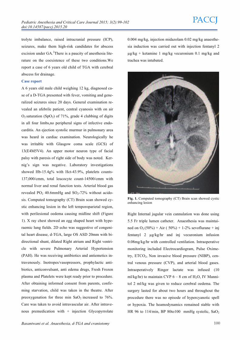
sis. Computed tomography (CT) Brain scan showed cy-
stic enhancing lesion in the left temporoparietal region,
with perilesional oedema causing midline shift (Figure
1). X ray chest showed an egg shaped heart with hype-
raemic lung fields. 2D echo was suggestive of congeni-
tal heart disease, d-TGA, large OS ASD 20mm with bi-
directional shunt, dilated Right atrium and Right ventri-
cle with severe Pulmonary Arterial Hypertension
(PAH). He was receiving antibiotics and antiemetics in-
travenously. Inotropes/vasopressors, prophylactic anti-
biotics, anticonvulsant, anti edema drugs, Fresh Frozen
plasma and Platelets were kept ready prior to procedure.
After obtaining informed consent from parents, confir-
ming starvation, child was taken in the theatre. After
preoxygenation for three min SaO2 increased to 76%.
Care was taken to avoid intravascular air. After intrave-
nous premedication with + injection Glycopyrrolate
0.004 mg/kg, injection midazolam 0.02 mg/kg anaesthe-
sia induction was carried out with injection fentanyl 2
µg/kg + ketamine 1 mg/kg vecuronium 0.1 mg/kg and
trachea was intubated.
Fig. 1. Computed tomography (CT) Brain scan showed cystic enhancing lesion
Right Internal jugular vein cannulation was done using
5.5 Fr triple lumen catheter. Anaesthesia was maintai-
ned on O2 (50%) + Air ( 50%) + 1-2% sevoflurane + inj
fentanyl 2 µg/kg/hr and inj vecuronium infusion
0.08mg/kg/hr with controlled ventilation. Intraoperative
monitoring included Electrocardiogram, Pulse Oxime-
try, ETCO2, Non invasive blood pressure (NIBP), cen-
tral venous pressure (CVP), and arterial blood gases.
Intraoperatively Ringer lactate was infused (10
ml/kg/hr) to maintain CVP 6 – 8 cm of H2O, IV Manni-
tol 2 ml/kg was given to reduce cerebral oedema. The
surgery lasted for about two hours and throughout the
procedure there was no episode of hypercyanotic spell
or hypoxia. The heamodynamics remained stable with
HR 96 to 114/min, BP 80to100 mmHg systolic, SaO2
Pediatric Anesthesia and Critical Care Journal 2015; 3(2):99-102 doi:10.14587/paccj.2015.20
Basantwani et al. Anaesthesia, d-TGA and craniotomy 101
ranging between 82–86% and PaCO2 between 30–33
mmHg. Craniotomy with drainage of encapsulated ab-
scess cavity was performed by Neurosurgeon. At the
end of surgery, residual neuromuscular blockade was
reversed, patient extubated once awake and monitored
in Neurosurgery ICU for 24 hrs postoperatively. IV Pa-
racetamol 15 mg /kg body weight 8 hourly was used for
postoperative analgesia.
Discussion
Patients with cyanotic congenital heart disease (cCHD)
are prone to develop frequent brain abscesses. TGA if
uncorrected, has a 30% mortality rate in the first week
of life, 45% in the first month and 90% in the first year.
Those who survive this period present with the pro-
blems of preexisting hypoxia and cyanosis, polycythe-
mia, hypercoagulability, thrombotic complications, coa-
gulopathies. They have higher incidence of systemic in-
fections due to bypass of the filtering of pulmonary ca-
pillaries leading to brain abscess.2 Treatment of brain
abscess is with systemic antibiotics. However, abscesses
which are larger than 2 cms in diameter, causing midline
shift, multiple abscesses require surgical excision under
general anaesthesia in small children.3,4 Mortality rate
remains as high as 13% in patients of cyanotic congeni-
tal heart disease (cCHD) with brain abscess4. The risk
further increases under anaesthesia due to presence of
other extracardiac malformations (20-45%)4, difficult
airway, a fragile cardiopulmonary status and various sy-
stemic and coagulation complications, together with ab-
scess-induced problems such as raised intracranial pres-
sure (ICP), seizures, dehydration, electrolyte imbalance.
Anesthetizing children with cCHD and a brain abscess
necessitates use of an anesthesia regimen appropriate to
both cCHD and intracranial surgery. Intraoperative
goals include maintenance of hemodynamic stability
and oxygenation and prevention of cyanotic spells and
arrhythmias. Perioperative hypoxia leads to hypervisco-
sity of blood and coagulation abnormality requiring
phlebotomy if haematocrit is above 65% . If coagulation
abnormalities are present, platelet concentrates are nee-
ded. In our patient the PCV was 43.9%, and did not ha-
ve deranged coagulation profile. The amount of shun-
ting of blood in dTGA is determined by the ratio of the
systemic vascular resistance (SVR) to pulmonary vascu-
lar resistance (PVR). As SVR is increased, right to left
shunting decreases. However increase in PVR leads to
abrupt worsening of cyanosis, tachycardia, hypotension.
Hence, one should maintain systemic vascular resistan-
ce, minimize pulmonary vascular resistance,maintain
cardiovascular stability. The optimal induction regimen
for general anaesthesia in cCHD should aim to improve
arterial blood oxygen saturation (SaO2)5. Induction of
anaesthesia increases arterial saturation probably due to
high oxygen concentration plus decreased oxygen con-
sumption with induction of anaesthesia and muscle pa-
ralysis6. However, if systemic vasodilatation occurs, it
may exacerbate the right to left shunting and intensify
hypoxia. We used a combination of Fentanyl and keta-
mine in our case for induction of anaesthesia which ac-
tually improved arterial oxygenation and maintained it
between 82-86% intraoperatively. Ketamine decreases
the right to left shunt by increasing systemic vascular
resistance4,7,8.9 and is a suitable induction agent in such
cases. Hypotension, hypovolemia, acidosis, hypoxia,
and hypercarbia increase intraoperative shunting and
should be avoided. Hypocarbia and diuretic therapy are
usually employed to obtain lax brain. Diuretic should be
used carefully to avoid hypovolemia and reduced right
ventricular output. Mannitol in a small dose is ideal as it
reduces blood viscosity also. We avoided hypovolemia
by maintenance of intravascular volume7. Early extuba-
tion is preferred to prevent further reduction in pulmo-
nary blood flow due to prolonged ventilation. Adequate
post operative analgesia is essential as pain increases
pulmonaryvascular resistance and sudden increase in
oxygen demand.
Conclusion
Brain abscess is a known complication in patients with
cyanotic congenital heart disease, which must be dia-
gnosed early and treated aggressively. In patients with
Pediatric Anesthesia and Critical Care Journal 2015; 3(2):99-102 doi:10.14587/paccj.2015.20
Basantwani et al. Anaesthesia, d-TGA and craniotomy 102
un-operated cyanotic congenital heart disease, early cor-
rective surgery or palliative shunts i-e Blalock Taussig,
Bidirectional Glen shunt for these cardiac malforma-
tions would be a definitive way of reducing this compli-
cation. A carefully administered GA with controlled
ventilation, maintaining cardiac output, normal sinus
rhythm and keeping PVR relatively lower than SVR
along with lax brain is recommended.
References
1. Prusty GK. Brain abscesses in cyanotic heart disea-
se. Indian J Pediatr 1993; 60:43-51.
2. Castenada AR,Jonas RA,Mayer JE,et al. Cardiac
surgery of the neonate and infan,t Philadelphia: WB
Saunders, 1994:409-438
3. Mehnaz Atiq, Umair Syed Ahmed, Salman Saleem
Allana and Khalid N. Chishti. Brain Abscess in
Children. Indian Journal of Pediatrics, 2006:73:401-
404.
4. Pandian JD, Monma NV, Cherian PJ, Radhakrish-
nan K. Brainstem abscess complication tetralogy of
Fallot successfully treated with antibiotics alone.
Neurology India 2000; 48: 272-5.
5. Takeshita M, Kagawa M, Yonetani H, Izawa M,
Yato S, Nakanishi T, Monma K. Risk factors for
brain abscess in patients with congenital cyanotic
heart disease. Neurologia medico-Chirurgica 1992;
32: 667-70.
6. Aebi C, Kauffmann F, Schaad UB. Brain abscess in
childhood-long term experience. European Journal
of Pediatrics 1991; 150: 282-6.
7. Takeshita M, Kagawa M, Yato S, Izawa M, Onda
H, Takakura K, et al. Current treatment of brain ab-
scess in patients with congenital cyanotic heart di-
sease. Neurosurgery 1997; 41: 1270-9
8. Raha A, P. Ganjoo, A. Singh, M. Tandon. Surgery
for brain abscess in children with cyanotic heart di-
sease: an anesthetic challenge. Journal of Pediatric
Neurosciences 2012; 7:23-26.
9. S.Routray, K Raut , D. Mishra .Cerebral abscess in
8 years old with uncorrected tetralogy of Fallot:
Anaesthetic Challenge. International Journal of
Biomedical and Advance Research 2013; 4:843-
845.