Embed Size (px)
Citation preview

CASE REPORT Open Access
A case report of infantile cysticnephroblastomaNozomu Kurose1* , Michiho Takenaka2, Manabu Yamashita2, Chie Shimaguchi2, Mariko Nakano2, Bryant Britni3,Xin Guo1, Chizuru Futatsuya1, Akihiro Shioya1 and Sohsuke Yamada1*
Abstract
Background: Nephroblastoma (NB) is a malignant embryonal neoplasm derived from nephrogenic blastemal cells.NB usually forms a solid mass, but in extremely rare cases, it may show cystic changes.
Case presentation: A six-month-old girl with persistent high fevers was found to have pyuria and bacteriuria.Ultrasonography revealed multilocular cysts in the right kidney. Right nephrectomy was performed with cyst wallrupture during surgery. An intraoperative rapid diagnosis, based on peritoneal fluid cytology, confirmed threecomponents of blastemal, stromal, and epithelial cells. The blastemal cells were dyshesive, with scant to nocytoplasm and were the predominant cell type. The spindle-shaped stromal cells were arranged in fascicles. Theepithelial cells demonstrated tubular structures. Macroscopically, the resected cystic tumor measured 80 mm inmaximum diameter with a prominently thin cyst wall, but solid areas were also apparent. Histologically, the tumorwas diagnosed as cystic NB (blastemal-predominant) displaying a triphasic pattern. Hyperchromatic nuclei andapoptotic bodies were found. The clinical stage classification of Japan Wilms Tumor Study group was 3. The patientwas treated with chemotherapy and radiotherapy. Tumor recurrence and metastasis have not been observed in the8 months since surgery.
Conclusion: This is an extremely rare case of infantile cystic NB. We diagnosed the NB cells that appeared in theperitoneal fluid by intraoperative rapid cytology. Cytological examination proved to be a very useful technique fordetermining the clinical stage of NB. Additionally, we propose that massive tumor degeneration and necrosis beconsidered as a pathogenic mechanism of cyst formation in NB.
Keywords: Cyst, Nephroblastoma (NB), Infant, Peritoneal fluid, Cytology
BackgroundWilms tumor, also known as nephroblastoma (NB) is a ma-lignant embryonal neoplasm derived from nephrogenicblastemal cells. NB is the most common malignant renaltumor in children and 98% of cases occur under the age of10. The mean age at diagnosis is 37 months and 43 monthsamong males and females, respectively [1]. However,adult-onset cases have also been reported [2]. Grossly, NBusually forms a solitary and rounded-solid mass. Histologi-cally, the three components of blastemal, epithelial, andstromal cells are mixed in various proportions.
Renal cystic tumors in children can be benign, such ascystic nephroma (multilocular cyst), and malignant hom-ologous tumors, such as cystic partially differentiatedNB (CPDNB) [3]. Furthermore, very few case of cysticNBs in infants and adults have been reported [4–6].We herein report an extremely rare case of cystic NB
in an infant and discuss its pathogenic mechanism. Inaddition, we also describe the cytological findings of NBthat appeared in peritoneal fluid.
Case presentationA previously healthy six-month-old girl who was bornfull-term following an uncomplicated pregnancy pre-sented with persistent high fever and was found to havepyuria and bacteriuria. Prior to this, she had no signifi-cant medical history. No obvious gross malformationswere observed on physical examination nor was any
* Correspondence: [email protected]; [email protected] of Pathology and Laboratory Medicine, Kanazawa MedicalUniversity, 1-1 Daigaku, Uchinada, Ishikawa 920-0293, JapanFull list of author information is available at the end of the article
© The Author(s). 2018 Open Access This article is distributed under the terms of the Creative Commons Attribution 4.0International License (http://creativecommons.org/licenses/by/4.0/), which permits unrestricted use, distribution, andreproduction in any medium, provided you give appropriate credit to the original author(s) and the source, provide a link tothe Creative Commons license, and indicate if changes were made. The Creative Commons Public Domain Dedication waiver(http://creativecommons.org/publicdomain/zero/1.0/) applies to the data made available in this article, unless otherwise stated.
Kurose et al. Diagnostic Pathology (2018) 13:84 https://doi.org/10.1186/s13000-018-0761-5
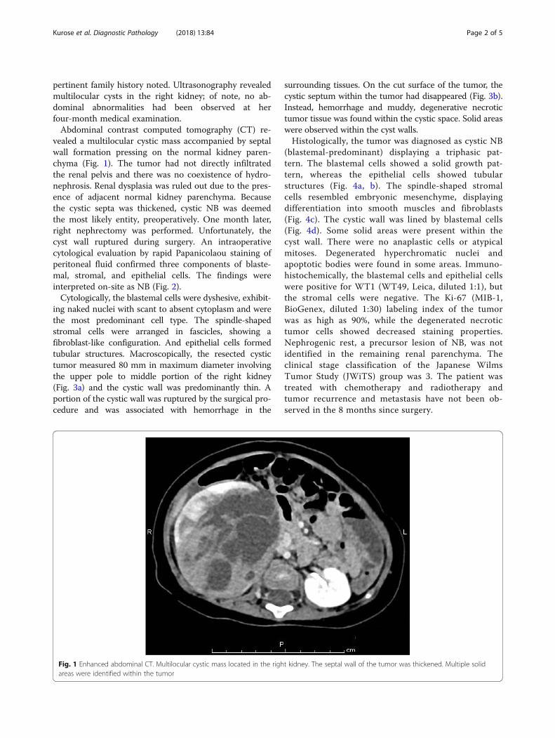
pertinent family history noted. Ultrasonography revealedmultilocular cysts in the right kidney; of note, no ab-dominal abnormalities had been observed at herfour-month medical examination.Abdominal contrast computed tomography (CT) re-
vealed a multilocular cystic mass accompanied by septalwall formation pressing on the normal kidney paren-chyma (Fig. 1). The tumor had not directly infiltratedthe renal pelvis and there was no coexistence of hydro-nephrosis. Renal dysplasia was ruled out due to the pres-ence of adjacent normal kidney parenchyma. Becausethe cystic septa was thickened, cystic NB was deemedthe most likely entity, preoperatively. One month later,right nephrectomy was performed. Unfortunately, thecyst wall ruptured during surgery. An intraoperativecytological evaluation by rapid Papanicolaou staining ofperitoneal fluid confirmed three components of blaste-mal, stromal, and epithelial cells. The findings wereinterpreted on-site as NB (Fig. 2).Cytologically, the blastemal cells were dyshesive, exhibit-
ing naked nuclei with scant to absent cytoplasm and werethe most predominant cell type. The spindle-shapedstromal cells were arranged in fascicles, showing afibroblast-like configuration. And epithelial cells formedtubular structures. Macroscopically, the resected cystictumor measured 80 mm in maximum diameter involvingthe upper pole to middle portion of the right kidney(Fig. 3a) and the cystic wall was predominantly thin. Aportion of the cystic wall was ruptured by the surgical pro-cedure and was associated with hemorrhage in the
surrounding tissues. On the cut surface of the tumor, thecystic septum within the tumor had disappeared (Fig. 3b).Instead, hemorrhage and muddy, degenerative necrotictumor tissue was found within the cystic space. Solid areaswere observed within the cyst walls.Histologically, the tumor was diagnosed as cystic NB
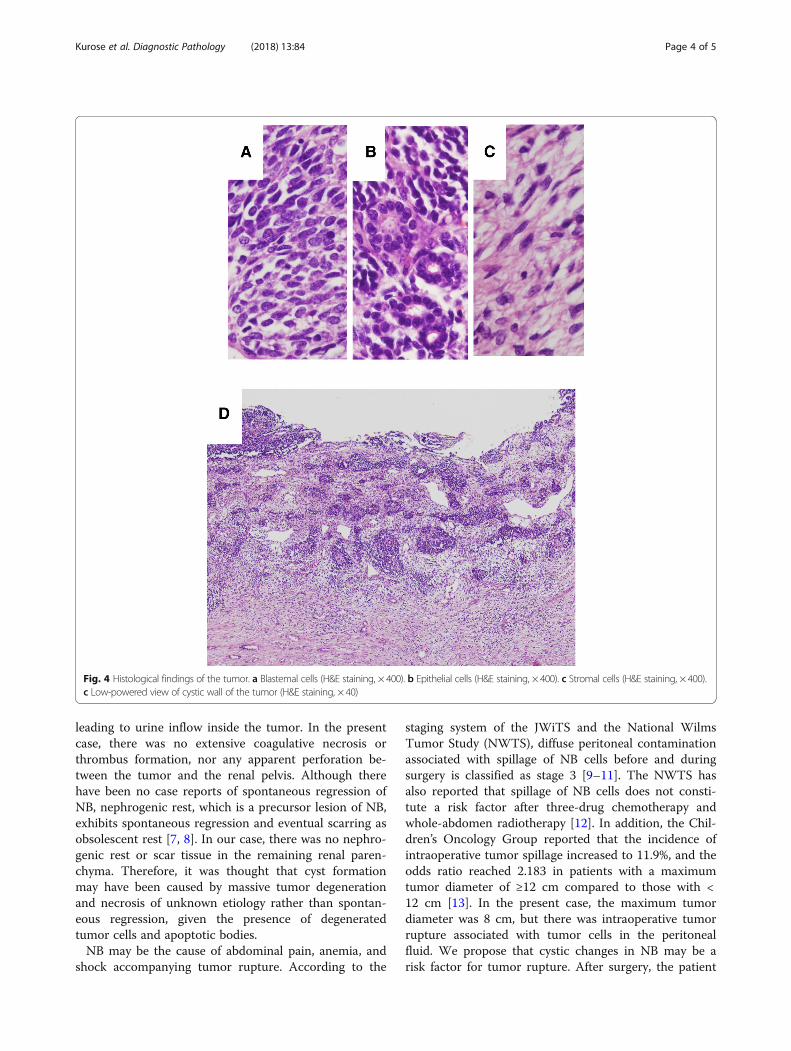
(blastemal-predominant) displaying a triphasic pat-tern. The blastemal cells showed a solid growth pat-tern, whereas the epithelial cells showed tubularstructures (Fig. 4a, b). The spindle-shaped stromalcells resembled embryonic mesenchyme, displayingdifferentiation into smooth muscles and fibroblasts(Fig. 4c). The cystic wall was lined by blastemal cells(Fig. 4d). Some solid areas were present within thecyst wall. There were no anaplastic cells or atypicalmitoses. Degenerated hyperchromatic nuclei andapoptotic bodies were found in some areas. Immuno-histochemically, the blastemal cells and epithelial cellswere positive for WT1 (WT49, Leica, diluted 1:1), butthe stromal cells were negative. The Ki-67 (MIB-1,BioGenex, diluted 1:30) labeling index of the tumorwas as high as 90%, while the degenerated necrotictumor cells showed decreased staining properties.Nephrogenic rest, a precursor lesion of NB, was notidentified in the remaining renal parenchyma. Theclinical stage classification of the Japanese WilmsTumor Study (JWiTS) group was 3. The patient wastreated with chemotherapy and radiotherapy andtumor recurrence and metastasis have not been ob-served in the 8 months since surgery.
Fig. 1 Enhanced abdominal CT. Multilocular cystic mass located in the right kidney. The septal wall of the tumor was thickened. Multiple solidareas were identified within the tumor
Kurose et al. Diagnostic Pathology (2018) 13:84 Page 2 of 5

DiscussionThis case report describes an extremely rare case of cys-tic NB that developed in an infant with no family historyor malformation. Furthermore, we describe the cyto-logical findings of the tumor observed intraoperativelyinvolving the peritoneal fluid.The differential diagnosis of cystic NB includes
CPDNB. CPDNB also exhibits multiloculated cysts, as inthis case, however these lesions should not have appar-ent solid or nodular mass formation. CPDNB containsmature or immature nephroblastic tissue, but conformsto the septum between cysts. In contrast, cystic NB has
solid NB components expanding the cyst wall, whichwere easily identified in this case.To our knowledge, only three cases of NB with cyst
formation have been reported in the English literature[4–6]. However, there were no case reports of rupturedcystic NB. One case was an infantile case (9.5 monthsold) and the other 2 cases were in adults (36 and 30 yearsold). The mechanism underlying the cyst formation isnot well understood. However, the following two pos-sible mechanisms have been proposed: 1) formation dueto tumor degeneration and necrosis and 2) formationdue to the tumor infiltrating into the renal pelvis,
Fig. 2 Cytology of the peritoneal fluid. Three components of blastemal, stromal and epithelial cells were seen. The blastemal cells showed decreasedcell adhesion (arrow), the stromal cells showed a fibroblast-like configuration (arrow head), and the epithelial cells showed a tubular structure (inset)(Papanicolaou staining, × 400)
A B
Fig. 3 Gross findings of the resected right kidney. a The tumor was localized from the upper pole to the middle portion. b The cystic septumwithin the tumor had disappeared. Some solid areas were seen (arrow head)
Kurose et al. Diagnostic Pathology (2018) 13:84 Page 3 of 5
leading to urine inflow inside the tumor. In the presentcase, there was no extensive coagulative necrosis orthrombus formation, nor any apparent perforation be-tween the tumor and the renal pelvis. Although therehave been no case reports of spontaneous regression ofNB, nephrogenic rest, which is a precursor lesion of NB,exhibits spontaneous regression and eventual scarring asobsolescent rest [7, 8]. In our case, there was no nephro-genic rest or scar tissue in the remaining renal paren-chyma. Therefore, it was thought that cyst formationmay have been caused by massive tumor degenerationand necrosis of unknown etiology rather than spontan-eous regression, given the presence of degeneratedtumor cells and apoptotic bodies.NB may be the cause of abdominal pain, anemia, and
shock accompanying tumor rupture. According to the
staging system of the JWiTS and the National WilmsTumor Study (NWTS), diffuse peritoneal contaminationassociated with spillage of NB cells before and duringsurgery is classified as stage 3 [9–11]. The NWTS hasalso reported that spillage of NB cells does not consti-tute a risk factor after three-drug chemotherapy andwhole-abdomen radiotherapy [12]. In addition, the Chil-dren’s Oncology Group reported that the incidence ofintraoperative tumor spillage increased to 11.9%, and theodds ratio reached 2.183 in patients with a maximumtumor diameter of ≥12 cm compared to those with <12 cm [13]. In the present case, the maximum tumordiameter was 8 cm, but there was intraoperative tumorrupture associated with tumor cells in the peritonealfluid. We propose that cystic changes in NB may be arisk factor for tumor rupture. After surgery, the patient
Fig. 4 Histological findings of the tumor. a Blastemal cells (H&E staining, × 400). b Epithelial cells (H&E staining, × 400). c Stromal cells (H&E staining, × 400).c Low-powered view of cystic wall of the tumor (H&E staining, × 40)
Kurose et al. Diagnostic Pathology (2018) 13:84 Page 4 of 5

received chemotherapy and radiation therapy accordingto the protocol of the NWTS. Because of the patient’sfavorable histologic features (lack of anaplasia) andexcellent response to therapy, this case is expected tohave a good prognosis.
ConclusionsWe present an extremely rare case of infantile cystic NB.The diagnosis was able to be made on-site by intraoper-ative rapid cytology of peritoneal fluid. This technique ofcytological examination proved to be very useful for de-termining the clinical stage of NB. Additionally, massivetumor degeneration and necrosis should be consideredas a possible mechanism of cyst formation in NB.
AbbreviationsCPDNB: Cystic partially differentiated nephroblastoma; CT: Computedtomography; JWiTS: Japanese Wilms Tumor Study; NB: Nephroblastoma;NWTS: National Wilms Tumor Study
AcknowledgmentsWe would like to thank Seiya Mizuguchi, Yumi Tsubata, Yoshiiku Ohkanemasa,Toshie Terauchi and Satoko Nakada for their expert technical assistance andhelpful comments.
FundingNone.
Availability of data and materialsThe dataset supporting the findings and conclusions of this case report isincluded within the article.
Authors’ contributionsNK and SY participated in the conception of the study and writing of themanuscript. MT, MY, CS, MN, BB, XG, CF and AS performed the clinicalimaging and/or pathological/cytological/immunohistochemicalinterpretation of this lesion. All of the authors have read and approvedthe final manuscript.
Authors’ informationNK, XG, CF, AS and SY belong to the Department of Pathology andLaboratory Medicine, Kanazawa Medical University, Ishikawa, Japan. MT,MY, CS and MN belong to the Department of Pathology, KanazawaMedical University, Ishikawa, Japan. BB belong to the Department ofPathology and Laboratory Medicine, University of Vermont College ofMedicine, Burlington, Vermont, USA.
Ethics approval and consent to participateNot applicable.
Consent for publicationWritten informed consent was obtained from the patient’s parents for thepublication of this case report and any accompanying images. A copy of thesigned consent is available for review by the editor of this journal.
Competing interestsThe authors declare that they have no competing interests.
Publisher’s NoteSpringer Nature remains neutral with regard to jurisdictional claims in publishedmaps and institutional affiliations.
Author details1Department of Pathology and Laboratory Medicine, Kanazawa MedicalUniversity, 1-1 Daigaku, Uchinada, Ishikawa 920-0293, Japan. 2Department ofPathology, Kanazawa Medical University, Ishikawa, Japan. 3Department of
Pathology and Laboratory Medicine, University of Vermont College ofMedicine, Burlington, VT, USA.
Received: 12 April 2018 Accepted: 10 October 2018
References1. Moch H, Humphrey PA, Ulbright TM, Reuter VE. WHO Classification of
Tumours of the urinary system and male genital organs, vol. 49. 4th ed.Lyon: IARC Press; 2016.
2. Huser J, Grignon DJ, Ro JY, Ayala AG, Shannon RL, Papadopoulos NJ. AdultWilms’ tumor: a clinicopathologic study of 11 cases. Mod Pathol. 1990;3:321–6.
3. Babut JM, Bawab F, Jouan H, Coeurdacier P, Treguier C, Fremond B. Renalcystic tumours in children-a diagnostic challenge. Eur J Pediatr Surg. 1993;3:157–60.
4. Bindhu J, Imtiaz A, Kumar RV, Thejaswini MD. Cystic variant of favorable-histology Wilms’ tumor presenting with osteolytic metastasis to the ribs. JPostgrad Med. 2010;56:28–30.
5. Fujita K, Nishimura K, Yasunaga Y, Miyake O, Hirota S, Okuyama A. AdultWilms’ tumor mimicking hemorrhagic renal cyst. Int J Urol. 2003;10:492–4.
6. Adjei ON, Tamura S, Sugimura H, Shimizu T, Kihara Y, Asato M, Qiang TX,Watanabe K, Marutsuka K, Hasui Y. Adult cystic Wilms' tumor(nephroblastoma): radiologic features with pathologic correlation. RadiatMed. 1996;14:287–91.
7. Beckwith JB. Precursor lesions of Wilms tumor: clinical and biologicalimplications. Med Pediatr Oncol. 1993;21:158–68.
8. William MM, David JG, Elizabeth JP. Tumors of the kidney, bladder andrelated urinary structures (AFIP atlas of tumor pathology 4th series).Washington, DC: AFIP Press; 2004. p. 38–47.
9. D’Angio GJ, Breslow N, Beckwith JB, Evans A, Baum H, deLorimier A,Fernbach D, Hrabovsky E, Jones B, Kelalis P. Treatment of Wilms’ tumor.Results of the third National Wilms’ tumor study. Cancer. 1989;64:349–60.
10. Fukuzawa H, Shiima Y, Mishima Y, Sekine S, Miura S, Yabe K, Yamaki S,Morita K, Okata Y, Hisamatsu C, Nakao M, Yokoi A, Maeda K, Kosaka Y.Predictive factor for intraoperative tumor rupture of Wilms tumor. PediatrSurg Int. 2017;33:91–5.
11. Khanna G, Naranjo A, Hoffer F, Mullen E, Geller J, Gratias EJ, Ehrlich PF,Perlman EJ, Rosen N, Grundy P, Dome JS. Detection of preoperative Wilmstumor rupture with CT: a report from the Children's oncology group.Radiology. 2013;266:610–7.
12. Ehrlich PF, Anderson JR, Ritchey ML, Dome JS, Green DM, Grundy PE,Perlman EJ, Kalapurakal JA, Breslow NE, Shamberger RC. Clinicopathologicfindings predictive of relapse in children with stage III favorable-histologyWilms tumor. J Clin Oncol. 2013;31:1196–201.
13. Gow KW, Barnhart DC, Hamilton TE, Kandel JJ, Chen MK, Ferrer FA, Price MR,Mullen EA, Geller JI, Gratias EJ, Rosen N, Khanna G, Naranjo A, Ritchey ML,Grundy PE, Dome JS, Ehrlich PF. Primary nephrectomy and intraoperativetumor spill: report from the Children’s oncology group (COG) renal tumorscommittee. J Pediatr Surg. 2013;48:34–8.
Kurose et al. Diagnostic Pathology (2018) 13:84 Page 5 of 5