Embed Size (px)
Citation preview

The Journal of Neuroscience, August 1991, If(e): 2499-2509
A Cellular Analysis of Inhibition in the Siphon Withdrawal Reflex of Aplysia
William G. Wright,” Emilie A. Marcus, and Thomas J. Carew
Departments of Biology and Psychology, Yale University, New Haven, Connecticut 06520
Recent behavioral experiments examining the siphon with- drawal reflex of Apiysia have revealed inhibitory effects of strong tail shock, a stimulus commonly used as an uncon- ditioned stimulus in studies of associative and nonassocia- tive learning in Aplysia.
We utilized a reduced preparation to perform a cellular analysis of tail shock-induced inhibition in the siphon with- drawal reflex. First, we carried out behavioral studies that showed that the reduced preparation exhibits a siphon with- drawal reflex to water jet stimuli, and that tail shock produces inhibitory behavioral effects comparable to those in the in- tact animal: (1) strong shock produces transient inhibition of nonhabituated responses, and (2) a habituated response is facilitated by weak shock, but not by strong shock, sug- gesting that increasing tail shock intensity recruits the in- hibitory process that competes with facilitation of habituated reflexes. Next, we carried out cellular studies that showed that the amplitude of the complex EPSP in siphon motor neurons elicited by water jet stimuli to the siphon also ex- hibits the inhibitory patterns produced by tail shock: (1) the nondecremented complex EPSP (a neural correlate of a non- habituated siphon withdrawal reflex) is significantly inhibited 90 set after strong tail shock and recovers to preshock lev- els 10 min later, and (2) the decremented complex EPSP (a neural correlate of a habituated reflex) is significantly facil- itated by weak shock, but is not facilitated by strong shock.
In addition to the complex EPSP, we simultaneously ex- amined the monosynaptic connection between siphon sen- sory neurons and siphon motor neurons. The monosynaptic EPSP does not show the pattern of inhibitory modulation by tail shock exhibited by the siphon withdrawal reflex and the complex EPSP: (1) the nondecremented monosynaptic EPSP is not inhibited 90 set after strong shock, but tends to be above preshock levels; and (2) the decremented monosyn- aptic EPSP is facilitated by weak as well as strong tail shock.
Our results suggest that an important component of the
Received Aug. 24, 1990; revised Mar. 12, I99 1; accepted Mar. 13, I99 1.
We thank Diana Blazis, David Cook, Kent Fitzgerald, and Mark Stopfer for their helpful comments on the manuscript and Elizabeth McCance for excellent assistance in the behavioral experiments. This work was supported by National Institute of Mental Health National Research Service Award l-F32-MH09397 to W.G.W., by a National Science Foundation graduate fellowship and National Institute of Mental Health National Research Service Award l-F31-MH09874- 01-BPN-2 to E.A.M., and by National Science Foundation Grant BNS8311300 and Office of Naval Research Grant N00014-87-K-0381 to T.J.C.
Corresoondence should be addressed to Thomas J. Carew. Deoartment of Psv- chology, -Yale University, P.O. Box 1 IA Yale Station, New’HaGen, CT 06520:
z Present address: Biology Department, Colorado State University, Fort Collins, CO 80521.
Copyright 0 1991 Society for Neuroscience 0270-6474/91/l 12498-12$03.00/O
inhibitory process triggered by strong tail shock is mediated by neural elements presynaptic to the siphon motor neurons. Because modulation of the monosynaptic connection be- tween identified siphon sensory and siphon motor neurons does not parallel the tail shock-induced inhibitory patterns observed in the siphon withdrawal reflex and in the complex EPSP, other synaptic connections are likely to play an im- portant role in mediating tail shock-induced inhibition in the siphon withdrawal reflex.
At both behavioral and cellular levels, a complete analysis of the processes that underlie learning requires understanding all of the possible effects of the stimuli used to produce the learning. In the marine mollusk Aplysia, a preparation well suited for a behavioral and cellular analysis of learning, an aversive electric shock to the tail has often been used as an unconditioned stim- ulus (US) to produce a variety of forms of both nonassociative and associative learning (Byrne, 1987; Carew, 1987; Hawkins et al., 1987). For a complete mechanistic understanding of leam- ing in Aplysia when tail shock is used as a US, it is important to understand the cellular mechanisms underlying all of the behavioral effects of tail shock. Tail shock has long been known to have facilitatory effects on a number of defensive reflexes, including siphon withdrawal, gill withdrawal, tail withdrawal, and escape locomotion, and in many cases a detailed under- standing of the cellular basis of these facilitatory effects has emerged (Klein and Kandel, 1980; Bemier et al., 1982; Siegel- baum et al., 1982; Occor et al., 1986; Walsh and Byrne, 1989; Baxter and Byrne, 1989; for reviews, see Kandel and Schwartz, 1982; Byrne, 1987; Carew, 1987; Hawkins et al., 1987). Re- cently, however, it has become clear that tail shock can also have inhibitory effects in some of these same reflexes (Krontiris- Litowitz et al., 1987; Mackey et al., 1987; Marcus et al., 1987, 1988; Rankin and Carew, 1987, 1989), In the present study, we have begun to explore the cellular basis of these inhibitory ef- fects.
In addition to providing insights into cellular mechanisms of inhibition in Apfysia, this article and its companion (Fitzgerald and Carew, 1991) are also aimed at a more general objective. Inhibitory processing has figured prominently in several general theories of learning (Mackintosh, 1974). However, the detailed cellular mechanisms by which inhibitory and excitatory pro- cesses interact to give rise to behavioral outcomes predicted by specific learning theories are very poorly understood. Because restricted neural networks in Aplysia, such as the neural circuit for the siphon withdrawal reflex examined in this article, are known to support several forms of learning, an understanding of the cellular mechanisms by which excitatory and inhibitory processes interact in these networks may provide general in-

The Journal of Neuroscience, August 1991, ff(8) 2499
sights into the specific roles that excitatory and inhibitory pro- cesses play in specific forms of learning.
Marcus et al. (1988) identified both excitatory and inhibitory effects of tail shock on the water jet-elicited siphon withdrawal reflex by systematically varying three parameters: (1) the initial state of the reflex (habituated or nonhabituated), (2) the strength of the modulatory tail shock, and (3) the time of testing after tail shock. A principal finding from that study was that strong tail shock recruited an inhibitory process, which could be ob- served directly as a transient decrease ofa nonhabituated siphon withdrawal reflex (see also Krontiris-Litowitz et al., 1987; Mack- ey et al., 1987; Rankin and Carew, 1987, 1989). The inhibitory process could also be inferred from the observation that, when the siphon withdrawal reflex was habituated just prior to tail shock, weak tail shock produced robust facilitation of the ha- bituated reflex (dishabituation), whereas strong shock produced no reflex facilitation. This suggested that increasing the tail shock intensity recruits the inhibitory process that competes with the facilitatory process of dishabituation (Marcus et al., 1988; see also Rankin and Carew, 1989).
Previous work on the siphon withdrawal reflex (Mackey et al., 1987) had proposed the monosynaptic connection between identified siphon sensory neurons (LE) and siphon motor neu- rons (LFS and LBS) as a possible locus mediating tail shock- induced inhibition. This suggestion was based on the observa- tion that tail shock produced transient inhibition of the mono- synaptic EPSP from sensory to motor neurons; the inhibition later gave way to facilitation. However, as pointed out by Mack- ey et al. (1987), though the sequence of the synaptic changes paralleled that of the behavioral changes, the time course of these monosynaptic changes differed from that ofthe behavioral changes. Specifically, inhibition ofthe monosynaptic connection gave way to facilitation within 90 set after tail shock, whereas behavioral inhibition of the reflex was observed to persist for several minutes after shock (Mackey et al., 1987). Thus, the synaptic changes observed at the level of the monosynaptic connection did not appear to account fully for the observed behavioral inhibition, implying that other cellular loci might be involved. In this article, we present a cellular analysis that es- tablishes neuronal correlates of the tail shock-induced inhibi- tory process in central neurons that mediate the siphon with- drawal reflex in Aplysia. We provide evidence that the inhibitory process in the siphon withdrawal reflex is not directly mediated by the LE sensory neuron-motor neuron connection, but rather appears to rely on input from intemeurons, or from other as yet unidentified sensory neurons.
Some of the results described in this article have previously been presented in preliminary form (Wright et al., 1988, 1989).
Materials and Methods Preparation. Adult Aplysia weighing 75-125 gm were obtained from Marinus, Inc., Long Beach, CA. Animals were anesthetized by injection of 120 ml of isotonic MgCl,, or by immersion in cold (-0°C) artificial sea water (ASW) for 30-50 min. The tail, mantle, gill, and siphon were dissected, leaving them attached to the CNS by their respective periph- eral nerves: the tail via the paired P9 nerves, and the mantle/gill/siphon via the siphon nerve (Fig. 1). The tail and mantle organs were then pinned loosely in a Sylgard-lined dish. In order to purge the tail of MgCl, and to deliver a supply of oxygen, the tail was constantly perfused with ASW solution (460 mM NaCl, 55 mM MgCI,, 11 mM CaCl,, 10 mM KCl, 10 mM Tris) via a hypodermic needle. In the behavioral experi- ments, approximately 60 ml of ASW was injected via hypodermic sy- ringe into the siphon 15-30 min before the start of each experiment.
Protocol. The various stimulus paradigms used in our experiments
TAIL SHOCK
Figure 1. The reduced preparation used to analyze the neuronal cor- relates of inhibition. See Materials and Methods for details.
are illustrated in Figure 2. Stimulus protocols equivalent to previous experiments with intact animals were used (Marcus et al., 1988): (1) A nondecremented (nonhabituated) protocol consisted of four water jet stimuli (two pretest and two test stimuli) delivered to the siphon at a IO-min interstimulus interval (ISI). The waterjet stimulus was produced by a Pica-Spritzer (General Valve Corp.), set at 25 psi and ‘IO-msec duration, delivered with a pipette (i.d., 5 mm) positioned approximately 5 mm from the internal surface of the siphon (see Fig. 1). This stimulus intensity was constant throughout each preparation. A strong electric shock (four I-set pulses of 50 mA AC, at a 1 -set interstimulus interval) was delivered to the tail 90 set before the first test stimulus. (2) The decremented (habituated) protocol was equivalent to dishabituation training and consisted of 2 1 water jet stimuli delivered at a rate (1 OO- set ISI) that produces habituation. Ninety seconds before the last water jet stimulus, an electric shock was delivered with one of two intensities: weak (a single 1 -set pulse of 10 mA AC) or strong (four 1 -set pulses of 50 mA AC).
Behavioralexperiments. In the nonhabituated protocol, animals were randomly assigned to shocked or unshocked groups. In the decremented protocol, animals were randomly assigned to weak- or strong-shock groups. Because some animals receiving strong shock produced ink, to maintain blind procedures we introduced ink taken from the ink gland of another animal into the chambers of all groups before the first post- shock test. An observer blind to the specific experimental conditions scored the duration of each water jet-elicited siphon withdrawal reflex (see Marcus et al., 1988) throughout the experiment.
Cellular experiments. The abdominal ganglion was pinned ventral side up in 50:50 isotonic MgCl,:ASW, and the left hemiganglion was desheathed. After rinsing the ganglion in ASW, a siphon motor neuron (LFS; Frost et al., 1988) and a siphon sensory neuron (LE; Byrne et al., 1974) were impaled with glass microelectrodes (10-20 MQ) filled with 3 M KC1 (Fig. 1). Before each test, the motor neuron was hyperpolarized 40-50 mV below rest to prevent spiking. During each test, we monitored the amplitude of two different synaptic inputs onto the motor neuron: (1) A complex (polysynaptic) EPSP was produced by a water jet stimulus to the siphon. Waterjet intensity was reduced in the cellular experiments to permit stable long-term recordings; relative to the behavioral exper- iments the reduced intensity was still capable of eliciting a brisk dis- charge from the motor neurons at rest, even though it did not activate

2500 Wright et al. l Cellular Analysis of Inhibition
NON-DECREMENTED (ISI f 10 min)
PRE TESTS
STRONG TAIL SHOCK
DECREMENTED (ISI = 100 set)
Figure 2. Schematic illustration of the paradigms used to examine the inhib- itory process. Each vertical tick mark represents a water jet stimulus to the siphon. Tail shock was delivered at the
WEAK TAIL SHOCK
arrow. The responses that were used to calculate the pretest baseline and the posttests are indicated by different lev- els of shading.
the LE sensory neurons. The amplitude of the complex EPSP was mea- sured as the maximum depolarization occurring within 800 msec after delivery of the water jet stimulus. (2) A monosynaptic EPSP was pro- duced by a single action potential in the sensory neuron (elicited by injecting depolarizing current). Typically, the complex EPSP was tested first. followed 30 set later bv a test of the monosynaptic EPSP; however, in some experiments the &onosynaptic EPSP was tested first, followed within 10 set by a test of the complex EPSP. Earlier pilot studies had demonstrated that there was no effect of the order of presentation of the two stimuli. In a separate experiment, just prior to the water jet- elicited complex EPSP, the input resistance of the hyperpolarized motor neuron was assessed by injection of 0.25 nA of hyperpolarizing current and measurement of the resulting electrotonic potential.
Statistical analysis. All test responses are expressed as a percentage ofbaseline responding obtained by dividing the test scores by the median of the two preshock scores in the nondecremented protocol, or the last three preshock scores (trials 18-20) in the decremented protocol. Be- cause some protocols gave rise to decremented behavioral and synaptic responses with amplitude scores approaching 0, the resultant non-nor- mal distribution of response amplitudes necessitated the use of non- parametric statistics. Between-group comparisons were made with the Wilcoxon two-sample test (test statistic W; Hollander and Wolfe, 1973); within-group comparisons were made with the Wilcoxon sign-rank test for repeated measures (test statistic T+; Hollander and Wolfe, 1973). In some cases, within- and between-group differences were modest, only achieving statistical significance with a one-tailed test (the use of a one- tailed test was appropriate because the direction of the effect had been predicted a priori). These few cases are indicated. In all other cases, probabilities are two-tailed. Data are expressed as medians f the in- terquartile range. The asymmetry of the interquartile range in some figures reflects the fact that this measure of variability need not be equally distributed around the median.
Results Behavioral analysis In order to carry out a cellular analysis of inhibitory modulation in the siphon withdrawal reflex, it was first important to dem- onstrate that the reduced preparation used for the cellular anal- ysis (Fig. 1) exhibited the essential behavioral features of in-
I STRONG
TAIL SHOCK
hibition that are observed in the intact animal (Marcus et al., 1988). To address this question, we explored inhibition in two ways: (1) by examining the effects of strong tail shock on non- habituated responses, and (2) by examining the effects of both weak and strong tail shock on habituated reflex responses (see Fig. 2).
Nonhabituated responses
The siphon withdrawal reflex of two randomly assigned groups (shocked, N = 12, and unshocked, N = 12) was tested at a nonhabituating 1 0-min IS1 before and after tail shock. As in the intact animal, stimulating the siphon at an interval of 10 min produced no habituation of the siphon withdrawal reflex be- tween the first and second tests. The two pretests were averaged yielding a median duration of 4.9 set in both groups. Moreover, 90 set after strong tail shock, the reflex was markedly inhibited (median, 2.7 set, 55% of baseline; Fig. 3A, test 1). The inhibited reflex was significantly lower than preshock levels (sign-rank test; N = 12; T+ = 2; p = 0.004) and was also significantly lower than the nonshocked control group (two-sample test; N = 12; W = 106; p = 0.012). The reflex recovered significantly from tail shock-induced inhibition 10 min later (median, 4.7 set, 95% of baseline; sign-rank test; T+ = 70; p = 0.003; Fig. 3A, test 2), at which time it was no longer significantly different from baseline levels (sign-rank test; T+ = 25; p = 0.29), nor from the nonshocked control group (two-sample test; W = 144; p = 0.75). Thus, in the reduced preparation, strong tail shock producks significant transient reflex inhibition comparable to that ob- served in intact animals (Marcus et al., 1988).
Habituated responses To examine the effects of tail shock on habituated responses, the siphon withdrawal reflex was habituated by 20 repeated

The Journal of Neuroscience, August 1991, 17(8) 2501
A NON-HABITUATED B HABITUATED
-40
a2 5
f -60 - -60 TEST1 TEST 2 TEST 1
Figure 3. Tail shock-induced modulation of the siphon withdrawal reflex in the reduced preparation. Duration of siphon withdrawal in response to water jet stimuli is shown relative to the preshock baseline. A, Nonhabituated reflex: stimuli were delivered with a lo-min ISI. Data are expressed as percent change from the median of the two preshock tests. Strong shock (N = 12) produced significant inhibition of the reflex 90 set postshock (TEST I) compared to a nonshocked control group (N = 12). This inhibition recovered within 10 min (TEST 2). B, Habituated reflex: 20 water jet test stimuli were delivered with a lOO-set IS1 prior to tail shock. Tail shock was delivered 10 set after the 20th siphon stimulus, and a single test stimulus (TEST I) was given 90 set later. Data are expressed as percent change from the median of the last three preshock tests (pretests 18- 20). Weak tail shock (N = 11) produced modest but significant facilitation in the siphon withdrawal reflex, whereas strong shock (N = 10) produced no significant change. The asterisk indicates a one-tailed probability value. In this and all subsequent figures, data are expressed as medians + the interquartile range. The asymmetry of the interquartile range in some figures reflects the fact that this measure of variability need not be equally distributed around the median.
water jet stimuli to the siphon (N = 21). This produced signif- icant habituation from a preshock median duration of 5.2 set to a habituated median of 3.0 set (combining both groups’ pre- test responses; sign-rank test comparing last three responses to initial response; T+ = 0; p < 0.001). Half of the animals (N = 11) then received weak tail shock, while the other half (N = 10) received strong shock. The behavioral response to the weak shock was a brief tail and siphon withdrawal; in response to the strong tail shock, both the tail and siphon withdrew much more vigorously and for a longer period of time, and in some cases inking occurred. In both conditions, the siphon returned to base- line prior to the posttest. The results are shown in Figure 3B. Weak tail shock produced modest but significant facilitation of the habituated reflex (median, 109% of baseline; sign-rank test; N = 11; T+ = 55; p = 0.028, one-tailed), while strong shock produced no facilitation (median, 95%; sign-rank test; N = 10; T+ = 20; p = 0.49, one-tailed; Fig. 38). Moreover, there was a modest but significant difference between weak- and strong- shock groups (two-sample test; N = 10, 11; W = 145; p = 0.049, one-tailed).
In summary, though the behavioral effects in the reduced preparation using the habituated protocol are modest, they are consistent with the observations in intact animals that weak tail shock facilitates habituated responses, whereas strong shock does not. Taken together with our results on the nondecremented reflex, these observations suggest that strong tail shock activates an inhibitory process that competes with the facilitatory process of dishabituation.
Cellular analysis
The results of the behavioral experiments described above dem- onstrate that strong tail shock produces an inhibitory pattern in the reduced preparation similar to that previously observed in
the intact animal. We therefore used this preparation to search for central neuronal correlates of this reflex inhibition. Specif- ically, we examined (1) whether strong tail shock produces in- hibition of nondecremented synaptic responses that recovers after 10 min, and (2) whether decremented synaptic responses are facilitated by weak tail shock but not by strong tail shock.
Nondecremented responses
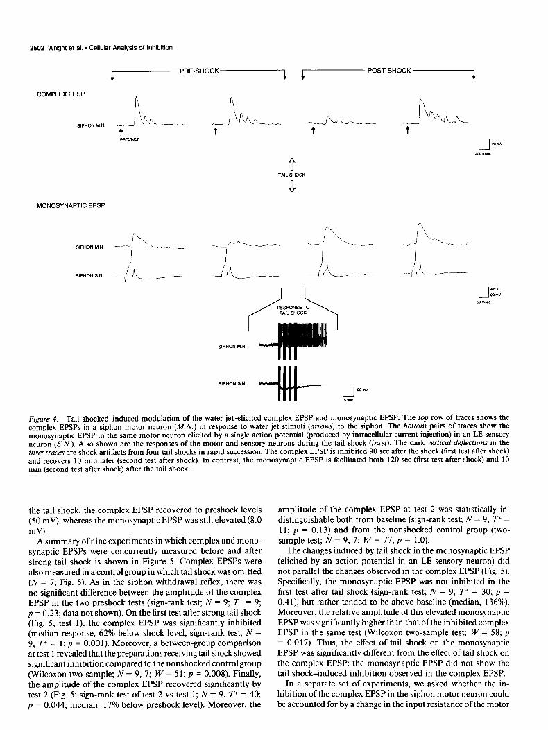
We first asked whether changes in nondecremented complex and monosynaptic EPSPs in siphon motor neurons paralleled the inhibition and recovery of the nonhabituated siphon with- drawal reflex after a strong tail shock. An example of our results is shown in Figure 4. The complex EPSP elicited by a water jet stimulus to the siphon at a IO-min IS1 was relatively stable in the two preshock tests (56 mV in pretest 1, 52 mV in pretest 2). However, the monosynaptic EPSP (elicited by a single action potential produced by intracellular current injection in an LE sensory neuron) in the same motor neuron showed considerable decrement (5.6 mV in pretest 1; 2.2 mV in pretest 2). Strong tail shock recruited intense excitatory synaptic activity and spik- ing in the motor neuron, and a transient hyperpolarization in the sensory neuron. It is likely that the excitatory effects of tail shock in the siphon motor neurons are mediated by interneurons in the pedal and pleural ganglia (Cleary and Byrne, 1985), and perhaps in the abdominal ganglion, as well. The inhibitory ef- fects of tail shock on the siphon sensory neurons are mediated, at least in part, by the identified inhibitory neuron L16, in the abdominal ganglion (Hawkins et al., 198 la; Wright and Carew, 1990).
In the first test after the tail shock, the complex EPSP was inhibited to less than a third of the preshock amplitude, from a baseline average of 56 mV to 16 mV. In contrast, the mono- synaptic EPSP was facilitated above the preshock levels, from a baseline average of 3.9 mV to 6.0 mV. Finally, 10 min after
2502 Wright et al. * Cellular Analysis of Inhibition
cp PRE-SHOCK-i cp POST-SHOCK 4
COMPLEX EPSP
MONOSYNAPTIC EPSP
Figure 4. Tail shocked-induced modulation of the water jet-elicited complex EPSP and monosynaptic EPSP. The top row of traces shows the complex EPSPs in a siphon motor neuron (MN.) in response to water jet stimuli (arrows) to the siphon. The bottom pairs of traces show the monosynaptic EPSP in the same motor neuron elicited by a single action potential (produced by intracellular current injection) in an LE sensory neuron (S.N.). Also shown are the responses of the motor and sensory neurons during the tail shock (inset). The dark vertical dejections in the inset truces are shock artifacts from four tail shocks in rapid succession. The complex EPSP is inhibited 90 set after the shock (first test after shock) and recovers 10 min later (second test after shock). In contrast, the monosynaptic EPSP is facilitated both 120 set (first test after shock) and 10 min (second test after shock) after the tail shock.
the tail shock, the complex EPSP recovered to preshock levels (50 mV), whereas the monosynaptic EPSP was still elevated (8.0 mV).
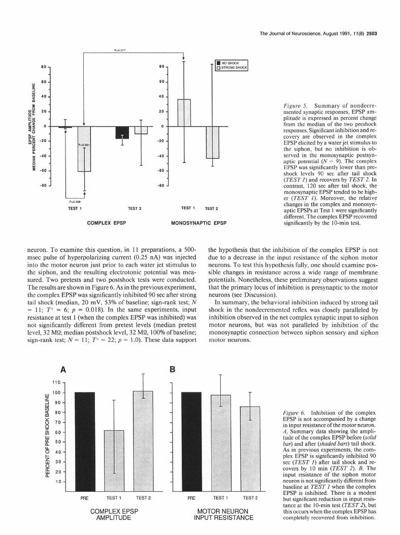
A summary of nine experiments in which complex and mono- synaptic EPSPs were concurrently measured before and after strong tail shock is shown in Figure 5. Complex EPSPs were also measured in a control group in which tail shock was omitted (N = 7; Fig. 5). As in the siphon withdrawal reflex, there was no significant difference between the amplitude of the complex EPSP in the two preshock tests (sign-rank test; N = 9; T+ = 9; p = 0.23; data not shown). On the first test after strong tail shock (Fig. 5, test l), the complex EPSP was significantly inhibited (median response, 62% below shock level; sign-rank test; N = 9, T+ = 1; p = 0.001). Moreover, a between-group comparison at test 1 revealed that the preparations receiving tail shock showed significant inhibition compared to the nonshocked control group (Wilcoxon two-sample; N = 9, 7; W = 5 1; p = 0.008). Finally, the amplitude of the complex EPSP recovered significantly by test 2 (Fig. 5; sign-rank test of test 2 vs test 1; N = 9, T+ = 40; p = 0.044; median, 17% below preshock level). Moreover, the
amplitude of the complex EPSP at test 2 was statistically in- distinguishable both from baseline (sign-rank test; N = 9, T+ = 11; p = 0.13) and from the nonshocked control group (two- sample test; N = 9, 7; W = 77; p = 1.0).
The changes induced by tail shock in the monosynaptic EPSP (elicited by an action potential in an LE sensory neuron) did not parallel the changes observed in the complex EPSP (Fig. 5). Specifically, the monosynaptic EPSP was not inhibited in the first test after tail shock (sign-rank test; N = 9; T+ = 30; p = 0.41) but rather tended to be above baseline (median, 136%). Moreover, the relative amplitude of this elevated monosynaptic EPSP was significantly higher than that ofthe inhibited complex EPSP in the same test (Wilcoxon two-sample test; W = 58; p = 0.017). Thus, the effect of tail shock on the monosynaptic EPSP was significantly different from the effect of tail shock on the complex EPSP: the monosynaptic EPSP did not show the tail shock-induced inhibition observed in the complex EPSP.
In a separate set of experiments, we asked whether the in- hibition of the complex EPSP in the siphon motor neuron could be accounted for by a change in the input resistance of the motor
I -80
t
P-0 008
TEST 1 TEST 2 TEST 1 TEST 2
COMPLEX EPSP MONOSYNAPTIC EPSP
The Journal of Neuroscience, AUgu8t 1991, 17(8) 2503
Figure 5. Summary of nondecre- mented synaptic responses. EPSP am- plitude is expressed as percent change from the median of the two preshock responses, Significant inhibition and re- covery are observed in the complex EPSP elicited by a water jet stimulus to the siphon, but no inhibition is ob- served in the monosynaptic postsyn- aptic potential (N = 9). The complex EPSP was significantly lower than pre- shock levels 90 set after tail shock (TEST 1) and recovers by TEST 2. In contrast, 120 set after tail shock, the monosynaptic EPSP tended to be high- er (TEST 1). Moreover, the relative changes in the complex and monosyn- aptic EPSPs at Test 1 were significantly different. The complex EPSP recovered significantly by the IO-min test.
neuron. To examine this question, in 11 preparations, a 500- msec pulse of hyperpolarizing current (0.25 nA) was injected into the motor neuron just prior to each water jet stimulus to the siphon, and the resulting electrotonic potential was mea- sured. Two pretests and two postshock tests were conducted. The results are shown in Figure 6. As in the previous experiment, the complex EPSP was significantly inhibited 90 set after strong tail shock (median, 20 mV, 53% of baseline; sign-rank test; N = 11; T+ = 6; p = 0.018). In the same experiments, input resistance at test 1 (when the complex EPSP was inhibited) was not significantly different from pretest levels (median pretest level, 32 MQ median postshock level, 32 MC& 100% of baseline; sign-rank test; N = 11; T+ = 22; p = 1 .O). These data support
the hypothesis that the inhibition of the complex EPSP is not due to a decrease in the input resistance of the siphon motor neurons. To test this hypothesis fully, one should examine pos- sible changes in resistance across a wide range of membrane potentials. Nonetheless, these preliminary observations suggest that the primary locus of inhibition is presynaptic to the motor neurons (see Discussion).
In summary, the behavioral inhibition induced by strong tail shock in the nondecremented reflex was closely paralleled by inhibition observed in the net complex synaptic input to siphon motor neurons, but was not paralleled by inhibition of the monosynaptic connection between siphon sensory and siphon motor neurons.
- i
\ lil 1: \
FE TEST 1 TEST 2 FfE TEST 1 TEST 2
COMPLEX EPSP MOTOR NEURON AMPLITUDE INPUT RESISTANCE
Figure 6. Inhibition of the complex EPSP is not accompanied by a change in input resistance of the motor neuron. A, Summary data showing the ampli- tude of the complex EPSP before (solid bar) and after (shaded bars) tail shock. As in previous experiments, the com- plex EPSP is significantly inhibited 90 set (TEST 1) after tail shock and re- covers by 10 min (TEST 2). B, The input resistance of the siphon motor neuron is not significantly different from baseline at TEST 1 when the complex EPSP is inhibited. There is a modest but significant reduction in input resis- tance at the IO-min test (TEST 2), but this occurs when the complex EPSP has completely recovered from inhibition.

2504 Wright et al. - Cellular Analysis of Inhibition
4 INITIAL-4 &- DECREMENTED -4 r POST-SHOCK7
COMPLEX EPSP
MONOSYNAPTIC EPSP
n
SIPHON S N.
t--
Figure 7. Effect of weak tail shock on decremented EPSPs. When first decremented by repeated stimuli, both complex and monosynaptic EPSPs are facilitated by weak tail shock. The top row of traces shows complex EPSPs elicited in a siphon motor neuron (MN.) by water jet stimuli (arrows) to the siphon. The bottom pairs of traces show monosynaptic EPSPs in the same motor neuron elicited by a single action potential in a siphon sensory neuron (S.N.). Complex EPSPs were decremented by repeated water jet stimuli; monosynaptic EPSPs were decremented by repeated single spikes in a sensory neuron elicited by intracellular current injection (1 00-set IS1 for both). Responses to the first (INITIAL), 19th, 20th (DECRE- MENTED), and 2 1 st (POST-SHOCK) stimuli are shown. Also shown are the responses in the motor neuron and sensory neuron during the tail shock (inset). Both decremented complex and monosynaptic EPSPs were facilitated by weak tail shock.
Decremented responses
We next examined the effects of weak and strong tail shock on decremented complex and monosynaptic EPSPs, in order to establish a neural correlate of our behavioral data demonstrating that weak shock produces dishabituation of the reflex response, whereas strong tail shock does not.
An example of our results with weak tail shock is shown in Figure 7. Twenty stimuli delivered at a lOO-set interval pro- duced synaptic decrement of both the complex EPSP and the monosynaptic EPSP, which paralleled behavioral habituation: the initial complex EPSP was 47 mV, and decremented to 22 and 18 mV on trials 19 and 20, respectively; likewise, the initial monosynaptic EPSP was 8.0 mV, and decremented to 1.6 and 3.8 mV on trials 19 and 20, respectively. Both complex and monosynaptic EPSPs showed facilitation 90 set after a weak electric shock to the tail (the complex EPSP facilitated to 42 mV; the monosynaptic EPSP, to 4.2 mV). Thus, both decre-
mented monosynaptic and complex EPSPs showed facilitation after weak shock that paralleled dishabituation to weak shock in behavioral experiments.
An example of our results examining the effects of strong shock on decremented synaptic responses is shown in Figure 8. Again, both complex EPSPs and monosynaptic EPSPs showed synaptic decrement (complex EPSP, from an initial value of 34 mV to 16 and 17 mV on trials 19 and 20, respectively; mono- synaptic EPSP, from 1.4 mV to 0.3 and 0.8 mV on trials 19 and 20, respectively). After strong tail shock, the complex EPSP remained at approximately its preshock (decremented) level (20 mV). In contrast, the decremented monosynaptic EPSP was facilitated more than twofold (to 1.8 mV).
The median EPSP amplitudes from 10 experiments using weak shock and 10 experiments using strong shock are shown in Figure 9. Consistent with the effects observed for the siphon withdrawal reflex in the intact and reduced preparations, after weak shock both complex and monosynaptic EPSPs were fa-

The Journal of Neuroscience, August 1991, 1 i(8) 2505
c ‘N’T’AL--i r---- DECREMENTED ------I c POST-SHmK--3
COMPLEX EPSP
SIPHONYN. h - -J----A k
t t t t
MONOSYNAPTIC EPSP
SIPHON IAN.
SIPHON S.N c c 7-:;:
SIPHON I . ! N
Figure 8. Effect of strong tail shock on decremented EPSPs. In contrast to weak tail shock, the decremented complex EPSP is not facilitated by strong tail shock, whereas the decremented monosynaptic EPSP is clearly facilitated by strong tail shock. See Figure 6 caption for details.
cilitated over their decremented baselines (complex EPSP: me- dian, 121%; sign-rank test; N = 10; T+ = 50; p = 0.025; mono- synaptic EPSP: median, 167%; sign-rank test; N = 9; T+ = 38; p = 0.038, one-tailed). The changes from baseline ofthe complex and monosynaptic EPSPs after weak shock were not significantly different from each other (two-sample test; N = 9, 10; W = 88; p = 0.348), suggesting that weak tail shock had a similar facil- itatory effect on both synaptic responses. However, after strong tail shock, only the complex EPSP paralleled the behavior, showing no significant change from its decremented baseline (median, 110%; sign-rank test; N = 10; T+ = 40; p = 0.22). In contrast, the decremented monosynaptic EPSP was significantly facilitated (median, 188%; sign-rank test; N = 9; T+ = 35; p = 0.02 1) by strong shock. Furthermore, the relative amplitude of the monosynaptic EPSP tended to be higher than that of the complex EPSP (two-sample test; W = 76; p = 0.055).
In summary, paralleling the effects of tail shock on the siphon withdrawal reflex, weak tail shock significantly facilitated decre- mented complex EPSPs, whereas strong tail shock failed to pro- duce facilitation. This inverse relationship between the degree of facilitation and tail shock intensity was not observed for the decremented monosynaptic EPSP.
5 I E 2 I
-50 1
Figure 9. Summary of decremented synaptic responses. EPSP ampli- tude is expressed as percent change from the median of the last three responses (trials 18-20) before the tail shock. Decremented complex EPSPs were significantly facilitated by weak but not by strong tail shock. In contrast, decremented monosynaptic EPSPs were facilitated by both weak and strong tail shock. The asterisk indicates a one-tailed proba- bility level.

2506 Wright et al. * Cellular Analysis of Inhibition
A SlPHO;NTWi;iDRAWAL: B SIPHON WITHDRAWAL: C COMPLEX EPSP:
ANIMAL REDUCED REDUCED
PREPARATION PREPARATION
-+T **
l *
* In
MONOSYNAPTIC EPSP: REDUCED
PREPARATION
Figure 10. Comparison of the effects of tail shock at different levels of analysis. For comparative purposes, all responses are expressed as percent change in response amplitude at test 1 (90-120 set after tail shock). Strong shock is indicated by solid bars; weak shock as open bars. Asterisks indicate within-group probability levels (*, one-tailed probability, p < 0.05; **, two-tailed probability, from p < 0.05 to p < 0.001).
SIPHON SENSORY NEURON
SIPHON MOTOR NEURONS
SIPHON
SIPHON MOTOR NEURONS
SIPHON SENSORY NEURON
Q L.E.
SIPHON
SIPHON MOTOR NEURONS
. . . . . ..-....‘.-.-........
SIPHON SENSORY NEURON
Q L.E
SPHON
SW’HON MOTOR NEURONS
Figure I I. Schematic representation of four classes of possible cellular loci (indicated by shading) underlying tail shock-induced inhibition of the siphon withdrawal reflex. See Discussion for details.
Discussion Neuronal correlates of inhibition in the siphon withdrawal reflex The primary goal of the present work was to explore the neural basis of tail shock-induced inhibitory modulation in the siphon withdrawal reflex in Aplysia. In these experiments, we employed a reduced preparation that allowed using the same stimuli that were used in the intact animal (Marcus et al., 1988): water jet stimuli to the siphon to elicit the reflex, and tail shock to mod- ulate the reflex. In order to identify neural correlates of inhi- bition in the neural circuit for siphon withdrawal, we examined two different sources of synaptic input onto siphon motor neu- rons: the complex EPSP elicited by water jet stimuli to the siphon, and the monosynaptic EPSP elicited by an action po- tential in a siphon sensory neuron.
Our results are summarized in Figure 10, which compares the effect of tail shock on decremented and nondecremented re- sponses in the siphon withdrawal reflex of the intact animal, the siphon withdrawal reflex of the reduced preparation, the complex EPSP in siphon motor neurons, and the monosynaptic connection between LE sensory neurons and the siphon motor neurons. In the intact animal (Fig. 1OA; redrawn from Marcus et al., 1988), strong tail shock produces significant inhibition of the nonhabituated (nondecremented) siphon withdrawal reflex. The same strong tail shock fails to produce facilitation of ha- bituated responses, suggesting the recruitment of an inhibitory process that competes with the facilitation produced by weak shock. This pattern (inhibition of nondecremented responses and a reduction in the amount of facilitation in the decremented responses) is paralleled in the reduced preparation, both at the level of the behavioral reflex and at the level of the complex EPSP elicited by water jet stimuli (Fig. 1OS.C). In contrast, tail

The Journal of Neuroscience, August 1991, 1 f(8) 2507
shock-induced modulation in the monosynaptic connection be- tween siphon sensory and siphon motor neurons does not reflect the inhibitory pattern (Fig. 1OD): at a time when the siphon withdrawal reflex and the complex EPSP are inhibited, strong tail shock produces no inhibition of noncremented monosyn- aptic EPSPs, and facilitation after strong shock is at least as great as after weak shock.
The data summarized in Figure 10 show that tail shock can produce opposite effects in complex and monosynaptic EPSPs in siphon motor neurons. Tail shock activates tail sensory neu- rons in the pleural ganglia (Walters et al., 1983a,b), which in turn modulate the siphon withdrawal reflex through intemeu- ronal pathways. For example, it is known that tail shock can activate serotonergic interneurons that project to the neural cir- cuit for siphon withdrawal (Mackey et al., 1989). These obser- vations raise the question of whether 5-HT, like tail shock, can also differentially affect synaptic inputs to siphon motor neu- rons. Fitzgerald and Carew (199 1) have examined this issue and found that S-HT can in fact inhibit the complex EPSP in siphon motor neurons, as well as the siphon withdrawal reflex, while simultaneously facilitating the monosynaptic EPSP from siphon sensory neurons. Thus, the dissociation of inhibition and facil- itation produced by tail shock in the neural circuit for siphon withdrawal (Fig. 10) is mimicked by the neuromodulator 5-HT. When the cellular loci of tail shock-induced inhibition are iden- tified, it will be interesting to examine whether 5-HT will sim- ilarly mimic the effects of tail shock at these sites.
Possible neuronal sites mediating the inhibitory process In considering inhibition in the siphon withdrawal reflex, four broad classes of hypotheses about the cellular locus of the in- hibitory process can be considered. These are schematically il- lustrated in Figure 11. We will discuss each of these hypotheses in light of our present results.
The first hypothesis is that the inhibition is produced by changes in the input resistance of the motor neuron (Fig. 11A). Specifically, tail shock could induce a large decrease in the input resistance of the motor neuron, which would shunt the synaptic current generated by the reflex input, thereby reducing the com- plex EPSP. Two lines of evidence suggest that this hypothesis is not likely to account for the inhibition: (1) the input resistance in the motor neuron (as measured by electrotonic potentials produced by current injected into the soma) does not change during the inhibition of the complex EPSP, and (2) following tail shock, when the complex EPSP is reduced, the monosynaptic EPSP is simultaneously increased. Thus, though it is possible that remote changes in input resistance could, in principle, con- tribute to inhibition of the complex EPSP, our results favor the idea that the principal site(s) of inhibition is presynaptic to the motor neuron.
The second hypothesis is that the direct monosynaptic con- nection between LE siphon sensory neurons and siphon motor neurons is diminished immediately following tail shock, so that it contributes less to the total synaptic input onto the motor neurons (Fig. 11B). Mackey et al. (1987) observed a decrease in this monosynaptic EPSP immediately after tail shock. How- ever, the inhibition they observed gave way to facilitation by 90 set after shock, whereas the behavioral inhibition persisted at least 2.5 min after shock. Thus, as Mackey et al. (1987) point out, there was a discrepancy between the time course of behav- ioral inhibition and the reduction in transmitter release from the LE sensory neurons. This discrepancy suggests that modu-
lation of the monosynaptic connection between LE sensory neu- rons and siphon motor neurons cannot, by itself, account for the behavioral inhibition observed in the siphon withdrawal reflex.
The results of the present study, in which the monosynaptic connection was monitored simultaneously with the water jet- elicited complex EPSP in siphon motor neurons, indicate that the inhibitory patterns observed in the complex EPSP are not paralleled by changes in the monosynaptic EPSP. First, 90 set after strong tail shock, when the behavioral reflex and the com- plex EPSP show significant inhibition, the monosynaptic EPSP is not inhibited; rather, consistent with the observations of Mackey et al. (1987), it tends to be facilitated. Second, the absence of facilitation of decremented responses after strong shock, which we observed in the reflex and the complex EPSP, was not observed in the monosynaptic connection. Indeed, strong tail shock produced a twofold facilitation of the decremented monosynaptic connection at the same time that the decremented complex EPSP showed no facilitation. Taken together, these results indicate that the monosynaptic connection between LE sensory neurons and siphon motor neurons cannot by itself account for tail shock-induced inhibition of either the water jet-elicited complex EPSP, or the siphon withdrawal reflex.
Although the tail shock-induced inhibition of the siphon withdrawal reflex we observed at 90 set cannot be accounted for by inhibition of the monosynaptic connection, it is known from work by Mackey et al. (1987) that this synapse is inhibited for about 30 set following tail shock. R. D. Hawkins and E. R. Kandel (personal communication) have recently found that, in the gill withdrawal reflex, tail shock-induced inhibition is more short-lived than in the siphon withdrawal reflex. Thus, the time course of inhibition in the gill withdrawal reflex is more closely paralleled by inhibition of the monosynaptic connection. This observation raises the possibility that the short-lasting reduction in transmitter release from the sensory neurons produced by tail shock (Mackey et al., 1987) contributes more to inhibition ofgill withdrawal than to that ofsiphon withdrawal. The broader implication of this possibility is that the neural circuits for the siphon withdrawal reflex and the gill withdrawal reflex, as well as plasticity within these circuits, may be less similar than pre- viously appreciated. It will therefore be of interest to examine the inhibitory process induced by tail shock, as well as other forms of modulation, simultaneously in the circuits for siphon withdrawal and gill withdrawal.
It is also possible that tail shock-induced increases in trans- mitter release from LE sensory neurons onto some of their fol- lower cells could contribute indirectly to the net inhibition of the complex EPSP in the siphon motor neurons. For example, if after tail shock the functional connections between LE sensory neurons and inhibitory interneurons (Hawkins et al., 1981a; Frost et al., 1988) were increased more than the connections to excitatory interneurons, then the increase in synaptic transmis- sion from the sensory neurons could, in principle, play a sig- nificant role in mediating tail shock-induced inhibition.
A third hypothesis for the locus of inhibition is illustrated in Figure 1 lC, which suggests the possibility that another (as yet unidentified) set of sensory neurons may be modulated by tail shock in ways consistent with the inhibition we observed in both the complex EPSP and the behavior. Such a possibility is supported by recent observations of Rosen and colleagues (Ro- sen et al., 1989), who examined sensory neurons in the cerebral ganglion ofAplysia. They showed that different classes ofsensory

2508 Wright et al. - Cellular Analysis of Inhibition
neurons responded oppositely to the same modulatory inputs: nerve shock or 5-HT application produced spike broadening and enhanced synaptic release in one class of sensory neurons, while simultaneously producing spike narrowing and dimin- ished synaptic release in another class. Moreover, Dubuc and Castellucci (199 1) have recently described several new popu- lations of sensory neurons in the abdominal ganglion. While some of these new sensory neurons closely resemble the LE sensory neurons in their sensitivity to a variety of neuromod- ulators, others do not. Thus, we are currently examining the possibility that another set of sensory neurons in the siphon withdrawal reflex may contribute to the inhibitory process that we observe.
A fourth hypothesis for the locus of inhibition is illustrated in Figure 11 D, which suggests the possibility that plasticity at central intemeurons might contribute significantly to the inhib- itory process. Many intemeurons (both excitatory and inhibi- tory) that have a role in mediating the siphon withdrawal reflex have already been identified (Hawkins et al., 1981a,b; Frost et al., 1988; Frost and Kandel, 1990). The synaptic efficacy ofthese cells, as well as that of other as yet unidentified intemeurons, may be altered by tail shock in ways that contribute to inhibition of the complex EPSP. For example, Hawkins et al. (198 1 b) first showed that intracellular activation of an identified inhibitory intemeuron (L16) can inhibit the complex EPSP elicited by siphon nerve shock. We have recently found that intracellular activation of L16 also inhibits the complex EPSP elicited by water jet stimuli to the siphon (Wright and Carew, 1990). Fur- thermore, we have found that tail shock produces strong spike activity in L16. Finally, hyperpolarizing or voltage clamping L16 (which prevents its firing in response to tail shock) signif- icantly diminishes or abolishes the inhibition of the complex EPSP produced by tail shock (Wright and Carew, 1990; W. G. Wright and T. J. Carew, in preparation). Thus, L16, as well as other intemeurons, may play a significant role in the reflex in- hibition we observe.
In conclusion, our results show that different functional ele- ments underlying a single reflex can be simultaneously modu- lated in opposing ways. Important insights into the cellular and subcellular basis of behavioral facilitation in the siphon with- drawal reflex have already been obtained (for review, see Byrne, 1987; Carew, 1987; Hawkins et al., 1987). Once the sites that mediate inhibition in the siphon withdrawal reflex have been identified, it will be ofconsiderable interest to examine similarly the cellular and subcellular mechanisms underlying the inhi- bition. Moreover, to understand fully the neuronal basis of tail shock-induced modulation in the reflex, it will be important to analyze how the nervous system integrates opposing modulatory changes to produce the net reflex output observed at motor neuronal and behavioral levels. This question of integration of opposing signals by cells within a circuit is analogous to ques- tions that have arisen from intensive study of integration within single cells (see, e.g., Abrams and Kandel, 1988). For example, within a single sensory neuron in Aplysia, second-messenger systems with opposing biophysical effects can be simultaneously activated by modulatory stimuli such as tail shock (Belardetti et al., 1987; Mackey et al., 1987; Piomelli et al., 1987). The results of the present study lend emphasis to the suggestion that an analysis of the principles of integration both within individ- ual neurons and between neurons in a functional neural circuit may provide insights into the mechanisms underlying learning and memory.
References Abrams TW, Kandel ER (1988) Is contiguity detection in classical
conditioning a system or a cellular property? Learning in Aplysia suggests a possible molecular site. Trends Neurosci 11: 128-l 35.
Baxter DA, Byrne JH (1989) Serotonergic modulation of two potas- sium currents in the pleural sensory neurons ofdplysiu. J Neurophysi- 01 62665-679.
Belardetti F, Kandel ER, Siegelbaum SA (1987) Neuronal inhibition by the peptide FMRFamide involves opening of S K+ channels. Na- ture 325: 153-l 56.
Bemier L, Castellucci VF, Kandel ER, Schwartz JH (1982) Facilitatory transmitter causes a selective and prolonged increase in adenosine 3’: 5’-monophosphate in sensory neurons mediating the gill and siphon withdrawal reflex in Aplysiu.. J Neurosci 2: 1682-l 69 1.
Bvme JH (1987) Cellular analvsis of associative learning. Phvsiol Rev .67:329i39.
- .
Byrne JH, Castehucci VF, Kandel ER (1974) Receptive fields and response properties of mechanoreceptor neurons innervating skin and mantle shelf of Aplysia. J Neurophysiol 37:1041-1064.
Carew TJ (1987) Cellular and molecular advances in the study of learning in Aplysiu. In: The neural and molecular basis of learning (Changeaux JP, Konishi M, eds), pp 177-204. New York: Wiley.
Cleary LJ, Byrne JH (1985) Intemeurons contributing to the mediation and modulation of the tail withdrawal reflex in Aplysia. Sot Neurosci Abstr 11:642.
Dubuc B, Castellucci VF (199 1) Receptive fields and properties of a new cluster of mechanoreceptor neurons innervating the mantle re- gion and branchial cavity of the marine mollusc Aplysiu culifornicu. J Exp Biol 156:3 15-334.
Fitzgerald KF, Carew TJ (199 1) Serotonin mimics tail shock in pro- ducing transient inhibition in the siphon withdrawal reflex of Aplysiu. J Neurosci 11:2510-2518.
Frost WN. Kandel ER (1990) Further characterization of intemeurons and motor neurons in the siphon withdrawal reflex circuit in Aplysiu culifornicu. J Neurophysiol,.in press.
Frost WN. Clark GA. Kandel ER (1988) Parallel orocessina of short- term memory for sensitization in Aplisiu. J Neurobiol 19297-334.
Hawkins RD, Castellucci VF, Kandel ER (198 1 a) Interneurons in- volved in mediation and modulation ofgill-withdrawal reflex in Aply- sia. I. Identification and characterization. J Neurophysiol 45:304- 314.
Hawkins RD, Castellucci VF, Kandel ER (1981b) Intemeurons in- volved in mediation and modulation ofgill-withdrawal reflex in Aply- siu. II. Identified neurons produce heterosynaptic facilitation con- tributing to behavioral sensitization. J Neurophysiol45:3 15-326.
Hawkins RD, Clark GA, Kandel ER (1987) Cell biological studies of learning in simple vertebrate and invertebrate systems. In: Handbook of physiology, Sect I, Higher functions of the nervous system, Vol 6 (Plum F, ed), pp 25-83. Bethesda, MD: American Physiological So- ciety.
Hollander M, Wolfe DA (1973) Non-parametric statistical methods. New York: Wiley.
Kandel ER, Schwartz JH (1982) Molecular biology of learning: mod- ulation of transmitter release. Science 2 18:4331143.
Klein M, Kandel ER (1980) Mechanism of calcium current modula- tion underlying presynaptic facilitation and behavioral sensitization in Aplysiu. Proc Nat1 Acad Sci USA 77:69 12-69 16.
Krontiris-Litowitz JK, Erikson MT, Walters ET (1987) Central sup- pression of defensive reflexes in Aplysiu by noxious stimulation and by factors released from the body wall. Sot Neurosci Abstr 13:8 15.
Mackey SL, Glanzman DL, Small SA, Dyke AM, Kandel ER, Hawkins RD (1987) Aversive stimuli produce inhibition as well as sensiti- zation ofthe siphon withdrawal reflex ofdplysiu: a possible behavioral role for oresynaptic inhibition mediated by the peptide FMRFamide. Proc Nail Acad-Sci USA 84:8730-8734.
Mackev SL. Kandel ER. Hawkins RD (1989) Identified serotonergic neurons LCB 1 and RCB 1 in the cerebral ganglia of Aplysiu produce presynaptic facilitation ofsiphon sensory neurons. J Neurosci 9:4227- 4235.
Mackintosh NJ (1974) Conditioning and associative learning. New York: Academic.
Marcus EA, Nolen TG, Rankin CH, Carew TJ (1987) Behavioral dissociation of dishabituation, sensitization and inhibition in the si- phon withdrawal reflex of adult Aplysiu. Sot Neurosci Abstr 13:8 16.

The Journal of Neuroscience, August 1991, 1 f(8) 2509
Marcus EA, Nolen TG, Rankin CH, Carew TJ (1988) Behavioral dissociation of dishabituation, sensitization, and inhibition of Aply- siu. Science 241:210-213.
Occor KA, Tabata M, Byrne JH (1986) Stimuli that produce sensi- tization lead to elevation of cyclic AMP levels in tail sensory neurons of Aplysiu. Brain Res 37 1: 190-192.
Piomelli D, Shapiro E, Feinmark SJ, Schwartz JH (1987) Metabolites of arachodonic acid in the nervous system of Aplysia: possible me- diators of synaptic modulation. J Neurosci 7:3675-3686.
Rankin CH. Carew TJ (1987) Analvsis of the developmental emer- gence of sensitization in Apiysiu reieals an inhibitory effect of a fa- cilitatory stimulus. Sot Neurosci Abstr 13:8 16.
Rankin CH, Carew TJ (1989) Developmental analysis in Aplysiu re- veals inhibitory as well as facilitatory effects of tail shock. Behav Neurosci 103:334-344.
Rosen SC, Susswein AJ, Cooper EC, Weiss KR, Kupferman I (1989) Selective modulation of spike duration by serotonin and the neuro- peptides FMRFamide, SCP,, buccalin and myomodulin in different classes of mechanoafferent neurons in the cerebral ganglion ofdplysiu. J Neurosci 9:390-402.
Siegelbaum SA, Camardo JS, Kandel ER (1982) Serotonin and cyclic
AMP close single K+ channels in Aplysiu sensory neurons. Nature 299:4131117.
Walsh JP, Byrne JH (1989) Modulation of a steady-state Ca*+-acti- vated K+ current in tail sensory neurons of Aplysiu: role of serotonin and CAMP. J Neurophysiol 61:32-44.
Walters ET, Byrne JH, Carew TJ, Kandel ER (1983a) Mechanoafferent neurons innervating the tail of Aplysiu. I. Response properties and synaptic connections. J Neurophysiol 50: 1522-l 542.
Walters ET, Byrne JH, Carew TJ, Kandel ER (1983b) Mechanoafferent neurons innervating the tail ofAplysiu. II. Modulation by sensitizing stimuli. J Neurophysiol 50: 1543-l 559.
Wright WG, Carew TJ (1990) Contributions of intemeurons to tail- shock induced inhibition of the siphon withdrawal reflex in Aplysiu. Sot Neurosci Abstr 16:20.
Wright WG, Marcus EA, Thaker H, Carew TJ (1988) A cellular anal- ysis of tail-shock induced inhibition in the siphon withdrawal reflex of Aplysiu. Sot Neurosci Abstr 14:84 I.
Wright WG, Marcus EA, Carew TJ (1989) Dissociation of monosyn- aptic and polysynaptic contributions to dishabituation, sensitization and inhibition in Aplysiu. Sot Neurosci Abstr 15: 1265.