Embed Size (px)
Citation preview
A CLINICAL, RADIOLOGICAL AND IN VITRO STRAIN INVESTIGATIONS OF MANDIBULAR IMPLANT OVERDENTURES
TAMEEM KHUDER JASSIM
FACULTY OF DENTISTRY
UNIVERSITY OF MALAYA KUALA LUMPUR
2018

A CLINICAL, RADIOLOGICAL AND IN VITRO STRAIN
INVESTIGATIONS OF MANDIBULAR IMPLANT OVERDENTURES
TAMEEM KHUDER JASSIM
THESIS SUBMITTED IN FULFILMENT OF THE REQUIREMENTS
FOR THE DEGREE OF DOCTOR OF PHILOSOPHY
FACULTY OF DENTISTRY
UNIVERSITY OF MALAYA
KUALA LUMPUR
2018
ii
UNIVERSITY OF MALAYA
ORIGINAL LITERARY WORK DECLARATION
Name of Candidate: TAMEEM KHUDER JASSIM
I.C/Passport No: A10355920
Registration/Matric No: DHA130018
Name of Degree: Doctor of Philosophy
Title of Thesis: A Clinical, Radiological and In Vitro Strain Investigations of
Mandibular Implant Overdentures.
Field of Study: Dentistry (Implant Prosthodontics).
I do solemnly and sincerely declare that:
(1) I am the sole author/writer of this work;
(2) This work is original;
(3) Any use of any work in which copyright exists was done by way of fair dealing
and for permitted purposes and any excerpt or extract from, or reference to or
reproduction of any copyright work has been disclosed expressly and
sufficiently and the title of the work and its authorship have been acknowledged
in this work;
(4) I do not have any actual knowledge nor do I ought reasonably to know that the
making of this work constitutes an infringement of any copyright work;
(5) I hereby assign all and every rights in the copyright to this work to the
University of Malaya (“UM”), who henceforth shall be owner of the copyright
in this work and that any reproduction or use in any form or by any means
whatsoever is prohibited without the written consent of UM having been first
had and obtained;
(6) I am fully aware that if in the course of making this work I have infringed any
copyright whether intentionally or otherwise, I may be subject to legal action
or any other action as may be determined by UM.
Candidate’s Signature Date:
Subscribed and solemnly declared before,
Witness’s Signature Date:
Name:
Designation:
iii
A CLINICAL, RADIOLOGICAL AND IN VITRO STRAIN INVESTIGATIONS
OF MANDIBULAR IMPLANT OVERDENTURES
ABSTRACT
Introduction: Conventional complete denture (CD) and two mandibular implant-
overdenture (IOD) prostheses with non-balanced occlusal scheme can induce residual
ridge resorption (RRR) and increase the risk of denture fracture. Nevertheless, the
association between occlusal force distribution to other clinical and mechanical factors
are not that clear. Objectives: To compare masticatory performance, occlusal force
distribution, and patient-reported outcomes in patients wearing IOD and CD, and to
investigate the effect of two mandibular implants and clinical parameters on maxillary
and mandibular residual ridge resorption (RRR). Finite element analysis (FEA) was
applied to investigate the in vitro strain distribution using loading simulation according
to the clinically determined occlusal force distribution. The fracture risk was predicted
based on tensile strain distribution at the implant attachment interface of IOD prostheses
and compared with the frequency of clinical complications recorded for two attachment
types, from the follow-up records of patients with IOD prostheses. Methods: Twenty-
three patients in each of the IOD (age 66 ± 8 years) and CD (age 65 ± 10 years) groups
were recalled 3-4 years after provision of the prostheses. Masticatory performance was
determined based on the patients’ ability to chew two-colored wax cubes and mixing
ability index (MAI) derived. The percentages of occlusal force (OF%) distribution at
anterior and posterior quadrants of the arch were recorded using a digital occlusal
analyzer (T-scan III). The radiographic images of the dental panoramic radiograph were
compared with the baseline for RRR determination using the bone area index
measurement method. Patient-reported outcomes were analyzed using the shortened
Malaysian version of Oral Health-related Quality of Life (OHIP-14) and denture
iv
satisfaction (DS) questionnaires. For FEA, maxillary and mandibular models were
constructed from cone-beam computed tomographic data of a patient. The anterior and
posterior vertical loads were simulated according to the OF% distribution obtained from
the respective patient groups. Compressive and tensile strains were compared with the
frequency of clinical complications recorded in IOD patients between the two implant
attachment types. Results were statistically analyzed with a 95% confidence interval.
Results: The mean MAI of the CD and IOD groups showed no significant difference
(P=0.134), while significant difference was observed in the mean OF% (P=0.002). For
patient-reported outcomes, the total scores of OHIP-14 (P=0.027) and DS (P=0.030)
between groups showed significant differences. The IOD showed significantly less bone
resorption compared to the CD groups in the maxillary anterior area (P=0.013).
Multivariate regression analyses showed significant association between RRR and OF%
at the anterior maxilla (P=0.000) and posterior mandible (P=0.023). The frequency of
mechanical complications clinically observed with telescopic were significantly greater
than with Locator attachments (P=0.005). The FEA results showed greater strain
concentration on the denture impression surface at the attachment-acrylic interface of
both telescopic- (561µε) and Locator- retained (323µε) IODs. Conclusions: Patients with
mandibular IODs showed better OHIP-14 and DS, but with no significant difference in
the masticatory performance compared to CD patients. Residual ridge resorption was
significantly associated with the occlusal force distribution and the type of prostheses. A
higher tensile strain produced by telescopic attachment compared to Locator attachment
interface supported the clinical observation of greater complication with telescopic IODs.
Keywords: Implant-supported overdenture, masticatory performance, T-scan III
system, bone resorption, finite element analysis.

v
A CLINICAL, RADIOLOGICAL AND IN VITRO STRAIN INVESTIGATIONS OF
MANDIBULAR IMPLANT OVERDENTURES
ABSTRAK
Pengenalan: Skima oklusi yang tidak seimbang untuk denture penuh (gigi palsu)
konvensional (CD) dan prostesis overdenture dengan dua implant mandibel (IOD) boleh
menyebabkan resorpsi batas residu (RRR) dan boleh meningkatkan risiko dentur
terfraktur. Walau bagaimanapun, pengagihan daya occlusal dan kaitannya dengan faktor
klinikal dan mekanikal lain adalah tidak jelas. Objektif: Untuk membandingkan prestasi
pengunyahan, pengagihan daya oklusi, dan hasil laporan pesakit yang memakai IOD dan
CD, dan untuk mengkaji kesan dua implan di mandibel dan parameter klinikal sosio-
demografi terhadap resorpsi batas residu mandibel dan maksila (RRR). Analisis elemen
finit (FEA) telah digunakan untuk menyiasat pengagihan strain in vitro menggunakan
simulasi daya occlusi klinikal yang telah dikenal pasti. Risiko fraktur diramalkan
berdasarkan pengagihan strain tensil pada antaramuka atakmen implant dan prostesis
IOD dan perbandingan dengan kekerapan komplikasi klinikal yang dicatatkan untuk dua
jenis atakmen dengan melihat pada rekod susulan pesakit dengan prostesis IOD.
Kaedah: Dua puluh tiga pesakit di setiap kumpulan IOD (umur 66 ± 8 tahun) dan CD
(umur 65 ± 10 tahun) telah dipanggil semula 3-4 tahun selepas penyediaan prostesis.
Prestasi pengunyahan ditentukan berdasarkan keupayaan pesakit mengunyah kiub lilin
dua warna untuk mendapatkan indeks keupayaan pencampuran (MAI). Peratusan
pengagihan daya oklusai (OF%) di kuadran anterior dan posterior rahang direkodkan
menggunakan analyser oklusi digital (T-scan III). Imej dari radiografi panorama
pergigian dibandingkan dengan garis dasar untuk penentuan RRR menggunakan kaedah
pengukuran indeks kawasan tulang. Hasil yang dilaporkan oleh pesakit dianalisa
menggunakan soal selidik yang berkaitan dengan Kualiti Kehidupan Kesihatan (OHIP-
vi
14, Versi pendek Malaysia) dan kepuasan penggunaan gigi palsu (DS). Bagi FEA, model
maksila dan mandibel telah dibina daripada data tomografi beam kon. Beban menegak
anterior dan posterior disimulasikan mengikut taburan OF% yang diperoleh dari
kumpulan pesakit masing-masing. Strain mampatan dan tegangan berbanding dengan
kekerapan komplikasi klinikal yang dicatatkan pada pesakit IOD antara kedua-dua jenis
atakmen implan. Keputusan dianalisa secara statistik dengan interval keyakinan 95%.
Keputusan: Purata MAI bagi kumpulan CD dan IOD tidak menunjukkan perbezaan yang
signifikan (P = 0.134), manakala perbezaan yang signifikan diperolehi dalam purata OF%
(P = 0.002). Bagi hasil yang dilaporkan oleh pesakit termasuk jumlah keseluruhan skor
OHIP-14 (P = 0.027) dan DS (P = 0.030) terdapat perbezaan signifikan di antara
kumpulan. Kumpulan IOD menunjukkan kurang resorpsi tulang berbanding dengan
kumpulan CD yang signifikan pada kawasan anterior maksila (P = 0.013). Analisa regresi
multivariat menunjukkan hubungan yang signifikan antara RRR dan OF% pada maxilla
anterior (P = 0.000) dan posterior mandibel (P = 0.023). Kekerapan komplikasi mekanikal
yang direkod secara klinikal dengan atakmen teleskopik lebih besar signifikannya
berbanding atakmen lokator (P = 0.005). Keputusan FEA menunjukkan penumpuan strain
yang lebih besar pada permukaan impresi dentur pada antaramuka kedua-dua denture
IOD teleskopik- (561με) dan Locator (323με) IODs. Kesimpulan: Pesakit dengan IOD
mandibel menunjukkan OHIP-14 dan DS yang lebih baik, tetapi tidak mempunyai kesan
yang signifikan terhadap prestasi pengunyahan berbanding pesakit dengan denture
penuhResopsi batas residu secara signifikan dikaitkan dengan pergagihan daya oklusi
dan jenis prosthesis. Tekanan tegangan yang lebih tinggi yang dihasilkan pada
antaramuka atakmen teleskopik berbanding atakmen Lokator, menyokong pemerhatian
klinikal di mana komplikasi yang lebih besar dengan IOD atakmen teleskopik.
Kata kunci: Overdenture dua mandibel implan, prestasi mastikatori, T-scan III sistem,
resorpsi tulang, analisa elemen finit.

vii
ACKNOWLEDGEMENTS
The process of dissertation accomplishment is a difficult and long process and it is a
combination of dedicated work of several peoples and not only by a single student.
First and foremost, I would like to thank my supervisor, Prof. Dr. Norsiah Yunus, for
the patience, direction, and endless support throughout my study time. I am extremely
lucky to have a supervisor who cared so much about my work and responded to my
questions so promptly. Without her, I would not have achieved my study goals.
I would like also to express my appreciation to my co-supervisors, Associate Prof. Dr.
Eshamsul Sulaiman and Lecturer Dr. Ali Dabbagh for their simultaneous encouragement,
advice, and support throughout my study work.
I wish to express sincere thanks to Prof. Dr. Roslan Saub, for his useful comments
regarding the oral health-related quality of life questionnaire, and for his valuable
statistical expertise and data interpretation.
I wish to acknowledge my deepest appreciation to Associate Prof. Dr. Norliza Ibrahim
for her keen scientific vision and her ability to place complex ideas into simple terms.
I would like to thank all the members and staff at the Department of Restorative Dentistry
for their generous support.
I must express my thankfulness to my mother, brothers and to my mother and father in
law for their endless support and engorgement during the years of my study.
Last but not the least, I am tremendously grateful to my lovely wife, Alyamama and my
sons Faisal and Abdullah. I couldn’t have accomplished this dissertation without their
love, support, and understanding.
I wish to dedicate this work to my father’s soul (may he rest in peace).
viii
TABLE OF CONTENTS
Abstract ............................................................................................................................ iii
Abstrak .............................................................................................................................. v
Acknowledgements ......................................................................................................... vii
Table of Contents ........................................................................................................... viii
List of Figures ................................................................................................................. xv
List of Tables................................................................................................................. xvii
List of Symbols and Abbreviations ................................................................................ xix
List of Appendices ......................................................................................................... xxi
CHAPTER 1: INTRODUCTION .................................................................................. 1
1.1 Background of the study .......................................................................................... 1
1.2 Problem statement ................................................................................................... 4
1.3 Null Hypothesis ....................................................................................................... 6
1.4 Aim of the study .......................................................................................................... 7
1.5 Objectives .................................................................................................................... 7
1.5.1 Clinical study .................................................................................... 7
1.5.2 Finite element analysis study ............................................................ 8
1.6 Rationale of the study.................................................................................................. 8
1.7 Study structure ......................................................................................................... 9
CHAPTER 2: LITERATURE REVIEW .................................................................... 10
2.1 Complete edentulism ............................................................................................. 10
2.2 Edentulism risk factors .......................................................................................... 11
2.2.1 Age ........................................................................................................... 11
2.2.2 Gender ...................................................................................................... 12
ix
2.2.3 Smoking .................................................................................................... 12
2.2.4 Asthma ...................................................................................................... 12
2.2.5 Diabetes .................................................................................................... 13
2.2.6 Rheumatoid arthritis ................................................................................. 13
2.2.7 Obesity ...................................................................................................... 14
2.2.8 Lower education and income.................................................................... 14
2.3 Rehabilitation of edentulous patients..................................................................... 14
2.3.1 Complete denture prostheses .................................................................... 14
2.3.2 Osseointegrated dental implants ............................................................... 15
2.3.3 Implant fixed prostheses ........................................................................... 16
2.3.4 Implant-supported prostheses ................................................................... 17
2.3.5 Number of implants used with implant-supported overdenture
prostheses………………………………………………………………..18
2.3.6 Attachments used with implant-supported overdenture prostheses ......... 19
2.3.6.1 Telescopic attachments ............................................................. 20
2.3.6.2 Locator attachments .................................................................. 20
2.4 Stress distribution according to attachment types ................................................. 21
2.5 Occlusal registration methods................................................................................ 22
2.5.1 Qualitative method ................................................................................... 22
2.5.1.1 Articulating paper/ribbon .......................................................... 23
2.5.1.2 Silk strips ................................................................................... 23
2.5.1.3 Foils……..………………………………………………………24
2.5.1.4 Impression materials ................................................................. 24
2.5.1.5 Occlusal indicator wax .............................................................. 24
2.5.2 Quantitative method ................................................................................. 25
2.5.2.1 Photo-occlusion system ............................................................. 25
x
2.5.2.2 T-scan system ............................................................................ 26
2.6 T-scan system development ................................................................................... 27
2.6.1 T-scan system applications in Dentistry ................................................... 29
2.6.1.1 Prosthodontics ........................................................................... 29
2.6.1.2 Trauma from occlusion ............................................................. 29
2.6.1.3 Implant prostheses ..................................................................... 30
2.6.2 T-scan system limitations ......................................................................... 30
2.7 Bone Resorption .................................................................................................... 31
2.7.1 Residual ridge resorption .......................................................................... 31
2.7.2 Consequences of residual ridge resorption ............................................... 32
2.7.3 Factors affecting residual ridge resorption ............................................... 32
2.7.3.1 Anatomic factors ....................................................................... 32
2.7.3.2 Prosthetic factors ....................................................................... 34
2.7.3.3 Role of inflammatory mediators ............................................... 34
2.7.3.4 Post-menopausal osteoporotic changes ..................................... 35
2.7.4 Methods to reduce residual ridge resorption after tooth loss ................... 35
2.7.4.1 Clinical and laboratory techniques ............................................ 35
2.7.4.2 Surgical techniques for highly resorbed ridges ......................... 36
2.7.4.3 Implant-supported overdentures treatment ................................ 37
2.7.5 Methods of ridge resorption measurements ............................................. 37
2.7.5.1 Dental cast measurement ........................................................... 37
2.7.5.2 Radiographical methods ............................................................ 38
2.7.6 Determination of alveolar bone resorption using panoramic radiographs 40
2.7.6.1 Linear method ........................................................................... 40
2.7.6.2 Area index method .................................................................... 41
2.8 Patient-reported outcomes ..................................................................................... 43
xi
2.8.1 Health-Related Quality of Life ................................................................. 43
2.8.2 Oral Health-Related Quality of Life ......................................................... 43
2.8.3 Oral Health Impact Profile (OHIP-49) ..................................................... 44
2.8.4 Oral Health Impact Profile (OHIP-14) ..................................................... 44
2.8.5 Oral Health Impact Profile [S-OHIP(M)] Malaysia ................................. 45
2.8.6 The effect prosthesis types on the Oral Health-Related Quality of Life .. 46
2.8.6.1 Quality of life for patients using conventional complete
dentures………………………………………………………..46
2.8.6.2 Quality of life for patients using implant-supported
overdentures…………..………………………………………47
2.8.7 Denture satisfaction .................................................................................. 47
2.9 Masticatory performance ....................................................................................... 48
2.10 Masticatory performance measurement methods .................................................. 48
2.11 Acrylic denture risk of fracture.............................................................................. 49
2.11.1 Finite element analysis method ................................................................ 50
2.11.2 Application of finite element analysis in dentistry ................................... 51
2.11.2.1 Implant stress analysis ............................................................... 51
2.11.2.2 Maxillary obturator ................................................................... 52
2.11.2.3 Post and core restorations .......................................................... 52
2.11.2.4 Trauma and fracture .................................................................. 52
2.11.2.5 Analysis of the biomechanical effects of occlusal loads on
implant prostheses ..................................................................... 53
2.11.3 Finite element analysis method assumptions ........................................... 53
2.11.3.1 Detailed geometry ..................................................................... 54
2.11.3.2 Material properties .................................................................... 54
2.11.3.3 Boundary conditions ................................................................. 55

xii
2.11.3.4 Loading conditions .................................................................... 55
2.11.4 Limitations of finite element analysis method ......................................... 55
CHAPTER 3: METHODOLOGY ............................................................................... 57
3.1 Patient selection ..................................................................................................... 57
3.2 Masticatory performance ....................................................................................... 58
3.3 Occlusal force distribution measurement using T-scan III .................................... 60
3.4 Residual ridge resorption measurements ............................................................... 62
3.5 Patient-reported outcomes ..................................................................................... 66
3.5.1 Patients OHRQoL evaluation ................................................................... 66
3.5.2 Denture satisfaction evaluation ................................................................ 67
3.6 Implant overdenture prostheses complications ...................................................... 67
3.7 In-vitro strain investigation using finite element analysis ..................................... 68
3.7.1 Hard and soft tissue simulations of edentulous maxilla and mandible .... 68
3.7.2 Acrylic denture prostheses, implants and attachments simulation ........... 70
3.7.3 Assembly simulation ................................................................................ 73
3.7.4 Boundary conditions determination ......................................................... 74
3.7.5 Load application ....................................................................................... 76
3.7.5.1 Relative load determination ...................................................... 76
3.7.5.2 Actual load distribution and direction ....................................... 76
3.7.6 Finite element analysis output .................................................................. 80
3.8 Statistical analyses ................................................................................................. 81
3.8.1 Sample size calculation ............................................................................ 81
3.8.2 Data analysis ............................................................................................. 82
3.8.2.1 Socio-demographic data ............................................................ 82
3.8.2.2 Masticatory performance data ................................................... 82
3.8.2.3 Occlusal parameters data ........................................................... 82

xiii
3.8.2.4 Residual ridge resorption data ................................................... 83
3.8.2.5 Patient-reported outcomes data ................................................. 83
3.8.2.6 Clinical attachments complication data ..................................... 83
CHAPTER 4: RESULTS .............................................................................................. 84
4.1 Patient socio-demographic data ............................................................................. 84
4.2 Masticatory performance ....................................................................................... 85
4.3 Occlusal parameters recorded using digital occlusal analyzer system T-scan III . 86
4.4 Residual ridge resorption ....................................................................................... 88
4.4.1 Reproducibility ......................................................................................... 88
4.4.2 The effect of arch locations and treatment groups on residual ridge
resorption .................................................................................................. 89
4.4.3 Residual ridge resorption at different locations ....................................... 90
4.4.4 Association of bone resorption with the OF%, treatment groups, and socio-
demographics ............................................................................................ 92
4.5 Patient-reported outcomes ..................................................................................... 94
4.5.1 OHIP-14 ................................................................................................... 94
4.5.2 Denture satisfaction .................................................................................. 95
4.5.3 Association of the patient-reported outcomes total score with OF%, MAI,
treatment groups and socio-demographics ............................................... 97
4.6 Clinical complications with implant overdenture prostheses ................................ 98
4.7 Finite element strain investigation ......................................................................... 99
4.7.1 Strain analysis in maxillary complete denture prostheses ........................ 99
4.7.2 Strain analysis in the mandibular prostheses .......................................... 101
4.7.3 Strain analysis at the acrylic/attachments interface area ........................ 105
xiv
CHAPTER 5: DISCUSSION ..................................................................................... 108
5.1 Introduction.......................................................................................................... 108
5.2 Study design and sample population ................................................................... 109
5.3 Masticatory performance ..................................................................................... 110
5.4 Occlusal parameters recorded using T-scan III ................................................... 112
5.5 Maxillary and mandibular residual ridge resorption at different locations and their
association with the occlusal force distribution ................................................... 115
5.6 Patient-reported outcomes and their association with the occlusal force
distribution..………………………………………………………….…………118
5.7 Demographics data .............................................................................................. 121
5.7.1 Age ......................................................................................................... 121
5.7.2 Gender .................................................................................................... 122
5.7.3 Years of edentulousness ......................................................................... 122
5.8 Strain investigation within denture prostheses and attachment clinical
complications ....................................................................................................... 123
5.9 Summary .............................................................................................................. 127
CHAPTER 6: CONCLUSIONS................................................................................. 129
6.1 Conclusions ......................................................................................................... 129
6.2 Limitations of the study ....................................................................................... 131
6.3 Study recommendations....................................................................................... 131
References .................................................................................................................... 133
List of Publications and Papers Presented ............................................................... 163
Appendix…………….…………………………………………………………….….165

xv
LIST OF FIGURES
Figure 1.1: Flow chart of the clinical and in vitro FEA investigation. ............................. 9
Figure 3.1: Intraoral view of the (a) telescopic and (b) Locator abutments in IOD patients.
...................................................................................................................... 57
Figure 3.2: Paraffin wax cube; (a) before, (b) after the masticatory test. ....................... 59
Figure 3.3: T-Scan III system, (a) handle, (b) supporting tray and (c) sensor. ............... 60
Figure 3.4: An example of 2-Dimensional representation of the occlusal contact and
occlusal force distribution at the maximal intercuspation position retrieved
from force/view pane of T-Scan III system at various quadrants. ............... 61
Figure 3.5: Radiographical landmarks as determined on DPR image of 2 implants
mandibular overdenture patient showing reference points; ......................... 63
Figure 3.6: Traced geometry on DPR image of IOD patient showing outlines of
anatomical and reference areas on the right side only; ................................ 65
Figure 3.7: Image J software scale setting on DPR image of IOD patient. .................... 65
Figure 3.8: Impression surface of mandibular IOD denture showing fracture incidence
around telescopic attachment. ...................................................................... 68
Figure 3.9: CBCT image data of a patient taken with surgical stent prior to implant
surgery. ......................................................................................................... 69
Figure 3.10: Maxillary and mandibular models with mucosa......................................... 70
Figure 3.11: Maxillary and mandibular dentures (a) original model, (b) simulation model,
attachments assembly; (c) telescopic and (d) Locator. ................................ 71
Figure 3.12: Schematics of the (a) telescopic, and (b) Locator attachments and their
positioning in the developed 3D model. ...................................................... 73
Figure 3.13: Schematics illustration of the model constraints. ....................................... 75
Figure 3.14: An example of T-scan III screen showing the (a, b) percentages of force
distribution, and (c, d) mean values of occlusal force at various quadrants of
(a, c) IOD, and (b, d) CD prostheses ............................................................ 78
Figure 3.15: Schematic illustration of the occlusal load distribution across the dental arch
following T-scan III percentage of occlusal forces at four quadrants in each
model. ........................................................................................................... 79
xvi
Figure 4.1: A plot graph of mean MAI for patients in CD and IOD............................... 86
Figure 4.2: Strain distribution on the polished (a), and impression (b) surfaces of the
maxillary CD opposing mandibular CD....................................................... 99
Figure 4.3: Strain distribution on the polished (a), and impression (b) surfaces of the
maxillary CD opposing mandibular IOD. .................................................. 100
Figure 4.4: Strain distribution on the impression (a), and polished (b) surfaces of the
mandibular CD. Circle showed enlarged view of the section. ................... 102
Figure 4.5: Strain distribution on the impression (a) and polished (b) surfaces of
mandibular IOD with two attachment types. Circles showed enlarged views
of the sections. ............................................................................................ 103
Figure 4.6: Strain distribution at acrylic/attachment interfaces of mandibular IOD with
(a) telescopic, and (b) Locator attachments. .............................................. 105
xvii
LIST OF TABLES
Table 3.1: Radiographical landmarks according to the maxillary and mandibular jaws.62
Table 3.2: Characteristics of the Ankylos dental implants, abutments, and attachments
used in the FEA study. ................................................................................. 72
Table 3.3: Bite force average of five patients on the right and left sides for total and
according to CD and IOD groups. ................................................................ 77
Table 3.4: Characteristics of the nodes and elements in the developed finite element
model. ........................................................................................................... 80
Table 3.5: Material properties applied in the FEA. ......................................................... 81
Table 4.1: Socio-demographic data of patients enrolled in the study for total and
according to CD and IOD groups. ................................................................ 84
Table 4.2: Mean MAI for total and according to CD and IOD groups. .......................... 85
Table 4.3: Distribution of mean % occlusal force (OF%) at anterior and posterior arches,
maximum occlusal force (%) and occlusal time (sec) for total and according
to CD and IOD groups. ................................................................................ 87
Table 4.4: Distribution of mean % occlusal force (OF%) at anterior and posterior arches
after pooling the right and left sides for total and according to CD and IOD
groups. .......................................................................................................... 88
Table 4.5: Reproducibility of bone resorption at various locations according to patient
groups (CD and IOD). .................................................................................. 89
Table 4.6: Interaction between bone resorption locations and patient groups (CD and
IOD). ............................................................................................................ 89
Table 4.7: Bone resorption at various locations for total and according to CD and IOD
groups. .......................................................................................................... 91
Table 4.8: Multivariate regression analysis of the association between bone resorption at
various locations with OF%, treatment group, gender, age and years of
edentulousness. ............................................................................................. 93
Table 4.9: Median OHIP-14 scores for total and according to CD and IOD treatment
groups. .......................................................................................................... 95
Table 4.10: Median denture satisfaction scores for total and according to CD and IOD
treatment groups. .......................................................................................... 96
xviii
Table 4.11: Multivariate regression analysis to detect the association of the OHIP-14 and
DS scores with OF%, treatment group, gender, age, years of edentulousness
and MAI. ...................................................................................................... 97
Table 4.12: Frequency of complications in telescopic and Locator implant attachments.
...................................................................................................................... 98
Table 4.13: Strain values (µε) distribution around midline area on polished and
impression surfaces of maxillary CDs opposing mandibular CD and IOD.
.................................................................................................................... 101
Table 4.14: Strain values (µε) distribution on polished and impression surfaces of
mandibular CD and IODs with Locator and telescopic attachments. ........ 104
Table 4.15: Strain values (µε) distribution on polished and impression surfaces of
telescopic and Locator attachments of mandibular IOD prostheses. ......... 107
xix
LIST OF SYMBOLS AND ABBREVIATIONS
ε : Strain
3D : 3-Dimensional
CBCT : Cone-beam computed tomography
CD : Complete dentures
CT : Cross-sectional imaging
DPR : Dental panoramic radiograph
DS : Denture satisfaction
FEA : Finite element analysis
HRQoL : Health-related quality of life
IFD Implant fixed denture
IOD : Implant overdentures
L-OHIP(M) : Long OHIP Malaysian version
MAI : Mixing ability index
OF : Occlusal force
OHIP : Oral Health Impact Profile
OHRQoL : Oral Health-Related Quality of Life
xx
QoL : Quality of life
RRR : Residual ridge resorption
SF-36 : General health-related quality of life 36-item
S-OHIP(M) : Short OHIP Malaysian version
STL : Standard triangle language
TMJ : Temporomandibular joint
WHO : World Health Organization

xxi
LIST OF APPENDICES
Appendix A: Patient information sheet……………………………...... 165
Appendix B: Patient consent form……………………………………. 167
Appendix C: Ethical approval ……………………………………….. 168
Appendix D: [S-OHIP (M)] questionnaire ………..……………….… 169
Appendix E: Denture satisfaction questionnaire ………….………… 175
Appendix F: First sample size estimation ……………………..…….. 177
Appendix G: Second sample size estimation ………………………... 178
Appendix H: Interaction between locations and treatment groups ….. 179
1
CHAPTER 1: INTRODUCTION
1.1 Background of the study
Edentulism is regarded as a “major oral disease”, which results in frequent functional,
psychologic, aesthetic, and economic complications. The extent of residual ridge
resorption (RRR) in mandibular and maxillary residual alveolar ridges following tooth
loss can vary among edentulous individuals (Atwood, 1971). Provision of removable
complete denture (CD) prostheses has been the standard of care as it has been shown to
be beneficial for the majority of patients who have lost all their teeth (Zarb & Bolender,
1997; Doundoulakis et al., 2003; Carlsson & Omar, 2010). The CDs derive support from
the mucosa and the underlying residual alveolar bone, where the occlusal load is
transmitted to the underlying bone tissues via the mucosa. An excessive level of the
occlusal load increases the bone resorption rate (Jaul et al., 1980; Maruo et al., 2010;
Fujiki et al., 2013; Ahmad et al., 2015), as the exerted force is beyond a limit that can
balance bone formation and resorption. From a biomechanical viewpoint, well-distributed
occlusal forces over maximal bearing area coverage of denture, minimizes the excessive
concentration of stress, which may result in progression of RRR (Maeda & Wood, 1989).
In relation to the occlusal force, it has been argued that the type of occlusal scheme for
a CD with nearly equal force distribution on both sides of the arch enhances the tissue
seat of the denture during mastication (Kerstein et al., 2013). Conventional CD and
mandibular-implant overdenture (IOD) prostheses were shown to become less stable with
asymmetrical occlusal forces. This pattern of symmetry in force distribution between the
left and right side of the arch in natural dentition has also been demonstrated (Maness &
Podoloff, 1989). Koos et al. (2012) also found a significantly higher force distribution on
the molar and premolar teeth compared to anterior teeth. Analysis of the dynamic occlusal
forces using computerized techniques such as the T-scan system has allowed these

2
occlusal parameters to be quantified with the occlusal load on multiple points in the
dentition to be measured in time sequence from initial to the maximum intercuspation
position (Maness, 1987b; Cartagena et al., 1997). The excessive occlusal forces with
unequal distribution recorded from the T-scan occlusal movie facilitates occlusal
adjustment with the help of articulating paper. Equally-distributed left and right occlusal
forces result in improved denture stability (Olivieri et al., 1998).
In regards to the patients' treatment outcome with CD prostheses, it was observed that
the individual complaints were mostly related to denture wearing (Allen & McMillan,
2003). In particular, more complaints were observed in mandibular dentures due to lack
of retention and stability, especially in severely-resorbed ridges (Tallgren, 1972;
Huumonen et al., 2012). Ill-fitting dentures disturb the patient’s capability to eat
satisfactorily, talk clearly, and smile spontaneously (Sheiham & Croog, 1981). Therefore,
an investigation utilizing standard epidemiological strategies to examine the
multidimensional characters of oral health status on clinical, social, and psychological
indicators is expected to give an experimental premise to dental health planning and
assessment (Reisine, 1988).
Fracture of acrylic dentures were previously, a very common complication (Beyli &
Von Fraunhofer, 1981; Darbar et al., 1994; Takamiya et al., 2012), which accounted for
more than half of the incidence of total causes of denture repair (Vallittu et al., 1993;
Narva et al., 2001). The fractured denture can be easily repaired, however additional visits
to the dentist and waiting for laboratory processes can cause inconvenience and
embarrassment for the patient who has to be without the denture while it is being repaired
(Gonda et al., 2010). Despite the improvement in the materials and technique used in CD
construction, the continual ridge resorption could lead to an ill-fitting denture, particularly
in patients who wear them over an extended length of time without being relined or
3
rebased. Lack of denture adaptation could make the dentures more susceptible to
deformation and fracture. It has been shown that stress concentration at the midline area
of the maxillary conventional CDs was relatively higher compared to the mandibular CDs
(Prombonas & Vlissidis, 2006). Denture fracture occurs mostly during mastication with
occlusal biting forces significantly lower than the static failure strength of the denture
base (Vallittu et al., 1993). However, repeated mastication loads lead to significant
deterioration of the mechanical properties and fatigue of the denture material in the long
term (Vallittu et al., 1994; Narva et al., 2005). Moreover, natural wetness of the oral
environment could intensify formation of microcracks due to the fatigue of the acrylic
denture material, which further propagate and result in denture fracture (Vallittu et al.,
1994).
In implant prostheses, the force distribution is expected to be different from that of
implant removable prostheses (Fontijn-Tekamp et al., 2000). A number of longitudinal
clinical reports have shown that IODs required a high level of prosthetic maintenance
(Goodacre et al., 2003; Andreiotelli et al., 2010). With the incorporation of attachments
on the fitting surface of the overdenture prostheses, the strain concentration around these
attachments may lead to increased fracture risk of these prostheses (Gonda et al., 2007;
Takahashi et al., 2015). The fracture risk of IOD prostheses is approximately 78%
(Walton & MacEntee, 1993), while the opposing conventional maxillary dentures show
approximately 38% frequency of fracture incidence (Walton & MacEntee, 1994).
There are various types and designs of implant attachment systems in the market, with
varied retention features (Alsabeeha et al., 2009). Several clinical complications were
observed with magnet attachment as compared to the ball attachment such as less
retention and plaque accumulation (Smith GA, 1983; Naert et al., 1998; Cune, 2005).
However, the ball attachment was observed to have a higher recurrence rate of technical
4
complications compared to the telescopic or Locator attachments (Krennmair et al.,
2011). The telescopic attachment consists of a telescopic crown abutment, and a
corresponding secondary outer coping (Langer et al., 2000), which functions based on a
friction grip between the inner and outer crowns. On the other hand, the Locator
attachment occupies less space in the denture base due to its shorter height (Evtimovska
et al., 2009), making it preferable in cases of limited inter-occlusal arch space (Pasciuta
et al., 2005).
1.2 Problem statement
Bilateral balanced occlusion is the preferred occlusal scheme for CD, which
effectively eliminates the unbalanced side to side torque of the prostheses. The T-scan
occlusal analysis system is an effective tool to monitor the restorative occlusal
adjustments and evaluate the equality of force distribution (Kerstein et al., 2013).
Furthermore, T-scan systems offer a chance for viewing the occlusal forces into different
zones (anterior and posterior) across the dental arch in maximum intercuspation
(Cartagena et al., 1997; Kumagai et al., 1999). However, studies investigating occlusion
of IODs or even comparing with conventional CD using the T-scan III occlusal system
are scarce in the literature.
The presence of interforamina implants in the anterior portion of the mandible could
inhibit ridge resorption with bone apposition reported in the severely-resorbed mandible
(Sennerby et al., 1988; Davis et al., 1999). Two-implants have been recommended for
mandibular IOD and the first choice of treatment for edentulous mandible (Feine et al.,
2002; Thomason et al., 2012). However, information in the literature comparing
mandibular bone resorption between conventional CD and IOD wearers showed a high
5
variability of the results. Kordatzis et al. (2003) showed lesser mandibular RRR in the
latter while Tymstra et al. (2011) could not find a significant difference between the
groups. With regards to the antagonistic edentulous maxilla, mandibular IOD seemed to
induce bone resorption in the anterior more than the posterior maxilla (Kreisler et al.,
2003). Ahmad et al. (2015) related an uneven hydrostatic pressure distribution underneath
the mandibular denture base distal to the implants and hence greater resorption in IOD,
while the antagonist maxillary ridge was not investigated. They used unilateral maximal
bite force measurement of one single tooth instead of multiple teeth. Since CD and IOD
did not remain stable when asymmetrical forces were applied, the assessment of occlusal
load distribution along the whole arch could be more relevant.
In regard to the patient-reported outcomes, patients wearing IOD experienced a greater
bite force and higher satisfaction scores than conventional CD wearers (Geckili et al.,
2012a). However, few studies have focused on bite force in relation to patients’ Oral
Health-Related Quality of Life and denture satisfaction among patients rehabilitated with
IODs or CDs (Lassila et al., 1985; Cune et al., 2005; Rismanchian et al., 2009; Geckili et
al., 2012a; Geckili et al., 2012b). Furthermore, most bite force measurements have been
performed using strain gauge devices, which could not be used routinely for clinical
assessment (Throckmorton et al., 2009). This is mainly due to the thickness of the utilized
measurement devices (Lassila et al., 1985; Rismanchian et al., 2009) as well as their
inability in performing measurement on both sides, which could result in the displacement
of the denture on the other side.
The strain distribution within the denture is often evaluated by applying a certain
amount of occlusal load at a specific location. However, this approach mostly fails to
accurately reproduce the clinical force applied intraorally on the denture (Cheng et al.,
2010; Gonda et al., 2010) due to the different magnitudes of occlusal forces at the anterior
6
and posterior quadrants in complete edentulous situations. Therefore, determining the
actual distribution of occlusal load generated clinically in various quadrants could assist
in achieving a more accurate simulation of the denture function. Most of the previous
studies have utilized strain gauges to investigate the denture fracture (Regli & Kydd,
1953; Regli & Gaskill, 1954; Wain, 1957; Lambrecht & Kydd, 1962; Johnson, 1965;
Obeid et al., 1982; Stafford & Glantz, 1991; Prombonas & Vlissidis, 2006; Cilingir et al.,
2013). However, strain gauges could only measure the surface strain and require a sealed
dry area to prevent short circuits of the gauge during measurements (Darbar et al., 1994).
Because of these limitations, Finite element analysis (FEA) has been recommended for
in vitro strain analyses (Darbar et al., 1994; Prombonas & Vlissidis, 2006). Nevertheless,
unrealistic force application compared to the clinical situation is considered as one of the
limitations associated with FEA, as it is simulated based on unilateral maximal bite force
measurement on one single tooth instead of multiple teeth. In addition, recent in vitro
studies of the CD fracture behavior using strain gauges (Prombonas & Vlissidis, 2006;
Cilingir et al., 2013) or FEA method (Cheng et al., 2010) have often investigated the
strain distribution within the denture by applying a certain amount of occlusal loads
without determining the load distribution across the arch.
1.3 Null Hypothesis
1. There are no significant differences in masticatory performance, occlusal force
distribution, and patient-reported outcomes between mandibular IOD and
conventional CD patients.
2. Mandibular and maxillary ridge resorption is not associated with the prostheses
type, occlusal force distribution, and patients’ socio-demographic factors.

7
3. There are no significant differences in the clinical complications between
telescopic and Locator attachments-retained mandibular IOD prostheses.
4. There are no differences in the in vitro strain distribution between maxillary and
mandibular CDs and maxillary CD opposing mandibular IOD with different
attachment systems.
5. There is no difference in the in vitro strain distribution between telescopic and
Locator attachment-retained mandibular IOD prostheses.
1.4 Aim of the study
The current study aimed to determine the masticatory performance and the occlusal
force distribution association with residual ridge resorption and patient-reported
outcomes and to investigate prostheses strain distribution using FEA in patients with
mandibular implant-overdentures and conventional complete denture prostheses.
1.5 Objectives
1.5.1 Clinical study
1. To compare the masticatory performance, occlusal force distribution and patient-
reported outcomes in edentulous patients with mandibular IOD and conventional
CD prostheses.
2. To determine factors associated with maxillary and mandibular residual ridge
resorption in edentulous patients with mandibular IOD and conventional CD
prostheses.
8
3. To compare the clinical complications between telescopic and Locator
attachments-retained mandibular IOD prostheses.
1.5.2 Finite element analysis study
1. To investigate the in vitro strain distribution in maxillary opposing mandibular CDs
and maxillary CD opposing mandibular IOD with different attachment systems.
2. To investigate the in vitro strain distribution between telescopic and Locator
attachment-retained mandibular IOD prostheses.
1.6 Rationale of the study
The provision of mandibular IODs supported by two implants has been shown as a
successful alternative treatment for conventional mandibular CDs. However, ridge
resorption, prostheses fracture, and high maintenance could affect patient-reported
outcomes.
The significance of this study was the utilization of the computerized T-scan III
occlusal analysis to determine the occlusal force distribution. The dental panoramic
radiograph images were also used to measure bone resorption in IOD patients. The control
group consisted of CD patients who had been wearing their prostheses for about the same
length of time as IOD patients.
Another intention was to investigate the strain concentration areas within mandibular
CD and IOD and the opposing maxillary CDs. The use of FEA could predict possible
areas of fracture in various prostheses and among IOD prostheses retained by different
9
attachment types. Moreover, the findings of strain pattern and distribution for different
attachments of IOD prostheses could help in the clinical decision making, as far as the
choice of attachment is concerned.
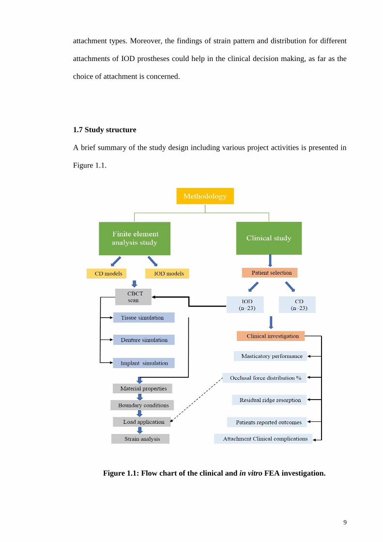
1.7 Study structure
A brief summary of the study design including various project activities is presented in
Figure 1.1.
Figure 1.1: Flow chart of the clinical and in vitro FEA investigation.
10
CHAPTER 2: LITERATURE REVIEW
2.1 Complete edentulism
Complete tooth loss or edentulism is a “debilitating disease and irreversible condition
and is described as the final marker of disease burden for oral health” (Cunha-Cruz et al.,
2007; Emami et al., 2013). It is a public health problem affecting older adults, and it has
been associated with socio-economic status and general well being (Cunha-Cruz et al.,
2007).
Edentulism disturbs diet intake due to masticatory dysfunction (Sheiham & Steele,
2001), which can directly lead to functional limitations, as well as physical,
psychological, and social disability and handicap (Locker, 1988). Therefore the
occurrence of edentulism should be monitored in different countries as an indicator of
population health and functioning (Thomson, 2012). Understanding the significance of
edentulism and its general effect on elderly population health can assist policymakers and
public health researchers in this area (Peltzer et al., 2014).
A systematic review and meta-analysis by Kassebaum et al. (2014) showed that the
prevalence of complete tooth loss has declined between 1990 to 2010. However, the
burden of complete tooth loss remains (Locker & Millar, 2005).
Complete edentulism is a worldwide health problem, mostly in the older age groups
(Felton, 2009). The overall rate of edentulism in Canada in the year 2010 was estimated
at 6.4% and among adults of age between 60–79 years old, it was 21.7% (Cooney, 2010).
In Ireland, the Netherlands, Iceland, Brazil, and Turkey the rate of edentulism among
adults between 65–74 years old was 48.3%, 65.4%, 54.7%, 71.5%, and 48.0%
11
respectively (Madléna et al., 2008; Felton, 2009; Ribeiro et al., 2011; Doğan & Gökalp,
2012), and it was 14% in Sweden for 55–84 years old (Österberg et al., 2010).
The rate of edentulism has a tendency to alter among various locales, even inside a
nation. For example in Canada, there was a wide variation amongst provinces, from
Quebec (14%) to Northwest Territories (5%), due to related factors such as access to
fluoridated water and smoking (Locker & Millar, 2005).
According to the Oral Health Division (2011) survey, it was reported that the rate of
edentulism in the Malaysian population had declined from 54% in 1974 to 32% in 2010.
However, the rate of edentulism could vary among different areas, wherein a study
conducted in Kelantan, the prevalence of edentulism was found to be 56%, and it was
associated with older age individuals (Shamdol et al., 2008).
2.2 Edentulism risk factors
2.2.1 Age
Deficient basic dental care for senior residents possibly leads to poor nutrition, dental
pain, medical complications and tooth loss (Wick, 2010). Tooth loss has been reliably
associated with increasing age (Islas-Granillo et al., 2016). As the vascularity of the
alveolar bone declines, bone resorption increase and reduces the capacity to repair
damage (Boskey, 2013). Additionally, the number of residual teeth diminishes with aging
because of the higher prevalence of periodontitis which increases with an older population
(Renvert et al., 2013).

12
2.2.2 Gender
Male and female genders were equally affected by tooth loss in some studies (Nelsen
et al., 2001; Takenaka et al., 2001; Humphrey & Bocaege, 2008; Bolhofner & Baker,
2012), and the association between gender and tooth loss could not be established (Russell
et al., 2013).
2.2.3 Smoking
Smoking has been recognized as a risk factor and can lead to tooth loss due to
peripheral vascular disease and chronic periodontal disease. In a study of 33,777 matured
Canadians aged 65 years or more, 48% of smokers were edentulous (Millar & Locker,
2007). Similarly, Xie & Ainamo (1999) examined the relationship of different factors
associated with tooth loss in senior citizens living at home in Helsinki, Finland. They
found that the individuals who smoked were two times more likely to be totally
edentulous after adjusting the age and gender factors.
2.2.4 Asthma
In addition to smoking, Xie & Ainamo (1999) found that asthmatic patients were ten
times more likely to be edentulous in the maxillary arch than non-asthmatics. The authors
hypothesized that patients with inhaled corticosteroids could have encountered systemic
effects causing suppression of bone development, and eventually bone and tooth loss of
the maxillary bone. In another investigation of 177 edentulous subjects aged 76 years,
Xie et al. (1997a) found that elderly patients suffering from asthma were six times more
13
likely to encounter severe reduction of the mandibular ridge compared to non-asthmatic
patients.
2.2.5 Diabetes
It has been estimated that by the year 2035 there would be about 600 million people
suffering from diabetes (Iversen et al., 2016). A cross-sectional study of 370 patients
revealed that edentulous older men had four times more risk for developing non-insulin
dependent diabetes mellitus, regardless of age or race than those with partial or complete
dentitions (Cleary & Hutton, 1995). Also, diabetes was found to be positively associated
with edentulism among adults aged 35 years and older in Mexico (Medina-Solís et al.,
2006).
2.2.6 Rheumatoid arthritis
Rheumatoid arthritis is a chronic inflammatory disease characterized by joint swelling,
joint tenderness, and destruction of synovial joints (Felton, 2009). Some clinical studies
have proposed a probable association between rheumatoid arthritis and tooth loss
(Mercado et al., 2001; Al-Shammari et al., 2005). While there was no positive association
found in other studies (Laurell et al., 1989; Yavuzyilmaz et al., 1992). de Pablo et al.
(2008) found edentulism in rheumatoid patients (56%) to be twice as common as in non-
rheumatoid patients (34%).

14
2.2.7 Obesity
Obesity is considered as a progressively serious health problem, due to its association
with diabetes (Andreyeva et al., 2007). A review by Österberg et al. (2010) found a
positive association between obesity and edentulism in the age group of 55–84 years and
the association was stronger in women than in men.
2.2.8 Lower education and income
People with higher salary possibly have more access to educational information and
measures that promote preventative health care, better dietary behaviors, and living
environments, and they gain a more prominent measure of oral hygiene products (Mendes
et al., 2012). Peoples in the lower occupational classes were thought to be at higher
possibility of health risk behaviors (Fukuda et al., 2005). Also, the prevalence of dental
caries in low-income individuals has been previously noted as a potential risk factor of
edentulism (Krustrup & Petersen, 2007).
2.3 Rehabilitation of edentulous patients
2.3.1 Complete denture prostheses
Complete denture rehabilitation method remains an important part of dental education
and practice (Zarb & Bolender, 1997). The most basic form of oral rehabilitation for
edentulous patients has been with CD, due to relatively affordable cost, has acceptable
aesthetics, and function, and is easy to clean (Doundoulakis et al., 2003). Until the

15
establishment of dental implants supporting IOD prostheses, the only accessible treatment
for edentulous patients was CDs (Carlsson & Omar, 2010).
Nevertheless, not all CD wearers are able to adapt to their dentures, even if the dentures
fulfill all conventional prosthodontic criteria. A retrospective study by Laurina &
Soboleva (2006) was conducted to determine possible causes of patients’ complains about
new CDs. The authors found that most of the patients frequently complained of denture
looseness, aesthetics, impaired masticatory function, and accumulation of food under the
denture.
Uram-Tuculescu et al. (2015) clinically investigated the differences in masticatory
muscle function using electromyographic (EMG) device during chewing of agar-based
food model between dentate patients and patients treated with conventional CD, IOD, and
implant fixed denture (IFD). The results showed a higher masticatory muscle activity for
denture wearers compared to the dentate subjects, due to extra mechanical efforts during
oral food processing to accommodate the use of dentures for preparing a bolus for
swallowing.
Similarly, a meta-analysis systematic review by Gracht et al. (2017). A higher chewing
values were observed in IFD or IOD groups compared to CD group, indicating improved
bite force and masticatory performance in the former groups.
2.3.2 Osseointegrated dental implants
Osseointegration is defined as “the apparent direct attachment or connection of
osseous tissue to an inert, alloplastic material without intervening fibrous connective
tissue” (The Glossary of Prosthodontic terms, 2017). Brånemark (1983) presented the

16
osseointegration term to describe this modality for stable fixation of titanium implants to
bone tissue. He described the chambers fabricated from metal titanium could become
permanently incorporated into living bone, and the bone fused with the titanium oxide
layer of the implant could not be separated (Brånemark, 1959).
Osseointegrated implants survival success rate of 87.5% was observed for individuals
in a 3 years longitudinal study conducted by Cox & Zarb (1987). Friberg & Jemt (2015)
investigated the implant/prosthesis survival rates, marginal bone loss, and clinical
complications in patients who received implants in one or two-surgery stages for 5 years
of follow-up. During the observation period, it was observed that out of 259 patients only
eight patients were observed with complete implant/prosthesis failures or bone loss.
It was also important for the implant not to be excessively loaded, as these loads could
interfere with osseointegration (Porter Jr et al., 2002). For the average tooth, the
periodontal ligament acts as an intermediate cushion to buffer the occlusal forces, while
in the osseointegrated dental implant, occlusal forces were transmitted immediately to the
surrounding bones (Weinberg, 1993).
2.3.3 Implant fixed prostheses
The use of 5-6 intreforaminal implants to support fixed prostheses for edentulous
patients has been introduced by Brånemark (1977) and Feine & Carlsson (2003). The
failure and survival rates of different modalities of implant prostheses have been reviewed
and the results indicated higher survival rates for IFD dental prostheses compared to other
types of implant prostheses (Muddugangadhar et al., 2015).
17
Implants lack the stress release associated with a periodontal ligament and hence they
are exposed to greater harmful loading effect on the restorative materials and the crestal
bone (Curtis et al., 2000). Dental implants are also thought to be predisposed to occlusal
overloading, which could explain for peri-implant bone loss and implant prostheses
failure (Kim et al., 2005).
Therefore it has been recommended that in order to minimize the incidence of
complications, reliable implant components and the restorative materials for IFDs
prostheses should be used in a well-structured maintenance system after treatment
(Pjetursson et al., 2012).
2.3.4 Implant-supported prostheses
The McGill consensus in the year 2002 on overdentures stated that: there is now
overwhelming evidence that a two-IOD should become the first choice of treatment for
the edentulous mandible, based on available scientific evidence (Feine et al., 2002).
In 2009, the York consensus statement was further support evidence demonstrating
that patients’ satisfaction and quality of life with implant-supported mandibular dentures.
Much of these evidences came from randomized controlled trials” (Thomason et al.,
2009). Due to their relatively lower cost and lower complication rate than fixed
prostheses, mandibular IODs have been popular (Feine & Carlsson, 2003; Carlsson et al.,
2004).
18
2.3.5 Number of implants used with implant-supported overdenture prostheses
Maxillary overdentures supported by four to six implants splinted with a bar (Slot et
al., 2010; Carlsson, 2014), have revealed effective functional results than two implants.
However, maxillary IODs on two implants were commonly found less effective than the
mandibular two-IODs (Mericske-Stern, 2003).
Merickske-Stern (1990) in a retrospective study on 67 patients who were provided
with mandibular two-IOD, has observed that two implants may sufficiently be used for
IOD retention, without the need of multiple implants or to implants splinted with a bar.
Regarding the peri-implant tissue condition, Batenburg et al. (1998) conducted a
prospective one year study to investigate patients with mandibular overdenture
prostheses, retained by two and four implants. They concluded that no difference was
observed in the peri-implant health between the groups and made a recommendation that
treatment with two implants should be sufficient to retain IOD in the mandible.
A recent randomized clinical trial on 20 participants with edentulous mandibular
ridges, who were randomly assigned into two groups; four implants installed in lateral-
canine and premolar regions, and two implants in lateral-canine regions. The authors
concluded however that increasing the number of implants from two to four in
mandibular IODs had no significant influence on implant stability (Wafa’a et al., 2017).
Therefore, following the McGill consensus two mandibular implants can be considered
a viable treatment option for edentulous mandible.
19
2.3.6 Attachments used with implant-supported overdenture prostheses
According to the Glossary of Oral and Maxillofacial Implants (Hjørting-Hansen et al.,
2007), an attachment system is “a design of a particular type of retentive mechanism
employing compatible matrix and patrix corresponding components. Matrix refers to the
receptacle component of the attachment system, and patrix refers to the portion that has
a frictional fit and engages the matrix”.
A wide variety of commercially-available attachment systems is used as retentive
components in implant overdentures. The retentive design can be splinted or unsplinted
using different attachments designs (Alsabeeha et al., 2009). The anatomy of the
edentulous arch with desired retention level, hygiene, maintenance capability, implants
parallelism, and cost considerations are important factors in choosing the appropriate
overdenture attachment type (Alsiyabi et al., 2005; Gulizio et al., 2005).
The attachment types can be classified into ball, Locator, magnet, bar, and telescopic
attachments. Plaque accumulation has been shown to be significantly higher in magnet
attachment than for ball attachment, because of frequent exchange of magnets, or wear
and corrosion of them (Naert et al., 1998). Besides, the use of magnet attachment provides
less retention as compared to the other attachment systems (Smith GA, 1983; Cune,
2005).
Between bar and ball attachments in the mandibular two-IODs it was observed that the
biological complications were not significantly different, however, a greater number of
technical complications per patient was recorded with bar than ball attachments
(Gotfredsen & Holm, 2000). Bar attachment requires the skill of technician in the
fabrication. At the same time, the splinted design of the bar could compromise hygiene.
A comparison between ball to telescopic attachments, however, was less favorable where
20
ball attachments showed higher recurrence of technical complications over five years
(Krennmair et al., 2011). Denture reinforcement when using ball attachments has been
suggested to increase fracture resistance of the base (ELsyad et al., 2016). From the
biological aspect, the soft tissue response around abutments in patients who use ball
attachments was less favorable compared to other unsplinted design of attachments (Kleis
et al., 2010).
2.3.6.1 Telescopic attachments
Telescopic crowns as retentive elements for overdenture prostheses, also known as
double-crowns, consist of an inner or primary telescopic coping, cemented to an
abutment, and the corresponding detachable outer or secondary telescopic crown (Langer
et al., 2000). This type of retainer provides excellent retention resulting from frictional fit
between the crown and the sleeve (Keller & Haase, 1991). The use of cemented, rigid
telescopic crowns was suggested to avoid disadvantages of screw-retained
superstructures, such as difficult access to the screw positions (Hoffmann et al., 2006).
Moreover, telescopic-retained restorations can be easily removed or inserted and are
considered as an effective treatment modality for geriatric patients (Heckmann et al.,
2004).
2.3.6.2 Locator attachments
The Locator attachment system is a system with a self-aligning feature and has dual
retention (inner and outer), which has been on the market since 2000. The Locator design
comprises; a patrix (male) which is the replaceable nylon attached to the denture base of
21
the overdenture, and a matrix (female) ) which is fixed as an overdenture abutment (Kleis
et al., 2010).
The nylon retentive element comes in different colors, to indicate different retentive
strength (Chung et al., 2004). Additional features are extended range attachments, which
can be used to correct implant angulation up to 20° (Evtimovska et al., 2009). The reduced
height of Locator attachment is the main advantageous in particular in cases with limited
inter-occlusal space or when retrofitting an existing old denture (Pasciuta et al., 2005).
2.4 Stress distribution according to attachment types
The way stress is applied to implants following osseointegration is one of the features
which should be investigated (Trakas et al., 2006). Telescopic IOD has been shown to
improve chewing efficiency compared to bar attachments (Elsyad & Khairallah, 2017).
However, with higher bite force, a higher stress could lead to greater risk of implant
fatigue and even fracture (Heckmann et al., 2001). Correspondingly, Elsyad et al. (2013)
in a 4 years retrospective study, observed an increased resorption and flabbiness of
maxillary anterior residual ridge when telescopic attachments used in mandibular two-
IOD.
Using an experimental resin model, Ichikawa et al. (1996) investigated the occlusal
stress distribution on the implants supports IOD, and the authors concluded that even with
the higher occlusal stress concentrated on implants especially in the distal areas, the
modified magnetic attachment with silicone provided optimal stress distribution.
Similarly, in vitro study by Gonda et al. (2004) observed that lateral stresses were reduced
22
on the abutment tooth under tooth-supported overdentures which were not rigidly
connected.
2.5 Occlusal registration methods
Any occlusal interference might not only affect implants and IOD prostheses. In fact,
reduced chewing stability, pain, gum disease, headaches, and temporomandibular joint
(TMJ) problems were possibly associated with occlusal force disturbances (Bicaj et al.,
2015). Therefore, occlusal recording devices and methods are important factors in
determining occlusal force distribution (Koc et al., 2010).
Various methods are employed to determine patients′ occlusal contact pattern after
restoration to allow harmonious contact with the opposing teeth. These methods also
provide information about the location, timing, direction, and magnitude of occlusal
contacts (Baba et al., 2000). Universally used techniques for occlusal registrations are
classified into qualitative and quantitative methods (Sharma et al., 2013).
2.5.1 Qualitative method
The qualitative method such as articulating paper requires an examiner to make his/her
choice to determine the occlusal contact points, however, the sequence or density of the
contacts could not be evaluated (Panigrahi et al., 2015). This method is concerned with
the usage of the following materials; articulating paper/ribbon, silk strips, foils,
impression, and occlusal indicator wax.

23
2.5.1.1 Articulating paper/ribbon
An articulating paper/ribbon can be a carbon paper, inked paper/ribbon or a
paper/ribbon coated with glossy-colored dye. These materials are commonly used in
clinical and laboratory settings to spot premature contacts in the occlusion. They are
manufactured in different thicknesses, shapes, and colors to facilitate clinical usage
(Oshida et al., 1994; Zuccari et al., 1996).
Excessive force or premature contact can be determined based on the marks on the
tooth. Large and dark marks signify heavy occlusal load, while smaller and lighter marks
are associated with reduced occlusal loads. Nevertheless, there is no scientific evidence
to indicate that articulating papers is an ideal choice of material for occlusal correction.
Clinical conclusions based on the heaviness of marks were reported to be an inexact
approach for assessment of the contact density (Kerstein, 2008). Moreover, the
articulating papers are demolished by saliva, resulting in a high percentage of pseudo
contact markings (Maness, 1987a; Saraçoǧlu & Özpinar, 2002). Therefore, application of
articulating paper is considered in determining the location of tooth contacts. However,
their low price and ease of use have made them the most widely used qualitative occlusal
registration materials (Afrashtehfar et al., 2013).
2.5.1.2 Silk strips
Silk strips are the soft flexible indicator materials with various range of thickness (26-
98 µm), which provide a higher reliability due to their texture. Furthermore, when
perfectly adjusted to cusps and fossae they do not create pseudo contact markings (Schelb
24
et al., 1985). One disadvantage with silk strip, however, the material could be altered and
distorted by the presence of saliva (Reiber et al., 1989).
2.5.1.3 Foils
Foils are the thinnest occlusal registration materials which provide extra precise
readings than paper and silk. However, their marking potential is diminished under
lowered pressure and on glossy surfaces. As a consequence, for their best usage, a larger
strip has to be used for occlusal detection (Reiber et al., 1989; Panigrahi et al., 2015).
2.5.1.4 Impression materials
Impression materials were used to record occlusal force by allowing biting without
resistance (Molligoda et al., 1986). Due to its elastic characteristics, the occlusal
impression material is used to mark the occlusal contacts and may be identified when the
material was removed after setting (Millstein & Maya, 2001).
Silicone putty impression materials maybe used as an inter-occlusal recording material
to evaluate occlusal contacts. The position of tooth contacts is displayed as perforations
in the silicone impression material (Ziebert & Donegan, 1979; Sharma et al., 2013).
2.5.1.5 Occlusal indicator wax
Occlusal indicator wax originates from a concept similar to impression materials
(Ehrlich & Taicher, 1981; Millstein, 1985; Kong et al., 1991). However, resistance exists
25
when biting into the wax, then the occlusal wax indicator is inspected ahead on a light
source. Each recording is situated on a diagnostic cast to visualize and confirm the
accurate site of each contact. Nevertheless, inaccuracy and difficult handling were some
drawbacks when wax was used as an occlusal material for clinical records (Ehrlich &
Taicher, 1981; Murray et al., 1999).
2.5.2 Quantitative method
In the qualitative method, an estimation can be derived from the density of the con-
tacts, for example, the degree of darkness of a tooth contact mark. This is however not
considered as a precise measure for evaluation of tooth contact due to its association with
human error (Sharma et al., 2013).
Photo-occlusion (Fitzig et al., 1985) and T-scan system (Maness, 1987b) are
quantitative techniques used for detecting occlusal relationships through which the
sequence and density of the contacts can be differentiated (Panigrahi et al., 2015).
2.5.2.1 Photo-occlusion system
Dental prescale system comprises a horseshoe-shaped bite foil of a pressure-sensitive
film and a computerized scanning system for the examination of the load. A thin photo
plastic film layer is positioned on the occlusal surface of the teeth where the patient bites
for 10–20 sec. The force is then spread over to the occlusal contact and an ordered color
is produced by a chemical reaction. The film layer is reviewed under a polariscope light
to find the relative tooth contact intensity measurement. The area and color intensity of
the red points are used to evaluate the occlusal contact area and pressure with automatic
26
estimation of the occlusal loads (Hidaka et al., 1999; Shinogaya et al., 2000; Miura et al.,
2001; Okiyama et al., 2003; Koc et al., 2010).
The dental prescale system, with a thin pressure sensitive foil of a 100 µm thickness
(Sadamori et al., 2007), was found to be excellent in measuring biting force as compared
to the ordinary measuring systems using a strain gauge transducer (Shinogaya &
Matsumoto, 1998; Shinogaya et al., 2000). The foil width allows the bite force
measurement close to the intercuspation position and instantaneous evaluation of load
distribution over dentition can be made (Shinogaya et al., 2000; Shinogaya et al., 2001).
However, this system has the disadvantage of its inability to record the occlusal contact
time orders and difficulty to analyze premature and laterotrusive or mediotrusive contacts
(Koos et al., 2010).
2.5.2.2 T-scan system
This type of computerized occlusal analysis system is a device that can reproduce the
actual occlusal contacts and it also has an advantage over silk marking ribbon in terms of
recording the time and force of each contact (Moini & Neff, 1991). Besides accuracy,
Cerna et al. (2015) examined the reliability of the T-scan III system under laboratory
conditions. The results showed a high degree of reliability when the T-scan III sensors of
two different production series were used to perform repeated measurements.
T-scan occlusal analysis technique was introduced as an uncomplicated, easy-to-use
method with high accuracy in determining the quantity of tooth contacts in maximum
intercuspation position. It allows the occlusal contact data to be quantitatively analyzed,

27
in contrast to other techniques which necessitate qualitative occlusal determination by the
operator (Gonzalez Sequeros et al., 1997).
2.6 T-scan system development
The T-scan electrical structure was developed from experimental touch sensors, which
were fabricated for robotic uses. Touch sensors were further developed by Purbrick
(1981) and Hillis (1982), where conductive rubber elements were used to detect pressure
at individual contact points. The material was constructed in such a manner that an
increase in pressure will lead to a diminution in its electrical resistance. A similar concept
is used with the T-scan, however, the conductive rubber base is replaced with an ultra-
thin layer of a pressure-sensitive ink.
Maness (1987a) developed a T-scan system to allow interpretation of the teeth contact
data and recording of stress magnitude at each contact by making use of time as the
fundamental diagnostic variable. With the T-scan system, it was possible to report various
occlusal parameters such as the timing of teeth contacts and their relative force. Maness
(1987b) also advocated its use in diagnosis and treatment of occlusion due to its accuracy
in detailing the tooth contact. This system is thus utilized to analyze the occlusal contact
information collected via directing the subject to occlude on intraoral recording sensor.
The system allows recognition of premature contacts or interferences in the dynamic
occlusion (Koos et al., 2010), making it applicable in occlusal diagnosis and treatment
(Reza & Neff, 1991; Kerstein, 2008; Throckmorton et al., 2009; Kerstein et al., 2013).
In 1995, the T-scan was further improved to T-scan II occlusal analysis system for
Windows. The modifications comprised a hardware, software, and additional sensor
28
developments that led to a third generation sensor (G3) with other sensor enhancements
(Kerstein, 2004). The G3 sensor scheme was again modified to enlarge the active sensel
size and reduce the inactive sensel area by 33% and 50%, respectively, in order to form
the fourth generation sensor advancement (G4) with a higher definition scheme (Kerstein,
2011). According to the T-scan III manufacturer, the sensor is 100 µm thick and
comprises around 2,500 sensing cells (sensels). The T-scan III G4 sensor contains two
layers of Mylar polyester resin sheets with the net of resistive ink rows and columns
printed between them. A row and a column are individually connected and signify one
sensel, which is the force and time recording portion of the computerized occlusal
analysis system. Both bigger sensels and lesser space between the sensels help to raise
the total volume of recording surface area and simultaneously reduce the amount of
inactive non-recording area between the sensels (Kerstein, 2011).
In 2008, T-scan III with Turbo recording was released. Turbo recording captured three
times more occlusal data for analysis than was possible with the previous versions. This
faster-scanning speed provides the clinician with greater ability to locate non-
simultaneous tooth contact sequences and any abnormal occlusal force concentrations. T-
scan III was further improved and in 2012 T-scan 8 was released, that was purposefully
designed to minimize the T-scan III’s user interface complexity by using fewer software
toolbar buttons and icons for simpler data display (Kerstein, 2015).
29
2.6.1 T-scan system applications in Dentistry
2.6.1.1 Prosthodontics
To determine whether T-scan could be used to replace remounting procedures in CD
reconstruction, a correlation between remounting and balance of occlusal load was
determined in 10 patients who had new CDs. The results showed a very strong correlation
in 9 out of 10 evaluated patients (Boening & Walter, 1992).
In another study, occlusion and force distribution characteristics of three CD patients,
whose dentures were constructed in the postgraduate prosthodontic clinic, were analyzed
in pre- and post-adjustment appointments using the T-scan II system. The computerized
occlusal analysis was found to provide information on occlusal load distribution and
adjustment, and it provides an efficient way for clinicians to improve denture stability
(Olivieri et al., 1998).
2.6.1.2 Trauma from occlusion
Trauma from heavy occlusal contacts can lead to loss of periodontal attachment with
increased tooth mobility (Harrel & Nunn, 2001). The T-scan III is a useful tool that can
be used to identify premature occlusal contacts on a hypermobile tooth. Also, it is a
valuable tool to identify contact intensity and to evaluate contact changes after occlusal
adjustment with a diamond bur (Nguyen et al., 2007), and in the diagnosis and treatment
follow-up until complete trauma resolution (Daing et al., 2012).
The T-scan III has also been used to determine centric and eccentric occlusion and
occlusal time of patients with dysfunctional cranial-mandibular syndrome (Oprea et al.,
2014).

30
2.6.1.3 Implant prostheses
Occlusal forces directed to implant prostheses with damaging component could reduce
the durability of the restoration. Material failures together with implant loss of
osseointegration could be linked to occlusal loading of dental implants (Kohavi, 1993;
Isidor, 1997).
With the advance in the computerized T-scan occlusal analysis system, occlusal forces
that are directed to implant prostheses can be quantitatively characterized for occlusal
corrections. The occlusal force equilibration can lead to less damaging forces towards the
prostheses and the underlying implant-bone interface (Cotruta et al., 2015).
Clinicians could obtain consistent and useful occlusal data for the placement, analysis,
and repair of a dental implant prosthesis (Garg, 2007). As premature contacts in
maximum intercuspation position must be eliminated when prosthesis is fixed to provide
better force control both on the bone-implant interface and implant-prostheses assembly
(Chapman, 1989).
A case of severe bruxism and masseteric hypertrophy, associated with repeated
implant loss was reported by Sukegawa et al. (2016). A successful management of
masseteric hypertrophy and subsequently avoidance of implant loss was achieved in this
case where occlusal analysis was preferred using T-scan III prior to dental implant
placement.
2.6.2 T-scan system limitations
It was revealed that thinner occlusal registration materials offer a more constant
reading of the contact points (Reiber et al., 1989). To achieve the technological
31
requirements, the T-scan sensors were prepared as thin as possible at 100 µm (Kerstein,
2007). However, these sensors are slightly thicker compared to other occlusal indicators
such as articulating silk, which has the thickness within the range of 26-98 µm.
The sensitivity of the T-scan sensors also appears to reduce or fade when the sensors
were used more than once. Previous studies have detected the absence of sensibility on
some of its parts of the sensor, which resulted in incorrect recordings (Patyk et al., 1989;
Yamamura et al. 1990; Harvey et al., 1991).
T-scan system measures force in percentages only instead of actual force in Newton.
A vitro study, with a limited clinical trial on two individuals, was carried out to measure
bite force by using the T-scan system by Lyons et al. (1991), concluded that the T-scan
system does not precisely determine actual bite force.
2.7 Bone Resorption
Great individual variation in bone loss of the alveolar process occurs after tooth
removal and it can be difficult to anticipate the extent of bone loss at the time of
extraction. Alveolar bone loss can be minimized by preserving a few teeth in the arch or
by implant placement to support the prostheses (Carlsson, 2014).
2.7.1 Residual ridge resorption
Bone remodeling that takes place after tooth removal occurs in two phases; an initial
and fast phase that can be detected in the first three months and the succeeding second
phase that is slow, and ongoing throughout life. During the early period of 6 and 12
32
months, there is new bone formation with the destruction of almost all of the alveolar
crest height and reduction of nearly two-thirds of the ridge width (Schropp et al., 2003).
The rate of resorption then decelerates to minimal levels and significant reduction in
bony dimensions could be seen particularly in patients become edentulous at younger age.
The rate of residual ridge resorption (RRR) differs, between individuals, at different
phases of life, and even at different sites in the same individual (D'Souza, 2012).
2.7.2 Consequences of residual ridge resorption
Residual ridge resorption is chronic and progressive, and in complete denture patients,
it can result with denture wearing. As it is cumulative, patients with this disease can
become dentally handicapped (Atwood, 1971).
An average rate of anterior vertical RRR of 0.5 mm per year could represent an average
loss of 5 mm in anterior ridge height in 10 years with a different variation among some
patients, resulting in a significant loss of vertical dimension, aesthetics, and comfort. Such
remarkable changes in edentulous people represent a staggering need for prosthodontic
care (Atwood & Coy, 1971).
2.7.3 Factors affecting residual ridge resorption
2.7.3.1 Anatomic factors
Anatomic factors include the amount and quality of bone. The amount of bone is not
a dependable prognostic factor for the rate of RRR since it was viewed that some large
ridges resorb quickly and certain knife-edge ridges could continue with slight variations
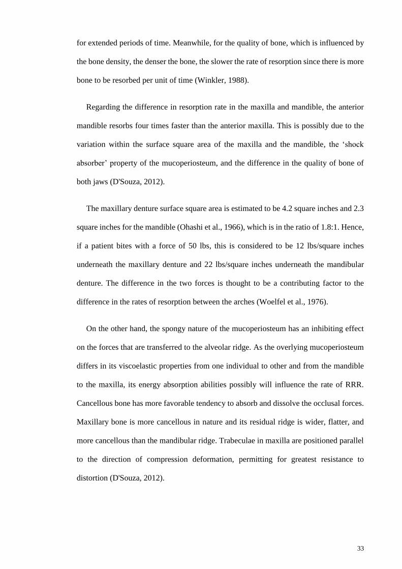
33
for extended periods of time. Meanwhile, for the quality of bone, which is influenced by
the bone density, the denser the bone, the slower the rate of resorption since there is more
bone to be resorbed per unit of time (Winkler, 1988).
Regarding the difference in resorption rate in the maxilla and mandible, the anterior
mandible resorbs four times faster than the anterior maxilla. This is possibly due to the
variation within the surface square area of the maxilla and the mandible, the ‘shock
absorber’ property of the mucoperiosteum, and the difference in the quality of bone of
both jaws (D'Souza, 2012).
The maxillary denture surface square area is estimated to be 4.2 square inches and 2.3
square inches for the mandible (Ohashi et al., 1966), which is in the ratio of 1.8:1. Hence,
if a patient bites with a force of 50 lbs, this is considered to be 12 lbs/square inches
underneath the maxillary denture and 22 lbs/square inches underneath the mandibular
denture. The difference in the two forces is thought to be a contributing factor to the
difference in the rates of resorption between the arches (Woelfel et al., 1976).
On the other hand, the spongy nature of the mucoperiosteum has an inhibiting effect
on the forces that are transferred to the alveolar ridge. As the overlying mucoperiosteum
differs in its viscoelastic properties from one individual to other and from the mandible
to the maxilla, its energy absorption abilities possibly will influence the rate of RRR.
Cancellous bone has more favorable tendency to absorb and dissolve the occlusal forces.
Maxillary bone is more cancellous in nature and its residual ridge is wider, flatter, and
more cancellous than the mandibular ridge. Trabeculae in maxilla are positioned parallel
to the direction of compression deformation, permitting for greatest resistance to
distortion (D'Souza, 2012).

34
2.7.3.2 Prosthetic factors
Lack of retention and/or instability of removable prostheses has been implicated in
higher rate of RRR (Atwood & Coy, 1971). Another study by Carlsson & Persson (1967)
showed the increase in RRR rate of the edentulous ridge in denture wearers who had been
using their denture for a long period.
Maxillary complete denture opposing mandibular natural teeth tended to have a
greater bone resorption in the anterior segment of the maxillary residual ridge (Kelly,
1972).
2.7.3.3 Role of inflammatory mediators
Numerous inflammatory mediators, mostly prostaglandins, have been suggested to
play a role in increasing the rate of RRR. When osteoblastic cells were exposed to cyclic
mechanical stresses in vitro, there was a significant escalation in prostaglandin E2
synthesis (Yeh & Rodan, 1984).
Adrenomedullin hormone has been also reported to be able to stimulate the
proliferation and migration of various cells including osteoblasts. The influence of local
adrenomedullin in rats tooth extraction socket after 4, 8, 12 weeks, suggested that local
application of adrenomedullin has the potential to preserve the residual alveolar ridge and
accelerate the alveolar bone remodeling (Wang et al., 2013).
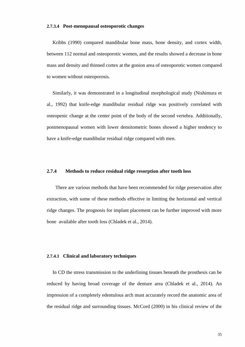
35
2.7.3.4 Post-menopausal osteoporotic changes
Kribbs (1990) compared mandibular bone mass, bone density, and cortex width,
between 112 normal and osteoporotic women, and the results showed a decrease in bone
mass and density and thinned cortex at the gonion area of osteoporotic women compared
to women without osteoporosis.
Similarly, it was demonstrated in a longitudinal morphological study (Nishimura et
al., 1992) that knife-edge mandibular residual ridge was positively correlated with
osteopenic change at the center point of the body of the second vertebra. Additionally,
postmenopausal women with lower densitometric bones showed a higher tendency to
have a knife-edge mandibular residual ridge compared with men.
2.7.4 Methods to reduce residual ridge resorption after tooth loss
There are various methods that have been recommended for ridge preservation after
extraction, with some of these methods effective in limiting the horizontal and vertical
ridge changes. The prognosis for implant placement can be further improved with more
bone available after tooth loss (Chladek et al., 2014).
2.7.4.1 Clinical and laboratory techniques
In CD the stress transmission to the underlining tissues beneath the prosthesis can be
reduced by having broad coverage of the denture area (Chladek et al., 2014). An
impression of a completely edentulous arch must accurately record the anatomic area of
the residual ridge and surrounding tissues. McCord (2000) in his clinical review of the
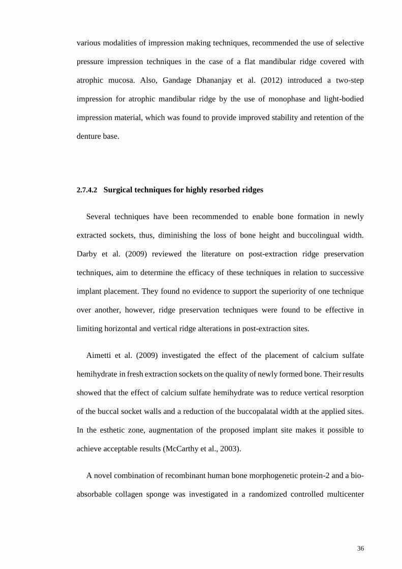
36
various modalities of impression making techniques, recommended the use of selective
pressure impression techniques in the case of a flat mandibular ridge covered with
atrophic mucosa. Also, Gandage Dhananjay et al. (2012) introduced a two-step
impression for atrophic mandibular ridge by the use of monophase and light-bodied
impression material, which was found to provide improved stability and retention of the
denture base.
2.7.4.2 Surgical techniques for highly resorbed ridges
Several techniques have been recommended to enable bone formation in newly
extracted sockets, thus, diminishing the loss of bone height and buccolingual width.
Darby et al. (2009) reviewed the literature on post-extraction ridge preservation
techniques, aim to determine the efficacy of these techniques in relation to successive
implant placement. They found no evidence to support the superiority of one technique
over another, however, ridge preservation techniques were found to be effective in
limiting horizontal and vertical ridge alterations in post-extraction sites.
Aimetti et al. (2009) investigated the effect of the placement of calcium sulfate
hemihydrate in fresh extraction sockets on the quality of newly formed bone. Their results
showed that the effect of calcium sulfate hemihydrate was to reduce vertical resorption
of the buccal socket walls and a reduction of the buccopalatal width at the applied sites.
In the esthetic zone, augmentation of the proposed implant site makes it possible to
achieve acceptable results (McCarthy et al., 2003).
A novel combination of recombinant human bone morphogenetic protein-2 and a bio-
absorbable collagen sponge was investigated in a randomized controlled multicenter
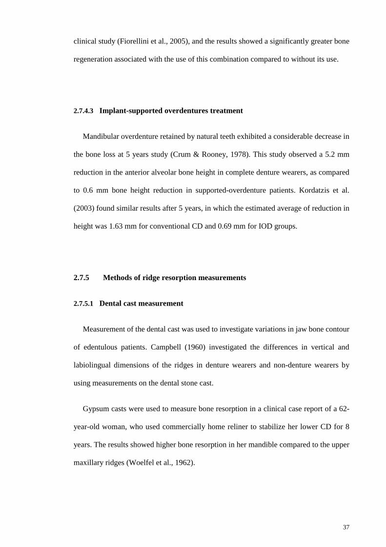
37
clinical study (Fiorellini et al., 2005), and the results showed a significantly greater bone
regeneration associated with the use of this combination compared to without its use.
2.7.4.3 Implant-supported overdentures treatment
Mandibular overdenture retained by natural teeth exhibited a considerable decrease in
the bone loss at 5 years study (Crum & Rooney, 1978). This study observed a 5.2 mm
reduction in the anterior alveolar bone height in complete denture wearers, as compared
to 0.6 mm bone height reduction in supported-overdenture patients. Kordatzis et al.
(2003) found similar results after 5 years, in which the estimated average of reduction in
height was 1.63 mm for conventional CD and 0.69 mm for IOD groups.
2.7.5 Methods of ridge resorption measurements
2.7.5.1 Dental cast measurement
Measurement of the dental cast was used to investigate variations in jaw bone contour
of edentulous patients. Campbell (1960) investigated the differences in vertical and
labiolingual dimensions of the ridges in denture wearers and non-denture wearers by
using measurements on the dental stone cast.
Gypsum casts were used to measure bone resorption in a clinical case report of a 62-
year-old woman, who used commercially home reliner to stabilize her lower CD for 8
years. The results showed higher bone resorption in her mandible compared to the upper
maxillary ridges (Woelfel et al., 1962).
38
Närhi et al. (2000) investigated the possible changes in the width of the maxillary
residual ridge on the diagnostic cast models after 6 years following mandibular IOD or
conventional CD insertion. They found a reduction of the ridge width with time,
regardless of the type of prostheses used. One concern with this method of measurement
was the variability of the pressure applied during the impression making (McGivney et
al., 1986), which could introduce some errors in the estimation.
2.7.5.2 Radiographical methods
Resorption of the alveolar ridge has been assessed with using radiographical images,
such as lateral cephalometric (Atwood & Coy, 1971; Woelfel et al., 1976; Mercier &
Lafontant, 1979; Tallgren et al., 1980; Jonkman et al., 1991) and panoramic radiographs
(Wical & Swoope, 1974; Van Waas, 1983; Swart & Allard, 1985; Wilding et al., 1987;
Humphries et al., 1989). The more recent imaging method using cone-beam computed
tomography (CBCT) allows 3D view and compared to conventional cross-sectional
imaging (CT), the CBCT reduced X-ray exposure (Angelopoulos et al., 2008).
(a) Cephalometric radiograph
The advantages of the cephalometric technique are that the vertical and horizontal
magnification factors are known, and both jawbones images can be clearly reproduced.
Nevertheless, the superimposition of the alveolar ridges renders it impossible to
differentiate the changes in resorption between the two sides (Xie et al., 1997b). An
oblique cephalometric technique has resolved this problem (Freihofer & Hoppenreys,
39
1986; Jonkman et al., 1991); however, multiple radiographs are required for the area of
investigation involves both the anterior and posterior regions on both sides of the arch.
(b) Panoramic radiograph
A panoramic radiograph is a curved plane tomographic radiography permits
visualization of the maxilla, mandible, TMJ joints and the lower one-half of the maxillary
sinuses on a single image. This radiographic procedure is possibly one of the most
operated diagnostic modality in implant dentistry (Dove & McDavid, 1993).
Panoramic radiography also provides valuable information about anatomic structures
such as the inferior alveolar nerves, and mental foramen (Lee et al., 2012). Additionally,
the examination of RRR in large numbers of patients with panoramic radiography is
achievable as it is usually used in large institutional practices as a method of screening
edentulous patients during routine treatment (Scandrett et al., 1973; Xie et al., 1997b).
Yet, there are some problems that limit the use of the panoramic radiography, for
instance; 2D distortion, variation in magnification, the difficulties in standardizing the
position of the head (Jacobs et al., 1993; Xie et al., 1997b).
(c) Cross-sectional imaging and cone-beam computed tomography scan radiographs
Owing to the fact that cone-beam computed tomography (CBCT) images were
reformatted in slices of the maxilla and mandible, they were free of magnification,
superimposition of neighboring structures, and other problems characteristic to
panoramic and cephalometric radiography. The recently introduced CBCT creates 3D
40
image data by a single scan around the imaging volume of interest and this may result in
very clear images representing the mandibular canal (Angelopoulos et al., 2008).
It was revealed that cross-sectional imaging (CT) images (Serhal et al., 2002; Jacobs
et al., 2004; Angelopoulos et al., 2008) is an alternative for the exact visualization of the
anatomical structures. However, panoramic radiographs were used due to its lower cost
(Dharmar, 1997), and most of its use in implant surgery planning is extensively
documented (Xie et al., 1997b).
2.7.6 Determination of alveolar bone resorption using panoramic radiographs
Panoramic radiography is considered a standard diagnostic procedure to evaluate
residual ridge changes (Jacobs et al., 1992), and compared to cephalometric radiographs,
it has no issues with overlapping of the left and right side images (Xie et al., 1997b).
Current studies for measuring residual ridge bone height loss used two methods; the linear
method and the area index method. However, panoramic radiographs still have some
magnification and head position drawbacks (Wilding et al., 1987; Kreisler et al., 2000).
2.7.6.1 Linear method
This method has been used by many researchers in evaluating the bone loss in the
mandible and maxilla. It is an easy method for recording the oral and related structures
(Wical & Swoope, 1974; Xie et al., 1997b; Güler et al., 2005; Panchbhai, 2013).
Sağlam (2002) determined the variation in maxillary and mandibular vertical
measurements based on the panoramic radiographic views for dentate and edentulous

41
jaws. Ninety-six dentate and edentulous patients were enrolled and the measurements
were made from reference lines drawn from anatomic landmarks on standardized
panoramic radiographs. It was found that the reductions in the height of the edentulous
mandible and maxilla were significantly more pronounced in women than in men.
2.7.6.2 Area index method
Area index method was developed by Wilding et al. (1987), to determine the accuracy
of mandibular bone loss measurements made by panoramic radiographs. A comparison
was made between measurements obtained directly from dry mandibles and those from
panoramic radiographs of the same mandible using either area index or linear vertical
methods. The area index method showed significant difference in the index between the
left and right sides of the mandible, while the liner method showed no significant
difference.
Kreisler et al. (2000) developed an in vitro method to enable the radiological
investigation of RRR in the maxilla. Determined experimental and reference areas in the
maxilla were drawn on transparent film laid over a panoramic radiograph and they were
digitized. All areas were measured with an integrated planimetry program and expressed
as a ratio. The effect of positioning errors on the reliability of the method was investigated
on dry skulls. The correlation between the change in ratio and actual bone loss was
examined by progressively reducing the height of an artificial residual ridge on one skull.
Comparison of the experimental area with the reference area on serial panoramic
radiographs appears suitable for the assessment of RRR in the maxilla.
42
Wright et al. (2002) investigated the change over time in the area of the posterior
mandibular residual ridge in patients wearing either i) mandibular overdentures stabilized
by two implants connected with a bar, or ii) mandibular fixed cantilever prostheses
stabilized on 5 or 6 implants. Proportional measurements were made to compare the area
of the residual ridge with an area of bone uninfluenced by resorption. Measurements were
made by digital tracings of panoramic radiographs that were taken shortly after implant
insertion and after follow-up of up to 7 years later. The results revealed that patients
rehabilitated with mandibular implant overdentures demonstrated low rates of posterior
mandibular RRR, while patients rehabilitated with implant-stabilized mandibular fixed
cantilever prostheses demonstrated bone apposition in the same area.
In a 5 years observational study, Kordatzis et al. (2003), found the average reduction
in the posterior mandibular residual ridge height was higher in conventional denture
patients than in the implant overdenture patients. Moreover, female was found to have a
higher risk factor for greater bone resorption.
Tymstra et al. (2011), utilized the proportional area measurements to determine RRR
in the maxillary anterior and mandibular posterior areas over a period of 10 years in
mandibular IOD, retained by two or four implants. Their results indicated that patients
rehabilitated with IOD were not subjected to more RRR in the anterior maxilla as
compared to patients wearing a conventional CD. While with respect to the mandibular
posterior residual ridge, resorption was independent of wearing an IOD or a conventional
CD.
Another study investigated the clinical and radiographic changes in the edentulous
maxilla in patients with either ball or telescopic attachments of mandibular IODs (Elsyad
et al., 2013). The proportional value between bone areas and of reference areas was
43
expressed as a ratio. Change in ratio immediately before baseline and 4 years after
overdenture insertion was calculated for the maxillary anterior and posterior regions.
Telescopic attachments for mandibular IODs were associated with increased maxillary
ridge resorption and flabbiness and decreased maxillary denture retention as compared to
IOD retained by ball attachments.
2.8 Patient-reported outcomes
2.8.1 Health-Related Quality of Life
World Health Organization (WHO) has defined health as “a complete state of physical,
mental, and social well-being and not just the absence of disease” (WHO, 1995). In the
last 30 years, there was an increase in the use of the terms ‘health-related quality of life’
(HRQoL) and ‘quality of life’ (QoL) in medicine, relative to the outcomes of health
conditions and therapy (Gill & Feinstein, 1994). QoL is defined as “an individual’s
perception of his or her position in life in the context of culture and value systems in
which they live, and in relation to their goals, expectations, standards, and concerns”
(Group, 1995). This emerged out of a growing recognition that traditional clinical
measures of health need to be supplemented by data obtained from patients and/or persons
that capture their experiences and concerns (Fitzpatric et al., 1998).
2.8.2 Oral Health-Related Quality of Life
Oral health-related quality of life (OHRQoL) is defined as “a multi-dimensional
construct that reflects people’s comfort when eating, sleeping, and engaging in social

44
interaction; their self-esteem; and their satisfaction with respect to their oral health”
(Health., 2006). It is a complex and multidimensional concept that focuses on the extent
to which the well-being of individuals and society as a whole is affected by oral problems.
Many variables influence the OHRQoL, including the association of patient’s age and
ethnicity (Atchison & Gift, 1997), gender, financial factors, current dentures, and
psychosocial factors (Locker, 2009).
2.8.3 Oral Health Impact Profile (OHIP-49)
The Oral Health Impact Profile (OHIP) was established with the purpose of providing
a full measure of self-reported dysfunction, discomfort, and disability attributed to oral
conditions. It provides information about the ‘burden of illness’ within populations and
the effectiveness of health services in reducing that burden of illness (Tugwell et al.,
1984).
This instrument was first introduced by Slade and Spencer (1994) in Australia as the
Oral Health Impact Profile (OHIP-49), following Locker's model of oral health (Locker,
1988). There are seven conceptual dimensions of impact; functional limitation, physical
pain, psychological discomfort, physical disability, psychological disability, social
disability, and handicap.
2.8.4 Oral Health Impact Profile (OHIP-14)
A shortened (14-item) version of the OHIP was developed by Slade (1997b). The
shortened version was considered for the reason that the original questionnaires take a
long time to complete, which place a burden on the patient and/or clinician. The
45
respondent burden may be an issue when the participants in a study are weak, ill, or
severely compromised and unable to cooperate for any length of time. Long scales are
also more likely to be subject to item nonresponse, giving rise to problems of how to
manage missing data (Locker & Allen, 2002).
Stewart et al. (1988) have described conceptual statistical and realistic requirements
for health status questionnaires’: 1. they should represent multiple health concepts and a
range of health states pertaining to general functioning and well-being; 2. they should
have good psychometric properties (reliability, validity, and precision); and 3. for clinic
settings, they should be simple and easy to use.
2.8.5 Oral Health Impact Profile [S-OHIP(M)] Malaysia
Oral health-related quality of life measure for the Malaysian adult population aged 18
and above was developed by a cross-cultural adoption of the OHIP-45 (Saub et al., 2007).
The OHIP was translated into the Malay language by using a forward-backward
translation technique. Based on the translation process and interview results, a Malaysian
version of the OHIP questionnaire that contained 45 items was produced. It was
designated as the long OHIP Malaysian version, L-OHIP(M). Subsequently, a short
version was developed, which contained 14 items by selecting the two most frequently
reported items from each domain, and it was designated as the short OHIP Malaysian
version S-OHIP(M). It has been shown to be valid, reliable, and appropriate for use in the
cross-sectional studies on Malaysian adult populations (Saub et al., 2005).

46
2.8.6 The effect prosthesis types on the Oral Health-Related Quality of Life
A relatively high number of patients wearing mucosa-supported dentures were likely
to be dissatisfied mostly with their mandibular dentures, due to stability and retention
problems (Lechner & Roessler, 2001).
Dental implant therapy has become a satisfactory form of treatment to replace missing
teeth and their associated infra-oral structures (Cooper, 2009). Once patients were
rehabilitated with the dental implant, it was easier for them to accept it as natural and as
a real part of their bodies (De Grandmont et al., 1994).
Higher levels of satisfaction have been reported for individuals rehabilitated through
IOD prostheses in terms of comfort, stability, and aesthetics as compared to those treated
with conventional CDs (De Grandmont et al., 1994; Awad & Feine, 1998; Vermylen et
al., 2003).
2.8.6.1 Quality of life for patients using conventional complete dentures
Completely edentulous patients meet WHO criteria for being: (1) physically impaired,
(2) disabled, and (3) handicapped (Felton, 2015). The OHIP and general HRQoL 36-item
(SF-36) questionnaires had been used to measure the quality of life of patients new CDs
(Kuoppala et al., 2013). Most participants reported medium satisfaction with their new
CDs. Meanwhile, in another retrospective study, OHRQoL was compared between
patients wearing both maxillary and mandibular CDs, and those with either the maxillary
or the mandibular CDs. Patients wearing CDs in both jaws were less satisfied than
patients who used a single CD (AlBaker, 2013).
47
2.8.6.2 Quality of life for patients using implant-supported overdentures
The inadequacy of conventional CD treatment in a certain group of edentulous patients
and with the introduction of implants in dentistry, mandibular two-IOD has now become
an alternative option in improving denture stability and retention (Boerrigter et al., 1995a;
Meijer et al., 1999). Short and long-term improvement with IOD in terms of patients′
OHRQoL had been demonstrated (Awad et al., 2000a).
Several studies had investigated the impact on QoL of individuals treated with either
conventional mandibular CDs or IODs. The results showed lower OHRQoL impacts in
patients treated with IODs than CDs (Allen et al., 2001; Awad et al., 2003; Heydecke et
al., 2003; Borges et al., 2010; Torres et al., 2011; Jabbour et al., 2012; Semyari et al.,
2013).
In a recent prospective study, the OHRQoL of a group of patients provided with
mandibular implant fixed partial prostheses and mandibular IOD prostheses.
Improvement in the QoL was observed following both types of prostheses, with both
groups demonstrated improved OHRQoL after implant rehabilitation. However, the
partially dentate group showed greater improvement compared to totally edentulous
group (Yunus et al., 2015).
2.8.7 Denture satisfaction
Significant improvement in stability, retention, bite force, chewing efficiency, in
patients with severe problems adapting to conventional dentures (Fontijn-Tekampl et al.,
1998; Pera et al., 1998; Awad et al., 2000a; Awad et al., 2003; Thomason et al., 2003)
had been shown when implant prostheses were provided. In several clinical trials, implant
48
therapy was compared to treatment with conventional dentures using patient-based
outcomes (Wismeijer et al., 1992; Boerrigter et al., 1995a; Burns et al., 1995). Results
from these studies suggested that implant treatment provided patients with more
satisfactory prostheses compared to conventional dentures. It could because of better
stability and retention of the mandibular denture, and hence better chewing experienced
by patients, and greater satisfaction with aesthetics could be explained by the denture not
visibly moving during function (Doundoulakis et al., 2003).
2.9 Masticatory performance
Simple and reliable methods for measuring masticatory performance would be useful
aids in evaluating the success of dental restorative procedures. One goal of dental
restoration is to improve the masticatory performance of patients who have lost their teeth
(Manly & Braley, 1950).
2.10 Masticatory performance measurement methods
Agerberg & Carlsson (1981) evaluated the chewing ability of 1106 individuals by
using questionnaires, and although the questionnaires were subjective, they found that
masticatory performance was closely related to the number of residual teeth.
The subjective method of measuring the masticatory performance can be evaluated by
questionnaires or personal interviews to assess an individual's chewing ability, while
objective method can provide more information about the efficiency and performance in
the mastication (Boretti et al., 1995).
49
Various methods had been developed and used for objective evaluation of masticatory
performance. There are however only a few methods which had both the validity and
reliability. Manly & Braley (1950) developed the sieving method for evaluation of
masticatory performance by using peanuts, shredded coconut, carrots, and raisins. Theses
test foods are natural food and some had used artificial food as they are easier to prepare
and suitable for analysis using sieving method. Edlund & Lamm (1980) used a silicone
compound (Optosil) as a test material for the sieving method because the variation of test-
retest consistency was smaller than that used with natural test food (Olthoff et al., 1984).
Several studies have developed a method to measure masticatory performance by
using two-colored chewing gum (Liedberg & Öwall, 1995; Matsui et al., 1996; Hayakawa
et al., 1998). Further development was achieved by Hayakawa et al. (1998), where they
used a spectrophotometer for analyzing the two-colored chewing gum. Reliability among
examiners was found, however, the validity of this method was not known.
Another method was developed by Sato et al. (2003) to measure masticatory
performance using the mixing ability index (MAI). This method used paraffin wax as a
test material, in which small cuboids having identical dimensions were made from molten
paraffin wax with either red or green dye. The chewed wax was digitally analyzed using
image analyzer which provided a simple masticatory evaluation (Sato et al., 2003).
2.11 Acrylic denture risk of fracture
Acrylic dentures fracture and repairs continue to present a challenge to clinicians as it
involved increased expenses to patients and health care (Darbar et al., 1994). Various
materials had been used as denture base, however, acrylic resins are extensively used due

50
to the aesthetic properties and ease of manufacture and repair. One of the drawbacks of
the acrylic resin is its low resistance to fracture (Uzun & Hersek, 2002).
Due to acrylic denture fractures several studies had been conducted to investigate the
stress and strain distributions in CD prostheses, they are listed as below:
1- Brittle lacquer coating (Wain, 1957).
2- Electrical strain gauges (Regli & Kydd, 1953; Regli & Gaskill, 1954; Wain, 1957;
Lambrecht & Kydd, 1962; Johnson, 1965; Obeid et al., 1982; Stafford & Glantz,
1991; Prombonas & Vlissidis, 2006; Cilingir et al., 2013).
3- Scanning electron microscope (Lamb et al., 1985; Vallittu, 1996).
4- Photoelastic method (Kubota, 1959; Craig et al., 1974; Koran & Craig, 1974).
5- Finite element analysis method (Farah et al., 1973; Maeda et al., 1986; Maeda &
Wood, 1989; Rees et al., 1990; Zhang, 1992; Kawano et al., 1993; Cheng et al.,
2010).
The finite element method, which is a numerical method, seems to overcome a large
portion of the problems related to different techniques and offers a wide potential for
stress analysis in dentistry (Darbar et al., 1994).
2.11.1 Finite element analysis method
The use of finite element analysis (FEA) method was adopted in solving sophisticated
geometric problems, for which it is extremely complicated to obtain an analytic answer.
FEA is a procedure for acquiring data on complicated mechanical problems by means of
dividing the problem domain (element) into a set of much smaller and less complicated
domains. FEA technique, rather than looking for an explanation of the entire domain,
51
construct an answer for each finite element and merges them to gain an answer to the total
physique. A mesh is required in FEA to divide the entire domain into small elements. The
procedure of creating the mesh, elements, their respective nodes, and defining boundary
conditions is termed ‘discretization’ (Geng et al., 2008).
Even though the FEA method was generally used earlier within the sector of structural
mechanics, it was efficaciously applied to answer several other forms of engineering
issues in civil engineering, mechanical engineering, nuclear engineering, biomedical
engineering, hydrodynamics, heat conduction, geomechanics, and others (Barkanov,
2001).
FEA has been used in medicine for the study of the biomechanical behavior of
orthopedic instruments including hip, knee, and spinal implants, under various loading
conditions (Crowninshield et al., 1983; Keyak & Falkinstein, 2003; Majumder et al.,
2005; Morra & Greenwald, 2005; Goel et al., 2006; Poggie et al., 2007).
In the past two decades, FEA has become a valuable method in dentistry for the
prediction of stress outcome on the implant and its surrounding bone. Vertical and
transverse loads occurred during mastication result in axial forces and bending moments
and their stress effects in the implant and surrounding bone (Geng et al., 2001).
2.11.2 Application of finite element analysis in dentistry
2.11.2.1 Implant stress analysis
The stress patterns in different implant structures and also in the peri-implant bone
were determined by quality and quantity of bone and also by the implant number,
52
diameter, length, thread profile, and material properties of implant components (Baggi et
al., 2008; Vairo & Sannino, 2013).
2.11.2.2 Maxillary obturator
Biomechanics of implant-supported maxillary obturator prostheses had been
investigated through FEA simulation, which was vital for predicting the endurance of
implant-supported prosthetic rehabilitation of patients next to the maxillectomy defect
(de Sousa & Mattos, 2014). The highest dislodgment of the implant-supported maxillary
obturator was shown to increase with the reduction in the area of bone support, implants
number and types of attachments.
2.11.2.3 Post and core restorations
FEA simulations could also be used to enhance the mechanical stability and durability
of post-and-core restorations. Liu et al. (2014) analyzed the occlusal forces on limited
coronal dentin areas with massive tissue loss. Preserving the remaining dentin had been
accomplished by lowering the lateral occlusal contact area.
2.11.2.4 Trauma and fracture
FEA of simulated traumatic loads can be utilized to understand the biomechanics of
fracture. Previously, cadaveric studies were used, but for ethical considerations, such
studies are now no more permitted. Moreover, as most studies speculated that the ordinary
age of patients with maxillofacial trauma is between 20–30 years, the age of death of a
53
cadaver frequently do not fit with that of an ordinary facial trauma patient (Roccia et al.,
2008; Elhammali et al., 2010).
2.11.2.5 Analysis of the biomechanical effects of occlusal loads on implant prostheses
Strain gauge measurements limited where the gauge can be attached, whereas the FEA
can be used to calculate stress and strain in any region of the simulated model (Baiamonte
et al., 1996; Cheng et al., 2010).
Implant-supported fixed prostheses with cantilevers aggregate additional causes that
can affect stress distribution. With a 3D FEA model of a bilateral distal cantilever fixed
prostheses supported with the aid of six implants in the mandible, Sertgöz & Güvener
(1996) proposed that maximum stresses would occur at the most distal bone-implant
interface on the loaded part and these stresses would increase drastically with the
cantilever size.
Liu et al. (2013) conducted a 3D FEA to assess stress distribution in peri-implant bone
and within the abutments of mandibular IOD anchored by limited numbers of implants
under various loading conditions. They concluded that the single-implant-retained
mandibular overdentures did not show harmful pressure within the bone around it.
2.11.3 Finite element analysis method assumptions
The fundamental problem in simulating the mechanical mode of dental implants is the
modeling of human bone tissue and its reaction to the mechanical load. Some assumptions
influence the accuracy of the FEA results. These include; detailed geometry of the bone
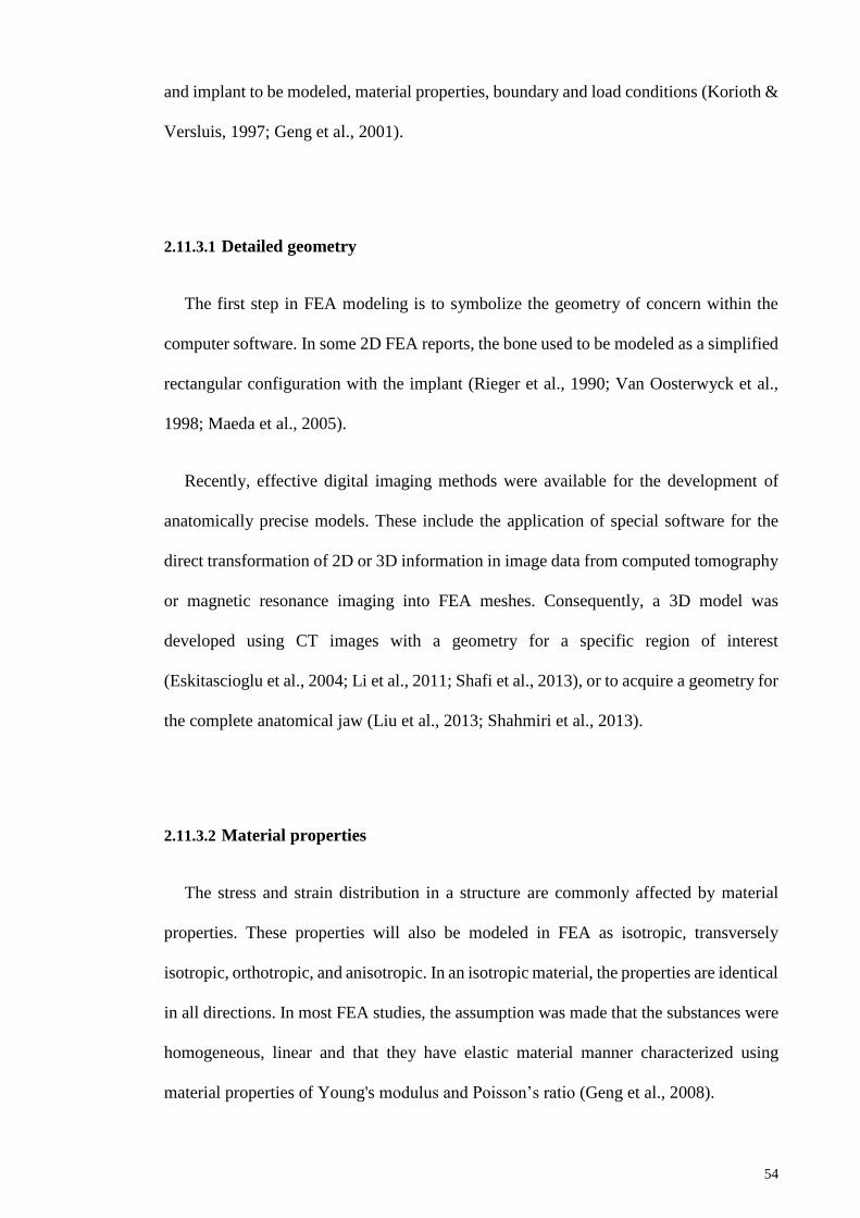
54
and implant to be modeled, material properties, boundary and load conditions (Korioth &
Versluis, 1997; Geng et al., 2001).
2.11.3.1 Detailed geometry
The first step in FEA modeling is to symbolize the geometry of concern within the
computer software. In some 2D FEA reports, the bone used to be modeled as a simplified
rectangular configuration with the implant (Rieger et al., 1990; Van Oosterwyck et al.,
1998; Maeda et al., 2005).
Recently, effective digital imaging methods were available for the development of
anatomically precise models. These include the application of special software for the
direct transformation of 2D or 3D information in image data from computed tomography
or magnetic resonance imaging into FEA meshes. Consequently, a 3D model was
developed using CT images with a geometry for a specific region of interest
(Eskitascioglu et al., 2004; Li et al., 2011; Shafi et al., 2013), or to acquire a geometry for
the complete anatomical jaw (Liu et al., 2013; Shahmiri et al., 2013).
2.11.3.2 Material properties
The stress and strain distribution in a structure are commonly affected by material
properties. These properties will also be modeled in FEA as isotropic, transversely
isotropic, orthotropic, and anisotropic. In an isotropic material, the properties are identical
in all directions. In most FEA studies, the assumption was made that the substances were
homogeneous, linear and that they have elastic material manner characterized using
material properties of Young's modulus and Poisson’s ratio (Geng et al., 2008).
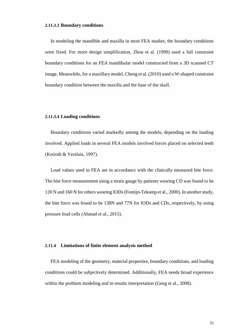
55
2.11.3.3 Boundary conditions
In modeling the mandible and maxilla in most FEA studies, the boundary conditions
were fixed. For more design simplification, Zhou et al. (1999) used a full constraint
boundary conditions for an FEA mandibular model constructed from a 3D scanned CT
image. Meanwhile, for a maxillary model, Cheng et al. (2010) used a W-shaped constraint
boundary condition between the maxilla and the base of the skull.
2.11.3.4 Loading conditions
Boundary conditions varied markedly among the models, depending on the loading
involved. Applied loads in several FEA models involved forces placed on selected teeth
(Korioth & Versluis, 1997).
Load values used in FEA are in accordance with the clinically measured bite force.
The bite force measurement using a strain gauge by patients wearing CD was found to be
120 N and 160 N for others wearing IODs (Fontijn-Tekamp et al., 2000). In another study,
the bite force was found to be 138N and 77N for IODs and CDs, respectively, by using
pressure load cells (Ahmad et al., 2015).
2.11.4 Limitations of finite element analysis method
FEA modeling of the geometry, material properties, boundary conditions, and loading
conditions could be subjectively determined. Additionally, FEA needs broad experience
within the problem modeling and in results interpretation (Geng et al., 2008).
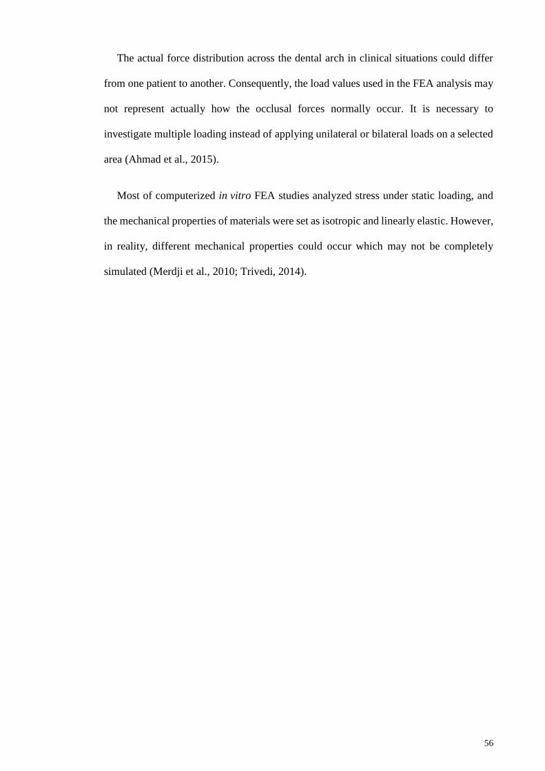
56
The actual force distribution across the dental arch in clinical situations could differ
from one patient to another. Consequently, the load values used in the FEA analysis may
not represent actually how the occlusal forces normally occur. It is necessary to
investigate multiple loading instead of applying unilateral or bilateral loads on a selected
area (Ahmad et al., 2015).
Most of computerized in vitro FEA studies analyzed stress under static loading, and
the mechanical properties of materials were set as isotropic and linearly elastic. However,
in reality, different mechanical properties could occur which may not be completely
simulated (Merdji et al., 2010; Trivedi, 2014).
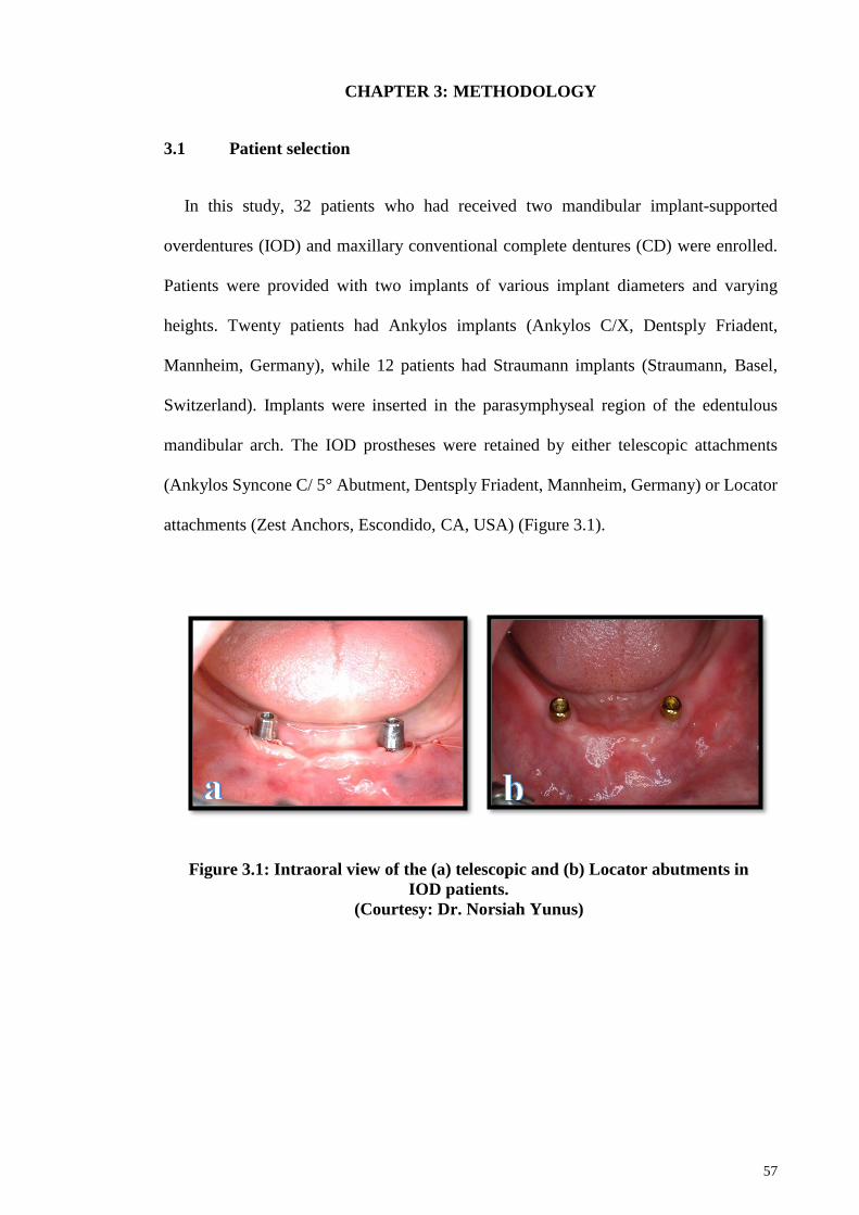
57
CHAPTER 3: METHODOLOGY
3.1 Patient selection
In this study, 32 patients who had received two mandibular implant-supported
overdentures (IOD) and maxillary conventional complete dentures (CD) were enrolled.
Patients were provided with two implants of various implant diameters and varying
heights. Twenty patients had Ankylos implants (Ankylos C/X, Dentsply Friadent,
Mannheim, Germany), while 12 patients had Straumann implants (Straumann, Basel,
Switzerland). Implants were inserted in the parasymphyseal region of the edentulous
mandibular arch. The IOD prostheses were retained by either telescopic attachments
(Ankylos Syncone C/ 5° Abutment, Dentsply Friadent, Mannheim, Germany) or Locator
attachments (Zest Anchors, Escondido, CA, USA) (Figure 3.1).
Figure 3.1: Intraoral view of the (a) telescopic and (b) Locator abutments in
IOD patients.
(Courtesy: Dr. Norsiah Yunus)
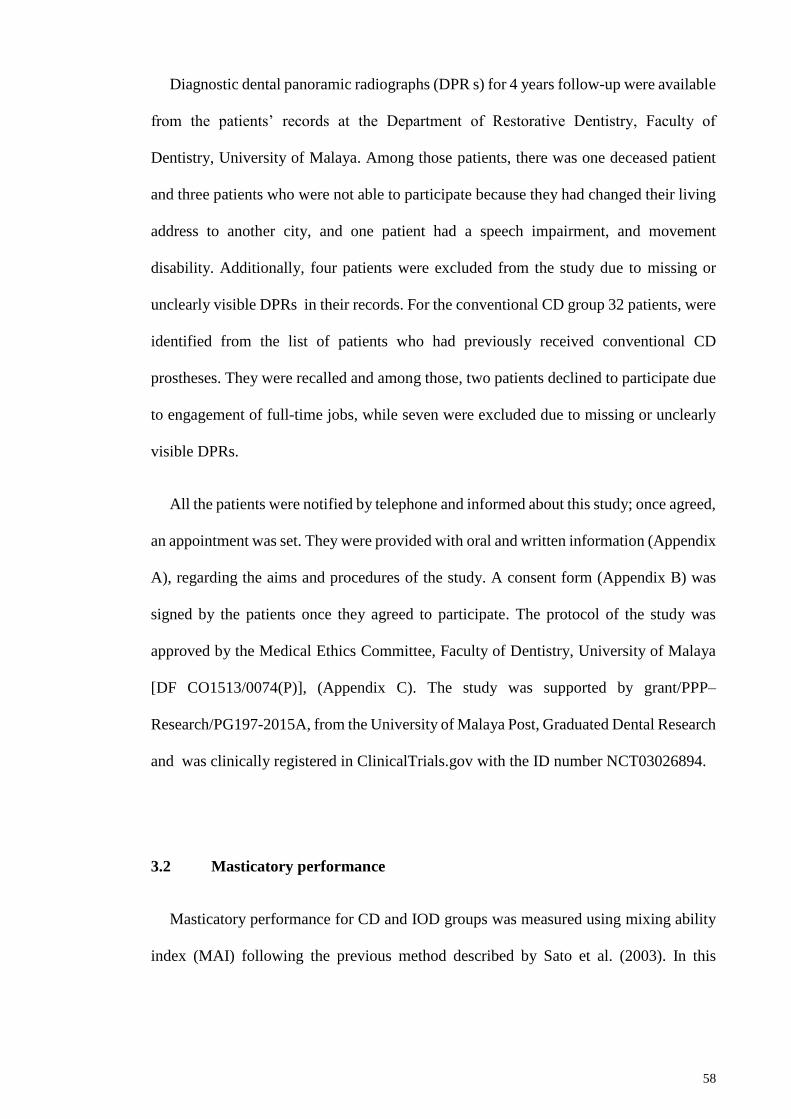
58
Diagnostic dental panoramic radiographs (DPR s) for 4 years follow-up were available
from the patients’ records at the Department of Restorative Dentistry, Faculty of
Dentistry, University of Malaya. Among those patients, there was one deceased patient
and three patients who were not able to participate because they had changed their living
address to another city, and one patient had a speech impairment, and movement
disability. Additionally, four patients were excluded from the study due to missing or
unclearly visible DPRs in their records. For the conventional CD group 32 patients, were
identified from the list of patients who had previously received conventional CD
prostheses. They were recalled and among those, two patients declined to participate due
to engagement of full-time jobs, while seven were excluded due to missing or unclearly
visible DPRs.
All the patients were notified by telephone and informed about this study; once agreed,
an appointment was set. They were provided with oral and written information (Appendix
A), regarding the aims and procedures of the study. A consent form (Appendix B) was
signed by the patients once they agreed to participate. The protocol of the study was
approved by the Medical Ethics Committee, Faculty of Dentistry, University of Malaya
[DF CO1513/0074(P)], (Appendix C). The study was supported by grant/PPP–
Research/PG197-2015A, from the University of Malaya Post, Graduated Dental Research
and was clinically registered in ClinicalTrials.gov with the ID number NCT03026894.
3.2 Masticatory performance
Masticatory performance for CD and IOD groups was measured using mixing ability
index (MAI) following the previous method described by Sato et al. (2003). In this
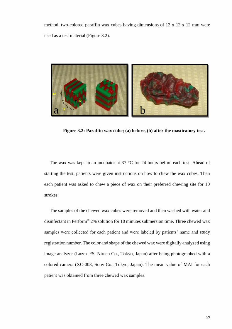
59
method, two-colored paraffin wax cubes having dimensions of 12 x 12 x 12 mm were
used as a test material (Figure 3.2).
The wax was kept in an incubator at 37 °C for 24 hours before each test. Ahead of
starting the test, patients were given instructions on how to chew the wax cubes. Then
each patient was asked to chew a piece of wax on their preferred chewing site for 10
strokes.
The samples of the chewed wax cubes were removed and then washed with water and
disinfectant in Perform® 2% solution for 10 minutes submersion time. Three chewed wax
samples were collected for each patient and were labeled by patients’ name and study
registration number. The color and shape of the chewed wax were digitally analyzed using
image analyzer (Luzex-FS, Nireco Co., Tokyo, Japan) after being photographed with a
colored camera (XC-003, Sony Co., Tokyo, Japan). The mean value of MAI for each
patient was obtained from three chewed wax samples.
12 mm
12 mm
12 mm b a
Figure 3.2: Paraffin wax cube; (a) before, (b) after the masticatory test.
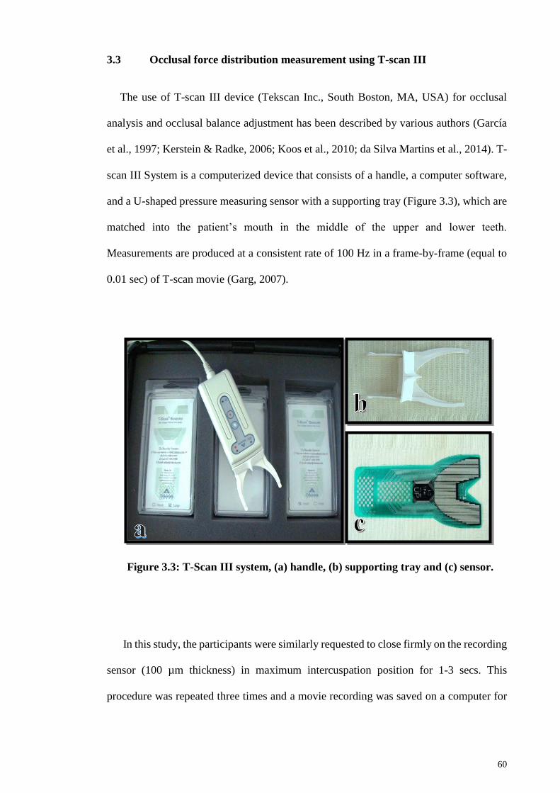
60
3.3 Occlusal force distribution measurement using T-scan III
The use of T-scan III device (Tekscan Inc., South Boston, MA, USA) for occlusal
analysis and occlusal balance adjustment has been described by various authors (García
et al., 1997; Kerstein & Radke, 2006; Koos et al., 2010; da Silva Martins et al., 2014). T-
scan III System is a computerized device that consists of a handle, a computer software,
and a U-shaped pressure measuring sensor with a supporting tray (Figure 3.3), which are
matched into the patient’s mouth in the middle of the upper and lower teeth.
Measurements are produced at a consistent rate of 100 Hz in a frame-by-frame (equal to
0.01 sec) of T-scan movie (Garg, 2007).
In this study, the participants were similarly requested to close firmly on the recording
sensor (100 µm thickness) in maximum intercuspation position for 1-3 secs. This
procedure was repeated three times and a movie recording was saved on a computer for
Figure 3.3: T-Scan III system, (a) handle, (b) supporting tray and (c) sensor.
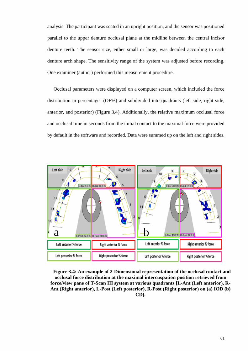
61
analysis. The participant was seated in an upright position, and the sensor was positioned
parallel to the upper denture occlusal plane at the midline between the central incisor
denture teeth. The sensor size, either small or large, was decided according to each
denture arch shape. The sensitivity range of the system was adjusted before recording.
One examiner (author) performed this measurement procedure.
Occlusal parameters were displayed on a computer screen, which included the force
distribution in percentages (OF%) and subdivided into quadrants (left side, right side,
anterior, and posterior) (Figure 3.4). Additionally, the relative maximum occlusal force
and occlusal time in seconds from the initial contact to the maximal force were provided
by default in the software and recorded. Data were summed up on the left and right sides.
b a
Figure 3.4: An example of 2-Dimensional representation of the occlusal contact and
occlusal force distribution at the maximal intercuspation position retrieved from
force/view pane of T-Scan III system at various quadrants [L-Ant (Left anterior), R-
Ant (Right anterior), L-Post (Left posterior), R-Post (Right posterior) on (a) IOD (b)
CD].
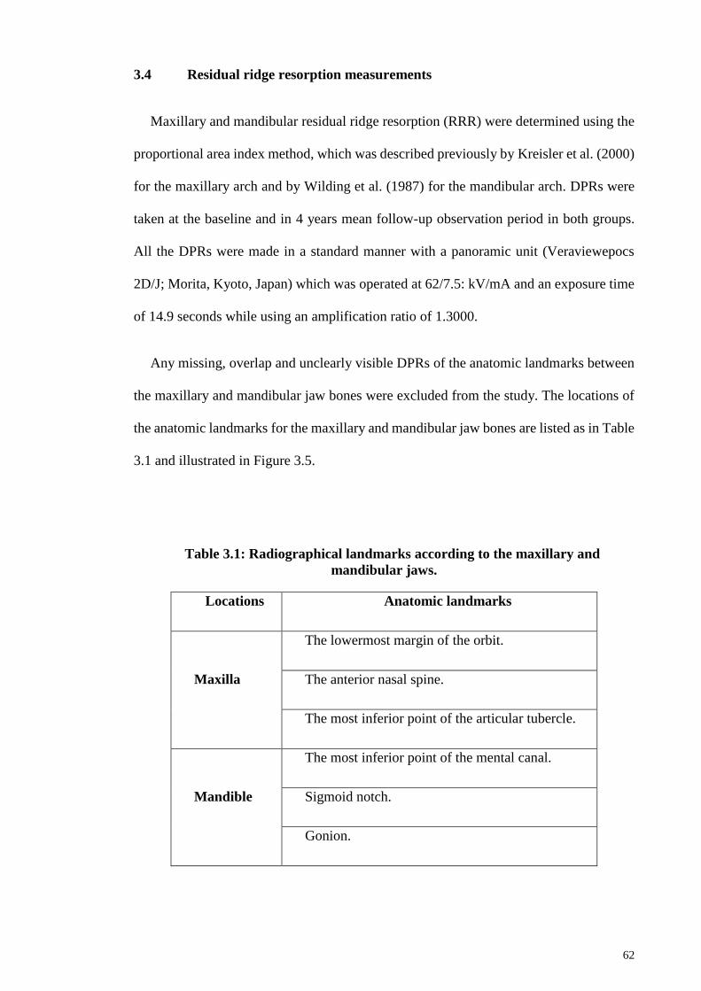
62
3.4 Residual ridge resorption measurements
Maxillary and mandibular residual ridge resorption (RRR) were determined using the
proportional area index method, which was described previously by Kreisler et al. (2000)
for the maxillary arch and by Wilding et al. (1987) for the mandibular arch. DPRs were
taken at the baseline and in 4 years mean follow-up observation period in both groups.
All the DPRs were made in a standard manner with a panoramic unit (Veraviewepocs
2D/J; Morita, Kyoto, Japan) which was operated at 62/7.5: kV/mA and an exposure time
of 14.9 seconds while using an amplification ratio of 1.3000.
Any missing, overlap and unclearly visible DPRs of the anatomic landmarks between
the maxillary and mandibular jaw bones were excluded from the study. The locations of
the anatomic landmarks for the maxillary and mandibular jaw bones are listed as in Table
3.1 and illustrated in Figure 3.5.
Table 3.1: Radiographical landmarks according to the maxillary and
mandibular jaws.
Locations Anatomic landmarks
Maxilla
The lowermost margin of the orbit.
The anterior nasal spine.
The most inferior point of the articular tubercle.
Mandible
The most inferior point of the mental canal.
Sigmoid notch.
Gonion.
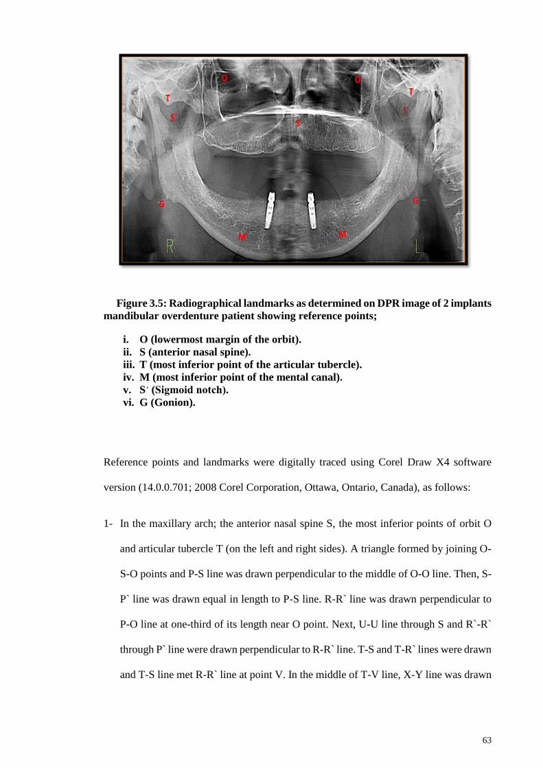
63
Figure 3.5: Radiographical landmarks as determined on DPR image of 2 implants
mandibular overdenture patient showing reference points;
i. O (lowermost margin of the orbit).
ii. S (anterior nasal spine).
iii. T (most inferior point of the articular tubercle).
iv. M (most inferior point of the mental canal).
v. S´ (Sigmoid notch).
vi. G (Gonion).
Reference points and landmarks were digitally traced using Corel Draw X4 software
version (14.0.0.701; 2008 Corel Corporation, Ottawa, Ontario, Canada), as follows:
1- In the maxillary arch; the anterior nasal spine S, the most inferior points of orbit O
and articular tubercle T (on the left and right sides). A triangle formed by joining O-
S-O points and P-S line was drawn perpendicular to the middle of O-O line. Then, S-
P` line was drawn equal in length to P-S line. R-R` line was drawn perpendicular to
P-O line at one-third of its length near O point. Next, U-U line through S and R`-R`
through P` line were drawn perpendicular to R-R` line. T-S and T-R` lines were drawn
and T-S line met R-R` line at point V. In the middle of T-V line, X-Y line was drawn
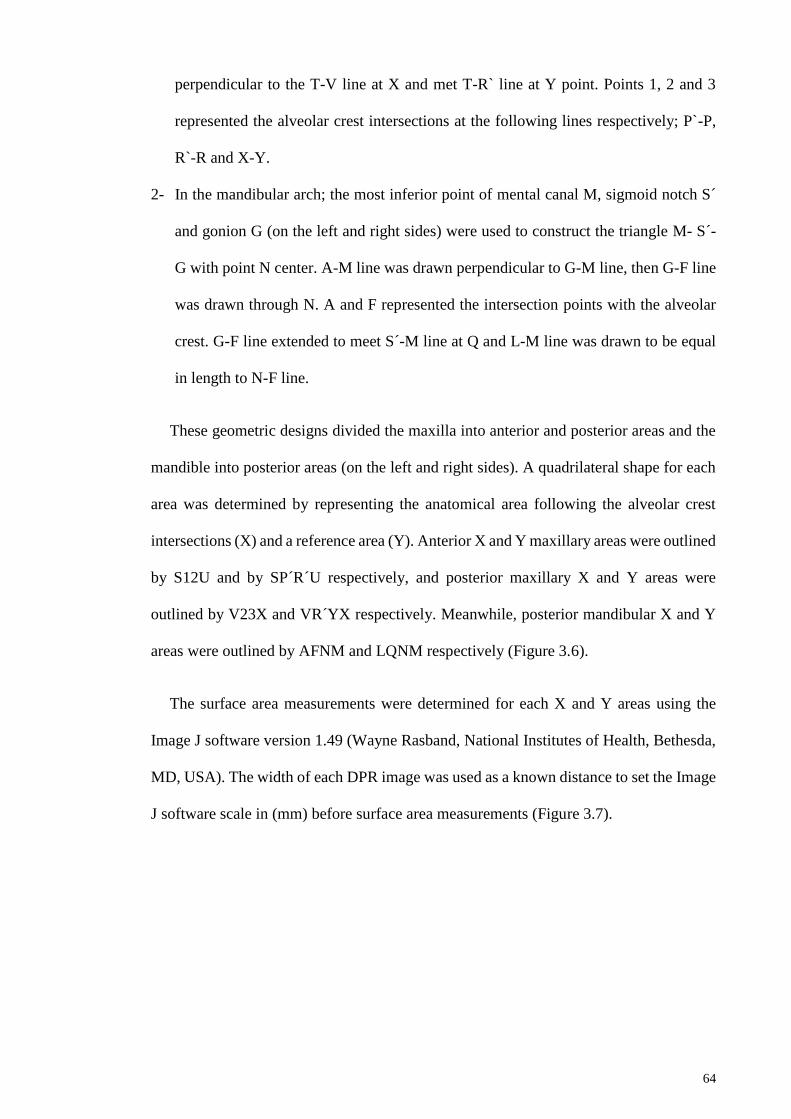
64
perpendicular to the T-V line at X and met T-R` line at Y point. Points 1, 2 and 3
represented the alveolar crest intersections at the following lines respectively; P`-P,
R`-R and X-Y.
2- In the mandibular arch; the most inferior point of mental canal M, sigmoid notch S´
and gonion G (on the left and right sides) were used to construct the triangle M- S´-
G with point N center. A-M line was drawn perpendicular to G-M line, then G-F line
was drawn through N. A and F represented the intersection points with the alveolar
crest. G-F line extended to meet S´-M line at Q and L-M line was drawn to be equal
in length to N-F line.
These geometric designs divided the maxilla into anterior and posterior areas and the
mandible into posterior areas (on the left and right sides). A quadrilateral shape for each
area was determined by representing the anatomical area following the alveolar crest
intersections (X) and a reference area (Y). Anterior X and Y maxillary areas were outlined
by S12U and by SP´R´U respectively, and posterior maxillary X and Y areas were
outlined by V23X and VR´YX respectively. Meanwhile, posterior mandibular X and Y
areas were outlined by AFNM and LQNM respectively (Figure 3.6).
The surface area measurements were determined for each X and Y areas using the
Image J software version 1.49 (Wayne Rasband, National Institutes of Health, Bethesda,
MD, USA). The width of each DPR image was used as a known distance to set the Image
J software scale in (mm) before surface area measurements (Figure 3.7).
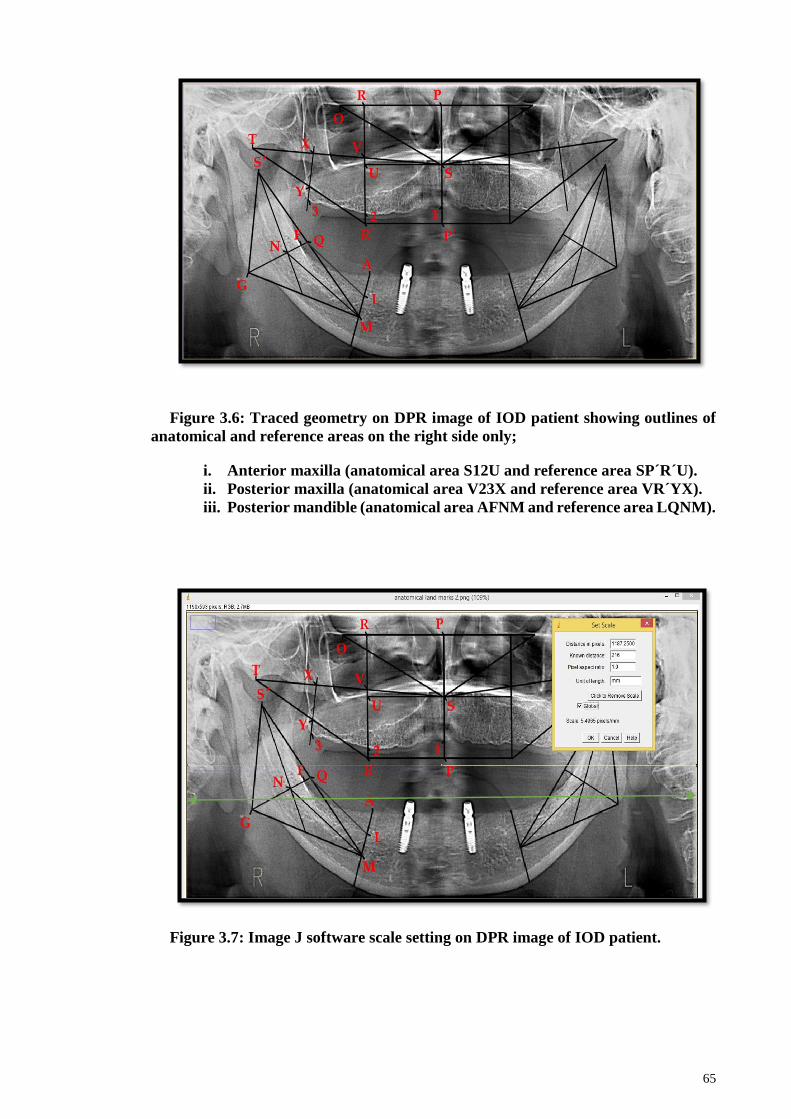
65
Figure 3.6: Traced geometry on DPR image of IOD patient showing outlines of
anatomical and reference areas on the right side only;
i. Anterior maxilla (anatomical area S12U and reference area SP´R´U).
ii. Posterior maxilla (anatomical area V23X and reference area VR´YX).
iii. Posterior mandible (anatomical area AFNM and reference area LQNM).
Figure 3.7: Image J software scale setting on DPR image of IOD patient.
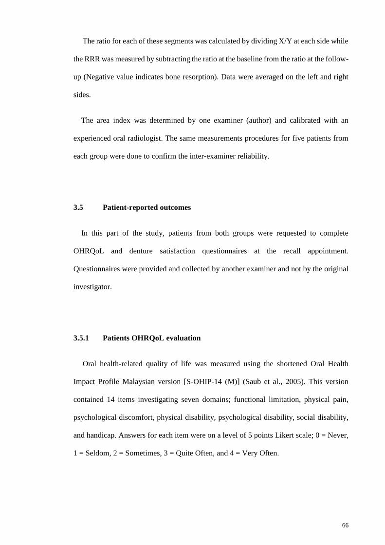
66
The ratio for each of these segments was calculated by dividing X/Y at each side while
the RRR was measured by subtracting the ratio at the baseline from the ratio at the follow-
up (Negative value indicates bone resorption). Data were averaged on the left and right
sides.
The area index was determined by one examiner (author) and calibrated with an
experienced oral radiologist. The same measurements procedures for five patients from
each group were done to confirm the inter-examiner reliability.
3.5 Patient-reported outcomes
In this part of the study, patients from both groups were requested to complete
OHRQoL and denture satisfaction questionnaires at the recall appointment.
Questionnaires were provided and collected by another examiner and not by the original
investigator.
3.5.1 Patients OHRQoL evaluation
Oral health-related quality of life was measured using the shortened Oral Health
Impact Profile Malaysian version [S-OHIP-14 (M)] (Saub et al., 2005). This version
contained 14 items investigating seven domains; functional limitation, physical pain,
psychological discomfort, physical disability, psychological disability, social disability,
and handicap. Answers for each item were on a level of 5 points Likert scale; 0 = Never,
1 = Seldom, 2 = Sometimes, 3 = Quite Often, and 4 = Very Often.
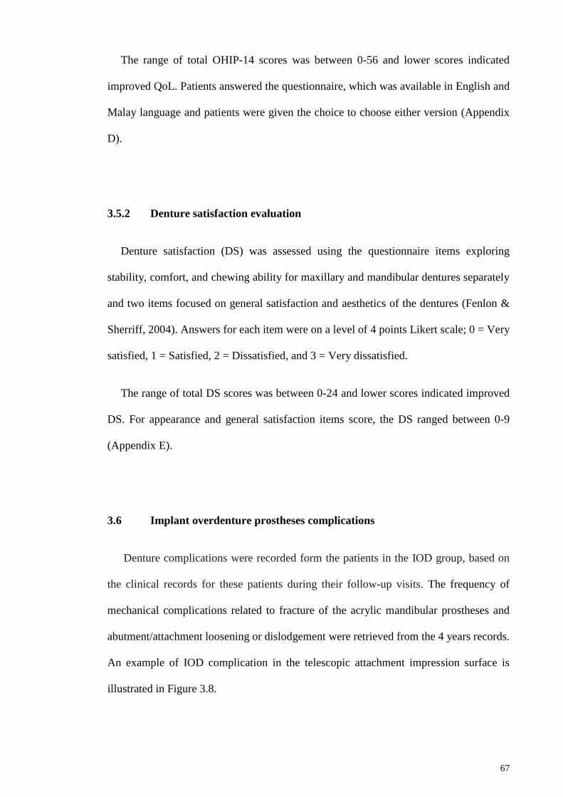
67
The range of total OHIP-14 scores was between 0-56 and lower scores indicated
improved QoL. Patients answered the questionnaire, which was available in English and
Malay language and patients were given the choice to choose either version (Appendix
D).
3.5.2 Denture satisfaction evaluation
Denture satisfaction (DS) was assessed using the questionnaire items exploring
stability, comfort, and chewing ability for maxillary and mandibular dentures separately
and two items focused on general satisfaction and aesthetics of the dentures (Fenlon &
Sherriff, 2004). Answers for each item were on a level of 4 points Likert scale; 0 = Very
satisfied, 1 = Satisfied, 2 = Dissatisfied, and 3 = Very dissatisfied.
The range of total DS scores was between 0-24 and lower scores indicated improved
DS. For appearance and general satisfaction items score, the DS ranged between 0-9
(Appendix E).
3.6 Implant overdenture prostheses complications
Denture complications were recorded form the patients in the IOD group, based on
the clinical records for these patients during their follow-up visits. The frequency of
mechanical complications related to fracture of the acrylic mandibular prostheses and
abutment/attachment loosening or dislodgement were retrieved from the 4 years records.
An example of IOD complication in the telescopic attachment impression surface is
illustrated in Figure 3.8.
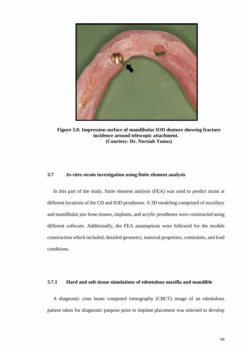
68
3.7 In-vitro strain investigation using finite element analysis
In this part of the study, finite element analysis (FEA) was used to predict strain at
different locations of the CD and IOD prostheses. A 3D modeling comprised of maxillary
and mandibular jaw bone tissues, implants, and acrylic prostheses were constructed using
different software. Additionally, the FEA assumptions were followed for the models
construction which included; detailed geometry, material properties, constraints, and load
conditions.
3.7.1 Hard and soft tissue simulations of edentulous maxilla and mandible
A diagnostic cone beam computed tomography (CBCT) image of an edentulous
patient taken for diagnostic purpose prior to implant placement was selected to develop
Figure 3.8: Impression surface of mandibular IOD denture showing fracture
incidence around telescopic attachment.
(Courtesy: Dr. Norsiah Yunus)
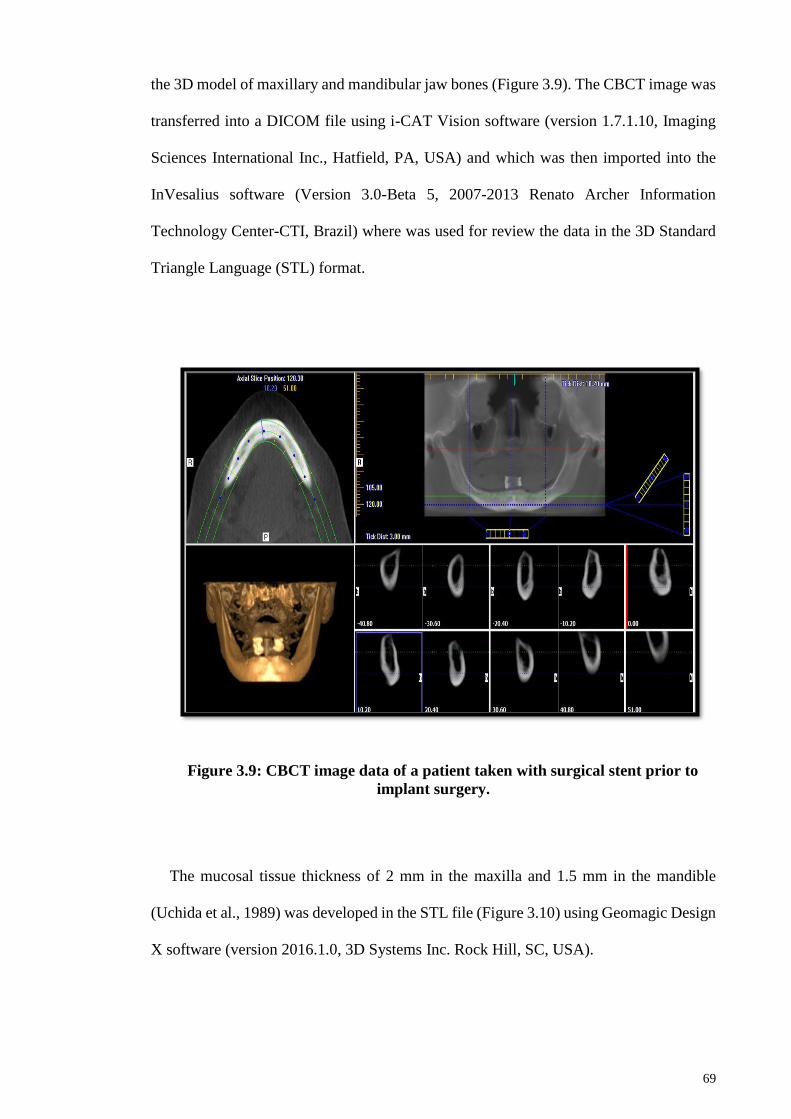
69
the 3D model of maxillary and mandibular jaw bones (Figure 3.9). The CBCT image was
transferred into a DICOM file using i-CAT Vision software (version 1.7.1.10, Imaging
Sciences International Inc., Hatfield, PA, USA) and which was then imported into the
InVesalius software (Version 3.0-Beta 5, 2007-2013 Renato Archer Information
Technology Center-CTI, Brazil) where was used for review the data in the 3D Standard
Triangle Language (STL) format.
Figure 3.9: CBCT image data of a patient taken with surgical stent prior to
implant surgery.
The mucosal tissue thickness of 2 mm in the maxilla and 1.5 mm in the mandible
(Uchida et al., 1989) was developed in the STL file (Figure 3.10) using Geomagic Design
X software (version 2016.1.0, 3D Systems Inc. Rock Hill, SC, USA).
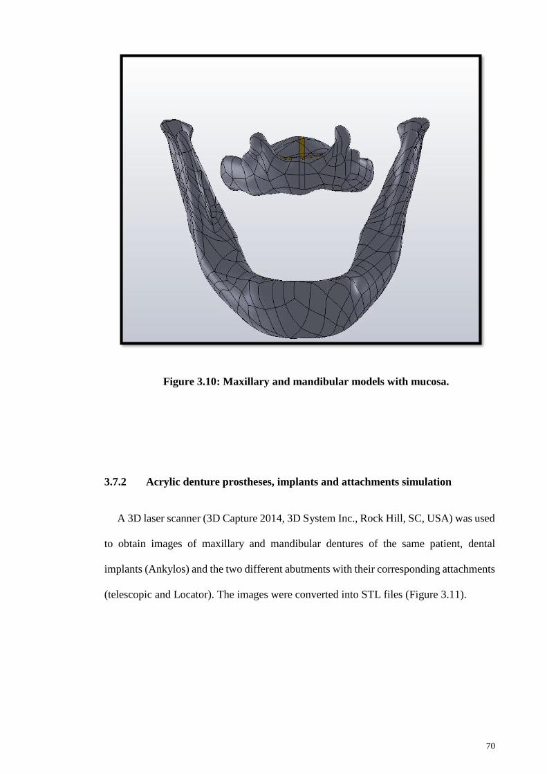
70
Figure 3.10: Maxillary and mandibular models with mucosa.
3.7.2 Acrylic denture prostheses, implants and attachments simulation
A 3D laser scanner (3D Capture 2014, 3D System Inc., Rock Hill, SC, USA) was used
to obtain images of maxillary and mandibular dentures of the same patient, dental
implants (Ankylos) and the two different abutments with their corresponding attachments
(telescopic and Locator). The images were converted into STL files (Figure 3.11).

71
The implant body was simulated with no threads in order to reduce the model
complexity and thus the analysis time. It was not the intention of this study to investigate
strain around the implants and surrounding bone. The dental implants, abutments, and
their attachments dimensions are illustrated in Table 3.2.
Figure 3.11: Maxillary and mandibular dentures (a) original model, (b)
simulation model, attachments assembly; (c) telescopic and (d) Locator.
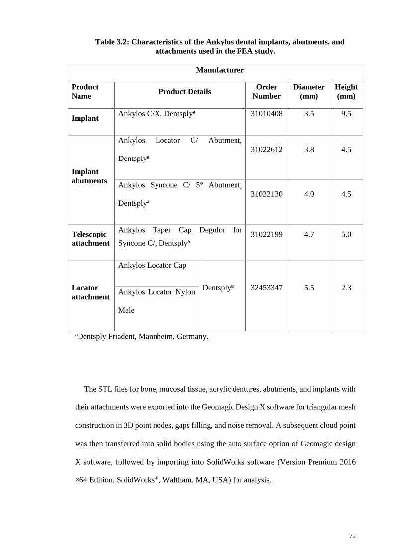
72
Table 3.2: Characteristics of the Ankylos dental implants, abutments, and
attachments used in the FEA study.
aDentsply Friadent, Mannheim, Germany.
The STL files for bone, mucosal tissue, acrylic dentures, abutments, and implants with
their attachments were exported into the Geomagic Design X software for triangular mesh
construction in 3D point nodes, gaps filling, and noise removal. A subsequent cloud point
was then transferred into solid bodies using the auto surface option of Geomagic design
X software, followed by importing into SolidWorks software (Version Premium 2016
×64 Edition, SolidWorks®, Waltham, MA, USA) for analysis.
Manufacturer
Product
Name Product Details
Order
Number
Diameter
(mm)
Height
(mm)
Implant Ankylos C/X, Dentsplya 31010408 3.5 9.5
Implant
abutments
Ankylos Locator C/ Abutment,
Dentsplya
31022612 3.8 4.5
Ankylos Syncone C/ 5° Abutment,
Dentsplya
31022130 4.0 4.5
Telescopic
attachment
Ankylos Taper Cap Degulor for
Syncone C/, Dentsplya
31022199 4.7 5.0
Locator
attachment
Ankylos Locator Cap
Dentsplya 32453347 5.5 2.3 Ankylos Locator Nylon
Male
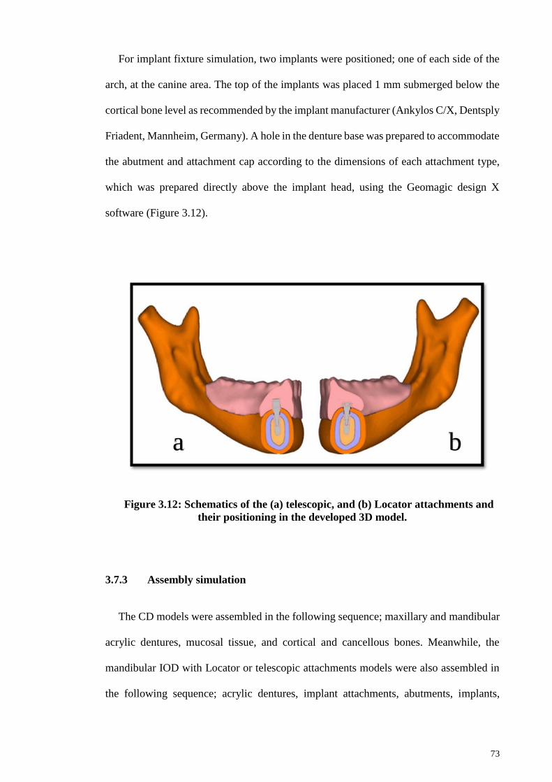
73
For implant fixture simulation, two implants were positioned; one of each side of the
arch, at the canine area. The top of the implants was placed 1 mm submerged below the
cortical bone level as recommended by the implant manufacturer (Ankylos C/X, Dentsply
Friadent, Mannheim, Germany). A hole in the denture base was prepared to accommodate
the abutment and attachment cap according to the dimensions of each attachment type,
which was prepared directly above the implant head, using the Geomagic design X
software (Figure 3.12).
Figure 3.12: Schematics of the (a) telescopic, and (b) Locator attachments and
their positioning in the developed 3D model.
3.7.3 Assembly simulation
The CD models were assembled in the following sequence; maxillary and mandibular
acrylic dentures, mucosal tissue, and cortical and cancellous bones. Meanwhile, the
mandibular IOD with Locator or telescopic attachments models were also assembled in
the following sequence; acrylic dentures, implant attachments, abutments, implants,
a b
74
mucosal tissue, and cortical and cancellous bones. The implant metal cap models for both
attachments were assembled following their structures according to the manufactures
instructions; a cylindrical cap for the telescopic and a cap with nylon male positioned
inside for the Locator attachments (Table 3.2).
The superimposed acrylic dentures for all models with the tissues, implants, and
implant attachments were sectioned for both maxillary and mandibular models. Each
section was corrected whenever necessary using the Cubify Sculpt Software (Version:
V2014. 64-Bit, Copyright 1993-2015 3D Systems, Inc. Rock Hill, SC, USA) to achieve
the optimal fit between the impression surface of the prostheses and underlying parts of
the attachment assembly.
3.7.4 Boundary conditions determination
Constraints for the maxillary denture were determined bilaterally following the union
between the maxilla with the base of the skull in a W shape (Cheng et al., 2010).
Meanwhile, the mandible was constrained bilaterally at its lower border (Uchida et al.,
1989), condyle, and angle areas (Marinescu et al., 2005) (Figure 3.13).
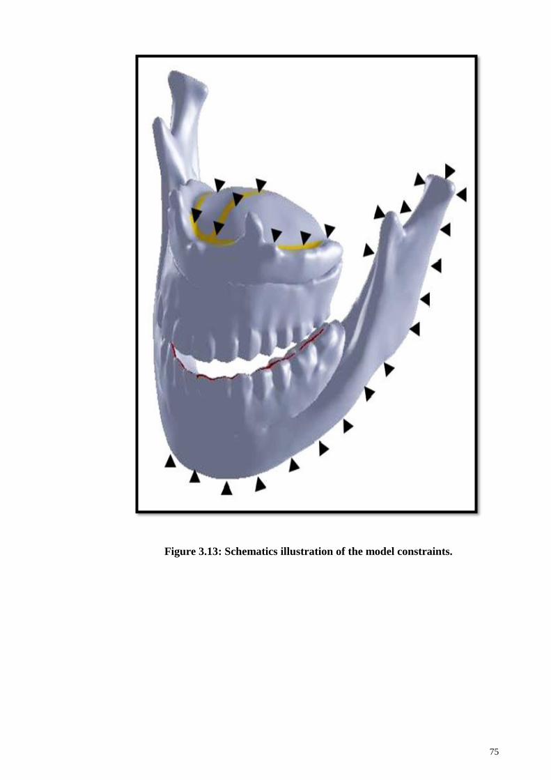
75
Figure 3.13: Schematics illustration of the model constraints.
76
3.7.5 Load application
3.7.5.1 Relative load determination
For the vertical occlusal load simulation, the mean percentage of occlusal force
distribution in 23 patients with conventional CD and 23 with mandibular IOD were
analyzed. This was achieved using the computerized occlusal analyzer T-scan III, where
patients were instructed to clench with their prostheses on a horseshoe-shaped metallic
sensor and the system analyzed the relative occlusal forces distribution at left and right,
anterior, and posterior quadrants. The data obtained from these two groups of patients are
as follows; anterior right and left from central incisor to canine, while posterior right and
left from the 1st premolar to 2nd molar teeth (Figure 3.14 a and b).
3.7.5.2 Actual load distribution and direction
A pilot study was conducted to measure the actual bite force for each group at two
locations and these values were used in the load simulation. Five patients from each group
were invited, seated in an upright position, instructed to bite bilaterally on the molar area
(left and right sides) using a digital dynamometer device (model IDDK 200, Kratos–
Equipamentos Industriais Ltd, Cotia, Sa˜o Paulo, Brazil). Three readings were measured
for each patient; then, data on the left and right sides were averaged. One examiner
(author) performed this measurement procedure for both groups. Non-significant
differences in the mean force value were observed. Therefore, a 150 N was considered as
an actual real load for the analysis (Table 3.3).
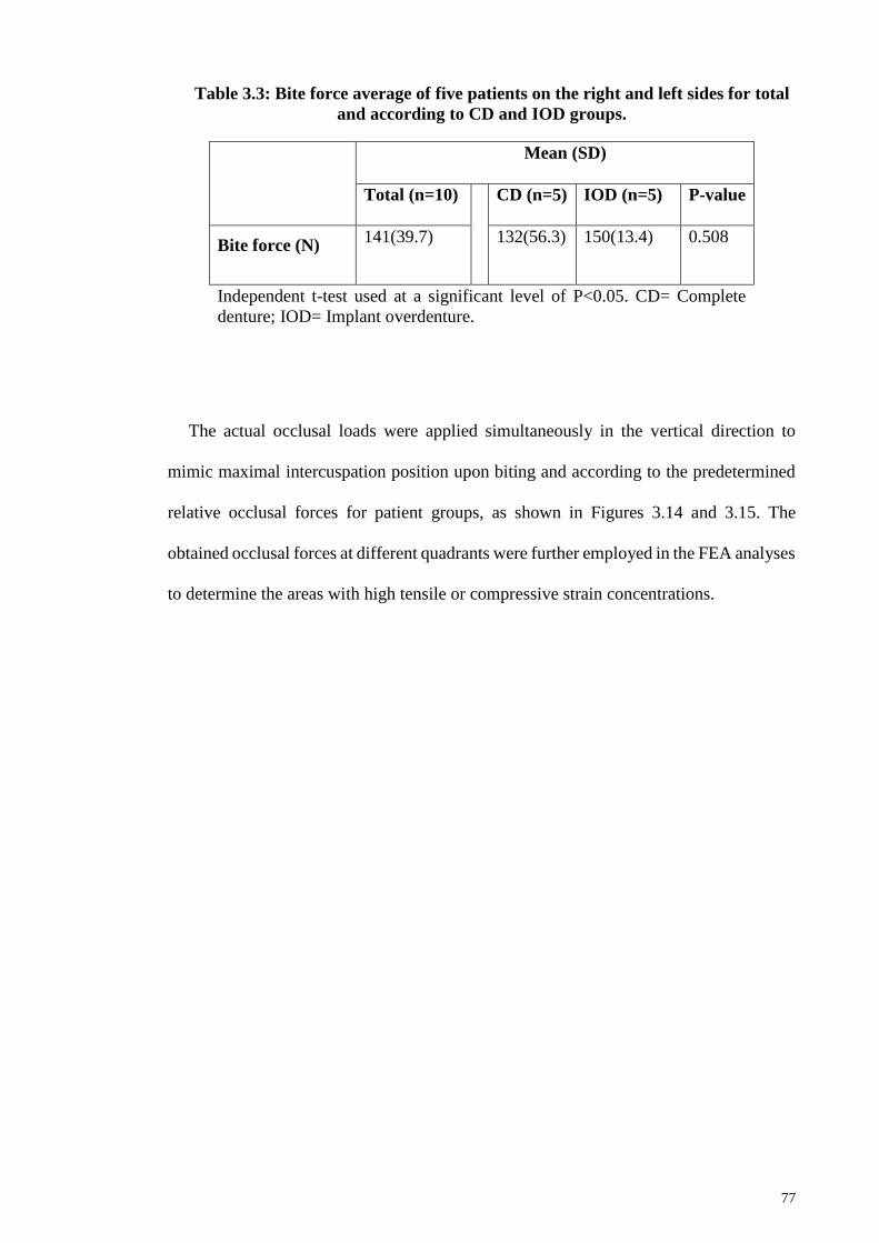
77
Table 3.3: Bite force average of five patients on the right and left sides for total
and according to CD and IOD groups.
Mean (SD)
Total (n=10)
CD (n=5) IOD (n=5) P-value
Bite force (N) 141(39.7) 132(56.3) 150(13.4) 0.508
Independent t-test used at a significant level of P<0.05. CD= Complete
denture; IOD= Implant overdenture.
The actual occlusal loads were applied simultaneously in the vertical direction to
mimic maximal intercuspation position upon biting and according to the predetermined
relative occlusal forces for patient groups, as shown in Figures 3.14 and 3.15. The
obtained occlusal forces at different quadrants were further employed in the FEA analyses
to determine the areas with high tensile or compressive strain concentrations.
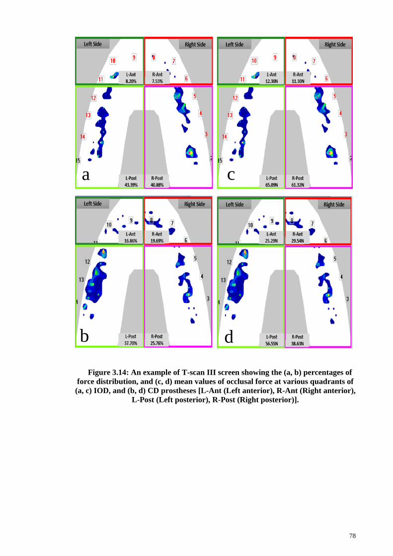
78
Figure 3.14: An example of T-scan III screen showing the (a, b) percentages of
force distribution, and (c, d) mean values of occlusal force at various quadrants of
(a, c) IOD, and (b, d) CD prostheses [L-Ant (Left anterior), R-Ant (Right anterior),
L-Post (Left posterior), R-Post (Right posterior)].
a
b
c
d
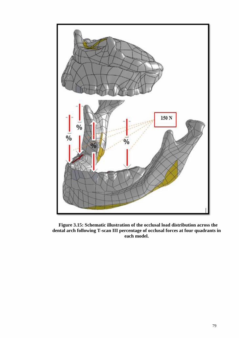
79
Figure 3.15: Schematic illustration of the occlusal load distribution across the
dental arch following T-scan III percentage of occlusal forces at four quadrants in
each model.
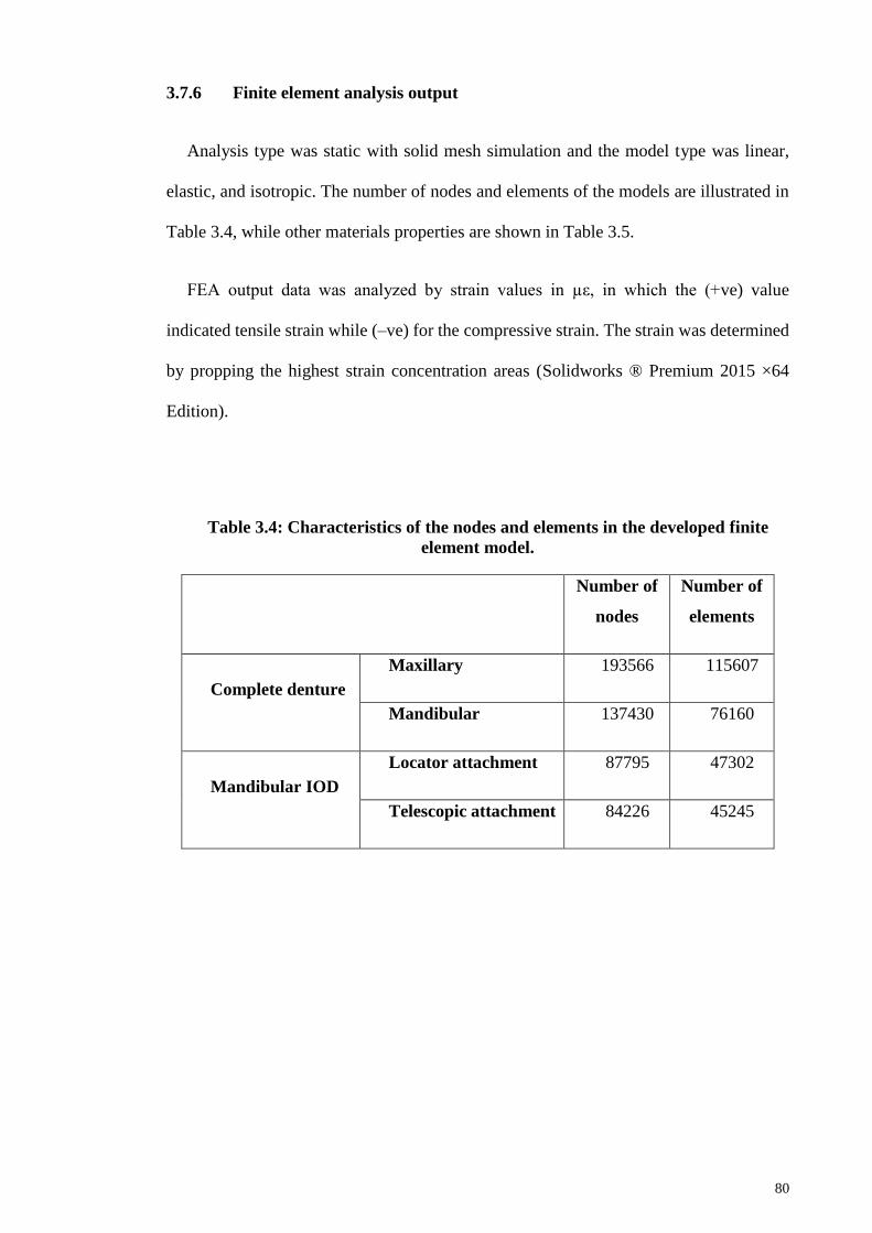
80
3.7.6 Finite element analysis output
Analysis type was static with solid mesh simulation and the model type was linear,
elastic, and isotropic. The number of nodes and elements of the models are illustrated in
Table 3.4, while other materials properties are shown in Table 3.5.
FEA output data was analyzed by strain values in µε, in which the (+ve) value
indicated tensile strain while (–ve) for the compressive strain. The strain was determined
by propping the highest strain concentration areas (Solidworks ® Premium 2015 ×64
Edition).
Table 3.4: Characteristics of the nodes and elements in the developed finite
element model.
Number of
nodes
Number of
elements
Complete denture
Maxillary 193566 115607
Mandibular 137430 76160
Mandibular IOD
Locator attachment 87795 47302
Telescopic attachment 84226 45245
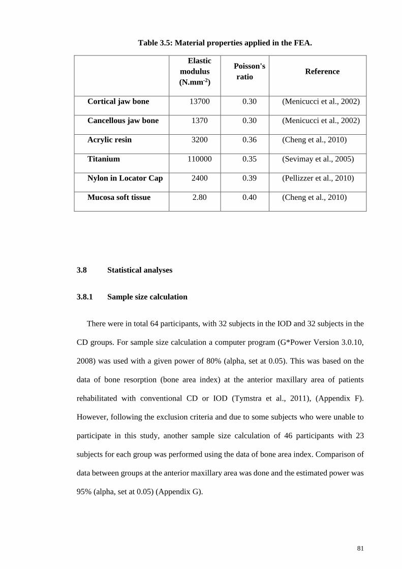
81
Table 3.5: Material properties applied in the FEA.
Elastic
modulus
(N.mm-2)
Poisson's
ratio Reference
Cortical jaw bone 13700 0.30 (Menicucci et al., 2002)
Cancellous jaw bone 1370 0.30 (Menicucci et al., 2002)
Acrylic resin 3200 0.36 (Cheng et al., 2010)
Titanium 110000 0.35 (Sevimay et al., 2005)
Nylon in Locator Cap 2400 0.39 (Pellizzer et al., 2010)
Mucosa soft tissue 2.80 0.40 (Cheng et al., 2010)
3.8 Statistical analyses
3.8.1 Sample size calculation
There were in total 64 participants, with 32 subjects in the IOD and 32 subjects in the
CD groups. For sample size calculation a computer program (G*Power Version 3.0.10,
2008) was used with a given power of 80% (alpha, set at 0.05). This was based on the
data of bone resorption (bone area index) at the anterior maxillary area of patients
rehabilitated with conventional CD or IOD (Tymstra et al., 2011), (Appendix F).
However, following the exclusion criteria and due to some subjects who were unable to
participate in this study, another sample size calculation of 46 participants with 23
subjects for each group was performed using the data of bone area index. Comparison of
data between groups at the anterior maxillary area was done and the estimated power was
95% (alpha, set at 0.05) (Appendix G).

82
3.8.2 Data analysis
The normality of the distribution of MAI, T-scan III, radiographic bone resorption,
OHIP-14, and DS data was determined using Shapiro-Wilk tests. Normally distributed
variables were reported as means and standard deviations, while for medians, 25% and
75% quartiles were reported for the variables that violated the normality assumption. Data
were analyzed by using SPSS (Version 20; IBM Corp., Armonk, NY, USA) with
significance set at p<0.05.
3.8.2.1 Socio-demographic data
Chi-square test was used to evaluate the gender difference between the groups. An
independent t-test was used for age evaluation and Mann-Whitney test for years of
edentulousness, between groups.
3.8.2.2 Masticatory performance data
Data for mixing ability index was analyzed using the independent t-test.
3.8.2.3 Occlusal parameters data
Mann-Whitney test was used to investigate occlusal parameters between the study
groups, while Wilcoxon Signed-test was used to investigate occlusal parameters between
anterior and posterior areas within the groups.
83
3.8.2.4 Residual ridge resorption data
Interclass correlation using Cronbach’s alpha was used to detect the correlation
between two examiners with regards to the RRR bone area indices, at various arch
locations in the two groups. Two-way factorial ANOVA was used to detect an interaction
between bone resorption locations and patient groups. Independent t-test was used to
investigate RRR between groups at the anterior and posterior maxillary areas. Mann-
Whitney test was used to investigate RRR at the posterior mandibular area. One-way
ANOVA and Tukey's posthoc test was used to determine within groups the differences
at various locations in the maxilla and mandible. The association between residual ridge
resorption with other variables was determined using multivariate regression analysis.
3.8.2.5 Patient-reported outcomes data
Total OHIP-14 and their individual domain scores between groups were evaluated
using the Mann-Whitney test. Total DS score was evaluated using independent t-test
while the Mann-Whitney test was used to evaluate their subscale scores. The OHIP-14
and the DS total scores association with other variables were determined using
multivariate regression analysis.
3.8.2.6 Clinical attachments complication data
Chi-square test was used to analyze the frequency of clinical complication between
telescopic and Locator attachments in IOD group.
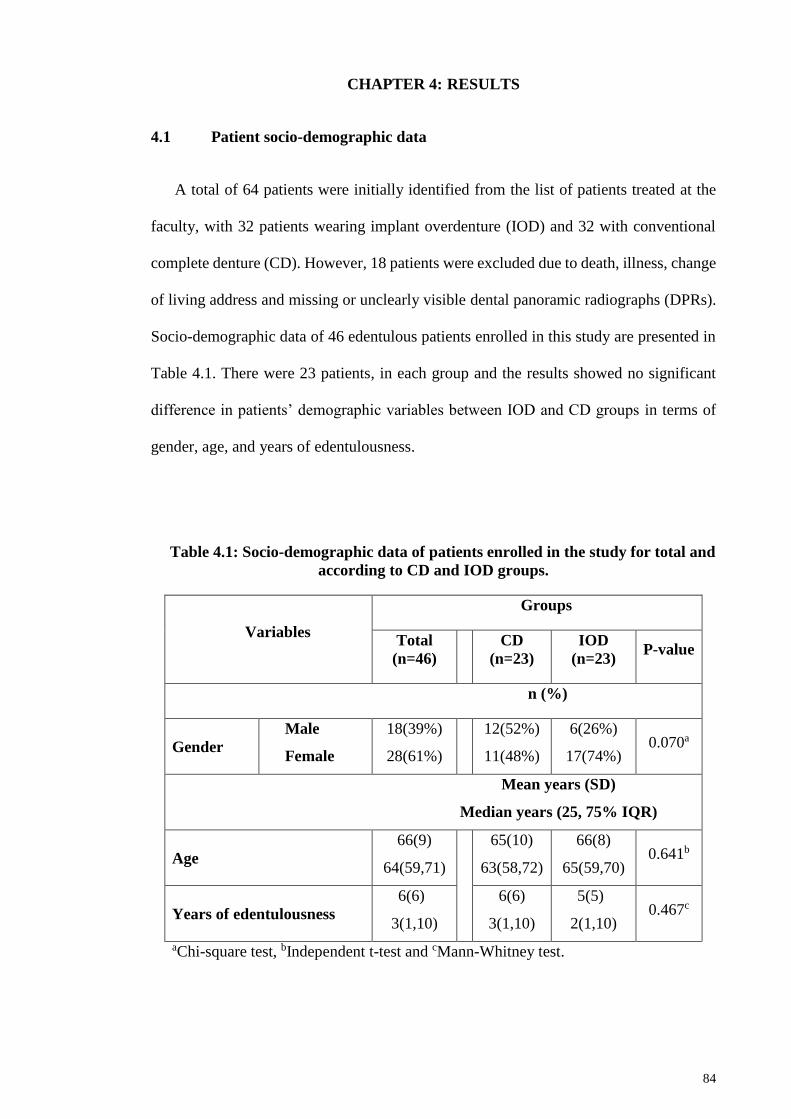
84
CHAPTER 4: RESULTS
4.1 Patient socio-demographic data
A total of 64 patients were initially identified from the list of patients treated at the
faculty, with 32 patients wearing implant overdenture (IOD) and 32 with conventional
complete denture (CD). However, 18 patients were excluded due to death, illness, change
of living address and missing or unclearly visible dental panoramic radiographs (DPRs).
Socio-demographic data of 46 edentulous patients enrolled in this study are presented in
Table 4.1. There were 23 patients, in each group and the results showed no significant
difference in patients’ demographic variables between IOD and CD groups in terms of
gender, age, and years of edentulousness.
Table 4.1: Socio-demographic data of patients enrolled in the study for total and
according to CD and IOD groups.
Variables
Groups
Total
(n=46)
CD
(n=23)
IOD
(n=23) P-value
n (%)
Gender
Male
Female
18(39%)
28(61%)
12(52%)
11(48%)
6(26%)
17(74%) 0.070a
Mean years (SD)
Median years (25, 75% IQR)
Age
66(9)
64(59,71)
65(10)
63(58,72)
66(8)
65(59,70) 0.641b
Years of edentulousness
6(6)
3(1,10)
6(6)
3(1,10)
5(5)
2(1,10) 0.467c
aChi-square test, bIndependent t-test and cMann-Whitney test.

85
4.2 Masticatory performance
Masticatory performance was evaluated by the mixing ability index (MAI) using 2-
colored wax cubes as a testing material. The results for the MAI showed no significant
difference between CD and IOD groups (Table 4.2).
Table 4.2: Mean MAI for total and according to CD and IOD groups.
Mean (SD)
Total (n=46)
CD (n=23) IOD (n=23) P-value
MAI 0.2(0.7) 0.0(0.7) 0.3(0.8) 0.134a
a Independent t-test.
Figure 4.1 showed a plot graph of MAI mean values for CD and IOD individual
patients. Positive values indicated good masticatory performance while negative values
referred to poor performance. Generally, patients of the IOD group showed a trend of
better MAI than the CD group.
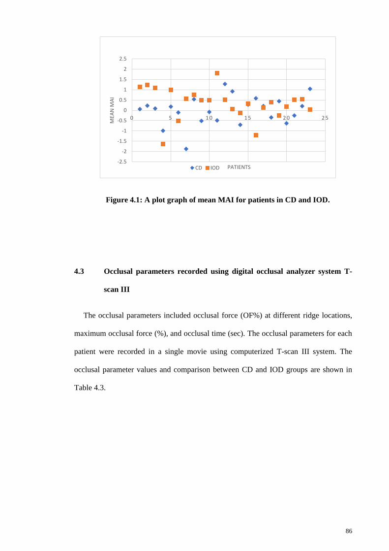
86
Figure 4.1: A plot graph of mean MAI for patients in CD and IOD.
4.3 Occlusal parameters recorded using digital occlusal analyzer system T-
scan III
The occlusal parameters included occlusal force (OF%) at different ridge locations,
maximum occlusal force (%), and occlusal time (sec). The occlusal parameters for each
patient were recorded in a single movie using computerized T-scan III system. The
occlusal parameter values and comparison between CD and IOD groups are shown in
Table 4.3.
-2.5
-2
-1.5
-1
-0.5
0
0.5
1
1.5
2
2.5
0 5 1 0 1 5 2 0 2 5M
EAN
MA
I
PATIENTSCD IOD
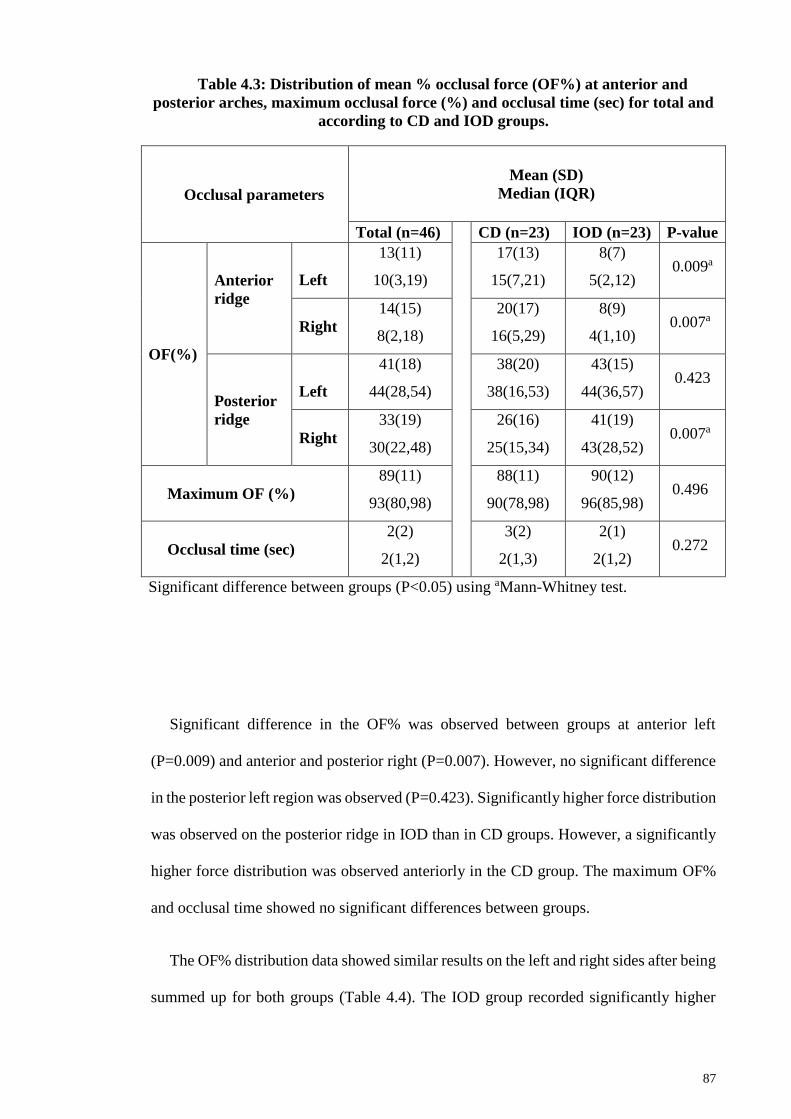
87
Table 4.3: Distribution of mean % occlusal force (OF%) at anterior and
posterior arches, maximum occlusal force (%) and occlusal time (sec) for total and
according to CD and IOD groups.
Occlusal parameters
Mean (SD)
Median (IQR)
Total (n=46)
CD (n=23) IOD (n=23) P-value
OF(%)
Anterior
ridge
Left
13(11)
10(3,19)
17(13)
15(7,21)
8(7)
5(2,12) 0.009a
Right
14(15)
8(2,18)
20(17)
16(5,29)
8(9)
4(1,10) 0.007a
Posterior
ridge
Left
41(18)
44(28,54)
38(20)
38(16,53)
43(15)
44(36,57) 0.423
Right
33(19)
30(22,48)
26(16)
25(15,34)
41(19)
43(28,52) 0.007a
Maximum OF (%)
89(11)
93(80,98)
88(11)
90(78,98)
90(12)
96(85,98) 0.496
Occlusal time (sec)
2(2)
2(1,2)
3(2)
2(1,3)
2(1)
2(1,2) 0.272
Significant difference between groups (P<0.05) using aMann-Whitney test.
Significant difference in the OF% was observed between groups at anterior left
(P=0.009) and anterior and posterior right (P=0.007). However, no significant difference
in the posterior left region was observed (P=0.423). Significantly higher force distribution
was observed on the posterior ridge in IOD than in CD groups. However, a significantly
higher force distribution was observed anteriorly in the CD group. The maximum OF%
and occlusal time showed no significant differences between groups.
The OF% distribution data showed similar results on the left and right sides after being
summed up for both groups (Table 4.4). The IOD group recorded significantly higher
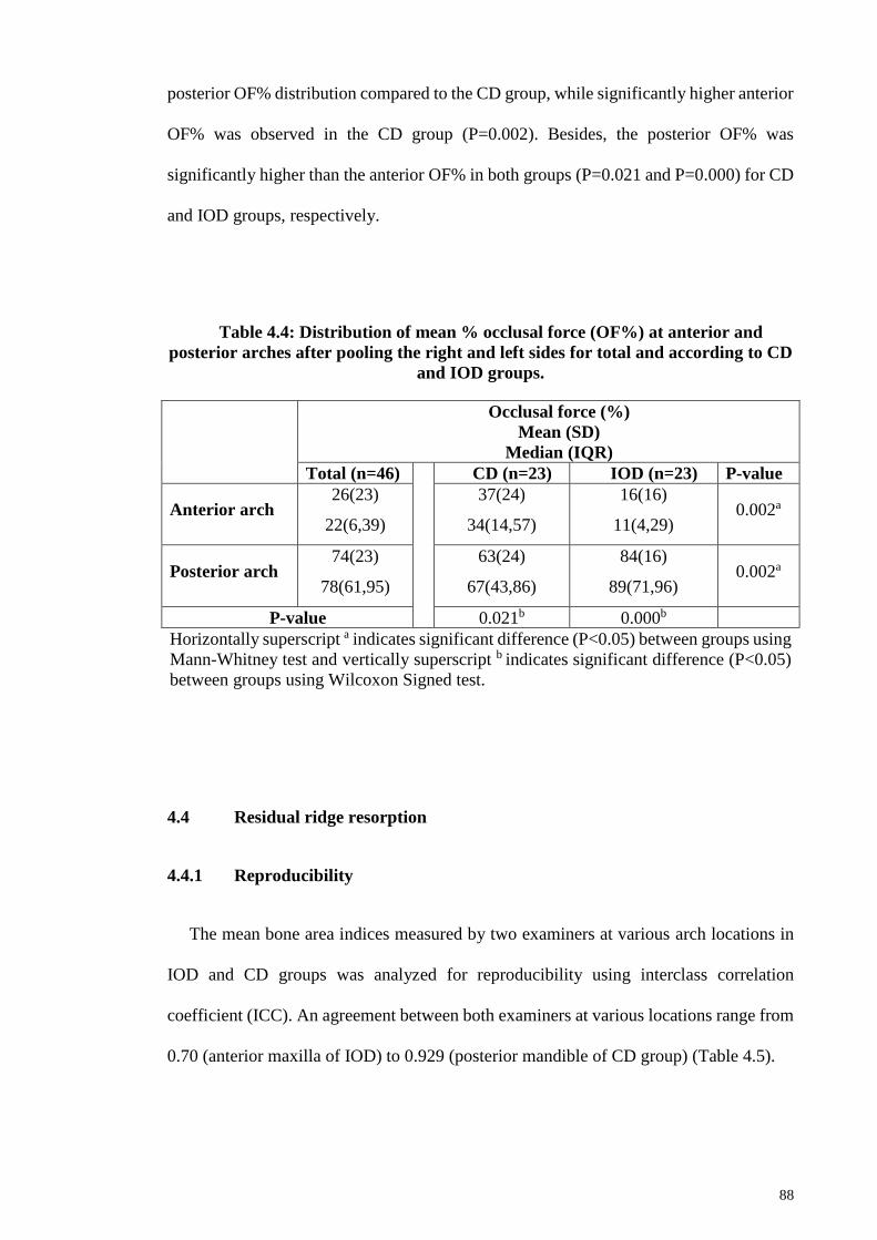
88
posterior OF% distribution compared to the CD group, while significantly higher anterior
OF% was observed in the CD group (P=0.002). Besides, the posterior OF% was
significantly higher than the anterior OF% in both groups (P=0.021 and P=0.000) for CD
and IOD groups, respectively.
Table 4.4: Distribution of mean % occlusal force (OF%) at anterior and
posterior arches after pooling the right and left sides for total and according to CD
and IOD groups.
Occlusal force (%)
Mean (SD)
Median (IQR)
Total (n=46)
CD (n=23) IOD (n=23) P-value
Anterior arch 26(23)
22(6,39)
37(24)
34(14,57)
16(16)
11(4,29) 0.002a
Posterior arch 74(23)
78(61,95)
63(24)
67(43,86)
84(16)
89(71,96) 0.002a
P-value 0.021b 0.000b
Horizontally superscript a indicates significant difference (P<0.05) between groups using
Mann-Whitney test and vertically superscript b indicates significant difference (P<0.05)
between groups using Wilcoxon Signed test.
4.4 Residual ridge resorption
4.4.1 Reproducibility
The mean bone area indices measured by two examiners at various arch locations in
IOD and CD groups was analyzed for reproducibility using interclass correlation
coefficient (ICC). An agreement between both examiners at various locations range from
0.70 (anterior maxilla of IOD) to 0.929 (posterior mandible of CD group) (Table 4.5).
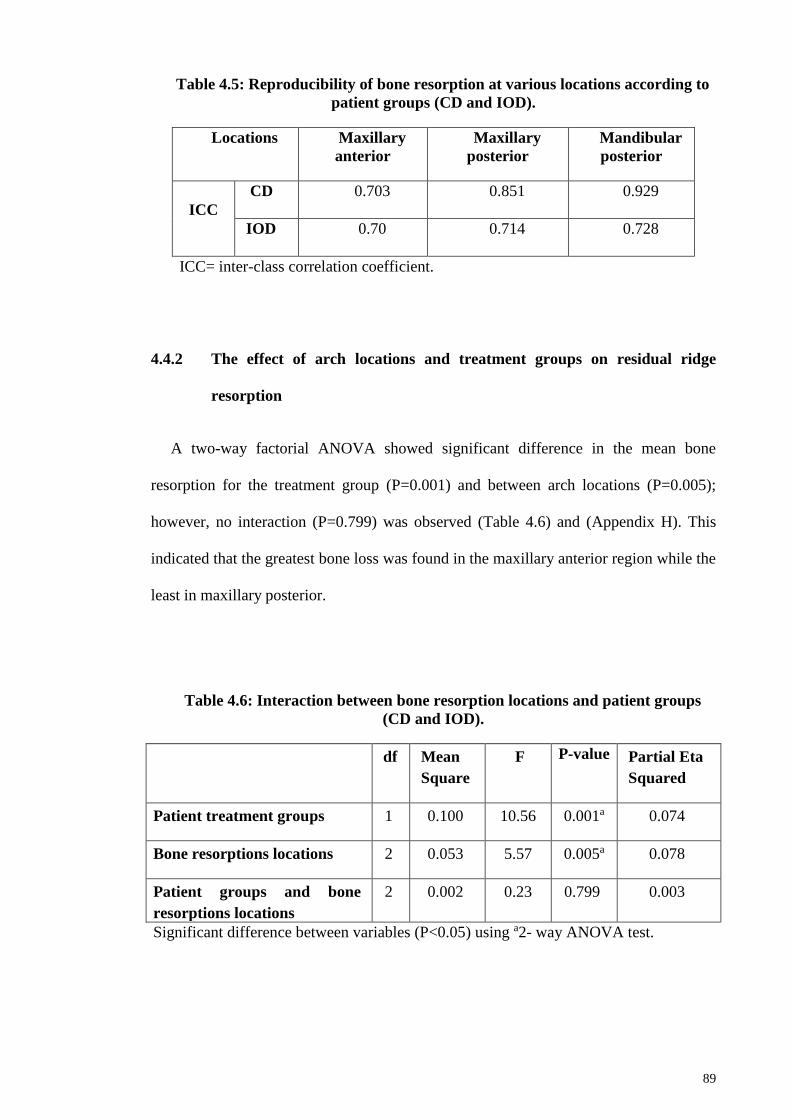
89
Table 4.5: Reproducibility of bone resorption at various locations according to
patient groups (CD and IOD).
Locations Maxillary
anterior
Maxillary
posterior
Mandibular
posterior
ICC
CD 0.703 0.851 0.929
IOD 0.70 0.714 0.728
ICC= inter-class correlation coefficient.
4.4.2 The effect of arch locations and treatment groups on residual ridge
resorption
A two-way factorial ANOVA showed significant difference in the mean bone
resorption for the treatment group (P=0.001) and between arch locations (P=0.005);
however, no interaction (P=0.799) was observed (Table 4.6) and (Appendix H). This
indicated that the greatest bone loss was found in the maxillary anterior region while the
least in maxillary posterior.
Table 4.6: Interaction between bone resorption locations and patient groups
(CD and IOD).
df Mean
Square
F P-value Partial Eta
Squared
Patient treatment groups 1 0.100 10.56 0.001a 0.074
Bone resorptions locations 2 0.053 5.57 0.005a 0.078
Patient groups and bone
resorptions locations
2 0.002 0.23 0.799 0.003
Significant difference between variables (P<0.05) using a2- way ANOVA test.

90
4.4.3 Residual ridge resorption at different locations
Bone resorption in the maxillary anterior area was significantly higher in CD than IOD
groups (P=0.013). While no significant differences between the groups were observed in
the maxillary (P=0.101) and mandibular (P=0.116) posterior areas. There were no
significant differences in bone resorption at various location in CD group (P=0.086);
however significant differences were observed in IOD group (P=0.038), at maxillary and
mandibular posterior (P=0.049) areas. In the IOD group, the mandibular posterior area
showed significantly greater resorption (Table 4.7).
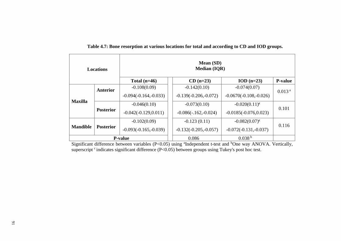
91
Table 4.7: Bone resorption at various locations for total and according to CD and IOD groups.
Locations
Mean (SD)
Median (IQR)
Total (n=46)
CD (n=23) IOD (n=23) P-value
Maxilla
Anterior
-0.108(0.09)
-0.094(-0.164,-0.033)
-0.142(0.10)
-0.139(-0.206,-0.072)
-0.074(0.07)
-0.0670(-0.108,-0.026) 0.013 a
Posterior
-0.046(0.10)
-0.042(-0.129,0.011)
-0.073(0.10)
-0.086(-.162,-0.024)
-0.020(0.11)c
-0.0185(-0.076,0.023) 0.101
Mandible Posterior
-0.102(0.09)
-0.093(-0.165,-0.039)
-0.123 (0.11)
-0.132(-0.205,-0.057)
-0.082(0.07)c
-0.072(-0.131,-0.037) 0.116
P-value 0.086 0.038 b
Significant difference between variables (P<0.05) using aIndependent t-test and bOne way ANOVA. Vertically,
superscript c indicates significant difference (P<0.05) between groups using Tukey's post hoc test.
91
92
4.4.4 Association of bone resorption with the OF%, treatment groups, and socio-
demographics
Multivariate linear regression analysis model was used to detect the association
between bone resorption as the dependent variable with OF%, treatment group, gender,
age, and years of edentulousness as predicted variables (Table 4.8).

93
Table 4.8: Multivariate regression analysis of the association between bone resorption at various locations with OF%, treatment
group, gender, age and years of edentulousness.
Variables
Bone resorption locations
Maxilla Mandible
Anterior Posterior Posterior
Coefficient
(SE)d
P-
value
Coefficient
(SE)d
P-
value
Coefficient
(SE)d P-value
Intercept 0.048(0.080) 0. 552 -0.005(0.152) 0.972 -0.008(0.128) 0.949
OF% -0.003(0.001) 0.000 a -0.002(0.001) 0.094 -0.002(0.001) 0.023 a
Treatment groupb 0.006(0.023) 0.785 0.085(0.038) 0.033 a 0.078(0.032) 0.021 a
Genderc -0.010(0.021) 0.646 -0.004(0.035) 0.906 0.008(0.029) 0.784
Age (Years) -0.001(0.001) 0.365 0.001(0.002) 0.725 0.000(0.002) 0.831
Years of edentulousness 0.001(0.002) 0.738 -0.001(0.003) 0.808 0.001(0.003) 0.567
R2 0.584 0.135 0.230
a Indicates significant difference between variables (P<0.05).
b 0 = CD; 1 = IOD, c 0 = Female; 1 = Male and d SE = Standard error.
93

94
The results revealed that the increase in the anterior and the posterior OF%
significantly showed increased bone resorption (negative value indicating bone
resorption) for the maxillary anterior area (P=0.000) and the mandibular posterior area
(P=0.023). However, the IOD group demonstrated less bone resorption compared to CD
in maxillary posterior (P=0.033) and mandibular posterior (P=0.021) areas. No
significant association was observed; however, between the two groups with respect to
the bone resorption in the anterior maxillary ridge, indicating that in this region, the bone
resorption was significantly associated only with the OF% exerted in the anterior maxilla.
Other patients’ predicted variables showed no significant associations with bone
resorption at any of the arch location areas.
4.5 Patient-reported outcomes
For this part of the study, shortened Oral Health Impact Profile Malaysian version [S-
OHIP-14 (M)] and denture satisfaction (DS) questionnaires were used to evaluate patient-
reported outcomes. Lower scores of OHIP-14 and DS represented better patient-reported
outcomes.
4.5.1 OHIP-14
The study groups were compared in relation to the OHIP-14 total scores and individual
domains which included functional limitation, physical pain, psychologic discomfort,
physical disability, psychologic disability, social disability and handicap (Table 4.9).
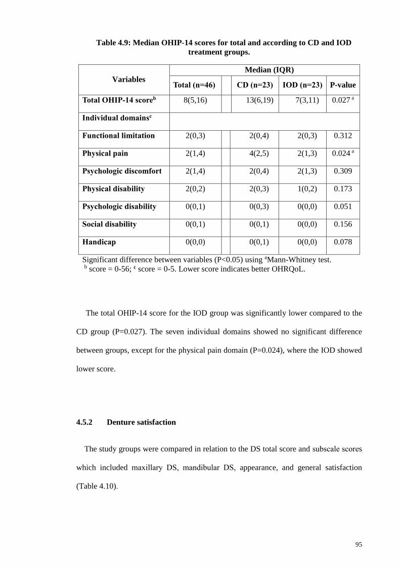
95
Table 4.9: Median OHIP-14 scores for total and according to CD and IOD
treatment groups.
Variables
Median (IQR)
Total (n=46) CD (n=23) IOD (n=23) P-value
Total OHIP-14 scoreb 8(5,16) 13(6,19) 7(3,11) 0.027 a
Individual domainsc
Functional limitation 2(0,3) 2(0,4) 2(0,3) 0.312
Physical pain 2(1,4) 4(2,5) 2(1,3) 0.024 a
Psychologic discomfort 2(1,4) 2(0,4) 2(1,3) 0.309
Physical disability 2(0,2) 2(0,3) 1(0,2) 0.173
Psychologic disability 0(0,1) 0(0,3) 0(0,0) 0.051
Social disability 0(0,1) 0(0,1) 0(0,0) 0.156
Handicap 0(0,0) 0(0,1) 0(0,0) 0.078
Significant difference between variables (P<0.05) using aMann-Whitney test.
b score = 0-56; c score = 0-5. Lower score indicates better OHRQoL.
The total OHIP-14 score for the IOD group was significantly lower compared to the
CD group (P=0.027). The seven individual domains showed no significant difference
between groups, except for the physical pain domain (P=0.024), where the IOD showed
lower score.
4.5.2 Denture satisfaction
The study groups were compared in relation to the DS total score and subscale scores
which included maxillary DS, mandibular DS, appearance, and general satisfaction
(Table 4.10).

96
Table 4.10: Median denture satisfaction scores for total and according to CD
and IOD treatment groups.
Variables
Mean (SD)
Median (IQR)
Total (n=46)
CD (n=23) IOD (n=23) P-value
Total denture satisfaction
scorec
9(5)
9(8,12)
11(4)
10(8,13)
8(4)
8(6,9) 0.030 a
Subscale scores
Maxillary denture satisfactiond 3(2)
3(2,4)
3(2)
3(3,4)
3(2)
3(2,5) 0.964
Mandibular denture
satisfactiond
4(2)
3(3,6)
5(2)
5(3,6)
3(2)
3(2,3) 0.001 b
Appearancee 1(1)
1(1,1)
1(1)
1(1,2)
1(1)
1(1,1) 0.091
General satisfactione 1(1)
1(1,2)
1(1)
1(1,2)
1(1)
1(1,1) 0.135
Significant difference between groups (P<0.05) using aIndependent samples t-test and bMann-Whitney test. cscore = 0-24; d score = 0-9; e score = 0-3. Lower score indicates greater satisfaction.
The total DS score for the IOD group showed significantly lower value than the CD
group (P=0.030). For DS subscale scores, there was a significantly lower score in relation
to satisfaction with mandibular prostheses in IOD than CD groups (P=0.001). No
difference was observed between the groups regarding maxillary DS, appearance, and
general satisfaction (Table 4.10).
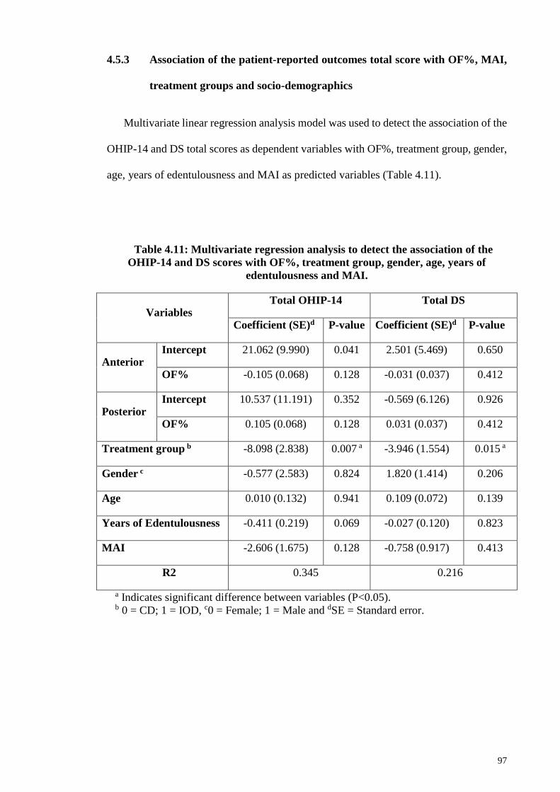
97
4.5.3 Association of the patient-reported outcomes total score with OF%, MAI,
treatment groups and socio-demographics
Multivariate linear regression analysis model was used to detect the association of the
OHIP-14 and DS total scores as dependent variables with OF%, treatment group, gender,
age, years of edentulousness and MAI as predicted variables (Table 4.11).
Table 4.11: Multivariate regression analysis to detect the association of the
OHIP-14 and DS scores with OF%, treatment group, gender, age, years of
edentulousness and MAI.
Variables Total OHIP-14 Total DS
Coefficient (SE)d P-value Coefficient (SE)d P-value
Anterior Intercept 21.062 (9.990) 0.041 2.501 (5.469) 0.650
OF% -0.105 (0.068) 0.128 -0.031 (0.037) 0.412
Posterior Intercept 10.537 (11.191) 0.352 -0.569 (6.126) 0.926
OF% 0.105 (0.068) 0.128 0.031 (0.037) 0.412
Treatment group b -8.098 (2.838) 0.007 a -3.946 (1.554) 0.015 a
Gender c -0.577 (2.583) 0.824 1.820 (1.414) 0.206
Age 0.010 (0.132) 0.941 0.109 (0.072) 0.139
Years of Edentulousness -0.411 (0.219) 0.069 -0.027 (0.120) 0.823
MAI -2.606 (1.675) 0.128 -0.758 (0.917) 0.413
R2 0.345 0.216
a Indicates significant difference between variables (P<0.05). b 0 = CD; 1 = IOD, c0 = Female; 1 = Male and dSE = Standard error.
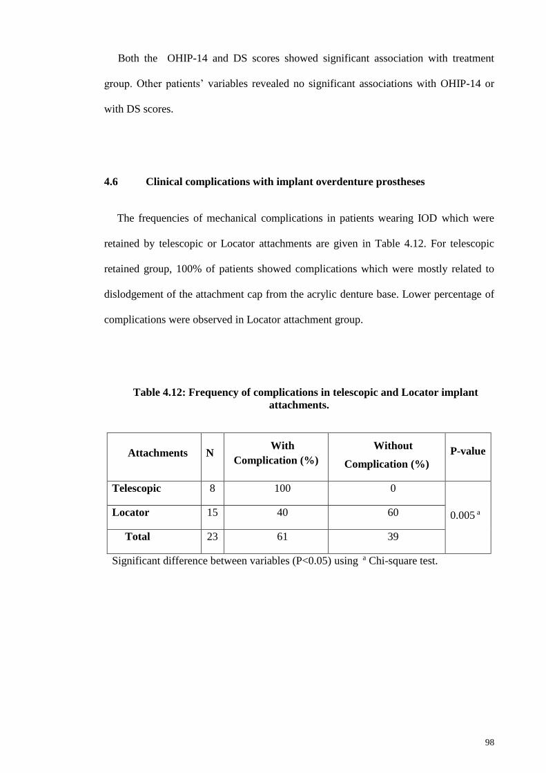
98
Both the OHIP-14 and DS scores showed significant association with treatment
group. Other patients’ variables revealed no significant associations with OHIP-14 or
with DS scores.
4.6 Clinical complications with implant overdenture prostheses
The frequencies of mechanical complications in patients wearing IOD which were
retained by telescopic or Locator attachments are given in Table 4.12. For telescopic
retained group, 100% of patients showed complications which were mostly related to
dislodgement of the attachment cap from the acrylic denture base. Lower percentage of
complications were observed in Locator attachment group.
Table 4.12: Frequency of complications in telescopic and Locator implant
attachments.
Attachments N With
Complication (%)
Without
Complication (%)
P-value
Telescopic 8 100 0
0.005 a Locator 15 40 60
Total 23 61 39
Significant difference between variables (P<0.05) using a Chi-square test.
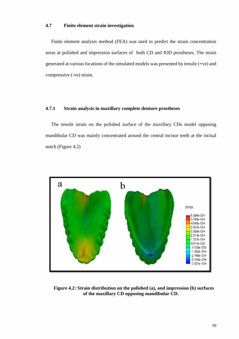
99
4.7 Finite element strain investigation
Finite element analysis method (FEA) was used to predict the strain concentration
areas at polished and impression surfaces of both CD and IOD prostheses. The strain
generated at various locations of the simulated models was presented by tensile (+ve) and
compressive (-ve) strain.
4.7.1 Strain analysis in maxillary complete denture prostheses
The tensile strain on the polished surface of the maxillary CDs model opposing
mandibular CD was mainly concentrated around the central incisor teeth at the incisal
notch (Figure 4.2)
Figure 4.2: Strain distribution on the polished (a), and impression (b) surfaces
of the maxillary CD opposing mandibular CD.
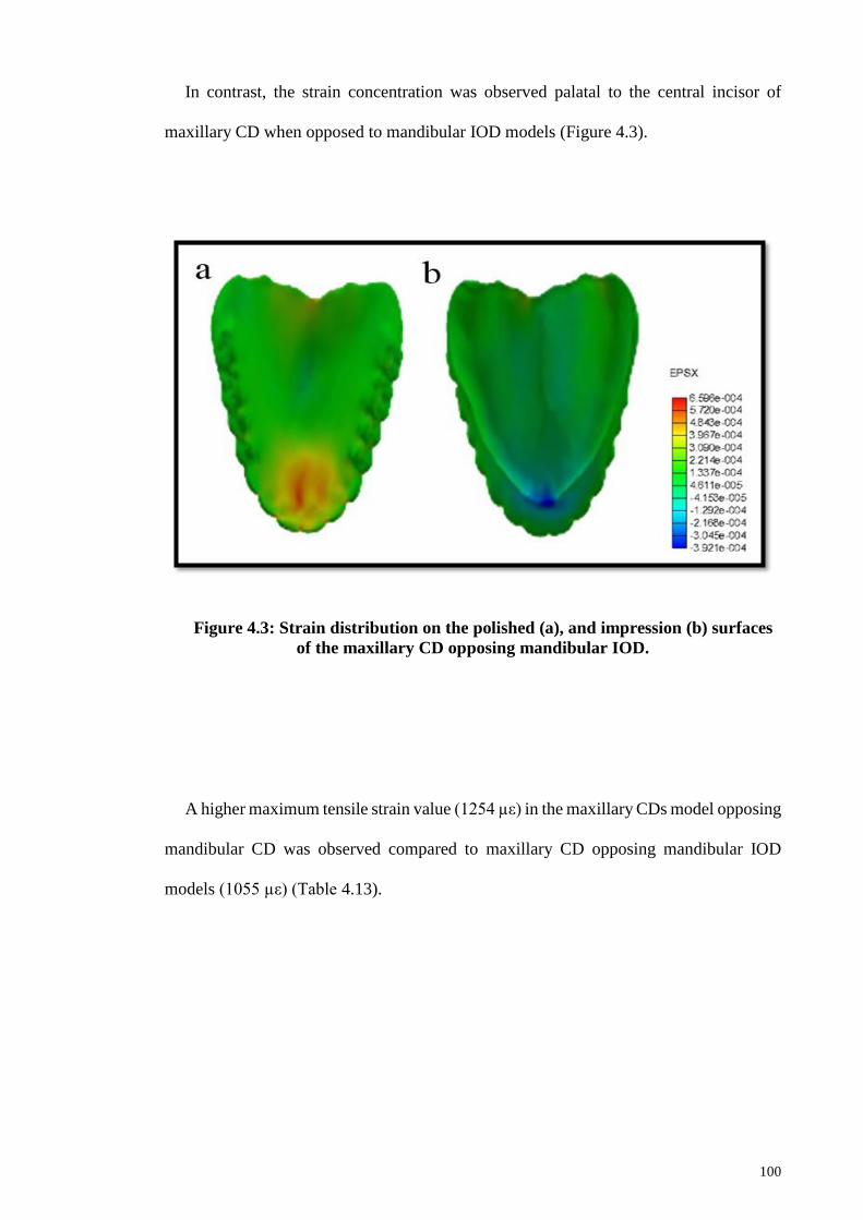
100
In contrast, the strain concentration was observed palatal to the central incisor of
maxillary CD when opposed to mandibular IOD models (Figure 4.3).
Figure 4.3: Strain distribution on the polished (a), and impression (b) surfaces
of the maxillary CD opposing mandibular IOD.
A higher maximum tensile strain value (1254 µε) in the maxillary CDs model opposing
mandibular CD was observed compared to maxillary CD opposing mandibular IOD
models (1055 µε) (Table 4.13).

101
Table 4.13: Strain values (µε) distribution around midline area on polished and
impression surfaces of maxillary CDs opposing mandibular CD and IOD.
4.7.2 Strain analysis in the mandibular prostheses
In the mandibular CD model, the tensile strain was mostly concentrated at the incisal
notch between the lateral incisor and canine, with less concentration at lingual flange
area, followed by the lower sulcus border of the lingual flange area. The strains at the
labial flange, labial frenal area polished surfaces, and denture impression surface were
compressive (Figure 4.4).
Locations
CD/CD CD/IOD
Polish
surface
Impression
surface
Polish
surface
Impression
surface
Incisal notch +1254 NA +847 NA
Labial Frenal notch NA -1250 NA -1642
Anterior palatal area +988 -559 +1055 -613
Posterior palatal area +883 -186 +723 -175
+ values indicate tensile strain. - values indicate compressive strain.
NA= Not applicable.
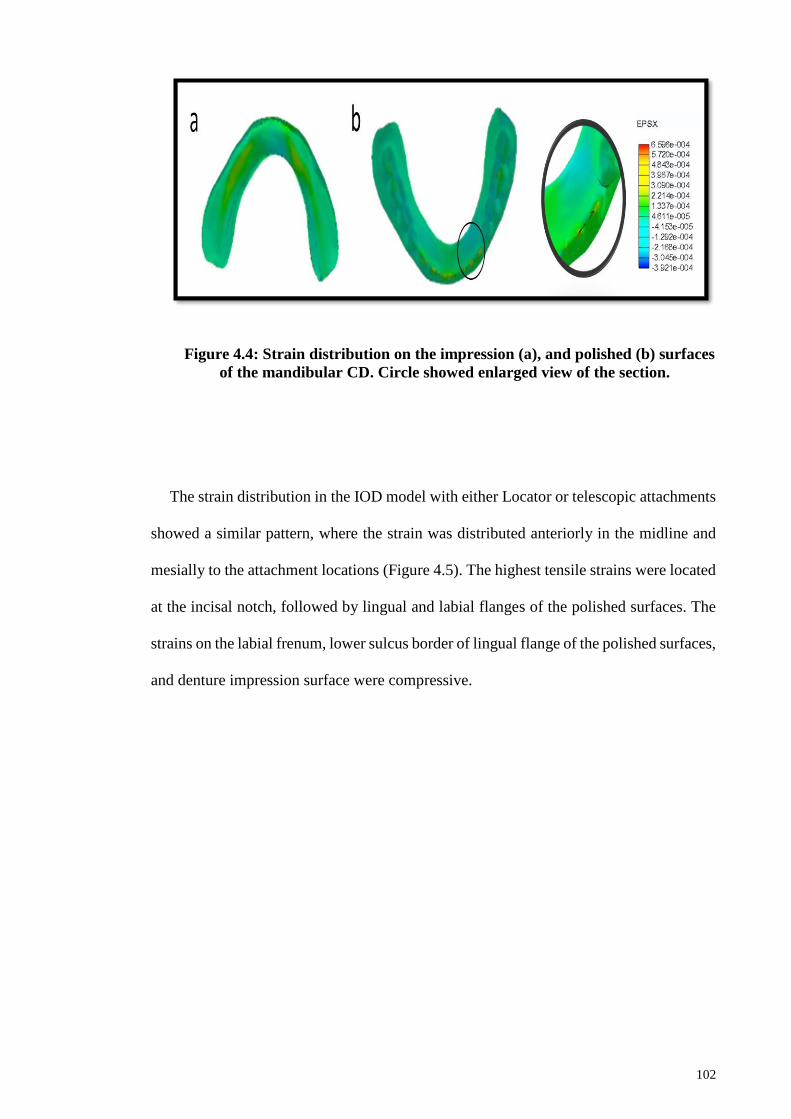
102
The strain distribution in the IOD model with either Locator or telescopic attachments
showed a similar pattern, where the strain was distributed anteriorly in the midline and
mesially to the attachment locations (Figure 4.5). The highest tensile strains were located
at the incisal notch, followed by lingual and labial flanges of the polished surfaces. The
strains on the labial frenum, lower sulcus border of lingual flange of the polished surfaces,
and denture impression surface were compressive.
Figure 4.4: Strain distribution on the impression (a), and polished (b) surfaces
of the mandibular CD. Circle showed enlarged view of the section.
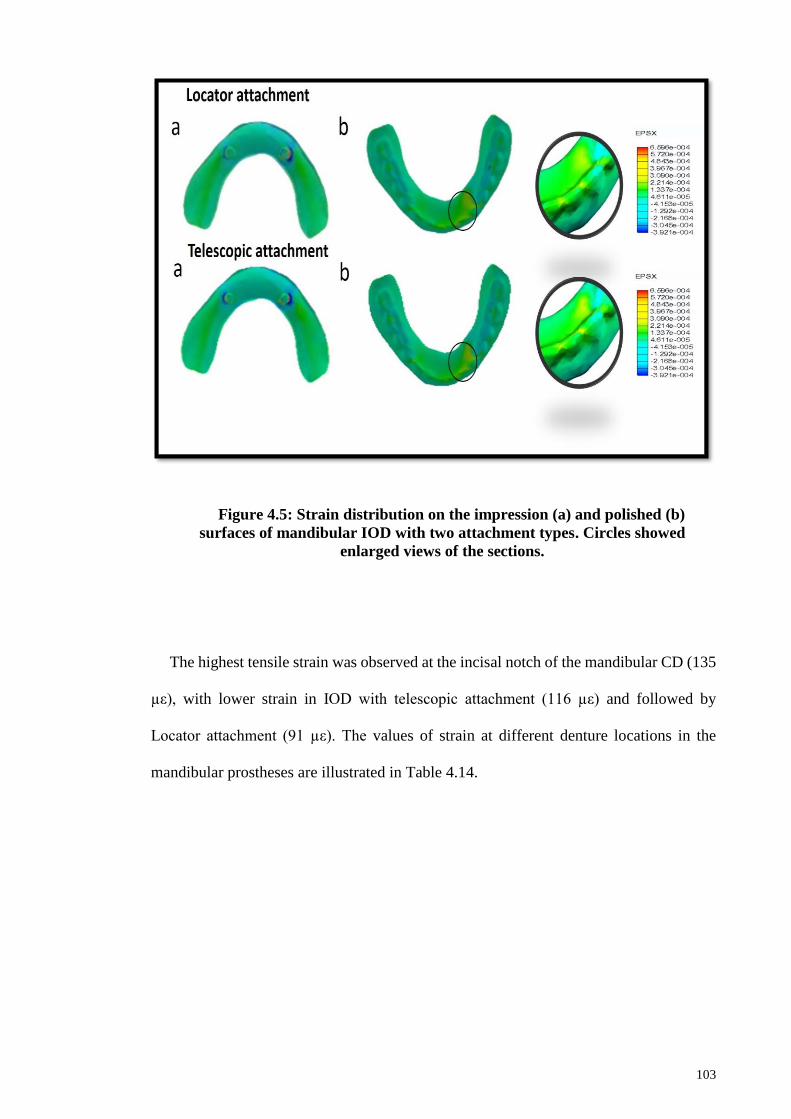
103
The highest tensile strain was observed at the incisal notch of the mandibular CD (135
µε), with lower strain in IOD with telescopic attachment (116 µε) and followed by
Locator attachment (91 µε). The values of strain at different denture locations in the
mandibular prostheses are illustrated in Table 4.14.
Figure 4.5: Strain distribution on the impression (a) and polished (b)
surfaces of mandibular IOD with two attachment types. Circles showed
enlarged views of the sections.
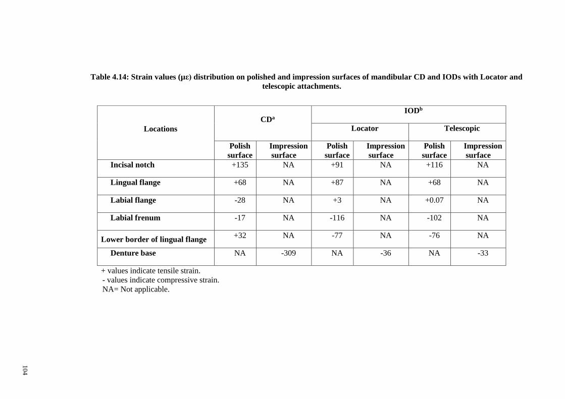
104
Table 4.14: Strain values (µε) distribution on polished and impression surfaces of mandibular CD and IODs with Locator and
telescopic attachments.
Locations
CDa
IODb
Locator Telescopic
Polish
surface
Impression
surface
Polish
surface
Impression
surface
Polish
surface
Impression
surface
Incisal notch +135 NA +91 NA +116 NA
Lingual flange +68 NA +87 NA +68 NA
Labial flange -28 NA +3 NA +0.07 NA
Labial frenum -17 NA -116 NA -102 NA
Lower border of lingual flange +32 NA -77 NA -76 NA
Denture base NA -309 NA -36 NA -33
+ values indicate tensile strain. - values indicate compressive strain.
NA= Not applicable.
10
4

105
4.7.3 Strain analysis at the acrylic/attachments interface area
For strain distributions in mandibular IOD prostheses with telescopic and Locator
attachments, the maximum tensile strain values in both attachments were found in the
acrylic denture-attachment interfaces on the impression surfaces (Figure 4.6).
Figure 4.6: Strain distribution at acrylic/attachment interfaces of mandibular
IOD with (a) telescopic, and (b) Locator attachments.
106
In the telescopic attachment model, lower strain value was observed at the lower
border of the lingual flange, compared to the lower borders of both lingual and labial
flanges of the Locator attachment in IOD models (Table 4.15). The maximum tensile
strain in the acrylic denture/telescopic attachment interface (561 µε) was higher than that
of Locator (323 µε) model. Hence fracture risk is higher with telescopic attachment was
IOD prostheses.

107
Table 4.15: Strain values (µε) distribution on polished and impression surfaces of telescopic and Locator attachments of
mandibular IOD prostheses.
IOD attachments
Locations
Locator Telescopic
Polish
surface
Impression
surface
Polish
surface
Impression
surface
Acrylic/attachments interface NA +323 NA +561
Incisal tip -61 NA +24 NA
Lower border of lingual flange +225 NA -259 NA
Lingual flange -188 NA +268 NA
Lower border of labial flange +191 NA -16 NA
+ values indicate tensile strain. - values indicate compressive strain.
NA= Not applicable.
10
7

108
CHAPTER 5: DISCUSSION
5.1 Introduction
As per the survey for Malaysian National Oral Health Plan conducted in 2011, 32% of
the estimated adult population above 65 years was edentulous (Oral Health Division,
2011). Hence, the concern for oral health in edentulous patients is considerable as well as
the concern of the rehabilitation of patients with conventional or implant prostheses still
remains. In some edentulous patients, providing mucosa-borne conventional complete
dentures (CD) is not considered as satisfactory treatment modality due to its lack of
retention and stability. Moreover, patients using conventional CD may be susceptible to
malnutrition due to their reduced capability of chewing (Awad et al., 2003).
The removable mandibular implant overdenture (IOD) is supported by both implant
and mucosa, where fewer implants are required as compared to fixed full-arch implant
prostheses. Additionally, removable mandibular IODs serves as a less complex and less
expensive option for edentulous patients (Johns et al., 1992). It has been shown that the
use of IOD may inhibit bone loss and may lead to bone apposition in the severely resorbed
mandible (Sennerby et al., 1988; Davis et al., 1999). However, the individual masticatory
occlusal muscle force may create pressure against the alveolar ridges leading to residual
ridge resorption (RRR). Therefore, occlusal force must be considered as a determining
factor for RRR. On the other hand, the chewing and biting functions in IOD patients are
similar to those in people with natural teeth (Mericske-Stern et al., 1993); however, are
better than patients using CD (Fontijn-Tekampl et al., 1998). This may be due to the
smaller cross-sectional area of masticatory muscles in edentulous patients, as compared
to dentate patients, which may affect their masticatory performance (Newton et al., 1993).
Therefore, a greater bite force and higher satisfaction scores were observed in IOD than
109
CD wearers (Geckili et al., 2012a). However, most studies focused on stress distribution
around the implants, and the way in which the presence of implants affects loading
patterns of the edentulous ridge is still not known (Kordatzis et al., 2003).
With many clinical complications related to denture fractures, the strain distribution
in acrylic dentures has been previously evaluated in different studies in vivo or in vitro
using strain gauges or finite element method for analysis (FEA) (Prombonas & Vlissidis,
2006; Cheng et al., 2010; Cilingir et al., 2013). However, the force that is clinically
distributed on the dentures and its relation with the clinical complications is still not
determined.
5.2 Study design and sample population
This study was a cross-sectional study to determine the occlusal force distribution in
edentulous patients wearing mandibular IOD and conventional maxillary CD and
comparison made with patients with conventional maxillary and mandibular CDs. The
findings of this study also provide an insight into the patient's masticatory performances,
bone resorption patterns across maxillary and mandibular ridges, and clinical
complications in mandibular IOD prostheses. While the subjective assessment included
the oral health-related quality of life (OHRQoL) and denture satisfaction, the acrylic
denture strain distribution was investigated using FEA.
The sample of this study comprised of 64 participants, gave a power of 80% (α was
set at 0.05) and was estimated based on the results comparing the bone resorption between
patients rehabilitated with conventional CD and IOD using area index method as
described by a previous study (Tymstra et al., 2011). Many studies have measured area

110
index in the mandibular (Wilding et al., 1987; Jacobs et al., 1992; Wright et al., 2002;
Kordatzis et al., 2003) or maxillary ridges (Jacobs et al., 1993; Kreisler et al., 2003),
whereas Tymstra et al. (2011) conducted a study that measured area index at both ridges.
Subsequently, owing to the exclusion criteria and some participants’ unwillingness to
participate, the sample size estimation was based on the preliminary results comparing
RRR for 10 patients wearing CD and IOD prostheses. The required number of 46 patients
participated in the study, thereby giving a power of 95% (α was set at 0.05).
5.3 Masticatory performance
Several different methods have been used to evaluate masticatory performances. A
subjective questionnaire method was employed to evaluate the masticatory performance
in CD and/or IOD wearers (Agerberg & Carlsson, 1981; Feine & Lund, 2006). Chewing
ability in the previous studies was determined based on the individual's own assessment
of his/her masticatory performance. However, many individuals evaluated the
masticatory performance of their dentures as good compared to dentate while a
comminution test resulted in much lower values than dentate subjects (Carlsson, 1984).
Hence, this subjective nature of the observation from the evaluation of patients’
satisfaction necessitated the use of an objective method. The objective methods employed
to measure masticatory performance include sieving with natural foods (Manly & Braley,
1950), sieving with artificial foods (Edlund & Lamm, 1980), two-colored chewing gums
(Liedberg & Öwall, 1995; Matsui et al., 1996; Hayakawa et al., 1998) or mixing ability
index (MAI) with two-colored paraffin wax cubes (Sato et al., 2003).
111
Ishikawa et al. (2007) investigated the correlations between two different objective
methods with the subjective outcome measures. They used the color change of the
chewing gum as the objective method and found a greater association with the subjective
evaluation outcomes, compared to the masticatory performance using sieving with natural
food objective method (Ishikawa et al., 2007). However, the chewing gum method
requires a special device (colorimeter CR-13) (Kamiyama et al., 2010), and the limitation
of this method was observed in its reliability and validity (Hama et al., 2014).
Masticatory performance, using MAI with two-colored paraffin wax cubes, integrated
the degree of color mixing and the shape of a chewed-food test into one-dimensional
value. Moreover, previously MAI was validated by Sato et al. (2003) for objective
measurement of masticatory performance in denture wearers for clinical use. Hence, it
was employed in the present study as an objective method to evaluate the masticatory
performance.
The MAI results in the present study showed no significant differences between CD
and IOD groups after four years of denture use, despite the better masticatory
performance trend observed in the IOD mean readings as compared to CD readings. The
greater retention and stability of an IOD could improve the masticatory performance (Van
Kampen et al., 2004). Similarly, in this study, we found better masticatory performance
in the IOD group as compared to the CD group.
However, our results contradict the previous studies that found a significant
improvement in the MAI for IOD prostheses, using the similar MAI method, for analyses
of the patients’ masticatory performance (Yunus et al., 2014; Bae et al., 2015). Two to
three months after patients had their CD dentures converted to IOD prostheses, the mean
MAI values of 0.66 (Yunus et al., 2014) and -0.03 (Bae et al., 2015) were reported. In the
112
present study, slightly different MAI mean values 0.0 and 0.3 were observed for CD and
IOD groups, respectively, after four years of wearing dentures, even though the same
MAI method was used for evaluation of masticatory performance. Our results probably
contradict the findings of the previous studies because the previous studies examined the
same group of patients using conventional CD dentures after conversion into IOD ones,
whereas we examined different groups of patients and the conventional CD patients did
not participate in any previous clinical study for implant rehabilitation. Moreover,
different attachment systems and sample sizes were used in the previous studies.
Seventeen patients wearing IOD, with telescopic attachments, participated in the study
by Yunus et al. (2014) while only eight patients wearing IOD, with Locator attachments,
participated in the study by Bae et al. (2015).
In the study conducted by Yunus et al. (2014), the mean MAI (0.86) for IOD, after the
patients wore their dentures for one year, did not show considerable improvement as
compared to the three months observation. This finding was in agreement with the results
of our study, following four years of wearing dentures. This finding could be explained
as most of the denture wearers are probably used to their dentures, thereby feeling
comfortable with chewing as with their mandibular and maxillary prostheses (Feine &
Lund, 2006; Yunus et al., 2014).
5.4 Occlusal parameters recorded using T-scan III
Many previous studies used strain-gauge transducers with a bite fork to clinically
measure the bite force of patients (Lassila et al., 1985; Fontijn-Tekampl et al., 1998;
Rismanchian et al., 2009). However, strain-gauge transducers measured the bite force
unilaterally and they had different thickness that ranged between 3–6 mm. Additionally,

113
strain-gauges are covered by certain materials, such as polyvinyl chloride, to protect the
teeth and appliances, and a plastic bag is also used to reduce moisture. These protection
requisites were found to reduce the bite force by 15% (Bakke et al., 1990). The T-scan
III occlusal analysis system, with its 100 µm thickness compared to strain gauges higher
thickness could be a more appropriate clinical method to detect patients’ occlusal force
distribution. Moreover, the U-shaped design of T-scan III enables it to measure the
occlusal force bilaterally.
In clinical conditions, there is a different force distribution across the arch in dentate
subjects, which could be manifested by the asymmetry in the left and right sides with
shifted the higher occlusal forces from anterior to posterior teeth (Koos et al., 2012).
Similarly, Kerstein et al. (2013) determined the occlusal force distribution in CD wearers
with maximum intercuspation. There was a higher force distribution in the posterior
(premolar and molar) areas than the anterior (incisor and canine) areas. Similarly, in this
study occlusal forces demonstrated different distributions, higher in the posterior areas of
the arch than anterior areas, in completely edentulous patients wearing mandibular IOD
more than patients wearing conventional CD prostheses. Patients wearing IOD might
exhibit a higher masseter muscles' electromyographic activity than patients with mucosa-
supported CD during maximum intercuspation (Shah et al., 2012). On the other hand, the
IOD showed lower force distribution anteriorly compared to the CD prostheses, which
may be because the patients with IOD had an open occlusal relationship in centric
occlusion to reduce stress on the implants, preserve premaxillary bone area and direct
occlusal forces towards the center of mandibular teeth (Becker et al., 1977; Denissen et
al., 1993).
Other occlusal parameters measured in this study include occlusal time—the time that
passes between the first and last occlusal contacts. The results of occlusal time in this
114
study showed an average of 2 secs which is longer than the occlusal time in dentate
subjects (˂0.2 secs) (Kerstein & Grundset, 2001). Edentulous patients might need longer
occlusal time compared to dentate patients, probably due to prostheses instability in
completely edentulous patients, especially for the mandibular prostheses (Van Waas,
1990). However, occlusion time was also found to be shorter (0.4 secs) in another study
with patients wearing conventional CD (Abdelnabi et al., 2016). These contradictory
values could be due to the improved stability of CD observed in the study by Abdelnabi
et al. (2016) after the application of denture adhesive, while in the present study no
adhesive was applied before occlusal parameters’ analyses.
Besides occlusal force distribution and occlusion time measurements, maximum
occlusal force percentage in maximum intercuspation was also determined. There were
similar maximum occlusal force results recorded for CD and IOD groups. These findings
are in accordance with the results obtained in our pilot study, which determined the bite
force using a strain gauge device. There was no significant difference between the CD
and IOD groups, in spite of the higher mean bite force (150 N) observed in the IOD than
the CD group (132 N). Similarly, in the study by Fontijn-Tekampl et al. (1998) the mean
maximum bite force values observed were 154 N and 123 N for IOD and CD,
respectively; however, the maximum bite force in patients with mandibular IOD was
significantly higher than in patients with conventional CD. These results could be due to
the limited number of patients (ten patients) enrolled in our pilot study. Also, Fontijn-
Tekampl et al. (1998) used trans-mandibular and mucosa-borne implant types to retain
their IOD patients.

115
5.5 Maxillary and mandibular residual ridge resorption at different locations
and their association with the occlusal force distribution
The present study employed an established method to determine RRR at mandibular
posterior ridge and opposing maxillary edentulous anterior and posterior ridges, in
patients wearing mandibular IOD and conventional CD prostheses. Using this method,
the bone resorption ratio (bone area/reference area) from dental panoramic radiographs
(DPRs) taken at intervals could be compared with minimal errors related to radiographic
magnification and positional variation between radiographs (Wilding et al., 1987;
Kreisler et al., 2000). As opposed to linear measurements, the proportional ratio of bone
area to reference area remained relatively constant with changes in the exposure angle of
X-ray beam (Kreisler et al., 2000).
The use of DPRs is well established; however, cone beam computed tomography
(CBCT) has been suggested as an alternative method with greater accuracy to measure
and determine the shape and size of alveolar ridges in edentulous patients (Varón de
Gaitán et al., 2014). On the other hand, repeated exposures to high doses of radiation and
extra costs might not justify the routine use of CBCT for follow-up evaluation. Moreover,
DPR was more routinely used for edentulous patients seeking CD treatment in this
establishment.
Higher levels of occlusal force increase the RRR rate as shown by many previous
studies (Jaul et al., 1980; Maruo et al., 2010; Fujiki et al., 2013; Ahmad et al., 2015).
However, with even force distribution and the concentration of occlusal forces over
maximal denture bearing area, this could reduce the unnecessary concentration of stress,
and hence slow the RRR (Maeda & Wood, 1989). Therefore, the effect of force
distribution on bone resorption at different areas across the dental arch was investigated
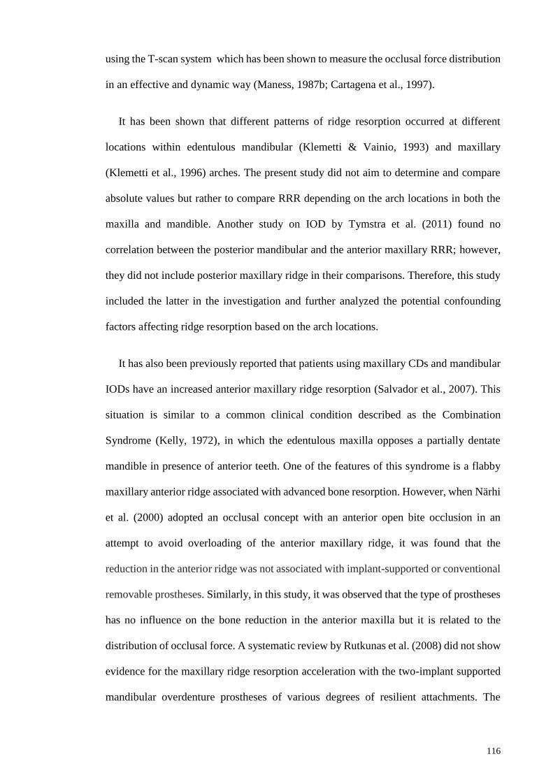
116
using the T-scan system which has been shown to measure the occlusal force distribution
in an effective and dynamic way (Maness, 1987b; Cartagena et al., 1997).
It has been shown that different patterns of ridge resorption occurred at different
locations within edentulous mandibular (Klemetti & Vainio, 1993) and maxillary
(Klemetti et al., 1996) arches. The present study did not aim to determine and compare
absolute values but rather to compare RRR depending on the arch locations in both the
maxilla and mandible. Another study on IOD by Tymstra et al. (2011) found no
correlation between the posterior mandibular and the anterior maxillary RRR; however,
they did not include posterior maxillary ridge in their comparisons. Therefore, this study
included the latter in the investigation and further analyzed the potential confounding
factors affecting ridge resorption based on the arch locations.
It has also been previously reported that patients using maxillary CDs and mandibular
IODs have an increased anterior maxillary ridge resorption (Salvador et al., 2007). This
situation is similar to a common clinical condition described as the Combination
Syndrome (Kelly, 1972), in which the edentulous maxilla opposes a partially dentate
mandible in presence of anterior teeth. One of the features of this syndrome is a flabby
maxillary anterior ridge associated with advanced bone resorption. However, when Närhi
et al. (2000) adopted an occlusal concept with an anterior open bite occlusion in an
attempt to avoid overloading of the anterior maxillary ridge, it was found that the
reduction in the anterior ridge was not associated with implant-supported or conventional
removable prostheses. Similarly, in this study, it was observed that the type of prostheses
has no influence on the bone reduction in the anterior maxilla but it is related to the
distribution of occlusal force. A systematic review by Rutkunas et al. (2008) did not show
evidence for the maxillary ridge resorption acceleration with the two-implant supported
mandibular overdenture prostheses of various degrees of resilient attachments. The

117
finding of the present study was also in agreement with another study (Tymstra et al.,
2011) that employed proportional area measurement and reported that patients
rehabilitated with an implant retained mandibular overdenture did not exhibit more RRR
as compared to patients wearing a conventional CD.
On the other hand, a different observation was found in the posterior arches, where
maxillary and mandibular ridge reduction was affected by the type of prostheses. There
was lesser posterior ridge resorption with implant prostheses compared to conventional
CDs, even though higher chewing forces and improved masticatory performances were
reported with implant therapy (Yunus et al., 2014; Bae et al., 2015). This observation
concurred with the results of Kordatzis et al. (2003), where they employed similar
proportional area measurements using panoramic radiographs to determine ridge
reduction within five years. The authors attributed this, lesser posterior ridge resorption,
to the more favorable loading and protection of the residual posterior ridge from an
excessive load of the mandibular anterior implants. Similarly, a recent systematic review
concluded alveolar ridge preservation with implant-assisted restorations (Khalifa et al.,
2016).
Regarding the within-group comparisons at various locations of the mandibular and
maxillary edentulous arches, the conventional CD group showed similar residual ridge
reduction at all locations, whereas the IOD group showed significantly greater posterior
ridge resorption in the mandible than in the maxilla. A greater percentage of occlusal
force was shifted to the posterior areas in IOD patients, and the posterior mandible
appeared to respond less favorably, hence, had a greater RRR as compared to the
antagonistic residual ridge. The maxillary denture base coverage provided more tissue
support for maxillary prostheses (Tallgren, 1972), compared to the smaller denture
bearing area available for mandibular dentures (Jivraj et al., 2006), thereby reducing the
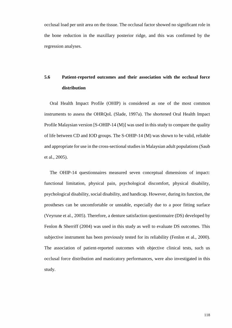
118
occlusal load per unit area on the tissue. The occlusal factor showed no significant role in
the bone reduction in the maxillary posterior ridge, and this was confirmed by the
regression analyses.
5.6 Patient-reported outcomes and their association with the occlusal force
distribution
Oral Health Impact Profile (OHIP) is considered as one of the most common
instruments to assess the OHRQoL (Slade, 1997a). The shortened Oral Health Impact
Profile Malaysian version [S-OHIP-14 (M)] was used in this study to compare the quality
of life between CD and IOD groups. The S-OHIP-14 (M) was shown to be valid, reliable
and appropriate for use in the cross-sectional studies in Malaysian adult populations (Saub
et al., 2005).
The OHIP-14 questionnaires measured seven conceptual dimensions of impact:
functional limitation, physical pain, psychological discomfort, physical disability,
psychological disability, social disability, and handicap. However, during its function, the
prostheses can be uncomfortable or unstable, especially due to a poor fitting surface
(Veyrune et al., 2005). Therefore, a denture satisfaction questionnaire (DS) developed by
Fenlon & Sherriff (2004) was used in this study as well to evaluate DS outcomes. This
subjective instrument has been previously tested for its reliability (Fenlon et al., 2000).
The association of patient-reported outcomes with objective clinical tests, such us
occlusal force distribution and masticatory performances, were also investigated in this
study.

119
Patients were assessed for their perception with regards to the experience using their
current prostheses. An overall lower scores were observed in OHIP-14 and DS total
scores for the IOD type more than CD type of prostheses, which could be due to better
stability and retention achieved with the IOD (Boerrigter et al., 1995a; Boerrigter et al.,
1995b; Meijer et al., 1999). This finding was in agreement with results of previous
clinical studies (Awad et al., 2000a; Awad et al., 2003; Heydecke et al., 2003; Torres et
al., 2011; Jabbour et al., 2012; Yunus et al., 2014).
Regarding the OHIP-14 individual domains, the patients in the IOD group reported
significantly lower score for physical pain than those in CD group, while there were no
differences in the other domains. This finding was in congruence with another similar
study conducted by Geckili et al. (2012a). The greater score in physical pain in CD group
may be attributed to the undesirable denture movements associated with the reduced
stability of CD, thereby resulting in increased pain sensation compared to the IOD group
(Cakir et al., 2014). Previous investigators have also shown a higher association between
complaint of pain and denture mucosal bearing areas in CD patients (Brunello &
Mandikos, 1998). The remaining OHIP-14 domains, however, showed no difference
between CD and IOD groups, which contradicts the findings of a study by Yunus et al.
(2014). This could be due to the different observation time because patients in the
previous study (Yunus et al., 2014) were evaluated one year after their dentures were
converted to IOD, while in the present study the patients were evaluated after four years.
Patients rehabilitated with CD need additional time to adapt to the new dentures than
patients with fixed or removable partial dentures, and the OHRQoL improvement was
observed following two years of prosthodontic treatment (Aarabi et al., 2015).
Regarding the denture satisfaction in IOD patients, there was greater satisfaction in
relation to stability, comfort, and chewing ability compared to those in CD patients.

120
Completely edentulous patients are frequently dissatisfied with their dentures (Lechner
& Roessler, 2001), especially with the mandibular dentures, due to compromised
retention, stability and masticatory performance (Van Waas, 1990). Patients’ habituation
to their mandibular dentures may explain greater satisfaction with mandibular compared
to maxillary prostheses (Fenlon & Sherriff, 2004).
In this study, no association was observed between the occlusal force distribution and
OHRQoL and denture satisfaction. This finding contradicted with other studies (Lassila
et al., 1985; Rismanchian et al., 2009) where they found the bite force was positively
correlated with patient satisfaction. However, our results were in line with other studies
(Cune et al., 2005; Geckili et al., 2012b) where increased bite force was not found to
enhance patient life expectation or satisfaction. These contradictory findings of bite force
association to patients satisfaction of the previous studies (Lassila et al., 1985;
Rismanchian et al., 2009), could be due to the variations in bite force measurement
devices used in these studies. These studies employed pezioelectronic strain gauges with
3-6 mm thickness; while in this study the thickness of sensor was 100 µm. The U-shaped
sensor used in the present study allowed bilateral balanced force measurements.
The objective method to determine masticatory performance was made using the
measurement of MAI. The association between masticatory performance and the patient-
reported outcomes was also determined. However, we found that MAI was neither
associated with the total OHIP-14 nor with total DS scores. This finding contradicts the
results of a study conducted by Bae et al. (2015), that investigated masticatory
performance, using MAI, and reported its correlation to the findings of a satisfaction
questionnaire in CD and IOD patients. Bae et al. (2015) found a positive correlation
between overall patients’ satisfaction and MAI values. These contradicting findings could
be due to the different sample sizes used in both studies because 46 patients were enrolled
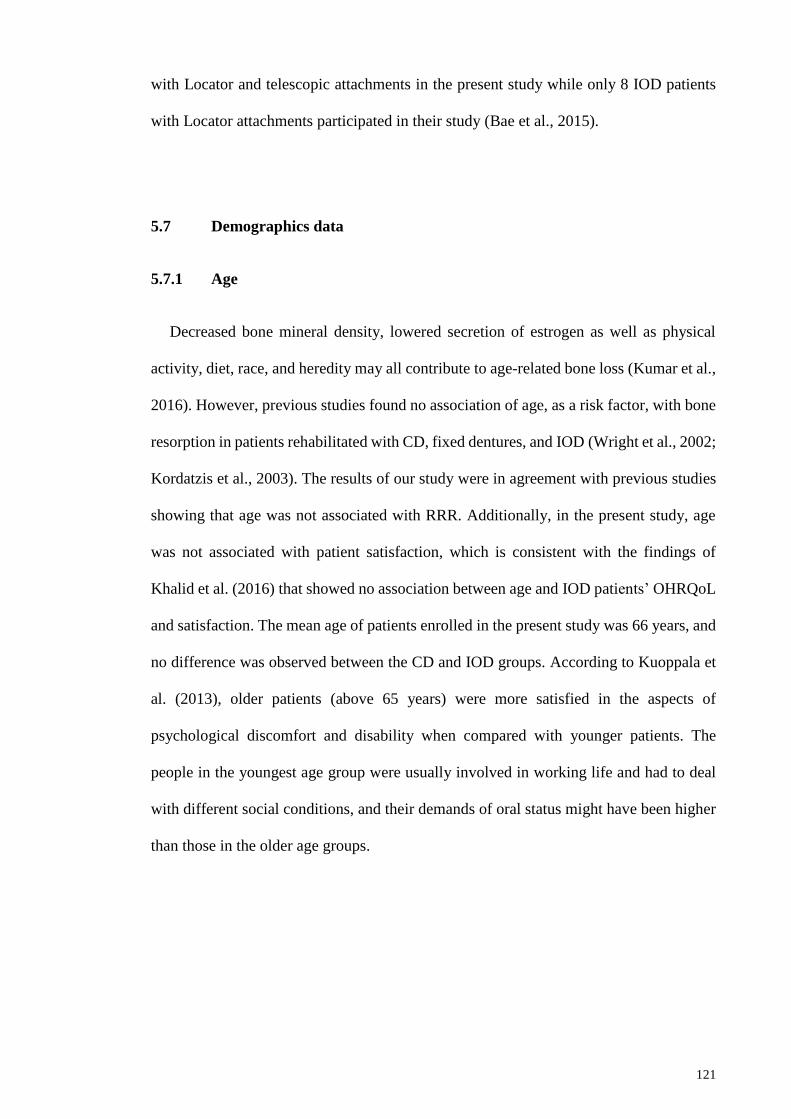
121
with Locator and telescopic attachments in the present study while only 8 IOD patients
with Locator attachments participated in their study (Bae et al., 2015).
5.7 Demographics data
5.7.1 Age
Decreased bone mineral density, lowered secretion of estrogen as well as physical
activity, diet, race, and heredity may all contribute to age-related bone loss (Kumar et al.,
2016). However, previous studies found no association of age, as a risk factor, with bone
resorption in patients rehabilitated with CD, fixed dentures, and IOD (Wright et al., 2002;
Kordatzis et al., 2003). The results of our study were in agreement with previous studies
showing that age was not associated with RRR. Additionally, in the present study, age
was not associated with patient satisfaction, which is consistent with the findings of
Khalid et al. (2016) that showed no association between age and IOD patients’ OHRQoL
and satisfaction. The mean age of patients enrolled in the present study was 66 years, and
no difference was observed between the CD and IOD groups. According to Kuoppala et
al. (2013), older patients (above 65 years) were more satisfied in the aspects of
psychological discomfort and disability when compared with younger patients. The
people in the youngest age group were usually involved in working life and had to deal
with different social conditions, and their demands of oral status might have been higher
than those in the older age groups.
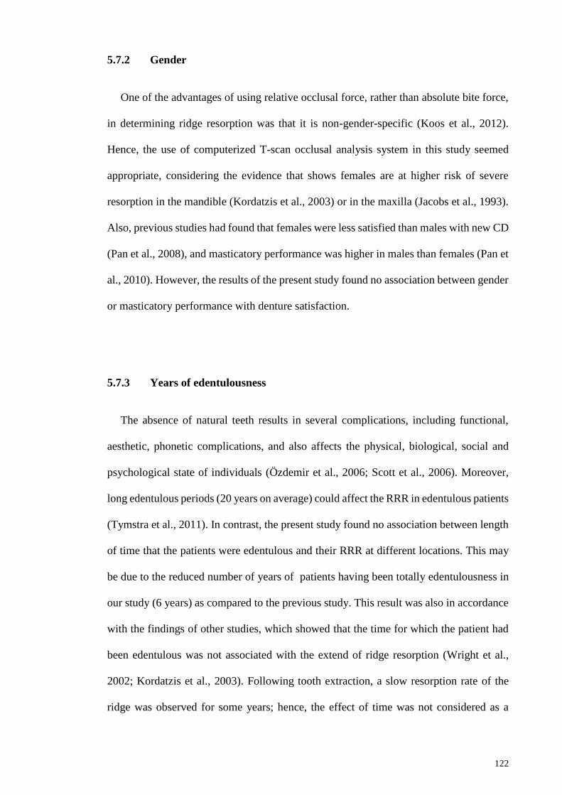
122
5.7.2 Gender
One of the advantages of using relative occlusal force, rather than absolute bite force,
in determining ridge resorption was that it is non-gender-specific (Koos et al., 2012).
Hence, the use of computerized T-scan occlusal analysis system in this study seemed
appropriate, considering the evidence that shows females are at higher risk of severe
resorption in the mandible (Kordatzis et al., 2003) or in the maxilla (Jacobs et al., 1993).
Also, previous studies had found that females were less satisfied than males with new CD
(Pan et al., 2008), and masticatory performance was higher in males than females (Pan et
al., 2010). However, the results of the present study found no association between gender
or masticatory performance with denture satisfaction.
5.7.3 Years of edentulousness
The absence of natural teeth results in several complications, including functional,
aesthetic, phonetic complications, and also affects the physical, biological, social and
psychological state of individuals (Özdemir et al., 2006; Scott et al., 2006). Moreover,
long edentulous periods (20 years on average) could affect the RRR in edentulous patients
(Tymstra et al., 2011). In contrast, the present study found no association between length
of time that the patients were edentulous and their RRR at different locations. This may
be due to the reduced number of years of patients having been totally edentulousness in
our study (6 years) as compared to the previous study. This result was also in accordance
with the findings of other studies, which showed that the time for which the patient had
been edentulous was not associated with the extend of ridge resorption (Wright et al.,
2002; Kordatzis et al., 2003). Following tooth extraction, a slow resorption rate of the
ridge was observed for some years; hence, the effect of time was not considered as a
123
significant factor (Kordatzis et al., 2003). In the present study, years of edentulousness
was not associated with OHRQoL and denture satisfaction. This lack of association was
similar to a study by Khalid et al. (2016), where they also observed that the years of
edentulousness had no association with the patient-reported outcomes. The groups of
patients in this study and their study (Khalid et al., 2016) were similar in this respect.
5.8 Strain investigation within denture prostheses and attachment clinical
complications
Fatigue of denture base polymer could be one of the main factors leading to acrylic
denture fractures—which is still one of the clinical problems faced in prosthetic dentistry
(Smith, 1961). Fatigue failure does not necessitate higher biting forces, and small forces
caused by the masticatory system, over a period of time, can ultimately lead to the
formation of small cracks which propagate in the dentures causing fractures (Farmer,
1983).
FEA is considered as a more suitable method for strain analysis as compared to the
strain gauges because FEA does not have special measure requirements such as a dry
field, which is required for the latter method (Darbar et al., 1994). Furthermore, FEA can
measure strain on two surfaces, unlike strain gauges which measure strain only on one
surface (Cheng et al., 2010). However, for vertical occlusal load simulations in FEA,
previous studies had applied 40% maximum bite force bilaterally on the first molars of
the prostheses, assuming a nearly symmetric loading condition at the left and right sides
of the arch (Gibbs et al., 1981; Ahmad et al., 2015). However, in this study, the application
of simulated load, at the anterior and posterior quadrants varied since the occlusal force
distribution was found to be different. A smaller proportion of the vertical occlusal force
124
was applied in anterior quadrants than posterior quadrants and this could be clinically
more relevant when the maxillary and mandibular teeth simultaneously come into
contact.
The T-scan occlusal force distribution can differentiate occlusal force area and helps
in the force analyses at different zones across the dental arch (Cartagena et al., 1997;
Kumagai et al., 1999). The occlusal force distribution between CD and IOD groups, in
this study, showed different strain concentrations and fracture initiation sites. Moreover,
different strain concentrations were observed between the maxillary and mandibular
prostheses.
The strains generated on polished and impression surfaces of the maxillary CD models
were tensile and compressive, respectively, regardless of the opposing mandibular
denture type. A previous study which used brittle lacquer coating materials (Matthews &
Wain, 1956), showed that a higher tensile stress was distributed at the mid-palatal
polished surface of the maxillary CD. Similarly, the FEA results of the present study
indicated that the maxillary CD was more susceptible to fractures in the mid-palatal area
of the polished surface when opposing the mandibular CD model. During mastication,
the maxillary CD is bent at the mid-palatal line as the fulcrum axis, thus making the
denture fractures more likely to initiate from this area (Anusavice et al., 2013). Improper
fitting of the dentures another factor that might increase the deflection of the removable
dentures during mastication; subsequently, predisposing the dentures to fatigue fractures
(Vallittu et al., 1994; Vallittu et al., 1996). A recent FEA study (Cheng et al., 2010) also
reported a similar area as the fracture initiation zone and the maximum tensile strain
measured at the tip of the incisal notch was 1917 με which was higher than that observed
in our study 1254 με. This difference could be due to the application of a bilateral 230 N
125
load on the posterior region in their study, while our study used a load of 150 N which
was heterogeneously distributed in the anterior and posterior areas.
When the maxillary CD model opposed the mandibular IOD models, a different strain
distribution scenario was observed where the fracture initiation areas were found to be
more palatal to the anterior teeth position, incisal notch, and the mid-posterior palatal
area. The strain distribution patterns in the maxillary CD models, when opposing
mandibular CD was different from the IOD models. This could be due to the different
occlusal force distributions between conventional and implant mandibular prostheses.
The occlusal force was relatively more homogeneous in the mandibular CD model. For
mandibular IOD models, higher occlusal forces were applied in the posterior quadrants
as compared to the anterior quadrants.
Investigations of the strain distribution in the mandibular CD model indicated a higher
tensile strain at the incisal notch of the lower incisors and canines. This finding was in
agreement with the results of previous studies, which investigated the fracture phenomena
using scanning electron microscope (Lamb et al., 1985; Vallittu, 1996). The initiation of
crack growth in the anterior zone is probably attributed to continuous flexing of dentures
from the lateral extension of their posterior part during the application of force (Vallittu,
1996).
The FEA results indicated that different tensile and compressive strain distributions
were generated in the mandibular IOD and the mandibular CD prostheses, due to their
different rotation axes around the intercepted areas between the attachments and the
acrylic denture bases (Solomons, 2000).
The strain concentration around the acrylic denture/attachment interface may
consequently lead to fractures, especially when self-cure resin materials are used in the
126
attachment pick-up procedures that results in a weak mechanical bond. This was in
agreement with some previous studies that used cast metal frameworks to minimize
denture base fractures in the root-retained overdentures and IOD prostheses (Langer &
Langer, 1991; Walton & MacEntee, 1994).
Regarding the attachment design, it was found that the Locator and telescopic
attachments possess their own unique mechanisms to retain the prostheses. With Locator
attachment, the maximum tensile strain was observed at the acrylic denture/attachment
interface with lesser strain at the lower sulcus border of the lingual and labial flange areas.
Telescopic attachment similarly demonstrated tensile strain around the interface, with
lesser strain at the top of the telescopic attachment. This indicated the likelihood of
fracture initiation from the acrylic denture/attachment area regardless of the attachment
type used.
From the clinical perspective of this study, it was observed that IOD retained by
telescopic attachments showed 100% complications, whereas only 40% complications
were observed in Locator attachments. As the tensile strain distribution around the
telescopic attachment was higher and significantly greater frequency of mechanical
complications was observed, seems to be there is a higher risk of clinical complications
with telescopic attachment compared to Locator attachment in IOD prostheses.
Telescopic attachment occupied greater space (5 mm) within the prosthesis compared to
Locator attachment (2.3 mm). The rigidity of the telescopic attachment is high as
compared to Locator attachment where the latter allows some degree of resilience in both
vertical and horizontal directions when in function (Evtimovska et al., 2009).
These findings contradict the previous results shown by Dong et al. (2006), who
undertook an FEA study in which the highest tensile strain was on top of the overdenture
127
copings regardless of their height. This could be due to the different load applications
seen in the previous study and their distributions across the arch. For instance, Dong et
al. (2006) applied a vertical force of 49 N individually on the first premolar, first molar,
and second molar areas; whereas in this study the force was vertically applied following
the relative occlusal force distribution obtained from digital occlusal analysis system T-
scan sensor.
Comparison of the maximum tensile strain values in all the studied models indicated
that maxillary CD had higher tensile strains than the opposing mandibular CD either with
Locator and telescopic attachment. Hence, fracture was more likely to occur in maxillary
denture. This was in agreement with a previous survey on the clinical denture repair,
which found two-fold higher incidences of fractures in maxillary to mandibular dentures
(Beyli & Von Fraunhofer, 1981).
5.9 Summary
Patients using maxillary CDs and mandibular IODs have an increased anterior
maxillary ridge resorption (Salvador et al., 2007) which was thought to be related to the
type of the prostheses used rather than the effect of occlusal force distribution. Hence,
this cross-sectional study determined the RRR in edentulous patients wearing mandibular
IOD or conventional CD, opposing conventional maxillary CDs. This study also
investigated the association of RRR with occlusal force distribution in different areas in
the maxillary and the mandibular arches as well as with other confounding factors, such
as age, gender, and years of edentulism. It was observed that the type of mandibular
prostheses has no influence on the bone resorption at the anterior maxilla. However, the
bone resorption was associated with the occlusal force distribution, and with the type of

128
mandibular prostheses at the posterior arches of the maxilla and mandible. Higher
chewing forces and improved masticatory performances were reported with IOD
prostheses, however, no association was observed between patient perceptions and the
occlusal force distribution in both IOD and conventional CD patients.
The risk of acrylic denture prostheses fracture has been previously investigated by
FEA simulation using specific occlusal load which was applied on molar teeth, however,
the occlusal load did not represent the actual clinical force distribution. Therefore, this
study retrieved the T-scan III occlusal force distribution at different locations across the
dental arch in the FEA load simulation. The results showed that there were different strain
concentrations and fracture initiation sites observed between CD and IOD models.
129
CHAPTER 6: CONCLUSIONS
6.1 Conclusions
Mandibular overdenture supported by two implants is more retentive and it has been
shown to improve patients OHRQoL and satisfaction over conventional complete denture
treatment. However, there is still a lack of information available about the occlusal force
distribution and its association to residual ridge resorption, patient-reported outcomes, as
well as the other mechanical and clinical factors. T-scan III occlusal analysis system was
used in this study to determine the percentage of occlusal force distribution. The findings
observed from this study could be useful for better prostheses design avoiding residual
ridge resorption, and minimizing the denture risk of fracture.
Within the limitation of this study, we can conclude that:
6.1.1 Clinical Study
1 There was no significant difference in the masticatory performance of patients
wearing conventional complete denture and mandibular implant-overdenture
prostheses.
2 The anterior and posterior occlusal force distribution showed significant
differences between conventional complete denture and mandibular implant-
overdenture prostheses. Significant differences between anterior and posterior
occlusal force distribution were also observed within each group.
3 Mandibular implant-overdenture patients showed better oral health-related
quality of life and denture satisfaction than conventional complete denture
patients.
130
4 Greater occlusal force percentage was associated with increased bone resorption
in the anterior maxilla and posterior mandibular area.
5 Mandibular implant-overdenture prostheses showed less residual ridge bone
resorption in the posterior maxilla and mandible areas.
6 Mandibular implant-overdenture with Locator attachment showed lower clinical
complications compared to the telescopic attachment.
6.1.2 Finite element analysis study
1. The risk of fracture incidence in the maxillary conventional complete denture
was higher than those of mandibular conventional complete denture and
mandibular implant-overdenture prostheses, especially at the anterior palatal
area of the polished surface of the acrylic denture base.
2. Mandibular conventional complete denture prostheses showed higher strain
values at the midline area of the polished surface in contrast to the mandibular
implant-overdenture prostheses with either Locator or telescopic attachments.
3. Mandibular implant-overdenture with Locator attachment showed lower
fracture susceptibility compared to the telescopic attachment at the denture-
attachment interface.
131
6.2 Limitations of the study
1- T-scan III system was only used to record the relative occlusal force distribution
rather than the actual force distribution
2- Utilization of combined force distribution values for telescopic and Locator
attachments is considered as one of the limitation observed in this study, due to
an unequal number of attachments used in the implant-overdenture group (15
patients having Locator and eight having telescopic attachments).
3- The implant fixture was simulated without threads in order to reduce the analysis
time and model complexity, as the major concern was to measure the strain
distribution of the acrylic denture base and its interference with the attachments.
4- The load value of 150 N used in FEA simulation might not present the actual force
applied for conventional complete denture and mandibular implant-overdenture
models since it is higher in the latter group (Ahmad et al., 2015). However, a pilot
study was conducted to measure this force and the results showed a non-
significant difference between the groups and the higher mean of force (150 N)
was applied for load simulation.
6.3 Study recommendations
1- Future studies could involve investigating residual ridge resorption in relation to
the distribution of force at various time sequences extending from the point of
initial tooth contact to maximal intercuspation position and to complete
disocclusion.
2- Investigation of the effect of eccentric occlusal movements in relation to residual
ridge resorption are suggested for future studies.
132
3- Future studies are necessary to investigate the stress distribution within the dental
implants and their supporting tissues in mandibular implant-overdenture retained
by telescopic or Locator attachments opposing conventional maxillary complete
denture based on their occlusal force distribution.

133
REFERENCES
Aarabi, G., John, M. T., Schierz, O., Heydecke, G. & Reissmann, D. R. (2015). The
course of prosthodontic patients’ oral health-related quality of life over a period
of 2 years. Journal of Dentistry. 43(2):261-268.
Abdelnabi, M. H., Swelem, A. A. & Al-Dharrab, A. A. (2016). Influence of denture
adhesives on occlusion and disocclusion times. Journal of Prosthetic Dentistry.
115(3):306-312.
Afrashtehfar, K. I., Akanksha Srivastava, B. & Esfandiari, S. (2013). A health technology
assessment report on the utility of digital occlusal analyzer system t-scan® in
temporomandibular disorders. (report no: S2013.02). Montreal, Canada: Faculty
of Dentistry, Mcgill university. Https://www.Mcgill.Ca/dentistryohs/courses-
and-events/dent-655/hta-reports:1-54.
Agerberg, G. & Carlsson, G. E. (1981). Chewing ability in relation to dental and general
health: Analyses of data obtained from a questionnaire. Acta Odontologica
Scandinavica. 39(3):147-153.
Ahmad, R., Chen, J., Abu-Hassan, M. I., Li, Q. & Swain, M. V. (2015). Investigation of
mucosa-induced residual ridge resorption under implant-retained overdentures
and complete dentures in the mandible. International Journal of Oral and
Maxillofacial Implants. 30(3):657-666.
Aimetti, M., Romano, F., Griga, F. & Godio, L. (2009). Clinical and histologic healing
of human extraction sockets filled with calcium sulfate. International Journal of
Oral and Maxillofacial Implant. 24(5):901-909.
Al-Shammari, K. F., Al-Khabbaz, A. K., Al-Ansari, J. M., Neiva, R. & Wang, H.-L.
(2005). Risk indicators for tooth loss due to periodontal disease. Journal of
Periodontology. 76(11):1910-1918.
AlBaker, A. M. (2013). The oral health‐related quality of life in edentulous patients
treated with conventional complete dentures. Gerodontology. 30(1):61-66.
Allen, P. F. & McMillan, A. S. (2003). A review of the functional and psychosocial
outcomes of edentulousness treated with complete replacement dentures. Journal
of the Canadian Dental Association 69(10):662-662.
Allen, P. F., McMillan, A. S. & Locker, D. (2001). An assessment of sensitivity to change
of the oral health impact profile in a clinical trial. Community Dentistry and Oral
Epidemiology. 29(3):175-182.
Alsabeeha, N. H., Payne, A. G. & Swain, M. V. (2009). Attachment systems for
mandibular two-implant overdentures: A review of in vitro investigations on
retention and wear features. International Journal of Prosthodontics. 22(5):429-
440.
Alsiyabi, A. S., Felton, D. A. & Cooper, L. F. (2005). The role of abutment‐attachment
selection in resolving inadequate interarch distance: A clinical report. Journal of
Prosthodontics. 14(3):184-190.
134
Andreiotelli, M., Att, W. & Strub, J.-R. (2010). Prosthodontic complications with implant
overdentures: A systematic literature review. International Journal of
Prosthodontics. 23(3):195-203.
Andreyeva, T., Michaud, P.-C. & Van Soest, A. (2007). Obesity and health in Europeans
aged 50 years and older. Public Health. 121(7):497-509.
Angelopoulos, C., Thomas, S., Hechler, S., Parissis, N. & Hlavacek, M. (2008).
Comparison between digital panoramic radiography and cone-beam computed
tomography for the identification of the mandibular canal as part of presurgical
dental implant assessment. Journal of Oral and Maxillofacial Surgery.
66(10):2130-2135.
Anusavice, K. J., Shen, C. & Rawls, H. R. (2013). Phillips' science of dental materials.
12th ed., ST. Louis, MO: Elsevier Sanders Co.
Atchison, K. & Gift, H. (1997). Perceived oral health in a diverse sample. Advances in
Dental Research. 11(2):272-280.
Atwood, D. A. (1971). Reduction of residual ridges: A major oral disease entity. Journal
of Prosthetic Dentistry. 26(3):266-279.
Atwood, D. A. & Coy, W. A. (1971). Clinical, cephalometric, and densitometric study of
reduction of residual ridges. Journal of Prosthetic Dentistry. 26(3):280-295.
Awad, M., Locker, D., Korner-Bitensky, N. & Feine, J. (2000a). Measuring the effect of
intra-oral implant rehabilitation on health-related quality of life in a randomized
controlled clinical trial. Journal of Dental Research. 79(9):1659-1663.
Awad, M. A. & Feine, J. S. (1998). Measuring patient satisfaction with mandibular
prostheses. Community Dentistry and Oral Epidemiology. 26(6):400-405.
Awad, M. A., Lund, J. P., Shapiro, S. H., Locker, D., Klemetti, E., Chehade, A., Savard,
A. & Feine, J. S. (2003). Oral health status and treatment satisfaction with
mandibular implant overdentures and conventional dentures: A randomized
clinical trial in a senior population. International Journal of Prosthodontics.
16(4):390-396.
Baba, K., Tsukiyama, Y. & Clark, G. T. (2000). Reliability, validity, and utility of various
occlusal measurement methods and techniques. Journal of Prosthetic Dentistry.
83(1):83-89.
Bae, J.-C., Jeong, S.-H., Jeong, C.-M. & Huh, J.-B. (2015). Comparison of the
masticatory functions of complete dentures and implant-retained overdentures.
International Journal of Prosthodontics. 28(4):345-347.
Baggi, L., Cappelloni, I., Di Girolamo, M., Maceri, F. & Vairo, G. (2008). The influence
of implant diameter and length on stress distribution of osseointegrated implants
related to crestal bone geometry: A three-dimensional finite element analysis.
Journal of Prosthetic Dentistry. 100(6):422-431.
135
Baiamonte, T., Abbate, M., Pizzarello, F., Lozada, J. & James, R. (1996). The
experimental verification of the efficacy of finite element modeling to dental
implant systems. Journal of Oral Implantology. 22(2):104-110.
Bakke, M., Holm, B., Jensen, B. L., Michler, L. & Møller, E. (1990). Unilateral, isometric
bite force in 8‐68‐year‐old women and men related to occlusal factors. European
Journal of Oral Sciences. 98(2):149-158.
Barkanov, E. (2001). Introduction to the finite element method. Institute of Materials and
Structures Faculty of Civil Engineering Riga Technical University.1-70.
Batenburg, R. H., Raghoebar, G. M., Van Oort, R. P., Heijdenrijk, K. & Boering, G.
(1998). Mandibular overdentures supported by two or four endosteal implants: A
prospective, comparative study. International Journal of Oral and Maxillofacial
Surgery. 27(6):435-439.
Becker, C. M., Swoope, C. C. & Guckes, A. D. (1977). Lingualized occlusion for
removable prosthodontics. Journal of Prosthetic Dentistry. 38(6):601-608.
Beyli, M. & Von Fraunhofer, J. (1981). An analysis of causes of fracture of acrylic resin
dentures. Journal of Prosthetic Dentistry. 46(3):238-241.
Bicaj, T., Pustina, T., Ahmedi, E., Dula, L., Lila, Z., Tmava-Dragusha, A., Ademi-Abdyli,
R. & Ajeti, N. (2015). The relation between the preferred chewing side and
occlusal force measured by t-scan iii system. Open Journal of Stomatology.
5(04):95-101.
Boening, K. W. & Walter, M. H. (1992). Computer-aided evaluation of occlusal load in
complete dentures. Journal of Prosthetic Dentistry. 67(3):339-344.
Boerrigter, E., Geertman, M., Van Oort, R., Bouma, J., Raghoebar, G., Van Waas, M.,
Van't Hof, M., Boering, G. & Kalk, W. (1995a). Patient satisfaction with implant-
retained mandibular overdentures. A comparison with new complete dentures not
retained by implants—a multicentre randomized clinical trial. British Journal of
Oral and Maxillofacial Surgery. 33(5):282-288.
Boerrigter, E., Stegenga, B., Raghoebar, G. & Boering, G. (1995b). Patient satisfaction
and chewing ability with implant-retained mandibular overdentures: A
comparison with new complete dentures with or without preprosthetic surgery.
Journal of Oral and Maxillofacial Surgery. 53(10):1167-1173.
Bolhofner, K. L. & Baker, B. J. (2012). Dental ablation in ancient nubia: Evulsion at the
ginefab school site. American Journal of Physical Anthropology. 147:102-102.
Boretti, G., Bickel, M. & Geering, A. H. (1995). A review of masticatory ability and
efficiency. Journal of Prosthetic Dentistry. 74(4):400-403.
Borges, T. F., Mendes, F. A., de Oliveira, T., Gomes, V. L., do Prado, C. J. & das Neves,
F. D. (2010). Mandibular overdentures with immediate loading: Satisfaction and
quality of life. International journal of prosthodontics. 24(6):534-539.
136
Boskey, A. L. (2013). Bone composition: Relationship to bone fragility and
antiosteoporotic drug effects. Bone Key Reports. 4(710):1-11.
Brånemark, P.-I. (1959). Vital microscopy of bone marrow in rabbit. Scandinavian
Journal of Clinical and Laboratory Investigation. 11:1-82.
Brånemark, P.-I. (1977). Osseointegrated implants in the treatment of the edentulous jaw.
Experience from a 10-year period. Scandinavian Journal of Plastic and
Reconstructive Surgery. 16:1-132.
Brånemark, P.-I. (1983). Osseointegration and its experimental background. Journal of
Prosthetic Dentistry. 50(3):399-410.
Brunello, D. L. & Mandikos, M. N. (1998). Construction faults, age, gender, and relative
medical health: Factors associated with complaints in complete denture patients.
Journal of Prosthetic Dentistry. 79(5):545-554.
Burns, D. R., Unger, J. W., Elswick, R. K. & Giglio, J. A. (1995). Prospective clinical
evaluation of mandibular implant overdentures: Part ii—patient satisfaction and
preference. Journal of Prosthetic Dentistry. 73(4):364-369.
Cakir, O., Kazancioglu, H. O., Celik, G., Deger, S. & Ak, G. (2014). Evaluation of the
efficacy of mandibular conventional and implant prostheses in a group of Turkish
patients: A quality of life study. Journal of Prosthodontics. 23(5):390-396.
Campbell, R. L. (1960). A comparative study of the resorption of the alveolar ridges in
denture-wearers and non-denture-wearers. Journal of the American Dental
Association. 60(2):143-153.
Carlsson, G. & Persson, G. (1967). Morphologic changes of the mandible after extraction
and wearing of dentures. A longitudinal, clinical, and x-ray cephalometric study
covering 5 years. Odontologisk Revy. 18(1): 27-54.
Carlsson, G. E. (1984). Masticatory efficiency: The effect of age, the loss of teeth and
prosthetic rehabilitation. International Dental Journal. 34(2):93-97.
Carlsson, G. E. (2014). Implant and root supported overdentures-a literature review and
some data on bone loss in edentulous jaws. Journal of Advanced Prosthodontics.
6(4):245-252.
Carlsson, G. E., Kronström, M., De Baat, C., Cune, M., Davis, D., Garefis, P., Heo, S. J.,
Jokstad, A., Matsuura, M. & Närhi, T. (2004). A survey of the use of mandibular
implant overdentures in 10 countries. Journal of Prosthetic Dentistry. 92(2):211-
217.
Carlsson, G. E. & Omar, R. (2010). The future of complete dentures in oral rehabilitation.
A critical review. Journal of Oral Rehabilitation. 37(2):143-156.
Cartagena, A. G., Sequeros, O. G. & Garcia, V. G. (1997). Analysis of two methods for
occlusal contact registration with the t‐scan system. Journal of Oral
Rehabilitation. 24(6):426-432.
137
Cerna, M., Ferreira, R., Zaror, C., Navarro, P. & Sandoval, P. (2015). Validity and
reliability of the t‐scan® iii for measuring force under laboratory conditions.
Journal of Oral Rehabilitation. 42(7):544-551.
Chapman, R. (1989). Principles of occlusion for implant prostheses: Guidelines for
position, timing, and force of occlusal contacts. Quintessence International.
20(7):473-480.
Cheng, Y. Y., Cheung, W. L. & Chow, T. W. (2010). Strain analysis of maxillary
complete denture with three-dimensional finite element method. Journal of
Prosthetic Dentistry. 103(5):309-318.
Chladek, G., Żmudzki, J. & Kasperski, J. (2014). Long-term soft denture lining materials.
Materials. 7(8):5816-5842.
Chung, K. H., Chung, C. Y., Cagna, D. R. & Cronin, R. J. (2004). Retention
characteristics of attachment systems for implant overdentures. Journal of
Prosthodontics. 13(4):221-226.
Cilingir, A., Bilhan, H., Baysal, G., Sunbuloglu, E. & Bozdag, E. (2013). The impact of
frenulum height on strains in maxillary denture bases. Journal of Advanced
Prosthodontics. 5(4):409-415.
Cleary, T. J. & Hutton, J. E. (1995). An assessment of the association between functional
edentulism, obesity, and NIDDM. Diabetes Care. 18(7):1007-1009.
Cooney, P. (2010). Report on the findings of the oral health component of the Canadian
health measures survey 2007–2009. Ottawa: Health Canada.1Á111.
Cooper, L. F. (2009). The current and future treatment of edentulism. Journal of
Prosthodontics. 18(2):116-122.
Cotruta, A. M., Mihaescu, C. S., Tănăsescu, L. A., Mărgărit, R. & Andrei, O. C. (2015).
Analyzing the morphology and intensity of occlusal contacts in implant-prosthetic
restorations using t-scan system. Romanian Journal of Morphology and
Embryology. 56(1):277-281.
Cox, J. & Zarb, G. (1987). The longitudinal clinical efficacy of osseointegrated dental
implants: A 3-year report. International Journal of Oral and Maxillofacial
Implant. 2(2):91-100.
Craig, R. G., Farah, J. W. & El-Tahawi, H. M. (1974). Three-dimensional photoelastic
stress analysis of maxillary complete dentures. Journal of Prosthetic Dentistry.
31(2):122-129.
Crowninshield, R., Brand, R. & Pedersen, D. (1983). A stress analysis of acetabular
reconstruction in protrusio acetabuli. Journal of Bone and Joint Surgery.
65(4):495-499.
Crum, R. J. & Rooney, G. E. (1978). Alveolar bone loss in overdentures: A 5-year study.
Journal of Prosthetic Dentistry. 40(6):610-613.
138
Cune, M., Van Kampen, F., Van der Bilt, A. & Bosman, F. (2005). Patient satisfaction
and preference with magnet, bar-clip, and ball-socket retained mandibular implant
overdentures: A cross-over clinical trial. International Journal of Prosthodontics.
18(2):99-105.
Cunha-Cruz, J., Hujoel, P. & Nadanovsky, P. (2007). Secular trends in socio-economic
disparities in edentulism: USA, 1972–2001. Journal of Dental Research.
86(2):131-136.
Curtis, D., Sharma, A., Finzen, F. & Kao, R. (2000). Occlusal considerations for implant
restorations in the partially edentulous patient. Journal of the California Dental
Association. 28(10):771-779.
D'Souza, D. (2012). Residual ridge resorption-revisited. In oral health care-
prosthodontics, periodontology, biology, research and systemic conditions.
Intech. INTECH Open Access Publisher.
da Silva Martins, M. J., Caramelo, F. J., da Fonseca, J. A. R. & Nicolau, P. M. G. (2014).
In vitro study on the sensibility and reproducibility of the new t‐scan® iii hd
system. Revista Portuguesa de Estomatologia, Medicina Dentária e Cirurgia
Maxillofacial. 55(1):14-22.
Daing, A., Singh, A., Dixit, J. & Anand, V. (2012). Management of periodontal lesion
associated with traumatic deep bite: A case report. International Journal of Dental
Case Reports. 2(3):48-53.
Darbar, U., Huggett, R. & Harrison, A. (1994). Stress analysis techniques in complete
dentures. Journal of Dentistry. 22(5):259-264.
Darby, I., Chen, S. T. & Buser, D. (2009). Ridge preservation techniques for implant
therapy. International Journal of Oral and Maxillofacial Implant. 24(s):260-271.
Davis, W. H., Lam, P. S., Marshall, M. W., Dorchester, W., Hochwald, D. A. &
Kaminishi, R. M. (1999). Using restorations borne totally by anterior implants to
preserve the edentulous mandible. Journal of the American Dental Association.
130(8):1183-1189.
De Grandmont, P., Feine, J., Tache, R., Boudrias, P., Donohue, W., Tanguay, R. & Lund,
J. (1994). Within-subject comparisons of implant-supported mandibular
prostheses: Psychometric evaluation. Journal of Dental Research. 73(5):1096-
1104.
de Pablo, P., Dietrich, T. & McAlindon, T. E. (2008). Association of periodontal disease
and tooth loss with rheumatoid arthritis in the US population. Journal of
Rheumatology. 35(1):70-76.
de Sousa, A. A. & Mattos, B. S. C. (2014). Finite element analysis of stability and
functional stress with implant-supported maxillary obturator prostheses. Journal
of Prosthetic Dentistry. 112(6):1578-1584.
139
Denissen, H., Kalk, W., Van Waas, M. & Van Os, J. (1993). Occlusion for maxillary
dentures opposing osseointegrated mandibular prostheses. International Journal
of Prosthodontics. 6(5):446-450.
Dharmar, S. (1997). Locating the mandibular canal in panoramic radiographs.
International Journal of Oral and Maxillofacial Implant. 12(1):1-7.
Doğan, B. G. & Gökalp, S. (2012). Tooth loss and edentulism in the Turkish elderly.
Archives of Gerontology and Geriatrics. 54(2):e162-e166.
Dong, J., Ikebe, K., Gonda, T. & Nokubi, T. (2006). Influence of abutment height on
strain in a mandibular overdenture. Journal of Oral Rehabilitation. 33(8):594-
599.
Doundoulakis, J. H., Eckert, S. E., Lindquist, C. C. & Jeffcoat, M. K. (2003). The implant-
supported overdenture as an alternative to the complete mandibular denture.
Journal of the American Dental Association. 134(11):1455-1458.
Dove, S. & McDavid, W. (1993). Digital panoramic and extraoral imaging. Dental
Clinics of North America. 37(4):541-551.
Edlund, J. & Lamm, C. (1980). Masticatory efficiency. Journal of Oral Rehabilitation.
7(2):123-130.
Ehrlich, J. & Taicher, S. (1981). Intercuspal contacts of the natural dentition in centric
occlusion. Journal of Prosthetic Dentistry. 45(4):419-421.
Elhammali, N., Bremerich, A. & Rustemeyer, J. (2010). Demographical and clinical
aspects of sports-related maxillofacial and skull base fractures in hospitalized
patients. International Journal of Oral and Maxillofacial Surgery. 39(9):857-862.
Elsyad, M. A., Errabti, H. M. & Mustafa, A. Z. (2016). Mandibular denture base
deformation with locator and ball attachments of implant‐retained overdentures.
Journal of Prosthodontics. 25(8):656-664.
Elsyad, M. A. & Khairallah, A. S. (2017). Chewing efficiency and maximum bite force
with different attachment systems of implant overdentures: A crossover study.
Clinical Oral Implants Research. 28(6):677-682.
Elsyad, M. A., Khairallah, A. S. & Shawky, A. F. (2013). Changes in the edentulous
maxilla with ball and telescopic attachments of implant-retained mandibular
overdentures: A 4-year retrospective study. Quintessence International.
44(7):487-495.
Emami, E., de Souza, R. F., Kabawat, M. & Feine, J. S. (2013). The impact of edentulism
on oral and general health. International Journal of Dentistry. 2013(2012):1-7.
Eskitascioglu, G., Usumez, A., Sevimay, M., Soykan, E. & Unsal, E. (2004). The
influence of occlusal loading location on stresses transferred to implant-supported
prostheses and supporting bone: A three-dimensional finite element study.
Journal of Prosthetic Dentistry. 91(2):144-150.
140
Evtimovska, E., Masri, R., Driscoll, C. F. & Romberg, E. (2009). The change in retentive
values of locator attachments and hader clips over time. Journal of
Prosthodontics. 18(6):479-483.
Farah, J., Craig, R. & Sikarskie, D. (1973). Photoelastic and finite element stress analysis
of a restored axisymmetric first molar. Journal of Biomechanics. 6(5):511-520.
Farmer, J. B. (1983). Preventive prosthodontics: Maxillary denture fracture. Journal of
Prosthetic Dentistry. 50(2):172-175.
Feine, J. & Carlsson, G. (2003). Implant overdentures. The standard of care for
edentulous patients. Chicago: Quintessence. 2003:145–154.
Feine, J., Carlsson, G., Awad, M., Chehade, A., Duncan, W., Gizani, S., Head, T., Lund,
J., MacEntee, M. & Mericske-Stern, R. (2002). The McGill consensus statement
on overdentures. Mandibular two-implant overdentures as first choice standard of
care for edentulous patients. Montreal, Quebec, May 24-25, 2002. International
Journal of Oral and Maxillofacial Implant. 17(4):601-602.
Feine, J. & Lund, J. (2006). Measuring chewing ability in randomized controlled trials
with edentulous populations wearing implant prostheses. Journal of Oral
Rehabilitation. 33(4):301-308.
Felton, D. A. (2009). Edentulism and comorbid factors. Journal of Prosthodontics.
18(2):88-96.
Felton, D. A. (2015). Complete edentulism and comorbid diseases: An update. Journal
of Prosthodontics. 25(1):5-20.
Fenlon, M. R. & Sherriff, M. (2004). Investigation of new complete denture quality and
patients' satisfaction with and use of dentures after two years. Journal of
Dentistry. 32(4):327-333.
Fenlon, M. R., Sherriff, M. & Douglas Walter, J. (2000). An investigation of factors
influencing patients' use of new complete dentures using structural equation
modeling techniques. Community Dentistry and Oral Epidemiology. 28(2):133-
140.
Fiorellini, J. P., Howell, T. H., Cochran, D., Malmquist, J., Lilly, L. C., Spagnoli, D.,
Toljanic, J., Jones, A. & Nevins, M. (2005). Randomized study evaluating
recombinant human bone morphogenetic protein-2 for extraction socket
augmentation. Journal of Periodontology. 76(4):605-613.
Fitzig, S., Serfaty, V. & Gazit, E. (1985). Photocclusion technique for simultaneous
qualitative and quantitative occlusal contact registration. Journal of Prosthetic
Dentistry. 53(3):413-414.
Fitzpatric, R., Davey, C., Buxton, M. & Jones, D. (1998). Evaluating patient based
outcome measures for use in clinical trial. Health Technology Assessment.
2(14):1-74.
141
Fontijn-Tekamp, F., Slagter, A., Van Der Bilt, A., Hof, M. V. T., Witter, D., Kalk, W. &
Jansen, J. (2000). Biting and chewing in overdentures, full dentures, and natural
dentitions. Journal of Dental Research. 79(7):1519-1524.
Fontijn-Tekampl, E., Slagter, A., Van't Hof, M., Geertman, M. & Kalk, W. (1998). Bite
forces with mandibular implant-retained overdentures. Journal of Dental
Research. 77(10):1832-1839.
Freihofer, H. P. M. & Hoppenreys, T. J. (1986). Mandibular ridge augmentation by visor
osteotomy combined with a subperiosteal rib graft. Journal of Maxillofacial
Surgery. 14(6):301-307.
Friberg, B. & Jemt, T. (2015). Rehabilitation of edentulous mandibles by means of
osseointegrated implants: A 5‐year follow‐up study on one or two‐stage surgery,
number of implants, implant surfaces, and age at surgery. Clinical Implant
Dentistry and Related Research. 17(3):413-424.
Fujiki, K., Aoki, K., Marcián, P., Borák, L., Hudieb, M., Ohya, K., Igarashi, Y. &
Wakabayashi, N. (2013). The influence of mechanical stimulation on osteoclast
localization in the mouse maxilla: Bone histomorphometry and finite element
analysis. Biomechanics and Modeling in Mechanobiology. 12(2):325-333.
Fukuda, Y., Nakamura, K. & Takano, T. (2005). Accumulation of health risk behaviours
is associated with lower socioeconomic status and women's urban residence: A
multilevel analysis in Japan. BioMed Central Public Health. 5(1):53-53.
Gandage Dhananjay, S., Kini Ashwini, Y., Gangadhar, S., Lagdive, S. & Pai, U. Y.
(2012). Two‐step impression for atrophic mandibular ridge. Gerodontology.
29(2):e1195-e1197.
García, V. G., Cartagena, A. G. & Sequeros, O. G. (1997). Evaluation of occlusal contacts
in maximum intercuspation using the t‐scan system. Journal of Oral
Rehabilitation. 24(12):899-903.
Garg, A. K. (2007). Analyzing dental occlusion for implants: Tekscan’s tscan iii. Dental
Implantology Update. 18(9):65-70.
Geckili, O., Bilhan, H., Mumcu, E., Dayan, C., Yabul, A. & Tuncer, N. (2012a).
Comparison of patient satisfaction, quality of life, and bite force between elderly
edentulous patients wearing mandibular two implant‐supported overdentures and
conventional complete dentures after 4 years. Special Care in Dentistry.
32(4):136-141.
Geckili, O., Bilhan, H., Mumcu, E. & Tuncer, N. (2012b). The influence of maximum
bite force on patient satisfaction and quality of life of patients wearing mandibular
implant overdentures. Journal of Oral Implantology. 38(3):271-277.
Geng, J.-P., Tan, K. B. & Liu, G.-R. (2001). Application of finite element analysis in
implant dentistry: A review of the literature. Journal of Prosthetic Dentistry.
85(6):585-598.
142
Geng, J., Yan, W. & Xu, W. (2008). Application of the finite element method in implant
dentistry. Springer Science & Business Media. Berlin. pp 3–6.
Gibbs, C. H., Mahan, P. E., Lundeen, H. C., Brehnan, K., Walsh, E. K. & Holbrook, W.
B. (1981). Occlusal forces during chewing and swallowing as measured by sound
transmission. Journal of Prosthetic Dentistry. 46(4):443-449.
Gill, T. M. & Feinstein, A. R. (1994). A critical appraisal of the quality of quality-of-life
measurements. Journal of the American Medical Association. 272(8):619-626.
Goel, V. K., Panjabi, M. M., Patwardhan, A. G., Dooris, A. P. & Serhan, H. (2006). Test
protocols for evaluation of spinal implants. The Journal of Bone and Joint
Surgery. 88(suppl 2):103-109.
Gonda, T., Ikebe, K., Dong, J. & Nokubi, T. (2007). Effect of reinforcement on
overdenture strain. Journal of Dental Research. 86(7):667-671.
Gonda, T., Ikebe, K., Ono, T. & Nokubi, T. (2004). Effect of magnetic attachment with
stress breaker on lateral stress to abutment tooth under overdenture. Journal of
Oral Rehabilitation. 31(10):1001-1006.
Gonda, T., Maeda, Y., Walton, J. N. & MacEntee, M. I. (2010). Fracture incidence in
mandibular overdentures retained by one or two implants. Journal of Prosthetic
Dentistry. 103(3):178-181.
Gonzalez Sequeros, O., García, V. G. & Cartagena, A. G. (1997). Study of occlusal
contact variability within individuals in a position of maximum intercuspation
using the t‐scan system. Journal of Oral Rehabilitation. 24(4):287-290.
Goodacre, C. J., Bernal, G., Rungcharassaeng, K. & Kan, J. Y. (2003). Clinical
complications with implants and implant prostheses. Journal of Prosthetic
Dentistry. 90(2):121-132.
Gotfredsen, K. & Holm, B. (2000). Implant-supported mandibular overdentures retained
with ball or bar attachments: A randomized prospective 5-year study.
International Journal of Prosthodontics. 13(2):125-130.
Gracht, I., Derks, A., Haselhuhn, K. & Wolfart, S. (2017). Emg correlations of edentulous
patients with implant overdentures and fixed dental prostheses compared to
conventional complete dentures and dentates: A systematic review and meta‐analysis. Clinical Oral Implants Research. 28(7):765-773.
Group, T. W. (1995). The world health organization quality of life assessment (whoqol):
Position paper from the world health organization. Social Science and Medicine.
41(10):1403-1409.
Güler, A., Sumer, M., Sumer, P. & Biçer, I. (2005). The evaluation of vertical heights of
maxillary and mandibular bones and the location of anatomic landmarks in
panoramic radiographs of edentulous patients for implant dentistry. Journal of
Oral Rehabilitation. 32(10):741-746.
143
Gulizio, M. P., Agar, J. R., Kelly, J. R. & Taylor, T. D. (2005). Effect of implant
angulation upon retention of overdenture attachments. Journal of Prosthodontics.
14(1):3-11.
Hama, Y., Kanazawa, M., Minakuchi, S., Uchida, T. & Sasaki, Y. (2014). Reliability and
validity of a quantitative color scale to evaluate masticatory performance using
color-changeable chewing gum. Journal of Medical and Dental Sciences. 61(1):1-
6.
Harrel, S. K. & Nunn, M. E. (2001). The effect of occlusal discrepancies on periodontitis.
Ii. Relationship of occlusal treatment to the progression of periodontal disease.
Journal of Periodontology. 72(4):495-505.
Harvey, W. L., Hatch, R. A. & Osborne, J. W. (1991). Computerized occlusal analysis:
An evaluation of the sensors. Journal of Prosthetic Dentistry. 65(1):89-92.
Hayakawa, I., Watanabe, I., Hirano, S., Nagao, M. & Seki, T. (1998). A simple method
for evaluating masticatory performance using a color-changeable chewing gum.
International Journal of Prosthodontics. 11(2):173-176.
Health U. D. O. & H., S. (2006). Oral health in America: A report of the surgeon general,
Rockville, MD: US Department of Health and Human Services, National Institute
of Dental and Craniofacial Research, National Institutes of Health; 2000.no 00-
4713.
Heckmann, S. M., Schrott, A., Graef, F., Wichmann, M. G. & Weber, H. P. (2004).
Mandibular two‐implant telescopic overdentures. Clinical Oral Implants
Research. 15(5):560-569.
Heckmann, S. M., Wichmann, M. G., Winter, W., Meyer, M. & Weber, H. P. (2001).
Overdenture attachment selection and the loading of implant and denture‐bearing
area. Part 2: A methodical study using five types of attachment. Clinical Oral
Implants Research. 12(6):640-647.
Heydecke, G., Locker, D., Awad, M. A., Lund, J. P. & Feine, J. S. (2003). Oral and
general health‐related quality of life with conventional and implant dentures.
Community Dentistry and Oral Epidemiology. 31(3):161-168.
Hidaka, O., Iwasaki, M., Saito, M. & Morimoto, T. (1999). Influence of clenching
intensity on bite force balance, occlusal contact area, and average bite pressure.
Journal of Dental Research. 78(7):1336-1344.
Hillis, W. D. (1982). A high-resolution imaging touch sensor. International Journal of
Robotics Research. 1(2):33-44.
Hjørting-Hansen, E., Laney, W., Broggini, N., Buser, D., Cochran, D., Garcia, L.,
Giannobile, W. & Taylor, T. (2007). Glossary of oral and maxillofacial implants.
Quintessence Publishing Ltd.
Hoffmann, O., Beaumont, C., Tatakis, D. N. & Zafiropoulos, G.-G. (2006). Telescopic
crowns as attachments for implant supported restorations: A case series. Journal
of Oral Implantology. 32(6):291-299.
144
Humphrey, L. T. & Bocaege, E. (2008). Tooth evulsion in the Maghreb: Chronological
and geographical patterns. African Archaeological Review. 25(1-2):109.
Humphries, S., Devlin, H. & Worthington, H. (1989). A radiographic investigation into
bone resorption of mandibular alveolar bone in elderly edentulous adults. Journal
of Dentistry. 17(2):94-96.
Huumonen, S., Haikola, B., Oikarinen, K., Söderholm, A. L., Remes‐Lyly, T. & Sipilä,
K. (2012). Residual ridge resorption, lower denture stability and subjective
complaints among edentulous individuals. Journal of Oral Rehabilitation.
39(5):384-390.
Ichikawa, T., Horiuchi, M., Wigianto, R. & Matsumoto, N. (1996). In vitro study of
mandibular implant-retained overdentures: The influence of stud attachments on
load transfer to the implant and soft tissue. International Journal of
Prosthodontics. 9(4):394-399.
Ishikawa, Y., Watanabe, I., Hayakawa, I., Minakuchi, S. & Uchida, T. (2007).
Evaluations of masticatory performance of complete denture wearers using color-
changeable chewing gum and other evaluating methods. Journal of Medical and
Dental Sciences. 54(1):65-70.
Isidor, F. (1997). Histological evaluation of peri‐implant bone at implants subjected to
occlusal overload or plaque accumulation. Clinical Oral Implants Research.
8(1):1-9.
Islas-Granillo, H., Borges-Yañez, A., Medina-Solis, C. E., Lucas-Rincón, S. E., de Jesús
Navarrete-Hernández, J., Villalobos-Rodelo, J. J., Casanova-Rosado, J. F. &
Maupomé, G. (2016). Tooth-loss experience and associated variables among adult
mexicans 60 years and older. Puerto Rico Health Sciences Journal. 35(2):88-92.
Iversen, M. M., Graue, M., Leksell, J., Smide, B., Zoffmann, V. & Sigurdardottir, A. K.
(2016). Characteristics of nursing studies in diabetes research published over three
decades in Sweden, Norway, Denmark, and Iceland: A narrative review of the
literature. Scandinavian Journal of Caring Sciences. 30(2):241-249.
Jabbour, Z., Emami, E., Grandmont, P., Rompre, P. & Feine, J. (2012). Is oral health‐related quality of life stable following rehabilitation with mandibular two‐implant
overdentures? Clinical Oral Implants Research. 23(10):1205-1209.
Jacobs, R., Mraiwa, N., van Steenberghe, D., Sanderink, G. & Quirynen, M. (2004).
Appearance of the mandibular incisive canal on panoramic radiographs. Surgical
and Radiologic Anatomy. 26(4):329-333.
Jacobs, R., Schotte, A., Van Steenberghe, D., Quirynen, M. & Naert, I. (1992). Posterior
jaw bone resorption in osseointegrated implant‐supported overdentures. Clinical
Oral Implants Research. 3(2):63-70.
Jacobs, R., van Steenberghe, D., Nys, M. & Naert, I. (1993). Maxillary bone resorption
in patients with mandibular implant-supported overdentures or fixed prostheses.
Journal of Prosthetic Dentistry. 70(2):135-140.
145
Jaul, D. H., McNamara, J. A., Carlson, D. S. & Upton, L. G. (1980). A cephalometric
evaluation of edentulous rhesus monkeys (macaca mulatta): A long-term study.
Journal of Prosthetic Dentistry. 44(4):453-460.
Jivraj, S., Chee, W. & Corrado, P. (2006). Treatment planning of the edentulous maxilla.
British Dental Journal. 201(5):261-279.
Johns, R. B., Jemt, T., Heath, M. R., Hutton, J. E., McKenna, S., McNamara, D. C., van
Steenberghe, D., Taylor, R., Watson, R. M. & Herrmann, I. (1992). A multicenter
study of overdentures supported by brånemark implants. International Journal of
Oral and Maxillofacial Implant. 7(4):98-114.
Johnson, W. (1965). A study of stress distribution in complete upper dentures. Dental
Practice. 15:374-379.
Jonkman, R. E., van Waas, M. A., Plooij, J. & Kalk, W. (1991). Measuring mandibular
ridge reduction on oblique cephalometric radiographs. Journal of Cranio-
Maxillofacial Surgery. 19(1):27-30.
Kamiyama, M., Kanazawa, M., Fujinami, Y. & Minakuchi, S. (2010). Validity and
reliability of a self-implementable method to evaluate masticatory performance:
Use of color-changeable chewing gum and a color scale. Journal of Prosthodontic
Research. 54(1):24-28.
Kassebaum, N., Bernabé, E., Dahiya, M., Bhandari, B., Murray, C. & Marcenes, W.
(2014). Global burden of severe tooth loss: A systematic review and meta-
analysis. Journal of Dental Research. 93(7-s):20s-28s.
Kawano, F., Koran III, A., Asaoka, K. & Matsumoto, N. (1993). Effect of soft denture
liner on stress distribution in supporting structures under a denture. International
Journal of Prosthodontics. 6(1):43-49.
Keller, U. & Haase, C. (1991). Care of edentulous mandible with implant stabilized
telescope complete denture. ZWR. 100(9):640-644, 646-647.
Kelly, E. (1972). Changes caused by a mandibular removable partial denture opposing a
maxillary complete denture. Journal of Prosthetic Dentistry. 27(2):140-150.
Kerstein, A. D. R. (2011). Healthy and harmonized function via computer-guided
occlusal force management. Cosmetic Dentistry. 5:6-12.
Kerstein, R. (2007). Computerized occlusal analysis technology and cerec case finishing.
International Journal of Computerized Dentistry. 11(1):51-63.
Kerstein, R. (2008). Articulating paper mark misconceptions and computerized occlusal
analysis technology. Dental Implantology Update. 19(6):41-46.
Kerstein, R. B. (2004). Combining technologies: A computerized occlusal analysis
system synchronized with a computerized electromyography system. Journal of
Craniomandibular Practice. 22(2):96-109.
146
Kerstein, R. B. (2015). History of the t-scan system development from 1984 to the present
day. Handbook of research on computerized occlusal analysis technology
applications in dental medicine. IGI Global.1-35.
Kerstein, R. B. & Grundset, K. (2001). Obtaining measurable bilateral simultaneous
occlusal contacts with computer-analyzed and guided occlusal adjustments.
Quintessence International. 32(1):7-18.
Kerstein, R. B. & Radke, J. (2006). The effect of disclusion time reduction on maximal
clench muscle activity levels. Journal of Craniomandibular Practice. 24(3):156-
165.
Kerstein, R. B., Thumati, P. & Padmaja, S. (2013). Force finishing and centering to
balance a removable complete denture prosthesis using the t-scan iii computerized
occlusal analysis system. Journal of Indian Prosthodontic Society. 13(3):184-188.
Keyak, J. H. & Falkinstein, Y. (2003). Comparison of in situ and in vitro CT scan-based
finite element model predictions of proximal femoral fracture load. Medical
Engineering and Physics. 25(9):781-787.
Khalid, T., Yunus, N., Ibrahim, N., Elkezza, A. & Masood, M. (2016). Patient‐reported
outcome and its association with attachment type and bone volume in mandibular
implant overdenture. Clinical Oral Implants Research. 28(5):535-542.
Khalifa, A. K., Wada, M., Ikebe, K. & Maeda, Y. (2016). To what extent residual alveolar
ridge can be preserved by implant? A systematic review. International Journal of
Implant Dentistry. 2(1):1-9.
Kim, Y., Oh, T. J., Misch, C. E. & Wang, H. L. (2005). Occlusal considerations in implant
therapy: Clinical guidelines with biomechanical rationale. Clinical Oral Implants
Research. 16(1):26-35.
Kleis, W. K., Kämmerer, P. W., Hartmann, S., Al‐Nawas, B. & Wagner, W. (2010). A
comparison of three different attachment systems for mandibular two‐implant
overdentures: One‐year report. Clinical Implant Dentistry and Related Research.
12(3):209-218.
Klemetti, E., Lassila, L. & Lassila, V. (1996). Biometric design of complete dentures
related to residual ridge resorption. Journal of Prosthetic Dentistry. 75(3):281-
284.
Klemetti, E. & Vainio, P. (1993). Effect of bone mineral density in skeleton and mandible
on extraction of teeth and clinical alveolar height. Journal of Prosthetic Dentistry.
70(1):21-25.
Koc, D., Dogan, A. & Bek, B. (2010). Bite force and influential factors on bite force
measurements: A literature review. European Journal of Dentistry. 4(2):223–232.
Kohavi, D. (1993). Complications in the tissue integrated prostheses components:
Clinical and mechanical evaluation. Journal of Oral Rehabilitation. 20(4):413-
422.
147
Kong, C. V., Yang, Y. & Maness, W. (1991). Clinical evaluation of three occlusal
registration methods for guided closure contacts. Journal of Prosthetic Dentistry.
66(1):15-20.
Koos, B., Godt, A., Schille, C. & Göz, G. (2010). Precision of an instrumentation-based
method of analyzing occlusion and its resulting distribution of forces in the dental
arch. Journal of Orofacial Orthopedics. 71(6):403-410.
Koos, B., Höller, J., Schille, C. & Godt, A. (2012). Time-dependent analysis and
representation of force distribution and occlusion contact in the masticatory cycle.
Journal of Orofacial Orthopedics. 73(3):204-214.
Koran, A. & Craig, R. (1974). Three‐dimensional photo‐elastic stress analysis of
maxillary and mandibular complete dentures. Journal of Oral Rehabilitation.
1(4):361-369.
Kordatzis, K., Wright, P. S. & Meijer, H. J. (2003). Posterior mandibular residual ridge
resorption in patients with conventional dentures and implant overdentures.
International Journal of Oral and Maxillofacial Implant. 18(3):447-452.
Korioth, T. & Versluis, A. (1997). Modeling the mechanical behavior of the jaws and
their related structures by finite element (FE) analysis. Critical Reviews in Oral
Biology and Medicine. 8(1):90-104.
Kreisler, M., Behneke, N., Behneke, A. & d’Hoedt, B. (2003). Residual ridge resorption
in the edentulous maxilla in patients with implant-supported mandibular
overdentures: An 8-year retrospective study. International Journal of
Prosthodontics. 16(3):295-300.
Kreisler, M., Schulze, R., Schalldach, F., d'Hoedt, B., Behneke, A. & Behneke, N. (2000).
A new method for the radiological investigation of residual ridge resorption in the
maxilla. Dento Maxillo Facial Radiology. 29(6):368-375.
Krennmair, G., Seemann, R., Weinländer, M. & Piehslinger, E. (2011). Comparison of
ball and telescopic crown attachments in implant-retained mandibular
overdentures: A 5-year prospective study. International Journal of Oral and
Maxillofacial Implant. 26(3):598-606.
Kribbs, P. J. (1990). Comparison of mandibular bone in normal and osteoporotic women.
Journal of Prosthetic Dentistry. 63(2):218-222.
Krustrup, U. & Petersen, P. E. (2007). Dental caries prevalence among adults in
Denmark-the impact of socio-demographic factors and use of oral health services.
Community Dental Health. 24(4):225-232.
Kubota, H. (1959). The relation between the position of artificial teeth and the alveolar
crest in dentures. Shikwa Gakuho. 59:377-384.
Kumagai, H., Suzuki, T., Hamada, T., Sondang, P., Fujitani, M. & Nikawa, H. (1999).
Occlusal force distribution on the dental arch during various levels of clenching.
Journal of Oral Rehabilitation. 26(12):932-935.
148
Kumar, T. A., Naeem, A., Verma, A., Mariyam, A., Krishna, D. & Kumar, P. K. (2016).
Residual ridge resorption: The unstoppable. Indian Journal of Applied Research.
2(2):169-171.
Kuoppala, R., Näpänkangas, R. & Raustia, A. (2013). Quality of life of patients treated
with implant-supported mandibular overdentures evaluated with the oral health
impact profile (OHIP-14): A survey of 58 patients. Journal of Oral and
Maxillofacial Research. 4(2):e4 doi:10.5037/jomr.2013.4204.
Lamb, D., Ellis, B. & Van Noort, R. (1985). The fracture topography of acrylic dentures
broken in service. Biomaterials. 6(2):110-112.
Lambrecht, J. R. & Kydd, W. L. (1962). A functional stress analysis of the maxillary
complete denture base. Journal of Prosthetic Dentistry. 12(5):865-872.
Langer, Y. & Langer, A. (1991). Root-retained overdentures: Part I-biomechanical and
clinical aspects. Journal of Prosthetic Dentistry. 66(6):784-789.
Langer, Y., Langer, A. & Maurice, T. (2000). Tooth-supported telescopic prostheses in
compromised dentitions: A clinical report. Journal of Prosthetic Dentistry.
84(2):129-132.
Lassila, V., Holmlund, I. & Koivumaa, K. K. (1985). Bite force and its correlations in
different denture types. Acta Odontologica Scandinavica. 43(3):127-132.
Laurell, L., Hugoson, A., Håkansson, J., Pettersson, B., Sjöström, L., Berglöf, F. &
Berglöf, K. (1989). General oral status in adults with rheumatoid arthritis.
Community Dentistry and Oral Epidemiology. 17(5):230-233.
Laurina, L. & Soboleva, U. (2006). Construction faults associated with complete denture
wearers’ complaints. Stomatologija. 8(2):61-64.
Lechner, S. & Roessler, D. (2001). Strategies for complete denture success: Beyond
technical excellence. Compendium of Continuing Education in Dentistry
(Jamesburg, NJ: 1995). 22(7):553-559; quiz 560.
Lee, S., Lee, S., Huh, K., Yi, W., Heo, M. & Choi, S. (2012). The effects of location of
alveolar crest on the vertical bone heights on panoramic radiographs.
Dentomaxillofacial Radiology. 41(2):117.
Lekovic, V., Camargo, P. M., Klokkevold, P. R., Weinlaender, M., Kenney, E. B.,
Dimitrijevic, B. & Nedic, M. (1998). Preservation of alveolar bone in extraction
sockets using bioabsorbable membranes. Journal of Periodontology. 69(9):1044-
1049.
Li, W., Lin, D., Rungsiyakull, C., Zhou, S., Swain, M. & Li, Q. (2011). Finite element
based bone remodeling and resonance frequency analysis for osseointegration
assessment of dental implants. Finite Elements in Analysis and Design. 47(8):898-
905.
Liedberg, B. & Öwall, B. (1995). Oral bolus kneading and shaping measured with
chewing gum. Dysphagia. 10(2):101-106.
149
Liu, J., Pan, S., Dong, J., Mo, Z., Fan, Y. & Feng, H. (2013). Influence of implant number
on the biomechanical behaviour of mandibular implant-retained/supported
overdentures: A three-dimensional finite element analysis. Journal of Dentistry.
41(3):241-249.
Liu, S., Liu, Y., Xu, J., Rong, Q. & Pan, S. (2014). Influence of occlusal contact and cusp
inclination on the biomechanical character of a maxillary premolar: A finite
element analysis. Journal of Prosthetic Dentistry. 112(5):1238-1245.
Locker, D. (1988). Measuring oral health: A conceptual framework. Community Dental
Health. 5(1):3-18.
Locker, D. (2009). Self‐esteem and socioeconomic disparities in self‐perceived oral
health. Journal of Public Health Dentistry. 69(1):1-8.
Locker, D. & Allen, P. F. (2002). Developing short‐form measures of oral health‐related
quality of life. Journal of Public Health Dentistry. 62(1):13-20.
Locker, D. & Millar, W. J. (2005). Edentulism and denture use. Health Reports. 17(1):55.
Madléna, M., Hermann, P., Jáhn, M. & Fejérdy, P. (2008). Caries prevalence and tooth
loss in Hungarian adult population: Results of a national survey. BioMed Central
Public Health. 8(1):364.
Maeda, Y., Sogo, M. & Tsutsumi, S. (2005). Efficacy of a posterior implant support for
extra shortened dental arches: A biomechanical model analysis. Journal of Oral
Rehabilitation. 32(9):656-660.
Maeda, Y., Tsutsumi, S., Okada, M., Ishii, K., Nokubi, T. & Okuno, Y. (1986). Rebase
and stress distribution in maxillary complete denture patients. Journal of Osaka
University Dental School. 26:113-122.
Maeda, Y. & Wood, W. (1989). Finite element method simulation of bone resorption
beneath a complete denture. Journal of Dental Research. 68(9):1370-1373.
Majumder, S., Roychowdhury, A. & Pal, S. (2005). Biomechanical analysis of human
pelvis under muscolo-skeletal load using 3d finite element method. International
Journal of Applied Mechanics and Engineering. 10(4):647-665.
Maness, W. (1987a). Computerized occlusal analysis: A new technology. Quintessence
International. 18:287-292.
Maness, W. L. (1987b). Computerized occlusal analysis: A new technology.
Quintessence International. 18:287-292.
Maness, W. L. & Podoloff, R. (1989). Distribution of occlusal contacts in maximum
intercuspation. Journal of Prosthetic Dentistry. 62(2):238-242.
Manly, R. S. & Braley, L. C. (1950). Masticatory performance and efficiency. Journal of
Dental Research. 29(4):448-462.
Marinescu, R., Daegling, D. J. & Rapoff, A. J. (2005). Finite‐element modeling of the
anthropoid mandible: The effects of altered boundary conditions. Anatomical
150
Record Part A: Discoveries in Molecular, Cellular, and Evolutionary Biology.
283(2):300-309.
Maruo, Y., Nishigawa, G., Irie, M., Oka, M., Hara, T., Suzuki, K. & Minagi, S. (2010).
Stress distribution prevents ischaemia and bone resorption in residual ridge.
Archives of Oral Biology. 55(11):873-878.
Matsui, Y., Ohno, K., Michi, K., Hata, H., Yamagata, K. & Ohtsuka, S. (1996). The
evaluation of masticatory function with low adhesive color-developing chewing
gum. Journal of Oral Rehabilitation. 23(4):251-256.
Matthews, E. & Wain, E. (1956). Stresses in denture bases. British Dental Journal.
100(7):167-171.
McCarthy, C., Patel, R., Wragg, P. & Brook, I. (2003). Dental implants and onlay bone
grafts in the anterior maxilla: Analysis of clinical outcome. International Journal
of Oral and Maxillofacial Implant. 18(2):238.
McCord, J. (2000). Impression making. British Dental Journal. 188(09):484.
McGivney, G. P., Haughton, V., Strandt, J. A., Eichholz, J. & Lubar, D. (1986). A
comparison of computer-assisted tomography and data-gathering modalities in
prosthodontics. International Journal of Oral and Maxillofacial Implant. 1(1):55-
68.
Medina-Solís, C. E., Pérez-Núñez, R., Maupomé, G. & Casanova-Rosado, J. F. (2006).
Edentulism among Mexican adults aged 35 years and older and associated factors.
American Journal of Public Health. 96(9):1578-1581.
Meijer, H. J., Raghoebar, G. M., Van't Hof, M. A., Geertman, M. E. & Van Oort, R. P.
(1999). Implant‐retained mandibular overdentures compared with complete
dentures; a 5‐years' follow‐up study of clinical aspects and patient satisfaction.
Clinical Oral Implants Research. 10(3):238-244.
Mendes, D. C., de Oliveira Poswar, F., de Oliveira, M. V. M., Haikal, D. S., da Silveira,
M. F., de Barros Lima Martins, A. M. & De Paula, A. M. B. (2012). Analysis of
socio‐demographic and systemic health factors and the normative conditions of
oral health care in a population of the Brazilian elderly. Gerodontology.
29(2):e206-e214.
Menicucci, G., Mossolov, A., Mozzati, M., Lorenzetti, M. & Preti, G. (2002). Tooth–
implant connection: Some biomechanical aspects based on finite element
analyses. Clinical Oral Implants Research. 13(3):334-341.
Mercado, F., Marshall, R. I., Klestov, A. & Bartold, P. (2001). Relationship between
rheumatoid arthritis and periodontitis. Journal of Periodontology. 72(6):779-787.
Mercier, P. & Lafontant, R. (1979). Residual alveolar ridge atrophy: Classification and
influence of facial morphology. Journal of Prosthetic Dentistry. 41(1):90-100.
151
Merdji, A., Bouiadjra, B. B., Achour, T., Serier, B., Chikh, B. O. & Feng, Z. (2010).
Stress analysis in dental prosthesis. Computational Materials Science. 49(1):126-
133.
Merickske-Stern, R. (1990). Clinical evaluation of overdenture restorations supported by
osseointegrated titanium implants: A retrospective study. International Journal of
Oral and Maxillofacial Implant. 5(4):375-383.
Mericske-Stern, R. (2003). Prosthodontic management of maxillary and mandibular
overdentures. Implant Overdentures: The Standard of Care for Edentulous
Patients. Chicago, IL: Quintessence Pub. Co.pp83-98.
Mericske-Stern, R., Hofmann, J., Wedig, A. & Geering, A. H. (1993). In vivo
measurements of maximal occlusal force and minimal pressure threshold on
overdentures supported by implants or natural roots: A comparative study, part 1.
International Journal of Oral and Maxillofacial Implant. 8(6):641-649.
Millar, W. J. & Locker, D. (2007). Smoking and oral health status. Journal of the
Canadian Dental Association. 73(2):155-162.
Millstein, P. & Maya, A. (2001). An evaluation of occlusal contact marking indicators:
A descriptive quantitative method. Journal of the American Dental Association.
132(9):1280-1286.
Millstein, P. L. (1985). An evaluation of occlusal indicator wax. Journal of Prosthetic
Dentistry. 53(4):570-572.
Miura, H., Watanabe, S., Isogai, E. & Miura, K. (2001). Comparison of maximum bite
force and dentate status between healthy and frail elderly persons. Journal of Oral
Rehabilitation. 28(6):592-595.
Moini, M. R. & Neff, P. A. (1991). Reproducibility of occlusal contacts utilizing a
computerized instrument. Quintessence International. 22(5):357-360.
Molligoda, M., Berry, D. & Gooding, P. (1986). Measuring diurnal variations in occlusal
contact areas. Journal of Prosthetic Dentistry. 56(4):487-492.
Morra, E. A. & Greenwald, A. S. (2005). Polymer insert stress in total knee designs during
high-flexion activities: A finite element study. Journal of Bone and Joint Surgery.
87(s2):120-124.
Muddugangadhar, B., Amarnath, G., Sonika, R., Chheda, P. S. & Garg, A. (2015). Meta-
analysis of failure and survival rate of implant-supported single crowns, fixed
partial denture, and implant tooth-supported prostheses. Journal of International
Oral Health. 7(9):11-17.
Murray, M., Smith, P., Watts, D. & Wilson, N. (1999). Occlusal registration: Science or
art? International Dental Journal. 49(1):41-46.
Naert, I., Gizani, S., Vuylsteke, M. & Van Steenberghe, D. (1998). A 5‐year randomized
clinical trial on the influence of splinted and unsplinted oral implants in the
152
mandibular overdenture therapy. Part i: Peri‐implant outcome. Clinical Oral
Implants Research. 9(3):170-177.
Närhi, T. O., Geertman, M. E., Hevinga, M., Abdo, H., Kalk, W., Nijmegen, T. N., of
Groningen, U. & Groningen, T. N. (2000). Changes in the edentulous maxilla in
persons wearing implant-retained mandibular overdentures. Journal of Prosthetic
Dentistry. 84(1):43-49.
Narva, K. K., Lassila, L. V. & Vallittu, P. K. (2005). Flexural fatigue of denture base
polymer with fiber-reinforced composite reinforcement. Composites Part A:
Applied Science and Manufacturing. 36(9):1275-1281.
Narva, K. K., Vallittu, P. K., Helenius, H. & Yli-Urpo, A. (2001). Clinical survey of
acrylic resin removable denture repairs with glass-fiber reinforcement.
International Journal of Prosthodontics. 14(3):219-224.
Nelsen, K., Tayles, N. & Domett, K. (2001). Missing lateral incisors in iron age south-
east Asians as possible indicators of dental agenesis. Archives of Oral Biology.
46(10):963-971.
Newton, J., Yemm, R., Abel, R. & Menhinick, S. (1993). Changes in human jaw muscles
with age and dental state. Gerodontology. 10(1):16-22.
Nguyen, K., Rincon, J. & Bickel, M. (2007). Association of strong premature contacts
and tooth mobility using t‐scan. Australian Dental Journal. 52(s4):49-49.
Nishimura, I., Hosokawa, R. & Atwood, D. A. (1992). The knive-edge tendency in
mandibular residual ridges in women. Journal of Prosthetic Dentistry. 67(6):820-
826.
Obeid, A., Stafford, G. & Bates, J. (1982). Clinical studies of strain behaviour of complete
dentures. Journal of Biomedical Engineering. 4(1):49-54.
Ohashi, M., Woelfel, J. B. & Paffenbarger, G. C. (1966). Pressures exerted on complete
dentures during swallowing. The Journal of the American Dental Association.
73(3):625-630.
Okiyama, S., Ikebe, K. & Nokubi, T. (2003). Association between masticatory
performance and maximal occlusal force in young men. Journal of Oral
Rehabilitation. 30(3):278-282.
Olivieri, F., Kang, K.-h., Hirayama, H. & Maness, W. L. (1998). New method for
analyzing complete denture occlusion using the center of force concept: A clinical
report. Journal of Prosthetic Dentistry. 80(5):519-523.
Olthoff, L., Van Der Bilt, A., Bosman, F. & Kleizen, H. (1984). Distribution of particle
sizes in food comminuted by human mastication. Archives of Oral Biology.
29(11):899-903.
Oprea, B., Cernusca-Mitariu, S., Dragomir, R., Boitan, M., Fleacă, R. & Cernuşcă-
Miţariu, M. (2014). Computer assistance harnessed in modern dentistry
treatments. Acta Medica Transilvanica. 19(2):298-301.
153
Oral Health Division, M. o. H. M. (2011). National oral health plan for Malaysia 2011-
2020.1-29.
Oshida, Y., Miyazaki, M., Osman, H. & Xirouchaki, L. (1994). Development of photo-
sensing bulge ductility tester. Bio-Medical Materials and Engineering. 5(2):125-
139.
Österberg, T., Dey, D. K., Sundh, V., Carlsson, G. E., Jansson, J.-O. & Mellström, D.
(2010). Edentulism associated with obesity: A study of four national surveys of
16 416 Swedes aged 55–84 years. Acta Odontologica Scandinavica. 68(6):360-
367.
Özdemir, A. K., Özdemir, H. D., Polat, N. T., Turgut, M. & Sezer, H. (2006). The effect
of personality type on denture satisfaction. International Journal of
Prosthodontics. 19(4):364-370.
Pan, S., Awad, M., Thomason, J. M., Dufresne, E., Kobayashi, T., Kimoto, S., Wollin, S.
D. & Feine, J. S. (2008). Sex differences in denture satisfaction. Journal of
Dentistry. 36(5):301-308.
Pan, S., Dagenais, M., Thomason, J. M., Awad, M., Emami, E., Kimoto, S., Wollin, S.
D. & Feine, J. S. (2010). Does mandibular edentulous bone height affect
prosthetic treatment success? Journal of Dentistry. 38(11):899-907.
Panchbhai, A. S. (2013). Quantitative estimation of vertical heights of maxillary and
mandibular jawbones in elderly dentate and edentulous subjects. Special Care in
Dentistry. 33(2):62-69.
Panigrahi, D., Satpathy, A., Patil, A. & Patel, G. (2015). Occlusion and occlusal
indicating materials. International Journal of Applied Dental Sciences. 1(4):23-
26.
Pasciuta, M., Grossmann, Y. & Finger, I. M. (2005). A prosthetic solution to restoring
the edentulous mandible with limited interarch space using an implant-tissue-
supported overdenture: A clinical report. Journal of Prosthetic Dentistry.
93(2):116-120.
Patyk, A., Lotzmann, U., Scherer, C. & Kobes, L. (1989). Comparative analytic occlusal
study of clinical use of t-scan systems. 98(9):752-755.
Pellizzer, E. P., Verri, F. R., Falcón-Antenucci, R. M., Goiato, M. C. & Gennari Filho, H.
(2010). Evaluation of different retention systems on a distal extension removable
partial denture associated with an osseointegrated implant. Journal of
Craniofacial Surgery. 21(3):727-734.
Peltzer, K., Hewlett, S., Yawson, A. E., Moynihan, P., Preet, R., Wu, F., Guo, G.,
Arokiasamy, P., Snodgrass, J. J. & Chatterji, S. (2014). Prevalence of loss of all
teeth (edentulism) and associated factors in older adults in China, Ghana, India,
Mexico, Russia and South Africa. International Journal of Environmental
Research and Public Health. 11(11):11308-11324.
154
Pera, P., Bassi, F., Schierano, G., Appendino, P. & Preti, G. (1998). Implant anchored
complete mandibular denture: Evaluation of masticatory efficiency, oral function
and degree of satisfaction. Journal of Oral Rehabilitation. 25(6):462-467.
Pjetursson, B. E., Thoma, D., Jung, R., Zwahlen, M. & Zembic, A. (2012). A systematic
review of the survival and complication rates of implant‐supported fixed dental
prostheses (fdps) after a mean observation period of at least 5 years. Clinical Oral
Implants Research. 23(s6):22-38.
Poggie, R. A., Turgeon, T. R. & Coutts, R. D. (2007). Failure analysis of a ceramic
bearing acetabular component. Journal of Bone and Joint Surgery. 89(2):367-375.
Porter Jr, J. A., Petropoulos, V. C. & Brunski, J. B. (2002). Comparison of load
distribution for implant overdenture attachments. International Journal of Oral
and Maxillofacial Implant. 17(5):651-662.
Prombonas, A. E. & Vlissidis, D. S. (2006). Comparison of the midline stress fields in
maxillary and mandibular complete dentures: A pilot study. Journal of Prosthetic
Dentistry. 95(1):63-70.
Purbrick, J. A. (1981). A force transducer employing conductive silicone rubber.
Proceedings of the 1st International Conference on Robot Vision and Sensory
Controls.73-80.
Rees, J., Huggett, R. & Harrison, A. (1990). Finite element analysis of the stress-
concentrating effect of fraenal notches in complete dentures. International
Journal of Prosthodontics. 3(3):238-240.
Regli, C. P. & Gaskill, H. L. (1954). Denture base deformation during function. Journal
of Prosthetic Dentistry. 4(4):548-554.
Regli, C. P. & Kydd, W. L. (1953). A preliminary study of the lateral deformation of
metal base dentures in relation to plastic base dentures. Journal of Prosthetic
Dentistry. 3(3):326-330.
Reiber, T., Fuhr, K., Hartmann, H. & Leicher, D. (1989). Recording pattern of occlusal
indicators. I. Influence of indicator thickness, pressure, and surface morphology.
Deutsche Zahnarztliche Zeitschrift. 44(2):90-93.
Reisine, S. T. (1988). The impact of dental conditions on social functioning and the
quality of life. Annual Review of Public Health. 9(1):1-19.
Renvert, S., Persson, R. E. & Persson, G. R. (2013). Tooth loss and periodontitis in older
individuals: Results from the Swedish national study on aging and care. Journal
of Periodontology. 84(8):1134-1144.
Ribeiro, M. T. F., Rosa, M. A. C. d., Lima, R. M. N. d., Vargas, A. M. D., Haddad, J. P.
A. & Ferreira e Ferreira, E. (2011). Edentulism and shortened dental arch in
Brazilian elderly from the national survey of oral health 2003. Revista de Saúde
Pública. 45(5):817-823.
155
Rieger, M., Mayberry, M. & Brose, M. (1990). Finite element analysis of six endosseous
implants. Journal of Prosthetic Dentistry. 63(6):671-676.
Rismanchian, M., Bajoghli, F., Mostajeran, Z., Fazel, A. & Eshkevari, P. (2009). Effect
of implants on maximum bite force in edentulous patients. Journal of Oral
Implantology. 35(4):196-200.
Roccia, F., Diaspro, A., Nasi, A. & Berrone, S. (2008). Management of sport-related
maxillofacial injuries. Journal of Craniofacial Surgery. 19(2):377-382.
Russell, S. L., Gordon, S., Lukacs, J. R. & Kaste, L. M. (2013). Sex/gender differences
in tooth loss and edentulism. Dental Clinics. 57(2):317-337.
Rutkunas, V., Mizutani, H., Peciuliene, V., Bendinskaite, R. & Linkevicius, T. (2008).
Maxillary complete denture outcome with two-implant supported mandibular
overdentures. A systematic review. Stomatologija. 10(1):10-15.
Sadamori, S., Kotani, H., Abekura, H. & Hamada, T. (2007). Quantitative analysis of
occlusal force balance in intercuspal position using the dental prescale system in
patients with temporomandibular disorders. International China Journal
Dentistry. 7:43-47.
Sağlam, A. (2002). The vertical heights of maxillary and mandibular bones in panoramic
radiographs of dentate and edentulous subjects. Quintessence International.
33(6):433-438.
Salvador, M. C. G., Valle, A. L. d., Ribeiro, M. C. M. & Pereira, J. R. (2007). Assessment
of the prevalence index on signs of combination syndrome in patients treated at
Bauru school of dentistry, University of Sao Paulo. Journal of Applied Oral
Science. 15(1):9-13.
Saraçoǧlu, A. & Özpinar, B. (2002). In vivo and in vitro evaluation of occlusal indicator
sensitivity. Journal of Prosthetic Dentistry. 88(5):522-526.
Sato, S., Fueki, K., Sato, H., Sueda, S., Shiozaki, T., Kato, M. & Ohyama, T. (2003).
Validity and reliability of a newly developed method for evaluating masticatory
function using discriminant analysis. Journal of Oral Rehabilitation. 30(2):146-
151.
Saub, R., Locker, D. & Allison, P. (2005). Derivation and validation of the short version
of the Malaysian oral health impact profile. Community Dentistry and Oral
Epidemiology. 33(5):378-383.
Saub, R., Locker, D., Allison, P. & Disman, M. (2007). Cross-cultural adaptation of the
oral health impact profile (OHIP) for the Malaysian adult population. Community
Dental Health. 24(3):166-175.
Scandrett, F. R., Tebo, H. G., Miller, J. T. & Quigley, M. B. (1973). Radiographic
examination of the edentulous patient: Part i. Review of the literature and
preliminary report comparing three methods. Oral Surgery, Oral Medicine, Oral
Pathology. 35(2):266-274.
156
Schelb, E., Kaiser, D. A. & Brukl, C. E. (1985). Thickness and marking characteristics of
occlusal registration strips. Journal of Prosthetic Dentistry. 54(1):122-126.
Schropp, L., Wenzel, A., Kostopoulos, L. & Karring, T. (2003). Bone healing and soft
tissue contour changes following single-tooth extraction: A clinical and
radiographic 12-month prospective study. International Journal of Periodontics
and Restorative Dentistry. 23(4):313-324.
Scott, B., Forgie, A. & Davis, D. (2006). A study to compare the oral health impact profile
and satisfaction before and after having replacement complete dentures
constructed by either the copy or the conventional technique. Gerodontology.
23(2):79-86.
Semyari, H., Heravi, M., Shayegh, S., Azar, R. & Bastami, F. (2013). Comparison of
quality of life between Iranians using two implant-retained overdenture prosthesis
and conventional complete denture. Avicenna Journal of Dental Research.
5(2):e20971.
Sennerby, L., Carlsson, G. E., Bergman, B. & Warfvinge, J. (1988). Mandibular bone
resorption in patients treated with tissue-integrated prostheses and in complete-
denture wearers. Acta Odontologica. 46(3):135-140.
Serhal, C. B., Jacobs, R., Flygare, L., Quirynen, M. & Van Steenberghe, D. (2002).
Perioperative validation of localization of the mental foramen. Dentomaxillofacial
Radiology. 31(1):39-43.
Sertgöz, A. & Güvener, S. (1996). Finite element analysis of the effect of cantilever and
implant length on stress distribution in an implant-supported fixed prosthesis.
Journal of Prosthetic Dentistry. 76(2):165-169.
Sevimay, M., Turhan, F., Kiliçarslan, M. & Eskitascioglu, G. (2005). Three-dimensional
finite element analysis of the effect of different bone quality on stress distribution
in an implant-supported crown. Journal of Prosthetic Dentistry. 93(3):227-234.
Shafi, A. A., Kadir, M. R. A., Sulaiman, E., Kasim, N. H. A. & Kassim, N. L. A. (2013).
The effect of dental implant materials and thread profiles-a finite element and
statistical study. Journal of Medical Imaging and Health Informatics. 3(4):509-
513.
Shah, F. K., Gebreel, A., Habib, A. A. & Porwal, A. (2012). Comparison of immediate
complete denture, tooth and implant-supported overdenture on vertical dimension
and muscle activity. Journal of Advanced Prosthodontics. 4(2):61-71.
Shahmiri, R., Aarts, J. M., Bennani, V., Atieh, M. A. & Swain, M. V. (2013). Finite
element analysis of an implant‐assisted removable partial denture. Journal of
Prosthodontics. 22(7):550-555.
Shamdol, Z., Ismail, N. M., Hamzah, N. T. & Ismail, A. R. (2008). Prevalence and
associated factors of edentulism among elderly Muslims in Kota Bharu, Kelantan,
Malaysia. Journal of the Islamic Medical Association of North America.
40(4):143-148.
157
Sharma, A., Rahul, G. R., Poduval, S. T., Shetty, K., Gupta, B. & Rajora, V. (2013).
History of materials used for recording static and dynamic occlusal contact marks:
A literature review. Journal of Clinical and Experimental Dentistry. 5(1):e48-e53.
Sheiham, A. & Croog, S. H. (1981). The psychosocial impact of dental diseases on
individuals and communities. Journal of Behavioral Medicine. 4(3):257-272.
Sheiham, A. & Steele, J. (2001). Does the condition of the mouth and teeth affect the
ability to eat certain foods, nutrient and dietary intake and nutritional status
amongst older people? Public Health Nutrition. 4(3):797-803.
Shinogaya, T., Bakke, M., Thomsen, C., Vilmann, A. & Matsumoto, M. (2000). Bite
force and occlusal load in healthy young subjects-a methodological study. The
European Journal of Prosthodontics and Restorative Dentistry. 8(1):11-15.
Shinogaya, T., Bakke, M., Thomsen, C., Vilmann, A., Sodeyama, A. & Matsumoto, M.
(2001). Effects of ethnicity, gender, and age on clenching force and load
distribution. Clinical Oral Investigations. 5(1):63-68.
Shinogaya, T. & Matsumoto, M. (1998). Evaluation of prosthodontic treatment by
occlusal force distribution: A methodological study. European Journal of
Prosthodontics and Restorative Dentistry. 6(3):121-125.
Slade, G. (1997a). Measuring oral health and the quality of life. Department of Dental
Ecology, School of Dentistry, University of North Carolina.93-104.
Slade, G. D. (1997b). Derivation and validation of a short‐form oral health impact profile.
Community Dentistry and Oral Epidemiology. 25(4):284-290.
Slot, W., Raghoebar, G. M., Vissink, A., Huddleston Slater, J. J. & Meijer, H. J. (2010).
A systematic review of implant‐supported maxillary overdentures after a mean
observation period of at least 1 year. Journal of Clinical Periodontology.
37(1):98-110.
Smith, D. (1961). The acrylic denture. Mechanical evaluation mid-line fracture. British
Dental Journal. 110(8):257-267.
Smith GA, L. W., Grant AA. (1983). Magnetic retention units for overdentures. Journal
of Oral Rehabilitation. 10(6):481-488.
Solomons, Y. F. (2000). Mandibular implant-supported overdentures: A prospective
evaluation of the burden of prosthodontic maintenance with 3 different attachment
systems. International Journal of Prosthodontics. 13(3):246-253.
Stafford, G. & Glantz, P.-O. (1991). Intraoral strain gauge measurements on complete
dentures: A methodological study. Journal of Dentistry. 19(2):80-84.
Stewart, A. L., Hays, R. D. & Ware, J. E. (1988). The mos short-form general health
survey: Reliability and validity in a patient population. Medical Care. 26(7):724-
735.
Sukegawa, S., Kanno, T., Akamatsu, H., Shibata, A., Takahashi, Y. & Furuki, Y. (2016).
Surgical management of severe bruxism and masseteric hypertrophy in the patient
158
with repeated implant failures: A case report. Journal of Oral and Maxillofacial
Surgery, Medicine, and Pathology. 28(5):416-420.
Swart, J. & Allard, R. (1985). Subperiosteal onlay augmentation of the mandible: A
clinical and radiographic survey. Journal of Oral and Maxillofacial Surgery.
43(3):183-187.
Takahashi, T., Gonda, T. & Maeda, Y. (2015). Influence of reinforcement on strains
within maxillary implant overdentures. International Journal of Oral and
Maxillofacial Implants. 30(6):1327-1332.
Takamiya, A. S., Monteiro, D. R., Marra, J., Compagnoni, M. A. & Barbosa, D. B.
(2012). Complete denture wearing and fractures among edentulous patients
treated in university clinics. Gerodontology. 29(2):e728-e734.
Takenaka, M., Mine, K., Tsuchimochi, K. & Shimada, K. (2001). Tooth removal during
ritual tooth ablation in the jomon period. Indo-Pacific Prehistory Association.
21:49-52.
Tallgren, A. (1972). The continuing reduction of the residual alveolar ridges in complete
denture wearers: A mixed-longitudinal study covering 25 years. Journal of
Prosthetic Dentistry. 27(2):120-132.
Tallgren, A., Lang, B., Walker, G. & Ash, M. (1980). Roentgen cephalometric analysis
of ridge resorption and changes in jaw and occlusal relationships in immediate
complete denture wearers. Journal of Oral Rehabilitation. 7(1):77-94.
The glossary of prosthodontic terms. (2017). Journal of Prosthetic Dentistry. 117(5):e1-
e105.
Thomason, J., Kelly, S., Bendkowski, A. & Ellis, J. (2012). Two implant retained
overdentures-a review of the literature supporting the McGill and York consensus
statements. Journal of Dentistry. 40(1):22-34.
Thomason, J., Lund, J., Chehade, A. & Feine, J. (2003). Patient satisfaction with
mandibular implant overdentures and conventional dentures 6 months after
delivery. International Journal of Prosthodontics. 16(5):467-473.
Thomason, J. M., Feine, J., Exley, C., Moynihan, P., Müller, F., Naert, I., Ellis, J. S.,
Barclay, C., Butterworth, C. & Scott, B. (2009). Mandibular two implant-
supported overdentures as the first choice standard of care for edentulous patients-
the York consensus statement. British Dental Journal. 207(4):185-186.
Thomson, W. M. (2012). Monitoring edentulism in older New Zealand adults over two
decades: A review and commentary. International Journal of Dentistry.
2012:375407-375407.
Throckmorton, G., Rasmussen, J. & Caloss, R. (2009). Calibration of t‐scan® sensors for
recording bite forces in denture patients. Journal of Oral Rehabilitation.
36(9):636-643.
159
Torres, B., Costa, F., Modena, C., Cota, L., Cortes, M. & Seraidarian, P. (2011).
Association between personality traits and quality of life in patients treated with
conventional mandibular dentures or implant‐supported overdentures. Journal of
Oral Rehabilitation. 38(6):454-461.
Trakas, T., Michalakis, K., Kang, K. & Hirayama, H. (2006). Attachment systems for
implant retained overdentures: A literature review. Implant Dentistry. 15(1):24-
34.
Trivedi, S. (2014). Finite element analysis: A boon to dentistry. Journal of Oral Biology
and Craniofacial Research. 4(3):200-203.
Tugwell, P., Bennett, K., Sackett, D. & Haynes, B. (1984). Relative risks, benefits, and
costs of intervention. Tropical and Geographical Medicine.1097-1113.
Tymstra, N., Raghoebar, G., Vissink, A. & Meijer, H. (2011). Maxillary anterior and
mandibular posterior residual ridge resorption in patients wearing a mandibular
implant‐retained overdenture. Journal of Oral Rehabilitation. 38(7):509-516.
Uchida, H., Kobayashi, K. & Nagao, M. (1989). Measurement in vivo of masticatory
mucosal thickness with 20 MHz b-mode ultrasonic diagnostic equipment. Journal
of Dental Research. 68(2):95-100.
Uram-Tuculescu, S., Cooper, L. F., Foegeding, E. A., Vinyard, C. J., De Kok, I. J. &
Essick, G. (2015). Electromyographic evaluation of masticatory muscles in
dentate patients versus conventional and implant-supported fixed and removable
denture wearers--a preliminary report comparing model foods. International
Journal of Prosthodontics. 28(1):79-92.
Uzun, G. & Hersek, N. (2002). Comparison of the fracture resistance of six denture base
acrylic resins. Journal of Biomaterials Applications. 17(1):19-28.
Vairo, G. & Sannino, G. (2013). Comparative evaluation of osseointegrated dental
implants based on platform-switching concept: Influence of diameter, length,
thread shape, and in-bone positioning depth on stress-based performance.
Computational and Mathematical Methods in Medicine. 2013:1-15
http://dx.doi.org/10.1155/2013/250929.
Vallittu, P. (1996). Fracture surface characteristics of damaged acrylic‐resinbased
dentures as analyzed by sem‐replica technique. Journal of Oral Rehabilitation.
23(8):524-529.
Vallittu, P., Lassila, V. & Lappalaine, R. (1996). The effect of notch shape and self‐cured
acrylic resin repair on the fatigue resistance of an acrylic resin denture base.
Journal of Oral Rehabilitation. 23(2):108-113.
Vallittu, P. K., Alakuijala, P., Lassila, V. P. & Lappalainen, R. (1994). In vitro fatigue
fracture of an acrylic resin-based partial denture: An exploratory study. Journal
of Prosthetic Dentistry. 72(3):289-295.
160
Vallittu, P. K., Lassila, V. P. & Lappalainen, R. (1993). Evaluation of damage to
removable dentures in two cities in Finland. Acta Odontologica Scandinavica.
51(6):363-369.
Van Kampen, F., Van Der Bilt, A., Cune, M., Fontijn-Tekamp, F. & Bosman, F. (2004).
Masticatory function with implant-supported overdentures. Journal of Dental
Research. 83(9):708-711.
Van Oosterwyck, H., Duyck, J., Vander Sloten, J., Van der Perre, G., De Coomans, M.,
Lieven, S. & Puers, R. (1998). The influence of bone mechanical properties and
implant fixation upon bone loading around oral implants. Clinical Oral Implants
Research. 9(6):407-418.
Van Waas, M. (1983). Ridge resorption in denture wearers after vestibuloplasty and
lowering of the floor of the mouth, measured on panoramic radiographs.
Dentomaxillofacial Radiology. 12(2):115-121.
Van Waas, M. A. (1990). The influence of psychologic factors on patient satisfaction
with complete dentures. Journal of Prosthetic Dentistry. 63(5):545-548.
Varón de Gaitán, A. C., Bustamante Mesa, J. C., Hoyos Galvis, A. C., Zapata Suárez, A.
F. & Plaza Ruíz, S. P. (2014). Tomographic determination of residual ridges shape
and size prevalence in edentate patients. Revista Facultad de Odontología
Universidad de Antioquia. 25(2):265-282.
Vermylen, K., Collaert, B., Lindén, U., Björn, A. L. & De Bruyn, H. (2003). Patient
satisfaction and quality of single‐tooth restorations. Clinical Oral Implants
Research. 14(1):119-124.
Veyrune, J., Tubert‐Jeannin, S., Dutheil, C. & Riordan, P. (2005). Impact of new
prostheses on the oral health related quality of life of edentulous patients.
Gerodontology. 22(1):3-9.
Wafa’a, R., Swelem, A. A. & Radi, I. A. (2017). The effect of 2 versus 4 implants on
implant stability in mandibular overdentures: A randomized controlled trial.
Journal of Prosthetic Dentistry.doi: 10.1016/j.prosdent.2016.1012.1008.
Wain, E. (1957). Stresses in denture bases. Dental Practice. 8:37-41.
Walton, J. & MacEntee, M. (1994). Problems with prostheses on implants: A
retrospective study. Journal of Prosthetic Dentistry. 71(3):283-288.
Walton, J. N. & MacEntee, M. I. (1993). A retrospective study on the maintenance and
repair of implant-supported prostheses. International Journal of Prosthodontics.
6(5):451-455.
Wang, L., Zheng, L., Li, C., Dong, S., Lan, A. & Zhou, Y. (2013). Adrenomedullin
delivery in microsphere-scaffold composite for remodeling of the alveolar bone
following tooth extraction: An experimental study in the rat. Biomedical
engineering online. 12(1):99.

161
Weinberg, L. A. (1993). The biomechanics of force distribution in implant-supported
prostheses. International Journal of Oral and Maxillofacial Implants. 8(1):451-
455.
WHO. (1995). Constitution of the world health organization.1-18.
Wical, K. E. & Swoope, C. C. (1974). Studies of residual ridge resorption. Part I. Use of
panoramic radiographs for evaluation and classification of mandibular resorption.
Journal of Prosthetic Dentistry. 32(1):7-12.
Wick, J. (2010). Oral health in the long-term care facility. The Consultant Pharmacist®.
25(4):214-224.
Wilding, R., Levin, I. & Pepper, R. (1987). The use of panoramic radiographs to measure
alveolar bone areas. Journal of Oral Rehabilitation. 14(6):557-567.
Winkler, S. (1988). Essentials of complete denture prosthodontics. Year Book Medical
Pub. 2nd ed. Littleton, Mass: PSG Pub Co, 1988.
Wismeijer, D., Vermeeren, J. I. & van Waas, M. A. (1992). Patient satisfaction with
overdentures supported by one-stage tps implants. International Journal of Oral
and Maxillofacial Implant. 7(1):168-177.
Woelfel, J. B., Kreider, J. A. & Berg, T. (1962). Deformed lower ridge caused by the
relining of a denture by a patient. Journal of the American Dental Association.
64(6):763-769.
Woelfel, J. B., Winter, C. M. & Igarashi, T. (1976). Five-year cephalometric study of
mandibular ridge resorption with different posterior occlusal forms. Part i.
Denture construction and initial comparison. Journal of Prosthetic Dentistry.
36(6):602-623.
Wright, P. S., Glantz, P. O., Randow, K. & Watson, R. M. (2002). The effects of fixed
and removable implant-stabilized prostheses on posterior mandibular residual
ridge resorption. Clinical Oral Implants Research. 13(2):169-174.
Xie, Q. & Ainamo, A. (1999). Association of edentulousness with systemic factors in
elderly people living at home. Community Dentistry and Oral Epidemiology.
27(3):202-209.
Xie, Q., Ainamo, A. & Tilvis, R. (1997a). Association of residual ridge resorption with
systemic factors in home-living elderly subjects. Acta Odontologica
Scandinavica. 55(5):299-305.
Xie, Q., Wolf, J. & Ainamo, A. (1997b). Quantitative assessment of vertical heights of
maxillary and mandibular bones in panoramic radiographs of elderly dentate and
edentulous subjects. Acta Odontologica. 55(3):155-161.
Yamamura, M., Takahashi, A., Aoki, H., Takeuchi, N., Endo, Y., Tamaki, K., Sano, Y.,
Fujiwara, S., Fujita, T. & Kusunoki, S. (1990). A study on display and accuracy
of occlusal contacts by means of t-scan system. Kanagawa shigaku. Journal of
the Kanagawa Odontological Society. 25(2):236-241.
162
Yavuzyilmaz, E., Yamalik, N., Çalgüner, M., Ersoy, F., Baykara, M. & Yeniay, I. (1992).
Clinical and immunological characteristics of patients with rheumatoid arthritis
and periodontal disease. Journal of Nihon University School of Dentistry.
34(2):89-95.
Yeh, C.-K. & Rodan, G. A. (1984). Tensile forces enhance prostaglandin e synthesis in
osteoblastic cells grown on collagen ribbons. Calcified Tissue International.
36(1):S67-S71.
Yunus, N., Masood, M., Saub, R., Al‐Hashedi, A. A., Taiyeb Ali, T. B. & Thomason, J.
M. (2015). Impact of mandibular implant prostheses on the oral health‐related
quality of life in partially and completely edentulous patients. Clinical Oral
Implants Research. 27(7):904-909.
Yunus, N., Saub, R., Bai Taiyeb Ali, T., Mohd Salleh, N. & Rustum Baig, M. (2014).
Patient-based and clinical outcomes of implant telescopic attachment–retained
mandibular overdentures: A 1-year longitudinal prospective study. International
Journal of Oral and Maxillofacial Implant. 29(5):1149-1156.
Zarb, G. A. & Bolender, C. L. (1997). Boucher's prosthodontic treatment for edentulous
patients. (vol. 275, p. 191). C.V.
Zhang, S. (1992). The analysis of the maxillary complete denture and its underlying
supporting tissues with three dimensional finite element method under the vertical
occlusal force in centric occlusion. Chinese Journal of Stomatology. 27(1):6-8,
61.
Zhou, X., Zhao, Z., Zhao, M. & Fan, Y. (1999). The boundary design of mandibular
model by means of the three-dimensional finite element method. West China
Journal of Stomatology. 17(1):29-32.
Ziebert, G. J. & Donegan, S. J. (1979). Tooth contacts and stability before and after
occlusal adjustment. Journal of Prosthetic Dentistry. 42(3):276-281.
Zuccari, A., Oshida, Y., Okamura, M. & Moore, B. (1996). Bulge ductility of several
occlusal contact measuring paper-based and plastic-based sheets. Bio-Medical
Materials and Engineering. 7(4):265-270.

163
LIST OF PUBLICATIONS AND PAPERS PRESENTED
Published papers
1- Khuder, T., Yunus, N., Sulaiman, E., Ibrahim, N., Khalid, T., & Masood, M.
(2017). Association between occlusal force distribution in implant overdenture
prostheses and residual ridge resorption. Journal of Oral Rehabilitation, 44(5),
398-404.
2- Khuder, T., Yunus, N., Sulaiman, E., & Dabbagh, A. (2017). Finite element
analysis and clinical complications in mandibular implant-overdentures opposing
maxillary dentures. Journal of the Mechanical Behavior of Biomedical Materials,
75, 97-104.
Conference papers
1- Event: The Regional Conference on Maxillofacial Prosthodontics, 8th-10th
August 2016, Faculty of Dentistry, University of Malaya, Malaysia.
Category: Poster Presentation.
Title: Occlusal Analysis Of Implant-Supported Prostheses Using Computerized
T-scan III System.
Rewards: Awarded the (Special Mention) in Poster Presentation.
2- Event: The 2nd International Conference on Innovative Dentistry (ICID), 13th-14th
August 2016, The Royal Chulan Damansara Hotel, Petaling Jaya, Selangor,
Malaysia.
Category: Poster Presentation.
Title: Occlusal Forces and Bone Resorption in Mandibular Implant-Supported
Overdentures.
Rewards: Awarded the (1st Runner-Up) in Poster Presentation.
164
3- Event: International Association for Dental Research Malaysian Section (South-
East Asian Division), Hotel Armada Petaling Jaya, Selangor on the 18th March
2017.
Category: Oral Presentation.
Title: Strain Distribution and Prostheses Complications with Mandibular
Implant-Supported Overdentures.
4- Event: International Association for Dental Research Malaysian Section (South-
East Asian Division), Holiday Villa Subang, Selangor on the 24th Feb 2018.
Category: Oral Presentation.
Title: Effect of Mandibular Prosthesis type on the strain distribution within
Maxillary Complete Dentures.
5- Event: University of Malaya three minutes thesis (UM3MT) competition, faculty
of dentistry university of Malaya Kuala lumper, on the 3rd April 2018.
Category: Oral Presentation.
Title: Clinical, Radiological and In Vitro Strain Investigations In Mandibular
Implant Overdentures.
Other Events
Event: IME SOLIDWORKS innovation day 2017.
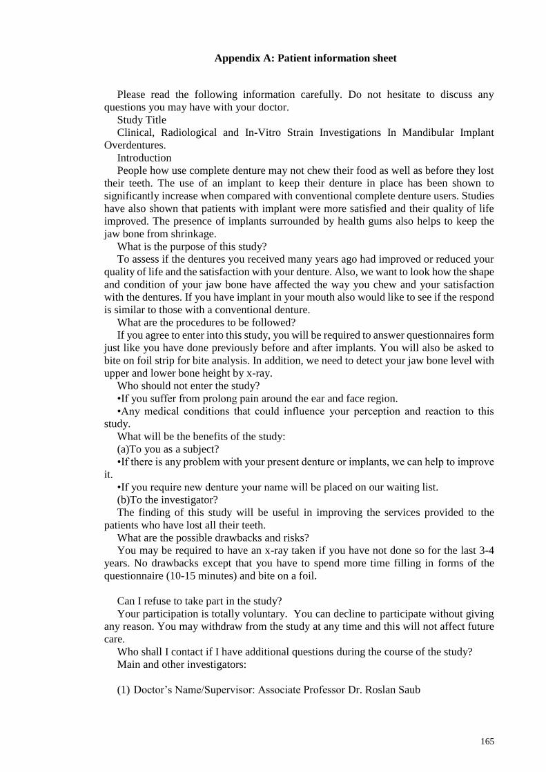
165
Appendix A: Patient information sheet
Please read the following information carefully. Do not hesitate to discuss any
questions you may have with your doctor.
Study Title
Clinical, Radiological and In-Vitro Strain Investigations In Mandibular Implant
Overdentures.
Introduction
People how use complete denture may not chew their food as well as before they lost
their teeth. The use of an implant to keep their denture in place has been shown to
significantly increase when compared with conventional complete denture users. Studies
have also shown that patients with implant were more satisfied and their quality of life
improved. The presence of implants surrounded by health gums also helps to keep the
jaw bone from shrinkage.
What is the purpose of this study?
To assess if the dentures you received many years ago had improved or reduced your
quality of life and the satisfaction with your denture. Also, we want to look how the shape
and condition of your jaw bone have affected the way you chew and your satisfaction
with the dentures. If you have implant in your mouth also would like to see if the respond
is similar to those with a conventional denture.
What are the procedures to be followed?
If you agree to enter into this study, you will be required to answer questionnaires form
just like you have done previously before and after implants. You will also be asked to
bite on foil strip for bite analysis. In addition, we need to detect your jaw bone level with
upper and lower bone height by x-ray.
Who should not enter the study?
•If you suffer from prolong pain around the ear and face region.
•Any medical conditions that could influence your perception and reaction to this
study.
What will be the benefits of the study:
(a)To you as a subject?
•If there is any problem with your present denture or implants, we can help to improve
it.
•If you require new denture your name will be placed on our waiting list.
(b)To the investigator?
The finding of this study will be useful in improving the services provided to the
patients who have lost all their teeth.
What are the possible drawbacks and risks?
You may be required to have an x-ray taken if you have not done so for the last 3-4
years. No drawbacks except that you have to spend more time filling in forms of the
questionnaire (10-15 minutes) and bite on a foil.
Can I refuse to take part in the study?
Your participation is totally voluntary. You can decline to participate without giving
any reason. You may withdraw from the study at any time and this will not affect future
care.
Who shall I contact if I have additional questions during the course of the study?
Main and other investigators:
(1) Doctor’s Name/Supervisor: Associate Professor Dr. Roslan Saub
166
Tel. No.: 03-79674888
Address: Community of community oral health & clinical prevention, Faculty of
Dentistry University of Malaya.
Email address: [email protected]
(2) Doctor’s Name/Supervisor: Professor Dr. Norsiah Yunus
Tel. No.: 0122866797
Address: Dept. Of Restorative Dentistry, University of Malaya.
Email address: [email protected]
(3) Doctor’s Name/Co-Supervisor: Lecturer Dr. Eshamsul Sulaiman
Tel. No.: 0126591408
Address: Dept. Of Restorative Dentistry, University of Malaya.
Email address: [email protected]
(4) Doctor’s Name/Researcher: Dr. Tameem Khuder Jassim
Tel. No.: 0182702776
Address: Dept. Of Restorative Dentistry, University of Malaya.
Email address: [email protected]
167
Appendix B: Patient consent form
CONSENT BY PATIENT FOR CLINICAL RESEARCH FACULTY OF
DENTISTRY, UM, K.L.
I, ……………………………………………………………Identity Card No.
……………………………… (Name of patient)
of…………………………………………………………………………………………
………………………… (Address)
hereby agree to take part in the clinical research ( clinical study ) specified below :
Title of Study : Clinical, Radiological and In-Vitro Strain Investigations In Mandibular
Implant Overdentures.
-The nature and purpose of which has been explained to me by Dr. Tameem Khuder /
Prof Norsiah Yunus… (Name & designation of doctor) and interpreted
by……………………………………………………(Name & designation of
interpreter)to the best of his/her ability in………………………. language/dialect.
I have been told about the nature of the clinical research in terms of methodology, possible
adverse effects, and complications ( as per the patient information sheet ). After knowing
and understanding all the possible advantages and disadvantages of this clinical research,
I voluntarily consent of my own free will to participate in the clinical research specified
above.
I understand that I can withdraw from this clinical research at any time without assigning
my reason whatsoever and in such a situation shall not be denied the benefits of usual
treatment by the attending doctors.
Date …………………………… Signature or
thumbprint……………………………………
(Patient)
IN THE PRESENCE OF
Name …………………………………………………,
I/C No. ……………………………………………….., Signature
………………………………………
(Witness for
signature of patient)
Designation …………………………………………... Date ............................
I confirm that I have explained to the patient the nature and purpose of the above
mentioned clinical research.
Date …………………………..... Signature
…………………………………………………..(Attending doctor)
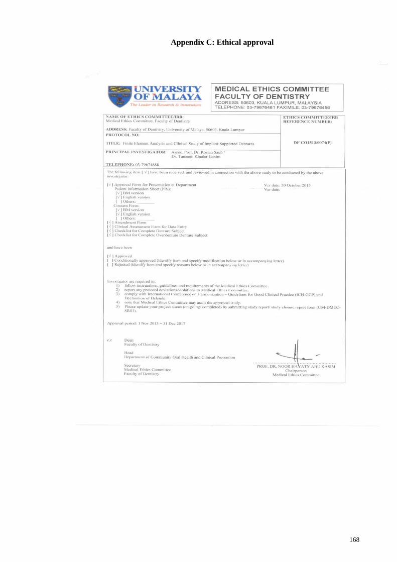
168
Appendix C: Ethical approval
169
Appendix D: Oral Health Impact Profile (Malaysia) S-OHIP (M) Questionnaire
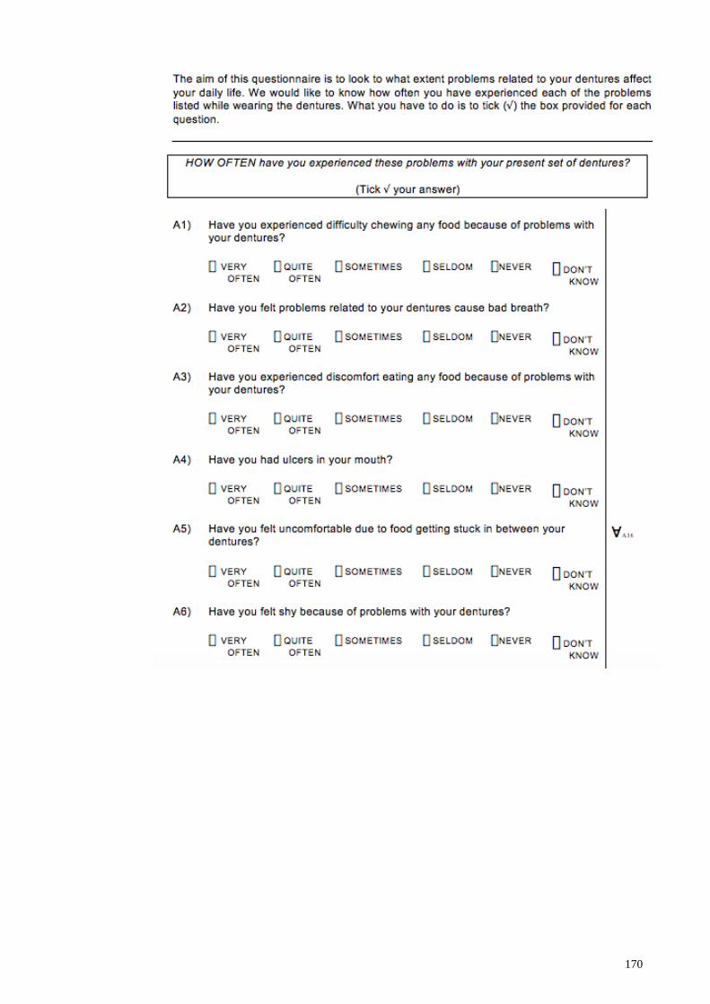
170
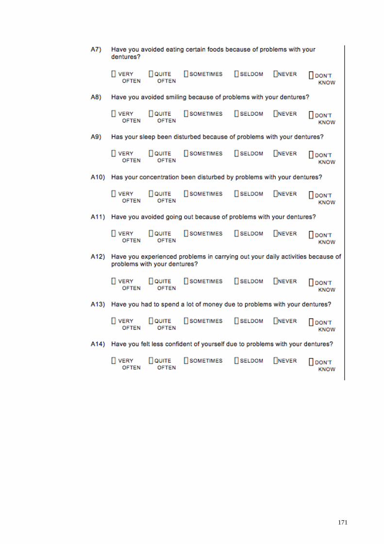
171
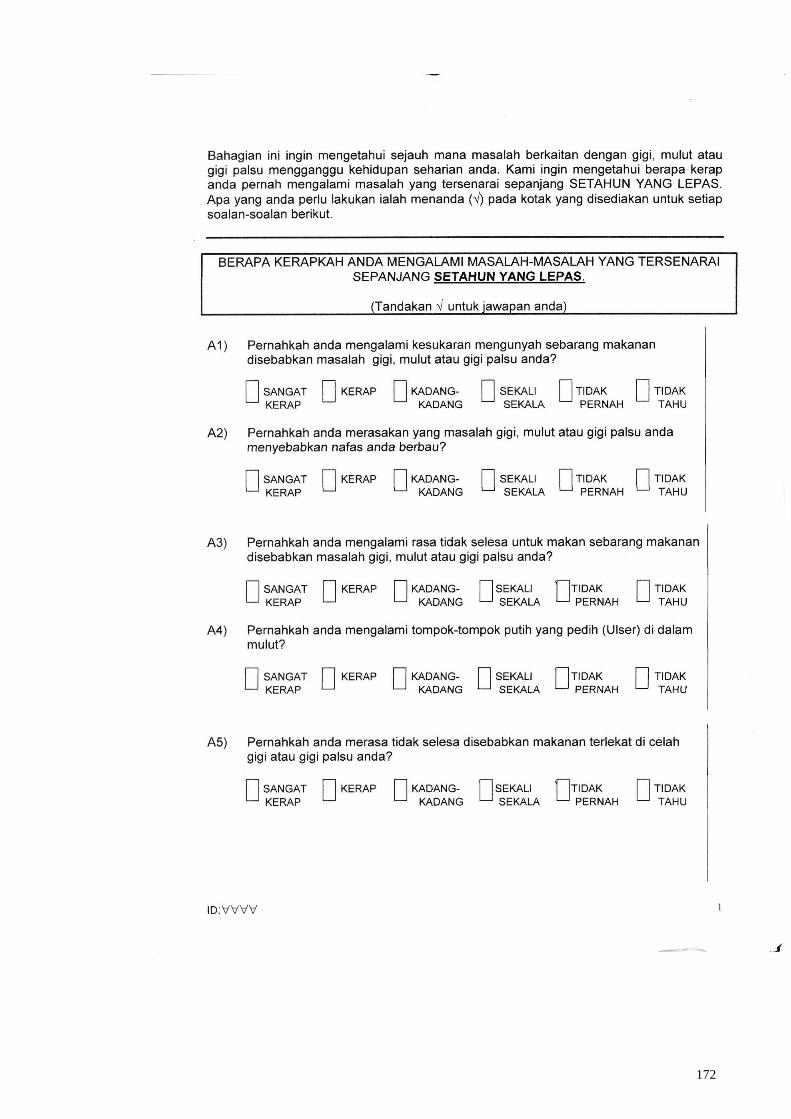
172
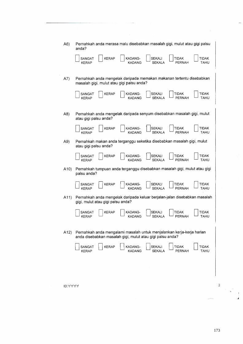
173

174
175
Appendix E: Denture Satisfaction (DS) Questionnaire
176
1. Are you wearing the last dentures made for you?
Yes No
2. How well does your present upper denture stay in position?
Very well Well Poorly Very poorly
3. How comfortable is your present upper denture?
Very comfortable Comfortable Uncomfortable Very uncomfortable
4. How well can you chew with your present upper denture?
Very well Well Poorly Very poorly
5. How well does your present lower denture stay in position?
Very well Well Poorly Very poorly
6. How comfortable is your present lower denture?
Very comfortable Comfortable Uncomfortable Very uncomfortable
7. How well can you chew with your present lower denture?
Very well Well Poorly Very poorly
8. How well do you like the appearance of your present set of dentures?
Very well Well Poorly Very poorly
9. How satisfied are you with your present set of dentures?
1. Very satisfied Satisfied Dissatisfied Very dissatisfied
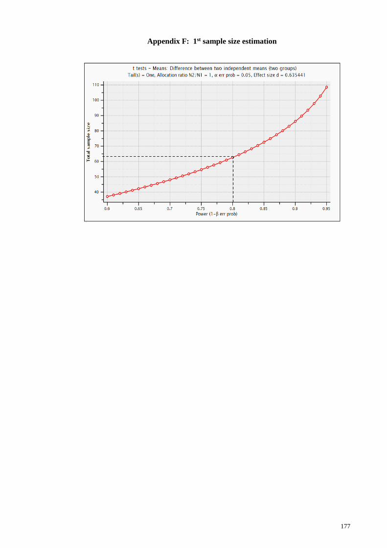
177
Appendix F: 1st sample size estimation
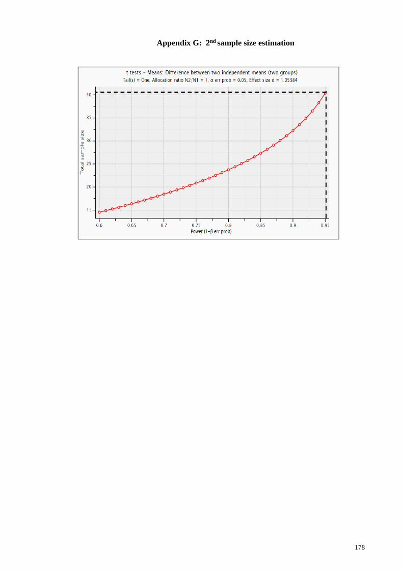
178
Appendix G: 2nd sample size estimation
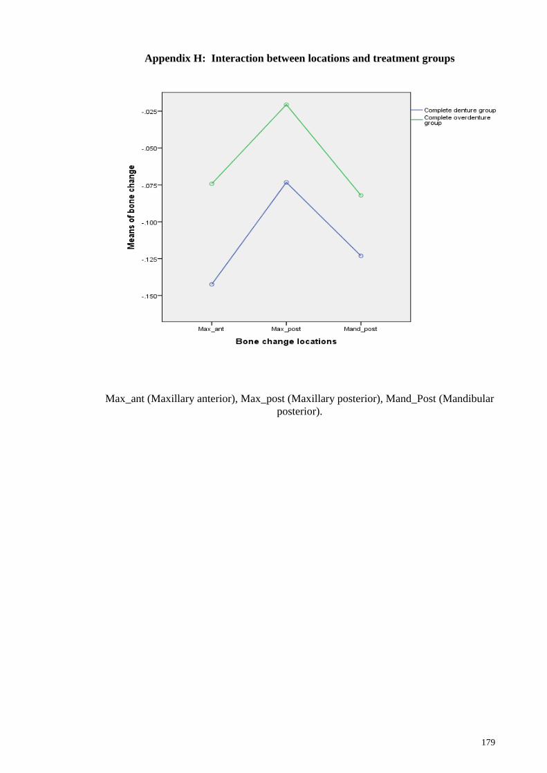
179
Appendix H: Interaction between locations and treatment groups
Max_ant (Maxillary anterior), Max_post (Maxillary posterior), Mand_Post (Mandibular
posterior).
180

181
182

183
184
185
186

187